Immunopathology and Infectious Disease A Role for Natural Regulatory T Cells in the Pathogenesis of Experimental Cerebral Malaria Fiona H. Amante,* Amanda C. Stanley,* Louise M. Randall,* † Yonghong Zhou,* Ashraful Haque,* Karli McSweeney,* Andrew P. Waters, ‡ Chris J. Janse, ‡ Michael F. Good,* Geoff R. Hill,* and Christian R. Engwerda* From the Queensland Institute of Medical Research,* Herston, Queensland, Australia; the School of Population Health, † University of Queensland, Herston, Queensland, Australia; and the Department of Parasitology, ‡ Leiden University Medical Centre, Leiden, The Netherlands Cerebral malaria (CM) is a serious complication of Plasmodium falciparum infection that is responsible for a significant number of deaths in children and nonimmune adults. A failure to control blood para- sitemia and subsequent sequestration of parasites to brain microvasculature are thought to be key events in many CM cases. Here , we show for the first time, to our knowledge, that CD4 CD25 Foxp3 natural reg- ulatory T (Treg) cells contribute to pathogenesis by modulating immune responses in P. berghei ANKA (PbA)-infected mice. Depletion of Treg cells with anti- CD25 monoclonal antibody protected mice from ex- perimental CM. The accumulation of parasites in the vasculature and brain was reduced in these animals , resulting in significantly lower parasite burdens com- pared with control animals. Mice lacking Treg cells had increased numbers of activated CD4 and CD8 T cells in the spleen and lymph nodes, but CD8 T-cell recruitment to the brain was selectively reduced in these mice. Importantly , a non-Treg-cell source of interleukin-10 was critical in preventing experimen- tal CM. Finally , we show that therapeutic administra- tion of anti-CD25 monoclonal antibody , even when blood parasitemia is established , can prevent disease , confirming a critical and paradoxical role for Treg cells in experimental CM pathogenesis. (Am J Pathol 2007, 171:548 –559; DOI: 10.2353/ajpath.2007.061033) Cerebral malaria (CM) is a major cause of death in people infected with Plasmodium falciparum, with most deaths oc- curring in young children in sub-Saharan Africa. An esti- mated 10 to 20% of children who develop CM die, and a significant proportion of survivors have permanent neuro- logical damage. 1–3 CM can be associated with sequestra- tion of parasitized red blood cells (pRBCs) in the brain microvasculature 4 and secretion of toxic molecules by par- asites, 5 as well as inflammatory components of the host immune response, including secretion of cytokines 6 and recruitment of activated leukocytes to the brain. 7–9 An ex- perimental model of CM (ECM) caused by infection of C57BL/6 and CBA mice with P. berghei ANKA (PbA) dis- plays many features of human CM and has allowed the identification of several important factors in CM pathogen- esis. Both CD4 and CD8 T cells contribute to the devel- opment of ECM, 10 –13 and the spleen seems to be a key site for priming of PbA-specific T-cell responses. 14 In addition, the proinflammatory cytokines interferon (IFN)-, 15,16 tumor necrosis factor, 17 and LT, 18 as well as perforin, 13 all seem to play a role in ECM pathogenesis. Although the risk factors that predispose individuals to develop CM remain largely unknown, high blood para- sitemia is significantly correlated with increased risk of CM. 19 Effective immune responses to Plasmodium blood stages only emerge in people living in malaria-endemic regions after several years of repeated malaria infec- tions. 20 Antibodies against the surface of the merozoite lifecycle stage of P. falciparum and cell-mediated immu- nity are both thought to be required for protective immu- nity, but they may also contribute to pathology. 21 Re- cently, CD4 CD25 regulatory T (Treg) cells were shown to be rapidly induced in vivo in humans following P. falci- parum infection, and this was associated with a burst of transforming growth factor- production, decreased par- asite-specific immune responses, and higher rates of Supported by grants from the Australian National Health and Medical Research Council (NHMRC). C.R.E. is an Australian NHMRC Career Development Fellow, and G.R.H. is a Wellcome Trust Senior Research Fellow. Accepted for publication April 24, 2007. Supplemental material for this article can be found on http://ajp. amjpathol.org. Address reprint requests to Dr. Christian Engwerda, Queensland Insti- tute of Medical Research, 300 Herston Rd., Herston, QLD 4006, Australia. E-mail: [email protected]. The American Journal of Pathology, Vol. 171, No. 2, August 2007 Copyright © American Society for Investigative Pathology DOI: 10.2353/ajpath.2007.061033 548

Welcome message from author
This document is posted to help you gain knowledge. Please leave a comment to let me know what you think about it! Share it to your friends and learn new things together.
Transcript
Immunopathology and Infectious Disease
A Role for Natural Regulatory T Cells in thePathogenesis of Experimental Cerebral Malaria
Fiona H. Amante,* Amanda C. Stanley,*Louise M. Randall,*† Yonghong Zhou,*Ashraful Haque,* Karli McSweeney,*Andrew P. Waters,‡ Chris J. Janse,‡
Michael F. Good,* Geoff R. Hill,* andChristian R. Engwerda*From the Queensland Institute of Medical Research,* Herston,
Queensland, Australia; the School of Population Health,†
University of Queensland, Herston, Queensland, Australia; and
the Department of Parasitology,‡ Leiden University Medical
Centre, Leiden, The Netherlands
Cerebral malaria (CM) is a serious complication ofPlasmodium falciparum infection that is responsiblefor a significant number of deaths in children andnonimmune adults. A failure to control blood para-sitemia and subsequent sequestration of parasites tobrain microvasculature are thought to be key eventsin many CM cases. Here, we show for the first time, toour knowledge, that CD4�CD25�Foxp3� natural reg-ulatory T (Treg) cells contribute to pathogenesis bymodulating immune responses in P. berghei ANKA(PbA)-infected mice. Depletion of Treg cells with anti-CD25 monoclonal antibody protected mice from ex-perimental CM. The accumulation of parasites in thevasculature and brain was reduced in these animals,resulting in significantly lower parasite burdens com-pared with control animals. Mice lacking Treg cellshad increased numbers of activated CD4� and CD8� Tcells in the spleen and lymph nodes, but CD8� T-cellrecruitment to the brain was selectively reduced inthese mice. Importantly, a non-Treg-cell source ofinterleukin-10 was critical in preventing experimen-tal CM. Finally, we show that therapeutic administra-tion of anti-CD25 monoclonal antibody, even whenblood parasitemia is established, can prevent disease,confirming a critical and paradoxical role for Tregcells in experimental CM pathogenesis. (Am J Pathol2007, 171:548–559; DOI: 10.2353/ajpath.2007.061033)
Cerebral malaria (CM) is a major cause of death in peopleinfected with Plasmodium falciparum, with most deaths oc-
curring in young children in sub-Saharan Africa. An esti-mated 10 to 20% of children who develop CM die, and asignificant proportion of survivors have permanent neuro-logical damage.1–3 CM can be associated with sequestra-tion of parasitized red blood cells (pRBCs) in the brainmicrovasculature4 and secretion of toxic molecules by par-asites,5 as well as inflammatory components of the hostimmune response, including secretion of cytokines6 andrecruitment of activated leukocytes to the brain.7–9 An ex-perimental model of CM (ECM) caused by infection ofC57BL/6 and CBA mice with P. berghei ANKA (PbA) dis-plays many features of human CM and has allowed theidentification of several important factors in CM pathogen-esis. Both CD4� and CD8� T cells contribute to the devel-opment of ECM,10–13 and the spleen seems to be a key sitefor priming of PbA-specific T-cell responses.14 In addition,the proinflammatory cytokines interferon (IFN)-�,15,16 tumornecrosis factor,17 and LT�,18 as well as perforin,13 all seemto play a role in ECM pathogenesis.
Although the risk factors that predispose individuals todevelop CM remain largely unknown, high blood para-sitemia is significantly correlated with increased risk ofCM.19 Effective immune responses to Plasmodium bloodstages only emerge in people living in malaria-endemicregions after several years of repeated malaria infec-tions.20 Antibodies against the surface of the merozoitelifecycle stage of P. falciparum and cell-mediated immu-nity are both thought to be required for protective immu-nity, but they may also contribute to pathology.21 Re-cently, CD4�CD25� regulatory T (Treg) cells were shownto be rapidly induced in vivo in humans following P. falci-parum infection, and this was associated with a burst oftransforming growth factor-� production, decreased par-asite-specific immune responses, and higher rates of
Supported by grants from the Australian National Health and MedicalResearch Council (NHMRC). C.R.E. is an Australian NHMRC CareerDevelopment Fellow, and G.R.H. is a Wellcome Trust Senior ResearchFellow.
Accepted for publication April 24, 2007.
Supplemental material for this article can be found on http://ajp.amjpathol.org.
Address reprint requests to Dr. Christian Engwerda, Queensland Insti-tute of Medical Research, 300 Herston Rd., Herston, QLD 4006, Australia.E-mail: [email protected].
The American Journal of Pathology, Vol. 171, No. 2, August 2007
Copyright © American Society for Investigative Pathology
DOI: 10.2353/ajpath.2007.061033
548
parasite growth.22 Treg cells have also been shown toenhance P. yoelii infection in BALB/c mice.23 Together,these reports support a detrimental role for Treg cells incontrolling parasites during malaria infections, althoughtheir effect on CM pathogenesis is unknown.
Naturally occurring CD25�CD4� Treg cells, constitut-ing 5 to 10% of peripheral CD4� T cells in mice andhumans, express the forkhead/winged helix transcriptionfactor Foxp3.24 They are produced in the thymus as adistinct and functionally mature population, but there isalso evidence that they are induced in the periphery.25
Treg cells play a critical role in the maintenance of im-munological self-tolerance, as well as the control of im-mune responses to pathogens,26 commensal microbes,and environmental antigens.24 Treg cells mediate theireffects by direct cell contact27 or the secretion of anti-inflammatory cytokines such as interleukin (IL)-10 andtransforming growth factor-�.28 Here, we show that Tregcells play an important role in modulating the host im-mune response to PbA during the pathogenesis of ECM.This is one of the first examples of Treg cells contributingto a pathogenic process during an infectious disease.
Materials and Methods
Mice
Female C57BL/6 and CBA/CaH mice 5 to 6 weeks of agewere purchased from the Australian Resource Centre(Canning Vale, Perth, Western Australia) and maintainedunder conventional conditions. Female C57BL/6 mice de-ficient in IL-10 (originally obtained from Jackson Labora-tories, Bar Harbor, ME) were bred and maintained inhouse. All animal procedures were approved and moni-tored by the Queensland Institute of Medical ResearchAnimal Ethics Committee.
Parasites and Infections
P. berghei ANKA (PbA) was used in all experiments afterone in vivo passage in mice. A transgenic PbA (231c1l)line expressing luciferase and green fluorescent proteinunder the control of the ef1-� promoter was used forexperiments involving in vivo imaging.29 All mice wereinfected by injecting 105 pRBCs intravenously (i.v.) viathe lateral tail vein. Blood parasitemia was monitored byexamination of Diff-Quick (Lab Aids, Narrabeen, NSW,Australia)-stained thin blood smears obtained from tailbleeds. Anemia was estimated by measuring hemoglobinlevels using a HemoCue Hb 201 analyzer according tothe manufacturer’s instructions (HemoCue AB, An-gelholm, Sweden). For serum cytokine analysis, 100 �l ofblood was collected via the lateral tail vein before infec-tion and 5 days after PbA infection. Blood was allowed toclot, and serum was collected and stored at �70°C untilrequired.
Disease Assessment
Mice were monitored twice daily after day 5 postinfection(p.i.), and clinical ECM evaluated. Clinical ECM scoreswere defined by the presentation of the following signs:ruffled fur, hunching, wobbly gait, limb paralysis, convul-sions, and coma. Each sign was given a score of 1.Animals with severe ECM (accumulative scores �4) weresacrificed by CO2 asphyxiation according to ethicsguidelines, and the day of death was deemed to be thefollowing day.
Antibodies
Allophycocyanin-conjugated anti-TCR� chain, phyco-erythrin (PE)-Cy5- or PE-conjugated anti-CD4, PE-conju-gated anti-CD69, anti-CD25-biotin (7D4), PE-Cy5-conju-gated anti-CD8, PE-conjugated anti-Ly6G, fluoresceinisothiocyanate-conjugated anti-Ly6C, allophycocyanin-conjugated anti-B220, fluorescein isothiocyanate-conju-gated anti-CD19, allophycocyanin-conjugated anti-CD11c,PE-Cy5-conjugated anti-CD11b, fluorescein isothiocya-nate-conjugated anti-CD45.2, biotin-conjugated anti-NK1.1, anti-intercellular adhesion molecule (ICAM)-1, an-ti-vascular cell adhesion molecule (VCAM)-1 monoclonalantibodies (mAbs), and Alexa Fluor 488-conjugatedstreptavidin were purchased from Biolegend (San Diego,CA) or BD Biosciences (Franklin Lakes, NJ). PE-Cy5-conjugated �-galactosylceramide (�GalCer) mouseCD1d tetramers were a generous gift from Dale Godfreyand Daniel Pellicci (University of Melbourne, Melbourne,VIC, Australia). PE-labeled anti-mouse Foxp3 mAb waspurchased from eBioscience (San Diego, CA). Anti-CD25(PC61; rat IgG1) and isotype control mAb (MAC49; ratIgG1) were purified from culture supernatants by proteinG column purification (Amersham, Uppsala, Sweden) fol-lowed by endotoxin removal (Mustang Membranes; PallLife Sciences, East Hills, NY). Purified control rat IgGwere also used in some experiments and purchased fromSigma-Aldrich (Castle Hill, NSW, Australia).
Preparation of Tissue Mononuclear Cells
Spleen cells were isolated by digesting tissue in collage-nase type 4 (1 mg/ml; Worthington Biochemical Corp.,Lakewood, NJ) and deoxyribonuclease I (0.5 mg/ml;Worthington Biochemical) at room temperature for 40minutes. Splenocytes and lymph node cells (from super-ficial cervical, axillary, brachial, mesenteric, and inguinallymph nodes) were isolated by passing tissue through a100-�m sieve and washing twice with phosphate-buff-ered saline supplemented with 2% (v/v) fetal calf serum(wash buffer). Red blood cells were then lysed using redcell lysis buffer (Sigma-Aldrich), according to the manu-facturer’s instructions, underlaid with fetal calf serum,and centrifuged at 443 � g for 5 minutes. Cell pelletswere washed once more with wash buffer and cellscounted. Brain mononuclear cells were isolated by di-gesting tissue as described above before passingthrough a 100-�m sieve and washing twice with wash
Treg Cells in Experimental Cerebral Malaria 549AJP August 2007, Vol. 171, No. 2
buffer. The cell pellet was resuspended in 33% (v/v)Percoll and centrifuged at 693 � g for 12 minutes at roomtemperature. Supernatant containing debris was re-moved, and the leukocyte pellet was washed once inwash buffer, depleted of red blood cells as describedabove, underlaid with fetal calf serum, and centrifuged at443 � g for 5 minutes. Cell pellets were washed oncemore with wash buffer and cells counted. Peripheralblood leukocytes were prepared from heparinized bloodthat was depleted of red blood cells by three or fourtreatments with red cell lysis buffer (Sigma-Aldrich)according to the manufacturer’s instructions.
Flow Cytometric Analysis
For the staining of cell surface antigens, cells were incu-bated with fluorochrome-conjugated or biotinylated mAbson ice for 30 minutes followed by Alexa Fluor 488-strepta-vidin incubation for an additional 30 minutes. Intracellularstaining for Foxp3 was performed on fixed/permeabilizedcells using PE-labeled anti-mouse Foxp3 kit (eBioscience),according to the manufacturer’s instructions. Data wereacquired on a FACSCalibur flow cytometer and analyzedusing Cell Quest Pro software (BD Biosciences). Cell pop-ulations in the spleen and brain were defined as follows:CD4� T cells (CD4�TCR�), CD8� T cells (CD8�TCR�),B cells (B220�CD19�), neutrophils (CD11b�Ly6G�),macrophages/monocytes (CD11b�Ly6C�), dendriticcells (DC; CD11chi), NK cells (NK1.1�TCR�), NK Tcells (CD1d �GalCer tetramer� NK1.1�), and microglia(CD45intermediate (int)CD11c� or CD11b�). Cytokines inserum samples collected 5 days p.i. were quantifiedusing the cytometric bead array (CBA) inflammatorykit (BD Biosciences) on a FACScan cytometerequipped with Cell Quest Pro and CBA software (BDBiosciences).
CD25� T-Cell Depletion
CD25� T-cell depletion was performed by intraperitoneal(i.p.) injection of 0.5 mg of anti-CD25 mAb (PC61) 1 dayor 14 days before PbA infection. The efficacy of CD25depletion was confirmed by fluorescence-activated cellsorting (FACS) analysis using anti-CD4, anti-CD25, andanti-Foxp3 antibodies.
IFN� ELISPOT
CD4� and CD8� T cells were positively selected fromRBC-depleted splenocytes using magnetic activated cellsorting according to protocols recommended by themanufacturer of the metallo-conjugated anti-CD4 and an-ti-CD8 antibodies and positive selection columns (Milte-nyi Biotec, Bergisch Gladbach, Germany). Cells isolatedby this procedure were greater than 98% pure as as-sessed by FACS. The IFN� ELISPOT was performed aspreviously described.18
In Vivo Bioluminescence Imaging
Luciferase-expressing PbA pRBCs were visualized byimaging whole bodies or dissected organs with an I-CCD
photon-counting video camera and in vivo imaging sys-tem (IVIS 100; Xenogen, Alameda, CA). On day 5 p.i.,when ECM symptoms were observed in infected controlanimals, mice were anesthetized with fluorothane andinjected subcutaneously with 0.1 ml of 5 mg/ml D-luciferinfirefly potassium salt (Xenogen). Images were then cap-tured on the IVIS 100 according to the manufacturer’sinstructions. Parasites were visualized in the brain afterremoval from mice that had been perfused with 20 ml ofsaline via the heart. Bioluminescence generated by lucif-erase transgenic PbA in mice or brain tissue was mea-sured according to the manufacturer’s instructions usingthe same regions of measurement for all samples beingcompared. The unit of measurement was photons/sec-ond/cm2/steer radiant (p/sec/cm2/sr).
Immunohistochemistry
ICAM-1 and VCAM-1 staining was conducted on 6-�macetone-fixed brain sections, and primary antibodies weredetected with appropriate secondary detection reagentsand horseradish peroxidase according to the manufactur-er’s instructions (Vector Laboratories, Peterborough, UK).Sections were dehydrated and mounted before micro-scopic examination. These sections were then used tocount ICAM-1- and VCAM-1-positive vessels in 25 consec-utive microscopic fields of view at a final magnification of400�.
Real-Time Reverse Transcriptase-PolymeraseChain Reaction
Total RNA was extracted from the spleen using TRIzolreagent (Invitrogen Life Technologies, Carlsbad, CA),and an RNeasy Mini Kit with on-column DNase digestion(Qiagen, Valencia, CA). RNA samples were reverse-tran-scribed into cDNA using the cDNA Archive Kit (AppliedBiosystems, Foster City, CA) according to the manufac-turer’s instructions. The number of IFN� and IL-10 cDNAmolecules in each sample were calculated by using Taq-Man gene expression assays (Applied Biosystems), andthe number of HPRT (forward: 5�-GTTGGATACAGGCCA-GACTTTGTTG-3�; reverse: 5�-GATTCAACCTTGCGCT-CATCTTAGGC-3�) (housekeeping gene) cDNA mole-cules in each sample were calculated by real-timereverse transcription-polymerase chain reaction usingPlatinum SYBR Green Master Mix (Invitrogen Life Tech-nologies). All reverse transcription-polymerase chain re-actions were performed on a Corbett Research RG-3000Rotor Gene (Corbett Life Sciences, Sydney, NSW, Aus-tralia). Standard curves were generated with knownamounts of cDNA for each gene, and the number ofcytokine molecules per 1000 HPRT molecules in eachsample was calculated.
Statistical Analysis
Differences in survival of treatment groups were analyzedusing the Kaplan-Meier log-rank test. Differences in par-asitemia, cytokine levels, and bioluminescence were an-alyzed using either the Mann-Whitney U-test or the Stu-
550 Amante et alAJP August 2007, Vol. 171, No. 2
dent’s t-test where indicated. For all statistical tests, P �0.05 was considered significant.
Results
Mice Depleted of CD4�CD25�Foxp3� TregCells Do Not Develop ECM
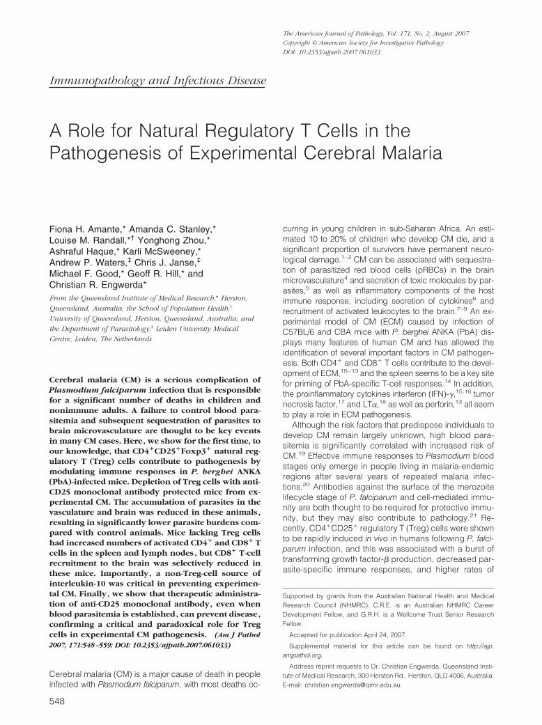
To establish whether natural Treg cells contribute to thedevelopment of ECM, we used an anti-CD25 mAb (PC61)to deplete CD4�CD25�Foxp3� Treg cells in C57BL/6mice. Specific depletion of CD4�CD25�Foxp3� Tregcells and no other major lymphocyte population in thespleen was confirmed by flow cytometry 24 hours afterantibody administration (Figure 1, A and B). Furthermore,we confirmed that CD4�CD25�Foxp3� Treg cells weredepleted and had not simply down-regulated CD25 ex-pression, as previously reported,30 because there was noaccumulation of CD4�CD25�Foxp3� Treg cells in micereceiving anti-CD25 mAb (data not shown). We also dem-onstrated that CD4�CD25�Foxp3� Treg cells were de-pleted by anti-CD25 mAb treatment in the lymph nodes,blood, and brain (see Supplemental Figure 1 at http://ajp.amjpathol.org). These cells remained at similar num-bers in the spleen and lymph nodes up until the time thatcontrol animals died with ECM but increased over thecourse of infection in the blood and brain (see Supple-mental Figure 1 at http://ajp.amjpathol.org).
Ninety percent of mice treated with an isotype controlantibody the day before PbA infection developed severe
neurological signs of ECM between days 6 and 9 p.i. andwere subsequently sacrificed (Figure 1C). In contrast,mice depleted of CD4�CD25�Foxp3� Treg cells the daybefore infection showed a significant (P � 0.001) in-crease in survival (80% survival on day 18 p.i.) (Figure1C) and a small but significant reduction (P � 0.05) inblood parasitemia at days 5, 6, and 7 p.i. compared withcontrol-treated mice (Figure 1D). Clinical scores werealso reduced in mice depleted of Treg cells (Figure 1E),and these animals developed no neurological signs ofECM and survived until 3 weeks after infection, when theydeveloped hyperparasitemia (Figure 1D) and severeanemia (hemoglobin levels �20 g/L). Histological exam-ination of the brain showed no cerebral hemorrhages inECM-resistant mice that received anti-CD25 mAb, unlikein control PbA-infected animals (data not shown).
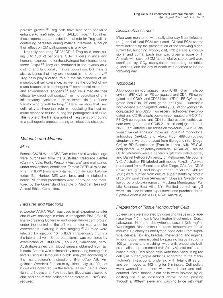
To exclude the possibility that anti-CD25 mAb mightdirectly affect emerging antiparasitic immune responses,including the generation of inducible Treg cells, we nextdepleted Treg cells 14 days before PbA infection, a timeperiod that allows continued Treg-cell depletion31 as wellas clearance of antibody from the blood32 (see Supple-mental Figure 2 at http://ajp.amjpathol.org). Specific de-pletion of CD4�CD25�Foxp3� Treg cells and no othermajor leukocyte population was confirmed by flow cytom-etry 13 days after anti-CD25 mAb administration and theday before infection (Figure 2, A and B), and we againdemonstrated that CD4�CD25�Foxp3� Treg cells weredepleted in the lymph nodes, blood, and brain using thisanti-CD25 mAb treatment regime (see Supplemental Fig-ure 1 at http://ajp.amjpathol.org).
Mice depleted of Treg cells 14 days before infectionfailed to develop ECM (Figure 2C), had significantly (P �0.05) lower blood parasitemia on day 6 p.i. (Figure 2D),and had lower clinical scores (Figure 2E) compared withcontrol mice. Again, these mice survived until the third
Figure 1. ECM is prevented in C57BL/6 mice lacking Treg cells. Mice wereinjected i.p. with 0.5 mg of anti-CD25 mAb or 0.5 mg of control mAb 1 daybefore infection with PbA. FACS profiles of CD25 and Foxp3 expression onsplenic leukocytes gated on CD4� T cells from control mice or Treg-depletedmice, as indicated, are shown 1 day after antibody administration (A).Analysis of splenic CD4� T cell, CD8� T cell, NK cell, NK T cell, and B cellnumbers from control mice (closed bars) or Treg-depleted mice (open bars)at the same time are also shown (B). Data are single representative samples(A) or mean � SEM (B) of individual samples from five mice per treatmentgroup. Results are from one representative experiment of three performed.Survival (C), parasitemia (mean � SEM; n � 5 mice per group) (D), andclinical disease (mean score � SEM; n � 5 mice per group) (E) weremonitored in mice that received anti-CD25 mAb (open triangles) or controlmAb (closed circles). Results in C represent data pooled from four separateexperiments yielding similar results (n � 20 mice per treatment group), andthe open box indicates the time when mice displayed ECM symptoms.Results in D and E are one representative experiment of four performed.Statistical differences of *P � 0.05 are indicated.
Figure 2. Anti-CD25 mAb treatment 14 days before PbA infection preventsECM in C57BL/6 mice. Mice were injected i.p. with 0.5 mg of anti-CD25 mAb(open bars or open triangles) or 0.5 mg of control mAb (closed bars or closedcircles) 14 days before infection with PbA. FACS analysis of splenic CD4�,CD8�, NK, NK T, and B cell numbers (A) as well as CD4�CD25�Foxp3� Tcells (B) on the day of infection are shown. Data represent the mean � SEMof individual samples from three mice per treatment group. Survival (C),parasitemia (mean � SEM) (D), and clinical disease (mean score � SEM) (E)were monitored. The open box in C–E indicates the time when micedisplayed ECM symptoms. Data are from one representative experiment offour performed. Statistical differences of *P � 0.05 are indicated.
Treg Cells in Experimental Cerebral Malaria 551AJP August 2007, Vol. 171, No. 2
week after infection with no signs of ECM and ultimatelydeveloped hyperparasitemia (Figure 2D) and severeanemia. Therefore, our data show that Treg-cell depletion14 days before PbA infection results in the same outcomeas Treg-cell depletion the day before infection and indi-cate a critical role for natural Treg cells, but not inducibleTreg cells, in ECM pathogenesis.
Prevention of Cerebral PbA Accumulation in theAbsence of Treg Cells
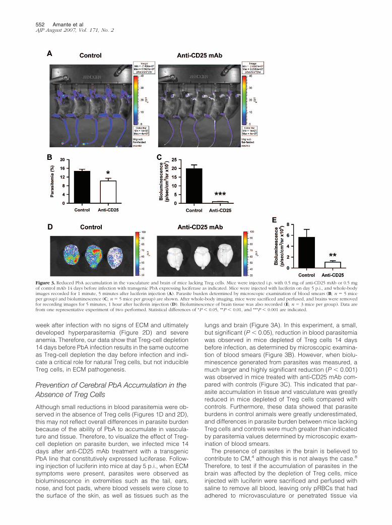
Although small reductions in blood parasitemia were ob-served in the absence of Treg cells (Figures 1D and 2D),this may not reflect overall differences in parasite burdenbecause of the ability of PbA to accumulate in vascula-ture and tissue. Therefore, to visualize the effect of Treg-cell depletion on parasite burden, we infected mice 14days after anti-CD25 mAb treatment with a transgenicPbA line that constitutively expressed luciferase. Follow-ing injection of luciferin into mice at day 5 p.i., when ECMsymptoms were present, parasites were observed asbioluminescence in extremities such as the tail, ears,nose, and foot pads, where blood vessels were close tothe surface of the skin, as well as tissues such as the
lungs and brain (Figure 3A). In this experiment, a small,but significant (P � 0.05), reduction in blood parasitemiawas observed in mice depleted of Treg cells 14 daysbefore infection, as determined by microscopic examina-tion of blood smears (Figure 3B). However, when biolu-minescence generated from parasites was measured, amuch larger and highly significant reduction (P � 0.001)was observed in mice treated with anti-CD25 mAb com-pared with controls (Figure 3C). This indicated that par-asite accumulation in tissue and vasculature was greatlyreduced in mice depleted of Treg cells compared withcontrols. Furthermore, these data showed that parasiteburdens in control animals were greatly underestimated,and differences in parasite burden between mice lackingTreg cells and controls were much greater than indicatedby parasitemia values determined by microscopic exam-ination of blood smears.
The presence of parasites in the brain is believed tocontribute to CM,4 although this is not always the case.8
Therefore, to test if the accumulation of parasites in thebrain was affected by the depletion of Treg cells, miceinjected with luciferin were sacrificed and perfused withsaline to remove all blood, leaving only pRBCs that hadadhered to microvasculature or penetrated tissue via
Figure 3. Reduced PbA accumulation in the vasculature and brain of mice lacking Treg cells. Mice were injected i.p. with 0.5 mg of anti-CD25 mAb or 0.5 mgof control mAb 14 days before infection with transgenic PbA expressing luciferase as indicated. Mice were injected with luciferin on day 5 p.i., and whole-bodyimages recorded for 1 minute, 5 minutes after luciferin injection (A). Parasite burden determined by microscopic examination of blood smears (B; n � 5 miceper group) and bioluminescence (C; n � 5 mice per group) are shown. After whole-body imaging, mice were sacrificed and perfused, and brains were removedfor recording images for 5 minutes, 1 hour after luciferin injection (D). Bioluminescence of brain tissue was also recorded (E; n � 3 mice per group). Data arefrom one representative experiment of two performed. Statistical differences of *P � 0.05, **P � 0.01, and ***P � 0.001 are indicated.
552 Amante et alAJP August 2007, Vol. 171, No. 2
hemorrhages. A striking and significant (P � 0.01) differ-ence was observed in the bioluminescence emergingfrom parasites in the brains of control mice and thosetreated with anti-CD25 mAb following removal and imag-ing (Figure 3, D and E). Few parasites were visualized inthe brains of mice lacking Treg cells. In contrast, randompatterns of intense parasite accumulation were observedin brain tissue from control animals. The same resultswere obtained when mice were treated with anti-CD25mAb 1 day before PbA infection (data not shown). To-gether, these data show that parasite accumulation in thebrain was associated with the onset of ECM in C57BL/6mice and that this process was prevented in the absenceof Treg cells. In addition, blood parasitemia values inmice with ECM symptoms underestimated total parasiteburden because of the accumulation of parasites in thebrain, vasculature, and other tissue sites.
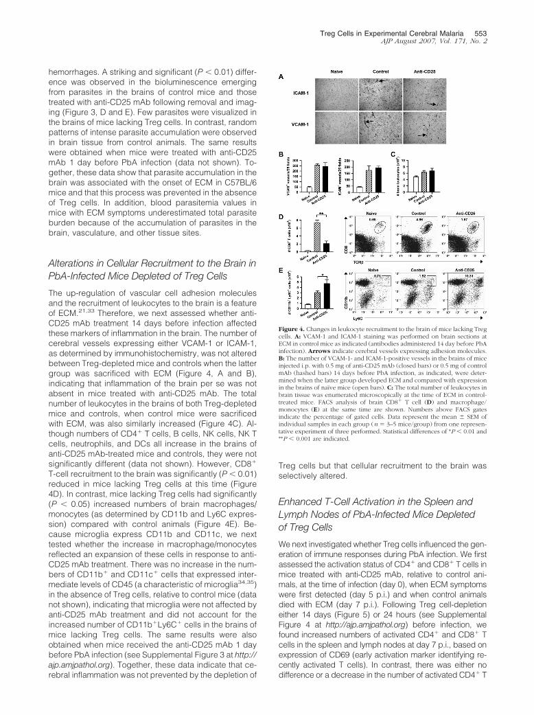
Alterations in Cellular Recruitment to the Brain inPbA-Infected Mice Depleted of Treg Cells
The up-regulation of vascular cell adhesion moleculesand the recruitment of leukocytes to the brain is a featureof ECM.21,33 Therefore, we next assessed whether anti-CD25 mAb treatment 14 days before infection affectedthese markers of inflammation in the brain. The number ofcerebral vessels expressing either VCAM-1 or ICAM-1,as determined by immunohistochemistry, was not alteredbetween Treg-depleted mice and controls when the lattergroup was sacrificed with ECM (Figure 4, A and B),indicating that inflammation of the brain per se was notabsent in mice treated with anti-CD25 mAb. The totalnumber of leukocytes in the brains of both Treg-depletedmice and controls, when control mice were sacrificedwith ECM, was also similarly increased (Figure 4C). Al-though numbers of CD4� T cells, B cells, NK cells, NK Tcells, neutrophils, and DCs all increase in the brains ofanti-CD25 mAb-treated mice and controls, they were notsignificantly different (data not shown). However, CD8�
T-cell recruitment to the brain was significantly (P � 0.01)reduced in mice lacking Treg cells at this time (Figure4D). In contrast, mice lacking Treg cells had significantly(P � 0.05) increased numbers of brain macrophages/monocytes (as determined by CD11b and Ly6C expres-sion) compared with control animals (Figure 4E). Be-cause microglia express CD11b and CD11c, we nexttested whether the increase in macrophage/monocytesreflected an expansion of these cells in response to anti-CD25 mAb treatment. There was no increase in the num-bers of CD11b� and CD11c� cells that expressed inter-mediate levels of CD45 (a characteristic of microglia34,35)in the absence of Treg cells, relative to control mice (datanot shown), indicating that microglia were not affected byanti-CD25 mAb treatment and did not account for theincreased number of CD11b�Ly6C� cells in the brains ofmice lacking Treg cells. The same results were alsoobtained when mice received the anti-CD25 mAb 1 daybefore PbA infection (see Supplemental Figure 3 at http://ajp.amjpathol.org). Together, these data indicate that ce-rebral inflammation was not prevented by the depletion of
Treg cells but that cellular recruitment to the brain wasselectively altered.
Enhanced T-Cell Activation in the Spleen andLymph Nodes of PbA-Infected Mice Depletedof Treg Cells
We next investigated whether Treg cells influenced the gen-eration of immune responses during PbA infection. We firstassessed the activation status of CD4� and CD8� T cells inmice treated with anti-CD25 mAb, relative to control ani-mals, at the time of infection (day 0), when ECM symptomswere first detected (day 5 p.i.) and when control animalsdied with ECM (day 7 p.i.). Following Treg cell-depletioneither 14 days (Figure 5) or 24 hours (see SupplementalFigure 4 at http://ajp.amjpathol.org) before infection, wefound increased numbers of activated CD4� and CD8� Tcells in the spleen and lymph nodes at day 7 p.i., based onexpression of CD69 (early activation marker identifying re-cently activated T cells). In contrast, there was either nodifference or a decrease in the number of activated CD4� T
Figure 4. Changes in leukocyte recruitment to the brain of mice lacking Tregcells. A: VCAM-1 and ICAM-1 staining was performed on brain sections atECM in control mice as indicated (antibodies administered 14 day before PbAinfection). Arrows indicate cerebral vessels expressing adhesion molecules.B: The number of VCAM-1- and ICAM-1-positive vessels in the brains of miceinjected i.p. with 0.5 mg of anti-CD25 mAb (closed bars) or 0.5 mg of controlmAb (hashed bars) 14 days before PbA infection, as indicated, were deter-mined when the latter group developed ECM and compared with expressionin the brains of naıve mice (open bars). C: The total number of leukocytes inbrain tissue was enumerated microscopically at the time of ECM in control-treated mice. FACS analysis of brain CD8� T cell (D) and macrophage/monocytes (E) at the same time are shown. Numbers above FACS gatesindicate the percentage of gated cells. Data represent the mean � SEM ofindividual samples in each group (n � 3–5 mice/group) from one represen-tative experiment of three performed. Statistical differences of *P � 0.01 and**P � 0.001 are indicated.
Treg Cells in Experimental Cerebral Malaria 553AJP August 2007, Vol. 171, No. 2
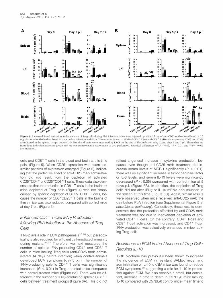
cells and CD8� T cells in the blood and brain at this timepoint (Figure 5). When CD25 expression was examined,similar patterns of expression emerged (Figure 5), indicat-ing that the protective effect of anti-CD25 mAb administra-tion did not result from the depletion of activatedCD25�CD4� or CD25�CD8� T cells. These data also dem-onstrate that the reduction in CD8� T cells in the brains ofmice depleted of Treg cells (Figure 4) was not simplycaused by specific depletion of CD25�CD8� T cells, be-cause the number of CD8�CD25� T cells in the brains ofthese mice was also reduced compared with control miceat day 7 p.i. (Figure 5).
Enhanced CD4� T-Cell IFN� Productionfollowing PbA Infection in the Absence of TregCells
IFN� plays a role in ECM pathogenesis15,16 but, paradox-ically, is also required for efficient cell-mediated immunityduring malaria.36,37 Therefore, we next measured thenumber of splenic IFN�-producing CD4� and CD8� Tcells in mice lacking Treg cells (anti-CD25 mAb admin-istered 14 days before infection) when control animalsdeveloped ECM symptoms (day 5 p.i.). The number ofIFN�-producing splenic CD4� T cells was significantlyincreased (P � 0.01) in Treg-depleted mice comparedwith control-treated mice (Figure 6A). There was no dif-ference in the number of IFN�-producing splenic CD8� Tcells between treatment groups (Figure 6A). This did not
reflect a general increase in cytokine production, be-cause even though anti-CD25 mAb treatment did in-crease serum levels of MCP-1 significantly (P � 0.01),there was no significant increase in tumor necrosis factoror IL-6 levels, and serum IL-10 levels were significantlydecreased (P � 0.05) compared with control mice at 5days p.i. (Figure 6B). In addition, the depletion of Tregcells did not alter IFN� or IL-10 mRNA accumulation inthe spleen at this time (Figure 6C). Again, similar resultswere observed when mice received anti-CD25 mAb theday before PbA infection (see Supplemental Figure 5 athttp://ajp.amjpathol.org). Collectively, these results dem-onstrate that the protection afforded by anti-CD25 mAbtreatment was not due to inadvertent depletion of acti-vated CD4� T cells. On the contrary, CD4� T-cell andCD8� T-cell activation was increased, and CD4� T-cellIFN� production was selectively enhanced in mice lack-ing Treg cells.
Resistance to ECM in the Absence of Treg CellsRequires IL-10
IL-10 blockade has previously been shown to increasethe incidence of ECM in resistant BALB/c mice, andadministration of IL-10 to CBA mice was found to reduceECM symptoms,38 suggesting a role for IL-10 in protec-tion against ECM. We also observe a small, but consis-tent, increase in time to death in C57BL/6 mice lackingIL-10 compared with C57BL/6 control mice (mean time to
Figure 5. Increased T-cell activation in the absence of Treg cells during PbA infection. Mice were injected i.p. with 0.5 mg of anti-CD25 mAb (closed bars) or 0.5mg of control mAb (hashed bars) 14 days before infection with PbA. The number (mean � SEM) of CD4� T (A) and CD8� T (B) cells expressing CD25 and CD69as indicated in the spleen, lymph nodes (LN), blood and brain were measured by FACS on the day of PbA infection (day 0) and days 5 and 7 p.i. These data arefrom three individual mice per group and are one representative experiment of two performed. Statistical differences of *P � 0.05, **P � 0.01, and ***P � 0.001are indicated.
554 Amante et alAJP August 2007, Vol. 171, No. 2
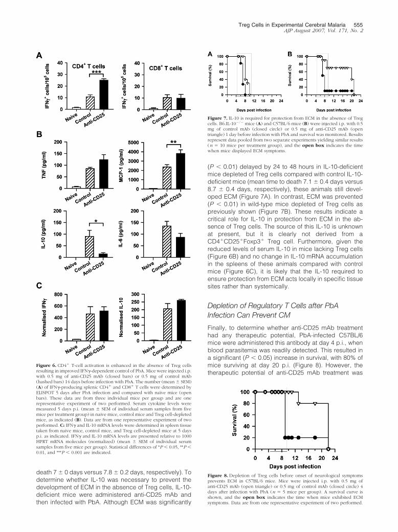
death 7 � 0 days versus 7.8 � 0.2 days, respectively). Todetermine whether IL-10 was necessary to prevent thedevelopment of ECM in the absence of Treg cells, IL-10-deficient mice were administered anti-CD25 mAb andthen infected with PbA. Although ECM was significantly
(P � 0.01) delayed by 24 to 48 hours in IL-10-deficientmice depleted of Treg cells compared with control IL-10-deficient mice (mean time to death 7.1 � 0.4 days versus8.7 � 0.4 days, respectively), these animals still devel-oped ECM (Figure 7A). In contrast, ECM was prevented(P � 0.01) in wild-type mice depleted of Treg cells aspreviously shown (Figure 7B). These results indicate acritical role for IL-10 in protection from ECM in the ab-sence of Treg cells. The source of this IL-10 is unknownat present, but it is clearly not derived from aCD4�CD25�Foxp3� Treg cell. Furthermore, given thereduced levels of serum IL-10 in mice lacking Treg cells(Figure 6B) and no change in IL-10 mRNA accumulationin the spleens of these animals compared with controlmice (Figure 6C), it is likely that the IL-10 required toensure protection from ECM acts locally in specific tissuesites rather than systemically.
Depletion of Regulatory T Cells after PbAInfection Can Prevent CM
Finally, to determine whether anti-CD25 mAb treatmenthad any therapeutic potential, PbA-infected C57BL/6mice were administered this antibody at day 4 p.i., whenblood parasitemia was readily detected. This resulted ina significant (P � 0.05) increase in survival, with 80% ofmice surviving at day 20 p.i. (Figure 8). However, thetherapeutic potential of anti-CD25 mAb treatment was
Figure 6. CD4� T-cell activation is enhanced in the absence of Treg cellsresulting in improved IFN�-dependent control of PbA. Mice were injected i.p.with 0.5 mg of anti-CD25 mAb (closed bars) or 0.5 mg of control mAb(hashed bars) 14 days before infection with PbA. The number (mean � SEM)(A) of IFN�-producing splenic CD4� and CD8� T cells were determined byELISPOT 5 days after PbA infection and compared with naıve mice (openbars). These data are from three individual mice per group and are onerepresentative experiment of two performed. Serum cytokine levels weremeasured 5 days p.i. (mean � SEM of individual serum samples from fivemice per treatment group) in naive mice, control mice and Treg cell-depletedmice, as indicated (B). Data are from one representative experiment of twoperformed. C: IFN� and IL-10 mRNA levels were determined in spleen tissuetaken from naive mice, control mice, and Treg cell-depleted mice at 5 daysp.i. as indicated. IFN� and IL-10 mRNA levels are presented relative to 1000HPRT mRNA molecules (normalized) (mean � SEM of individual serumsamples from five mice per group). Statistical differences of *P � 0.05, **P �0.01, and ***P � 0.001 are indicated.
Figure 7. IL-10 is required for protection from ECM in the absence of Tregcells. B6.IL-10�/� mice (A) and C57BL/6 mice (B) were injected i.p. with 0.5mg of control mAb (closed circle) or 0.5 mg of anti-CD25 mAb (opentriangle) 1 day before infection with PbA and survival was monitored. Resultsrepresent data pooled from two separate experiments yielding similar results(n � 10 mice per treatment group), and the open box indicates the timewhen mice displayed ECM symptoms.
Figure 8. Depletion of Treg cells before onset of neurological symptomsprevents ECM in C57BL/6 mice. Mice were injected i.p. with 0.5 mg ofanti-CD25 mAb (open triangle) or 0.5 mg of control mAb (closed circle) 4days after infection with PbA (n � 5 mice per group). A survival curve isshown, and the open box indicates the time when mice exhibited ECMsymptoms. Data are from one representative experiment of two performed.
Treg Cells in Experimental Cerebral Malaria 555AJP August 2007, Vol. 171, No. 2
lost when initiated after the onset of neurological symp-toms (data not shown).
Discussion
In this study, we found that administration of anti-CD25mAb before PbA infection resulted in the depletion ofTreg cells, associated with reduced parasite load andprotection from ECM. Imaging of PbA in situ (Figure 3, Aand D) demonstrated that the overall parasite burden incontrol PbA-infected mice was much greater than indi-cated by blood smears due to accumulation of parasitesin peripheral tissue sites and in the vasculature. There-fore, the effect of Treg depletion on parasite burdens wassubstantially greater than revealed from counting bloodsmears (Figures 1D and 2D). The difficulty in estimatingthe number of parasites sequestered in tissues has longhampered clinical investigations of malaria, because it isthese parasites that are thought to be critical for diseasepathogenesis.39,40 Our data show that Treg depletionresults in a major reduction in tissue parasites that mayexplain why these animals fail to develop ECM.
Recently, it was reported that treatment with anti-CD25mAb protected mice from ECM when administered theday before and the day after PbA infection, but not whengiven 30 days before infection.41 The protective effect ofanti-CD25 mAb was attributed to the depletion of acti-vated T cells, but this was not investigated. Our dataindicate that the numbers of activated CD4� and CD8� Tcells in PbA-infected mice increase significantly in sev-eral different tissue sites following anti-CD25 mAb treat-ment either 1 or 14 days before infection and that thisenhanced T-cell activation allows improved control ofparasitemia at a critical time in ECM pathogenesis, aswell as reduced tissue accumulation and vascularizationof pRBCs. One possible explanation that anti-CD25 mAbtreatment 30 days before PbA infection failed to protectmice41 is the rapid emergence of Treg cells after infectionwith this treatment regime. Recently, it was reported thatTreg cells expand rapidly following P. yoelli infection,42
and this is also found in PbA infection41 (see Supplemen-tal Figure 1 at http://ajp.amjpathol.org). Hence, the longeranti-CD25 mAb pretreatment period of 30 days in theprevious study may have enabled the rapid emergenceof Treg cells that prevented the enhanced T-cell activa-tion and parasite control observed in our study. Thisremains to be tested experimentally.
Although the anti-CD25 mAb treatment reduced para-site burden significantly, it did not allow ultimate control ofparasite growth. Instead, these animals go on to die withsevere anemia and hyperparasitemia, as has beenreported for all other mouse strains that are resistant toECM induced by PbA, such as those deficient in IFN�receptor16 and LT�.18 Several parasite-mediatedchanges to the host immune response may contribute tothe failure of anti-CD25 mAb-treated mice to ultimatelycontrol PbA growth. First, parasite-specific CD4� T cellsare depleted via an IFN�-dependent mechanism afterPbA infection but before elimination of parasites.43 Sec-ond, CD8� DCs become nonresponsive after PbA infec-
tion, resulting in a failure to cross-prime CD8� T cells andpossibly present parasite antigen to CD4� T cells.44 Fi-nally, in murine P. chabaudi infection, CD8� DCs becomethe major antigen presenting cell population later in in-fection.45 If this change takes place during PbA infectionand there are differences in the effectiveness of immuneresponses generated by these DCs, they may alter theability of the host to control parasite growth. Together,these changes are likely to contribute to PbA escapingcell-mediated immune responses after the phase of ce-rebral malaria induction has passed in resistant mice.Although there has been little work on the role of antibodyin this model, it is also possible that a failure to generateeffective antiparasitic antibodies may also contribute tothe failure to control PbA later in infection.
The depletion of Treg cells prevented ECM and theaccumulation of parasites in the cerebral microvascula-ture, an event concomitant with many, but not all, humanCM cases.8 Because control-treated PbA-infected micedevelop brain hemorrhages,18 whereas mice depleted ofTreg cells do not (data not shown), it is unclear whetherparasite accumulation in the brains of control animalsresulted from the trapping of parasites in hemorrhages,vascularization, or sequestration. In another study usingPbA that expressed luciferase under the schizont-spe-cific AMA-1 promoter, very few schizonts were seques-tered in the brain during ECM.29 The PbA line used in thecurrent study expressed luciferase under the control ofthe ef1-� promoter that allows luciferase expression in allblood stages.46 Therefore, it is likely that the intensebioluminescence observed in the brains taken from micewith ECM in our study arises from the accumulation ofring forms, trophozoites, and young schizonts in the hem-orrhages associated with the cerebral vasculature ofthese animals. Nevertheless, the presence of detectablenumbers of PbA pRBCs in the brain was always associ-ated with the onset of ECM in our studies, and removal ofTreg cells prevented this accumulation, possibly by pre-venting the formation of cerebral hemorrhages. It is in-creasingly recognized that CM is not a homogeneouscondition but is a disease syndrome that comprises anumber of pathological correlates and pathogenic pro-cesses.47 The C57BL/6 ECM model seems to reflectsome aspects of the human disease, including damageto the cerebral vasculature leading to perivascular hem-orrhages. The prevention of microvascular endothelialcell damage in brain in the absence of Treg cells indi-cates a strong immunological involvement in this patho-genic process.
Both CD4� and CD8� T cells are involved in the patho-genesis of ECM10–13 but are also required for the effec-tive control of malaria parasites,48 emphasizing the deli-cate balance that exists between host-mediated controlof infection and disease development. CD8� T cells havebeen shown to sequester to the brain following PbA in-fection at the onset of cerebral symptoms12,13 and arepostulated to cause damage to the brain endothelium viathe production of perforin.13 We found reduced CD8�
T-cell recruitment to the brains of PbA-infected mice de-pleted of Treg cells. This effect was not caused by afailure in CD8� T-cell activation in mice treated with anti-
556 Amante et alAJP August 2007, Vol. 171, No. 2

CD25 mAb, because CD69 expression was increasedand there was no reduction in IFN� production comparedwith CD8� T cells from control-treated animals after PbAinfection. In addition, the increase in CD25�CD8� T cellsin the spleen and lymph nodes at day 7 p.i. in micelacking Treg cells demonstrates that the anti-CD25 mAbwas not simply depleting these cells. This conclusion isalso supported by the reduction in CD25�CD8� T cells inthe brains of Treg cell-depleted mice compared withcontrols, further confirming that there was no selectiveeffect on CD25�CD8� T cells. Therefore, Treg cells seemto play an important role in establishing the immuneconditions necessary for the recruitment of CD8� T cellsto the brain during ECM. This recruitment mechanism iscurrently under investigation.
The influence of Treg cells on CD8� T-cell recruitmentto the brain during ECM was selective as the recruitmentof no other major leukocyte population was reduced inthe absence of Treg cells. The reduced accumulation ofCD8� T cells in the brain of PbA-infected mice lackingTreg cells could either result from changes in the initialpriming of parasite-specific CD8� T cells after infectionor subsequent recruitment to the brain. The degree ofCD8� T-cell trafficking following Listeria monocytogenesinfection has been shown to depend on the length ofantigen stimulus during the early stages of infection,49
suggesting that early activation of CD8� T cells followingPbA infection in the absence of Treg cells could alter thetissue homing properties of these cells. In addition, par-asites or parasite products might induce local tissuechanges in the brain to induce CD8� T-cell accumula-tion. Studies to investigate these possibilities areunderway.
Importantly, the decrease in CD8� T-cell recruitmentdid not reflect a general reduction in brain inflammation inmice depleted of Treg cells, because total brain leuko-cyte accumulation and increased expression of ICAM-1and VCAM-1 on cerebral vascular endothelium was notaltered relative to control mice. Furthermore, the numberof macrophage/monocytes (CD11b�Ly6C�) in the brainof mice that received anti-CD25 mAb was increasedcompared with control mice at the onset of ECM in thelatter group. The increase in this cell population wasunlikely to be caused by a relative increase due to re-duced numbers of CD8� T cells because the numbers ofno other leukocyte population in the brain increased in asimilar way. Macrophages have potential anti-inflamma-tory activity and the ability to reduce pathology causedby infection.50 However, whether the accumulation ofthese cells in the brain of mice lacking Treg cells contrib-utes to survival is unknown at this time. Nevertheless,these data do suggest selective changes to the leuko-cytes recruited to the brain in PbA-infected mice de-pleted of Treg cells.
IFN� has been shown to play a key role in ECM patho-genesis.15,16 Human CD4� T cells are a significantsource of IFN� after exposure to P. falciparum antigens,51
and IFN� production by peripheral blood mononuclearcells in response to liver-stage or blood-stage antigens isassociated with resistance to P. falciparum infection anddisease.36,37 Recently, elevated plasma IFN� levels and
the presence of IFN� gene polymorphisms involved inincreased gene transcription were also found to be as-sociated with protection from CM in African children.52
These data again reinforce the delicate balance thatdetermines whether host immune factors such as IFN�promote antiparasitic immunity or mediate disease pa-thology during malaria. The data in our study indicate thatIFN� can either promote or inhibit ECM, depending onthe specific cellular interaction occurring in the host afterPbA infection and the timing of this interaction. In partic-ular, the presence of Treg cells following PbA infectionskews the role of IFN� from antiparasitic to pathogenic.
The resistance to ECM in mice depleted of Treg cellswas also dependent on the presence of the regulatorycytokine IL-10. However, mice lacking Treg cells hadreduced levels of serum IL-10 (Figure 6B) and no changein IL-10 mRNA accumulation in the spleen (Figure 6C)after PbA infection. Therefore, it is likely that IL-10 isproduced and acts in very specific tissue locations toprevent ECM. The source of this IL-10, the relevant tissuewhere it is produced, and the target of its activity arecurrently under investigation. A recent study has shownthat transforming growth factor-� production and thegeneration of inducible Treg cells following P. falciparuminfection suppresses antiparasitic immunity.22 NaturalTreg cells but not inducible Treg cells were involved inECM pathogenesis, as indicated by our studies that de-pleted Treg cells 14 days before infection. However, thedata do not exclude the possibility that inducible Tregscould contribute to protection from ECM, potentially viaproduction of regulatory cytokines such as IL-10. Alter-natively, conventional parasite-specific CD4� T cells maybe a critical source of this cytokine, as recently reportedin experimental models of cutaneous leishmaniasis53 andtoxoplasmosis.54 The ability of proinflammatory cytokinessuch as IFN� and regulatory cytokines like IL-10 to eitherpromote or protect from severe malaria are likely to de-pend on many factors, including host and parasite ge-netics and the immune status of an infected individual.Identifying the source of these cytokines during malariainfection will be an important first step in defining thecellular targets that either suppress antiparasitic immu-nity or protect from developing pathology. The specifictissue microenvironments where cytokine production andresponses occur during malaria will also have a majorimpact on disease outcome.
The IL-2 receptor comprises the CD25 molecule andhas been found on the myelin sheath of the mouse centralnervous system.55 Up-regulation of CD25 has also beenreported in the brain tissue of mice after lipopolysaccha-ride stimulation.56 Furthermore, IL-2 can modulate thefunction and behavior of neurons, glia, and oligodendro-cytes in vitro.57 In fact, there has been a report thatanti-CD25 mAb could ameliorate the signs and symp-toms of multiple sclerosis, although the mechanism re-sponsible for this effect was not reported.58 Therefore,one possibility for the lack of brain pathology in the cur-rent study was that the administration of anti-CD25 mAbhad direct effects on the brain following PbA infection.However, we believe this explanation is unlikely becausemice depleted of Treg cells 14 days before infection did
Treg Cells in Experimental Cerebral Malaria 557AJP August 2007, Vol. 171, No. 2
not develop ECM. In these naıve mice, antibody would beunable to cross the blood-brain barrier,59 and it is unlikelythat anti-CD25 mAb would be available when the micewere infected32 (see Supplemental Figure 2 at http://ajp.amjpathol.org). In addition, given that mice depletedof Treg cells displayed no breakdown in the blood-brainbarrier as discussed above, no residual antibody wouldbe able to cross this barrier following infection. Further-more, we observed no difference in microglia numbers inthe brain of control and anti-CD25 mAb-treated mice.Hence, the most likely cause of mice failing to developECM in the absence of Treg cells was an effect on theevolving host immune response to the parasite.
Finally, we demonstrated that the depletion of Tregcells has therapeutic potential for preventing CM whenblood parasitemia is detected in blood smears if con-ducted before the onset of CM symptoms. Reagents forsuch a therapy in humans are already in clinical use.However, the use of such a therapy in humans withsevere malaria would need to consider the potential forTreg depletion to modulate pre-existing host immunity26
and break immunological tolerance to self-antigens.24
Interestingly, mice that were depleted of Treg cells beforethe onset of ECM symptoms but after PbA infection hadbeen established, had no reduction in blood parasitemiacompared with control animals (data not shown). Thissuggests that anti-CD25 mAb therapy may directly mod-ulate host mediators of pathology when administeredafter the establishment of infection.
In conclusion, we found that Treg-cell depletion inmice with anti-CD25 mAb resulted in protection fromECM. This protection was associated with improvedCD4� and CD8� T-cell activation, reduced accumulationof PbA in the vasculature and brain, and selectivechanges to cerebral leukocyte recruitment. The results inthis study highlight the delicate balance that exists be-tween immunity and pathology during malaria, and haveimportant implications for understanding the pathogene-sis of CM and the development of vaccines and therapiesto prevent severe malaria. Significantly, this is the firstreport showing that Treg cells can contribute to patho-genesis during infectious disease by suppressing antip-arasitic immunity.
Acknowledgments
We thank Grace Chojnowski and Paula Hall for expertassistance with flow cytometry and cell sorting; MarkSmyth, Ian Clark, Ray Steptoe, and Bill Heath for helpfuldiscussions; and Heather Mathews for assistance withthe preparation of artwork.
References
1. Snow RW, Trape JF, Marsh K: The past, present and future of child-hood malaria mortality in Africa. Trends Parasitol 2001, 17:593–597
2. Mung’Ala-Odera V, Snow RW, Newton CR: The burden of the neuro-cognitive impairment associated with Plasmodium falciparum malariain sub-Saharan Africa. Am J Trop Med Hyg 2004, 71:64–70
3. Carter JA, Ross AJ, Neville BG, Obiero E, Katana K, Mung’ala-Odera
V, Lees JA, Newton CR: Developmental impairments following severefalciparum malaria in children. Trop Med Int Health 2005, 10:3–10
4. MacPherson GG, Warrell MJ, White NJ, Looareesuwan S, Warrell DA:Human cerebral malaria: a quantitative ultrastructural analysis of par-asitized erythrocyte sequestration. Am J Pathol 1985, 119:385–401
5. Schofield L, Hackett F: Signal transduction in host cells by a glyco-sylphosphatidylinositol toxin of malaria parasites. J Exp Med 1993,177:145–153
6. Clark IA, Rockett KA: The cytokine theory of human cerebral malaria.Parasitol Today 1994, 10:410–412
7. Patnaik JK, Das BS, Mishra SK, Mohanty S, Satpathy SK, Mohanty D:Vascular clogging, mononuclear cell margination, and enhanced vas-cular permeability in the pathogenesis of human cerebral malaria.Am J Trop Med Hyg 1994, 51:642–647
8. Clark IA, Awburn MM, Whitten RO, Harper CG, Liomba NG, MolyneuxME, Taylor TE: Tissue distribution of migration inhibitory factor andinducible nitric oxide synthase in falciparum malaria and sepsis inAfrican children. Malar J 2003, 2:6
9. Taylor TE, Fu WJ, Carr RA, Whitten RO, Mueller JG, Fosiko NG,Lewallen S, Liomba NG, Molyneux ME: Differentiating the pathologiesof cerebral malaria by postmortem parasite counts. Nat Med 2004,10:143–145
10. Yanez DM, Manning DD, Cooley AJ, Weidanz WP, van der Heyde HC:Participation of lymphocyte subpopulations in the pathogenesis ofexperimental murine cerebral malaria. J Immunol 1996, 157:1620–1624
11. Hermsen C, van de Wiel T, Mommers E, Sauerwein R, Eling W:Depletion of CD4� or CD8� T-cells prevents Plasmodium bergheiinduced cerebral malaria in end-stage disease. Parasitology 1997,114:7–12
12. Belnoue E, Kayibanda M, Vigario AM, Deschemin JC, van Rooijen N,Viguier M, Snounou G, Renia L: On the pathogenic role of brain-sequestered �� CD8� T cells in experimental cerebral malaria. J Im-munol 2002, 169:6369–6375
13. Nitcheu J, Bonduelle O, Combadiere C, Tefit M, Seilhean D, Mazier D,Combadiere B: Perforin-dependent brain-infiltrating cytotoxic CD8� Tlymphocytes mediate experimental cerebral malaria pathogenesis.J Immunol 2003, 170:2221–2228
14. Hermsen CC, Mommers E, van de Wiel T, Sauerwein RW, Eling WM:Convulsions due to increased permeability of the blood-brain barrierin experimental cerebral malaria can be prevented by splenectomy oranti-T cell treatment. J Infect Dis 1998, 178:1225–1227
15. Grau GE, Heremans H, Piguet PF, Pointaire P, Lambert PH, Billiau A,Vassalli P: Monoclonal antibody against interferon � can preventexperimental cerebral malaria and its associated overproduction oftumor necrosis factor. Proc Natl Acad Sci USA 1989, 86:5572–5574
16. Amani V, Vigario AM, Belnoue E, Marussig M, Fonseca L, Mazier D,Renia L: Involvement of IFN-g� receptor-medicated signaling in pa-thology and anti-malarial immunity induced by Plasmodium bergheiinfection. Eur J Immunol 2000, 30:1646–1655
17. Grau GE, Fajardo LF, Piguet PF, Allet B, Lambert PH, Vassalli P:Tumor necrosis factor (cachectin) as an essential mediator in murinecerebral malaria. Science 1987, 237:1210–1212
18. Engwerda CR, Mynott TL, Sawhney S, De Souza JB, Bickle QD, KayePM: Locally up-regulated lymphotoxin �, not systemic tumor necrosisfactor �, is the principle mediator of murine cerebral malaria. J ExpMed 2002, 195:1371–1377
19. Molyneux ME, Taylor TE, Wirima JJ, Borgstein A: Clinical features andprognostic indicators in paediatric cerebral malaria: a study of 131comatose Malawian children. Q J Med 1989, 71:441–459
20. Greenwood BM, Bradley AK, Greenwood AM, Byass P, Jammeh K,Marsh K, Tulloch S, Oldfield FS, Hayes R: Mortality and morbidity frommalaria among children in a rural area of The Gambia, West Africa.Trans R Soc Trop Med Hyg 1987, 81:478–486
21. Good MF, Xu H, Wykes M, Engwerda CR: Development and regula-tion of cell-mediated immune responses to the blood stages ofmalaria: implications for vaccine research. Annu Rev Immunol 2005,23:69–99
22. Walther M, Tongren JE, Andrews L, Korbel D, King E, Fletcher H,Andersen RF, Bejon P, Thompson F, Dunachie SJ, Edele F, de SouzaJB, Sinden RE, Gilbert SC, Riley EM, Hill AV: Upregulation of TGF-beta, FOXP3, and CD4�CD25� regulatory T cells correlates withmore rapid parasite growth in human malaria infection. Immunity2005, 23:287–296
23. Hisaeda H, Maekawa Y, Iwakawa D, Okada H, Himeno K, Kishihara
558 Amante et alAJP August 2007, Vol. 171, No. 2
K, Tsukumo S, Yasutomo K: Escape of malaria parasites from hostimmunity requires CD4�CD25� regulatory T cells. Nat Med 2004,10:29–30
24. Sakaguchi S: Naturally arising CD4� regulatory T cells for immuno-logic self-tolerance and negative control of immune responses. AnnuRev Immunol 2004, 22:531–562
25. Kretschmer K, Apostolou I, Hawiger D, Khazaie K, Nussenzweig MC,von Boehmer H: Inducing and expanding regulatory T cell popula-tions by foreign antigen. Nat Immunol 2005, 6:1219–1227
26. Belkaid Y, Rouse BT: Natural regulatory T cells in infectious disease.Nat Immunol 2005, 6:353–360
27. Thornton AM, Shevach EM: Suppressor effector function ofCD4�CD25� immunoregulatory T cells is antigen nonspecific. J Im-munol 2000, 164:183–190
28. Powrie F, Read S, Mottet C, Uhlig H, Maloy K: Control of immunepathology by regulatory T cells. Novartis Found Symp 2003,252:92–114
29. Franke-Fayard B, Janse CJ, Cunha-Rodrigues M, Ramesar J,Buscher P, Que I, Lowik C, Voshol PJ, den Boer MA, van Duinen SG,Febbraio M, Mota MM, Waters AP: Murine malaria parasitesequestration: CD36 is the major receptor, but cerebral pathology isunlinked to sequestration. Proc Natl Acad Sci USA 2005,102:11468–11473
30. Kohm AP, McMahon JS, Podojil JR, Begolka WS, DeGutes M,Kasprowicz DJ, Ziegler SF, Miller SD: Cutting Edge: Anti-CD25 mono-clonal antibody injection results in the functional inactivation, notdepletion, of CD4�CD25� T regulatory cells. J Immunol 2006,176:3301–3305
31. Oldenhove G, de Heusch M, Urbain-Vansanten G, Urbain J, MaliszewskiC, Leo O, Moser M: CD4�CD25� regulatory T cells control T helper celltype 1 responses to foreign antigens induced by mature dendritic cellsin vivo. J Exp Med 2003, 198:259–266
32. Loughry A, Fairchild S, Athanasou N, Edwards J, Hall FC: Inflamma-tory arthritis and dermatitis in thymectomized, CD25� cell-depletedadult mice. Rheumatology (Oxford) 2005, 44:299–308
33. Engwerda C, Belnoue E, Gruner AC, Renia L: Experimental models ofcerebral malaria. Curr Top Microbiol Immunol 2005, 297:103–143
34. Ford AL, Goodsall AL, Hickey WF, Sedgwick JD: Normal adult rami-fied microglia separated from other central nervous system macro-phages by flow cytometric sorting: phenotypic differences definedand direct ex vivo antigen presentation to myelin basic protein-reactive CD4� T cells compared. J Immunol 1995, 154:4309–4321
35. Sedgwick JD, Schwender S, Imrich H, Dorries R, Butcher GW, terMeulen V: Isolation and direct characterization of resident microglialcells from the normal and inflamed central nervous system. Proc NatlAcad Sci USA, 1991 88:7438–7442
36. Luty AJ, Lell B, Schmidt-Ott R, Lehman LG, Luckner D, Greve B,Matousek P, Herbich K, Schmid D, Migot-Nabias F, Deloron P,Nussenzweig RS, Kremsner PG: Interferon-� responses are associ-ated with resistance to reinfection with Plasmodium falciparum inyoung African children. J Infect Dis 1999, 179:980–988
37. Reece WH, Pinder M, Gothard PK, Milligan P, Bojang K, Doherty T,Plebanski M, Akinwunmi P, Everaere S, Watkins KR, Voss G,Tornieporth N, Alloueche A, Greenwood BM, Kester KE, McAdam KP,Cohen J, Hill AV: A CD4� T-cell immune response to a conservedepitope in the circumsporozoite protein correlates with protectionfrom natural Plasmodium falciparum infection and disease. Nat Med2004, 10:406–410
38. Kossodo S, Monso C, Juillard P, Velu T, Goldman M, Grau GE:Interleukin-10 modulates susceptibility in experimental cerebral ma-laria. Immunology 1997, 91:536–540
39. Burgner D, Xu W, Rockett K, Gravenor M, Charles IG, Hill AV,Kwiatkowski D: Inducible nitric oxide synthase polymorphism andfatal cerebral malaria. Lancet 1998, 352:1193–1194
40. Dondorp AM, Desakorn V, Pongtavornpinyo W, Sahassananda D,Silamut K, Chotivanich K, Newton PN, Pitisuttithum P, Smithyman AM,White NJ, Day NP: Estimation of the total parasite biomass in acutefalciparum malaria from plasma PfHRP2. PLoS Med 2005, 2:e204
41. Vigario AM, Gorgette O, Dujardin HC, Cruz T, Cazenave PA, Six A,Bandeira A, Pied S: Regulatory CD4�CD25�Foxp3� T cells expand
during experimental Plasmodium infection but do not prevent cere-bral malaria. Int J Parasitol 2007, [Epub ahead of print]
42. Couper KN, Blount DG, de Souza JB, Suffia I, Belkaid Y, Riley EM:Incomplete depletion and rapid regeneration of Foxp3� regulatory Tcells following anti-CD25 treatment in malaria-infected mice. J Immu-nol 2007, 178:4136–4146
43. Xu H, Wipasa J, Yan H, Zeng M, Makobongo MO, Finkelman FD,Kelso A, Good MF: The mechanism and significance of deletion ofparasite-specific CD4� T cells in malaria infection. J Exp Med 2002,195:881–892
44. Wilson NS, Behrens GM, Lundie RJ, Smith CM, Waithman J, Young L,Forehan SP, Mount A, Steptoe RJ, Shortman KD, de Koning-Ward TF,Belz GT, Carbone FR, Crabb BS, Heath WR, Villadangos JA: Sys-temic activation of dendritic cells by Toll-like receptor ligands ormalaria infection impairs cross-presentation and antiviral immunity.Nat Immunol 2006, 7:165–172
45. Sponaas AM, Cadman ET, Voisine C, Harrison V, Boonstra A, O’GarraA, Langhorne J: Malaria infection changes the ability of splenic den-dritic cell populations to stimulate antigen-specific T cells. J Exp Med2006, 203:1427–1433
46. Franke-Fayard B, Trueman H, Ramesar J, Mendoza J, van der KeurM, van der Linden R, Sinden RE, Waters AP, Janse CJ: A Plasmodiumberghei reference line that constitutively expresses GFP at a highlevel throughout the complete life cycle. Mol Biochem Parasitol 2004,137:23–33
47. Mackintosh CL, Beeson JG, Marsh K: Clinical features and patho-genesis of severe malaria. Trends Parasitol 2004, 20:597–603
48. Weidanz WP, Melancon-Kaplan J, Cavacini LA: Cell-mediated immu-nity to the asexual blood stages of malarial parasites: animal models.Immunol Lett 1990, 25:87–95
49. Williams MA, Bevan MJ: Shortening the infectious period does notalter expansion of CD8 T cells but diminishes their capacity to differ-entiate into memory cells. J Immunol 2004, 173:6694–6702
50. Herbert DR, Holscher C, Mohrs M, Arendse B, Schwegmann A,Radwanska M, Leeto M, Kirsch R, Hall P, Mossmann H, Claussen B,Forster I, Brombacher F: Alternative macrophage activation is essen-tial for survival during schistosomiasis and downmodulates T helper 1responses and immunopathology. Immunity 2004, 20:623–635
51. Scragg IG, Hensmann M, Bate CA, Kwiatkowski D: Early cytokineinduction by Plasmodium falciparum is not a classical endotoxin-likeprocess. Eur J Immunol 1999, 29:2636–2644
52. Cabantous S, Poudiougou B, Traore A, Keita M, Cisse MB, DoumboO, Dessein AJ, Marquet S: Evidence that interferon-� plays a protec-tive role during cerebral malaria. J Infect Dis 2005, 192:854–860
53. Anderson CF, Oukka M, Kuchroo VJ, Sacks D:CD4�CD25�Foxp3�TH1 cells are the source of IL-10-mediated im-mune suppression in chronic cutaneous leishmaniasis. J Exp Med2007, 204:285–297
54. Jankovic D, Kullberg MC, Feng CG, Goldszmid RS, Collazo CM,Wilson M, Wynn TA, Kamanaka M, Flavell RA, Sher A: ConventionalT-bet�Foxp3�Th1 cells are the major source of host-protective reg-ulatory IL-10 during intracellular protozoan infection. J Exp Med,204:273–283
55. Chakraborty G, Reddy R, Drivas A, Ledeen RW: Interleukin-2 recep-tors and interleukin-2-mediated signaling in myelin: activation of di-acylglycerol kinase and phosphatidylinositol 3-kinase. Neuroscience2003, 122:967–973
56. Utsuyama M, Hirokawa K: Differential expression of various cytokinereceptors in the brain after stimulation with LPS in young and oldmice. Exp Gerontol 2002, 37:411–420
57. Otero GC, Merrill JE: Response of human oligodendrocytes to inter-leukin-2. Brain Behav Immun 1997, 11:24–38
58. Bielekova B, Richert N, Howard T, Blevins G, Markovic-Plese S,McCartin J, Frank JA, Wurfel J, Ohayon J, Waldmann TA, McFarlandHF, Martin R: Humanized anti-CD25 (daclizumab) inhibits diseaseactivity in multiple sclerosis patients failing to respond to interferon �.Proc Natl Acad Sci USA 2004, 101:8705–8708
59. Bullard DE, Bourdon M, Bigner DD: Comparison of various methodsfor delivering radiolabeled monoclonal antibody to normal rat brain.J Neurosurg 1984, 61:901–911
Treg Cells in Experimental Cerebral Malaria 559AJP August 2007, Vol. 171, No. 2
Related Documents


![Cerebral Malaria [Dr. Usman Sp. S]](https://static.cupdf.com/doc/110x72/577d1d971a28ab4e1e8c97e2/cerebral-malaria-dr-usman-sp-s.jpg)


