Review A review of glycosylated carriers for drug delivery Keerti Jain, Prashant Kesharwani, Umesh Gupta, Narendra K. Jain * Pharmaceutics Research Laboratory, Department of Pharmaceutical Sciences, Dr. H. S. Gour University, Sagar (M.P.) 470003, India article info Article history: Received 27 January 2012 Accepted 16 February 2012 Available online 6 March 2012 Keywords: Carbohydrate Glycosylated carriers Lectin receptors Ligand abstract Carbohydrates not only represent a vast potential as structure building blocks of living cells but also have proved as a promising candidate for drug delivery. Glycosylation of nanocarriers instructs some gratifying characteristic, which leads to the evolution of promising delivery systems. Some path-breaking advantages of glycosylated carriers include the engineered release profile of bioactives when introduced into biological system. Being natural product of living system these carriers also upshots as a multifaceted drug delivery vehicle and reduces the toxicity associated with unmodified drug carrier and therapeutic agent. An additional attribute of these carriers is to alter the pharmacokinetic profile of drugs positively with stabilization of drug carrier. The presence of lectin receptors on different cell surfaces makes the glyco- sylated carrier appreciable for targeted delivery of drugs to improve their therapeutic index. Active participation of some lectin receptors in immune responses to antigen overlaid the application of glyco- sylated carriers in delivery of antigen and immunotherapy for treatment of ailments like cancer. These advantages revealed the promising potential of glycosylated carriers in each perspective of drug delivery. Collectively this review presents an overview of different applications of glycosylated carriers, with a focus on their applicability in development of a nanoconstruct with GRAS status. Ó 2012 Elsevier Ltd. All rights reserved. 1. Introduction Development of safe, effective and stable dosage form is the foremost requirement in the perspective of drug delivery. Therapy of an ailment aims at complete elimination of causative agent without harming the normal cells of the living being. Hence, an intense prominence needs to be placed on development of the strategies that selectively and preferentially deliver the therapeutic agents to the target site simultaneously reduceing the access to non-target site. For this purpose one of the attractive strategies is use of “ligand” that will facilitate the homing of the therapeutic moieties to the target tissues [1]. Carbohydrates are one of the ligand that can be explored for receptor targeted drug delivery [1e3]. Besides targeting of drug, conjugation of carbohydrate to various carriers like nanoparticles, liposomes, dendrimers etc may also provide different beneficial properties in the field of drug delivery including stealth characteristics, bioadhesive property, biostability, solubility and reduced toxicity [4,5]. All these proper- ties are very important in the milieu of a formulation acceptable by patient, appreciable by physician and pharmacist. Including this oligosaccharides and glycoconjugates can also be exploited as a biopharmaceutical tool in the drug design [6]. Thus the use of carbohydrate as carrier and as ligand may assist in the development of a safe and effective as well as a stable formulation. In this review the various aptitudes of carbohydrates as drug carrier and as ligand have been discussed with an assortment of therapeutic applications of glycosylated carrier. Simultaneously a brief account of lectin receptors, which interact with carbohy- drate ligands and methods for conjugation of carbohydrate moie- ties to drug carrier to form glycosylated carrier or glycoconjugates, has also been presented. Glycosylated carriers have been success- fully utilized to deliver various therapeutic agents including anti- viral drugs like azidothymidine, stavudine, anticancer drugs such as methotrexate, doxorubicin, antitubercular drugs for example rifampicin, antigens for instance HBsAg, genes etc [7e15]. In study with various therapeutic agents glycosylated carriers have emerged as potential delivery system by providing various pharmaceutical benefits including sustained and controlled release, stabilization and reduction in toxicity of carrier and drug with improved phar- macokinetic profile and superior therapeutic index via selective delivery of drug to target tissues. 2. Lectin receptors A number of cells express the receptors on the surface which can bind carbohydrates; these receptors are known as membrane * Corresponding author. Tel./fax: þ91 7582 264712. E-mail address: [email protected] (N.K. Jain). Contents lists available at SciVerse ScienceDirect Biomaterials journal homepage: www.elsevier.com/locate/biomaterials 0142-9612/$ e see front matter Ó 2012 Elsevier Ltd. All rights reserved. doi:10.1016/j.biomaterials.2012.02.033 Biomaterials 33 (2012) 4166e4186

Welcome message from author
This document is posted to help you gain knowledge. Please leave a comment to let me know what you think about it! Share it to your friends and learn new things together.
Transcript

at SciVerse ScienceDirect
Biomaterials 33 (2012) 4166e4186
Contents lists available
Biomaterials
journal homepage: www.elsevier .com/locate/biomateria ls
Review
A review of glycosylated carriers for drug delivery
Keerti Jain, Prashant Kesharwani, Umesh Gupta, Narendra K. Jain*
Pharmaceutics Research Laboratory, Department of Pharmaceutical Sciences, Dr. H. S. Gour University, Sagar (M.P.) 470003, India
a r t i c l e i n f o
Article history:Received 27 January 2012Accepted 16 February 2012Available online 6 March 2012
Keywords:CarbohydrateGlycosylated carriersLectin receptorsLigand
* Corresponding author. Tel./fax: þ91 7582 264712E-mail address: [email protected] (N.K. Jain).
0142-9612/$ e see front matter � 2012 Elsevier Ltd.doi:10.1016/j.biomaterials.2012.02.033
a b s t r a c t
Carbohydrates not only represent a vast potential as structure building blocks of living cells but also haveproved as a promising candidate for drug delivery. Glycosylation of nanocarriers instructs some gratifyingcharacteristic, which leads to the evolution of promising delivery systems. Some path-breaking advantagesof glycosylated carriers include the engineered release profile of bioactiveswhen introduced into biologicalsystem. Being natural product of living system these carriers also upshots as a multifaceted drug deliveryvehicle and reduces the toxicity associated with unmodified drug carrier and therapeutic agent. Anadditional attribute of these carriers is to alter the pharmacokinetic profile of drugs positively withstabilization of drug carrier. The presence of lectin receptors on different cell surfaces makes the glyco-sylated carrier appreciable for targeted delivery of drugs to improve their therapeutic index. Activeparticipation of some lectin receptors in immune responses to antigen overlaid the application of glyco-sylated carriers in delivery of antigen and immunotherapy for treatment of ailments like cancer. Theseadvantages revealed the promising potential of glycosylated carriers in each perspective of drug delivery.Collectively this review presents an overview of different applications of glycosylated carriers, with a focuson their applicability in development of a nanoconstruct with GRAS status.
� 2012 Elsevier Ltd. All rights reserved.
1. Introduction
Development of safe, effective and stable dosage form is theforemost requirement in the perspective of drug delivery. Therapyof an ailment aims at complete elimination of causative agentwithout harming the normal cells of the living being. Hence, anintense prominence needs to be placed on development of thestrategies that selectively and preferentially deliver the therapeuticagents to the target site simultaneously reduceing the access tonon-target site. For this purpose one of the attractive strategies isuse of “ligand” that will facilitate the homing of the therapeuticmoieties to the target tissues [1]. Carbohydrates are one of theligand that can be explored for receptor targeted drug delivery[1e3]. Besides targeting of drug, conjugation of carbohydrate tovarious carriers like nanoparticles, liposomes, dendrimers etc mayalso provide different beneficial properties in the field of drugdelivery including stealth characteristics, bioadhesive property,biostability, solubility and reduced toxicity [4,5]. All these proper-ties are very important in the milieu of a formulation acceptable bypatient, appreciable by physician and pharmacist. Including thisoligosaccharides and glycoconjugates can also be exploited as
.
All rights reserved.
a biopharmaceutical tool in the drug design [6]. Thus the use ofcarbohydrate as carrier and as ligandmay assist in the developmentof a safe and effective as well as a stable formulation.
In this review the various aptitudes of carbohydrates as drugcarrier and as ligand have been discussed with an assortment oftherapeutic applications of glycosylated carrier. Simultaneouslya brief account of lectin receptors, which interact with carbohy-drate ligands and methods for conjugation of carbohydrate moie-ties to drug carrier to form glycosylated carrier or glycoconjugates,has also been presented. Glycosylated carriers have been success-fully utilized to deliver various therapeutic agents including anti-viral drugs like azidothymidine, stavudine, anticancer drugs such asmethotrexate, doxorubicin, antitubercular drugs for examplerifampicin, antigens for instance HBsAg, genes etc [7e15]. In studywith various therapeutic agents glycosylated carriers have emergedas potential delivery system by providing various pharmaceuticalbenefits including sustained and controlled release, stabilizationand reduction in toxicity of carrier and drug with improved phar-macokinetic profile and superior therapeutic index via selectivedelivery of drug to target tissues.
2. Lectin receptors
A number of cells express the receptors on the surfacewhich canbind carbohydrates; these receptors are known as membrane
DELL
Highlight

K. Jain et al. / Biomaterials 33 (2012) 4166e4186 4167
lectins and because of these receptors different carbohydrates maybe used as ligand to target the therapeutic agents [2,6,16,17].Interaction of lectin with glycosylated compound can be studied byexamining the interaction of lectins such as Ricinus communisagglutinin and concanavalin A (Con A) with glycosylatedcompounds [18,19]. Targeting via carbohydrate ligand uses inter-action of endogenous ligands with different sugar moieties likemannose, galactose, fructose, fucose and lactose [1,20]. When thesecarbohydrate moieties are conjugated to different drug carriers theresultant glycosylated carriers having carbohydrate as surfaceligands are recognized and endocytosed by lectin receptors. Manyscientists have reported that the glycosylated carriers havingsurface carbohydrate ligand are successfully recognized by lectinreceptors. LecB, a fucose-specific lectin is associated with tissuebinding and biofilm formation during the infection of Pseudomonasaeruginosa. Kolomiets et al. have developed glycopeptidesdendrimers containing C-fucosyl peptide dendrimers with thebasic structure (CFuc-X(6)X(5)X(4))(4)(LysX(3)X(2)X(1))(2)LysIle-HisNH(2) (CFuc ¼ alpha-L-fucosyl acetic acid, X(1-6)¼ amino acids,Lys ¼ lysine branching) for developing LecB inhibitors as an anti-bacterial agents [21]. Strong lectin binding affinity was observed forthese glycopeptides dendrimers with fucose terminals in enzyme-linked lectin assay (ELLA). Binding affinity for tetravalent andoctavalent ligand was 440 times compared to fucose. This fact hasbeen explored for targeting various therapeutic agents via lectinreceptor-mediated uptake of glycosylated carrier. Similarly it wasfound that glycopeptide dendrimers with C-fucosyl residues arespecific for binding with Ulex europaeus lectin UEA-I and have highaffinity for PA-IIL lectin from P. aeruginosa [22,23]. This forms thebasis for the biomedical application of glycosylated carriers.
Different type of lectin receptors are articulated by an assort-ment of cell surfaces and a brief summary of different lectinreceptors is presented in Table 1. Liver cells express receptors fora variety of carbohydrate molecules. The parenchymal cells of liveri.e. hepatocytes recognize galactose or moieties having galactosylresidues due to presence of asialoglycoprotein receptors while thenon-parenchymal cells, explicitly Kupffer cells, and endothelialcells of liver, have receptors for mannose [24e26]. Hepatocytes inliver possess asialoglycoprotein receptor, which recognizes
Table 1Summary of lectin receptors.
Receptor (kDa) Expression or cell type Organ Ligand
Asialoglycoproteinreceptors
Parenchymal cells(Hepatocytes)
Liver Galactos
Mannose receptors(C-type lectinreceptor)
Non-parenchymalcells (Endothelialcells and Kupffer cells)
Liver Glycoproresidues
Macrophages Macrophages rich organs(liver, spleen, brain, bonemarrow and lungs)
Mannoseglycolpro
Dendritic cells Spleen, lymph nodes,blood
Complexmannose
Fucose receptors Kupffer cells Liver Glycoproresidues
Galactosyl receptor Hepatic endothelialand Kupffer cells
Liver Particle hresidues
Lectin-like receptorsContainingcarbohydrates likeSialic Lewisx
Malignant cells Tumor Carbohydlactose, m(3-AminSialyl-Le
Lectin receptors Oral epithelium,buccal cells
Buccal cavity ConsiderparticulaPisum sa
Lectin receptors Corneal andconjunctival cells
Eye N-acetylN-acetyl
galactose as ligand [27,28]. Mannose receptors present on liverendothelial cells and macrophages illustrate a rapid internalizationof mannose-terminated glycoproteins via receptor-mediatedendocytosis. In a study with sinusoidal endothelial cells of ratliver, a very rapid mannose receptor-mediated endocytosis wasobserved for a mannose-terminated glycoprotein; ovalbumin withan endocytotic rate constant of 4.12 min�1 corresponding to a veryshort half life of 10 s for the surface pool of receptor-ligandcomplexes. According to the authors this was the highest re-ported Ke for a receptor-mediated endocytosis system [25]. Plasmamembrane of macrophages expresses various receptors includingmannose receptors, Fc-receptors, integrins, scavenger receptors,stearylamine receptors, which plays important role in variousphysiological activities of mononuclear phagocyte system such asgrowth, differentiation, activation, migration, antigen recognitionand endocytosis. Mannose receptors are highly expressed on thealveolar macrophages, Kupffer cells, splenic macrophages, perito-neal macrophages, macrophages of brain i.e. the two types of glialcells; astrocytes and microglia and monocyte-derived dendriticcells [29e33]. Mannose receptor expressed by macrophages anddendritic cells is a C-type lectin 175 kDa transmembrane protein[34,35]. These mannose receptors have a sound influence on hostimmune response by active participation in phagocytosis, pro-cessing and presentation of antigen, cell migration and intracellularsignalling [36e39]. Particularly the surface of dendritic cellsenunciates high density of mannose receptors [40,41]. Kupffer cellsof liver also express fucose receptors, which can internalize fuco-sylated drug carriers and galactosyl receptors, which can uptakeparticles having galactose residues [42e45]. Neoplastic cells oftumor also express lectin-like receptors containing carbohydrateslike Sialic Lewis-X having high affinity for carbohydrate moietieslike mannose, galactose, fructose and lactose [1,46]. Apart fromthese, lectin receptors have also been detected on oral epithelium,buccal cells, corneal epithelium and conjunctiva [47,48].
3. Conjugation methods for glycosylation
Various chemical reactions and conjugation methods have beenapplied for derivatizing the surface of different carriers with
Functions References
yl residues Targeted delivery of therapeuticagents (drugs, genes) tohepatocytes
[24, 26, 125]
teins with mannose Receptor-mediated rapidendocytosis
[25]
-terminatedteins
Phagocytosis; Internalization bymacrophages, treatment of diseaselocalized in macrophages likeGaucher disease
[29, 34, 39]
with terminalgroups
Targeted DNA delivery to triggercellular immunity; pathogenrecognition; vaccination
[34, 39, 40]
teins having fucose May be explored for delivery oftherapeutic agents to Kupffer cells
[44, 45, 97]
aving galactose Endocytosis; clearance of sulfatedglycoproteins from blood
[42, 43]
rates like galactose,annose, fucose, SiaLex
opropyl glycosides ofwisx)
Targeting of antineoplastic drugs tomalignant tissue
[1, 46]
able affinity for lectinsrly for lectins fromtivum and Arachis hypogaea
Selective delivery of drugs to oralcavity
[47]
-D-glucosamine and-D-galactosamine
Ocular delivery of therapeuticagnets
[48]

K. Jain et al. / Biomaterials 33 (2012) 4166e41864168
carbohydrate ligands. Dutta et al. conjugatedmannose to the amineterminals of polypropyleneimine (PPI) dendrimers through thio-glycosidic linkage. In this method mannose conjugation of PPIdendrimers was accomplished by first synthesizing the acetylatedthioglycoside of mannose by reacting mannose with thiopropionicacid, followed by activation with N-hydroxysuccinimide (NHS).Finally, conjugation was ended by reacting PPI dendrimers withactivated NHS ester of acetylated thioglycoside of mannose withsubsequent deacetylation with sodium methoxide [49]. Agasheet al. also conjugated mannose and lactose to PPI dendrimers usingthe same method [50]. This method for conjugation of mannose tocarriers having surface amino groups is presented schematically inFig. 1. Espuelas et al. developed a conjugate consisting of
Fig. 1. Conjugation sch
a tetramannosyl head group attached to lipid moiety via poly-ethylene glycol spacer. Then authors incorporated this amphiphilicmolecule into liposomes bilayers and observed substantial bindingaffinity of these multivalent mannose residues exposed on lipo-somes for Con A compared to monomannosyl analogue [51].Agrawal et al. conjugated dextran to the surface amino groups ofPPI dendrimers [52]. In this reaction dextranwas first oxidized withpotassium iodate to form oxidized or polyaldehyde dextran, thanPPI dendrimers was added very slowly to curtail cross-linking andto assist conjugation of dendrimers on dextran, followed byreduction with sodium borohydride (NaBH4) (Fig. 2). Galactose canbe conjugated directly to the carrier surface provided the givencarrier has surface amino groups. In this conjugation, galactose is
eme for mannose.

Fig. 2. Conjugation scheme for dextran.
K. Jain et al. / Biomaterials 33 (2012) 4166e4186 4169
first dissolved in buffer solution followed by addition of carrier.-Galactose conjugation is then achieved by agitating the mixture fortwo days at ambient temperature [53e55] (Fig. 3).
Garg et al. prepared galactosylated, fucosylated and mannosy-lated liposomes in two steps;first, synthesis of O-palmitoylgalactose(OPG), O-palmitoylfucose (OPF) and O-palmitoylmannose (OPM),and second, bindingofO-palmitoylated ester of galactose, fucose andmannose to liposome. In this conjugation method galactose, fucoseand mannose were esterified with palmlitoyl chloride in dime-thylformamide to prepare OPG, OPF and OPM, respectively. ThenOPG, OPF and OPM was allowed to react with liposomes containingphosphatidylethanolamine to develop galactosylated, fucosylatedand mannosylated liposomes, respectively, using the fact that OPG,OPF and OPM can easily bind with phosphatidylethanolamine ofliposomes (Fig. 4) [8]. Frisch et al. has explored a ‘click chemistry’based bioconjugation reaction, using Cu(I)-catalyzed azide-alkynecyclo-addition in aqueous media, to attach various ligands to thesurface of preformed liposomes [56]. The authors applied thismodelclick reaction for single step conjugation of a-1-thiomannosylligands functionalized with an azide group to liposomes containing
a terminal alkyne-functionalized lipidanchor. In this reactionawatersoluble copper-ion chelator, bathophenanthrolinedisulphonate wasused as a catalyst. This catalyst increased the coupling yield tosubstantial level. The scientists observed that this conjugationreaction did not induce any vesicle leakage and also the mannoseligands coupled by this reaction were exposed at the surface of theliposomes. Cu(I)-catalyzed reaction is limited to saturated phos-pholipids containing liposomes however this limitation could bealleviated by using Cu free azide-alkyne click reaction. Similarlya unique method was developed by Maruyama and coworkers forintroducing high density of carbohydrate chains on the surface ofnanoparticles. In this method the scientists attached the carbohy-drates on the surface of nanoparticles composed of poly(lactic acid)or polystyrene using carbohydrate-carrying polystyrene derivativevia solvent evaporation method. In this procedure polystyrenederivative carrying carbohydrate served as emulsifier as well assurface coating. The attachment of carbohydrate to thenanoparticlessurface was confirmed by aggregation with carbohydrate-specificlectin and also the surface carbohydrate chains were distinguishedby hepatocytes in in vitro study with isolated rat hepatocytes [57].

Fig. 3. Conjugation scheme for galactose.
K. Jain et al. / Biomaterials 33 (2012) 4166e41864170
Various methods have been adopted by different scientists forconjugating a number of carbohydrates to different types of carriersystems to develop glycosylated carrier or glycoconjugates. Mac-millan and Daines have reviewed promising methods for devel-oping glycoconjugate assembly, screening the carbohydrate ligandand glycoconjugate based drug design and drug delivery [58].
4. Characteristic features of glycosylation in drug delivery
Glycosylation is a speculated strategy in the field of drugdelivery owing to the some exciting features of glycosylated carrier.Targeting via glycosylated carriers exploit highly specific interac-tions of multiple carbohydrate ligands with endogenous lectinreceptors. On themolecular level proteins such asmannose bindingproteins are involved in lectin receptor-mediated endocytosis [20].Molecular recognition of glycosylated carrier is particularly influ-enced by terminal sugar units of participating oligosaccharidechain. So it is very important to engineer the carrier in such a waythat the key sugar units necessary for molecular recognition bylectin receptors should be incorporated in the carrier and be
available for binding with lectin receptors. For example, a glycosy-lated carrier with terminal galactose moiety can be designed asa template for selective delivery of bioactives to liver cells viarecognition by asialoglycoprotein receptors of liver cells [59].Another aspect which makes glycosylated macromolecular carrieran important candidate in medicinal field is the involvement ofproteinecarbohydrate interactions as the initiating step in thepathological conditions including cancer, bacterial, fungal and viralinfections. The involvement of carbohydrate to such extent in thenumerous intercellular recognition events of various diseasesincluding cancer cell aggregation, metastatic spread of cancer,bacterial invasion, viral adhesion, agglutination of red blood cellsetc has paved the way for utilization of glycosylation as an amazingstrategy in drug delivery [60]. Considering the importance ofcarbohydrate in various biological processes and intercellularrecognition, glycosylation is a natural route for the development ofnew therapeutic carrier. The applications of these carriers asdelivery vehicle for drug and other bioactives, as diagnostic agent,as ecological carrier etc will be discussed in the following sectionsof this review.

Fig. 4. Scheme for conjugation of galactose, fucose and mannose to liposomes via esterification with Palmitoyl chloride.
K. Jain et al. / Biomaterials 33 (2012) 4166e4186 4171
5. Therapeutic applications of glycosylated carriers
Carbohydrates have been utilized in pharmaceuticals both as anactive moiety having therapeutic activity and as a carrier for ther-apeutic agents. Various applications of glycosylated carrier inpharmaceutical field are summarized in Table 2 and presentedschematically in Fig. 5.
5.1. As drug carrier
Various drugs have been delivered through glycosylated carrierwith successful inferences. Dutta and Jain evaluated mannoseconjugated 5.0G PPI dendrimers for delivery of an antiretroviraldrug, lamivudine and concluded that it could serve as a promisingdelivery agent for antiretroviral drugs as in in vitro drug releasestudy mannosylated dendrimers prolonged the release up to 144 hin contrast to plain PPI dendrimers, which released the drug by24 h. Also 43.27� 0.13% of lamivudinewas found to be entrapped inmannosylated carrier whereas 35.69 � 0.2% of drug was entrappedby plain PPI dendrimers [18]. Including this, improved anti-HIVactivity was observed with drug loaded mannosylated den-drimers compared to free drug, lamivudine, due to enhancedcellular uptake via mannose receptors. Similarly galactosylatedliposomes have been found suitable for delivery of antiviral drug,azidothymidine [9]. Modification of b-cyclodextrins with galactosegives galactosylated cyclodextrin, which binds specifically withlectin because these galactosylated cyclodextrin aggregates interactmore efficiently with lectin binding sites due to presence of smallmicelles. Also galactose competes with the galactosylated cyclo-dextrin by inhibiting the binding sites of lectin [61]. Methotrexate
conjugated to the mannosylated human serum albumin showsimproved pharmacokinetic profile with preferential distribution ofdrug to the Kupffer cells to act on Leishmania parasites [62]. Surianoet al. evaluated the d-glucose, d-galactose and d-mannose con-taining amphiphilic block glycopolymers for application in drugdelivery. These glycopolymers were synthesized by means ofmetal-free organocatalyzed ring-opening polymerization of func-tional cyclic carbonates giving finely dispersed products withcontrolled molecular weight and end group loyalty. From theresults of this investigation the authors concluded that these gly-copolymers can serve as promising drug delivery module withenhanced uptake by liver cells [63]. Oda et al. developed theconjugate between d-galactose and b-cyclodextrin as a drugdelivery molecule. In this study the authors found that the devel-oped conjugate served as a successful delivery module for drugdoxorubicin [64]. Jain and Jain synthesized the galactosylated lowmolecular weight chitosan nanoparticles and evaluated for hepa-tocyte targeting and drug delivery propensity [11]. The developednanoparticles showed the increase in doxorubicin entrapment withincrease in galactose substitution. In the in vitro drug release study,the galactosylated nanoparticles revealed the initial burst releasefollowed by sustained release. Thus conjugation of differentcarbohydrates with drug delivery vehicles provides the glycosy-lated carriers, which can be successfully explored for delivery oftherapeutic agents.
5.2. Per oral delivery
Oral route for administration of therapeutic agents is preferredbecause this route is convenient to patients, requires reduced cost

Table 2Summary of therapeutic applications of glycosylated carriers.
Therapeutic applications Carrier Ligand Active Observations References
Enhanced cellularuptake
Poly-L-lysine Fucose Phosphorothioateoligonucleotide
Increase in uptake and biological anti-sense activityof oligonucleotides.
[15]
Liposomes Galactose Azidothymidine Cellular uptake of azidothymidine was substantiallyaugmented with galactosylated liposomes. Uptake ofdrug was 8.5 and 1.8 times with galactosylatedliposomes compared to free drug and uncoatedliposomes, respectively.
[9]
PPI dendrimers Mannose Efavirenz Cellullar uptake of efavirenz was 12 and 5.5 timeshigher with mannosylated PPI dendrimers comparedto free drug and t-Boc-glycine conjugated PPIdendrimers at 48 h, respectively.
[49]
Vaccine delivery Niosomes Mannan Recombinant HBsAg DNA vaccine carrier and adjuvant-carrier system fororal genetic immunization.
[10]
Nanoparticles Mannan Human basic fibroblastgrowth factor (bFGF)
Improved humoral immunity was observed due totargeted delivery to dendritic cells, hence mannanmodified nanoparticles may proved as promisingvaccine delivery systems.
[126]
Biocompatible drugcarrier
Dendrimer Mannose Rifampicin Provides biocompatibility to the carrier. [14]
Delivery of anticanceragent
Drug polymerconjugate
Mannan Methotrexate Improved antitumor activity compared to freemethotrexate.
[7]
Nanoparticles Galactose Paclitaxel In cytotoxicity test HepG2 cells were more sensitiveto paclitaxel loaded galactosylated nanoparticlesthan free paclitaxel due to presence of ASGP receptors.
[127]
Selective delivery toliver cells
Sterically stabilizedliposomes
Galactose e Could be explored for delivery of drugs, enzymes,genetic materials, anti-sense oligonucleotidesselectively to liver parenchymal cells.
[102]
Mannose e For selective delivery of therapeutic agents to liverKupffer cells.
Superpara-magneticiron oxide (SPIO)nanoparticles
Galactose e Can serve as a potential carrier for targetingASGP receptor expressing cells, hepatocytes.
[128]
Microcapsules Galactose Acyclovir Provide a promising way to encapsulate and delivervarious therapeutic agents to hepatic cells.
[129]
Liposomes Galactose lipid Doxorubicin Sustained liver targeting, improved anti-proliferativeeffect.
[130]
Targeted delivery toalveolar macrophages
Liposomes Mannose e Efficient aerosolized delivery to alveolar macrophagecomparative to bared liposomes.
[95]
Dendrimer Mannose Rifampicin Site-specific delivery to alveolar macrophages. [14]Liposomes Mannose e Selective internalization by alveolar macrophages
on intratracheal administration.[96]
Gene transfection Cationic liposomes Mannose Plasmid DNA encodingluciferase gene(pCMV-Luc)
Highly efficient non-viral gene transfer both in vivoand in vitro via recognition by mannose receptors.
[12]
Gene therapy of tumor CpG DNA Lipoplex Mannose CpG DNA Inhibition of tumor cells proliferation in the greateromentum and the mesentery.
[13]
Stabilization of drugcarrier
Liposomes Galactose Stavudine Uncoated liposomes are stable only at 4 � 2 �C forone month only whereas glycosylated carrier arestable at 25 � 2 �C/60% � 5% RH and 40 � 2 �C/75% � 5% RH.
[8]FucoseMannose
Improvement inpharmacokineticprofile of drug
Liposomes Galactose Azidothymidine Prolonged release was obtained with galactosylatedliposomes with greater distribution of drug to tissueshaving galactose receptors.
[9]
PPI dendrimers Mannose Lamivudine PPI released the almost complete drug by 24 h whereasmannosylated dendrimers prolonged the release up to144 h.
[18]
Reduction in toxicity ofdrug and carrier
Poly-L-lysinedendrimers
Galactose Chloroquinephosphate
Hemolytic toxicity of poly-L-dendrimers was found tobe reduced on conjugation with galactose.
[53]
Liposomes Galactose Azidothymidine Substantial changes in the leucocyte number, erythrocytenumber, Hb content and bone marrow cellularity wasobserved with Azidothymidine solution and uncoatedliposomes encapsulating Azidothymidine, while variationwas insignificant in case of galactosylated formulation.
[9]
PPI dendrimers Mannose Efavirenz Negligible cytotoxicity in human hepatoma (HepG2)cell lines was observed with Mannose conjugateddendrimers. Mannose conjugated dendrimers showedscanty hemolytic toxicity (i.e. 2.8 � 0.04% and 5.3 � 0.03%at a concentration of 1 mg/ml, respectively) whereas PPIdendrimers showed prominent hemolytic activity (i.e.89 � 1.3% at a concentration of 1 mg/ml)
[49]
Polyphosphor-amidatenanoparticles
Galactose DNA Reduction in cytotoxicity was observed with increasein galactose substitution of nanoparticles.
[131]
MPS targeted drugdelivery
Liposomes Galactose Stavudine Prolonged accumulation in mononuclear phagocytesystem (MPS)-rich organs with reduced uptake in bone.
[117]
K. Jain et al. / Biomaterials 33 (2012) 4166e41864172
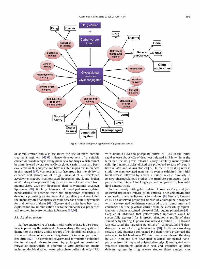
Fig. 5. Various therapeutic applications of glycosylated carriers.
K. Jain et al. / Biomaterials 33 (2012) 4166e4186 4173
of administration and also facilitates the use of more chronictreatment regimens [65,66]. Hence development of a suitablecarrier for oral delivery is always beneficial for drugs, which cannotbe administered by oral route. Glycosylated carriers have also beenevaluated for this purpose and have resulted in positive inferencesin this regard [67]. Mannose as a surface group has the ability toenhance oral absorption of drugs. Pukanud et al. developedacyclovir entrapped mannosylated liposomes and found higherin vitro drug absorptions through everted sacs of mice ileum frommannosylated acyclovir liposomes than conventional acyclovirliposomes [68]. Similarly, Salman et al. developed mannosylatednanoparticles to describe their gut bioadhesive properties todevelop a promising carrier for oral drug delivery and concludedthatmannosylated nanoparticles could serve as a promising vehiclefor oral delivery of drugs [69]. Glycosylated carrier have been alsoexplored for oral immunization due to their bioadhesive propertiesand resulted in overwhelming inferences [69,70].
5.3. Sustained release
Surface engineering of carriers with carbohydrate is also bene-ficial in providing the sustained release of drugs. The conjugation ofdextran to the surface amino groups of PPI dendrimers results insustained release of anticancer drug doxorubicin in comparison tofree drug [52]. The developed glycosylated formulation exhibitedthe initial rapid release followed by prolonged and sustainedrelease of doxorubicin in different in vitro dissolution mediaincluding double distilled water, phosphate buffer saline (pH 7.4)
with albumin (1%) and phosphate buffer (pH 6.8). In the initialrapid release about 40% of drug was released in 3 h, while in thelater half the drug was released slowly. Similarly mannosylatedsolid lipid nanoparticles elicited the prolonged release of drug inboth in vitro and in vivo studies [72]. In the in vitro drug releasestudy the mannosylated nanometric system exhibited the initialburst release followed by slower sustained release. Similarly inin vivo pharmacokinetic studies the mannose conjugated nano-particles was retained for longer period compared to plain solidlipid nanoparticles.
In their study with galactosylated liposomes Garg and Jainobserved prolonged release of an antiviral drug azidothymidinecompared to uncoated liposomal formulation [9]. Similarly Agrawalet al. also observed prolonged release of Chloroquine phosphatewith galactosylated dendrimers compared to plain dendrimers andconcluded that the galactose carrier could be successfully capital-ized on to obtain sustained release of Chloroquine phosphate [53].Garg et al. observed that galactosylated liposomes could besuccessfully explored for improved therapeutic profile of drugstavudine by altering its pharmacokinetic disposition [8]. Dutta andJain evaluated the targeting potential of mannosylated PPI den-drimers for anti-HIV drug lamivudine [18]. In the in vitro drugrelease study mannose conjugated PPI dendrimers prolonged therelease up to 144 h whereas PPI dendrimers has released the drugby 24 h. Kim and Kim developed galactose conjugated nano-particles from biotinylated poly(ethylene glycol) conjugated withgalactose containing lactobionic acid and evaluated as drugdelivery system. In drug release studies these nanoparticles

K. Jain et al. / Biomaterials 33 (2012) 4166e41864174
displayed the pseudo zero-order release pattern during a period ofone month. From all these studies the author postulated that theglycosylation of drug delivery system may result in a promisingdrug carrier to provide sustained release of drugs [73].
5.4. In treatment of cancer
Malignant transformation occurs as a result of many molecularchanges, and glycosylation of glycoproteins and glycolipids is one ofthem [74]. This abnormal glycosylation of cancerous tissue escortsto expression of various surface binding lectin receptors havinghigh affinity for ligands having surface carbohydrate molecules [1].This piece of evidence landed an important application of glyco-conjugated carriers in targeted delivery of anticancer agents tocancerous tissue [75]. Conjugation of dextran to amine terminals ofdendrimers enhances the uptake into the cancer cells [52]. Incytotoxicity study performed in xenograft model of A549 tumorbearing cells, doxorubicin loaded PPI dendrimers displayed theincreased cytotoxicity compared to free doxorubicin. The increasein cytotoxicity was due to the better uptake of dextran conjugatedPPI dendrimers by the neoplastic cells through ligand receptor-mediated internalization [52]. Similarly mannosylated solid lipidnanoparticles showed higher uptake by A549 tumor cells in cellularuptake study and higher delivery to tumor mass in in vivo bio-distribution studies due to lectin receptor-mediated recognitionfollowed by endocytosis [4]. Galactose conjugated nanoparticleswere found efficient in targeting the anticancer drug, paclitaxel toliver cancer [76].
Tang et al. have explored the potential of oxidized and reducedmannan in receptor-mediated gene transfer for cancer immuno-therapy. Introduction of DNA encoding ovalbumin via mannan-poly-L-lysine was found to lead to protection of mice from tumorsin in vivo studies in C57BL/6 mice [39]. Kuramoto et al. evaluatedthe efficiency of glycosylated carrier in treatment of tumor viaeffective inhibition of peritoneal dissemination via immunotherapyusing immune-stimulatory CpG DNA. For this study the number oftumor cells was quantitatively evaluated by measuring luciferaseactivity in colon26/Luc cell lines. On intraperitoneal administrationto peritoneal dissemination model mice, mannosylated CpG DNAlipoplex were found more effective in inhibiting tumor cell prolif-eration in greater omentum and mesentery than CpG DNA lipoplexand even than galactosylated CpG DNA lipoplex. The high efficacyof mannosylated CpG DNA lipoplex in inhibition of proliferation oftumor cell was attributed to mannose receptor-mediated CpG DNAtransfer [13].
Matsui and coworkers developed oligomannose-coated lipo-somes as delivery system for anticancer drugs and found that thesecarriers were successfully taken up by mouse peritoneal macro-phages and the anticancer drugs were carried to metastatic sites inthe peritoneal cavity in mice. Further the scientist checked theclinical application of this delivery system to gastric cancer patientsby investigating whether the oligomannose-coated liposomescould be uptaken by human peripheral blood monocytes (PBMs)and human peritoneal macrophages (PEMs) to carry them tomicrometastatic foci in the mouse omentum and resectedomentum from cancer patients in ex vivo studies. For this investi-gation the authors incubated the oligomannose-coated liposomeswith PBMs obtained from four healthy volunteers and PEMs fromperitoneal washes of five gastric cancer patients. With PBMs theauthors found that on an average 88% of CD14-positive PBMs wastaken up the oligomannose-coated liposomes and an average 63%uptake rate was obtained with CD14-positive PEMs. Theseoligomannose-coated liposome incorporated PBMs and PEMs weresignificantly accumulated in micrometastatic foci at the omentumformed after intraperitoneal injection of GFP-tagged gastric cancer
cells into mice and in ex vivo studies to tumor foci in the surgicallyresected human omentum. Finally the authors suggested that theseoligomannose-coated liposomes could be successfully exploited aspotential carrier to deliver anticancer drugs to peritoneal micro-metastasis in the omentum of gastric cancer patients using humanmonocytes/macrophages as a cellular vehicle [77]. Galactoseconjugated fluorescent nanoparticles have also shown promise asa probe in detection of live liver cancer cells in a mixed cell system[78]. Peng and coworkers conjugated the lactobionic acid to amino-modified fluorescent silica nanoparticles through EDAC linkage todevelop galactose conjugated fluorescent nanoparticles. Thesegalactose nanoparticles were found efficient in identificantion ofliver cancer cells in the blood precisely as demonstrated by laserconfocal scanning microscopy and flow cytometric analysis.
The above accounts clearly establish that the glycosylatedcarriers have potential implication in cancer therapy as a deliverysystem for anticancer drugs, as targeted carrier for selectivedelivery of therapeutic agents in the vicinity of cancer cells and alsoas identification probe in the recognition of cancer cells in mixedcell system.
6. Biomedical applications of glycosylated carriers
Glycosylated carriers have been explored for cell-specificdelivery of genes, DNA, oligonucleotides and found advantageousin this application because of their ability to deliver the geneticmaterial to the intracellular compartment through lectin receptor-mediated endocytosis [79e81] (Figs. 6and 7). Also glycosylation hasbeen explored for reducing the cytotoxicity and imparting thebiocompatibility to non-viral transfection agents [81].
6.1. In gene delivery
Diebold et al. successfully evaluated the mannose conjugatedpolyethylenimine (PEI) as a transfection complex [40]. Thiscomplex was found efficient in transfecting the DNA in cellsexpressing mannose receptors. Conjugation of phosphorothioatedoligonucleotides to poly-L-lysine containing fucose moleculesresulted in increase in cellular uptake of oligonucleotide by 15times compared to free phosphorothioated oligonucleotides [15].Hashida et al. reviewed the gene delivery potential of glycosylatedliposomes and polymers. The authors compared the gene deliveryefficiency of galactosylated liposomes, poly (amino acids) andmannosylated liposomes. DNA-galactosylated cationic liposomecomplexes were found to be more efficient than DNA-cationicliposome complexes in DNA uptake and gene expression in theliver parenchymal cells, whereasmannosylated carriers were foundto be able to provide specific delivery of genes to non-parenchymalliver cells. Galactosylated and mannosylated carriers are internal-ized by asialoglycoprotein and mannose receptor positive cells,respectively and can be explored for cell-specific delivery of genes[79]. Glycosylated cationic liposomes have been used successfullyfor cell selective targeting of plasmid DNA, siRNA and NFkappaBdecoy [80,81]. Mannosylation of nanoparticles is accomplished todesign non-toxic nanoparticles for DNA transfection. Park et al.developed mesoporous silica nanoparticles to transfect plasmidDNA. These nanoparticles were also coupled with mannosylatedpolyethylenimine (MP) to enhance transfection efficiency ofnanoparticles through mannose receptors to target macrophagecells. Transfection efficiency of these complexes was evaluated onRaw 264.7 and HeLa cell lines and mannose conjugated complexesshowed enhanced transfection efficacy. This increase in trans-fection efficacy was attributed to mannose receptor-mediatedendocytosis [81]. Similarly oxidized mannan and reducedmannan conjugated to poly-L-lysine also resulted in improved gene

Fig. 6. Formation of glycosylated DNA-Carrier complex.
K. Jain et al. / Biomaterials 33 (2012) 4166e4186 4175
transfection aptitude as studied by Tang et al. Finally it can beconcluded that glycosylation of carriers result in enhanced trans-fection efficiency with biocompatibility thereforegene delivery isan important attribute in the therapeutic prospective of thesecarriers [39].
6.2. In vaccine delivery
Glycosylated carriers also have been used successfully as vaccinedelivery systems. Macrophages and dendritic cells expressmannose receptors, which play important role in immuneresponses to antigen by taking active part in phagocytosis, pro-cessing and presentation of antigen, cell migration and intracellularsignalling [34]. Jiang et al. have developed chitosan microspheresand mannose conjugated chitosan microspheres loaded with Bor-detella bronchiseptica antigens containing dermonecrotoxin (BBD).They evaluated the immune-stimulating activity of these vaccinedelivery systems on murine macrophages (RAW264.7 cells). In thisstudy BBD-loaded mannosylated chitosan microsphere showedbinding with mannose receptors present on the macrophages. Thein vivo immune-stimulating activity was evaluated in mice afterintranasal immunization with BBD-loaded chitosan and man-nosylated chitosan microspheres. The immune-stimulating activityof mannosylated chitosan microspheres was superior to bare chi-tosan microspheres. Again mice immunized with BBD-loadedmannosylated chitosan microspheres showed significantly higherIgA antibody response in saliva and serum. Hence mannoseconjugated carriers may serve as promising carrier for mucosaldelivery of antigen via interaction between mannose and mannosereceptors on macrophages [82]. Tang et al. also postulated thatoxidized and reduced mannan can be efficiently utilized as DNAvaccine for cancer immunotherapy [39].
Un et al. developed ultrasound-responsive and mannosemodified carrier, Man-PEG2000 bubble lipoplexes for gene trans-fection into antigen presenting cells (APCs). This method leads toabout 500e800 fold increase in gene expression in APCs in in-vivostudies compared to conventional lipofectionmethod. This increasewas attributed to ability of mannose conjugated carriers to selec-tively deliver the gene into targeted organs. Further ultrasoundexposure increased the transfection of nucleic acids into cytoplasm,which is themajor obstacle in delivery of gene via non-viral vectors.Finally the authors suggested that this delivery system could beexploited for vaccine therapy and anti-inflammation therapy toAPCs. These authors also found that this combination method was12 times more efficient in gene transfection to hepatic non-parenchymal cells than the hepatic parenchymal cells. It wasconcluded that the combination method based on mannosylatedlipoplexes and bubble lipoplexes with ultrasound exposure is verysignificant for gene transfection to macrophages and dendritic cells[83,84].
Glycosylated carriers could also be exploited as carrier for oralimmunization. Fievez et al. evaluated the potential of mannose asligand in oral immunization with nanoparticles. Potential of non-peptidic ligand in targeting of nanoparticles to M-cells for oralimmunization was investigated. Mannose was incorporated inPLGA-based nanoparticles after grafting on PEG chains. The authorsfound higher production of IgG antibodies on intraduodenalimmunization with mannose-labeled nanoparticles in comparisionto free ovalbumin given intramuscularly or non-targeted or RGD-nanoparticles administered via intraduodenal route. The highproduction of IgG antibodies was ascribed to higher uptake bymacrophages. So the grafting of mannose on the surface nano-particles can result in a carrier suitable for oral immunization andcan influence the elicitation of immune response positively [70].

Fig. 7. Glycosylated carrier mediated delivery of oligonucleotide via lectin receptor-mediated endocytosis.
K. Jain et al. / Biomaterials 33 (2012) 4166e41864176
Also conjugation of mannose to bioadhesive polyanhydride nano-particles resulted in strong long lasting systemic and mucosalimmune responses on oral immunization using ovalbumin asantigen model. On subcutaneous as well as oral administration themannose coated bioadhesive nanoparticles educed systemic andmucosal immune responses attributable to elevated and balancedantibody responses [71].
7. Diagnostic applications
Apart from the therapeutic and biomedical applications, glyco-sylated carriers have also showed potential as diagnostic agent.Sialic acid and fucose terminating glycotopes expressed by plasmafibronectins amniotic fluids play important role in implantation,growth and differentiation of fetal tissues. The different importantstages of pregnancy including second and third trimester, perinatalperiod, delivery and post-date pregnancy are associated withchanges in the exhibition of terminal sialic acid and fucose isoformsin the amniotic fluid glycoconjugates to ensure homoeostasis
during pregnancy and to protect the fetus. So presence of glyco-topes on the seminal fibronectins, amniotic fluid and importantrole of these glycotopes in pregnancy and fetus maturity ensurestheir aptitude as clinically applicable biomarkers to monitor preg-nancy, in vitro fertilization and to evaluate the ejaculate of infertilemen in obstetrics and andrology [85,86].
In vitro organ imaging is another perspective in the diagnositicapplication of glycosylated carriers. Kikkeri et al. developed d-mannose, d-galactose and d-galactosamine capped PEGylatedquantum dots and evaluated for in vitro imaging and in vivo livertargeting [87]. Benito et al. developed the mannosylated dendriticb-cyclodextrin and evaluated for the binding affinity with thetetrameric plant lectin Con A and mannose/fucose specific receptorof macrophages. In addition the authors also observed that theresultant complex has high solubilization capacity for anticanceragent docetaxel along with proficient affinity for lectin receptors.This insinuates applicability of glycosylated carrier in solubilizationof poorly soluble drugs [88]. Glycosylated carriers have also beenevaluated for photodynamic therapeutic efficiency. For this

K. Jain et al. / Biomaterials 33 (2012) 4166e4186 4177
micelles based on amphiphilic copolymer galactosyl and mono-aminoporphyrin incorporated poly(2-aminoethyl methacrylate)-polycaprolactone and conjugated with porphyrin and galactosewas developed and evaluated biologically in human laryngealcarcinoma (HEp2) and hepatocellular liver carcinoma (HepG2)cells. It was observed that porphyrin and galactosyl conjugatedpolymer micelles showed higher targeting and photodynamictherapeutic efficacy in HepG2 cells in comparison to HEp2 cells[89]. The result of this study entails the possible application ofglycosylated carriers in photodynamic therapy with porphyrin.
8. Reduction in the toxicity
Nanometric delivery systems like nanoparticles, dendrimersbrought great development in the area of drug delivery, but besidesof their biomedical application they also induce toxicity because oftheir effective and specific interaction with vital cell components
Fig. 8. Reduction in toxicity of drug carrier wit
like plasma membrane, mitochondria, nucleus, endosome andenzymes owing to their nanometric size i.e. 1e100 nm. Surfaceengineering of these carriers with different non-toxic and biode-gradable ligands is one of the strategies to alleviate these toxicities[72] (Fig. 8). Different carbohydrate moieties like mannose, lactose,dextran, fucose, lactose and galactose have been conjugated withvarious drug delivery systems and elucidated significantly reducedtoxicity [8,50,53,90,91] (Table 3).
8.1. Drug carriers
Lherm et al. evaluated the cytotoxicity of cyanoacrylate nano-particles and also the effect of alkyl chain length on the toxicity ofcyanoacrylate nanoparticles. The authors ascertained the cytotox-icity of cyanoacrylate particles in L929 fibroblast cell cultures andfound that the ethyl and isobutyl cyanoacrylate nanoparticles weremost toxic followed by methyl derivative with the isohexyl
h surface cationic charge via glycosylation.

Table 3Effect of glycosylation on the hemolytic toxicity, cytotoxicity and hematological toxicity of various drug carriers.
Type of carrier Type of study Observations References
Formulation Leucocytes(�103/ml)
Erythrocytes(�103/ml)
Hb (g/dl)
Galactosylatedliposomes
Hematologicaltoxicity in malealbino rats(Sprague-Dawleystrain)
Control 10.4 � 1.3 9.2 � 0.7 13.4 � 0.2 [9]Free azidothymidinesolution
5.9 � 0.2(After 10 days)
6.0 � 0.6(After 10 days)
8.2 � 0.5(After 10 days)
Galactosylatedliposomalazidothymidine
10.1 � 0.3(After 10 days)
9.2 � 0.2(After 10 days)
13.3 � 0.6(After 10 days)
Type of carrier Type of study Observations References
Formulation Bone marrow cells (femur �106)
Galactosylatedliposomes
Bone marrowcellularity in malealbino rats(Sprague-Dawleystrain)
Control 12.9 � 1.2 [9]Free azidothymidinesolution
8.0 � 0.6(After 10 days)
Galactosylatedliposomalazidothymidine
12.8 � 0.5(After 10 days)
Type of carrier Type of study Observations References
Formulation % Haemolysis(after 4 h at 1 mg/mlconcentration)
CarbohydrateconjugatedPPI-5.0 Gdendrimers
Hemolytic toxicity PPI-5.0 G dendrimers 86.2 � 0.6 [50]Mannose conjugatedPPI-5.0 G dendrimers
2.9 � 0.6
Lactose conjugatedPPI-5.0 G dendrimers
2.2 � 0.3
Type of carrier Type of study Observations References
Formulation % Cell viability (after 72 h at1 mg/ml concentration)
HepG2 cell lines COS-7 cell lines
CarbohydrateconjugatedPPI-5.0 Gdendrimers
Cytotoxicity inHepG2 and COS-7cell lines
PPI-5.0 G dendrimers 1.7 � 1.1 2.3 � 1.2 [50]Mannose conjugatedPPI-5.0 G dendrimers
97.6 � 1.2 96.7 � 1.2
Lactose conjugatedPPI-5.0 G dendrimers
94.7 � 1.1 95.8 � 1.3
Type of carrier Type of study Observations References
Formulation RBC WBC Hb HCT MCH
CarbohydrateconjugatedPPI-5.0 Gdendrimers
Hematologicaltoxicity in malealbino rats(Sprague-Dawleystrain)
Control 7.15 � 1.37 8.25 � 1.67 14.89 � 1.35 41.69 � 5.51 20.76 � 1.35 [50]PPI-5.0 G dendrimers 4.43 � 1.15 12.01 � 1.23 8.26 � 1.39 27.06 � 3.16 18.82 � 1.06Mannose conjugatedPPI-5.0 G dendrimers
7.36 � 1.01 8.24 � 1.89 13.46 � 1.78 41.82 � 4.32 18.41 � 0.53
Lactose conjugatedPPI-5.0 G dendrimers
7.24 � 1.24 8.37 � 1.46 13.01 � 1.59 41.02 � 4.62 18.15 � 1.21
Type of carrier Type of study Observations References
Formulation RBC count(�106/ml)
WBC count(�106/ml)
Galactosylatedpoly-L-lysine(PLL) 4.0Gdendrimers
Hematologicaltoxicity in malealbino rats
Control 9.1 � 0.3 10.8 � 0.6 [53]Drug (Chloroquine) 8.4 � 0.2 10.7 � 0.5PLL-Chloroquine 7.5 � 0.3 15.2 � 0.4Galactose conjugatedPLL-Chloroquine
8.8 � 0.2 11.3 � 0.4
K. Jain et al. / Biomaterials 33 (2012) 4166e41864178
cyanoacrylate nanoparticles being the least toxic. The mechanismof cytotoxicity was also evaluated and the degradation of nano-particles in the culture medium and adherence of particles tocell membrane was found to be the main reason for toxicity ofnanoparticles. So the possible toxicity of these carriers may bealleviated by using the carriers based on the material likepoly(hydroxybutyric acid) and poly(lactic acid), which undergodegradation comparatively slowly [90]. This toxicity problemassociated with nanocarriers may be reduced by two strategies,first one being the use of biodegradable material for preparation ofnanocarriers, and the second is surface modification of carrier withsome non-toxic ligands like carbohydrates, polyethylene glycol etc
[72]. When Amphotericin B is loaded in gelatin nanoparticles,where gelatin is an example of biodegradable polymer, the result-ing gelatin nanoparticles showed the reduction in toxicity. Inhematological toxicity studies reduction in the amount of hemo-globin concentration and hematocrit was found to be non-significant. Simultaneously nephrotoxicity, which was elucidatedby increase in blood urea nitrogen and serum creatinine levels, wasalso found to be insignificant with gelatin nanoparticles whereasAmphotericin B had significant nephrotoxicity [91]. Surface engi-neering of carrier with natural ligands also decreases the toxicity.The effect of galactosylated liposomes on the hepatic toxicity,cellular uptake and pharmacokinetics of stavudine and

K. Jain et al. / Biomaterials 33 (2012) 4166e4186 4179
azidothymidine was investigated [8,9]. The authors found that thegalactosylated liposomes entrapped stavudine did not show thehematological and hepatic toxicity with increased uptake of gal-actosylated formulations by liver parenchymal cells harvested frommale albino rats in ex vivo cellular uptake studies. Similarlyhematological toxicity was not observed for 10 days with azido-thymidine loaded galactosylated vesicles.
8.2. Drug
With reduction in toxicity of carriers conjugation of carbohy-drates to drug carriers also reduces toxicity related to drug. Agrawalet al. investigated the potential of dextran conjugated PPI den-drimers as carrier for a model anticancer agent, doxorubicinhydrochloride [52]. The authors investigated the hemolytic toxicityand cytotoxicity of developed formulation. Plain PPI dendrimersand doxorubicin showed considerable hemolytic toxicity i.e. 19.8%and 16.7% respectively; whereas doxorubicin loaded dextranconjugated PPI dendrimers displayed hemolytic toxicity up to12.3%, which was lesser than plain dendrimers and drug at equiv-alent concentration. The reduction in toxicity was ascribed toshielding of surface amino groups of dendrimers and drug bydextran. Similarly chloroquine phosphate loaded galactosylatedpoly-L-lysine dendrimers elicited lower hemolytic toxicitycompared to plain drug chloroquine phosphate and plain poly-L-lysine dendrimers. This reduction in toxicity of carrier and drugwasattributed to shielding of drug in glycosylated dendrimers. Jainet al. prepared mannosylated solid lipid nanoparticles and evalu-ated for site-specific delivery of anticancer drug doxorubicinhydrochloride. In the hemolytic toxicity study the authors observed17.1 � 0.3%, 11.4 � 0.4% and 6.1 � 0.2% hemolysis with doxorubicinhydrochloride, solid lipid nanoparticles and mannosylated solid
Fig. 9. Scheme for lectin receptor-mediated delivery of glycosylated carriers (a) association oEarly endosome, (d) Endosomal disruption, (e) release of drug molecules in to cytoplasm, a
lipid nanoparticles, respectively. Evidently mannosylation ofnanoparticles reduced the hemolytic toxicity of drug and solid lipidnanoparticles because of biocompatibility and biodegradability ofmannose. In addition to reduction in hemolytic toxicity; nephro-toxicity and hepatotoxic effects of doxorubicin hydrochloride werealso reduced significantly with mannosylated solid lipid nano-particles. The nephro- and hepatic toxicity of doxorubicin hydro-chloridewas assessed bymeasuring the level of urea/creatinine andALT/AST/ALP in serum. Significant increase in creatinine and urealevel, ALT/AST/ALP level was observed with plain doxorubicintreated group of Balb/c mice whereas increase was minimal withmannosylated solid lipid nanoparticles [4].
All these studiesclearly establish the beneficial role of glycosy-lated carriers in reduction of toxicity of drug and as well as drugcarrier because of their biodegradable and macromolecular nature.
9. Site-specific delivery
As already discussed the different cell surfaces express lectinreceptors for various carbohydrate molecules and these receptorscan internalizes the carriers having carbohydrate ligands on theirsurface (Fig. 9). This forms the basis for attaining the targeteddelivery to different organs exploring glycosylated carriers.
9.1. Lungs
Mannose receptors are prominently expressed on the surface ofalveolar macrophages [35,92]. Kumar et al. developed mannosy-lated 5.0G EDA-cored PPI dendrimers for selective delivery ofrifampicin to alveolar macrophages for treatment of tuberculosis.The developed formulation was evaluated for drug loading, in-vitrorelease profile, hemolytic toxicity, cytotoxicity and phagocytic
f glycosylated carrier with lectin receptors, (b) lectin receptor-mediated endocytosis, (c)nd (f) recycling of lectin receptors.

K. Jain et al. / Biomaterials 33 (2012) 4166e41864180
alveolar macrophage uptake and from the results of all thesestudies the authors concluded that the developed dendrimericarchitecture could serve as promising delivery system for selectivedelivery of rifampicin to lung alveolar macrophages, which are thetarget site for tuberculosis [14]. Chono et al. also observed thatmannosylation increases the drug targeting efficiency of liposomesto alveolar macrophages for treatment of respiratory intracellularparasitic infections. On pulmonary administration the targetingefficiency of ciprofloxacin loaded mannosylated liposomes wasconsiderably greater than ciprofloxacin loaded unmodified lipo-somes [93].
Inhalable microparticles facilitate drug targeting to lungs andparticularly to alveolar macrophages [94]. Chono et al. investigatedthe influence of mannose conjugation on aerosolized liposomaldrug delivery to alveolar macrophages. In the in vitro and in vivoexperimental studies the higher uptake rate was observed formannosylated liposomes over bared liposomes. The uptake ofmannosylated liposomes by the alveolar macrophages was inter-ceded by mannose receptor-mediated endocytosis. The mannosy-lated liposomes were selectively delivered to the alveolarmacrophages but did not harm the lung tissues [95]. Wijagkanalanet al. evaluated the efficiency of mannose in targeting alveolarmacrophages on intratracheal administration of mannosylatedliposomes. In this study the authors administered the bared lipo-somes and mannosylated liposomes in rats via intratracheal routeand observed appreciably selective targeting to alveolar macro-phages through mannose receptor-mediated endocytosis [96].
9.2. Liver
The parenchymal cells (hepatocytes) and non-parenchymal cells(Kupffer cells and endothelial cells) of liver express the receptorsfor galactose and mannose [24,25,27]. Kupffer cells also articulatefucose receptors having affinity for glycoproteins with fucoseresidues [45,97]. These receptors have been explored by manyscientists for targeted delivery of various therapeutic agents to livercells.
Akamatsu et al. evaluated the drug or gene carrier potential ofglycosylated poly (amino acids) to the liver cells. The scientistsconjugated the galactoside and mannoside to poly-L-glutamic acidand poly-L-lysine to develop galactosylated and mannosylatedcarriers, respectively. Both of the carriers were found to be able todeliver the drugs and/or polynucleotides to the liver but to thedifferent types of liver cells. Galactosylated derivatives wereselectively taken up by the liver parenchymal cells whereas man-nosylated carriers were preferentially taken up by liver non-parenchymal cells [98]. In the in vivo pharmacokinetic and dispo-sition study performed by Yeeprae and coworkers, the hepaticuptake clearances of mannosylated and fucosylated emulsionswere respectively, 3.3 and 4.0 times higher in comparison to thebare emulsions. Uptake ratio of 2.0 and 2.9 was observed formannosylated and fucosylated emulsions by non-parenchymal aswell as parenchymal cells [99]. The results of pharmacokineticstudies supports liver targeted perspectives of mannosylated andfucosylated emulsions. Further mannosylated emulsion has beenexploited to target the drug to liver non-parenchymal cells viamannose receptor-mediated internalization. Increasing the densityof mannose in mannosylated emulsion lead to preferential accu-mulation to the liver non-parenchymal cells compared to emulsionwith low density of mannose and plain emulsion [100]. Kawakamiet al. compared the biodistribution pattern of mannosylated,fucosylated and galactosylated liposomes in mice. Liposomessynthesized for this study was composed of dis-tearoylphosphatidylcholine (DSPC) and cholesterol. The gal-actosylated, mannosylated and fucosylated liposomes were
prepared using cholesten-5-yloxy-N-(4-((1-imino-2-beta-D-thio-galactosyle thyl)amino)alkyl)formamide (Gal-C4-Chol), cholesten-5-yloxy-N-(4-((1-imino-2-beta-D-thiomannosyle thyl)amino)alkyl)formamide (Man-C4-Chol) and cholesten-5-yloxy-N-(4-((1-imino-2-beta-D-thiofucosyle thyl)amino)alkyl)formamide (Fuc-C4-Chol),respectively. In biodistribution studies in mice, unmodified lipo-somes consisting of DSPC and cholesterol were found to be retainedin the circulating blood for long time whereas glycosylated lipo-somes were found to be rapidly eliminated from circulating bloodand recovered in liver preferentially. The uptake by different typesof liver cells was different for mannosylated, galactosylated andfucosylated liposomes. Galactosylated liposomes were preferen-tially taken up by parenchymal cells whereas mannosylated andfucosylated liposomes were preferentially taken up by non-parenchymal cells of liver. Further it was reported that galactosy-lated, mannosylated and fucosylated liposomes were taken up bythe asialoglycoprotein receptors in parenchymal cells, mannosereceptors in non-parenchymal cells and fucose receptors in non-parenchymal cells, respectively. The uptake ratio by parenchymalcells and non-parenchymal cells were found to be 0.2, 0.6 and 15.1for fucosylated, mannosylated and galactosylated liposomesrespectively [101]. Nag and Ghosh also assessed the targetingpotential of mannosylated and galactosylated sterically stabilizedliposomes to different cell types of mouse liver and observed thatgalactosylated liposomes were preferentially taken up by paren-chymal cells of liver whereas mannosylated liposomes shifted thedistribution preferentially to Kupffer cells [102].
Fiume et al. explored the presence of lectin receptors on livercells to optimize the treatment of chronic hepatitis B by antiviraldrugs. The authors conjugated the antiviral nucleoside drugs withtwo glycoproteins having galactosyl terminal; asialofetuin andlactosaminated albumin, and observed selective delivery of theseconjugates to liver cells [103]. Frese et al. reviewed the potential ofglycoprotein carriers for targeted delivery of genes to hepatocytesvia ligand receptor-mediated uptake [104]. Julyan et al. evaluatedthe targeting efficiency of galactosylated carrier in phase I clinicaltrial study of HPMA copolymers bearing doxorubicin and galac-tosamine. In this study the pharmacokinetics was determined bygradient high performance liquid chromatography (HPLC) assess-ment of plasma, urine; and 123I-based imaging in the patient havingmultifocal hepatoma and enrolled first in phase I clinical trial study.In thewhole body imaging it was observed that 30% of the drugwasdelivered in the hepatic region. Finally scientists concluded thatdrugs can be effectively targeted to the hepatic cells using gal-actosylated carriers [105]. Garg et al. loaded an antiviral drug, sta-vudine, in galactosylated liposomes and observed that the resultantformulation showed enhanced uptake by liver parenchymal cells(27.96 � 2.41 pg d4T/million cells) [8]. Nishikawa et al. haveexplored the potential of a delivery system having galactoseterminals in targeted delivery of gene to hepatocytes. In this studyauthors synthesized a multifunctional gene delivery system byconjugating galactose followed by fusigenic peptide (mHA2) toa cationic polymer, poly(L-ornithine) and observed a 280-foldincrease in transgenic product (luciferase) compared to lip-oplexes. This increase in gene transfection efficiency was attributedto asialoglycoprotein receptor-mediated endocytosis of gene intothe hepatocytes [26]. Cho et al. again exploited galactose for tar-geting hepatocyte via asialoglycoprotein receptors. The researchersdeveloped the galactose coated and all-trans-retinoic acid (ATRA)loaded poly(L-lactic acid) nanoparticles for studying the effect ofretinoic acid released from nanoparticles on the morphology andDNA synthesis of hepatocytes. The nanoparticles were prepared bydiafiltration method. Receptor-mediated endocytosis of nano-particles was checked by fluorescence and confocal laser micros-copy and it was concluded that the galactose coated nanoparticles

Fig. 10. Lectin receptor-mediated targeting to macrophages.
K. Jain et al. / Biomaterials 33 (2012) 4166e4186 4181
were internalized via receptor mediate endocytosis by hepatocytes.Also, the retinoic acid released from drug loaded nanoparticlesresults in greater stimulation of hepatocyte DNA synthesiscompared to free retinoic acid in the presence of epidermal growthfactor (EGF). This increase in efficiency was attributed to controlledrelease of retinoic acid from galactosylated nanoparticles [106]. Seoet al. evaluated the Poly(N-p-vinylbenzyl-4-o-b-d-galactopyr-anosyl-d-gluconamide) (PVLA) nanoparticles as a candidate fortargeting hepatocytes. In this study the potential of PVLA nano-particles for receptor-mediated delivery of ATRA was investigated.It is well known that ATRA is important in regulation of CYP26 geneexpression in hepatocytes. ATRA-loaded PVLA nanoparticlesinduced the CYP26A1 gene in hepatocytes, as analyzed by reversetranscription-polymerase chain reaction. This induction of genewas attributed to targeted delivery of ATRA to hepatocytes viaasialoglycoprotein receptor-mediated endocytosis of deliverysystem. Galactose has also been explored with solid lipid nano-particles (SLNs) for targeting hepatocytes [107]. Xu et al. preparedliver hepatoma targeted SLNs with galactosylated dio-leoylphosphatidyl ethanolamine. The developed targeted SLNswere evaluated for encapsulation efficiency and drug release,cellular toxicity and cellular uptake, therapeutic effect and toxicity,biodistribution and histology. Greater than 90% of anticancer drug,docetaxel, was encapsulated in the galactosylated SLNs. In thein vitro drug release study a low burst effect for the first day fol-lowed by sustained release for next 29 days was observed. Also,these targeted SLNs were found superior over non-targeted SLNs incytotoxiciy study in hepatocellular carcinoma cell line BEL 7402.The increase in antitumor efficacy was also observed in cellularuptake and biodistribution studies due to increased accumulationand better cellular uptake in hepatoma cell. These targeted SLNshave not shown any destructive effect on healthy liver as well asliver with fibrosis in histological examination. Finally, authorsconcluded that galactosylated SLNs could successfully utilize fortreatment of hepatocellular carcinoma and as carrier for docetaxel[108].
Higuchi et al. evaluated the fucosylated cationic liposomes fortargeted delivery of NFkappaB decoy to Kupffer cells. NFkappaBdecoy inhibits NFkappaB-mediated transcription, which leads toproduction of cytokine causing severe liver injury in endotoxicsyndrome. In intrahepatic distribution study it was monitored thatafter intravenous injection the Fuc-liposomes/NFkappaB decoycomplexes were preferentially accumulated in the non-parenchymal cells of the liver and the serum levels of TNFalpha,IFNgamma, ALT and AST were considerably reduced as judgedagainst NFkappaB decoy [109]. In overall conclusion glycosylatedcarriers can serve as promising drug delivery modules for selectivedelivery of drugs, enzymes, genes and oligonucleotides to variouscells of liver for treatment of hepatic disorders.
9.3. Monocyte macrophage system
Monocytes and macrophages express different types of recep-tors including scavenger receptors, integrins, mannose receptorsand Fc-receptors. Hence it should be possible to target these MPScells by conjugating various drug carriers with the ligands for thesereceptors (Fig. 10). Such targeting may play crucial role in thetreatment of different ailments, as monocytes and macrophagesplay significant role in a range of diseases, for example cancer,atherosclerosis, HIV and chronic inflammation because these cellsare intrinsically involved in innate immunity and perform thefunction of phagocytosis by engulfing their targets throughcomplement and Fc-receptor-mediated opsonin receptor-dependent mechanism and lectin, scavenger and stearylaminereceptors mediated opsonin receptor-independent mechanism
[31,110,111]. Along with this, in case of parasitic infection, theingested microorganisms are killed by lysosomes, but somemicroorganisms can survive and multiply intracellularly within themacrophages including Brucella abortus, Candida albicans, Leish-mania spp., Listeria monocytogens, Mycobacterium avium, Myco-bacterium leprae, Mycobacterium tuberculosis, Neisseriagonorrhoeae, Salmonella typhimurium [112]. Hence targeting ofantiparasitic drugs to infected macrophages could be an attractiveapproach in improving the therapeutic efficacy and reducing thetoxicity associated with these drug. This strategy appears prom-ising and worth exploring clinically.
Mannose receptors are possessed by the monocyte macro-phages, alveolar macrophages, astrocytes in brain, hepatocytes inliver etc [12,13,18,32,95]. Grafting of mannose on the surface ofnanoparticles influences the macrophage uptake positively andcould serve as promising targeted drug delivery system [70]. Fortreatment of Gaucher’s disease two enzymes; Ceredase containingremodeled human placental-derived b-glucocerebrosidase andCerezyme, a recombinant form of b-glucocerebrosidase are used.For effective enzyme replacement therapy in Gaucher’s disease,Ceredase and Cerezyme are remodeled to expose core mannoseresidues by removal of terminal sialic acid, galactose, and N-acetyl-glucosamine sugars. This remodeling facilitates the delivery ofenzyme to macrophages, which are the target site for macrophagesvia mannose receptor-mediated endocytosis by macrophages [29].
Macrophages express mannose receptors, which can recognizecarbohydrates and function as immune receptors that engulfglycoproteins via endocytosis [113]. This piece of evidence has beenexplored by many scientists for targeting various therapeuticagents to macrophages. Doxorubicin was actively targeted to themacrophages via mannosylated liposomes for treatment of exper-imental visceral leishmaniasis by Kole et al. [114]. Doxorubicinloaded mannosylated liposomes were found to be more efficient intargeting doxorubicin to macrophages than doxorubicin loadedliposomes or free doxorubicin [114]. Further, Kuramoto et al.observed mannosylated CpG DNA lipoplex to be more effective inproduction of tumor necrosis factor (TNF)-a and in inhibition oftumor cell proliferation in greater omentum and mesenteryin peritoneal dissemination model mice on intraperitoneal
K. Jain et al. / Biomaterials 33 (2012) 4166e41864182
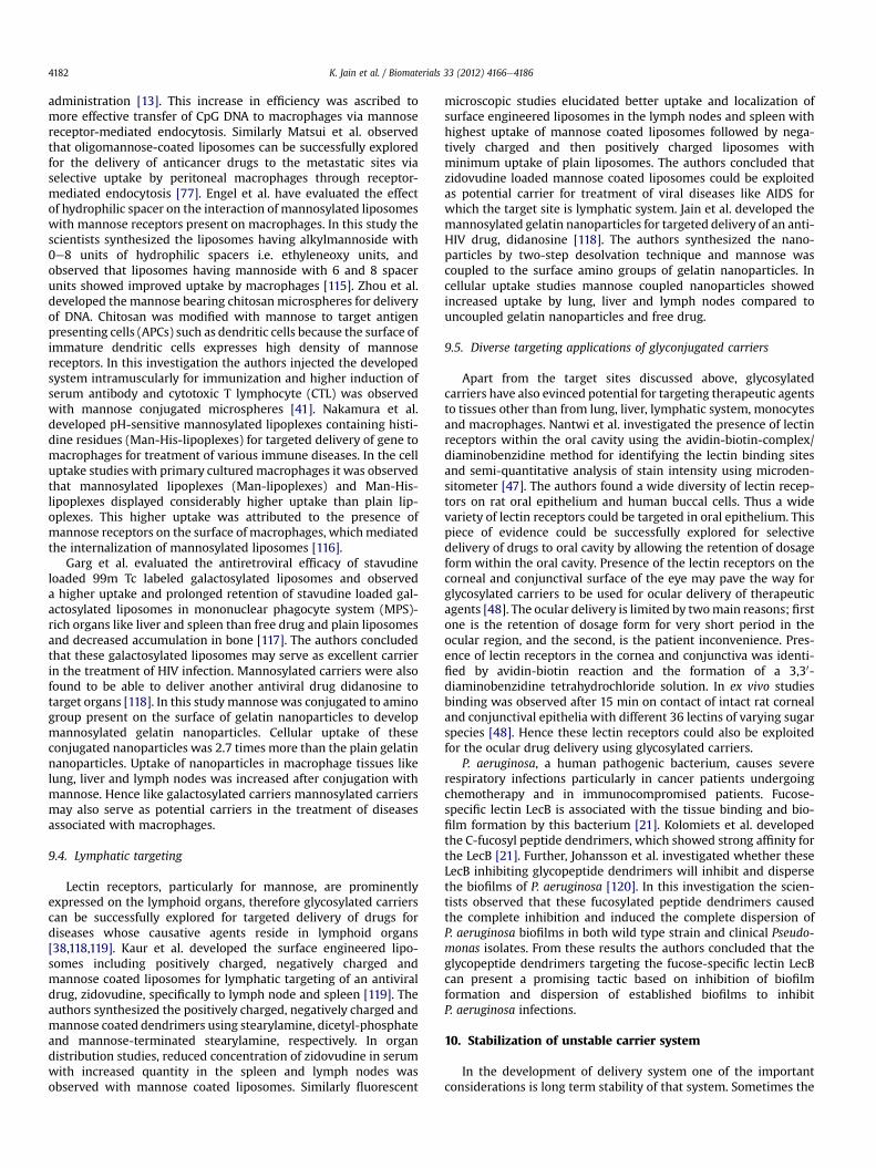
administration [13]. This increase in efficiency was ascribed tomore effective transfer of CpG DNA to macrophages via mannosereceptor-mediated endocytosis. Similarly Matsui et al. observedthat oligomannose-coated liposomes can be successfully exploredfor the delivery of anticancer drugs to the metastatic sites viaselective uptake by peritoneal macrophages through receptor-mediated endocytosis [77]. Engel et al. have evaluated the effectof hydrophilic spacer on the interaction of mannosylated liposomeswith mannose receptors present on macrophages. In this study thescientists synthesized the liposomes having alkylmannoside with0e8 units of hydrophilic spacers i.e. ethyleneoxy units, andobserved that liposomes having mannoside with 6 and 8 spacerunits showed improved uptake by macrophages [115]. Zhou et al.developed the mannose bearing chitosan microspheres for deliveryof DNA. Chitosan was modified with mannose to target antigenpresenting cells (APCs) such as dendritic cells because the surface ofimmature dendritic cells expresses high density of mannosereceptors. In this investigation the authors injected the developedsystem intramuscularly for immunization and higher induction ofserum antibody and cytotoxic T lymphocyte (CTL) was observedwith mannose conjugated microspheres [41]. Nakamura et al.developed pH-sensitive mannosylated lipoplexes containing histi-dine residues (Man-His-lipoplexes) for targeted delivery of gene tomacrophages for treatment of various immune diseases. In the celluptake studies with primary cultured macrophages it was observedthat mannosylated lipoplexes (Man-lipoplexes) and Man-His-lipoplexes displayed considerably higher uptake than plain lip-oplexes. This higher uptake was attributed to the presence ofmannose receptors on the surface of macrophages, whichmediatedthe internalization of mannosylated liposomes [116].
Garg et al. evaluated the antiretroviral efficacy of stavudineloaded 99m Tc labeled galactosylated liposomes and observeda higher uptake and prolonged retention of stavudine loaded gal-actosylated liposomes in mononuclear phagocyte system (MPS)-rich organs like liver and spleen than free drug and plain liposomesand decreased accumulation in bone [117]. The authors concludedthat these galactosylated liposomes may serve as excellent carrierin the treatment of HIV infection. Mannosylated carriers were alsofound to be able to deliver another antiviral drug didanosine totarget organs [118]. In this study mannosewas conjugated to aminogroup present on the surface of gelatin nanoparticles to developmannosylated gelatin nanoparticles. Cellular uptake of theseconjugated nanoparticles was 2.7 times more than the plain gelatinnanoparticles. Uptake of nanoparticles in macrophage tissues likelung, liver and lymph nodes was increased after conjugation withmannose. Hence like galactosylated carriers mannosylated carriersmay also serve as potential carriers in the treatment of diseasesassociated with macrophages.
9.4. Lymphatic targeting
Lectin receptors, particularly for mannose, are prominentlyexpressed on the lymphoid organs, therefore glycosylated carrierscan be successfully explored for targeted delivery of drugs fordiseases whose causative agents reside in lymphoid organs[38,118,119]. Kaur et al. developed the surface engineered lipo-somes including positively charged, negatively charged andmannose coated liposomes for lymphatic targeting of an antiviraldrug, zidovudine, specifically to lymph node and spleen [119]. Theauthors synthesized the positively charged, negatively charged andmannose coated dendrimers using stearylamine, dicetyl-phosphateand mannose-terminated stearylamine, respectively. In organdistribution studies, reduced concentration of zidovudine in serumwith increased quantity in the spleen and lymph nodes wasobserved with mannose coated liposomes. Similarly fluorescent
microscopic studies elucidated better uptake and localization ofsurface engineered liposomes in the lymph nodes and spleen withhighest uptake of mannose coated liposomes followed by nega-tively charged and then positively charged liposomes withminimum uptake of plain liposomes. The authors concluded thatzidovudine loaded mannose coated liposomes could be exploitedas potential carrier for treatment of viral diseases like AIDS forwhich the target site is lymphatic system. Jain et al. developed themannosylated gelatin nanoparticles for targeted delivery of an anti-HIV drug, didanosine [118]. The authors synthesized the nano-particles by two-step desolvation technique and mannose wascoupled to the surface amino groups of gelatin nanoparticles. Incellular uptake studies mannose coupled nanoparticles showedincreased uptake by lung, liver and lymph nodes compared touncoupled gelatin nanoparticles and free drug.
9.5. Diverse targeting applications of glyconjugated carriers
Apart from the target sites discussed above, glycosylatedcarriers have also evinced potential for targeting therapeutic agentsto tissues other than from lung, liver, lymphatic system, monocytesand macrophages. Nantwi et al. investigated the presence of lectinreceptors within the oral cavity using the avidin-biotin-complex/diaminobenzidine method for identifying the lectin binding sitesand semi-quantitative analysis of stain intensity using microden-sitometer [47]. The authors found a wide diversity of lectin recep-tors on rat oral epithelium and human buccal cells. Thus a widevariety of lectin receptors could be targeted in oral epithelium. Thispiece of evidence could be successfully explored for selectivedelivery of drugs to oral cavity by allowing the retention of dosageform within the oral cavity. Presence of the lectin receptors on thecorneal and conjunctival surface of the eye may pave the way forglycosylated carriers to be used for ocular delivery of therapeuticagents [48]. The ocular delivery is limited by twomain reasons; firstone is the retention of dosage form for very short period in theocular region, and the second, is the patient inconvenience. Pres-ence of lectin receptors in the cornea and conjunctiva was identi-fied by avidin-biotin reaction and the formation of a 3,30-diaminobenzidine tetrahydrochloride solution. In ex vivo studiesbinding was observed after 15 min on contact of intact rat cornealand conjunctival epithelia with different 36 lectins of varying sugarspecies [48]. Hence these lectin receptors could also be exploitedfor the ocular drug delivery using glycosylated carriers.
P. aeruginosa, a human pathogenic bacterium, causes severerespiratory infections particularly in cancer patients undergoingchemotherapy and in immunocompromised patients. Fucose-specific lectin LecB is associated with the tissue binding and bio-film formation by this bacterium [21]. Kolomiets et al. developedthe C-fucosyl peptide dendrimers, which showed strong affinity forthe LecB [21]. Further, Johansson et al. investigated whether theseLecB inhibiting glycopeptide dendrimers will inhibit and dispersethe biofilms of P. aeruginosa [120]. In this investigation the scien-tists observed that these fucosylated peptide dendrimers causedthe complete inhibition and induced the complete dispersion ofP. aeruginosa biofilms in both wild type strain and clinical Pseudo-monas isolates. From these results the authors concluded that theglycopeptide dendrimers targeting the fucose-specific lectin LecBcan present a promising tactic based on inhibition of biofilmformation and dispersion of established biofilms to inhibitP. aeruginosa infections.
10. Stabilization of unstable carrier system
In the development of delivery system one of the importantconsiderations is long term stability of that system. Sometimes the

K. Jain et al. / Biomaterials 33 (2012) 4166e4186 4183
issue related to the stability of delivery system creates majorobstacle in clinical application of formulation and hence greatemphasis need be placed in discovering the strategies to alleviatethe problem of instability of a drug delivery system. Glycosylationof drug carrier is also one of the promising strategies to solve theinstability problems of drug carriers. Applications of multifaceteddrug carrier liposomes are hindered by their instability. The baredliposomes are stable only for a month even if stored at a tempera-ture of 4 �C, which poses a major challenge in the clinical use ofliposomes. Coating of liposomes with carbohydrate molecules suchas galactose, mannose and fucose has been found to increase thestability of liposomes and these galactosylated, mannosylated andfucosylated liposomes were found to be stable at a temperature of25 �C � 2 �C with relative humidity of 60% � 5% and at 40 �C � 2 �Cwith 75% � 5% relative humidity [8]. The stability of formulationwas estimated by evaluating the influence of temperature andhumidity on the parameters including vesicle size, zeta potential,drug content, in vitro ligand agglutination assay, oxidation indexand thiobarbituric acid-reactive species determination. In all thesestudies carbohydrate coated dendrimers showed sufficient stabilityin comparison to uncoated liposomes.
11. Conclusion and future clinical prognosis
In the development of a safe and effective formulation one of theimportant aspects is the selective delivery of drug to the target siteand this can be achieved by receptor-mediated targeting of drug viaglycosylated carrier. Another vista in improving targeting efficiencyof these versatile carriers is based on receptor up-regulation.Dexamethasone up-regulates the mannose receptors present onmacrophages by increasing mRNA level induced synthesis of newmannose receptor protein [121,122]. This fact has been exploited forselective delivery of glucocerebrosidase to macrophages, which arethe affected cells in Gaucher’s disease [123]. Surfactant protein Aalso up-regulates the activity of the mannose receptor on humanmacrophages [124]. Discovery and development of some otheragent, which can up-regulate the lectin receptors, may further aidin designing of a dreamt formulation having therapeutic effect butdevoid of any adverse effect.
By way of chronological development of drug delivery systemfrom a simple drug carrier to engineered nanocarrier it may bepossible to realize the concept of complete targeting. The surfaceengineering of nanocarriers with carbohydrate ligands requiressimple conjugation reactions. Even a single step bioconjugationreaction based on click chemistry for glycosylation of drug carrier isalso possible. Various carbohydrate moieties such as mannose,lactose, galactose, fucose, dextran etc have been explored as ligandfor glycosylation but out of all these the most explored ligand ismannose, which has been surveyed for all the therapeutic potentialapplications of glycosylated carrier.
The discovery of high levels of lectin receptor expression onmacrophages, hepatocytes, liver endothelial cells, Kupffer cells,alveolar macrophages, monocytes and dendritic cells has renderedthe carbohydrate an attractive ligand for targeting therapeuticagents to these sites. It being the natural components of biologicalenvironment, carbohydrates serve as multifaceted carriers fortherapeutic agents. By providing biocompatibility and biodegrad-ability they help in alleviating the toxicity associated with nano-carriers. The prevalence of lectin receptors overexpression makes ita good marker for internalization of carbohydrates via receptor-mediated endocytosis. Also the glycosylation of drug carriers aidsin increasing the stability of unstable carriers like liposomes.Engineering with carbohydrate ligands assists in providing longcirculating properties to drug carriers, which results in sustainedrelease characteristics. Apart from providing targeting efficiency
and stability, glycosylation of drug carriers also aids in alteration ofphysicochemical properties to provide suitable pharmacokineticprofile to the delivery system.
Finally in the recapitulation of the therapeutic potential of gly-cosylated carrier, it seems to be a highly promising approach in thearena of novel drug delivery. Whatever the case may be, asmacromolecular tool glycosylated carriers appear to be the prom-ising in drug delivery as targeting ligand, non-cytotoxic andbiodegradable carrier with sufficient stability. The key to thedevelopment of an optimized formulation lies in fine tuning of thecarrier and its ligand. The glycosylated carriers have proved to beexcellent in this regard because they can be designed with a highgrade of sophistication, selectivity and specificity. Therefore,glycosylation approach holds great promise for future clinicaldiagnostic and therapeutic applications. Available evidence clearlysuggests glycosylation to be a valuable arsenal in the combatagainst some dreadful diseases.
List of abbreviations
APCs Antigen presenting cellsATRA All-trans-retinoic acidBBD Bordetella bronchiseptica antigens containing
dermonecrotoxinCon A Concanavalin ACTL Cytotoxic T lymphocyteDSPC DistearoylphosphatidylcholineEGF Epidermal growth factorELLA Enzyme-linked lectin assayGRAS Generally Referred As SafeHEp2 Human laryngeal carcinomaHepG2 Hepatocellular liver carcinomaHPLC High performance liquid chromatographyPEI PolyethylenimineMPS Mononuclear phagocyte systemNHS N-hydroxysuccinimideOPF O-palmitoylfucoseOPG O-palmitoylgalactoseOPM O-palmitoylmannosePBMs Peripheral blood monocytesPEMs Human peritoneal macrophagesPPI PolypropyleneiminePVLA Poly(N-p-vinylbenzyl-4-o-b-d-galactopyranosyl-d-
gluconamide)SLNs Solid lipid nanoparticlesTNF Tumor necrosis factor
References
[1] Agrawal A, Saraf S, Asthana A, Gupta U, Gajbhiye V, Jain NK. Ligand baseddendritic systems for tumor targeting. Int J Pharm 2008;350:3e13.
[2] Monsigny M, Roche AC, Midoux P, Mayer R. Glycoconjugates as carriers forspecific delivery of therapeutic drugs and genes. Adv Drug Deliv Rev 1994;14:1e24.
[3] Wang Q, Zhang L, Hu W, Hu ZH, Bei YY, Xu JY, et al. Norcantharidin-asso-ciated galactosylated chitosan nanoparticles for hepatocyte-targeteddelivery. Nanomed 2010;6:371e81.
[4] Jain K, Kesharwani P, Gupta U, Jain NK. Dendrimer toxicity: Let’s meet thechallenge. Int J Pharm 2010;394:122e42.
[5] Nahar M, Dutta T, Murugesan S, Asthana A, Mishra D, Rajkumar V, et al.Functional polymeric nanoparticles: an efficient and promising tool foractive delivery of bioactives. Crit Rev Ther Drug Carrier Syst 2006;23:259e318.
[6] Unverzagt C, Andre S, Seifert J, Kojima S, Fink C, Srikrishna G, et al. Structure-activity profiles of complex biantennary glycans with core fucosylation andwith/without additional a2,3/ a2,6 sialylation: synthesis of neoglycoproteinsand their properties in lectin assays, cell binding, and organ uptake. J MedChem 2002;45:478e91.

K. Jain et al. / Biomaterials 33 (2012) 4166e41864184
[7] Budzynska R, Nevozhay D, Kanska U, Jagiello M, Opolski A, Wietrzyk J, et al.Antitumor activity of mannan-methotrexate conjugate in vitro and in vivo.Oncol Res 2007;16:415e21.
[8] Garg M, Dutta T, Jain NK. Reduced hepatic toxicity, enhanced cellular uptakeand altered pharmacokinetics of stavudine loaded galactosylated liposomes.Eur J Pharm Biopharm 2007;67:76e85.
[9] Garg M, Jain NK. Reduced hematopoietic toxicity, enhanced cellular uptakeand altered pharmacokinetics of azidothymidine loaded galactosylatedliposomes. J Drug Target 2006;14:1e11.
[10] Jain S, Singh P, Mishra V, Vyas SP. Mannosylated niosomes as adjuvant-carrier system for oral genetic immunization against hepatitis B. ImmunolLett 2005;101:41e9.
[11] Jain NK, Jain SK. Development and in vitro characterization of galactosylatedlow molecular weight chitosan nanoparticles bearing doxorubicin. AAPSPharm Sci Tech 2010;11:686e97.
[12] Kawakami S, Sato A, Nishikawa M, Yamashita F, Hashida M. Mannosereceptor-mediated gene transfer into macrophages using novel mannosy-lated cationic liposomes. Gene Ther 2000;7:292e9.
[13] Kuramoto Y, Kawakami S, Zhou S, Fukuda K, Yamashita F, Hashida M. Use ofmannosylated cationic liposomes/immunostimulatory CpG DNA complex foreffective inhibition of peritoneal dissemination in mice. J Gene Med 2008;10:392e9.
[14] Kumar PV, Asthana A, Dutta T, Jain NK. Intracellular macrophage uptake ofrifampicin loaded mannosylated dendrimers. J Drug Target 2006;14:546e56.
[15] Stewart AJ, Pichon C, Meunier L, Midoux P, Monsigny M, Roche AC. Enhancedbiological activity of antisense oligonucleotides complexed with glycosylatedpoly-L-lysine. Mol Pharmacol 1996;50:1487e94.
[16] Bies C, Lehr CM, Woodley JF. Lectin-mediated drug targeting: history andapplications. Adv Drug Deliv Rev 2004;56:425e35.
[17] Wadhwa MS, Rice KG. Receptor mediated glycotargeting. J Drug Target 1995;3:111e27.
[18] Dutta T, Jain NK. Targeting potential and anti-HIV activity of lamivudineloaded mannosylated poly (propyleneimine) dendrimer. Biochim BiophysActa 2007;1770:681e6.
[19] Wang J, Duan T, Sun L, Liu D, Wang Z. Functional gold nanoparticles forstudying the interaction of lectin with glycosyl complex on living cellularsurfaces. Anal Biochem 2009;392:77e82.
[20] Davis BG, Robinson MA. Drug delivery systems based on sugar macromole-cule conjugates. Curr Opin Drug Discov Dev 2002;5:279e88.
[21] Kolomiets E, Swiderska MA, Kadam RU, Johansson EM, Jaegerr KE, Darbre T,et al. Glycopeptide dendrimers with high affinity for the fucose-bindinglectin LecB from Pseudomonas aeruginosa. Chem Med Chem 2009;4:562e9.
[22] Darbre T, Reymond JL. Glycopeptide dendrimers for biomedical applications.Curr Top Med Chem 2008;8:1286e93.
[23] Kolomiets E, Johansson EM, Renaudet O, Darbre T, Reymond JL. Neo-glycopeptide dendrimer libraries as a source of lectin binding ligands. OrgLett 2007;9:1465e8.
[24] Lin A, Liu Y, Huang Y, Sun J, Wu Z, Zhang X, et al. Glycyrrhizin surface-modified chitosan nanoparticles for hepatocyte-targeted delivery. Int JPharm 2008;359:247e53.
[25] Magnusson S, Berg T. Extremely rapid endocytosis mediated by the mannosereceptor of sinusoidal endothelial rat liver cells. Biochem J 1989;257:651e6.
[26] Nishikawa M, Yamauchi M, Morimoto K, Ishida E, Takakura Y, Hashida M,et al. Hepatocyte-targeted in vivo gene expression by intravenous injectionof plasmid DNA complexed with synthetic multi-functional gene deliverysystem. Gene Ther 2000;7:548e55.
[27] Lin WJ, Chen MH. Synthesis of multifunctional chitosan with galactose asa targeting ligand for glycoprotein receptor. Carbohydr Polym 2007;67:474e80.
[28] Sagara K, Kim SW. A new synthesis of galactose-poly(ethylene glycol)-poly-ethylenimine for gene delivery to hepatocytes. J Control Rel 2002;79:271e81.
[29] Friedman BA, Vaddi K, Preston C, Mahon E, Cataldo JR, McPherson JM.A comparison of the pharmacological properties of carbohydrate remodeledrecombinant and placental-derived b-glucocerebrosidase: implications forclinical efficacy in treatment of Gaucher disease. Blood 1999;93:2807e16.
[30] Gordon S, Perry VH, Rabinowitz S, Chung LP, Rosen H. Plasma membranereceptors of themononuclear phagocyte system. J Cell Sci Suppl 1988;9:1e26.
[31] Kelly C, Jefferies C, Cryan SA. Targeted liposomal drug delivery to monocytesand macrophages. J Drug Deliv 2010;2011:1e11.
[32] Regnier-Vigouroux A. The mannose receptor in the brain. Int Rev Cytol 2003;226:321e42.
[33] Takagi H, Numazaki M, Kajiwara T, Abe Y, Ishii M, Kato C, et al. Cooperationof specific ICAM-3 grabbing nonintegrin-related 1 (SIGNR1) and complementreceptor type 3 (CR3) in the uptake of oligomannose-coated liposomes bymacrophages. Glycobiology 2009;19:258e66.
[34] Gazi U, Martinez PL. Influence of the mannose receptor in host immuneresponses. Immunobiology 2009;214:554e61.
[35] Wileman TE, Lennartz MR, Stahl PD. Identification of the macrophagemannose receptor as a 175-kDa membrane protein. Proc Natl Acad Sci 1986;83:2501e5.
[36] Egan BS, Abdolrasulnia R, Shepherd VL. IL-4 modulates transcriptionalcontrol of the mannose receptor in mouse FSDC dendritic cells. Arch Bio-chem Biophys 2004;428:119e30.
[37] Irache JM, Salman HH, Gamazo C, Espuelas S. Mannose-targeted systems forthe delivery of therapeutics. Expert Opin Drug Deliv 2008;5:703e24.
[38] McKenzie EJ, Taylor PR, Stillion RJ, Lucas AD, Harris J, Gordon S, et al.Mannose receptor expression and function define a new population ofmurine dendritic cells. J Immunol 2007;178:4975e83.
[39] Tang CK, Lodding J, Minigo G, Pouniotis DS, Plebanski M, Scholzen A, et al.Mannan-mediated gene delivery for cancer immunotherapy. Immunol 2007;120:325e35.
[40] Diebold SS, Kursa M, Wagner E, Cotton M, Zenke M. Mannose poly-ethylenimine conjugates for targeted DNA delivery into dendritic cells. J BiolChem 1999;274:19087e94.
[41] Zhou X, Liu B, Yu X, Zha X, Zhang X, Chen Y, et al. Controlled release of PEI/DNA complexes from mannose-bearing chitosan microspheres as a potentdelivery system to enhance immune response to HBV DNA vaccine. J ControlRel 2007;121:200e7.
[42] Fiete D, Srivastava V, Hindsgaul O, Baenziger JU. A hepatic reticuloendo-thelial cell receptor specific for SO4-4GalNAc beta 1,4Glc NAc beta 1,2Manalpha that mediates rapid clearance of lutropin. Cell 1991;67:1103e10.
[43] Kempka G, Roos PH, Kolb-Bachofen V. A membrane-associated form of C-reactive protein is the galactose-specific particle receptor on rat livermacrophages. J Immunol 1990;144:1004e9.
[44] Opanasopit P, Shirashi K, Nishikawa M, Yamashita F, Takakura Y, Hashida M.In vivo recognition of mannosylated proteins by hepatic mannose receptorsand mannan-binding protein. Am J Physiol Gastrointest Liver Physiol 2001;280:G879e89.
[45] Shepherd VL, Lee YC, Schlesinger PH, Stahl PD. L-fucose-terminated glyco-conjugates are recognized by pinocytosis receptors on macrophages. ProcNatl Acad Sci USA 1981;78:1019e22.
[46] Vodovozova EL, Moiseeva EV, Grechko GK, Gayenko GP, Nifant’ev NE,Bovin NV, et al. Antitumour activity of cytotoxic liposomes equipped withselectin ligand SiaLe(X), in a mouse mammary adenocarcinoma model. Eur JCancer 2000;36:942e9.
[47] Nantwi PK, Cook DJ, Rogers DJ, Smart JD. Lectins for drug delivery within theoral cavity-investigation of lectin binding to oral mucosa. J Drug Target 1997;5:45e55.
[48] Nicholls TJ, Green KL, Rogers DJ, Cook JD, Wolowacz S, Smart JD. Lectins inocular drug delivery: an investigation of lectin binding sites on the cornealand conjunctival surfaces. Int J Pharm 1996;138:175e83.
[49] Dutta T, Agashe HB, Garg M, Balasubramanium P, Kabra M, Jain NK. Poly(propyleneimine) dendrimer based nanocontainers for targeting of efavir-enz to human monocytes/macrophages in vitro. J Drug Target 2007;15:89e98.
[50] Agashe HB, Dutta TD, Garg M, Jain NK. Investigations on the toxicologicalprofile of functionalized fifth-generation poly (propylene imine) dendrimer.J Pharm Pharmacol 2006;58:1491e8.
[51] Espuelas S, Haller P, Schuber F, Frisch B. Synthesis of an amphiphilic tet-raantennary mannosyl conjugate and incorporation into liposome carriers.Bioorg Med Chem Lett 2003;13:2557e60.
[52] Agrawal A, Gupta U, Asthana A, Jain NK. Dextran conjugated dendriticnanoconstructs as potential vectors for anti-cancer agent. Biomaterials 2009;30:3588e96.
[53] Agrawal P, Gupta U, Jain NK. Glycoconjugated peptide dendrimers-basednanoparticulate system for the delivery of chloroquine phosphate. Bioma-terials 2007;28:3349e59.
[54] Bhadra D, Yadav AK, Bhadra S, Jain NK. Glycodendrimeric nanoparticulatecarriers of primaquine phosphate for liver targeting. Int J Pharm 2005;295:221e33.
[55] Mitchell JP, Roberts KD, Langley J, Koentegen F, Lambert JN. A direct methodfor the formation of peptide and carbohydrate dendrimers. Biol Med ChemLett 1999;9:2785e8.
[56] Frisch B, Hassane FS, Schuber F. Conjugation of ligands to the surface ofpreformed liposomes by click chemistry. Methods Mol Biol 2010;605:267e77.
[57] Maruyama A, Ishihara T, Adachi N, Akaike T. Preparation of nanoparticlesbearing high density carbohydrate chains using carbohydrate-carryingpolymers as emulsifier. Biomaterials 1994;15:1035e42.
[58] Macmillan D, Daines AM. Recent developments in the synthesis anddiscovery of oligosaccharides and glycoconjugates for the treatment ofdisease. Curr Med Chem 2003;10:2733e73.
[59] Okada M. Molecular design and syntheses of glycopolymers. Prog Polym Sci2001;26:67e104.
[60] Schlick KH, Cloninger MJ. Inhibition binding studies of glycodendrimer/lectin interactions using surface Plasmon resonance. Tetrahedron 2010;66:5305e10.
[61] Mazzaglia A, Valerio A, Villari V, Rencurosi A, Lay L, Spadaro S, et al. Probingspecific protein recognition by size-controlled glycosylated cyclodextrinnanoassemblies. New J Chem 2006;30:1662e8.
[62] Sett R, Sarkar HS, Das PK. Pharmacokinetics and biodistribution of metho-trexate conjugated to mannosyl human serum albumin. J Antimicrob Che-mother 1993;31:151e9.
[63] Suriano F, Pratt R, Tan JP, Wiradharma N, Nelson A, Yang YY, et al. Synthesisof a family of amphiphilic glycopolymers via controlled ring-opening poly-merization of functionalized cyclic carbonates and their application in drugdelivery. Biomaterials 2010;31:2637e45.
[64] Oda Y, Yanagisawa H, Maruyama M, Hattori K, Yamanoi T. Design, synthesisand evaluation of D-galactose-beta-cyclodextrin conjugates as drug-carryingmolecules. Bioorg Med Chem 2008;16:8830e40.

K. Jain et al. / Biomaterials 33 (2012) 4166e4186 4185
[65] Malingre MM, Beijnen JH, Schellens JHM. Oral delivery of taxanes. InvestNew Drugs 2001;19:155e62.
[66] Ponchel G, Irache JM. Specific and non-specific bioadhesive particulatesystems for oral delivery to the gastrointestinal tract. Adv Drug Del Rev;1998:191e219.
[67] Gabor F, Bogner E, Weissenboeck A, Wirth M. The lectin-cell interaction andits implications to intestinal lectin-mediated drug delivery. Adv Drug DelivRev 2004;56:459e80.
[68] Pukanud P, Peungvicha P, Sarisuta N. Development of mannosylated lipo-somes for bioadhesive oral drug delivery via M cells of Peyer’s patches. DrugDeliv 2009;16:289e94.
[69] Salman HH, Gamazo C, Campanero MA, Irache JM. Bioadhesive mannosy-lated nanoparticles for oral drug delivery. J Nanosci Nanotechnol 2006;6:3203e9.
[70] Fievez V, Plapied L, Rieux A, Pourcelle V, Freichels H, Wascotte V, et al.Targeting nanoparticles to M cells with non-peptidic ligands for oral vacci-nation. Eur J Pharm Biopharm 2009;73:16e24.
[71] Salman HH, Irache JM, Gamazo C. Immunoadjuvant capacity of flagellin andmannosamine-coated poly(anhydride) nanoparticles in oral vaccination.Vaccine 2009;27:4784e90.
[72] Jain A, Agrawal A, Majumder S, Lariya N, Kharya A, Agrawal H, et al. Man-nosylated solid lipid nanoparticles as vectors for site-specific delivery of ananti-cancer drug. J Control Rel 2010;148:359e67.
[73] Kim IS, Kim SH. Development of polymeric nanoparticulate drug deliverysystems: evaluation of nanoparticles based on biotinylated poly(ethyleneglycol) with sugar moiety. Int J Pharm 2003;257:195e203.
[74] Dennis JW, Granovsky M, Warren CE. Glycoprotein glycosylation and cancerprogression. Biochim Biophys Acta 1999;1473:21e34.
[75] Kesharwani P, Tekade RK, Gajbhiye V, Jain K, Jain NK. Cancer targetingpotential of some ligand-anchored poly(propylene imine) dendrimers:a comparison. Nanomedicine 2011;7:295e304.
[76] Liang HF, Chen CT, Chen SC, Kulkarni AR, Chiu YL, Chen MC, et al. Paclitaxel-loaded poly(g-glutamic acid)-poly(lactide) nanoparticles as a targeted drugdelivery system for the treatment of liver cancer. Biomaterials 2006;27:2051e9.
[77] Matsui M, Shimizu Y, Kodera Y, Kondo E, Ikehara Y, Nakanishi H. Targeteddelivery of oligomannose-coated liposome to the omental micrometastasisby peritoneal macrophages from patients with gastric cancer. Cancer Sci2010;101:1670e7.
[78] Peng J, Wang K, Tan W, He X, He C, Wu P, et al. Identification of live livercancer cells in a mixed cell system using galactose-conjugated fluorescentnanoparticles. Talanta 2007;71:833e40.
[79] Hashida M, Nishikawa M, Yamashita F, Takakura Y. Cell-specificdelivery of genes with glycosylated carriers. Adv Drug Deliv Rev 2001;52:187e96.
[80] Kawakami S. Development and application of glycosylated particulatecarriers for delivery of nucleic acid medicine. J Pharma Soc Jpn 2008;128:1743e9.
[81] Park IY, Kim IY, Yoo MK, Choi YJ, Cho MH, Cho CS. Mannosylated poly-ethylenimine coupled mesoporous silica nanoparticles for receptor-mediated gene delivery. Int J Pharm 2008;359:280e7.
[82] Jiang HL, Kang ML, Quan JS, Kang SG, Akaike T, Yoo HS, et al. The potential ofmannosylated chitosan microspheres to target macrophage mannosereceptors in an adjuvant-delivery system for intranasal immunization.Biomaterials 2008;29:1931e9.
[83] Un K, Kawakami S, Suzuki R, Maruyama K, Yamashita F, Hashida M. Devel-opment of an ultrasound-responsive and mannose modified gene carrier forDNA vaccine therapy. Biomaterials 2010;31:7813e26.
[84] Un K, Kawakami S, Suzuki R, Maruyama K, Yamashita F, Hashida M.Enhanced transfection efficiency into macrophages and dendritic cells bya combination method using mannosylated lipoplexes and bubble liposomeswith ultrasound exposure. Hum Gene Ther 2010;21:65e74.
[85] Katnik-Prastowska I, Orczyk-Pawliowicz M. Expression and potential bio-logical role of a(1,2)fucosylated glycotopes on amniotic and seminal fibro-nectins. Biochem Soc Trans 2011;39:355e9.
[86] Orczyk-Pawilowicz M, Katnik-Prastowska I. Terminal monosaccharideexpression on amniotic glycoproteins as biomarkers of fetus maturity. Bio-chem Soc Trans 2011;39:344e8.
[87] Kikkeri R, Lepenies B, Adibekian A, Laurino P, Seeberger PH. In vitro imagingand in vivo liver targeting with carbohydrate capped quantum dots. J AmChem Soc 2009;131:2110e2.
[88] Benito JM, Gomez-Garcia M, Ortiz Mellet C, Baussanne I, Defaye J, GarciaFernandez JM. J Am Chem Soc 2004;126:10355e63.
[89] Wu DQ, Li ZY, Li C, Fan JJ, Lu B, Chang C, et al. Porphyrin and galactosylconjugated micelles for targeting photodynamic therapy. Pharm Res 2010;27:187e99.
[90] Lherm C, Muller RH, Puisieux F, Couvreur P. Alkylcyanoacrylate drug carriers:cytotoxicity of cyanoacrylate nanoparticles with different alkyl chain length.Int J Pharm 1992;84:13e22.
[91] Nahar M, Mishra D, Dubey V, Jain NK. Development, characterization, andtoxicity evaluation of amphotericin B-loaded gelatin nanoparticles. Nano-medicine 2008;4:252e61.
[92] Stahl PD, Rodman JS, Miller MJ, Schlesinger PH. Evidence for receptor-mediated binding of glycoproteins, glycoconjugates, and lysosomal glycosi-dases by alveolar macrophages. Proc Natl Acad Sci USA 1978;75:1399e403.
[93] Chono S, Tanino T, Seki T, Morimoto K. Efficient drug targeting to rat alveolarmacrophages by pulmonary administration of ciprofloxacin incorporatedinto mannosylated liposomes for treatment of respiratory intracellularparasitic infections. J Control Rel 2008;127:50e8.
[94] Verma RK, Kaur J, Kumar K, Yadav AB, Misra A. Intracellular time course,pharmacokinetics, and biodistribution isoniazid and rifabutin followingpulmonary delivery of inhalable microparticles to mice. Antimicrob AgentsChemother 2008;52:3195e201.
[95] Chono S, Kaneko K, Yamamoto E, Togami K, Morimoto K. Effect of surface-mannose modification on aerosolized liposomal delivery to alveolarmacrophages. Drug Dev Ind Pharm 2009;36:102e7.
[96] Wijagkanalan W, Kawakami S, Takenaga M, Igarashi R, Yamashita F,Hashida M. Efficient targeting to alveolar macrophages by intratrachealadministration of mannosylated liposomes in rats. J Control Rel 2008;125:121e30.
[97] Opanasopit P, Nishikawa M, Yamashita F, Takakura Y, Hashida M. Pharma-cokinetic analysis of lectin-dependent biodistribution of fucosylated bovineserum albumin: a possible carrier for Kupffer cells. J Drug Target 2001;9:341e51.
[98] Akamatsu K, Imai M, Yamasaki Y, Nishikawa M, Takakura Y, Hashida M.Disposition characteristics of glycosylated poly (amino acids) as liver cell-specific drug carrier. J Drug Target 1998;6:229e39.
[99] Yeeprae W, Kawakami S, Higuchi Y, Yamashita F, Hashida M. Biodistributioncharacteristics of mannosylated and fucosylated O/W emulsions in mice.J Drug Target 2005;13:479e87.
[100] Yeeprae W, Kawakami S, Yamashita F, Hashida M. Effect of mannose densityon mannose receptor-mediated cellular uptake of mannosylated O/Wemulsions by macrophages. J Control Rel 2006;114:193e201.
[101] Kawakami S, Wong J, Sato A, Hattori Y, Yamashita F, Hashida M. Bio-distribution characteristics of mannosylated, fucosylated and galactosylatedliposomes in mice. Biochim Biophys Acta 2000;1524:258e65.
[102] Nag A, Ghosh PC. Assessment of targeting potential of galactosylated andmannosylated sterically stabilized liposomes to different cell types of mouseliver. J Drug Target 1999;6:427e38.
[103] Fiume L, Busi C, Di Stefano G, Mattioli A. Targeting of antiviral drugs to theliver using glycoprotein carriers. Adv Drug Del Rev 1994;14:51e65.
[104] Frese Jr J, Wu CH, Wu GY. Targeting of genes to the liver with glycoproteincarriers. Adv Drug Del Rev 1994;14:137e52.
[105] Julyan PJ, Seymour LW, Ferry DR, Daryani S, Boivin CM, Doran J, et al.Preliminary clinical study of the distribution of HPMA copolymers bearingdoxorubicin and galactosamine. J Control Rel 1999;57:281e90.
[106] Cho CS, Cho KY, Park IK, Kim SH, Sasagawa T, Uchiyama M, et al. Receptor-mediated delivery of all trans-retinoic acid to hepatocyte using poly(L-lacticacid) nanoparticles coated with galactose-carrying polystyrene. J Control Rel2001;77:7e15.
[107] Seo SJ, Moon HS, Guo DD, Kim SH, Akaike T, Cho CS. Receptor mediateddelivery of all-trans-retinoic acid (ATRA) to hepatocytes from ATRA-loadedpoly(N-p-vinylbenzyl-4-o-b-d-galactopyranosyl-d-gluconamide) nano-particles. Mater Sci Eng C 2006;26:136e41.
[108] Xu Z, Chen L, Gu W, Gao Y, Lin L, Zhang Z, et al. The performance ofdocetaxel-loaded solid lipid nanoparticles targeted to hepatocellular carci-noma. Biomaterials 2009;30:226e32.
[109] Higuchi Y, Kawakami S, Yamashita F, Hashida M. The potential role offucosylated cationic liposome/NFkappaB decoy complexes in the treatmentof cytokine-related liver disease. Biomaterials 2007;28:532e9.
[110] Auger MJ, Ross JA. The biology of the macrophage. In: Lewis CE, Mcgee JOD,editors. The natural immune system: the macrophage. 1st ed. New York:Oxford University Press; 1992.
[111] Ravichandran KS, Lorenz U. Engulfment of apoptotic cells: signals for a goodmeal. Nat Rev Immunol 2007;7:964e74.
[112] Owais M, Gupta CM. Targeted drug delivery to macrophages in parasiticinfections. Curr Drug Del 2005;2:311e8.
[113] McGreal EP, Martinez-Pomares L, Gordon S. Divergent roles for C-type lec-tins expressed by cells of the innate immune system. Mol Immunol 2004;41:1109e21.
[114] Kole L, Das L, Das PK. Synergistic effect of interferon-gamma and man-nosylated liposome-incorporated doxorubicin in the therapy of experi-mental visceral leishmaniasis. J Infect Dis 1999;180:811e20.
[115] Engel A, Chatterjee SK, Al-arifi A, Riemann D, Langner J, Nuhn P. Influence ofspacer length on interaction of mannosylated liposomes with humanphagocytic cells. Pharm Res 2003;20:51e7.
[116] Nakamura K, Kuramoto Y, Mukai H, Kawakami S, Higuchi Y, Hashida M.Enhanced gene transfection in macrophages by histidine-conjugated man-nosylated cationic liposomes. Biol Pharm Bull 2009;32:1628e31.
[117] Garg M, Garg BR, Jain S, Mishra P, Sharma RK, Mishra AK, et al. Radiolabeling,pharmacoscintigraphic evaluation and antiretroviral efficacy of stavudineloaded 99m Tc labeled galactosylated liposomes. Eur J Pharm Sci 2008;33:271e81.
[118] Jain SK, Gupta Y, Jain A, Saxena AR, Khare P, Jain A. Mannosylated gelatinnanoparticles bearing an anti-HIV drug didanosine for site-specific delivery.Nanomedicine 2008;4:41e8.
[119] Kaur CD, Nahar M, Jain NK. Lymphatic targeting of zidovudine using surface-engineered liposomes. J Drug Target 2008;16:798e805.
[120] Johansson EM, Crusz SA, Kolomiets E, Buts L, Kadam RU, Cacciarini M, et al.Inhibition and dispersion of Pseudomonas aeruginosa biofilms by

K. Jain et al. / Biomaterials 33 (2012) 4166e41864186
glycopeptides dendrimers targeting the fucose-specific lectin LecB. ChemBiol 2008;15:1249e57.
[121] Shepherd VL, Konish MG, Stahl P. Dexamethasone increases expression ofmannose receptors and decreases extracellular lysosomal enzyme accumu-lation in macrophages. J Biol Chem 1985;260:160e4.
[122] Cowan HB, Vick S, Conary JT, Shepherd VL. Dexamethasone up-regulatesmannose receptor activity by increasing mRNA levels. Arc Biochem Bio-phys 1992;296:314e20.
[123] Zhu Y, Xuemei L, Schuchman EH, Desnick RJ, Cheng SH. Dexamethasone-mediated up-regulation of the mannose receptor improves the delivery ofrecombinant glucocerebrosidase to gaucher macrophages. J Pharmacol ExpTher 2004;308:705e11.
[124] Beharka AA, Gaynor CD, Kang BK, Voelker DR, McCormack FX, Schlesinger LS.Pulmonary surfactant protein A up-regulates activity of the mannosereceptor, a pattern recognition receptor expressed on human macrophages.J Immunol 2002;169:3563e73.
[125] Fiume L, Di Stefano G. Lactosaminated human albumin, a hepatotropiccarrier of drugs. Eur J Pharm Sci 2010;40:253e62.
[126] Gou ML, Dai M, Li XY, Yang L, Huang MJ, Wang YS, et al. Preparation ofmannan modified anionic PCL-PEG-PCL nanoparticles at one-step for bFGFantigen delivery to improve humoral immunity. Colloids Surf B Biointerfaces2008;64:135e9.
[127] Jeong Y-II, Seo SJ, Park IK, Lee HC, Kang IC, Akaike T, et al. Cellular recognitionof paclitaxel-loaded polymeric nanoparticles composed of poly(g-benzyl 1-glutamate) and poly(ethylene glycol) diblock copolymer endcapped withgalactose moiety. Int J Pharm 2005;296:151e61.
[128] Huang G, Diakur J, Xu Z, Wiebe LI. Asialoglycoprotein receptor-targetedsuperparamagnetic iron oxide nanoparticles. Int J Pharm 2008;360:197e203.
[129] Zhang F, Wu Q, Chen ZC, Zhang M, Lin XF. Hepatic-targeting microcapsulesconstruction by self-assembly of bioactive galactose-branched polyelectrolytefor controlled drug release system. J Colloid Interface Sci 2008;317:477e84.
[130] Wang S, XuH, Xu J, Zhang Y, Liu Y, Deng YH, et al. Sustained liver targeting andimproved antiproliferative effect of doxorubicin liposomes modified withgalactosylated lipid and PEG-lipid. AAPS Pharm Sci Tech 2010;11:870e7.
[131] Zhang XQ, Wang XL, Zhang PC, Liu ZL, Zhuo RX, Mao HQ, et al. J Control Rel2005;102:749e63.
Related Documents