J. Clin. Med. 2022, 11, 487. https://doi.org/10.3390/jcm11030487 www.mdpi.com/journal/jcm Review A Review of Current and Emerging Trends in Donor Graft‐Quality Assessment Techniques Natalia Warmuzińska, Kamil Łuczykowski and Barbara Bojko * Department of Pharmacodynamics and Molecular Pharmacology, Faculty of Pharmacy, Collegium Medicum in Bydgoszcz, Nicolaus Copernicus University in Torun, 85‐089 Bydgoszcz, Poland; [email protected] (N.W.); [email protected] (K.Ł.) * Correspondence: [email protected] Abstract: The number of patients placed on kidney transplant waiting lists is rapidly increasing, resulting in a growing gap between organ demand and the availability of kidneys for transplantation. This organ shortage has forced medical professionals to utilize marginal kidneys from expanded criteria donors (ECD) to broaden the donor pool and shorten wait times for patients with end‐stage renal disease. However, recipients of ECD kidney grafts tend to have worse outcomes compared to those receiving organs from standard criteria donors (SCD), specifically increased risks of delayed graft function (DGF) and primary nonfunction incidence. Thus, representative methods for graft‐quality assessment are strongly needed, especially for ECDs. Currently, graft‐quality evaluation is limited to interpreting the donor’s recent laboratory tests, clinical risk scores, the visual evaluation of the organ, and, in some cases, a biopsy and perfusion parameters. The last few years have seen the emergence of many new technologies designed to examine organ function, including new imaging techniques, transcriptomics, genomics, proteomics, metabolomics, lipidomics, and new solutions in organ perfusion, which has enabled a deeper understanding of the complex mechanisms associated with ischemia‐reperfusion injury (IRI), inflammatory process, and graft rejection. This review summarizes and assesses the strengths and weaknesses of current conventional diagnostic methods and a wide range of new potential strategies (from the last five years) with respect to donor graft‐quality assessment, the identification of IRI, perfusion control, and the prediction of DGF. Keywords: kidney transplantation; graft quality assessment; biomarkers; machine perfusion; IRI; DGF 1. Introduction Kidney transplantation (KTx) is a life‐saving treatment for patients with end‐stage renal dysfunction that is characterized by higher survival rates and greater quality of patient life compared to dialysis treatment [1]. Unfortunately, the number of patients placed on kidney transplant waiting lists is rapidly increasing, resulting in a growing gap between organ demand and the availability of kidneys for transplantation. Standard criteria donors (SCD) are preferred for kidney transplants because organs from these individuals typically result in more favourable outcomes compared to other donor types [2]. However, the shortage of available kidneys has forced medical professionals to utilize marginal kidneys from expanded criteria donors (ECD) to broaden the donor pool and shorten wait times for patients with end‐stage renal disease. Nonetheless, it is well known that donor organ quality affects long‐term outcomes for renal transplant recipients, and ECD kidney grafts have been shown to have worse outcomes compared to SCD grafts, including an increased risk of delayed graft function (DGF) and primary nonfunction incidence (PNF) [2,3]. Thus, representative methods of assessing graft‐quality are urgently needed, especially for ECDs. Currently, the surgeon decides whether to accept Citation: Warmuzińska, N.; Łuczykowski, K.; Bojko, B. A Review of Current and Emerging Trends in Donor Graft‐Quality Assessment Techniques. J. Clin. Med. 2022, 11, 487. https://doi.org/10.3390/jcm11030487 Academic Editor: Eytan Mor Received: 13 December 2021 Accepted: 14 January 2022 Published: 18 January 2022 Publisher’s Note: MDPI stays neutral with regard to jurisdictional claims in published maps and institutional affiliations. Copyright: © 2022 by the authors. Licensee MDPI, Basel, Switzerland. This article is an open access article distributed under the terms and conditions of the Creative Commons Attribution (CC BY) license (https://creativecommons.org/license s/by/4.0/).

Welcome message from author
This document is posted to help you gain knowledge. Please leave a comment to let me know what you think about it! Share it to your friends and learn new things together.
Transcript
J. Clin. Med. 2022, 11, 487. https://doi.org/10.3390/jcm11030487 www.mdpi.com/journal/jcm
Review
A Review of Current and Emerging Trends in Donor
Graft‐Quality Assessment Techniques
Natalia Warmuzińska, Kamil Łuczykowski and Barbara Bojko *
Department of Pharmacodynamics and Molecular Pharmacology, Faculty of Pharmacy, Collegium Medicum
in Bydgoszcz, Nicolaus Copernicus University in Torun, 85‐089 Bydgoszcz, Poland;
[email protected] (N.W.); [email protected] (K.Ł.)
* Correspondence: [email protected]
Abstract: The number of patients placed on kidney transplant waiting lists is rapidly increasing,
resulting in a growing gap between organ demand and the availability of kidneys for
transplantation. This organ shortage has forced medical professionals to utilize marginal kidneys
from expanded criteria donors (ECD) to broaden the donor pool and shorten wait times for patients
with end‐stage renal disease. However, recipients of ECD kidney grafts tend to have worse
outcomes compared to those receiving organs from standard criteria donors (SCD), specifically
increased risks of delayed graft function (DGF) and primary nonfunction incidence. Thus,
representative methods for graft‐quality assessment are strongly needed, especially for ECDs.
Currently, graft‐quality evaluation is limited to interpreting the donor’s recent laboratory tests,
clinical risk scores, the visual evaluation of the organ, and, in some cases, a biopsy and perfusion
parameters. The last few years have seen the emergence of many new technologies designed to
examine organ function, including new imaging techniques, transcriptomics, genomics, proteomics,
metabolomics, lipidomics, and new solutions in organ perfusion, which has enabled a deeper
understanding of the complex mechanisms associated with ischemia‐reperfusion injury (IRI),
inflammatory process, and graft rejection. This review summarizes and assesses the strengths and
weaknesses of current conventional diagnostic methods and a wide range of new potential
strategies (from the last five years) with respect to donor graft‐quality assessment, the identification
of IRI, perfusion control, and the prediction of DGF.
Keywords: kidney transplantation; graft quality assessment; biomarkers; machine perfusion; IRI;
DGF
1. Introduction
Kidney transplantation (KTx) is a life‐saving treatment for patients with end‐stage
renal dysfunction that is characterized by higher survival rates and greater quality of
patient life compared to dialysis treatment [1]. Unfortunately, the number of patients
placed on kidney transplant waiting lists is rapidly increasing, resulting in a growing gap
between organ demand and the availability of kidneys for transplantation. Standard
criteria donors (SCD) are preferred for kidney transplants because organs from these
individuals typically result in more favourable outcomes compared to other donor types
[2]. However, the shortage of available kidneys has forced medical professionals to utilize
marginal kidneys from expanded criteria donors (ECD) to broaden the donor pool and
shorten wait times for patients with end‐stage renal disease. Nonetheless, it is well known
that donor organ quality affects long‐term outcomes for renal transplant recipients, and
ECD kidney grafts have been shown to have worse outcomes compared to SCD grafts,
including an increased risk of delayed graft function (DGF) and primary nonfunction
incidence (PNF) [2,3]. Thus, representative methods of assessing graft‐quality are
urgently needed, especially for ECDs. Currently, the surgeon decides whether to accept
Citation: Warmuzińska, N.;
Łuczykowski, K.; Bojko, B. A Review
of Current and Emerging Trends
in Donor Graft‐Quality
Assessment Techniques.
J. Clin. Med. 2022, 11, 487.
https://doi.org/10.3390/jcm11030487
Academic Editor: Eytan Mor
Received: 13 December 2021
Accepted: 14 January 2022
Published: 18 January 2022
Publisher’s Note: MDPI stays
neutral with regard to jurisdictional
claims in published maps and
institutional affiliations.
Copyright: © 2022 by the authors.
Licensee MDPI, Basel, Switzerland.
This article is an open access article
distributed under the terms and
conditions of the Creative Commons
Attribution (CC BY) license
(https://creativecommons.org/license
s/by/4.0/).
J. Clin. Med. 2022, 11, 487 2 of 36
or decline a kidney based on their interpretation of the donor’s recent laboratory tests and
a visual evaluation of the organ, with a biopsy being employed in some cases for direct
tissue analysis [4,5]. Notably, the rapid emergence of techniques such as imaging, omics,
and organ perfusion has provided surgeons with a wide range of new potential tools and
biomarkers that could be used to evaluate graft quality.
In this paper, we review and evaluate the limits and advantages of current
conventional diagnostic methods and a range of new potential tools (from the last five
years) with respect to donor graft‐quality assessment, the identification of ischemia‐
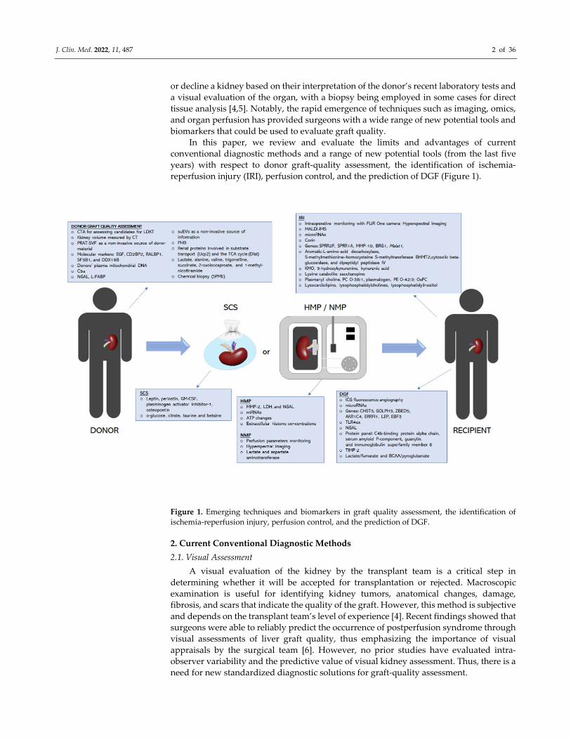
reperfusion injury (IRI), perfusion control, and the prediction of DGF (Figure 1).
Figure 1. Emerging techniques and biomarkers in graft quality assessment, the identification of
ischemia‐reperfusion injury, perfusion control, and the prediction of DGF.
2. Current Conventional Diagnostic Methods
2.1. Visual Assessment
A visual evaluation of the kidney by the transplant team is a critical step in
determining whether it will be accepted for transplantation or rejected. Macroscopic
examination is useful for identifying kidney tumors, anatomical changes, damage,
fibrosis, and scars that indicate the quality of the graft. However, this method is subjective
and depends on the transplant team’s level of experience [4]. Recent findings showed that
surgeons were able to reliably predict the occurrence of postperfusion syndrome through
visual assessments of liver graft quality, thus emphasizing the importance of visual
appraisals by the surgical team [6]. However, no prior studies have evaluated intra‐
observer variability and the predictive value of visual kidney assessment. Thus, there is a
need for new standardized diagnostic solutions for graft‐quality assessment.
J. Clin. Med. 2022, 11, 487 3 of 36
2.2. Clinical Risk Scores
Clinical information and laboratory results for a potential donor are crucial for an
initial assessment of organ quality. Consequently, several scoring systems have been
created to comprehensively analyse the risk of long‐term graft failure or DGF [7–10]. At
present, the Kidney Donor Risk Index (KDRI) and the Kidney Donor Profile Index (KDPI)
are recognized as the most effective systems for scoring kidney graft quality. The KDRI
was created by Rao et al., to quantify the risk of graft failure from deceased donors (DDs)
based on donor and transplant variables, such as age, serum creatinine (CR), diabetes,
HCV status, and cause of death [10]. The KDPI is a percentile measure based on the KDRI
that was designed to assess how long a kidney from a DD is expected to function relative
to all kidneys recovered in the U.S. during the previous year. The KDPI score is calculated
based on ten variably weighted donor parameters that relevantly affect organ quality,
with an emphasis on nephron mass. Lower KDPI scores are linked with longer estimated
organ function, while higher KDPI scores are associated with a shorter estimated organ
lifespan [11,12]. The KDRI and KDPI are regarded as reliable predictors of graft outcomes,
and they are expected to increase the prevalence of marginal kidney grafting and reduce
the unnecessary discard rate [11,13]. However, these indexes are not intended to be used
as the only metric for determining donor suitability; rather, they should be utilized as a
part of a comprehensive assessment along with other factors, including pre‐implant
biopsy histopathology and hypothermic mechine perfusion (HMP) parameters [11,14].
Because age is the most influential factor in calculating the KDRI and KDPI scores, it is
unclear whether the scores for these indexes can be applied to elderly and pediatric DDs.
Recent studies suggest that the KDPI does not precisely predict pediatric kidney graft
survival, while the KDRI has been found to be more reliable for elderly DDs. Overall,
more research is needed to assess how reliably KDPI and KDRI scores predict
postoperative renal function for grafts using kidneys from pediatric and elderly donors
[13,15].
2.3. Biopsy
Pretransplant biopsy is currently one of the most widely used diagnostic methods
and is recognized as the gold standard for confirming allograft injury. However, the
frequency with which biopsies are performed varies between medical facilities and
countries. In the United States, up to 85% of higher‐risk kidneys are biopsied, whereas
pretransplant biopsies are rarely conducted in European medical facilities. Histological
evaluation is usually applied selectively, predominantly in ECD and donor after cardiac
death (DCD) kidneys, and can help surgeons decide whether a kidney should be selected
for transplantation or rejected [4,5,16].
In contrast to most laboratory data, histopathological assessments of biopsies do not
yield a single value; rather, they produce comprehensive diagnoses that consider all
available information. Although glomerulosclerosis, vascular disease, and interstitial
fibrosis are the most frequently reported kidney parameters associated with worse graft
outcomes [4,16], there is no consensus on the relative importance of each factor and which
threshold values should be used to define the acceptable limit values. A further difficulty
is the low reproducibility of kidney biopsy evaluations between on‐call pathologists and
renal pathologists described in many prior studies. The clear need to improve
reproducibility and to objectivize the procedure and reporting of results prompted the
development of several new composite histopathological scoring systems, including the
Remuzzi score, the Maryland Aggregate Pathology Index, Banff criteria, and the Chronic
Allograft Damage Index. Nevertheless, even with all these scoring systems, there are still
doubts relating to the sampling, processing, and evaluation of biopsies [4,5,16].
In daily practice, it may be necessary to obtain quick results. In such circumstances,
frozen section (FS) evaluation is often used for decision making. Producing paraffin
sections (PS) is time consuming, which can cause histological evaluations to require up to
J. Clin. Med. 2022, 11, 487 4 of 36
3 h to complete, even with the use of high‐speed processing methods [5,17,18]. However,
reports of reproducibility and prognostic value are based on paraffin‐embedded tissue
[18]. Recent studies have shown discrepancies in the results obtained with the use of FS
and PS, but these variances had no significant impact on the outcomes for the transplanted
organs [18]. Observed changes could be subtler in frozen sections than in paraffin sections,
which may be a limitation, particularly in the hands of inexperienced pathologists [17,19].
On the other hand, it is also critical to consider logistics when choosing an optimal biopsy
technique. For instance, FS is able to provide a diagnosis in less than 30 min, whereas PS
requires at least 3 h. In selecting the proper technique, it is important to strike a balance
between the benefits and risks associated with increased cold ischemia [4,18].
A lack of uniformity with respect to procedural standards has resulted in the use of
a variety of biopsy techniques. The majority of medical facilities seem to prefer wedge
biopsy (WB) over needle biopsy (NB) because NB carries a greater risk of injuring larger
blood vessels, potentially resulting in uncontrolled bleeding after reperfusion. However,
most recent reports comparing WB and NB have found that NB provides a much better
evaluation of vascular lesions and has a higher overall correlation with the state of the
whole kidney [5,16,17].
Ultimately, the most crucial factor is how the histopathological results correlate with
long‐term graft survival. Many studies have attempted to address the predictive value of
renal biopsy with respect to graft outcomes, but the results of these studies have been
predominantly inconclusive [20–23]. For instance, Traynor et al., conducted a
retrospective study that examined kidney transplants over a 10‐year period to determine
whether pretransplant histology is able to predict graft outcomes at 5 years, and whether
donor histology adds incremental data to the current clinical parameters. While the results
of these reports suggest that that histological assessment adds little additional prognostic
information aside from clinical parameters [20], Yap et al., found that the histological
evaluation of ECD kidneys was associated with improved long‐term graft survival. Their
results suggest that pretransplant biopsy assessment can enable ECD kidneys to be used
as a safe and viable option during persistent shortages of kidney donors [21]. The
divergence between recent studies highlights the need for a prospective controlled trial to
evaluate the predictive value of pretransplant biopsies. Until a standardized and
comprehensive evaluation protocol has been developed, biopsy findings remain only one
component of a donor organ assessment and should not be taken as the sole determinant
in deciding whether to discard or transplant donor kidneys [19,24,25].
2.4. Perfusion Control
Static cold storage (SCS) and HMP are the main techniques of kidney graft
preservation [26]. HMP has become a frequently and widely used procedure in kidney
transplantation over the past few years [26–28]. Indeed, several reports have shown that
the HMP reconditioning effect results in better postoperative outcomes with respect to
reducing DGF and better long‐term graft survival after transplantation [29–31]. An
important benefit of HMP is that it enables the monitoring of perfusion parameters that
could predict post‐transplant organ viability. In particular, flow rate and renal resistance
(RR) have been among the most frequently used perfusion parameters in predicting post‐
transplant function [27,32–34]. Previous studies have produced findings suggesting that
real‐time RR detection provides good predictive value. As Bissolati et al., showed, the RR
trend during HMP can be used to predict post‐transplantation outcomes, especially in
relation to kidneys procured from ECD [28]. Patel et al., conducted a retrospective study
that included 190 kidneys in order to evaluate the prognostic utility of HMP in DD
transplantation. Their findings showed that resistances at two hours and beyond
predicted DGF, while initial resistance to machine perfusion predicted one‐year graft
survival post‐transplantation [35]. On the other hand, some studies found no association
between hemodynamic parameters during HMP and the development of DGF [27]. Thus,
due to these inconclusive results, the perfusion parameters cannot be regarded as stand‐
J. Clin. Med. 2022, 11, 487 5 of 36
alone criteria. However, the undoubted advantage of perfusion parameters is that they
are easy to obtain in a non‐invasive manner. As such, Jochmans et al., and Zheng et al.,
have suggested that HMP parameters should be included as part of a comprehensive graft
assessment [14,32]. DGF has a complex pathogenesis and cannot be predicted with
precision using the HMP parameters as a stand‐alone assessment tool. However, RR
represents an additional source of information that can help clinicians in their decision‐
making process. Attaining more accurate predictions of graft outcomes will require
integrating the perfusion parameters into multifactorial graft quality scoring systems. A
combination of the donor’s clinical data, kidney pre‐implant histopathology, and HMP
parameters may provide a more effective prediction of DGF than any of the measures
alone [14,32].
2.5. Microbiological Analysis of Preservation Fluid
Organ transplant recipients are prone to infectious complications, and despite many
advances, post‐operative infections remain associated with significant morbidity and
mortality [36–38]. Early post‐transplant infections among kidney transplant recipients
may be transmitted via the donor, or the donated organ may be contaminated during the
transplantation procedure [36,38]. Moreover, pathogens can be transmitted via
preservation solution, which is required to maintain kidney viability, but due to its
biochemical characteristics, it can also keep microorganisms alive and serve as an infection
vector [36,38,39]. For that reason, some transplant centres collect preservation fluid for
microbiological analysis in addition to standard screening for donor infections. However,
there are no widely accepted recommendations for managing positive preservation fluid
cultures [36,38]. Moreover, it remains unanswered whether intra‐operative preservation
fluid routine screening should be performed because the clinical impact of this practice is
still not well established. Some studies have evaluated the risk factors associated with
culture‐positive preservation fluid and determined the benefit of routine screening of
preservation solutions for the management of kidney transplant recipients [36–38,40].
Corbel et al., demonstrated that 24% of DD preservation fluid cultures were positive, and
these contaminations were mainly a consequence of procurement procedures [37].
Reticker et al. [36] and Oriol et al. [38] showed that the prevalence of culture‐positive
preservation fluid was up to 60%; however, the vast majority of microbial growth was
consistent with skin flora or low‐virulence pathogens. In addition, Oriol et al., indicated
that pre‐emptive antibiotic therapy for recipients with high‐risk culture‐positive
preservation fluid might improve the outcomes and help to avoid preservation‐fluid‐
related infections [38]. Moreover, Stern et al., reported that fungal contamination of
preservation fluid was infrequent, although yeast contamination of preservation solutions
was associated with high mortality [40]. In parallel, Reticker et al., suggested that
antibiotic therapy for recipients with preservation solutions contaminated by low
virulence pathogens may not be necessary, reducing antibiotic overuse [36]. In conclusion,
routine screening of preservation solutions could improve graft outcomes and pre‐
emptive antibiotic therapy and be helpful to avoid preservation‐fluid‐related infections.
However, future studies are needed to establish guidelines for preservation fluid
microbiological analysis and handling culture‐positive preservation fluid.
3. Emerging Techniques
3.1. Imaging
Diagnostic imaging methods are mainly used to evaluate kidneys from living donors
(LD) prior to acceptance for transplantation, as well as for assessing post‐renal transplant
complications. In the case of living donor surgeries, non‐invasive preoperative evaluation
of the quality of the graft organ is especially critical, which allows surgeons to assess
certain vital features, such as size, the presence/absence of focal cystic or solid lesions, and
the condition of vascular structures, to establish whether it is appropriate for
J. Clin. Med. 2022, 11, 487 6 of 36
transplantation. While most of these features can be visualized via Doppler ultrasound,
computed tomography angiography (CTA) is usually necessary for a more accurate
assessment of the vascular anatomy [41–43]. However, given the critical role of careful
evaluation and suitable preparation when dealing with living donor transplantation, it
will be imperative to continue to conduct new research aimed at improving
transplantation outcomes.
Sarier et al., conducted a retrospective study wherein they compared pretransplant
CTA images to intraoperative findings to evaluate renal artery variations in a large sample
of LD. They found that laparoscopic donor nephrectomy enabled the detection of the same
number of renal arteries as CTA in 97.9% of the analysed kidneys, but less than CTA in
the remaining 2.1%. Notably, a greater number of renal arteries were not detected in any
of the studied kidneys via nephrectomy compared to CTA. These results indicate that
CTA is more accurate than intraoperative findings, and is an effective method for
evaluating candidate donors for living donor kidney transplantation (LDKT), as well as
for identifying renovascular variations [42].
Al‐Adra et al., employed computed tomography (CT) scans to assess the influence of
donor kidney volume on recipient estimated glomerular filtration rate (eGFR) in a large
cohort of patients undergoing LDKT. The resultant statistical models showed a significant
correlation between donor kidney volume and recipient eGFR at 1, 3, and 6 months (p <
0.001). These findings indicate that donor kidney volume is a strong independent
predictor of recipient eGFR in LDKT and may therefore be a valuable addition to
predictive models of eGFR after transplantation. Further research could examine whether
addition of donor kidney volume in matching algorithms can improve recipient outcomes
[43].
Although the ability to monitor graft status intraoperatively is limited at present,
several novel solutions have been proposed over the past few years to evaluate graft
quality during transplantation and predict DGF.
In 2019, Fernandez et al., proposed a novel approach that utilized infrared imaging to
monitor the reperfusion phase during kidney transplantation in real‐time. To this end, they
used a long‐wave infrared camera (FLIR One) with a visual resolution of 1440 × 1080 pixels
and a thermal resolution of 160 × 120 to study the grafts in 10 pediatric patients undergoing
kidney transplantation. During the study, images were acquired at several key time points.
The authors observed a correlation between changes in intraoperative graft temperature
and decreases in postoperative creatinine levels in all of the analysed subjects. Given these
results, Fernandez et al., concluded that infrared thermal imaging could be a promising
option for non‐invasive graft perfusion monitoring. However, additional work is required
to confirm Fernandez et al.’s results because they were somewhat limited due to the
relatively small number of patients included and the short follow‐up period [44].
In another study, Sucher et al., employed Hyperspectral Imaging (HSI) as a
noncontact, non‐invasive, and non‐ionizing method of acquiring quantitative information
relating to kidney viability and performance during transplantation. Specifically, they
used HSI to study seventeen consecutive deceased donor kidney transplants prior to
transplantation, while stored on ice, and again at 15 and 45 min after reperfusion. After
computation time of less than 8 s, the analysis software was able to provide an RGB image
and 4 false color images representing the physiological parameters of the recorded tissue
area, namely, tissue oxygenation, perfusion, organ hemoglobin, and tissue water index.
The obtained results revealed that allograft oxygenation and microperfusion were
significantly lower in patients with DGF. Future applications might also utilize HSI
during donor surgery to assess kidney quality prior to cold perfusion and procurement.
However, HSI can only be used intraoperatively and requires a direct view of the kidney
because the maximum penetration depth for microcirculation measurements is currently
4–6 millimetres, making transcutaneous applications impossible. Thus, this technique’s
main limitations are its inability to provide continuous or intermittent transcutaneous
J. Clin. Med. 2022, 11, 487 7 of 36
follow‐up measurements, as well as its small sample size. Thus, further studies are
required to confirmed these results [45].
In the recent article, Gerken et al., documented a prospective diagnostic study that they
had conducted in two German transplantation centres wherein allograft microperfusion
was assessed intraoperatively via near‐infrared fluorescence angiography with indocyanine
green (ICG). While previous studies have shown that ICG fluorescence angiography can be
applied safely during kidney transplantation, none have provided a quantitative
assessment of the use of fluorescence video. To fill this gap, Gerken et al., evaluated the
benefits of coupling quantitative intraoperative fluorescence angiography with ICG to
predict post‐operative graft function and the occurrence of DGF. Their findings indicated
that the impairment of intraoperative microperfusion in the allograft cortex is a risk factor
for the occurrence of DGF, and that ICG Ingress is an independent predictor of DGF. Further
studies are warranted to analyse the effect of applying early therapeutic approaches to
prevent DGF in kidney transplant recipients, thus improving long‐term graft success [46].
The use of imaging techniques to diagnose post‐renal transplant complications has
been discussed extensively in recent reviews [47–49]; therefore, the present work will only
examine a few of the most recent studies in this field. Promising results have been
reported with respect to combining positron emission tomography (PET) with CT or
magnetic resonance imaging (MRI) using the glucose analogue radiotracer, 2‐deoxy‐2‐
fluoro‐D‐glucose (FDG), to detect acute kidney allograft rejection, for diagnostic
applications, for the functional assessment of grafts, and for therapeutic monitoring
[50,51]. In another study, the utility of arterial spin labeling (ASL) magnetic resonance
imaging was evaluated for its ability to identify kidney allografts with underlying
pathologies. ASL uses endogenous water as a tracer, and it has previously been used in
applications relating to the brain. Moreover, there have been reports demonstrating that
ASL can be used to categorize stages of chronic kidney disease [52]. Wang et al.,
demonstrated that ASL might be a non‐invasive tool for differentiating kidneys with
subclinical pathology from those with stable graft function. However, more research
should be performed to verify these findings [53].
3.2. Omics
The last few years has seen the emergence of many new technologies that examine
organ function on a molecular level, which has enabled the discovery of numerous
potential biomarkers of renal injury. High‐throughput omics technologies allow
researchers to obtain a large amount of data about specific types of molecules, providing
a holistic picture that captures the complex and dynamic interactions within a biological
system. These innovative methods, including transcriptomics, genomics, proteomics,
metabolomics, and lipidomics, provide a deeper understanding of the complex
mechanisms associated with IRI, inflammatory processes, and graft rejection [5,54]. This
section surveys some promising methods and techniques that could be successfully
translated to clinical settings in the foreseeable future (Table 1).
3.2.1. Transcriptomics/Genomics
Several studies have examined how graft quality and donor category impact graft
and patient survival. Giraud et al., proposed an open‐ended approach based on
microarray technology to understand IRI occurring in DCD kidneys in a preclinical
porcine model that had been subjected to warm ischemia (WI) followed by cold ischemia.
Giraud et al.’s findings indicated that hundreds of cortex and corticomedullary junction
genes were significantly regulated after WI or after WI followed by cold storage compared
to healthy kidneys. In addition, they also analysed the kinetics of the most differentially
expressed genes. They hypothesized that these genes played a key role in IRI and could
be divided into eight categories: mitochondria and redox state regulation; inflammation
and apoptosis; and protein folding and proteasome; cell cycle, cellular differentiation and
proliferation; nucleus genes and transcriptional regulation; transporters; metabolism
J. Clin. Med. 2022, 11, 487 8 of 36
regulation; mitogen‐activated protein kinase and GTPase (guanosine triphosphate, GTP)
activity [55].
Boissier et al., performed a comparative study of cellular components,
transcriptomics, and the vasculogenic profiles obtained from 22 optimal donors and 31
deceased ECDs. They hypothesized that as an easily accessible source of donor‐derived
material, perirenal adipose tissue (PRAT) can be used to assess the quantitative and
functional features that characterize donor cells. In addition, adipose tissue can be
enzymatically processed to obtain stromal vascular fraction (SVF), which is a
heterogeneous cellular mixture free of adipocytes. In their study, Bossier et al., performed
a transcriptomic analysis in order to differentiate the PRAT‐SVF molecular transcript in
ECD and other donors. The upregulated genes demonstrated a strong association with
the inflammatory response, cytokine secretion, and circulatory system development,
while the downregulated genes were associated with regulating metabolic processes and
circulatory system development. Importantly, Bossier et al.’s findings provide new
evidence that PRAT‐SVF serves as a non‐invasive source of donor material that can be
highly valuable in the assessment of inflammatory features affecting the quality and
function of the graft [56].
The midterm outcomes of kidney transplant recipients with early borderline changes
between ECD, SCD, and LD were compared in a retrospective observational study. In the
ECD group, microarray analysis showed a higher expression of 244 transcripts than the
SCD group, and 437 more than the LD group. Compared to both the SCD and LD groups,
gene annotation analysis of transcripts with elevated expression in ECD group revealed
enhancement in the inflammatory response, the response to wounding, the defence
response, and the ECM‐receptor interaction pathway. ECD‐related transcripts were likely
increased by already occurred vascular changes compared to SCD group, and, similarly
in SCD group, by longer ischemia compared with LD group. Therefore, chronic vascular
changes and cold ischemia time enhance inflammation and thus contribute to poor
outcomes for these grafts [57].
Another novel organ‐evaluation tool was proposed in a retrospective open‐cohort
study that examined donors’ plasma mitochondrial DNA (mtDNA), which can be easily
and non‐invasively assayed in the pre‐transplant period, and may be a promising
predictive biomarker for allograft function [58]. The mtDNA levels in the plasma of DCD
were determined via real‐time polymerase chain reaction (RT‐PCR) and then statistically
analysed in relation to the recipient’s mtDNA levels and DGF. The linear prediction
model, which included plasma mtDNA, donor serum creatinine, and warm ischemia time
(WIT), showed high predictive value for reduced graft function. Moreover, the findings
indicated that plasma mtDNA might be a novel non‐invasive predictor of DGF and
allograft function at six months after transplantation, in addition to correlating to allograft
survival. Furthermore, mtDNA may serve as a surrogate predictive marker for PNF [58].
The vast majority of studies aiming to identify novel biomarkers involved in IRI have
used murine or rat models. A growing body of evidence indicates that the aberrant
expression of microRNAs (miRNA/miR) is closely associated with IRI pathogenesis [59–
64]. MiRNAs are small, noncoding RNAs that mediate mRNA cleavage, translational
repression, or mRNA destabilization [59]. For instance, Chen et al.’s findings suggest that
miR‐16 may serve as a potential biomarker of IRI‐induced acute kidney injury (AKI) [59],
while Zhu et al., found that miR‐142‐5p and miR‐181a might be responsible for
modulating renal IRI development [63]. On the other hand, some studies have pointed
that miR‐17‐92, miR‐139‐5p, and miR‐27a may play a protective role in IRI [61,62,64]. For
example, Song et al., suggest that the overexpression of miR‐17‐92 could partly reverse
the side‐effects of IRI on the proximal tubules in vivo [61]. Furthermore, Wang et al., have
reported that the overexpression of miR‐27a results in the downregulation of toll‐like
receptor 4 (TLR4), which in turn inhibits inflammation, cell adhesion, and cell death in IRI
[62].
J. Clin. Med. 2022, 11, 487 9 of 36
Other murine‐model‐based studies have explored new candidate genes associated
with renal IRI. In one such study, Su et al., found that IRI caused the upregulation of
SPRR2F, SPRR1A, MMP‐10, and long noncoding RNA (lncRNA) Malat1 in kidney tissues.
These genes are involved in keratinocyte differentiation, regeneration, and the repair of
kidney tissues; extracellular matrix degradation and remodeling; inflammation; and cell
proliferation in renal IRI [64]. In a separate study, Liu et al., investigated the role of BRG1 in
IRI‐induced AKI with a focus on its role in regulating IL‐33 expression in endothelial cells.
Their findings revealed that endothelial BRG1 deficiency reduces renal inflammation
following ischemia‐reperfusion in mice with a simultaneous reduction in IL‐33 levels [65].
Comparisons of IRI in murine‐based models and clinical studies have yielded
valuable results [66,67]. For instance, Cippà et al., employed RNA‐sequencing‐mediated
transcriptional profiling and machine learning computational approaches to analyse the
molecular responses associated with IRI, which emphasized early markers of kidney
disease progression and outlined transcriptional programs involved in the transition to
chronic injury [66]. Other studies have demonstrated that Corin is downregulated in renal
IRI and may be associated with DGF after kidney transplantation. Researchers have also
screened differentially expressed genes in a murine model of IRI, with findings identifying
Corin as one of the most relevant downregulated genes among 2218 differentially expressed
genes. Moreover, 11 recipients with complications due to DGF and 16 without DGF were
recruited for an ELISA to determine their plasma Corin concentrations. The findings of this
study showed downregulation of plasma Corin concentrations in transplant recipients with
DGF complications, indicating that Corin could be a potential biomarker of DGF [67]. DGF
may result from early ischemic injury and potentially contribute to poor long‐term survival
following kidney transplantation [68,69]. For this reason, much research has been devoted
to devising reliable methods for predicting the extent of IRI, and hence, DGF.
Hence, as with the IRI, miRNA was evaluated as a biomarker of DGF. In one study,
Khalid et al., quantified microRNAs in urine samples from kidney transplant patients to
determine whether this approach can be used to predict who will develop DGF following
kidney transplantation. To this end, they used unbiased profiling to identify microRNAs
that are predictive of DGF following kidney transplantation (i.e., miR‐9, ‐10a, ‐21, ‐29a, ‐221,
and ‐429), and afterward confirmed their findings by measuring specific microRNAs via
RT‐PCR. The biomarker panel was then assessed using an independent cohort at a separate
transplant centre, with urine samples being collected at varying times during the first week
after transplantation. When considered individually, all miRs in the panel showed a trend
towards an increase or relevant increase in patients with DGF [68].
Wang et al., used high‐throughput sequencing to investigate the miRNA expression
profiling of exosomes in the peripheral blood of kidney recipients with and without DGF,
and explain the regulation of miRNAs in the DGF pathogenesis [69]. Exosomes are cell‐
derived membrane vesicles present in numerous bodily fluids that play a crucial role in
processes such as the regulation of cellular activity, intercellular communication, and waste
management [69,70]. Wang et al., identified 52 known and 5 conserved exosomal miRNAs
specifically expressed in transplant recipients with DGF. Additionally, their findings
showed that transplant recipients with DGF also exhibited the upregulation of three co‐
expressed miRNAs: hsa‐miR‐33a‐5p R‐1, hsa‐miR‐98‐5p, and hsa‐miR‐151a‐5p. Moreover,
hsa‐miR‐151a‐5p was positively correlated with the kidney recipients’ serum CR, blood urea
nitrogen (BUN), and uric acid (UA) levels in the first week post‐transplantation [69].
MicroRNA expression in kidney transplant recipients with DGF has also been
assessed in another recently published study [71]. In this work, the researchers employed
RT‐PCR to analyse the expression of miRNA‐146‐5p in peripheral blood and renal tissue
obtained from kidney transplant recipients who had undergone a surveillance graft
biopsy during the DGF period. In the renal tissue, the expression of miR‐146a‐5p was
significantly increased among the DGF patients compared to the stable and acute rejection
(AR) patients. Similarly, microRNA 146a‐5p had heightened expression in the peripheral
J. Clin. Med. 2022, 11, 487 10 of 36
blood samples from the DGF group compared to those of the acute rejection and stable
groups; however, these differences were not statistically significant (p = 0.083) [71].
Overall, all these reports indicate that miRNAs are emerging as essential biomarkers
in the molecular diagnosis of DGF. The above‐discussed findings identify biomarkers that
could contribute to the development of tools for predicting DGF and, as such, represent
an important area of focus for future research.
Zmonarski et al., applied PCR to nonstimulated peripheral blood mononuclear cells
(PBMCs) to examine the averaged mRNA toll‐like receptor 4 expression (TLR4ex). The
sample for this study consisted of 143 kidney transplant patients, 46 of whom had a history
of DGF, and a control group of 38 healthy volunteers. The patients with a history of DGF
were divided into two subgroups based on the median TLRex: low‐TLR4 expression and
high‐TLR expression. Zmonarski et al.’s findings showed that patients with DGF had a
much lower TLR4ex and worse parameters of kidney function. In addition, while a
comparison of the DGF patients with low and high TLR4ex revealed no initial differences
in kidney transplant function, differences were observed in the post‐follow‐up period.
Furthermore, regression analysis showed that TLR4ex was related to recipient age,
tacrolimus concentration, and uremic milieu. Consequently, the authors concluded that the
low TLR4 expression in patients with DGF may be associated with poor graft‐capacity
prognosis, and that analysis of changes in TLR4ex may be valuable for assessing
immunosuppression efficacy [72].
Another study aiming to identify potential biomarkers of DGF and AKI was recently
conducted by Bi et al. [73]. In this study, the authors obtained two mRNA expression
profiles from the National Center of Biotechnology Information Gene Expression
Omnibus repository, including 20 DGF and 68 immediate graft function (IGF) samples.
Differentially expressed genes (DEGs) in the DGF and IGF groups were identified, and
pathway analysis of these DEGs was conducted using the Gene Ontology and Kyoto
Encyclopedia of Genes and Genomes. Next, a protein–protein interaction analysis
extracted hub genes. The essential genes were then searched in the literature and cross‐
validated based on the training dataset. In total, 330 DEGs were identified in the DGF and
IGF samples, including 179 upregulated and 151 downregulated genes. Of these, OLIG3,
EBF3, and ETV1 were transcription factor genes, while LEP, EIF4A3, WDR3, MC4R,
PPP2CB, DDX21, and GPT served as hub genes in the PPI network. In addition, the
findings suggested that EBF3 may be associated with the development of AKI following
renal transplantation because it was significantly upregulated in the validation dataset
(GSE139061), which is consistent with the initial gene differential expression analysis.
Moreover, the authors found that LEP had a good diagnostic value for AKI (AUC = 0.740).
Overall, these findings provided more profound insights into the diagnosis of AKI
following kidney transplantation [73].
Elsewhere, McGuinness et al., combined epigenetic and transcriptomic data sets to
determine a molecular signature for loss of resilience and impaired graft function. Notably,
at a translational level, this study also provided a platform for developing a universal IRI
signature and the ability to link it to post‐transplant outcomes. Furthermore, McGuinness
et al.’s findings relate DNA methylation status to reperfusion injury and DGF outcome. In
this study, 24 paired pre‐ and post‐perfusion renal biopsies defined as either meeting the
extreme DGF phenotype or exhibiting IGF were selected for analysis. The findings of this
analysis showed that the molecular signature contained 42 specific transcripts, related
through IFNγ signaling, which, in allografts displaying clinically impaired function (DGF),
exhibited a major change in transcriptional amplitude and increased expression of
noncoding RNAs and pseudogenes, which is consistent with increased allostatic load. This
phenomenon was attended by an increase in DNA methylation within the promoter and
intragenic regions of the DGF panel in pre‐perfusion allografts with IGF. Overall,
McGuinness et al.’s findings suggest that kidneys exhibiting DGF suffer from an impaired
ability to restore physiological homeostasis in response to stress that is commensurate to
J. Clin. Med. 2022, 11, 487 11 of 36
their biological age and associated allostatic load. This outcome is reflected in changes in the
epigenome and transcriptome, as well as in the dysregulation of RNA metabolism [3].
3.2.2. Proteomics
Proteomics approaches have also been used to identify donor biomarkers that may
predict graft dysfunction in order to alleviate organ shortages and address the lack of
representative methods for assessing graft quality. To date, several studies have focused
on identifying novel proteomic biomarkers of graft quality in donor urine [74–77]. Koo et
al.’s study aimed to investigate the viability of using the levels of neutrophil gelatinase‐
associated lipocalin (NGAL), kidney injury molecule‐1 (KIM‐1), and L‐type fatty acid
binding protein (L‐FABP) in donor urine samples to predict reduced graft function (RGF).
In addition, Koo et al., also created a prediction model of early graft dysfunction based on
these donor biomarkers. This model, which includes donor urinary NGAL, L‐FABP, and
serum CR, has been shown to provide better predictive value for RGF than donor serum
CR alone. Based on this model, a nomogram for a scoring method to predict RGF was
created to help guide the allocation of DD and maximize organ utilization [74]. On the
other hand, another large prospective study has shown that donor injury biomarkers such
as microalbumin, NGAL, KIM‐1, IL‐18, and L‐FABP have limited utility in predicting
outcomes among kidney transplant recipients [75]. This study evaluated the associations
between injury biomarkers in the urine of DD and donor AKI, recipient DGF, and
recipient six‐month eGFR. Each of the tested biomarkers was strongly associated with
donor AKI in the adjusted analyses. However, although the levels of all five donor
biomarkers were higher in recipients with DGF than in those without DGF, the fully
adjusted analyses revealed an association between higher donor urinary NGAL
concentrations and a modest increase in the relative risk of recipient DGF. Moreover, the
results of this study indicated that donor urinary biomarkers add minimal value in
predicting recipient allograft function at six months post‐transplantation [75]. In both
studies, the tested biomarkers were strongly associated with donor AKI, while NGAL
concentration was associated with DGF. A potential explanation for the different
conclusions of these studies may be that Koo et al., used RGF as an outcome in their study,
while Reese et al., used DGF due to different donor characteristics. Furthermore, it is
worth emphasizing that, while these proteins are upregulated and secreted in urine in
response to tubular injury, they were reported to have low specificity for tubular epithelial
cell injury and were observed to increase in patients with urinary tract infections and
sepsis [78,79].
In another study, the potential utility of C3a and C5a in DD urine samples as
biomarkers for early post‐transplant outcomes was investigated [76]. The results of this
large, prospective, observational cohort study indicated a three‐fold increase in C5a
concentrations in urine samples from donors with stage 2 and 3 AKI compared to donors
without AKI. In addition, donor C5a was positively correlated with the occurrence of DGF
in recipients. In adjusted analyses, C5a remained independently correlated with recipient
DGF only for donors without AKI. Moreover, the authors observed a tendency to indicate
better 12‐month organ functioning from donors with the lowest urinary C5a [76].
Monocyte chemoattractant protein‐1 (MCP‐1) has also been proposed as a potential
biomarker of donor kidney quality. For example, Mansour et al., evaluated the association
between graft outcomes and levels of MCP‐1 in urine from DD at the time of organ
procurement. In particular, they measured MCP‐1 concentration to determine its
correlation to donor AKI, recipient DGF, six‐month estimated eGFR, and graft failure.
Unfortunately, Mansour et al.’s results suggested that urinary MCP‐1 has minimal clinical
utility. Although median urinary MCP‐1 concentrations were elevated in donors with AKI
compared to those without AKI, higher MCP‐1 levels were independently associated with
a higher six‐month eGFR in those without DGF. However, MCP‐1 was not independently
associated with DGF, and no independent associations between MCP‐1 and graft failure
were observed over a median follow‐up of ~two years [77].
J. Clin. Med. 2022, 11, 487 12 of 36
Recently, Braun et al., demonstrated the potential of using small urinary extracellular
vesicles (suEVs) as a non‐invasive source of data regarding early molecular processes in
transplant biology. Their unbiased proteomic analysis revealed temporal patterns in the
signature of suEV proteins, as well as cellular processes involved in both early response
and longer‐term graft adaptation. In addition, a subsequent correlative analysis identified
potential prognostic markers of future graft function, such as phosphoenol pyruvate
carboxykinase (PCK2). However, while Braun et al.’s study showed the potential of suEVs
as biomarkers, the small number of patients in their sample did not allow for a conclusive
statement on the predictive value of suEV PCK2. Therefore, the potential use of this
biomarker will depend on larger trials in the future [80].
Studies focusing on the use of kidney tissue as a sample matrix to evaluate donor
organ quality have also been performed. Using a rabbit model of brain death (BD), Li et
al., employed two‐dimensional gel electrophoresis and Matrix Assisted Laser
Desorption/Ionization Time‐of‐Flight Mass Spectrometry (MALDI‐TOF‐MS)‐based
comparative proteomic analysis to profile the differentially‐expressed proteins between
BD and renal tissue collected from a control group. The authors were able to acquire five
downregulated proteins and five upregulated proteins, which were then classified
according to their function, including their association with proliferation and
differentiation, signal transduction, protein modification, electron transport chain, and
oxidation‐reduction. Moreover, immunohistochemical analysis indicated that the
expression of prohibitin (PHB) gradually elevated in a time‐dependent manner. These
data showed alterations in the levels of certain proteins in the organs from the BD group,
even in the case of non‐obvious functional and morphological changes. Given their
results, Li et al., suggested that PHB may be an innovative biomarker for the primary
assessment of the quality of kidneys from BD donors [81].
Conversely, van Erp et al., used a multi‐omics approach and a rat model to investigate
organ‐specific responses in the kidneys and liver during BD. The application of proteomics
analysis enabled them to quantify 50 proteins involved in oxidative phosphorylation,
tricarboxylic acid (TCA) cycle, fatty acid oxidation (FAO), substrate transport, and several
antioxidant enzymes in isolated hepatic and renal mitochondria. The most relevant changes
were observed in the reduced peptide levels in the kidneys, which were related to complex
I (Ndufs1), the TCA cycle (Aco2, Fh, and Suclg2), FAO (Hadhb), and the connection between
FAO and the electron transport chain (Etfdh). The expression of two renal proteins, which
were associated with substrate transport (Ucp2) and the TCA cycle (Dlat), was significantly
increased in samples from the BD group compared to the sham‐operated group.
Interestingly, van Erp et al.’s findings showed that BD pathophysiology affects systemic
metabolic processes, alongside organ‐explicit metabolic changes, manifest in the kidneys by
metabolic shutdown and suffering from oxidative stress, and a shift to anaerobic energy
production, while kidney perfusion decreases. Ultimately, van Erp et al., concluded that an
organ‐specific strategy focusing on metabolic changes and graft perfusion should be part of
novel procedures for assessing graft quality in organs from brain‐dead donor, and may be
the key to improving transplantation outcomes [82].
The vast majority of studies focusing on IRI have used animal models. In one proteo‐
metabolomics study using rat models, coagulation, complement pathways, and fatty acid
(FA) signaling were observed following the elevation of proteins belonging to acute phase
response due to IRI. Moreover, after 4 h of reperfusion, analysis of metabolic changes
showed an increase in glycolysis, lipids, and FAs, while mitochondrial function and
adenosine triphosphate (ATP) production were impaired after 24 h [83]. The authors of
another study that used a porcine model of IRI found that integrative proteome analysis
can provide a panel of potential—and predominantly renal—biomarkers at many levels,
as changes occurring in the tissue are reflected in serum and urine protein profiles. This
conclusion was based on the use of urine, serum, and renal cortex samples. In the renal
cortex proteome, the authors observed an elevation in the synthesis of proteins in the
ischemic kidney (vs. the contralateral kidney), which was highlighted by transcription
J. Clin. Med. 2022, 11, 487 13 of 36
factors and epithelial adherens junction proteins. Intersecting the set of proteins up‐ or
downregulated in the ischemic tissue with both serum and urine proteomes, authors
identified six proteins in the serum that may provide a set of targets of kidney injury. In
addition, four urinary proteins with predominantly renal gene expression were also
identified: aromatic‐L‐amino‐acid decarboxylase (AADC), S‐methylmethionine–
homocysteine S‐methyltransferase BHMT2 (BHMT2), cytosolic beta‐glucosidase (GBA3),
and dipeptidyl peptidase IV (DPPIV) [84]. Recent research by Moser et al., has examined
kidney preservation injury and the nephroprotective activity of doxycycline (Doxy). In
this work, rat kidneys were cold perfused with and without Doxy for 22 h, followed by
the extraction of proteins from the renal tissue. Subsequent analysis showed a significant
difference in eight enzymes involved in cellular and mitochondrial metabolism.
Interestingly, the levels of N(G),N(G)‐dimethylarginine dimethylaminohydrolase and
phosphoglycerate kinase 1 decreased during cold perfusion on its own but increased
during cold perfusion with Doxy [85]. The influence of perfusion type on graft quality has
also been evaluated by Weissenbacher et al., who applied proteomics analysis to
determine the differences between normothermically perfused (normothermic machine
perfusion, NMP) human kidneys with urine recirculation (URC) and urine replacement
(UR). Their findings revealed that damage‐associated patterns in the kidney tissue
decreased after 6 h of NMP with URC, suggesting decreased inflammation. Furthermore,
they also observed that vasoconstriction in the kidneys was also attenuated with URC, as
indicated by a reduction in angiotensinogen levels. The kidneys became metabolically
active during NMP, which could be improved and prolonged by applying URC. The
application of URC also enhanced mitochondrial succinate dehydrogenase enzyme levels
and carbonic anhydrase, which contributed to pH stabilization. Key enzymes involved in
glucose metabolism increased after 12 and 24 h of NMP with URC, including
mitochondrial malate dehydrogenase and glutamic‐oxaloacetic transaminase,
predominantly in DCD tissue. The authors concluded that NMP with URC can prolong
organ preservation and revitalize metabolism to possibly better mitigate IRI in discarded
kidneys [86].
Ischemic injury may result in DGF, which is associated with a more complicated post‐
operative course, including a higher risk of AR [87]. Therefore, the early evaluation of
kidney function following transplantation is essential for predicting graft outcomes [88].
Several studies have applied proteomic analysis to recipient urine samples in an attempt
to identify protein biomarkers of DGF [87–89]. For instance, Lacquaniti et al., evaluated
the usefulness of NGAL levels both for the early detection of DGF and as a long‐term
predictor of graft outcome. Their findings revealed that serum and urine samples from
DGF patients contained high levels of NGAL beginning the first day after transplantation.
Moreover, in patients who had received a kidney from a living related donor with
excellent allograft function, NGAL concentrations lowered quickly during the first 24 h
post‐transplant period, reflecting a more pronounced reversible short‐term injury.
Importantly, NGAL levels in urine provided a better diagnostic profile than serum NGAL.
Hence, urinary biomarkers on day 1 post‐transplant may not only be useful in predicting
who will need dialysis within one week, but they may also allow clinicians to discriminate
between more subtle allograft recovery patterns [88]. However, as mentioned above,
NGAL is characterized by low specificity; hence, its clinical application is limited due to
inconclusive results [78,79]. Williams et al., used a Targeted Urine Proteome Assay
(TUPA) to identify biomarkers of DGF following kidney transplantation. After employing
data quality consideration and rigorous statistical analysis, they identified a panel of the
top 4 protein biomarkers, including the C4b‐binding protein alpha chain, serum amyloid
P‐component, guanylin, and immunoglobulin superfamily member 8, which had an AUC
of 0.891, a specificity of 82.6%, and a sensitivity of 77.4% [87]. Similarly, urinary tissue
inhibitor of metalloproteinases‐2 (TIMP‐2) and insulin‐like growth factor binding protein‐
7 (IGFBP7) have been evaluated as biomarkers for DGF [89]. The findings of these studies
indicated that TIMP‐2 was able to adequately identify patients with DGF and prolonged
J. Clin. Med. 2022, 11, 487 14 of 36
DGF (AUC 0.89 and 0.77, respectively), whereas IGFBP7 was not. Moreover, correcting
TIMP‐2 for urine osmolality improved predictability (AUC 0.91 for DGF, AUC 0.80 for
prolonged DGF), and 24‐h urinary CR excretion and TIMP‐2/mOsm were found to be
significant predictors of DGF, with an AUC of 0.90. Hence, the obtained results indicated
that TIMP‐2 might be a promising, non‐invasive indicator for predicting the occurrence
and duration of DGF in individual patients [89].
3.2.3. Metabolomics and Lipidomics
In the absence of good quantitative biomarkers correlating to pre‐transplantation
organ quality, van Erp et al., examined metabolic alterations during BD using
hyperpolarized magnetic resonance (MR) spectroscopy and ex vivo graft glucose
metabolism during normothermic isolated perfused kidney (IPK) machine perfusion [90].
To this end, they employed hyperpolarized 13C‐labeled pyruvate MR spectroscopy to
quantify pyruvate metabolism in the kidneys and liver at three time points during BD in
a rat model. Following BD, glucose oxidation was measured using tritium‐labeled glucose
(D‐6‐3H‐glucose) during IPK reperfusion. In addition, enriched 13C‐pyruvate was injected
repetitively to evaluate the metabolic profile at T = 0, T = 2, and T = 4 h via the relative
conversion of pyruvate into lactate, alanine, and bicarbonate. The rats showed
significantly higher lactate levels immediately following the induction of BD, with alanine
production decreasing in the kidneys 4 h post‐BD. However, it should be emphasized that
this study’s results did not assess whether these metabolic alterations can be associated
with graft quality, or if they are suitable predictors of transplant outcome [90].
Another study using a rodent model of IRI examined the potential of using
Hyperpolarized 13C‐labeled pyruvate to evaluate the metabolic profile directly in the
kidneys [91]. The in vivo responses observed at 24 h and 7 d following ischemic injury
demonstrated a similar trend towards a general decrease in the overall metabolism in the
ischemic kidney and a compensatory increase in anaerobic metabolism, which is
evidenced by elevated lactate production, compared to aerobic metabolism. In addition,
a correlation was found between the intra‐renal metabolic profile 24 h after reperfusion
and 7 d after injury induction, as well as a correlation with the plasma CR. As a result, the
authors suggest that using hyperpolarized 13C‐labeled pyruvate to identify the balance
between anaerobic and aerobic metabolism has great future potential as a prognostic
biomarker [91].
Increased lactate levels due to IRI were also observed in another study [92]. However,
analysis of urine samples via nuclear magnetic resonance (NMR) spectroscopy showed
higher levels of valine and alanine and decreased levels of metabolites such as
trigonelline, succinate, 2‐oxoisocaproate, and 1‐methyl‐nicotinamide following IRI, which
was likely due to altered kidney function or metabolism [92].
A novel and minimally invasive metabolomic and lipidomic diagnostic protocol
based on solid‐phase microextraction (SPME) has been proposed to address the lack of
representative methods of assessing graft quality [93,94]. The small size of the SPME probe
allows the performance of chemical biopsy, which enables metabolites to be extracted
directly from the kidney without any tissue collection. Furthermore, SPME’s minimally
invasive nature permits multiple analyses over time. For instance, ischemia‐induced
alterations in the metabolic profile of the kidneys and oxidative stress as a function of cold
storage were observed in one study that used an animal model, with the most pronounced
alterations being observed in the levels of essential amino acids and purine nucleosides
[93]. However, more work is required to discriminate a set of characteristic compounds
that could serve as biomarkers of graft quality and indicators of possible development of
organ dysfunction.
In response to reports that the pharmacological inhibition of kynurenine 3‐
monooxygenase (KMO), and, separately, the transcriptional blockage of the Kmo gene,
reduces 3‐hydroxykynurenine formation and protects against secondary AKI, Zheng et
al., investigated whether mice lacking functional KMO (Kmonull mice) are protected from
J. Clin. Med. 2022, 11, 487 15 of 36
AKI experimentally induced by the direct induction of renal IRI [95]. KMO plays a crucial
role in kynurenine metabolism. Kynurenine metabolites are generated by tryptophan
catabolism and are involved in the regulation of various biological processes, including
host‐microbiome signaling, immune cell response, and neuronal excitability. The
kynurenine pathway diverges into two distinct branches, which are regulated by
kynurenine aminotransferases (KATs) and KMO, respectively. KMO is the only route of
3‐hydroxykynurenine production that is known to be injurious to cells and tissue.
Kynurenine may also be metabolized into kynurenic acid by KATs and to anthranilic acid
by kynureninase [95]. Following the experimental induction of AKI via renal IRI, Zheng
et al., observed that the Kmonull mice had kept renal function, decreased renal tubular cell
injury, and fewer infiltrating neutrophils than the wild‐type control mice. Given these
results, they suggested that KMO is a critical regulator of renal IRI. Moreover, higher
levels of kynurenine and kynurenic acid were observed in the Kmonull IRI mice compared
to the Kmonull sham‐operated mice. This result may indicate that these metabolites help to
protect against AKI after renal IRI, particularly because kynurenic acid has been
demonstrated to have protective properties in other inflammatory situations due to its
activity at glutamate receptors [95].
A 12.5‐fold increase in the lysine catabolite saccharopine in IRI kidneys was observed
in a recent study examining the differences between renal allograft acute cellular rejection
(ACR) and IRI. The findings of this work indicated that the accumulation of saccharopine
causes mitochondrial toxicity and may contribute to IRI pathophysiology. Moreover,
similar to other reports, increased levels of itaconate and kynurenine were also observed
in ACR kidneys. However, the detected changes in metabolites seemed to be unique for
IRI and ACR, respectively, indicating that these two conditions have distinct tissue
metabolomic signatures [96].
Several reports have also demonstrated that IRI can alter the lipidome. For example,
Rao et al., evaluated lipid changes in an IRI mouse model using sequential window
acquisition of all theoretical spectra‐mass spectrometry (SWATH‐MS) lipidomics. Their
findings indicated that four lipids increased significantly at 6 h after IRI: plasmanyl
choline, phosphatidylcholine (PC) O‐38:1 (O‐18:0, 20:1), plasmalogen, and
phosphatidylethanolamine (PE) O‐42:3 (O‐20:1, 22:2). As anticipated, statistically
significant changes were observed in many more lipids at 24 h after IRI. Interestingly,
elevated levels of PC O‐38:1 persisted at 24 h post‐IRI, while renal levels of PE O‐42:3
decreased alongside all ether PEs detected by SWATH‐MS at this later time point. Overall,
the authors found that coupling SWATH‐MS lipidomics with MALDI‐IMS (Imaging Mass
Spectrometry, IMS) for lipid localization provided a better understanding of the role
played by lipids in the pathobiology of acute kidney injury [97].
Researchers have also tested whether oxidized phosphatidylcholine (OxPC)
molecules are generated following renal IRI. Solati et al., identified fifty‐five distinct OxPC
molecules in rat kidneys following IRI, including various fragmented (aldehyde and
carboxylic‐acid‐containing species) and nonfragmented products. Among these, 1‐
stearoyl‐2‐linoleoyl‐phosphatidylcholine (SLPC‐OH) and 1‐palmitoyl‐2‐azelaoyl‐sn‐
glycero‐3‐phosphocholine (PAzPC) were the most abundant after 6 h and 24 h IRI,
respectively. The total number of fragmented aldehyde OxPC molecules was significantly
elevated in the 6 h and 24 h IRI groups compared to the sham‐operated group, while an
increase in the level of fragmented carboxylic acid was observed in the 24 h group
compared to the sham and 6 h groups. In addition, fragmented OxPC levels were found
to be significantly correlated with CR levels [98].
In their recent paper, van Smaalen et al., introduced and employed an interesting
new approach based on IMS to rapidly and accurately evaluate acute ischemia in kidney
tissue from a porcine model. First, ischemic tissue damage was systematically evaluated
by two pathologists; this was followed by the application of MALDI‐IMS to study the
spatial distributions and compositions of lipids in the same tissues. Whereas the
histopathological analysis revealed no significant difference between the tested groups,
J. Clin. Med. 2022, 11, 487 16 of 36
the MALDI‐IMS analysis provided detailed discrimination of severe and mild ischemia
based on the differential expression of characteristic lipid‐degradation products
throughout the tissue. In particular, elevated levels of lysolipids, including
lysocardiolipins, lysophosphatidylcholines, and lysophosphatidylinositol, were present
after severe ischemia. This data shows IMS’s potential for use in differentiating and
identifying early ischemic injury molecular patterns, and as a future tool that can be
deployed in kidney assessment [99].
Because ischemia and reperfusion are inevitable consequences of kidney
transplantation, and because DGF is a manifestation of IRI, Wijermars et al., used kidney
transplantation as a clinical model of IRI to evaluate the role of the hypoxanthine‐xanthine
oxidase (XO) axis in human IRI. The sample group for this study consisted of patients
undergoing renal allograft transplantation (n = 40), who were classified into three groups
based on the duration of ischemia: short, intermediate, and prolonged. The results of the
analysis confirmed the progressive accumulation of hypoxanthine during ischemia.
However, differences in arteriovenous concentrations of UA and an in situ enzymography
of XO did not indicate relevant XO activity in IRI kidney grafts. Moreover, renal
malondialdehyde and isoprostane levels and allantoin formation were assessed during
the reperfusion period to determine whether a putative association exists between
hypoxanthine accumulation and renal oxidative stress. The absence of the release of these
markers indicated the lack of an association between ischemic hypoxanthine
accumulation and post‐reperfusion oxidative stress. Based on these results, the authors
suggest that the hypoxanthine‐xanthine oxidase axis is not involved in the initial phase of
clinical IRI [100]. In their clinical study, Kostidis et al., employed NMR spectroscopy to
analyse the urinary metabolome of DCD transplant recipients at multiple time points in
an attempt to identify markers that predict the prolonged duration of functional DGF [79].
To this end, urine samples were collected at 10, 42, 180, and 360 days post‐transplantation.
Their analysis revealed that samples collected on day 10 had a different profile than
samples obtained at the other time points. At day 10, D‐glucose, 2‐aminobutyrate, valine,
p‐hydroxyhippurate, fumarate, 2‐ethylacrylate, leucine, and lactate were significantly
elevated in patients with DGF compared to those without DGF, while asparagine, DMG,
3‐hydroxyisobutyrate, 3‐hydroxyisovalerate, 2‐hydroxy‐isobutyrate, and histidine were
significantly reduced in the DGF group. Urine samples from patients with prolonged DGF
(≥21 days) showed increased levels of lactate and lower levels of pyroglutamate compared
to participants with limited DGF (<21 days). Moreover, the ratios of all metabolites were
analysed via logistic regression analysis in an attempt to further distinguish prolonged
DGF from limited DGF. The results of this analysis showed that the combination of
lactate/fumarate and branched chain amino acids (BCAA)/pyroglutamate provided the
best outcome, predicting prolonged DGF with an AUC of 0.85. Given these results, the
authors concluded that it is possible to identify kidney transplant recipients with DGF
based on their altered urinary metabolome, and that it may also be possible to use these
two ratios to predict prolonged DGF [79].
In another study, Lindeman et al., examined possible metabolic origins of clinical IRI
by integrating data from 18 pre‐ and post‐reperfusion tissue biopsies with 36 sequential
arteriovenous blood samplings from grafts in three groups of subjects, including LD and
DD grafts with and without DGF. The integration of metabolomics data enabled Lindeman
et al., to determine a discriminatory profile that can be used to identify future DGF. This
profile was characterized by impaired recovery of the high‐energy phosphate‐buffer,
phosphocreatine, in DGF grafts post‐reperfusion, as well as by persistent post‐reperfusion
ATP/GTP catabolism and significant ongoing tissue damage. The impaired recovery of
high‐energy phosphate occurred despite the activation of glycolysis, fatty acid oxidation,
glutaminolysis, autophagia and was found to be related to a defect at the level of the
oxoglutarate dehydrogenase complex in the Krebs cycle. Hence, Lindeman et al.’s findings
suggest that DGF is preceded by a post‐reperfusion metabolic collapse, leading to an
J. Clin. Med. 2022, 11, 487 17 of 36
inability to sustain the organ’s energy requirements. Thus, efforts aimed at preventing DGF
should aim to preserve or restore metabolic competence [101].
3.3. New Solutions in Perfusion Control
Organ‐preservation technologies have been garnering significant interest for graft
quality assessment, advanced organ monitoring, and treating transplanted kidneys
during machine perfusion. As mentioned above, SCS and HMP are two of the more
common methods of hypothermic preservation applied in clinical settings at present. In
SCS, the kidney is submerged in a cold preservation fluid and placed on ice in an icebox;
in HMP, a device pumps cold preservation fluid through the renal vasculature, which has
been revealed to improve post‐transplant outcomes [102]. NMP is another dynamic
preservation strategy that involves the circulation of a perfusion solution through the
kidney. The NMP conditions are designed to nearly replicate physiological conditions,
which makes a real‐life assessment of the graft possible prior to transplantation [103,104].
NMP has been recently translated into clinical practice, but this application is still at an
experimental stage. However, early clinical results are promising [103,105]. Because
preservation/perfusion solutions serve as a non‐invasive source for the analysis of
biomarkers, numerous studies have employed it for the purposes of graft quality
assessment. In this section of this paper, we summarize the latest findings and studies that
have used preservation/perfusion fluid and perfusion control in kidney transplantation
(Table 1).
Coskun et al., used proteomic techniques to analyse the protein profiles of
preservation fluid used in SCS kidneys. Their findings revealed significant correlations
between protein levels and donor age (23 proteins), cold ischemia time (5 proteins),
recipients’ serum BUN (12 proteins), and CR levels (7 proteins). The identified proteins
belonged to groups related to the structural constituent of the cytoskeleton, serine‐type
endopeptidase inhibitor activity, peptidase inhibitor activity, cellular component
organization or biogenesis, and cellular component morphogenesis, among others [106].
In another proteomic study of preservation fluid, five potential biomarkers (leptin,
periostin, granulocyte‐macrophage colony‐stimulating factor (GM‐CSF), plasminogen
activator inhibitor‐1, and osteopontin) were identified in a discovery panel for
differentiating kidneys with immediate function from those with DGF. Further analysis
yielded a prediction model based on leptin and GM‐CSF. Receiver‐operating
characteristic analysis revealed an AUC of 0.87, and the addition of recipient BMI
significantly increased the model’s predictive power, resulting in an AUC of 0.89 [107].
The metabolomic study compared the level of metabolites in perfusate samples collected
prior to transplantation, during static cold storage, and between the allografts exhibiting
DGF and IGF, while an integrated NMR‐based analysis revealed a significant elevation in
α‐glucose and citrate levels, and significant decreases in taurine and betaine levels in the
perfusate of DGF allografts [108].
In the last few years, several studies have documented the benefits of HMP over SCS,
including improved short‐term outcomes and reduced risk of DGF [109–111]. However,
reports suggesting that HMP improves long‐term graft function are inconclusive
[102,111]. Some research groups have compared HMP with SCS to evaluate HMP’s
potential to improve kidney‐graft outcomes [109,112] and to better understand the long‐
term benefits associated with its use [111,113]. At the same time, other groups have
investigated how the use of oxygenated HMP impacts post‐transplant outcomes, and how
it can be used to further optimize kidney preservation, thereby expanding the number of
organs available for transplant [102,114]. Furthermore, perfusion solution has been used
in the search for useful biomarkers of graft quality and potential therapeutic targets. The
analysis of perfusates from donor after brain death (DBD), DCD, and LD kidneys showed
that DCD kidneys contained the highest levels of matrix metalloproteinase‐2 (MMP‐2),
lactate dehydrogenase (LDH), and NGAL, followed by DBD and LD kidneys,
respectively, suggesting a greater amount of injury in the DCD kidneys. Moreover, the
J. Clin. Med. 2022, 11, 487 18 of 36
DCD kidney perfusate contained significantly higher levels of protein compared to the
DBD and LD perfusates, with quantitative analysis of the protein spots revealing
significant differences between the groups in relation to seven spots: peroxiredoxin‐2,
FABP, A1AT, heavy chain of immunoglobulin, serum albumin, fragment of collagen 1,
and protein deglycase (DJ‐1) [115]. In another proteomic study, perfusate analysis of DBD
kidneys preserved via HMP was performed to identify differences between the proteomic
profiles of kidneys with good (GO) and suboptimal outcomes (SO) one‐year post‐
transplantation. Analysis of samples collected 15 min after the start of HMP (T1) and
before the termination of HMP (T2) indicated that the 100 most abundant proteins
demonstrated discrimination between grafts, with a GO and SO at T1. Increased proteins
were involved in classical complement cascades at both T1 and T2, while a decreased
abundance of lipid metabolism at T1 and cytoskeletal proteins at T2 in GO (vs. SO) was
also observed. Perfusate analysis at T1 revealed a predictive value of 91% for ATP‐citrate
synthase and fatty acid‐binding protein 5, and analysis at T2 showed a predictive value
of 86% for immunoglobulin heavy variable 2–26 and desmoplakin. In summary, HMP
perfusate profiles for DBD kidneys can distinguish between outcomes one‐year post‐
transplantation, providing a potential non‐invasive method of assessing donor organ
quality [2].
MicroRNAs in kidney machine perfusion solutions have also been considered as new
biomarkers for graft function. For instance, Gómez dos Santos et al., conducted a
prospective cohort study to investigate graft dysfunction in kidney transplantation from
ECD. To this end, they employed a mean expression value approach, which confirmed
the significance of a subset of the miRNAs previously identified with the development of
delayed graft function, namely, miR‐486‐5p, miR‐144‐3p, miR‐142‐5p, and miR‐144‐5p.
These results confirmed that perfusion fluid can be a valuable pre‐transplantation source
of organ‐viability biomarkers [116].
In another study, Tejchman et al., assessed oxidative stress markers from the
hypothermic preservation of transplanted kidneys. In particular, they sought to analyse
the activity of enzymes and levels of non‐enzymatic compounds involved in antioxidant
defense mechanisms. These compounds, which included glutathione (GSH), glutathione
peroxidase (GPX), catalase (CAT), superoxide dismutase (SOD), glutathione reductase
(GR), glutathione transferase (GST), thiobarbituric acid reactive substances (TBARS),
malondialdehyde (MDA), were measured in preservation solutions before the
transplantation of human kidneys grafted from DBD. The study group was divided into
two groups based on the method of kidney storage, with Group 1 consisting of HMP
kidneys (n = 26) and Group 2 consisting of SCS kidneys (n = 40). There were aggregations
of significant correlations between kidney function parameters after KTx and oxidative
stress markers, namely: diuresis and CAT; Na+ and CAT; K+ and GPX; and urea and GR.
Moreover, there were aggregations of correlations between recipient blood count and
oxidative stress markers, including CAT and monocyte count; SOD and white blood cell
count; and SOD and monocyte count. However, there was an issue of unequivocal
interpretation because none of the observed aggregations constituted conditions that
supported the authors’ hypothesis that kidney function after KTx can be predicted based
on oxidative stress markers measured during preservation. Moreover, it would be hard
to conclude that the blood count alterations observed in the repeated measurements after
KTx were unrelated to factors other than oxidative stress or acidosis. As the authors
suggest, many other factors may modify blood count, including operative stress, bleeding,
immunosuppression, and microaggregation [117].
Longchamp et al., presented an interesting and non‐invasive method of assessing
graft quality during perfusion based on the use of 31P pMRI spectroscopy to detect high‐
energy phosphate metabolites, such as ATP. Thus, pMRI can be used to predict the energy
state of a kidney and its viability before transplantation. In addition, Longchamp et al.,
also performed gadolinium perfusion sequences, which allowed them to observe the
internal distribution of the flow between the cortex and the medulla. pMRI showed that
J. Clin. Med. 2022, 11, 487 19 of 36
warm ischemia caused a reduction in ATP levels, but not its precursor, adenosine
monophosphate (AMP). Moreover, they found that ATP levels and cortical and medullary
gadolinium elimination were inversely correlated with the severity of kidney histological
injury. Thus, the measured parameters may be considered as biomarkers of kidney injury
after warm ischemia, and Longchamp et al.’s method provides an innovative non‐invasive
approach to assessing kidney viability prior to transplantation [118].
Other researchers have examined whether a correlation exists between the level of
extracellular histones in machine perfusates and the viability of DD kidneys. Extracellular
histone levels were significantly elevated in the perfusates of kidneys with post‐transplant
graft dysfunction, and they were considered an independent risk factor for DGF and one‐
year graft failure, but not for PNF. One‐year graft survival was 12% higher in the low‐
histone‐concentration group (p = 0.008) compared to the higher‐histone‐concentration
group. Hence, the quantitation of extracellular histones might contribute to the evaluation
of post‐transplant graft function and survival [119].
NMP is an emerging approach for donor organ preservation and functional
improvements in kidney transplantation. However, methods for evaluating organs via
NMP have yet to be developed, and the development of novel graft quality assessment
solutions has only recently come into focus.
Kaths et al., used a porcine model to investigate whether NMP is suitable for graft
quality assessment prior to transplantation. They found that intra‐renal resistance was
lowest in the HBD group and highest in the severely injured DCD group (60 min of warm
ischemia), and that the initiation of NMP was correlated with post‐operative renal
function. Markers of acid‐base homeostasis (pH, HCO3–, base excess) correlated with post‐
transplantation renal function. Furthermore, concentrations of lactate and aspartate
aminotransferase were lowest in perfusate from non‐injured grafts (vs. DCD kidneys) and
were correlated with post‐transplantation kidney function. Kaths et al., found that
perfusion characteristics and clinically available perfusate biomarkers during NMP were
correlated with post‐transplantation kidney graft injury and function. However, further
research is needed to identify perfusion parameter thresholds for DGF and PNF [120].
HSI combined with NMP was introduced as a novel approach for monitoring
physiological kidney parameters. The experimental results of an HSI‐based oxygen‐
saturation calculation indicated that HSI is useful for monitoring oxygen saturation
distribution and identifying areas with a reduced oxygen supply prior to transplantation.
Moreover, camera‐based measurements are easy to integrate with a perfusion setup and
allow the fast and non‐invasive measurement of tissue characteristics [121]. Subsequent
research has explored how to improve algorithms for determining kidney oxygen
saturation [122]. Unfortunately, the application of HSI is limited by the propagating light’s
low penetration depth, which makes it impossible to detect deeper tissue injuries.
However, based on the fact that most metabolic activity occurs in the kidney cortex, the
combined use of HSI and NMP offers a promising and easy‐to‐use method for assessing
the status of the organ and for chemical imaging [121,122].
Hyperpolarized MRI and spectroscopy (MRS) using pyruvate and other 13C‐labeled
molecules offers a novel approach to monitoring the state of ex vivo perfused kidneys. In
one study, the state of a porcine kidney was quantified using acquired anatomical,
functional, and metabolic data. The findings showed an apparent reduction in pyruvate
turnover during renal metabolism compared with the typical in vivo levels observed in
pigs, while perfusion and blood gas parameters were found to be in the normal ex vivo
range. Mariager et al.’s findings demonstrate the applicability of these techniques for
monitoring ex vivo graft metabolism and function in a large animal model that resembles
human renal physiology [123].
In another study, researchers sought to investigate the link between the urinary
biomarkers, endothelin‐1 (ET‐1), NGAL, and KIM‐1, and NMP parameters in order to
improve kidney assessment prior to transplantation. Fifty‐six kidneys from DD were used
in this work, with each kidney being subjected to 1 h of NMP, followed by assessment
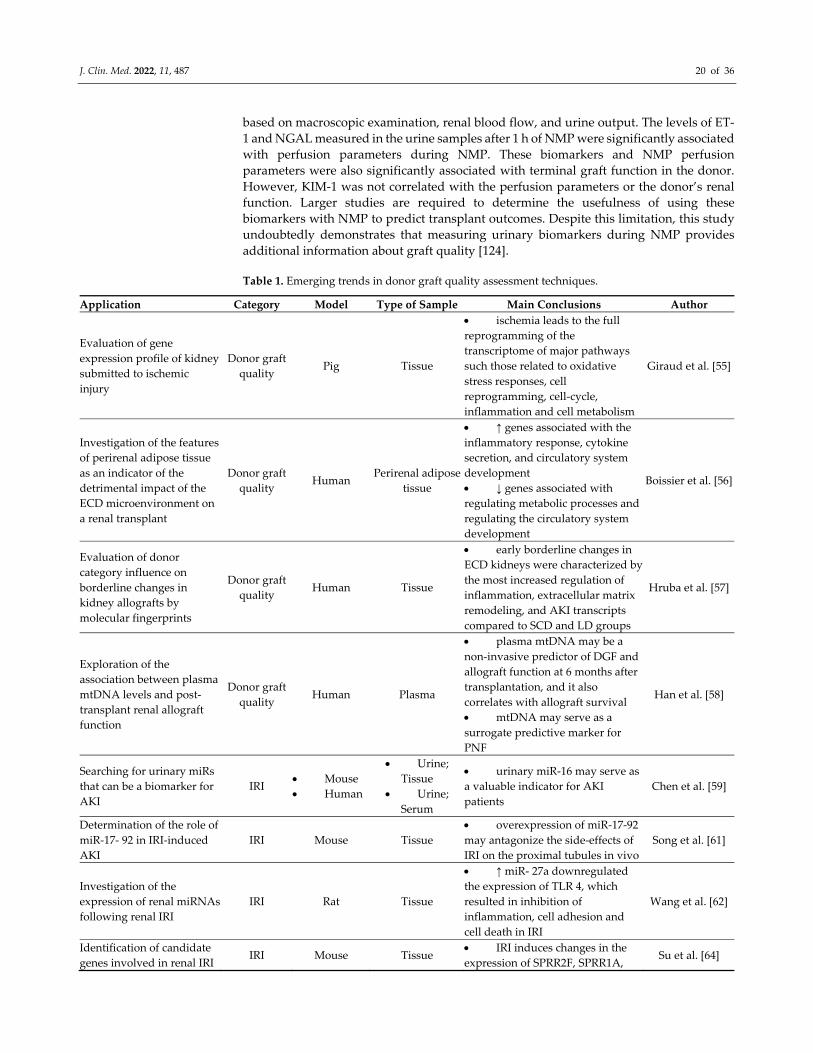
J. Clin. Med. 2022, 11, 487 20 of 36
based on macroscopic examination, renal blood flow, and urine output. The levels of ET‐
1 and NGAL measured in the urine samples after 1 h of NMP were significantly associated
with perfusion parameters during NMP. These biomarkers and NMP perfusion
parameters were also significantly associated with terminal graft function in the donor.
However, KIM‐1 was not correlated with the perfusion parameters or the donor’s renal
function. Larger studies are required to determine the usefulness of using these
biomarkers with NMP to predict transplant outcomes. Despite this limitation, this study
undoubtedly demonstrates that measuring urinary biomarkers during NMP provides
additional information about graft quality [124].
Table 1. Emerging trends in donor graft quality assessment techniques.
Application Category Model Type of Sample Main Conclusions Author
Evaluation of gene
expression profile of kidney
submitted to ischemic
injury
Donor graft
quality Pig Tissue
ischemia leads to the full
reprogramming of the
transcriptome of major pathways
such those related to oxidative
stress responses, cell
reprogramming, cell‐cycle,
inflammation and cell metabolism
Giraud et al. [55]
Investigation of the features
of perirenal adipose tissue
as an indicator of the
detrimental impact of the
ECD microenvironment on
a renal transplant
Donor graft
quality Human
Perirenal adipose
tissue
↑ genes associated with the
inflammatory response, cytokine
secretion, and circulatory system
development
↓ genes associated with
regulating metabolic processes and
regulating the circulatory system
development
Boissier et al. [56]
Evaluation of donor
category influence on
borderline changes in
kidney allografts by
molecular fingerprints
Donor graft
quality Human Tissue
early borderline changes in
ECD kidneys were characterized by
the most increased regulation of
inflammation, extracellular matrix
remodeling, and AKI transcripts
compared to SCD and LD groups
Hruba et al. [57]
Exploration of the
association between plasma
mtDNA levels and post‐
transplant renal allograft
function
Donor graft
quality Human Plasma
plasma mtDNA may be a
non‐invasive predictor of DGF and
allograft function at 6 months after
transplantation, and it also
correlates with allograft survival
mtDNA may serve as a
surrogate predictive marker for
PNF
Han et al. [58]
Searching for urinary miRs
that can be a biomarker for
AKI
IRI Mouse
Human
Urine;
Tissue
Urine;
Serum
urinary miR‐16 may serve as
a valuable indicator for AKI
patients
Chen et al. [59]
Determination of the role of
miR‐17‐ 92 in IRI‐induced
AKI
IRI Mouse Tissue
overexpression of miR‐17‐92
may antagonize the side‐effects of
IRI on the proximal tubules in vivo
Song et al. [61]
Investigation of the
expression of renal miRNAs
following renal IRI
IRI Rat Tissue
↑ miR‐ 27a downregulated
the expression of TLR 4, which
resulted in inhibition of
inflammation, cell adhesion and
cell death in IRI
Wang et al. [62]
Identification of candidate
genes involved in renal IRI IRI Mouse Tissue
IRI induces changes in the
expression of SPRR2F, SPRR1A, Su et al. [64]
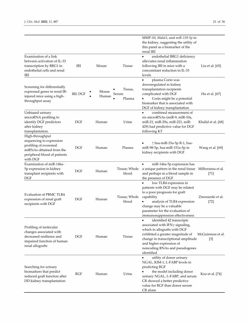
J. Clin. Med. 2022, 11, 487 21 of 36
MMP‐10, Malat1, and miR‐139‐5p in
the kidney, suggesting the utility of
this panel as a biomarker of the
renal IRI
Examination of a link
between activation of IL‐33
transcription by BRG1 in
endothelial cells and renal
IRI
IRI Mouse Tissue
endothelial BRG1 deficiency
alleviates renal inflammation
following IRI in mice with a
concomitant reduction in IL‐33
levels
Liu et al. [65]
Screening for differentially
expressed genes in renal IR‐
injured mice using a high‐
throughput assay
IRI; DGF Mouse
Human
Tissue,
Serum
Plasma
plasma Corin was
downregulated in kidney
transplantation recipients
complicated with DGF
Corin might be a potential
biomarker that is associated with
DGF of kidney transplantation
Hu et al. [67]
Unbiased urinary
microRNA profiling to
identify DGF predictors
after kidney
transplantation.
DGF Human Urine
combined measurement of
six microRNAs (miR‐9, mIR‐10a,
miR‐21, miR‐29a, miR‐221, miR‐
429) had predictive value for DGF
following KT
Khalid et al. [68]
High‐throughput
sequencing to expression
profiling of exosomal
miRNAs obtained from the
peripheral blood of patients
with DGF
DGF Human Plasma
↑ hsa‐miR‐33a‐5p R‐1, hsa‐
miR‐98‐5p, hsa‐miR‐151a‐5p in
kidney recipients with DGF
Wang et al. [69]
Examination of miR‐146a‐
5p expression in kidney
transplant recipients with
DGF
DGF Human Tissue; Whole
blood
miR‐146a‐5p expression has
a unique pattern in the renal tissue
and perhaps in a blood sample in
the presence of DGF
Milhoransa et al.
[71]
Evaluation of PBMC TLR4
expression of renal graft
recipients with DGF
DGF Human Tissue; Whole
blood
low TLR4 expression in
patients with DGF may be related
to a poor prognosis for graft
capability
analysis of TLR4 expression
change may be a valuable
parameter for the evaluation of
immunosuppression effectiveness
Zmonarski et al.
[72]
Profiling of molecular
changes associated with
decreased resilience and
impaired function of human
renal allografts
DGF Human Tissue
identified 42 transcripts
associated with IFNγ signaling,
which in allografts with DGF
exhibited a greater magnitude of
change in transcriptional amplitude
and higher expression of
noncoding RNAs and pseudogenes
identified
McGuinness et al.
[3]
Searching for urinary
biomarkers that predict
reduced graft function after
DD kidney transplantation
RGF Human Urine
utility of donor urinary
NGAL, KIM‐1, L‐FABP levels in
predicting RGF
the model including donor
urinary NGAL, L‐FABP, and serum
CR showed a better predictive
value for RGF than donor serum
CR alone
Koo et al. [74]
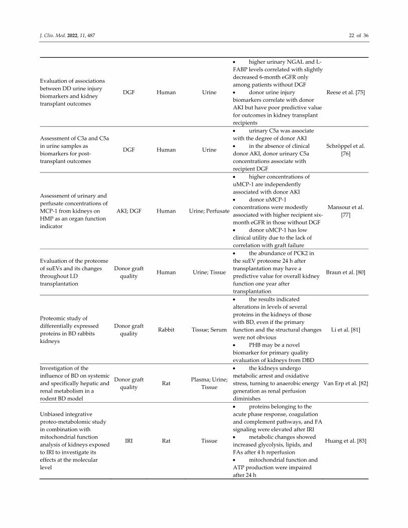
J. Clin. Med. 2022, 11, 487 22 of 36
Evaluation of associations
between DD urine injury
biomarkers and kidney
transplant outcomes
DGF Human Urine
higher urinary NGAL and L‐
FABP levels correlated with slightly
decreased 6‐month eGFR only
among patients without DGF
donor urine injury
biomarkers correlate with donor
AKI but have poor predictive value
for outcomes in kidney transplant
recipients
Reese et al. [75]
Assessment of C3a and C5a
in urine samples as
biomarkers for post‐
transplant outcomes
DGF Human Urine
urinary C5a was associate
with the degree of donor AKI
in the absence of clinical
donor AKI, donor urinary C5a
concentrations associate with
recipient DGF
Schröppel et al.
[76]
Assessment of urinary and
perfusate concentrations of
MCP‐1 from kidneys on
HMP as an organ function
indicator
AKI; DGF Human Urine; Perfusate
higher concentrations of
uMCP‐1 are independently
associated with donor AKI
donor uMCP‐1
concentrations were modestly
associated with higher recipient six‐
month eGFR in those without DGF
donor uMCP‐1 has low
clinical utility due to the lack of
correlation with graft failure
Mansour et al.
[77]
Evaluation of the proteome
of suEVs and its changes
throughout LD
transplantation
Donor graft
quality Human Urine; Tissue
the abundance of PCK2 in
the suEV proteome 24 h after
transplantation may have a
predictive value for overall kidney
function one year after
transplantation
Braun et al. [80]
Proteomic study of
differentially expressed
proteins in BD rabbits
kidneys
Donor graft
quality Rabbit Tissue; Serum
the results indicated
alterations in levels of several
proteins in the kidneys of those
with BD, even if the primary
function and the structural changes
were not obvious
PHB may be a novel
biomarker for primary quality
evaluation of kidneys from DBD
Li et al. [81]
Investigation of the
influence of BD on systemic
and specifically hepatic and
renal metabolism in a
rodent BD model
Donor graft
quality Rat
Plasma; Urine;
Tissue
the kidneys undergo
metabolic arrest and oxidative
stress, turning to anaerobic energy
generation as renal perfusion
diminishes
Van Erp et al. [82]
Unbiased integrative
proteo‐metabolomic study
in combination with
mitochondrial function
analysis of kidneys exposed
to IRI to investigate its
effects at the molecular
level
IRI Rat Tissue
proteins belonging to the
acute phase response, coagulation
and complement pathways, and FA
signaling were elevated after IRI
metabolic changes showed
increased glycolysis, lipids, and
FAs after 4 h reperfusion
mitochondrial function and
ATP production were impaired
after 24 h
Huang et al. [83]
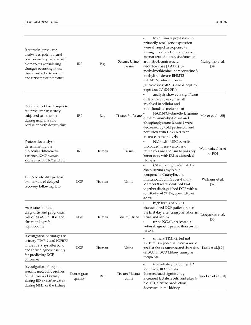
J. Clin. Med. 2022, 11, 487 23 of 36
Integrative proteome
analysis of potential and
predominantly renal injury
biomarkers considering
changes occurring in the
tissue and echo in serum
and urine protein profiles
IRI Pig Serum; Urine;
Tissue
four urinary proteins with
primarily renal gene expression
were changed in response to
managed kidney IRI and may be
biomarkers of kidney dysfunction:
aromatic‐L‐amino‐acid
decarboxylase (AADC), S‐
methylmethionine–homocysteine S‐
methyltransferase BHMT2
(BHMT2), cytosolic beta‐
glucosidase (GBA3), and dipeptidyl
peptidase IV (DPPIV)
Malagrino et al.
[84]
Evaluation of the changes in
the proteome of kidney
subjected to ischemia
during machine cold
perfusion with doxycycline
IRI Rat Tissue; Perfusate
analysis showed a significant
difference in 8 enzymes, all
involved in cellular and
mitochondrial metabolism
N(G),N(G)‐dimethylarginine
dimethylaminohydrolase and
phosphoglycerate kinase 1 were
decreased by cold perfusion, and
perfusion with Doxy led to an
increase in their levels
Moser et al. [85]
Proteomics analysis
determinating the
molecular differences
between NMP human
kidneys with URC and UR
IRI Human Tissue
NMP with URC permits
prolonged preservation and
revitalizes metabolism to possibly
better cope with IRI in discarded
kidneys
Weissenbacher et
al. [86]
TUPA to identify protein
biomarkers of delayed
recovery following KTx
DGF Human Urine
C4b‐binding protein alpha
chain, serum amyloid P‐
component, Guanylin, and
Immunoglobulin Super‐Family
Member 8 were identified that
together distinguished DGF with a
sensitivity of 77.4%, specificity of
82.6%
Williams et al.
[87]
Assessment of the
diagnostic and prognostic
role of NGAL in DGF and
chronic allograft
nephropathy
DGF Human Serum; Urine
high levels of NGAL
characterized DGF patients since
the first day after transplantation in
urine and serum
urine NGAL presented a
better diagnostic profile than serum
NGAL
Lacquaniti et al.
[88]
Investigation of changes of
urinary TIMP‐2 and IGFBP7
in the first days after KTx
and their diagnostic utility
for predicting DGF
outcomes
DGF Human Urine
urinary TIMP‐2, but not
IGFBP7, is a potential biomarker to
predict the occurrence and duration
of DGF in DCD kidney transplant
recipients
Bank et al.[89]
Investigation of organ‐
specific metabolic profiles
of the liver and kidney
during BD and afterwards
during NMP of the kidney
Donor graft
quality Rat
Tissue; Plasma;
Urine
immediately following BD
induction, BD animals
demonstrated significantly
increased lactate levels, and after 4
h of BD, alanine production
decreased in the kidney
van Erp et al. [90]
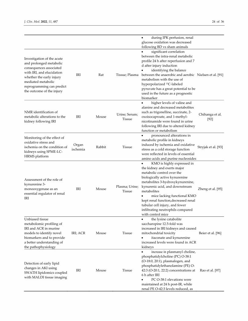
J. Clin. Med. 2022, 11, 487 24 of 36
during IPK perfusion, renal
glucose oxidation was decreased
following BD vs sham animals
Investigation of the acute
and prolonged metabolic
consequences associated
with IRI, and elucidation
whether the early injury
mediated metabolic
reprogramming can predict
the outcome of the injury
IRI Rat Tissue; Plasma
significant correlation
between the intra‐renal metabolic
profile 24 h after reperfusion and 7
d after injury induction
identifying the balance
between the anaerobic and aerobic
metabolism with the use of
hyperpolarized 13C‐labeled
pyruvate has a great potential to be
used in the future as a prognostic
biomarker
Nielsen et al. [91]
NMR identification of
metabolic alterations to the
kidney following IRI
IRI Mouse Urine; Serum;
Tissue
higher levels of valine and
alanine and decreased metabolites
such as trigonelline, succinate, 2‐
oxoisocaproate, and 1‐methyl‐
nicotinamide were found in urine
following IRI due to altered kidney
function or metabolism
Chihanga et al.
[92]
Monitoring of the effect of
oxidative stress and
ischemia on the condition of
kidneys using SPME‐LC‐
HRMS platform
Organ
ischemia Rabbit Tissue
pronounced alterations in
metabolic profile in kidneys
induced by ischemia and oxidative
stress as a cold storage function
were reflected in levels of essential
amino acids and purine nucleosides
Stryjak et al. [93]
Assessment of the role of
kynurenine 3‐
monooxygenase as an
essential regulator of renal
IRI
IRI Mouse Plasma; Urine;
Tissue
KMO is highly expressed in
the kidney and exerts major
metabolic control over the
biologically active kynurenine
metabolites 3‐hydroxykynurenine,
kynurenic acid, and downstream
metabolites
mice lacking functional KMO
kept renal function,decreased renal
tubular cell injury, and fewer
infiltrating neutrophils compared
with control mice
Zheng et al. [95]
Unbiased tissue
metabolomic profiling of
IRI and ACR in murine
models to identify novel
biomarkers and to provide
a better understanding of
the pathophysiology
IRI; ACR Mouse Tissue
the lysine catabolite
saccharopine 12.5‐fold was
increased in IRI kidneys and caused
mitochondrial toxicity
itaconate and kynurenine
increased levels were found in ACR
kidneys
Beier et al. [96]
Detection of early lipid
changes in AKI using
SWATH lipidomics coupled
with MALDI tissue imaging
IRI Mouse Tissue
increase in plasmanyl choline,
phosphatidylcholine (PC) O‐38:1
(O‐18:0, 20:1), plasmalogen, and
phosphatidylethanolamine (PE) O‐
42:3 (O‐20:1, 22:2) concentrations at
6 h after IRI
PC O‐38:1 elevations were
maintained at 24 h post‐IR, while
renal PE O‐42:3 levels reduced, as
Rao et al. [97]
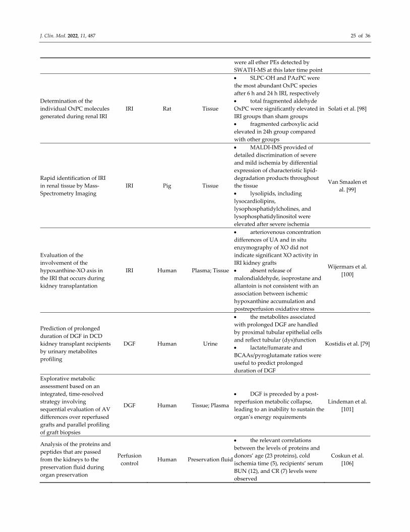
J. Clin. Med. 2022, 11, 487 25 of 36
were all ether PEs detected by
SWATH‐MS at this later time point
Determination of the
individual OxPC molecules
generated during renal IRI
IRI Rat Tissue
SLPC‐OH and PAzPC were
the most abundant OxPC species
after 6 h and 24 h IRI, respectively
total fragmented aldehyde
OxPC were significantly elevated in
IRI groups than sham groups
fragmented carboxylic acid
elevated in 24h group compared
with other groups
Solati et al. [98]
Rapid identification of IRI
in renal tissue by Mass‐
Spectrometry Imaging
IRI Pig Tissue
MALDI‐IMS provided of
detailed discrimination of severe
and mild ischemia by differential
expression of characteristic lipid‐
degradation products throughout
the tissue
lysolipids, including
lysocardiolipins,
lysophosphatidylcholines, and
lysophosphatidylinositol were
elevated after severe ischemia
Van Smaalen et
al. [99]
Evaluation of the
involvement of the
hypoxanthine‐XO axis in
the IRI that occurs during
kidney transplantation
IRI Human Plasma; Tissue
arteriovenous concentration
differences of UA and in situ
enzymography of XO did not
indicate significant XO activity in
IRI kidney grafts
absent release of
malondialdehyde, isoprostane and
allantoin is not consistent with an
association between ischemic
hypoxanthine accumulation and
postreperfusion oxidative stress
Wijermars et al.
[100]
Prediction of prolonged
duration of DGF in DCD
kidney transplant recipients
by urinary metabolites
profiling
DGF Human Urine
the metabolites associated
with prolonged DGF are handled
by proximal tubular epithelial cells
and reflect tubular (dys)function
lactate/fumarate and
BCAAs/pyroglutamate ratios were
useful to predict prolonged
duration of DGF
Kostidis et al. [79]
Explorative metabolic
assessment based on an
integrated, time‐resolved
strategy involving
sequential evaluation of AV
differences over reperfused
grafts and parallel profiling
of graft biopsies
DGF Human Tissue; Plasma
DGF is preceded by a post‐
reperfusion metabolic collapse,
leading to an inability to sustain the
organ’s energy requirements
Lindeman et al.
[101]
Analysis of the proteins and
peptides that are passed
from the kidneys to the
preservation fluid during
organ preservation
Perfusion
control Human Preservation fluid
the relevant correlations
between the levels of proteins and
donors’ age (23 proteins), cold
ischemia time (5), recipients’ serum
BUN (12), and CR (7) levels were
observed
Coskun et al.
[106]
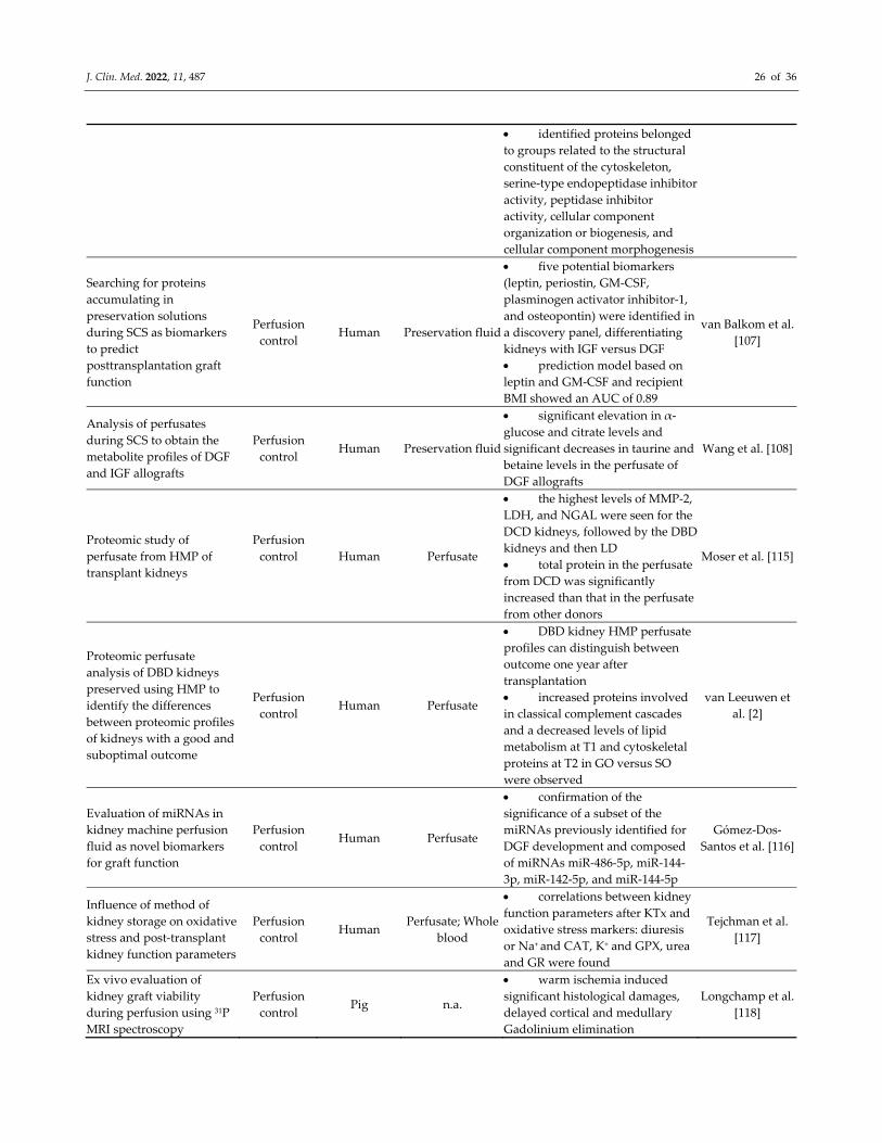
J. Clin. Med. 2022, 11, 487 26 of 36
identified proteins belonged
to groups related to the structural
constituent of the cytoskeleton,
serine‐type endopeptidase inhibitor
activity, peptidase inhibitor
activity, cellular component
organization or biogenesis, and
cellular component morphogenesis
Searching for proteins
accumulating in
preservation solutions
during SCS as biomarkers
to predict
posttransplantation graft
function
Perfusion
control Human Preservation fluid
five potential biomarkers
(leptin, periostin, GM‐CSF,
plasminogen activator inhibitor‐1,
and osteopontin) were identified in
a discovery panel, differentiating
kidneys with IGF versus DGF
prediction model based on
leptin and GM‐CSF and recipient
BMI showed an AUC of 0.89
van Balkom et al.
[107]
Analysis of perfusates
during SCS to obtain the
metabolite profiles of DGF
and IGF allografts
Perfusion
control Human Preservation fluid
significant elevation in α‐
glucose and citrate levels and
significant decreases in taurine and
betaine levels in the perfusate of
DGF allografts
Wang et al. [108]
Proteomic study of
perfusate from HMP of
transplant kidneys
Perfusion
control
Human Perfusate
the highest levels of MMP‐2,
LDH, and NGAL were seen for the
DCD kidneys, followed by the DBD
kidneys and then LD
total protein in the perfusate
from DCD was significantly
increased than that in the perfusate
from other donors
Moser et al. [115]
Proteomic perfusate
analysis of DBD kidneys
preserved using HMP to
identify the differences
between proteomic profiles
of kidneys with a good and
suboptimal outcome
Perfusion
control Human Perfusate
DBD kidney HMP perfusate
profiles can distinguish between
outcome one year after
transplantation
increased proteins involved
in classical complement cascades
and a decreased levels of lipid
metabolism at T1 and cytoskeletal
proteins at T2 in GO versus SO
were observed
van Leeuwen et
al. [2]
Evaluation of miRNAs in
kidney machine perfusion
fluid as novel biomarkers
for graft function
Perfusion
control Human Perfusate
confirmation of the
significance of a subset of the
miRNAs previously identified for
DGF development and composed
of miRNAs miR‐486‐5p, miR‐144‐
3p, miR‐142‐5p, and miR‐144‐5p
Gómez‐Dos‐
Santos et al. [116]
Influence of method of
kidney storage on oxidative
stress and post‐transplant
kidney function parameters
Perfusion
control Human
Perfusate; Whole
blood
correlations between kidney
function parameters after KTx and
oxidative stress markers: diuresis
or Na+ and CAT, K+ and GPX, urea
and GR were found
Tejchman et al.
[117]
Ex vivo evaluation of
kidney graft viability
during perfusion using 31P
MRI spectroscopy
Perfusion
control Pig n.a.
warm ischemia induced
significant histological damages,
delayed cortical and medullary
Gadolinium elimination
Longchamp et al.
[118]
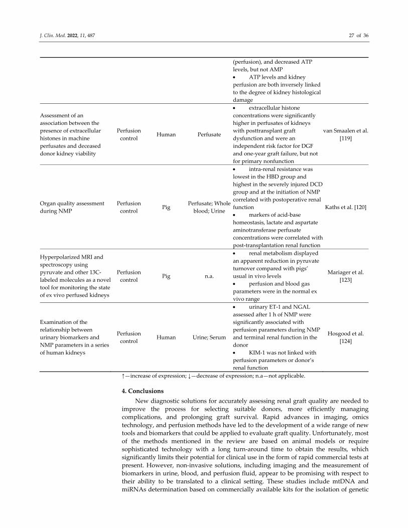
J. Clin. Med. 2022, 11, 487 27 of 36
(perfusion), and decreased ATP
levels, but not AMP
ATP levels and kidney
perfusion are both inversely linked
to the degree of kidney histological
damage
Assessment of an
association between the
presence of extracellular
histones in machine
perfusates and deceased
donor kidney viability
Perfusion
control Human Perfusate
extracellular histone
concentrations were significantly
higher in perfusates of kidneys
with posttransplant graft
dysfunction and were an
independent risk factor for DGF
and one‐year graft failure, but not
for primary nonfunction
van Smaalen et al.
[119]
Organ quality assessment
during NMP
Perfusion
control Pig
Perfusate; Whole
blood; Urine
intra‐renal resistance was
lowest in the HBD group and
highest in the severely injured DCD
group and at the initiation of NMP
correlated with postoperative renal
function
markers of acid‐base
homeostasis, lactate and aspartate
aminotransferase perfusate
concentrations were correlated with
post‐transplantation renal function
Kaths et al. [120]
Hyperpolarized MRI and
spectroscopy using
pyruvate and other 13C‐
labeled molecules as a novel
tool for monitoring the state
of ex vivo perfused kidneys
Perfusion
control Pig n.a.
renal metabolism displayed
an apparent reduction in pyruvate
turnover compared with pigs’
usual in vivo levels
perfusion and blood gas
parameters were in the normal ex
vivo range
Mariager et al.
[123]
Examination of the
relationship between
urinary biomarkers and
NMP parameters in a series
of human kidneys
Perfusion
control Human Urine; Serum
urinary ET‐1 and NGAL
assessed after 1 h of NMP were
significantly associated with
perfusion parameters during NMP
and terminal renal function in the
donor
KIM‐1 was not linked with
perfusion parameters or donor’s
renal function
Hosgood et al.
[124]
↑—increase of expression; ↓—decrease of expression; n.a—not applicable.
4. Conclusions
New diagnostic solutions for accurately assessing renal graft quality are needed to
improve the process for selecting suitable donors, more efficiently managing
complications, and prolonging graft survival. Rapid advances in imaging, omics
technology, and perfusion methods have led to the development of a wide range of new
tools and biomarkers that could be applied to evaluate graft quality. Unfortunately, most
of the methods mentioned in the review are based on animal models or require
sophisticated technology with a long turn‐around time to obtain the results, which
significantly limits their potential for clinical use in the form of rapid commercial tests at
present. However, non‐invasive solutions, including imaging and the measurement of
biomarkers in urine, blood, and perfusion fluid, appear to be promising with respect to
their ability to be translated to a clinical setting. These studies include mtDNA and
miRNAs determination based on commercially available kits for the isolation of genetic
J. Clin. Med. 2022, 11, 487 28 of 36
material in combination with the RT‐PCR technique widely used in laboratory practice. A
similar clinical potential is demonstrated by the determination of biomarkers such as
NGAL, KIM‐1, L‐FABP and C5a in urine by ELISA, also routinely used in diagnostics.
Nevertheless, the translation of biomarkers from the discovery stage to clinical practice is
still challenging due to the complex and multifactorial type of injuries, the absence of
standard guidelines for method validation, and adequate prospective and retrospective
cohort studies. Larger, multi‐centre validation studies are needed before new solutions
can be widely implemented in clinics. Moreover, it will be imperative for future research
to explore new technologies and integrate molecular measurements from large data sets
reported in different experiments.
Author Contributions: Writing—original draft preparation, N.W.; designing writing—review and
editing, N.W., K.Ł., B.B.; funding acquisition, B.B. All authors have read and agreed to the published
version of the manuscript.
Funding: This study was funded by National Science Centre, grant Opus number
2017/27/B/NZ5/01013.
Data Availability Statement: No new data were created or analyzed in this study. Data sharing is
not applicable to this article.
Conflicts of Interest: The authors declare no conflict of interest.
Abbreviations
ACR acute cellular rejection
AKI acute kidney injury
AMP adenosine monophosphate
AR acute rejection
ASL arterial spin labelling
ATP adenosine triphosphate
AUC area under the curve
BCAA branched chain amino acids
BD brain death
BMI body mass index
BUN blood urea nitrogen
CAT catalase
CR creatinine
CTA computed tomography
CTA computed tomography angiography
DBD donor after brain death
DCD donor after cardiac death
DD deceased donors
DEG Differentially expressed genes
DGF delayed graft function
DJ‐1 protein deglycase
Doxy doxycycline
ECD expanded criteria donors
eGFR estimated glomerular filtration rate
ET‐1 endothelin‐1
FA fatty acid
FABP fatty acid‐binding protein
FAO fatty acid oxidation
FC fold change
FS frozen section
GM‐CSF Granulocyte‐macrophage colony‐stimulating factor
J. Clin. Med. 2022, 11, 487 29 of 36
GO good outcome
GPX glutathione peroxidase
GR glutathione reductase
GSH glutathione
GST glutathione transferase
GTP guanosine triphosphate
HMP Hypothermic machine perfusion
HSI Hyperspectral Imaging
ICG indocyanine green
IGF immediate graft function
IGFBP7 insulin‐like growth factor binding protein‐7
IL‐18 Interleukin‐18
IPK normothermic isolated perfused kidney
IRI ischemia‐reperfusion injury
KATs kynurenine aminotransferases
KDPI Kidney Donor Profile Index
KDRI Kidney Donor Risk Index
KIM‐1 kidney injury molecule‐1
KMO kynurenine 3‐monooxygenase
KTx Kidney transplantation
LD living donor
LDH lactate dehydrogenase
LDKT living donor kidney transplantation
L‐FABP L‐type fatty acid binding protein
lncRNA long noncoding RNA
MALDI‐IMS Matrix Assisted Laser Desorption/Ionization‐Imaging Mass
Spectrometry
MALDI‐TOF‐
MS
Matrix Assisted Laser Desorption/Ionization Time‐of‐Flight Mass
Spectrometry
MCP‐1 Monocyte chemoattractant protein‐1
MDA malondialdehyde
miRNA/miR microRNA
MMP‐2 matrix metalloproteinase‐2
MR magnetic resonance
MRI magnetic resonance imaging
MRS magnetic resonance spectroscopy
MS mass spectrometry
MSI mass spectrometry imaging
mtDNA mitochondrial DNA
NB needle biopsy
NGAL neutrophil gelatinase‐associated lipocalin
NMP normothermic machine perfusion
NMR nuclear magnetic resonance
OxPC oxidized phosphatidylcholine
PBMC peripheral blood mononuclear cells
PC phosphatidylcholine
PCK2 phosphoenol pyruvate carboxykinase
PE phosphatidylethanolamine
PET positron emission tomography
PHB prohibitin
pMRI 31P magnetic resonance imaging
J. Clin. Med. 2022, 11, 487 30 of 36
PNF primary nonfunction
PRAT perirenal adipose tissue
PS paraffin sections
RGB Red Green Blue (colour model)
RGF reduced graft function
RR renal resistance
RT‐PCR real‐time polymerase chain reaction
SCD standard criteria donors
SCS Static cold storage
SO suboptimal outcome
SOD superoxide dismutase
SPME solid‐phase microextraction
suEVs small urinary extracellular vesicles
SVF stromal vascular fraction
SWATH‐MS sequential window acquisition of all theoretical spectra‐mass
spectrometry
TBARS thiobarbituric acid reactive substances
TCA tricarboxylic acid
TIMP‐2 tissue inhibitor of metalloproteinases‐2
TLR4 Toll‐like receptor 4
TUPA Targeted Urine Proteome Assay
UA uric acid
UR urine replacement
URC urine recirculation
WB wedge biopsy
WI warm ischemia
WIT warm ischemia time
XO hypoxanthine‐xanthine oxidase
References
1. Swanson, K.J.; Aziz, F.; Garg, N.; Mohamed, M.; Mandelbrot, D.; Djamali, A.; Parajuli, S. Role of novel biomarkers in kidney
transplantation. World J. Transplant. 2020, 10, 230–255, doi:10.5500/wjt.v10.i9.230.
2. van Leeuwen, L.L.; Spraakman, N.A.; Brat, A.; Huang, H.; Thorne, A.M.; Bonham, S.; van Balkom, B.W.M.; Ploeg, R.J.; Kessler,
B.M.; Leuvenink, H.G.D. Proteomic analysis of machine perfusion solution from brain dead donor kidneys reveals that elevated
complement, cytoskeleton and lipid metabolism proteins are associated with 1‐year outcome. Transpl. Int. 2021, 34, 1618–1629,
doi:10.1111/tri.13984.
3. McGuinness, D.; Mohammed, S.; Monaghan, L.; Wilson, P.A.; Kingsmore, D.B.; Shapter, O.; Stevenson, K.S.; Coley, S.M.; Devey,
L.; Kirkpatrick, R.B.; et al. A molecular signature for delayed graft function. Aging Cell 2018, 17, 1–16, doi:10.1111/acel.12825.
4. Dare, A.J.; Pettigrew, G.J.; Saeb‐parsy, K. Preoperative Assessment of the Deceased‐Donor Kidney : From Macroscopic
Appearance to Molecular Biomarkers. Transplantation 2014, 97, 797–807, doi:10.1097/01.TP.0000441361.34103.53.
5. Moeckli, B.; Sun, P.; Lazeyras, F.; Morel, P.; Moll, S.; Pascual, M.; Bühler, L.H. Evaluation of donor kidneys prior to
transplantation: An update of current and emerging methods. Transpl. Int. 2019, 32, 459–469, doi:10.1111/tri.13430.
6. Kork, F.; Rimek, A.; Andert, A.; Becker, N.J.; Heidenhain, C.; Neumann, U.P.; Kroy, D.; Roehl, A.B.; Rossaint, R.; Hein, M. Visual
quality assessment of the liver graft by the transplanting surgeon predicts postreperfusion syndrome after liver transplantation:
A retrospective cohort study. BMC Anesthesiol. 2018, 18, 1–11, doi:10.1186/s12871‐018‐0493‐9.
7. Nyberg, S.L.; Matas, A.J.; Kremers, W.K.; Thostenson, J.D.; Larson, T.S.; Prieto, M.; Ishitani, M.B.; Sterioff, S.; Stegall, M.D.
Improved scoring system to assess adult donors for cadaver renal transplantation. Am. J. Transplant. 2003, 3, 715–721,
doi:10.1034/j.1600‐6143.2003.00111.x.
8. Schold, J.D.; Kaplan, B.; Baliga, R.S.; Meier‐Kriesche, H.‐U. The broad spectrum of quality in deceased donor kidneys. Am. J.
Transplant. 2005, 5, 757–765, doi:10.1111/j.1600‐6143.2005.00770.x.
9. Watson, C.J.E.; Johnson, R.J.; Birch, R.; Collett, D.; Bradley, J.A. A simplified donor risk index for predicting outcome after
deceased donor kidney transplantation. Transplantation 2012, 93, 314–318, doi:10.1097/TP.0b013e31823f14d4.
10. Rao, P.S.; Schaubel, D.E.; Guidinger, M.K.; Andreoni, K.A.; Wolfe, R.A.; Merion, R.M.; Port, F.K.; Sung, R.S. A comprehensive
risk quantification score for deceased donor kidneys: The kidney donor risk index. Transplantation 2009, 88, 231–236,
J. Clin. Med. 2022, 11, 487 31 of 36
doi:10.1097/TP.0b013e3181ac620b.
11. U.S. Department of Health and Human Services. Organ Procurement and Transplantation Network: KDPI Calculator. Available
online: https://optn.transplant.hrsa.gov/resources/guidance/kidney‐donor‐profile‐index‐kdpi‐guide‐for‐clinicians (accessed on
9 November 2021).
12. Dahmen, M.; Becker, F.; Pavenstädt, H.; Suwelack, B.; Schütte‐Nütgen, K.; Reuter, S. Validation of the Kidney Donor Profile
Index (KDPI) to assess a deceased donor’s kidneys’ outcome in a European cohort. Sci. Rep. 2019, 9, 11234, doi:10.1038/s41598‐
019‐47772‐7.
13. Jun, H.; Yoon, H.E.; Lee, K.W.; Lee, D.R.; Yang, J.; Ahn, C.; Han, S.Y. Kidney Donor Risk Index Score Is More Reliable Than
Kidney Donor Profile Index in Kidney Transplantation From Elderly Deceased Donors. Transplant. Proc. 2020, 52, 1744–1748,
doi:10.1016/j.transproceed.2020.03.005.
14. Zheng, J.; Hu, X.; Ding, X.; Li, Y.; Ding, C.; Tian, P.; Xiang, H.; Feng, X.; Pan, X.; Yan, H.; et al. Comprehensive assessment of
deceased donor kidneys with clinic al characteristics, pre‐implant biopsy histopathology and hypothermic mechanical
perfusion parameters is highly predictive of delayed graft function. Ren. Fail. 2020, 42, 369–376,
doi:10.1080/0886022X.2020.1752716.
15. Parker, W.F.; Thistlethwaite Jr, J.R.; Ross, L.F. Kidney Donor Profile Index (KDPI) Does Not Accurately Predict the Graft
Survival of Pediatric Deceased Donor Kidneys. Transplantation 2016, 100, 2471–2478, doi:10.1097/TP.0000000000001028.Kidney.
16. Hopfer, H.; Kemény, E. Assessment of donor biopsies. Curr. Opin. Organ. Transplant. 2013, 18, 306–312,
doi:10.1097/MOT.0b013e3283607a6e.
17. Naesens, M. Zero‐time renal transplant biopsies: A comprehensive review. Transplantation 2016, 100, 1425–1439,
doi:10.1097/TP.0000000000001018.
18. Sagasta, A.; Sánchez‐Escuredo, A.; Oppenheimer, F.; Paredes, D.; Musquera, M.; Campistol, J.M.; Solé, M. Pre‐implantation
analysis of kidney biopsies from expanded criteria donors: Testing the accuracy of frozen section technique and the adequacy
of their assessment by on‐call pathologists. Transpl. Int. 2016, 29, 234–240, doi:10.1111/tri.12709.
19. Cooper, M.; Formica, R.; Friedewald, J.; Hirose, R.; O’Connor, K.; Mohan, S.; Schold, J.; Axelrod, D.; Pastan, S. Report of National
Kidney Foundation Consensus Conference to Decrease Kidney Discards. Clin. Transplant. 2019, 33, e13419, doi:10.1111/ctr.13419.
20. Traynor, C.; Saeed, A.; O’Ceallaigh, E.; Elbadri, A.; O’Kelly, P.; de Freitas, D.G.; Dorman, A.M.; Conlon, P.J.; O’Seaghdha, C.M.
Pre‐transplant histology does not improve prediction of 5‐year kidney allograft outcomes above and beyond clinical
parameters. Ren. Fail. 2017, 39, 671–677, doi:10.1080/0886022X.2017.1363778.
21. Yap, Y.T.; Ho, Q.Y.; Kee, T.; Ng, C.Y.; Chionh, C.Y. Impact of pre‐transplant biopsy on 5‐year outcomes of expanded criteria
donor kidney transplantation. Nephrology 2021, 26, 70–77, doi:10.1111/nep.13788.
22. De Vusser, K.; Lerut, E.; Kuypers, D.; Vanrenterghem, Y.; Jochmans, I.; Monbaliu, D.; Pirenne, J.; Naesens, M. The predictive
value of kidney allograft baseline biopsies for long‐term graft survival. J. Am. Soc. Nephrol. 2013, 24, 1913–1923,
doi:10.1681/ASN.2012111081.
23. Phillips, B.L.; Kassimatis, T.; Atalar, K.; Wilkinson, H.; Kessaris, N.; Simmonds, N.; Hilton, R.; Horsfield, C.; Callaghan, C.J.
Chronic histological changes in deceased donor kidneys at implantation do not predict graft survival: A single‐centre
retrospective analysis. Transpl. Int. 2019, 32, 523–534, doi:10.1111/tri.13398.
24. Liapis, H.; Gaut, J.P.; Klein, C.; Bagnasco, S.; Kraus, E.; Farris III, A.B.; Honsova, E.; Perkowska‐Ptasinska, A.; David, D.;
Goldberg, J.; et al. Banff Histopathological Consensus Criteria for Preimplantation Kidney Biopsies. Am. J. Transplant. 2017, 17,
140–150, doi:10.1111/ajt.13929.
25. Hall, I.E.; Parikh, C.R.; Schröppel, B.; Weng, F.L.; Jia, Y.; Thiessen‐Philbrook, H.; Reese, P.P.; Doshi, M.D. Procurement biopsy
findings versus kidney donor risk index for predicting renal allograft survival. Transplant. Direct 2018, 4, e373,
doi:10.1097/TXD.0000000000000816.
26. Peng, P.; Ding, Z.; He, Y.; Zhang, J.; Wang, X.; Yang, Z. Hypothermic Machine Perfusion Versus Static Cold Storage in Deceased
Donor Kidney Transplantation: A Systematic Review and Meta‐Analysis of Randomized Controlled Trials. Artif. Organs 2019,
43, 478–489, doi:10.1111/aor.13364.
27. Peris, A.; Fulceri, G.E.; Lazzeri, C.; Bonizzoli, M.; Li Marzi, V.; Serni, S.; Cirami, L.; Migliaccio, M.L. Delayed graft function and
perfusion parameters of kidneys from uncontrolled donors after circulatory death. Perfusion 2021, 36, 299–304,
doi:10.1177/0267659120938928.
28. Bissolati, M.; Gazzetta, P.G.; Caldara, R.; Guarneri, G.; Adamenko, O.; Giannone, F.; Mazza, M.; Maggi, G.; Tomanin, D.; Rosati,
R.; et al. Renal Resistance Trend During Hypothermic Machine Perfusion Is More Predictive of Postoperative Outcome Than
Biopsy Score: Preliminary Experience in 35 Consecutive Kidney Transplantations. Artif. Organs 2018, 42, 714–722,
doi:10.1111/aor.13117.
29. Moers, C.; Smits, J.M.; Maathuis, M.‐H.J.; Treckmann, J.; Gelder, F. van; Napieralski, B.P.; van Kasterop‐Kutz, M.; van der Heide,
J.J.H.; Squifflet, J.‐P.; van Heurn, E.; et al. Machine Perfusion or Cold Storage in Deceased‐Donor Kidney Transplantation. N.
Engl. J. Med. 2009, 360, 7–19, doi:10.1056/NEJMoa0802289.
30. Lindell, S.L.; Muir, H.; Brassil, J.; Mangino, M.J. Hypothermic Machine Perfusion Preservation of the DCD Kidney: Machine
Effects. J. Transplant. 2013, 2013, 802618, doi:10.1155/2013/802618.
31. De Deken, J.; Kocabayoglu, P.; Moers, C. Hypothermic machine perfusion in kidney transplantation. Curr. Opin. Organ.
Transplant. 2016, 21, 294–300, doi:10.1097/MOT.0000000000000306.
32. Jochmans, I.; Moers, C.; Smits, J.M.; Leuvenink, H.G.D.; Treckmann, J.; Paul, A.; Rahmel, A.; Squifflet, J.P.; van Heurn, E.;
J. Clin. Med. 2022, 11, 487 32 of 36
Monbaliu, D.; et al. The prognostic value of renal resistance during hypothermic machine perfusion of deceased donor kidneys.
Am. J. Transplant. 2011, 11, 2214–2220, doi:10.1111/j.1600‐6143.2011.03685.x.
33. Mozes, M.F.; Skolek, R.B.; Korf, B.C. Use of perfusion parameters in predicting outcomes of machine‐preserved kidneys.
Transplant. Proc. 2005, 37, 350–351, doi:10.1016/j.transproceed.2005.01.058.
34. Bunegin, L.; Tolstykh, G.P.; Gelineau, J.F.; Cosimi, A.B.; Anderson, L.M. Oxygen consumption during oxygenated hypothermic
perfusion as a measure of donor organ viability. ASAIO J. 2013, 59, 427–432, doi:10.1097/MAT.0b013e318292e865.
35. Patel, S.K.; Pankewycz, O.G.; Nader, N.D.; Zachariah, M.; Kohli, R.; Laftavi, M.R. Prognostic utility of hypothermic machine
perfusion in deceased donor renal transplantation. Transplant. Proc. 2012, 44, 2207–2212, doi:10.1016/j.transproceed.2012.07.129.
36. Reticker, A.; Lichvar, A.; Walsh, M.; Gross, A.E.; Patel, S. The Significance and Impact of Screening Preservation Fluid Cultures
in Renal Transplant Recipients. Prog. Transplant. 2021, 31, 40–46, doi:10.1177/1526924820978608.
37. Corbel, A.; Ladrière, M.; Le Berre, N.; Durin, L.; Rousseau, H.; Frimat, L.; Thilly, N.; Pulcini, C. Microbiological epidemiology
of preservation fluids in transplanted kidney: A nationwide retrospective observational study. Clin. Microbiol. Infect. 2020, 26,
475–484, doi:10.1016/j.cmi.2019.07.018.
38. Oriol, I.; Sabe, N.; Càmara, J.; Berbel, D.; Ballesteros, M.A.; Escudero, R.; Lopez‐Medrano, F.; Linares, L.; Len, O.; Silva, J.T.; et
al. The impact of culturing the organ preservation fluid on solid organ transplantation: A prospective multicenter cohort study.
Open Forum Infect. Dis. 2019, 6, 1–7, doi:10.1093/ofid/ofz180.
39. Yu, X.; Wang, R.; Peng, W.; Huang, H.; Liu, G.; Yang, Q.; Zhou, J.; Zhang, X.; Lv, J.H.; Lei, W.; et al. Incidence, distribution and
clinical relevance of microbial contamination of preservation solution in deceased kidney transplant recipients: A retrospective
cohort study from China. Clin. Microbiol. Infect. 2019, 25, 595–600, doi:10.1016/j.cmi.2018.12.040.
40. Stern, S.; Bezinover, D.; Rath, P.M.; Paul, A.; Saner, F.H. Candida contamination in kidney and liver organ preservation solution:
Does it matter? J. Clin. Med. 2021, 10, doi:10.3390/jcm10092022.
41. Sjekavica, I.; Novosel, L.; Rupčić, M.; Smiljanić, R.; Muršić, M.; Duspara, V.; Lušić, M.; Perkov, D.; Hrabak‐Paar, M.; Zidanić,
M.; et al. Radiological imaging in renal transplantation. Acta Clin. Croat. 2018, 57, 694–712, doi:10.20471/acc.2018.57.04.12.
42. Sarier, M.; Callioglu, M.; Yuksel, Y.; Duman, E.; Emek, M.; Usta, S.S. Evaluation of the Renal Arteries of 2,144 Living Kidney
Donors Using Computed Tomography Angiography and Comparison with Intraoperative Findings. Urol. Int. 2020, 104, 637–
640, doi:10.1159/000507796.
43. Al‐Adra, D.P.; Lambadaris, M.; Barbas, A.; Li, Y.; Selzner, M.; Singh, S.K.; Famure, O.; Kim, S.J.; Ghanekar, A. Donor kidney
volume measured by computed tomography is a strong predictor of recipient eGFR in living donor kidney transplantation.
World J. Urol. 2019, 37, 1965–1972, doi:10.1007/s00345‐018‐2595‐x.
44. Fernandez, N.; Lorenzo, A.; Chua, M.; Koyle, M.A.; Farhat, W.; Matava, C. Real‐time kidney graft perfusion monitoring using
infrared imaging during pediatric kidney transplantation. J. Pediatr. Urol. 2019, 15, 222.e1–222.e7,
doi:10.1016/j.jpurol.2019.03.016.
45. Sucher, R.; Wagner, T.; Köhler, H.; Sucher, E.; Guice, H.; Recknagel, S.; Lederer, A.; Hau, H.M.; Rademacher, S.; Schneeberger,
S.; et al. Hyperspectral Imaging (HSI) of Human Kidney Allografts. Ann. Surg. 2020, doi:10.1097/sla.0000000000004429.
46. Gerken, A.L.H.; Nowak, K.; Meyer, A.; Weiss, C.; Krüger, B.; Nawroth, N.; Karampinis, I.; Heller, K.; Apel, H.; Reissfelder, C.;
et al. Quantitative Assessment of Intraoperative Laser Fluorescence Angiography with Indocyanine Green Predicts Early Graft
Function after Kidney Transplantation. Ann. Surg. 2020, doi:10.1097/sla.0000000000004529.
47. Yu, Y.M.; Ni, Q.Q.; Wang, Z.J.; Chen, M.L.; Zhang, L.J. Multiparametric functional magnetic resonance imaging for evaluating
renal allograft injury. Korean J. Radiol. 2019, 20, 894–908, doi:10.3348/kjr.2018.0540.
48. Schutter, R.; Lantinga, V.A.; Borra, R.J.H.; Moers, C. MRI for diagnosis of post‐renal transplant complications: Current state‐of‐
the‐art and future perspectives. Magn. Reson. Mater. Physics, Biol. Med. 2020, 33, 49–61, doi:10.1007/s10334‐019‐00813‐8.
49. Jehn, U.; Schuette‐Nuetgen, K.; Kentrup, D.; Hoerr, V.; Reuter, S. Renal allograft rejection: Noninvasive ultrasound‐ and mri‐
based diagnostics. Contrast Media Mol. Imaging 2019, 2019, 3568067, doi:10.1155/2019/3568067.
50. Pajenda, S.; Rasul, S.; Hacker, M.; Wagner, L.; Geist, B.K. Dynamic 2‐deoxy‐2[18F] fluoro‐D‐glucose PET/MRI in human renal
allotransplant patients undergoing acute kidney injury. Sci. Rep. 2020, 10, 8270, doi:10.1038/s41598‐020‐65267‐8.
51. Jadoul, A.; Lovinfosse, P.; Bouquegneau, A.; Weekers, L.; Pottel, H.; Hustinx, R.; Jouret, F. Observer variability in the assessment
of renal 18F‐FDG uptake in kidney transplant recipients. Sci. Rep. 2020, 10, 4617, doi:10.1038/s41598‐020‐61032‐z.
52. Cai, Y.; Li, Z.; Zuo, P.; Pfeuffer, J.; Li, Y.; Liu, F.; Liu, R. Diagnostic value of renal perfusion in patients with chronic kidney
disease using 3D arterial spin labeling. J. Magn. Reson. Imaging 2017, 46, 589–594, doi:10.1002/jmri.25601.
53. Wang, W.; Yu, Y.; Li, X.; Chen, J.; Zhang, Y.; Zhang, L.; Wen, J. Early detection of subclinical pathology in patients with stable
kidney graft function by arterial spin labeling. Eur. Radiol. 2021, 31, 2687–2695, doi:10.1007/s00330‐020‐07369‐5.
54. Bontha, S.V.; Maluf, D.G.; Mueller, T.F.; Mas, V.R. Systems Biology in Kidney Transplantation: The Application of Multi‐Omics
to a Complex Model. Am. J. Transplant. 2017, 17, 11–21, doi:10.1111/ajt.13881.
55. Giraud, S.; Steichen, C.; Allain, G.; Couturier, P.; Labourdette, D.; Lamarre, S.; Ameteau, V.; Tillet, S.; Hannaert, P.; Thuillier, R.;
et al. Dynamic transcriptomic analysis of Ischemic Injury in a Porcine Pre‐Clinical Model mimicking Donors Deceased after
Circulatory Death. Sci. Rep. 2018, 8, 5986, doi:10.1038/s41598‐018‐24282‐6.
56. Boissier, R.; François, P.; Tellier, B.G.; Meunier, M.; Lyonnet, L.; Simoncini, S.; Magalon, J.; Legris, T.; Arnaud, L.; Giraudo, L.;
et al. Perirenal Adipose Tissue Displays an Age‐Dependent Inflammatory Signature Associated With Early Graft Dysfunction
of Marginal Kidney Transplants. Front. Immunol. 2020, 11, 445, doi:10.3389/fimmu.2020.00445.
57. Hruba, P.; Krejcik, Z.; Dostalova Merkerova, M.; Klema, J.; Stranecky, V.; Slatinska, J.; Maluskova, J.; Honsova, E.; Viklicky, O.
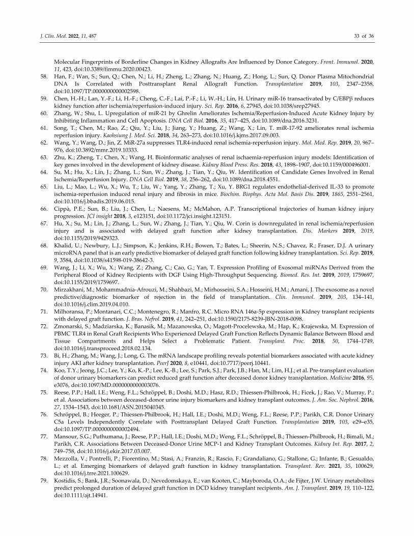
J. Clin. Med. 2022, 11, 487 33 of 36
Molecular Fingerprints of Borderline Changes in Kidney Allografts Are Influenced by Donor Category. Front. Immunol. 2020,
11, 423, doi:10.3389/fimmu.2020.00423.
58. Han, F.; Wan, S.; Sun, Q.; Chen, N.; Li, H.; Zheng, L.; Zhang, N.; Huang, Z.; Hong, L.; Sun, Q. Donor Plasma Mitochondrial
DNA Is Correlated with Posttransplant Renal Allograft Function. Transplantation 2019, 103, 2347–2358,
doi:10.1097/TP.0000000000002598.
59. Chen, H.‐H.; Lan, Y.‐F.; Li, H.‐F.; Cheng, C.‐F.; Lai, P.‐F.; Li, W.‐H.; Lin, H. Urinary miR‐16 transactivated by C/EBPβ reduces
kidney function after ischemia/reperfusion‐induced injury. Sci. Rep. 2016, 6, 27945, doi:10.1038/srep27945.
60. Zhang, W.; Shu, L. Upregulation of miR‐21 by Ghrelin Ameliorates Ischemia/Reperfusion‐Induced Acute Kidney Injury by
Inhibiting Inflammation and Cell Apoptosis. DNA Cell Biol. 2016, 35, 417–425, doi:10.1089/dna.2016.3231.
61. Song, T.; Chen, M.; Rao, Z.; Qiu, Y.; Liu, J.; Jiang, Y.; Huang, Z.; Wang, X.; Lin, T. miR‐17‐92 ameliorates renal ischemia
reperfusion injury. Kaohsiung J. Med. Sci. 2018, 34, 263–273, doi:10.1016/j.kjms.2017.09.003.
62. Wang, Y.; Wang, D.; Jin, Z. MiR‐27a suppresses TLR4‐induced renal ischemia‐reperfusion injury. Mol. Med. Rep. 2019, 20, 967–
976, doi:10.3892/mmr.2019.10333.
63. Zhu, K.; Zheng, T.; Chen, X.; Wang, H. Bioinformatic analyses of renal ischaemia‐reperfusion injury models: Identification of
key genes involved in the development of kidney disease. Kidney Blood Press. Res. 2018, 43, 1898–1907, doi:10.1159/000496001.
64. Su, M.; Hu, X.; Lin, J.; Zhang, L.; Sun, W.; Zhang, J.; Tian, Y.; Qiu, W. Identification of Candidate Genes Involved in Renal
Ischemia/Reperfusion Injury. DNA Cell Biol. 2019, 38, 256–262, doi:10.1089/dna.2018.4551.
65. Liu, L.; Mao, L.; Wu, X.; Wu, T.; Liu, W.; Yang, Y.; Zhang, T.; Xu, Y. BRG1 regulates endothelial‐derived IL‐33 to promote
ischemia‐reperfusion induced renal injury and fibrosis in mice. Biochim. Biophys. Acta Mol. Basis Dis. 2019, 1865, 2551–2561,
doi:10.1016/j.bbadis.2019.06.015.
66. Cippà, P.E.; Sun, B.; Liu, J.; Chen, L.; Naesens, M.; McMahon, A.P. Transcriptional trajectories of human kidney injury
progression. JCI insight 2018, 3, e123151, doi:10.1172/jci.insight.123151.
67. Hu, X.; Su, M.; Lin, J.; Zhang, L.; Sun, W.; Zhang, J.; Tian, Y.; Qiu, W. Corin is downregulated in renal ischemia/reperfusion
injury and is associated with delayed graft function after kidney transplantation. Dis. Markers 2019, 2019,
doi:10.1155/2019/9429323.
68. Khalid, U.; Newbury, L.J.; Simpson, K.; Jenkins, R.H.; Bowen, T.; Bates, L.; Sheerin, N.S.; Chavez, R.; Fraser, D.J. A urinary
microRNA panel that is an early predictive biomarker of delayed graft function following kidney transplantation. Sci. Rep. 2019,
9, 3584, doi:10.1038/s41598‐019‐38642‐3.
69. Wang, J.; Li, X.; Wu, X.; Wang, Z.; Zhang, C.; Cao, G.; Yan, T. Expression Profiling of Exosomal miRNAs Derived from the
Peripheral Blood of Kidney Recipients with DGF Using High‐Throughput Sequencing. Biomed. Res. Int. 2019, 2019, 1759697,
doi:10.1155/2019/1759697.
70. Mirzakhani, M.; Mohammadnia‐Afrouzi, M.; Shahbazi, M.; Mirhosseini, S.A.; Hosseini, H.M.; Amani, J. The exosome as a novel
predictive/diagnostic biomarker of rejection in the field of transplantation. Clin. Immunol. 2019, 203, 134–141,
doi:10.1016/j.clim.2019.04.010.
71. Milhoransa, P.; Montanari, C.C.; Montenegro, R.; Manfro, R.C. Micro RNA 146a‐5p expression in Kidney transplant recipients
with delayed graft function. J. Bras. Nefrol. 2019, 41, 242–251, doi:10.1590/2175‐8239‐JBN‐2018‐0098.
72. Zmonarski, S.; Madziarska, K.; Banasik, M.; Mazanowska, O.; Magott‐Procelewska, M.; Hap, K.; Krajewska, M. Expression of
PBMC TLR4 in Renal Graft Recipients Who Experienced Delayed Graft Function Reflects Dynamic Balance Between Blood and
Tissue Compartments and Helps Select a Problematic Patient. Transplant. Proc. 2018, 50, 1744–1749,
doi:10.1016/j.transproceed.2018.02.134.
73. Bi, H.; Zhang, M.; Wang, J.; Long, G. The mRNA landscape profiling reveals potential biomarkers associated with acute kidney
injury AKI after kidney transplantation. PeerJ 2020, 8, e10441, doi:10.7717/peerj.10441.
74. Koo, T.Y.; Jeong, J.C.; Lee, Y.; Ko, K.‐P.; Lee, K.‐B.; Lee, S.; Park, S.J.; Park, J.B.; Han, M.; Lim, H.J.; et al. Pre‐transplant evaluation
of donor urinary biomarkers can predict reduced graft function after deceased donor kidney transplantation. Medicine 2016, 95,
e3076, doi:10.1097/MD.0000000000003076.
75. Reese, P.P.; Hall, I.E.; Weng, F.L.; Schröppel, B.; Doshi, M.D.; Hasz, R.D.; Thiessen‐Philbrook, H.; Ficek, J.; Rao, V.; Murray, P.;
et al. Associations between deceased‐donor urine injury biomarkers and kidney transplant outcomes. J. Am. Soc. Nephrol. 2016,
27, 1534–1543, doi:10.1681/ASN.2015040345.
76. Schröppel, B.; Heeger, P.; Thiessen‐Philbrook, H.; Hall, I.E.; Doshi, M.D.; Weng, F.L.; Reese, P.P.; Parikh, C.R. Donor Urinary
C5a Levels Independently Correlate with Posttransplant Delayed Graft Function. Transplantation 2019, 103, e29–e35,
doi:10.1097/TP.0000000000002494.
77. Mansour, S.G.; Puthumana, J.; Reese, P.P.; Hall, I.E.; Doshi, M.D.; Weng, F.L.; Schröppel, B.; Thiessen‐Philbrook, H.; Bimali, M.;
Parikh, C.R. Associations Between Deceased‐Donor Urine MCP‐1 and Kidney Transplant Outcomes. Kidney Int. Rep. 2017, 2,
749–758, doi:10.1016/j.ekir.2017.03.007.
78. Mezzolla, V.; Pontrelli, P.; Fiorentino, M.; Stasi, A.; Franzin, R.; Rascio, F.; Grandaliano, G.; Stallone, G.; Infante, B.; Gesualdo,
L.; et al. Emerging biomarkers of delayed graft function in kidney transplantation. Transplant. Rev. 2021, 35, 100629,
doi:10.1016/j.trre.2021.100629.
79. Kostidis, S.; Bank, J.R.; Soonawala, D.; Nevedomskaya, E.; van Kooten, C.; Mayboroda, O.A.; de Fijter, J.W. Urinary metabolites
predict prolonged duration of delayed graft function in DCD kidney transplant recipients. Am. J. Transplant. 2019, 19, 110–122,
doi:10.1111/ajt.14941.
J. Clin. Med. 2022, 11, 487 34 of 36
80. Braun, F.; Rinschen, M.; Buchner, D.; Bohl, K.; Plagmann, I.; Bachurski, D.; Späth, M.R.; Antczak, P.; Göbel, H.; Klein, C.; et al.
The proteomic landscape of small urinary extracellular vesicles during kidney transplantation. J. Extracell. Vesicles 2020, 10,
e12026, doi:10.1002/jev2.12026.
81. Li, L.; Li, N.; He, C.; Huang, W.; Fan, X.; Zhong, Z.; Wang, Y.; Ye, Q. Proteomic analysis of differentially expressed proteins in
kidneys of brain dead rabbits. Mol. Med. Rep. 2017, 16, 215–223, doi:10.3892/mmr.2017.6609.
82. Van Erp, A.C.; Rebolledo, R.A.; Hoeksma, D.; Jespersen, N.R.; Ottens, P.J.; Nørregaard, R.; Pedersen, M.; Laustsen, C.;
Burgerhof, J.G.M.; Wolters, J.C.; et al. Organ‐specific responses during brain death: Increased aerobic metabolism in the liver
and anaerobic metabolism with decreased perfusion in the kidneys. Sci. Rep. 2018, 8, 4405, doi:10.1038/s41598‐018‐22689‐9.
83. Huang, H.; Van Dullemen, L.F.A.; Akhtar, M.Z.; Faro, M.‐L. Lo; Yu, Z.; Valli, A.; Dona, A.; Thézénas, M.‐L.; Charles, P.D.;
Fischer, R.; et al. Proteo‐metabolomics reveals compensation between ischemic and non‐injured contralateral kidneys after
reperfusion. Sci. Rep. 2018, 8, 8539, doi:10.1038/s41598‐018‐26804‐8.
84. Malagrino, P.A.; Venturini, G.; Yogi, P.S.; Dariolli, R.; Padilha, K.; Kiers, B.; Gois, T.C.; Cardozo, K.H.M.; Carvalho, V.M.;
Salgueiro, J.S.; et al. Proteome analysis of acute kidney injury—Discovery of new predominantly renal candidates for biomarker
of kidney disease. J. Proteomics 2017, 151, 66–73, doi:10.1016/j.jprot.2016.07.019.
85. Moser, M.A.J.; Sawicka, K.; Sawicka, J.; Franczak, A.; Cohen, A.; Bil‐Lula, I.; Sawicki, G. Protection of the transplant kidney
during cold perfusion with doxycycline: Proteomic analysis in a rat model. Proteome Sci. 2020, 18, 3, doi:10.1186/s12953‐020‐
00159‐3.
86. Weissenbacher, A.; Huang, H.; Surik, T.; Faro, M.L.L.; Ploeg, R.J.; Coussios, C.C.; Friend, P.J.; Kessler, B.M. Urine recirculation
prolongs normothermic kidney perfusion via more optimal metabolic homeostasis—a proteomics study. Am. J. Transplant. 2020,
21, 1740–1753, doi:10.1111/ajt.16334.
87. Williams, K.R.; Colangelo, C.M.; Hou, L.; Chung, L.; Belcher, J.M.; Abbott, T.; Hall, I.E.; Zhao, H.; Cantley, L.G.; Parikh, C.R.
Use of a Targeted Urine Proteome Assay (TUPA) to identify protein biomarkers of delayed recovery after kidney transplant.
Proteomics Clin. Appl. 2017, 11, 10.1002/prca.201600132, doi:10.1002/prca.201600132.
88. Lacquaniti, A.; Caccamo, C.; Salis, P.; Chirico, V.; Buemi, A.; Cernaro, V.; Noto, A.; Pettinato, G.; Santoro, D.; Bertani, T.; et al.
Delayed graft function and chronic allograft nephropathy: Diagnostic and prognostic role of neutrophil gelatinase‐associated
lipocalin. Biomarkers 2016, 21, 371–378, doi:10.3109/1354750X.2016.1141991.
89. Bank, J.R.; Ruhaak, R.; Soonawala, D.; Mayboroda, O.; Romijn, F.P.; Van Kooten, C.; Cobbaert, C.M.; De Fijter, J.W. Urinary
TIMP‐2 Predicts the Presence and Duration of Delayed Graft Function in Donation after Circulatory Death Kidney Transplant
Recipients. Transplantation 2019, 103, 1014–1023, doi:10.1097/TP.0000000000002472.
90. van Erp, A.C.; Qi, H.; Jespersen, N.R.; Hjortbak, M.V.; Ottens, P.J.; Wiersema‐Buist, J.; Nørregaard, R.; Pedersen, M.; Laustsen,
C.; Leuvenink, H.G.D.; et al. Organ‐specific metabolic profiles of the liver and kidney during brain death and afterwards during
normothermic machine perfusion of the kidney. Am. J. Transplant. 2020, 20, 2425–2436, doi:10.1111/ajt.15885.
91. Nielsen, P.M.; Qi, H.; Bertelsen, L.B.; Laustsen, C. Metabolic reprogramming associated with progression of renal ischemia
reperfusion injury assessed with hyperpolarized [1‐13C]pyruvate. Sci. Rep. 2020, 10, 8915, doi:10.1038/s41598‐020‐65816‐1.
92. Chihanga, T.; Ma, Q.; Nicholson, J.D.; Ruby, H.N.; Edelmann, R.E.; Devarajan, P.; Kennedy, M.A. NMR spectroscopy and
electron microscopy identification of metabolic and ultrastructural changes to the kidney following ischemia‐reperfusion injury.
Am. J. Physiol. Ren. Physiol. 2018, 314, F154–F166, doi:10.1152/ajprenal.00363.2017.
93. Stryjak, I.; Warmuzińska, N.; Bogusiewicz, J.; Łuczykowski, K.; Bojko, B. Monitoring of the influence of long‐term oxidative
stress and ischemia on the condition of kidneys using solid‐phase microextraction chemical biopsy coupled with liquid
chromatography–high‐resolution mass spectrometry. J. Sep. Sci. 2020, 43, 1867–1878, doi:10.1002/jssc.202000032.
94. Stryjak, I.; Warmuzińska, N.; Łuczykowski, K.; Hamar, M.; Urbanellis, P.; Wojtal, E.; Masztalerz, M.; Selzner, M.; Włodarczyk,
Z.; Bojko, B. Using a chemical biopsy for graft quality assessment. J. Vis. Exp. 2020, e60946, doi:10.3791/60946.
95. Zheng, X.; Zhang, A.; Binnie, M.; McGuire, K.; Webster, S.P.; Hughes, J.; Howie, S.E.M.; Mole, D.J. Kynurenine 3‐
monooxygenase is a critical regulator of renal ischemia–reperfusion injury. Exp. Mol. Med. 2019, 51, 1–14, doi:10.1038/s12276‐
019‐0210‐x.
96. Beier, U.H.; Hartung, E.A.; Concors, S.; Hernandez, P.T.; Wang, Z.; Perry, C.; Baur, J.A.; Denburg, M.R.; Hancock, W.W.; Gade,
T.P.; et al. Tissue metabolic profiling shows that saccharopine accumulates during renal ischemic‐reperfusion injury, while
kynurenine and itaconate accumulate in renal allograft rejection. Metabolomics 2021, 16, 65, doi:10.1007/s11306‐020‐01682‐
2.Tissue.
97. Rao, S.; Walters, K.B.; Wilson, L.; Chen, B.; Bolisetty, S.; Graves, D.; Barnes, S.; Agarwal, A.; Kabarowski, J.H. Early lipid changes
in acute kidney injury using SWATH lipidomics coupled with MALDI tissue imaging. Am. J. Physiol. Ren. Physiol. 2016, 310,
F1136–F1147, doi:10.1152/ajprenal.00100.2016.
98. Solati, Z.; Edel, A.L.; Shang, Y.; Karmin, O.; Ravandi, A. Oxidized phosphatidylcholines are produced in renal ischemia
reperfusion injury. PLoS ONE 2018, 13, e0195172, doi:10.1371/journal.pone.0195172.
99. van Smaalen, T.C.; Ellis, S.R.; Mascini, N.E.; Siegel, T.P.; Cillero‐Pastor, B.; Hillen, L.M.; van Heurn, L.W.E.; Peutz‐Kootstra, C.J.;
Heeren, R.M.A. Rapid Identification of Ischemic Injury in Renal Tissue by Mass‐Spectrometry Imaging. Anal. Chem. 2019, 91,
3575–3581, doi:10.1021/acs.analchem.8b05521.
100. Wijermars, L.G.M.; Bakker, J.A.; de Vries, D.K.; van Noorden, C.J.F.; Bierau, J.; Kostidis, S.; Mayboroda, O.A.; Tsikas, D.;
Schaapherder, A.F.; Lindeman, J.H.N. The hypoxanthine‐xanthine oxidase axis is not involved in the initial phase of clinical
transplantation‐related ischemia‐reperfusion injury. Am. J. Physiol. Ren. Physiol. 2017, 312, F457–F464,
J. Clin. Med. 2022, 11, 487 35 of 36
doi:10.1152/ajprenal.00214.2016.
101. Lindeman, J.H.; Wijermars, L.G.; Kostidis, S.; Mayboroda, O.A.; Harms, A.C.; Hankemeier, T.; Bierau, J.; Gupta, K.B.S.S.; Giera,
M.; Reinders, M.E.; et al. Results of an explorative clinical evaluation suggest immediate and persistent post‐reperfusion
metabolic paralysis drives kidney ischemia reperfusion injury. Kidney Int. 2020, 98, 1476–1488, doi:10.1016/j.kint.2020.07.026.
102. Jochmans, I.; Brat, A.; Davies, L.; Hofker, H.S.; van de Leemkolk, F.E.M.; Leuvenink, H.G.D.; Knight, S.R.; Pirenne, J.; Ploeg, R.J.
Oxygenated versus standard cold perfusion preservation in kidney transplantation (COMPARE): A randomised, double‐blind,
paired, phase 3 trial. Lancet 2020, 396, 1653–1662, doi:10.1016/S0140‐6736(20)32411‐9.
103. Hosgood, S.A.; Hoff, M.; Nicholson, M.L. Treatment of transplant kidneys during machine perfusion. Transpl. Int. 2021, 34, 224–
232, doi:10.1111/tri.13751.
104. Resch, T.; Cardini, B.; Oberhuber, R.; Weissenbacher, A.; Dumfarth, J.; Krapf, C.; Boesmueller, C.; Oefner, D.; Grimm, M.;
Schneeberger, S. Transplanting Marginal Organs in the Era of Modern Machine Perfusion and Advanced Organ Monitoring.
Front. Immunol. 2020, 11, doi:10.3389/fimmu.2020.00631.
105. Hamar, M.; Selzner, M. Ex‐vivo machine perfusion for kidney preservation. Curr. Opin. Organ. Transplant. 2018, 23, 369–374,
doi:10.1097/MOT.0000000000000524.
106. Coskun, A.; Baykal, A.T.; Kazan, D.; Akgoz, M.; Senal, M.O.; Berber, I.; Titiz, I.; Bilsel, G.; Kilercik, H.; Karaosmanoglu, K.; et al.
Proteomic analysis of kidney preservation solutions prior to renal transplantation. PLoS ONE 2016, 11, e0168755,
doi:10.1371/journal.pone.0168755.
107. van Balkom, B.W.M.; Gremmels, H.; Ooms, L.S.S.; Toorop, R.J.; Dor, F.J.M.F.; de Jong, O.G.; Michielsen, L.A.; de Borst, G.J.; De
Jager, W.; Abrahams, A.C.; et al. Proteins in preservation fluid as predictors of delayed graft function in kidneys from donors
after circulatory death. Clin. J. Am. Soc. Nephrol. 2017, 12, 817–824, doi:10.2215/CJN.10701016.
108. Wang, Z.; Yang, H.; Zhao, C.; Wei, J.; Wang, J.; Han, Z.; Tao, J.; Xu, Z.; Ju, X.; Tan, R.; et al. Proton nuclear magnetic resonance
(1H‐NMR)‐based metabolomic evaluation of human renal allografts from donations after circulatory death. Med. Sci. Monit.
2017, 23, 5472–5479, doi:10.12659/MSM.905168.
109. Nath, J.; Smith, T.B.; Patel, K.; Ebbs, S.R.; Hollis, A.; Tennant, D.A.; Ludwig, C.; Ready, A.R. Metabolic differences between cold
stored and machine perfused porcine kidneys: A 1H NMR based study. Cryobiology 2017, 74, 115–120,
doi:10.1016/j.cryobiol.2016.11.006.
110. Adani, G.L.; Pravisani, R.; Crestale, S.; Baccarani, U.; Scott, C.A.; D’Alì, L.; Demaglio, G.; Tulissi, P.; Vallone, C.; Isola, M.; et al.
Effects of delayed hypothermic machine perfusion on kidney grafts with a preliminary period of static cold storage and a total
cold ischemia time of over 24 hours. Ann. Transplant. 2020, 25, e918997, doi:10.12659/AOT.918997.
111. Foucher, Y.; Fournier, M.‐C.; Legendre, C.; Morelon, E.; Buron, F.; Girerd, S.; Ladrière, M.; Mourad, G.; Garrigue, V.; Glotz, D.;
et al. Comparison of machine perfusion versus cold storage in kidney transplant recipients from expanded criteria donors: A
cohort‐based study. Nephrol. Dial. Transplant. 2020, 35, 1051–1059, doi:10.1093/ndt/gfz175.
112. Tejchman, K.; Sierocka, A.; Kotowski, M.; Zair, L.; Pilichowska, E.; Ostrowski, M.; Sieńko, J. Acid‐Base Balance Disorders During
Kidney Preservation in Cold Ischemia. Transplant. Proc. 2020, 52, 2036–2042, doi:10.1016/j.transproceed.2020.01.099.
113. He, N.; Li, J.‐H.; Jia, J.‐J.; Xu, K.‐D.; Zhou, Y.‐F.; Jiang, L.; Lu, H.‐H.; Yin, S.‐Y.; Xie, H.‐Y.; Zhou, L.; et al. Hypothermic Machine
Perfusion’s Protection on Porcine Kidney Graft Uncovers Greater Akt‐Erk Phosphorylation. Transplant. Proc. 2017, 49, 1923–
1929, doi:10.1016/j.transproceed.2017.05.011.
114. Patel, K.; Smith, T.B.; Neil, D.A.H.; Thakker, A.; Tsuchiya, Y.; Higgs, E.B.; Hodges, N.J.; Ready, A.R.; Nath, J.; Ludwig, C. The
Effects of Oxygenation on Ex Vivo Kidneys Undergoing Hypothermic Machine Perfusion. Transplantation 2019, 103, 314–322,
doi:10.1097/TP.0000000000002542.
115. Moser, M.A.J.; Sawicka, K.; Arcand, S.; O’Brien, P.; Luke, P.; Beck, G.; Sawicka, J.; Cohen, A.; Sawicki, G. Proteomic analysis of
perfusate from machine cold perfusion of transplant kidneys: Insights into protection from injury. Ann. Transplant. 2017, 22,
730–739, doi:10.12659/AOT.905347.
116. Gómez‐Dos‐Santos, V.; Ramos‐Muñoz, E.; García‐Bermejo, M.L.; Ruiz‐Hernández, M.; Rodríguez‐Serrano, E.M.; Saiz‐González,
A.; Martínez‐Perez, A.; Burgos‐Revilla, F.J. MicroRNAs in Kidney Machine Perfusion Fluid as Novel Biomarkers for Graft
Function. Normalization Methods for miRNAs Profile Analysis. Transplant. Proc. 2019, 51, 307–310,
doi:10.1016/j.transproceed.2018.09.019.
117. Tejchman, K.; Sierocka, A.; Kotfis, K.; Kotowski, M.; Dolegowska, B.; Ostrowski, M.; Sienko, J. Assessment of oxidative stress
markers in hypothermic preservation of transplanted kidneys. Antioxidants 2021, 10, 1263, doi:10.3390/antiox10081263.
118. Longchamp, A.; Klauser, A.; Songeon, J.; Agius, T.; Nastasi, A.; Ruttiman, R.; Moll, S.; Meier, R.P.H.; Buhler, L.; Corpataux, J.‐
M.; et al. Ex Vivo Analysis of Kidney Graft Viability Using 31P Magnetic Resonance Imaging Spectroscopy. Transplantation 2020,
104, 1825–1831, doi:10.1097/TP.0000000000003323.
119. Van Smaalen, T.C.; Beurskens, D.M.H.; Hoogland, E.R.P.; Winkens, B.; Christiaans, M.H.L.; Reutelingsperger, C.P.; van Heurn,
L.W.E.; Nicolaes, G.A.F. Presence of Cytotoxic Extracellular Histones in Machine Perfusate of Donation after Circulatory Death
Kidneys. Transplantation 2017, 101, e93–e101, doi:10.1097/TP.0000000000001590.
120. Kaths, J.M.; Echeverri, J.; Chun, Y.M.; Cen, J.Y.; Goldaracena, N.; Linares, I.; Dingwell, L.S.; Yip, P.M.; John, R.; Bagli, D.; et al.
Continuous Normothermic Ex Vivo Kidney Perfusion Improves Graft Function in Donation after Circulatory Death Pig Kidney
Transplantation. Transplantation 2017, 101, 754–763, doi:10.1097/TP.0000000000001343.
J. Clin. Med. 2022, 11, 487 36 of 36
121. Tetschke, F.; Markgraf, W.; Gransow, M.; Koch, S.; Thiele, C.; Kulcke, A.; Malberg, H. Hyperspectral imaging for monitoring
oxygen saturation levels during normothermic kidney perfusion. J. Sensors Sens. Syst. 2016, 5, 313–318, doi:10.5194/jsss‐5‐313‐
2016.
122. Markgraf, W.; Feistel, P.; Thiele, C.; Malberg, H. Algorithms for mapping kidney tissue oxygenation during normothermic
machine perfusion using hyperspectral imaging. Biomed. Tech. 2018, 63, 557–566, doi:10.1515/bmt‐2017‐0216.
123. Mariager, C.Ø.; Hansen, E.S.S.; Bech, S.K.; Munk, A.; Kjærgaard, U.; Lyhne, M.D.; Søberg, K.; Nielsen, P.F.; Ringgaard, S.;
Laustsen, C. Graft assessment of the ex vivo perfused porcine kidney using hyperpolarized [1‐13C]pyruvate. Magn. Reson. Med.
2020, 84, 2645–2655, doi:10.1002/mrm.28363.
124. Hosgood, S.A.; Nicholson, M.L. An assessment of urinary biomarkers in a series of declined human kidneys measured during
ex vivo normothermic kidney perfusion. Transplantation 2017, 101, 2120–2125, doi:10.1097/TP.0000000000001504.
Related Documents