61 A redescription of the adult male of Caecognathia cryptopais (Barnard, 1925) (Crustacea: Isopoda: Gnathiidae) from southern Africa Nico J. Smit, Linda Basson and Jo G. Van As Department of Zoology and Entomology, University of the Free State, PO Box 339, Bloemfontein, 9300, South Africa Key words: Caecognathia cryptopais, redescription, taxonomy, morphology Abstract. A redescription of the adult male of Caecognathia cryptopais (Barnard, 1925) is provided from syntypes and other material deposited in the South African Museum. The generic status of Caecognathia cryptopais is also revised. This redescription is based on light and scanning electron microscopy. From their phylogenetic analysis of the family Gnathiidae, Cohen and Poore (1994), established 10 genera for the family. One of their significant taxonomic changes was the resurrection of the genus Caecognathia Dollfus, 1901. This genus is closely related to the genus Gnathia Leach, 1814. Cohen and Poore (1994) transferred many of the species described as belonging to the genus Gnathia to Caecognathia. Most of the species of Monod’s (1926) Sectio Productae of the genus Gnathia were also moved to Caecognathia. The main taxonomic character distinguishing species of Caecognathia from those of Gnathia is the presence of a produced frontal border lacking any frontal processes. All four gnathiid species from southern Africa have been described by Barnard (1914a,b, 1920, 1925a,b) as belonging to the genus Gnathia (Gnathia africana Barnard, 1914; Gnathia spongicola Barnard, 1920; Gnathia disjuncta Barnard, 1920 and Gnathia cryptopais Barnard, 1925). Due to the lack of sufficient information on their morphology, the southern African species were not included in Cohen and Poore’s (1994) phylogenetic analysis and thus their generic status was not revised. In the present study the type material in the South African Museum, Cape Town as well as other material in their collection was used to do a redescription of Caecognathia cryptopais and a revision of its generic status. MATERIALS AND METHODS For scanning electron microscopy specimens were rehydrated in a descending sequence of ethanol and washed in tap water in order to get rid of debris. They were then dehydrated to absolute ethanol and critical point dried. Specimens were mounted on aluminium stubs, sputter coated with gold and studied with the aid of a JEOL WINSEM JSM 6400 at 10 kV. Temporary slides were prepared of whole mounts as well as dissected mouthparts and pereopods. These were examined with the aid of a Leitz Laborlux D compound as well as a Wild M5 dissection microscope and drawings made from projections using drawing attachments. RESULTS Caecognathia cryptopais (Barnard, 1925) Figs. 1-17 Syn.: Gnathia cryptopais Barnard, 1925 Description: Total length of syntype: 2 mm (SAM- A6051). Total lengths of other material: 2-2.35 mm (mean 2.15 mm, n = 4) (SAM-A43161), 2.05 and 2.15 mm (n = 2) (SAM-A19310), 3.80 mm (n = 1) (SAM- A19311), 3.65 and 3.75 mm (n = 2) (SAM-A19312), 3.74 and 3.94 mm (n = 2) (SAM-A14602). Cephalon. Rectangular, one and a half times as wide as long, posterior margin concave, lateral margins convex (Figs. 1, 12). Shallow dorsal sulcus, extending to median tubercle (Figs. 1, 11). Dorsal and ventral cephalon armed with numerous randomly distributed wart-like tubercles closely associated with long simple setae. Well developed oval-shaped, prominent bulbous, compound eyes on lateral margin of cephalon, slightly elevated and encircled by a smooth rim, length of eye slightly more than a fifth of cephalon (Figs. 3, 15). No paraocular ornamentation (Figs. 3, 11). Posterior medi- an tubercle from middle of cephalon to posterior margin. Frontal border. Rounded, produced for a fifth of cephalon’s length, lacking any frontal processes (Figs. 2, 13). Median groove on produced border, six to seven long simple setae on dorsal surface of median producing frontal border, rest of border covered with short simple setae. External scissura deeply excavated (Fig. 2). Supraocular lobe prominent, convex, extending laterally with five to seven tubercles. Address for correspondence: N.J. Smit, Department of Zoology and Entomology, PO Box 339, University of the Free State, Bloemfontein, 9300, South Africa. Phone: ++27 51 401 2440; Fax: ++ 27 51 448 8711; E-mail: [email protected] FOLIA PARASITOLOGICA 47: 61-66, 2000

Welcome message from author
This document is posted to help you gain knowledge. Please leave a comment to let me know what you think about it! Share it to your friends and learn new things together.
Transcript

61
A redescription of the adult male of Caecognathia cryptopais(Barnard, 1925) (Crustacea: Isopoda: Gnathiidae) from southernAfrica
Nico J. Smit, Linda Basson and Jo G. Van As
Department of Zoology and Entomology, University of the Free State, PO Box 339, Bloemfontein, 9300, South Africa
Key words: Caecognathia cryptopais, redescription, taxonomy, morphology
Abstract. A redescription of the adult male of Caecognathia cryptopais (Barnard, 1925) is provided from syntypes and othermaterial deposited in the South African Museum. The generic status of Caecognathia cryptopais is also revised. Thisredescription is based on light and scanning electron microscopy.
From their phylogenetic analysis of the familyGnathiidae, Cohen and Poore (1994), established 10genera for the family. One of their significanttaxonomic changes was the resurrection of the genusCaecognathia Dollfus, 1901. This genus is closelyrelated to the genus Gnathia Leach, 1814. Cohen andPoore (1994) transferred many of the species describedas belonging to the genus Gnathia to Caecognathia.Most of the species of Monod’s (1926) SectioProductae of the genus Gnathia were also moved toCaecognathia. The main taxonomic characterdistinguishing species of Caecognathia from those ofGnathia is the presence of a produced frontal borderlacking any frontal processes.
All four gnathiid species from southern Africa havebeen described by Barnard (1914a,b, 1920, 1925a,b) asbelonging to the genus Gnathia (Gnathia africanaBarnard, 1914; Gnathia spongicola Barnard, 1920;Gnathia disjuncta Barnard, 1920 and Gnathiacryptopais Barnard, 1925). Due to the lack of sufficientinformation on their morphology, the southern Africanspecies were not included in Cohen and Poore’s (1994)phylogenetic analysis and thus their generic status wasnot revised.
In the present study the type material in the SouthAfrican Museum, Cape Town as well as other materialin their collection was used to do a redescription ofCaecognathia cryptopais and a revision of its genericstatus.
MATERIALS AND METHODS
For scanning electron microscopy specimens wererehydrated in a descending sequence of ethanol and washed intap water in order to get rid of debris. They were thendehydrated to absolute ethanol and critical point dried.Specimens were mounted on aluminium stubs, sputter coatedwith gold and studied with the aid of a JEOL WINSEM JSM6400 at 10 kV.
Temporary slides were prepared of whole mounts as wellas dissected mouthparts and pereopods. These were examinedwith the aid of a Leitz Laborlux D compound as well as aWild M5 dissection microscope and drawings made fromprojections using drawing attachments.
RESULTS
Caecognathia cryptopais (Barnard, 1925) Figs. 1-17
Syn.: Gnathia cryptopais Barnard, 1925Description: Total length of syntype: 2 mm (SAM-
A6051). Total lengths of other material: 2-2.35 mm(mean 2.15 mm, n = 4) (SAM-A43161), 2.05 and 2.15mm (n = 2) (SAM-A19310), 3.80 mm (n = 1) (SAM-A19311), 3.65 and 3.75 mm (n = 2) (SAM-A19312),3.74 and 3.94 mm (n = 2) (SAM-A14602).
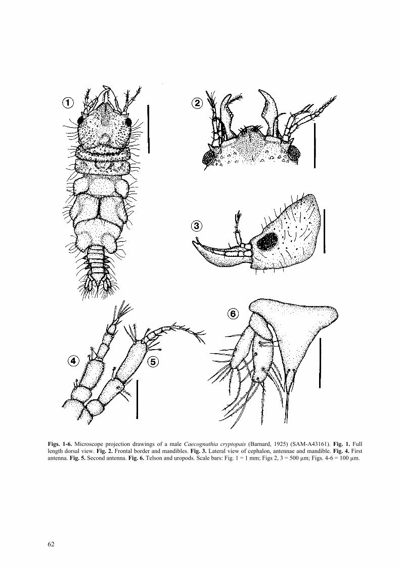
Cephalon. Rectangular, one and a half times as wideas long, posterior margin concave, lateral marginsconvex (Figs. 1, 12). Shallow dorsal sulcus, extendingto median tubercle (Figs. 1, 11). Dorsal and ventralcephalon armed with numerous randomly distributedwart-like tubercles closely associated with long simplesetae. Well developed oval-shaped, prominent bulbous,compound eyes on lateral margin of cephalon, slightlyelevated and encircled by a smooth rim, length of eyeslightly more than a fifth of cephalon (Figs. 3, 15). Noparaocular ornamentation (Figs. 3, 11). Posterior medi-an tubercle from middle of cephalon to posteriormargin.
Frontal border. Rounded, produced for a fifth ofcephalon’s length, lacking any frontal processes (Figs.2, 13). Median groove on produced border, six to sevenlong simple setae on dorsal surface of median producingfrontal border, rest of border covered with short simplesetae. External scissura deeply excavated (Fig. 2).Supraocular lobe prominent, convex, extending laterallywith five to seven tubercles.
Address for correspondence: N.J. Smit, Department of Zoology and Entomology, PO Box 339, University of the Free State, Bloemfontein, 9300,South Africa. Phone: ++27 51 401 2440; Fax: ++ 27 51 448 8711; E-mail: [email protected]
FOLIA PARASITOLOGICA 47: 61-66, 2000
62
Figs. 1-6. Microscope projection drawings of a male Caecognathia cryptopais (Barnard, 1925) (SAM-A43161). Fig. 1. Fulllength dorsal view. Fig. 2. Frontal border and mandibles. Fig. 3. Lateral view of cephalon, antennae and mandible. Fig. 4. Firstantenna. Fig. 5. Second antenna. Fig. 6. Telson and uropods. Scale bars: Fig. 1 = 1 mm; Figs 2, 3 = 500 µm; Figs. 4-6 = 100 µm.
Smit et al.: Caecognathia cryptopais from southern Africa
63
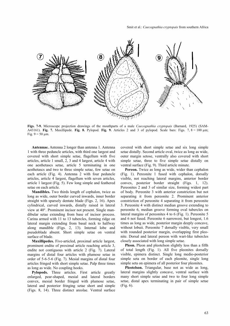
Figs. 7-9. Microscope projection drawings of the mouthparts of a male Caecognathia cryptopais (Barnard, 1925) (SAM-A43161). Fig. 7. Maxillipede. Fig. 8. Pylopod. Fig. 9. Articles 2 and 3 of pylopod. Scale bars: Figs. 7, 8 = 100 µm;Fig. 9 = 50 µm.
Antennae. Antenna 2 longer than antenna 1. Antenna1 with three peduncle articles, with third one largest andcovered with short simple setae, flagellum with fivearticles, article 1 small, 2, 3 and 4 largest, article 4 withone aesthetascs setae, article 5 terminating in oneaesthetascs and two to three simple setae, few setae oneach article (Fig. 4). Antenna 2 with four pedunclearticles, article 4 largest, flagellum with seven articles,article 1 largest (Fig. 5). Few long simple and featheredsetae on each article.
Mandibles. Two thirds length of cephalon, twice aslong as wide, outer border curved inwards, inner borderstraight with sparsely dentate blade (Figs. 2, 16). Apexcylindrical, curved inwards, distally raised in lateralview at 40°. Prominent incisor not present. Single man-dibular setae extending from base of incisor process.Carina armed with 11 to 13 tubercles, forming ridge onlateral margin extending from basal neck to halfwayalong mandible (Figs. 2, 13). Internal lobe andpseudoblade absent. Short simple setae on ventralsurface of blade.
Maxillipedes. Five-articled, proximal article largest,prominent endite of proximal article reaching article 3,endite not contiguous with article 2 (Fig. 7). Lateralmargins of distal four articles with plumose setae inorder of 5-6-5-6 (Fig. 7). Mesial margins of distal fourarticles fringed with short simple setae. Palp three timesas long as wide. No coupling hooks.
Pylopods. Three articles. First article greatlyenlarged, pear-shaped, mesial and lateral bordersconvex, mesial border fringed with plumose setae,lateral and posterior fringing setae short and simple(Figs. 8, 14). Three distinct areolae. Ventral surface
covered with short simple setae and six long simplesetae distally. Second article oval, twice as long as wide,outer margin setose, ventrally also covered with shortsimple setae, three to five simple setae distally onventral surface (Fig. 9). Third article minute.
Pereon. Twice as long as wide, wider than cephalon(Fig. 1). Pereonite 1 fused with cephalon, dorsallyvisible, not reaching lateral margins, anterior borderconvex, posterior border straight (Figs. 1, 12).Pereonites 2 and 3 of similar size, forming widest partof body. Pereonite 3 with anterior constriction but notseparating it from pereonite 2. Prominent anteriorconstriction of pereonite 4 separating it from pereonite3. Pereonite 4 with distinct median groove extending topereonite 6, median groove forming oval tubercles onlateral margins of pereonites 4 to 6 (Fig. 1). Pereonite 5and 6 not fused. Pereonite 6 narrowest, but longest, 1.6times as long as wide, posterior margin deeply concave,without lobuii. Pereonite 7 dorsally visible, very smallwith rounded posterior margin, overlapping first pleo-nite. Dorsal and lateral pereon with wart-like tuberclesclosely associated with long simple setae.
Pleon. Pleon and pleotelson slightly less than a fifthof total length (Fig. 1). All five pleonites dorsallyvisible, epimera distinct. Single long medio-posteriorsimple seta on border of each pleonite, single longsimple seta on epimera of all posterior four pleonites.
Pleotelson. Triangular, base not as wide as long,lateral margins slightly concave, ventral surface withmany short simple setae and two to four long simplesetae, distal apex terminating in pair of simple setae(Fig. 6).

64
Fig. 10. Microscope projection drawings of pereopods 2 to 6 (P2–P6) of a male Caecognathia cryptopais (Barnard, 1925) (SAM-A43161). Scale bar = 200 µm.
Uropods. Extending to apex of pleotelson, endopodlonger and wider than exopod, both with long fringingsetae, endopod with inner six plumose setae, four to sixsimple setae on dorsal surface, exopod with inner fourplumose setae, rest of setae simple (Fig. 6).
Pereopods. Pereopod 2 consists of elongated basiswith nine to eleven simple setae anterior and four toseven on posterior side, ischium two thirds length ofbasis, with three to five anterior setae and posteriortubercles with simple setae in between (Fig. 10). Merusis half the length of ischium with anterior bulbous
protrusion, simple setae on bulbous protrusion, posteriormargin with tubercles as well as simple setae. Carpus ofalmost same size and shape as merus, but withoutanterior bulbous protrusion single plumose seta on distalpart of anterior margin. Basis, ischium, merus andcarpus covered with short simple setae. Propodus abouttwice the length of carpus, prominent tubercles fringeposterior side, two elongated denticulated compoundspines ending in sharp points situated on middle anddistal part of posterior margin respectively, only a fewsimple setae anteriorly. Dactylus half the length of

Smit et al.: Caecognathia cryptopais from southern Africa
65
Figs. 11-17. Scanning electron micrographs of a male Caecognathia cryptopais (Barnard, 1925) (SAM-A43161). Fig. 11.Anterio-lateral view of cephalon. Fig. 12. Dorsal view of cephalon. Fig. 13. Dorsal view of produced frontal border with simplesetae. Fig. 14. Ventral view of cephalon and pylopods. Fig. 15. Dorsal view of left eye and supraocular lobe. Fig. 16. Dorsalview of left mandible. Fig. 17. Ventral view of penes and first pleopods. Scale bars: Figs. 12, 13, 15, 17 = 100 µm; Fig. 16 =30 µm.

66
propodus, terminates in sharp posterior pointing unguis,prominent spine on posterior side proximal to unguiswith few simple setae on dorsal and ventral sides.Pereopods 3 to 6 similar to pereopod 2 (Fig. 10),differing only in direction, pereopods 4 to 6 are directedposteriorly and pereopods 2 and 3 anteriorly.
Pleopods. Five pairs of similar exo- and endopods,without fringing setae, short simple setae on distalborders (Fig. 17).
Penes. Large, one and a half times as long as wide,consist of two contiguous papillae (Fig. 17).T y p e m a t e r i a l : Syntypes: In the collection of the South
African Museum, Cape Town (SAM-A6051). Originalauthor designated no holotype.
T y p e l o c a l i t y : Duminy Point, off Saldanha Bay (E. ×N. 0.5 N., distant 8 nautical miles)
O t h e r l o c a l i t i e s : Off East London (32°14.9’S,29°10.43’E), (32°29.5’S, 28°57.1’E) and (32°28.6’S,28°58.8’E). Off Port Alfred (33°39.3’S, 27°11.6’E).
T y p e h o s t : Unknown.M a t e r i a l e x a m i n e d : Type material in the collection
of the South African Museum, Cape Town (1 male, 1 larva,SAM-A6051).
O t h e r m a t e r i a l : In the collection of the South AfricanMuseum, Cape Town (2 males, SAM-A14602) (2 males,SAM-A19310) (1 male, SAM-A19311), (2 males, SAM-A19312), (4 males, SAM-A43161).
DISCUSSION
According to Cohen and Poore (1994) the genusCaecognathia is characterised by a produced frontalborder without frontal processes, cephalon withoutparaocular ornamentation, pereonite immersed incephalon, pylopod two- or three-articled, with article 1enlarged and article 3 small or absent. Caecognathiacryptopais conforms to all these specifications for the
genus and hence its transfer from the genus Gnathia toCaecognathia.
Caecognathia cryptopais differs from the othersouthern African species in that it is the only specieswith a rounded produced frontal border without anyfrontal processes, many wart-like tubercles on the body,no long simple setae on the distal margins of pleopodsand very big penes in comparison with the othersouthern African species. According to Barnard(1925a,b, 1940), C. cryptopais is closely related toCaecognathia elongata (Kröyer, 1847) (syn. Gnathiacerina Stimpson, 1853) but differs in the presence ofwart-like tubercles associated with long simple setae allover the dorsal and lateral surface of the body.Caecognathia elongata also has no prominent dorsalsulcus on pereonites 4 to 6 forming oval lobi lateraleson pereonites 4 to 6. Monod (1926) as well as Barnard(1940) pointed out that C. cryptopais seems to besimilar to Caecognathia antarctica (Studer, 1883). Onexamination of a single specimen of C. antarcticadeposited in the South African Museum (SAM-A16158)it was found that it can be distinguished from C.cryptopais by its different shaped mandible, broadercephalon, a more prominent median tubercle and theindistinct epimera of the pleon. All the above mentionedspecies related to C. cryptopais were also transferred byCohen and Poore (1994) from the genus Gnathia toCaecognathia.
Acknowledgements. The authors thank Ms. Michelle van derMerwe of the South African Museum, Cape Town, for makingthe gnathiid material available for examination and Dr. GaryPoore, Museum of Victoria, Australia, for confirming the newgeneric status of Caecognathia cryptopais. This study wasfunded by the marine resource program of the NationalResearch Foundation (NRF) of South Africa.
REFERENCES
BARNARD K.H. 1914a: Contributions to the crustaceanfauna of South Africa. 1. Additions to the marine Isopoda.Ann. S. Afr. Mus. 10: 197-230.
BARNARD K.H. 1914b: Contributions to the crustaceanfauna of South Africa. 3. Additions to the marine Isopoda,with notes on some previously incompletely knownspecies. Ann. S. Afr. Mus. 10: 325a-358a, 359-442.
BARNARD K.H. 1920: Contributions to the crustacean faunaof South Africa. 6. Further additions to the list of marineIsopoda. Ann. S. Afr. Mus. 17: 319-438.
BARNARD K.H. 1925a: Description of a new species ofGnathia (Crustacea, Isopoda) from South Africa. Ann.Mag. Nat. Hist. 15: 417-418.
BARNARD K.H. 1925b: Contributions to the crustacean
fauna of South Africa. 9. Further additions to the list ofIsopoda. Ann. S. Afr. Mus. 20: 381-412.
BARNARD K.H. 1940: Contributions to the crustacean faunaof South Africa. 12. Further additions to the Tanaidacea,Isopoda, and Amphipoda, together with keys foridentification of the hitherto recorded marine and fresh-water species. Ann. S. Afr. Mus. 32: 381-543.
COHEN B.F., POORE G.C.B. 1994: Phylogeny andbiogeography of the Gnathiidae (Crustacea: Isopoda) withdescriptions of new genera and species, most from south-eastern Australia. Mem. Mus. Victoria 54: 271-397.
MONOD T. 1926: Les Gnathiidae. Essai monographique(Morphologie, Biologie, Systématique). Mem. Soc. Sci.Nat. Maroc 13: 1-668.
Received 27 May 1999 Accepted 14 July 1999
Related Documents