REVIEW ARTICLE A Reappraisal on Lidocaine-Sensitive Repetitive, Uniform Atrial Tachycardia Hugo A. Garro, M.D., ∗ Marcelo V. Elizari, M.D., F.A.C.C., ∗ Adrian Baranchuk, M.D., F.A.C.C., F.R.C.P.C.,† Francisco Femen´ ıa, M.D.,‡ and Pablo A. Chiale, M.D. ∗ From the ∗ Centro de Arritmias Card´ ıacas de la Ciudad de Buenos Aires, Division of Cardiology, Hospital J. M. Ramos Mej´ ıa, and Pontificia Universidad Cat´ olica Argentina “Santa Mar´ ıa de los Buenos Aires”, Buenos Aires, Argentina; †Heart Rhythm Service, Kingston General Hospital, Queen’s University, Kingston, Ontario, Canada; and ‡Servicio de Arritmias, Hospital Espa ˜ nol, Mendoza, Argentina Background: Lidocaine sensitive, repetitive atrial tachycardia is an unusual arrhythmia whose electrophysiologic substrate remains undefined. We aimed to analyze the electropharmacologic characteristics of this arrhythmia with emphasis on its cellular substrate and response to drug challenges. Methods: We retrospectively analyzed a series of 18 patients from an electrocardiographic and electrophysiologic perspective and the response to pharmacological challenge. Results: There was no evidence of structural heart disease in 12 patients, 4 patients presented with systemic hypertension; one patient had a prior myocardial infarction and one a mitral valve prolapse. The arrhythmia depicted a consistent pattern in nine patients. The first initiating ectopic beat showed a long coupling interval, the cycle length of the second atrial ectopic beat presented the shortest cycle length and a further prolongation was apparent towards the end of the atrial salvos. Conversely, in the other nine cases, the atrial tachycardia cycle length was erratic. The arrhythmia was suppressed by asynchronous atrial pacing at cycle lengths longer than those of the atrial tachycardia. Intravenous lidocaine eliminated the arrhythmia in all patients, but intravenous verapamil suppressed the atrial tachycardia in only two patients while adenosine caused a transient disappearance in 2/8 patients. Only one patient responded to all the three agents. Radiofrequency ablation was successfully performed in 10 patients. Conclusions: Repetitive uniform atrial tachycardia can be sensitive to lidocaine. In few cases, this rare focal arrhythmia may be also suppressed by adenosine and/or verapamil, which suggests a diversity of electrophysiologic substrates that deserve to be accurately identified. Ann Noninvasive Electrocardiol 2013;18(1):1–11 atrial tachycardia; lidocaine-sensitive Atrial tachycardias are a variety of supraven- tricular tachycardias that do not require the atrio-ventricular junction, accessory pathways, or ventricular tissue for initiation and perpetuation of the fast heart rhythm. The cardiac rate during atrial tachycardia encompasses a broad range (120– 250 beats per minute); atrial rhythm is usually regular whereas ventricular rhythm may become irregular depending on the conducting properties of the atrio-ventricular node, which may give rise to Address for correspondence: Hugo A. Garro, M.D., Centro de Arritmias Card´ıacas de la Ciudad Aut´ onoma de Buenos Aires, Division Cardiologia. Hospital J.M. Ramos Mej´ıa, Buenos Aires, Argentina. Fax: 054 11 4956 2102; E-mail: [email protected] diverse conduction patterns (fixed or a combination of 2:1, 3:1, 4:1 atrio-ventricular conduction or Mobitz I periodicities). QRS complexes are usually narrow, unless bundle branch block aberration or ventricular preexcitation are also present. The P-wave morphology may provide useful clues to determine the anatomic site of origin and, eventually, the intrinsic mechanism of the arrhythmia. However, it has a limited spatial resolution since atrial pacing at sites as far apart as C 2012, Wiley Periodicals, Inc. DOI:10.1111/anec.12014 1

Welcome message from author
This document is posted to help you gain knowledge. Please leave a comment to let me know what you think about it! Share it to your friends and learn new things together.
Transcript
REVIEW ARTICLE
A Reappraisal on Lidocaine-Sensitive Repetitive,Uniform Atrial Tachycardia
Hugo A. Garro, M.D.,∗ Marcelo V. Elizari, M.D., F.A.C.C.,∗Adrian Baranchuk, M.D., F.A.C.C., F.R.C.P.C.,† Francisco Femenıa, M.D.,‡and Pablo A. Chiale, M.D.∗From the ∗Centro de Arritmias Cardıacas de la Ciudad de Buenos Aires, Division of Cardiology, Hospital J. M.Ramos Mejıa, and Pontificia Universidad Catolica Argentina “Santa Marıa de los Buenos Aires”, Buenos Aires,Argentina; †Heart Rhythm Service, Kingston General Hospital, Queen’s University, Kingston, Ontario, Canada;and ‡Servicio de Arritmias, Hospital Espanol, Mendoza, Argentina
Background: Lidocaine sensitive, repetitive atrial tachycardia is an unusual arrhythmia whoseelectrophysiologic substrate remains undefined. We aimed to analyze the electropharmacologiccharacteristics of this arrhythmia with emphasis on its cellular substrate and response to drugchallenges.
Methods: We retrospectively analyzed a series of 18 patients from an electrocardiographic andelectrophysiologic perspective and the response to pharmacological challenge.
Results: There was no evidence of structural heart disease in 12 patients, 4 patients presentedwith systemic hypertension; one patient had a prior myocardial infarction and one a mitral valveprolapse. The arrhythmia depicted a consistent pattern in nine patients. The first initiating ectopicbeat showed a long coupling interval, the cycle length of the second atrial ectopic beat presentedthe shortest cycle length and a further prolongation was apparent towards the end of the atrialsalvos. Conversely, in the other nine cases, the atrial tachycardia cycle length was erratic. Thearrhythmia was suppressed by asynchronous atrial pacing at cycle lengths longer than those of theatrial tachycardia. Intravenous lidocaine eliminated the arrhythmia in all patients, but intravenousverapamil suppressed the atrial tachycardia in only two patients while adenosine caused a transientdisappearance in 2/8 patients. Only one patient responded to all the three agents. Radiofrequencyablation was successfully performed in 10 patients.
Conclusions: Repetitive uniform atrial tachycardia can be sensitive to lidocaine. In few cases,this rare focal arrhythmia may be also suppressed by adenosine and/or verapamil, which suggests adiversity of electrophysiologic substrates that deserve to be accurately identified.
Ann Noninvasive Electrocardiol 2013;18(1):1–11
atrial tachycardia; lidocaine-sensitive
Atrial tachycardias are a variety of supraven-tricular tachycardias that do not require theatrio-ventricular junction, accessory pathways, orventricular tissue for initiation and perpetuationof the fast heart rhythm. The cardiac rate duringatrial tachycardia encompasses a broad range (120–250 beats per minute); atrial rhythm is usuallyregular whereas ventricular rhythm may becomeirregular depending on the conducting properties ofthe atrio-ventricular node, which may give rise to
Address for correspondence: Hugo A. Garro, M.D., Centro de Arritmias Cardıacas de la Ciudad Autonoma de Buenos Aires, DivisionCardiologia. Hospital J.M. Ramos Mejıa, Buenos Aires, Argentina. Fax: 054 11 4956 2102; E-mail: [email protected]
diverse conduction patterns (fixed or a combinationof 2:1, 3:1, 4:1 atrio-ventricular conduction orMobitz I periodicities). QRS complexes are usuallynarrow, unless bundle branch block aberration orventricular preexcitation are also present.
The P-wave morphology may provide usefulclues to determine the anatomic site of originand, eventually, the intrinsic mechanism of thearrhythmia. However, it has a limited spatialresolution since atrial pacing at sites as far apart as
C©2012, Wiley Periodicals, Inc.DOI:10.1111/anec.12014
1
2 � A.N.E. � January 2013 � Vol. 18, No. 1 � Garro, et al. � Lidocaine-Sensitive Repetitive Atrial Tachycardia
32 mm in the coronary sinus and 17 mm in the rightatrium causes morphologically identical P waves.1
Atrial tachycardias may be classified on thebasis of their clinical and electrocardiographiccharacteristics, the underlying electrophysiologicmechanisms and the anatomic site of origin.
Clinical sustained uniform atrial tachycardia isrelatively infrequent; the prevalence in symp-tomatic patients is lower than 1%, while it accountsfor around 10% in adults undergoing electrophys-iological studies.2 Bursts of atrial tachycardia arecommon in Holter ECG recordings of aged individ-uals. Repetitive, almost incessant, uniform atrialtachycardia that is a rare variety of the arrhythmia,described in detail in this review, accounts for onlya 0.3% among all supraventricular tachycardiasreferred to our center for evaluation and treatment.Multifocal atrial tachycardia shows an atrial ratehigher than 100 beats per minute, with at leastthree morphologically different P-waves, irregularP–P intervals and an isoelectric baseline betweenthe P waves.
Based on endocardial activation, atrial tachycar-dias may be grouped into focal, arising from a lo-calized area in the atria, and macroreentrant, mostcommonly occurring in patients with structuralheart disease, particularly after surgery involvingincisions, or scarring in the atria.
The three putative underlying electrophysio-logic mechanisms of focal atrial tachycardia areenhanced automaticity, triggered activity, andmicroreentry.3–7 Although a significant overlapbetween the electrophysiologic characteristics ofatrial tachycardias caused by triggered activity andmicroreentry is a limiting factor in the analysisof their mechanisms, a previous report showedthat adenosine insensitive focal atrial tachycardiasare caused by micro-reentry.7 Focal sustainedatrial tachycardias are usually refractory to vagalstimulating maneuvers and some of them may besensitive to adenosine and/or verapamil. Regardinglong-term treatment, most antiarrhythmic agents,such as Class I A and I C sodium channelblockers, β adrenergic blockers and calciumchannel blockers, have a very limited efficacywhile Class III agents may provide better results.
Lidocaine, a class Ib antiarrhythmic drug hasbeen used as a first line therapy for ventriculararrhythmias occurring in the setting of theacute myocardial infarction. Despite its negligibleelectrophysiologic effects on atrial, sinus node andatrio-ventricular nodal tissues, Cotoi and Luca8
showed more than three decades ago that lidocaine
may terminate diverse atrial tachyarrhythmias, andlater, Markowitz et al.9 described a patient witha repetitive form of atrial tachycardia that wasresponsive to lidocaine.
In this manuscript we will revisit our findingsregarding a very peculiar and rare variety of focalrepetitive atrial tachycardia whose electrophysio-logic substrate is still intriguing and it was shownto be highly sensitive to intravenous lidocaine andusually refractory to most other antiarrhythmicagents.10,11
LIDOCAINE-SENSITIVE, RATERELATED, REPETITIVE, UNIFORM
ATRIAL TACHYCARDIA
This is a peculiar form of chronic, iterative,uniform atrial tachycardia whose unique elec-trophysiologic characteristics and pharmacologicresponses might indicate an unusual arrhythmo-genic mechanism operating in human atrial tissue.Since 1995 to the present we identified 18 casesof this arrhythmia, which were selected for thispresentation from 24 patients referred for study ofchronic, repetitive, uniform atrial tachycardias.
CLINICAL ANDELECTROCARDIOGRAPHIC
CHARACTERISTICS
Clinical Findings
Table 1 depicts the main clinical characteristicsof 18 patients with lidocaine sensitive, rate related,repetitive, uniform atrial tachycardia.
Sixteen patients complained about palpitations(associated with dizziness and a syncopal episodein one of them and with chest pain in another two);two patients were asymptomatic, and the arrhyth-mia was fortuitously diagnosed during a routineclinical examination. No evidence of structuralheart disease was found in twelve patients (one ofthem had multiform ventricular premature beats),four patients suffered from systemic hypertension,one patient had a previous myocardial infarction,and the remaining one had a mitral valve prolapse.The B mode echocardiogram was normal in twelvepatients and showed mild left ventricular dilationin two young patients, concentric left ventricularhypertrophy with mild left atrial enlargement inthree hypertensive patients, and a typical mitralvalve prolapse in one patient.
A.N.E. � January 2013 � Vol. 18, No. 1 � Garro, et al. � Lidocaine-Sensitive Repetitive Atrial Tachycardia � 3
Table 1. Clinical Findings in Eighteen Patients with Repetitive Atrial Tachycardia
Age ECG in Sinus B-Mode AntiarrhythmicCase (years) Gender Symptoms Rhythm Echocardiogram Treatment
1 14 F Palpitations Normal Normal A, D, Fle, P, Q2 16 M Palpitations Normal Mild LV Dilation A, At, Fle, Q, V3 21 M Palpitations Normal Normal A, At, D, Fle, V4 22 M Palpitations Normal Normal A, At, Fle, Q5 24 F Palpitations Multiform PVC´s Mild LV Dilation At, Fle, P, V6 28 M Palpitations Normal Normal A, At, P7 60 F Asymptomatic LVH Mild LAE; Mild LVH A, At8 56 M Asymptomatic Inferior and Posterior Normal N
Fibrosis9 62 M Palpitations LVH Mild LAE; Mild LVH –10 53 F Palpitations Normal Normal B, Fle, D11 83 F Palpitations Normal Mild LAE;Mild LVH At, d,l sotalol, Fle12 48 M Palpitations Normal Normal P13 37 F Palpitations Normal MVP Digoxin, A14 20 M Palpitations Normal Normal Fle, d,l sotalol15 28 M Palpitations Normal Normal –16 34 M Palpitations Normal Normal –17 25 M Palpitations; Normal Normal –
chest pain18 19 M Palpitations Normal Normal Nevibolol, Iv
A = amiodarone; At = atenolol; B = bisoprolol; D = digoxin; D = diltiazem; F = female; Fle = flecainide; LAE = left atriumenlargement; LV = left ventricle; LVH = left ventricle hypertrophy; M = male; MVP = mitral valve prolapse; N = nadolol; P =propafenone; PVCs = premature ventricular contractions; Q = quinidine; S: d,l-sotalol; V = verapamil; Iv = ivabradine.
Electrocardiographic Findings
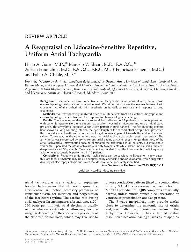
In all patients, the baseline ECG showed almostincessant bursts of self-limited atrial tachycardia(single or double atrial premature beats wereinfrequently observed) separated by one to threebeats of sinus node origin (Fig. 1).
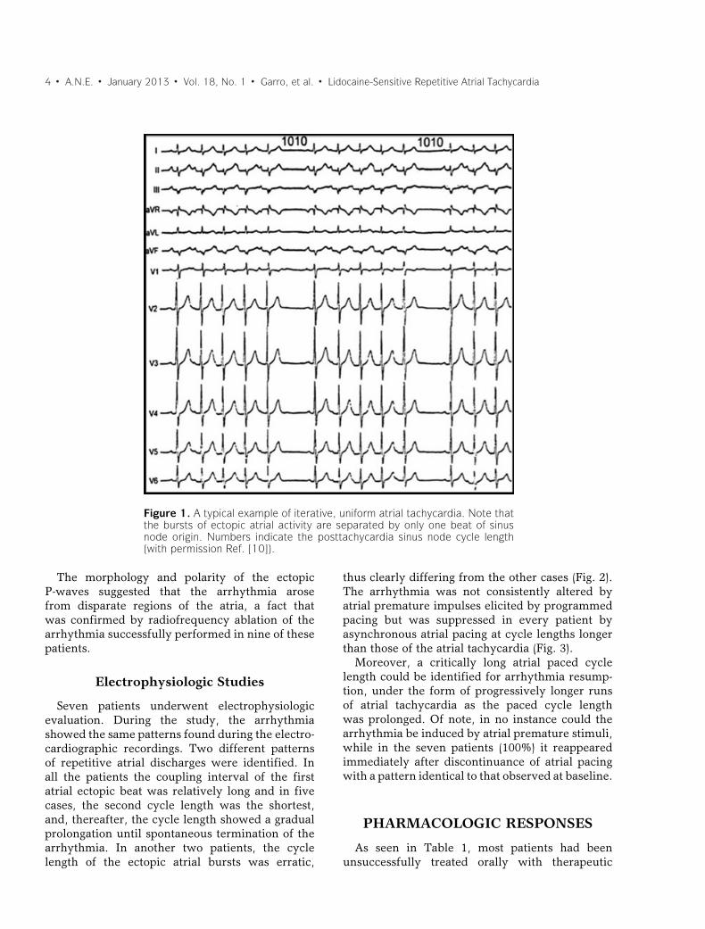
Duration of atrial tachycardia bursts was vari-able (between three or four beats and severalseconds), even in the same patient, and mostof them showed 1:1 AV conduction. Rate-relatedchanges in the PR interval (due to P–P intervalvariations during the runs of atrial tachycardia)and, eventually, Mobitz type I atrioventricularblock were observed. The arrhythmia exhibiteda stereotyped pattern in nine patients. In fact, inall cases the initiating atrial ectopic beat had arelatively long coupling interval, whereas in thosenine patients the cycle length of the second atrialpremature beat was the shortest and a furtherprolongation was apparent to the end of atrialsalvos (Fig. 2A and B). Conversely, in the other ninecases the atrial tachycardia cycle length showederratic variations (Fig. 2C and D).
In addition, shortening of the sinus node cyclelength induced by mild exercising suppressedor abbreviated the repetitive atrial discharges
while prolongation of the sinus node cyclelength, obtained by vagal stimulating maneuvers,tended to prolong the bursts of ectopic atrialactivity. These changes were not associated withany discernible modification in atrial tachycardiacycle length. Furthermore, the suppressive effectwas consistently observed at sinus node ratesslower than that of the atrial tachycardias. Thus,it was not due to an overdrive suppressionmechanism.
The same relationship between the sinus noderate and the atrial ectopic activity was also clearlydocumented in 24-hour ECG Holter recordingsperformed in 12 of our patients. In fact, in nineof them the runs of atrial tachycardia were presentwhen the sinus node rate was slow, or relativelyslow, and disappeared or were abbreviated athigher sinus node rates. However, in three patientsthe arrhythmia was not only suppressed bymoderate increments of sinus node rate but alsoby sinus node slowing during sleep, indicating thatappropriate changes in cardiac rate that reflectthe predominance of either the sympathetic or thevagal influences on the heart may have the samesuppressing action on the arrhythmia mechanism.In no instance was the arrhythmia found todeteriorate into atrial flutter or fibrillation.
4 � A.N.E. � January 2013 � Vol. 18, No. 1 � Garro, et al. � Lidocaine-Sensitive Repetitive Atrial Tachycardia
Figure 1. A typical example of iterative, uniform atrial tachycardia. Note thatthe bursts of ectopic atrial activity are separated by only one beat of sinusnode origin. Numbers indicate the posttachycardia sinus node cycle length(with permission Ref. [10]).
The morphology and polarity of the ectopicP-waves suggested that the arrhythmia arosefrom disparate regions of the atria, a fact thatwas confirmed by radiofrequency ablation of thearrhythmia successfully performed in nine of thesepatients.
Electrophysiologic Studies
Seven patients underwent electrophysiologicevaluation. During the study, the arrhythmiashowed the same patterns found during the electro-cardiographic recordings. Two different patternsof repetitive atrial discharges were identified. Inall the patients the coupling interval of the firstatrial ectopic beat was relatively long and in fivecases, the second cycle length was the shortest,and, thereafter, the cycle length showed a gradualprolongation until spontaneous termination of thearrhythmia. In another two patients, the cyclelength of the ectopic atrial bursts was erratic,
thus clearly differing from the other cases (Fig. 2).The arrhythmia was not consistently altered byatrial premature impulses elicited by programmedpacing but was suppressed in every patient byasynchronous atrial pacing at cycle lengths longerthan those of the atrial tachycardia (Fig. 3).
Moreover, a critically long atrial paced cyclelength could be identified for arrhythmia resump-tion, under the form of progressively longer runsof atrial tachycardia as the paced cycle lengthwas prolonged. Of note, in no instance could thearrhythmia be induced by atrial premature stimuli,while in the seven patients (100%) it reappearedimmediately after discontinuance of atrial pacingwith a pattern identical to that observed at baseline.
PHARMACOLOGIC RESPONSES
As seen in Table 1, most patients had beenunsuccessfully treated orally with therapeutic

A.N.E. � January 2013 � Vol. 18, No. 1 � Garro, et al. � Lidocaine-Sensitive Repetitive Atrial Tachycardia � 5
Figure 2. The different behavior of repetitive uniform atrial tachycardia cyclelength. The coupling interval of the initiating beat is always relatively long; inA and B, the following cycle length is the shortest and then it shows a gradualprolongation until the end of atrial salvos, while in C and D the ectopic atrialcycle length is erratic. HRA = high right atrium electrogram. (with permissionRef. [10]).
doses of different antiarrhythmic agents, includingβ adrenergic blockers, sodium channel blockers(Class I A and I C), calcium channel blockers,d,l-sotalol, amiodarone, ivabradine, and digoxin.Intravenous lidocaine and calcium channel block-ers were tested in all patients (verapamil in17 patients and diltiazem in the remaining one)and intravenous adenosine was administered to10 patients. Intravenous lidocaine (0.5–1.0 mg/kgin 1 minute) produced a rapid suppression of thearrhythmia (between 30 and 120 seconds afterthe end of injection) in all patients, an effectthat persisted for 8–25 minutes and disappearedgradually. In no case was the suppression of thearrhythmia by lidocaine related to sinus nodeacceleration. In fact, sinus node rate during thepeak effect of the drug was slower than the criticalheart rate that consistently caused the arrhythmiadisappearance in each of the seven patients whounderwent an electrophysiologic study, and nota single atrial ectopic impulse was observed
during sinus node slowing by bradycardia-inducingmaneuvers. Figure 4 depicts the interruption of therepetitive atrial ectopic discharges after lidocaineadministration.
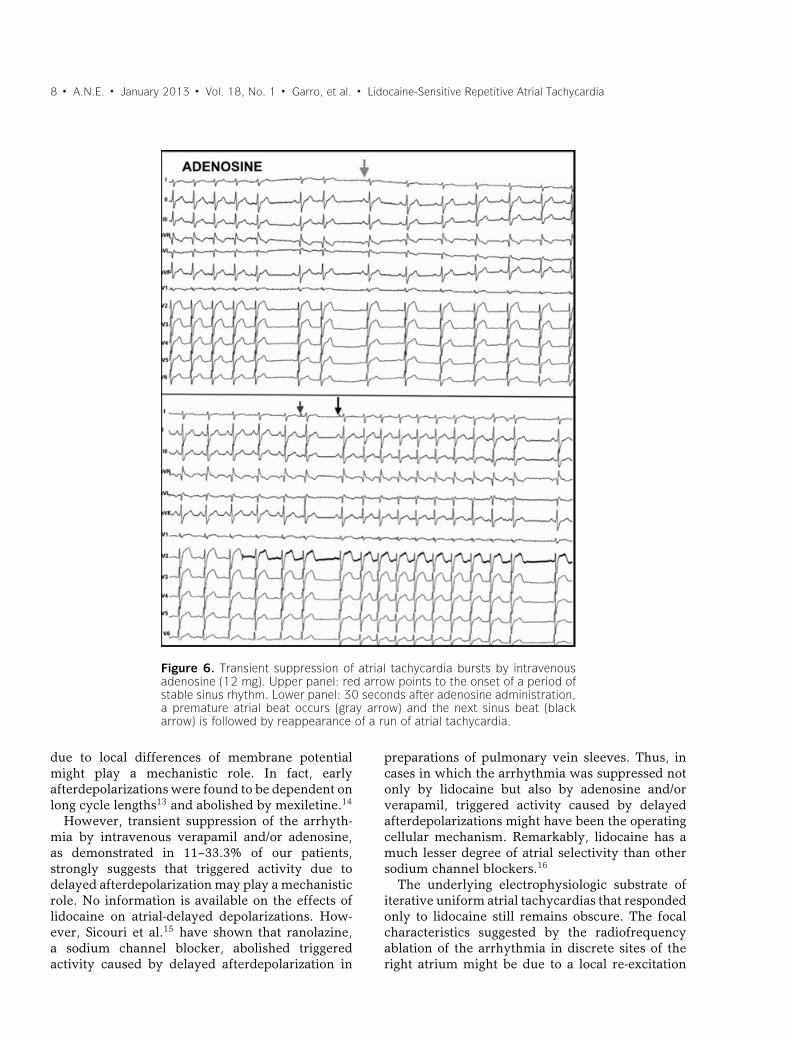
In contrast, intravenous verapamil (10 mg in60 seconds) suppressed the arrhythmia in onlytwo patients (Fig. 5) and was ineffective in fifteenpatients; although a Mobitz type I and evenrepetitive AV block during the bursts of atrialtachycardia were documented in nine of them.At the time of atrial tachycardia termination, thesinus node cycle length did not differ from that atbaseline and normal sinus rhythm still persisted atslower heart rates, and the arrhythmia was absenteven after pauses lasting up to 1250 and 1620ms, respectively, induced by vagal stimulatingmaneuvers. In these two patients, atrial ectopicactivity resumed 15 and 25 minutes after verapamiladministration. A quite unexpected response wasobserved in one patient after intravenous diltiazemadministration. The repetitive e irregular salvos
6 � A.N.E. � January 2013 � Vol. 18, No. 1 � Garro, et al. � Lidocaine-Sensitive Repetitive Atrial Tachycardia
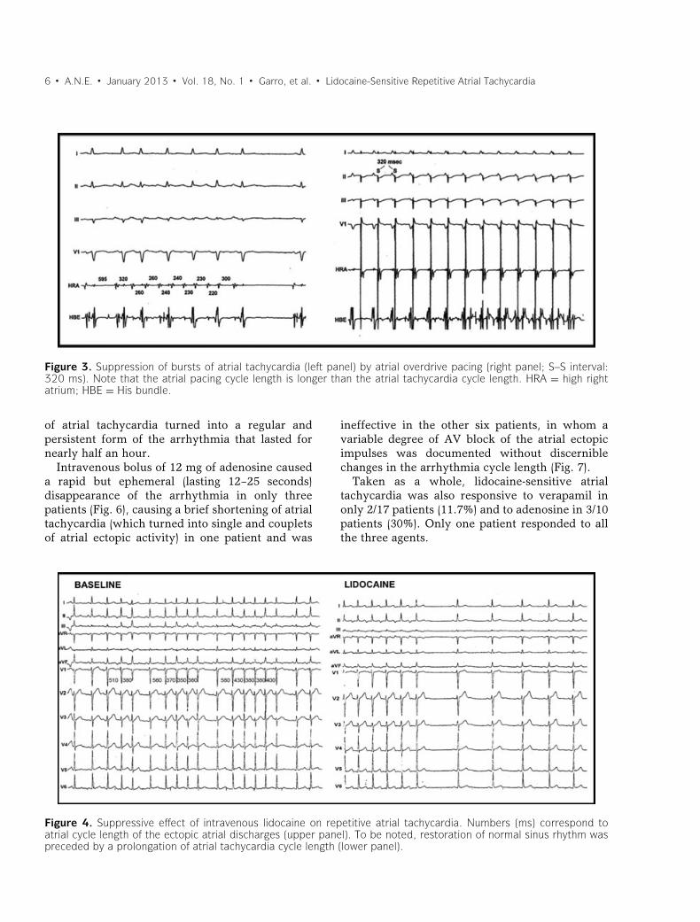
Figure 3. Suppression of bursts of atrial tachycardia (left panel) by atrial overdrive pacing (right panel; S–S interval:320 ms). Note that the atrial pacing cycle length is longer than the atrial tachycardia cycle length. HRA = high rightatrium; HBE = His bundle.
of atrial tachycardia turned into a regular andpersistent form of the arrhythmia that lasted fornearly half an hour.
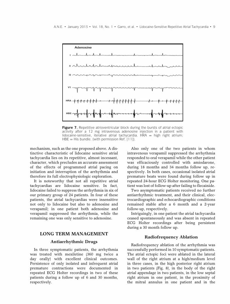
Intravenous bolus of 12 mg of adenosine causeda rapid but ephemeral (lasting 12–25 seconds)disappearance of the arrhythmia in only threepatients (Fig. 6), causing a brief shortening of atrialtachycardia (which turned into single and coupletsof atrial ectopic activity) in one patient and was
ineffective in the other six patients, in whom avariable degree of AV block of the atrial ectopicimpulses was documented without discerniblechanges in the arrhythmia cycle length (Fig. 7).
Taken as a whole, lidocaine-sensitive atrialtachycardia was also responsive to verapamil inonly 2/17 patients (11.7%) and to adenosine in 3/10patients (30%). Only one patient responded to allthe three agents.
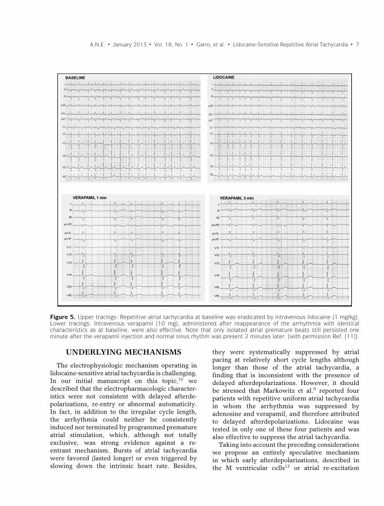
Figure 4. Suppressive effect of intravenous lidocaine on repetitive atrial tachycardia. Numbers (ms) correspond toatrial cycle length of the ectopic atrial discharges (upper panel). To be noted, restoration of normal sinus rhythm waspreceded by a prolongation of atrial tachycardia cycle length (lower panel).
A.N.E. � January 2013 � Vol. 18, No. 1 � Garro, et al. � Lidocaine-Sensitive Repetitive Atrial Tachycardia � 7
Figure 5. Upper tracings: Repetitive atrial tachycardia at baseline was eradicated by intravenous lidocaine (1 mg/kg).Lower tracings: Intravenous verapamil (10 mg), administered after reappearance of the arrhythmia with identicalcharacteristics as at baseline, were also effective. Note that only isolated atrial premature beats still persisted oneminute after the verapamil injection and normal sinus rhythm was present 2 minutes later. (with permission Ref. [11]).
UNDERLYING MECHANISMS
The electrophysiologic mechanism operating inlidocaine-sensitive atrial tachycardia is challenging.In our initial manuscript on this topic,10 wedescribed that the electropharmacologic character-istics were not consistent with delayed afterde-polarizations, re-entry or abnormal automaticity.In fact, in addition to the irregular cycle length,the arrhythmia could neither be consistentlyinduced nor terminated by programmed prematureatrial stimulation, which, although not totallyexclusive, was strong evidence against a re-entrant mechanism. Bursts of atrial tachycardiawere favored (lasted longer) or even triggered byslowing down the intrinsic heart rate. Besides,
they were systematically suppressed by atrialpacing at relatively short cycle lengths althoughlonger than those of the atrial tachycardia, afinding that is inconsistent with the presence ofdelayed afterdepolarizations. However, it shouldbe stressed that Markowitz et al.9 reported fourpatients with repetitive uniform atrial tachycardiain whom the arrhythmia was suppressed byadenosine and verapamil, and therefore attributedto delayed afterdepolarizations. Lidocaine wastested in only one of these four patients and wasalso effective to suppress the atrial tachycardia.
Taking into account the preceding considerationswe propose an entirely speculative mechanismin which early afterdepolarizations, described inthe M ventricular cells12 or atrial re-excitation
8 � A.N.E. � January 2013 � Vol. 18, No. 1 � Garro, et al. � Lidocaine-Sensitive Repetitive Atrial Tachycardia
Figure 6. Transient suppression of atrial tachycardia bursts by intravenousadenosine (12 mg). Upper panel: red arrow points to the onset of a period ofstable sinus rhythm. Lower panel: 30 seconds after adenosine administration,a premature atrial beat occurs (gray arrow) and the next sinus beat (blackarrow) is followed by reappearance of a run of atrial tachycardia.
due to local differences of membrane potentialmight play a mechanistic role. In fact, earlyafterdepolarizations were found to be dependent onlong cycle lengths13 and abolished by mexiletine.14
However, transient suppression of the arrhyth-mia by intravenous verapamil and/or adenosine,as demonstrated in 11–33.3% of our patients,strongly suggests that triggered activity due todelayed afterdepolarization may play a mechanisticrole. No information is available on the effects oflidocaine on atrial-delayed depolarizations. How-ever, Sicouri et al.15 have shown that ranolazine,a sodium channel blocker, abolished triggeredactivity caused by delayed afterdepolarization in
preparations of pulmonary vein sleeves. Thus, incases in which the arrhythmia was suppressed notonly by lidocaine but also by adenosine and/orverapamil, triggered activity caused by delayedafterdepolarizations might have been the operatingcellular mechanism. Remarkably, lidocaine has amuch lesser degree of atrial selectivity than othersodium channel blockers.16
The underlying electrophysiologic substrate ofiterative uniform atrial tachycardias that respondedonly to lidocaine still remains obscure. The focalcharacteristics suggested by the radiofrequencyablation of the arrhythmia in discrete sites of theright atrium might be due to a local re-excitation
A.N.E. � January 2013 � Vol. 18, No. 1 � Garro, et al. � Lidocaine-Sensitive Repetitive Atrial Tachycardia � 9
Figure 7. Repetitive atrioventricular block during the bursts of atrial ectopicactivity after a 12 mg intravenous adenosine injection in a patient withlidocaine-sensitive, iterative atrial tachycardia. HRA = high right atrium;HBE = His bundle. (with permission Ref. [11]).
mechanism, such as the one proposed above. A dis-tinctive characteristic of lidocaine sensitive atrialtachycardia lies on its repetitive, almost incessant,character, which precludes an accurate assessmentof the effects of programmed atrial pacing oninitiation and interruption of the arrhythmia andtherefore its full electrophysiologic exploration.
It is noteworthy that not all repetitive atrialtachycardias are lidocaine sensitive. In fact,lidocaine failed to suppress the arrhythmia in six ofour primary group of 24 patients. In four of thesepatients, the atrial tachycardias were insensitivenot only to lidocaine but also to adenosine andverapamil; in one patient both adenosine andverapamil suppressed the arrhythmia, while theremaining one was only sensitive to adenosine.
LONG TERM MANAGEMENT
Antiarrhythmic Drugs
In three symptomatic patients, the arrhythmiawas treated with mexiletine (360 mg twice aday orally) with excellent clinical outcomes.Persistence of only isolated and infrequent atrialpremature contractions were documented inrepeated ECG Holter recordings in two of thesepatients during a follow up of 6 and 30 months,respectively.
Also only one of the two patients in whomintravenous verapamil suppressed the arrhythmiaresponded to oral verapamil while the other patientwas efficaciously controlled with amiodarone,during 18 months and 34 months follow up, re-spectively. In both cases, occasional isolated atrialpremature beats were found during follow up inrepeated 24-hour ECG Holter monitoring. One pa-tient was lost of follow-up after failing to flecainide.
Two asymptomatic patients received no furtherantiarrhythmic treatment, and their clinical, elec-trocardiographic and echocardiographic conditionsremained stable after a 6 month and a 2-yearfollow-up, respectively.
Intriguingly, in one patient the atrial tachycardiaceased spontaneously and was absent in repeatedECG Holter recordings after being persistentduring a 30 month follow up.
Radiofrequency Ablation
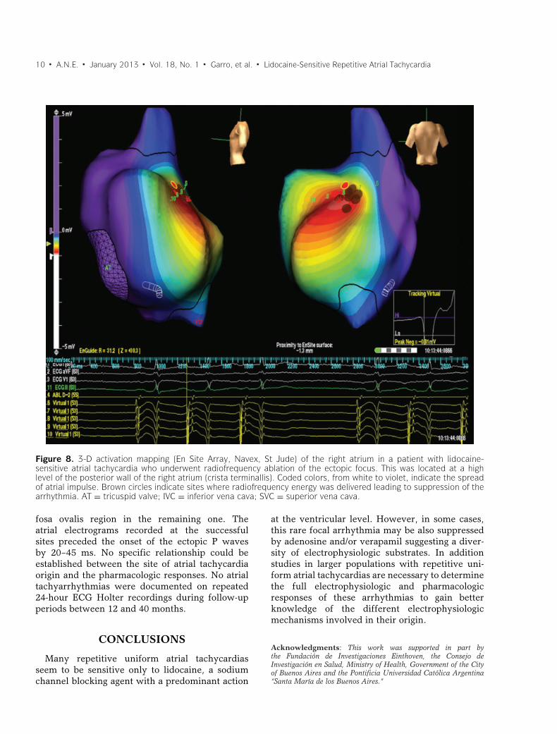
Radiofrequency ablation of the arrhythmia wassuccessfully performed in 10 symptomatic patients.The atrial ectopic foci were ablated in the lateralwall of the right atrium at a high/medium levelin three cases, in the high posterior right atriumin two patients (Fig. 8), in the body of the rightatrial appendage in two patients, in the low septalright atrium in one patient, in the proximity ofthe mitral annulus in one patient and in the
10 � A.N.E. � January 2013 � Vol. 18, No. 1 � Garro, et al. � Lidocaine-Sensitive Repetitive Atrial Tachycardia
Figure 8. 3-D activation mapping (En Site Array, Navex, St Jude) of the right atrium in a patient with lidocaine-sensitive atrial tachycardia who underwent radiofrequency ablation of the ectopic focus. This was located at a highlevel of the posterior wall of the right atrium (crista terminallis). Coded colors, from white to violet, indicate the spreadof atrial impulse. Brown circles indicate sites where radiofrequency energy was delivered leading to suppression of thearrhythmia. AT = tricuspid valve; IVC = inferior vena cava; SVC = superior vena cava.
fosa ovalis region in the remaining one. Theatrial electrograms recorded at the successfulsites preceded the onset of the ectopic P wavesby 20–45 ms. No specific relationship could beestablished between the site of atrial tachycardiaorigin and the pharmacologic responses. No atrialtachyarrhythmias were documented on repeated24-hour ECG Holter recordings during follow-upperiods between 12 and 40 months.
CONCLUSIONS
Many repetitive uniform atrial tachycardiasseem to be sensitive only to lidocaine, a sodiumchannel blocking agent with a predominant action
at the ventricular level. However, in some cases,this rare focal arrhythmia may be also suppressedby adenosine and/or verapamil suggesting a diver-sity of electrophysiologic substrates. In additionstudies in larger populations with repetitive uni-form atrial tachycardias are necessary to determinethe full electrophysiologic and pharmacologicresponses of these arrhythmias to gain betterknowledge of the different electrophysiologicmechanisms involved in their origin.
Acknowledgments: This work was supported in part bythe Fundacion de Investigaciones Einthoven, the Consejo deInvestigacion en Salud, Ministry of Health, Government of the Cityof Buenos Aires and the Pontificia Universidad Catolica Argentina“Santa Marıa de los Buenos Aires.”
A.N.E. � January 2013 � Vol. 18, No. 1 � Garro, et al. � Lidocaine-Sensitive Repetitive Atrial Tachycardia � 11
REFERENCES1. Man KC, Chan KK, Kovack P, et al. Spatial resolution of
atrial pace mapping as determined by unipolar atrial pacingat adjacent sites. Circulation 1996;94:1357–1364.
2. Poutiainen AM, Koistinen MJ, Airaksinen KE, et al.Prevalence and natural course of ectopic atrial tachycardia.Eur Heart J 1999;20:694–700.
3. Engelstein ED, Lippman N, Stein KM, et al. Mechanism-specific effects of adenosine on atrial tachycardia. Circula-tion 1994;89:2645–2654.
4. Kistler PM, Kalman JM. Atrial tachycardia: mechanisms,diagnosis and management. Curr Prob Cardiol 2005;30:529–573.
5. Chen S-A, Chiang CE, Yang CJ, et al. Sustained atrial tachy-cardia in adult patients. Electrophysiological characteristics,pharmacological response, possible mechanisms and effectsof radiofrequency ablation. Circulation 1994;90:1262–1278.
6. Mehta AV, Sanchez GR, Sacks EJ, et al. Ectopic automaticatrial tachycardia in children: Clinical characteristics,management and follow-up. J Am Coll Cardiol 1988;11:379–385.
7. Markowitz SM, Nemirovksy D, Stein KM, et al. Adenosine-insensitive focal atrial tachycardia. Evidence for de novomicro–re-entry in the human atrium. J Am Coll Cardiol2007;49:1324–1333.
8. Cotoi S, Luca C. Effetti della lidocaina nelle aritmiesopraventriculare. G Ital Cardiol 1976;6:433–439.
9. Markowitz SM, Stein KM, Mittal S, et al. Differentialeffects of adenosine on focal and macroreentrant atrial
tachycardia. J Cardiovasc Electrophysiol 1999;10:489–502.
10. Chiale PA, Selva HO, Militello CA, et al. Lidocaine-sensitive, rate-related, repetitive atrial tachycardia: Anew arrhythmogenic syndrome. J Am Coll Cardiol.2000;36:1637–1645.
11. Chiale PA, Faivelis L, Garro HA, et al. Distinct phar-macologic substrate in lidocaine-sensitive, repetitive atrialtachycardia. J Cardiovasc Pharmacol Ther 2012;17:146–152.
12. Sicouri S, Antzelevitch C. A subpopulation of cells withunique electrophysiological properties in the deep subepi-cardium of the canine ventricle. Circ Res. 1991;68:1729–1741.
13. Sicouri S, Fish J, Antzelevitch C. Distribution of Mcells in the canine ventricle. J Cardiovasc Electrophysiol1994;5:834–837.
14. Sicouri S, Antzelevitch D, Heilmann C, et al. Effectof sodium channel block with mexiletine to reverseaction potential prolongation in vitro models of the longQT syndrome. J Cardiovasc Electrophysiol 1997;8:1280–1290.
15. Sicouri S, Glass A, Belardinelli L, Antzelevitch C. Antiar-rhythmic effects of ranolazine in canine pulmonary veinsleeve preparations. Heart Rhythm 2008;5:1019–1026.
16. Burashnikov A, Di Diego JM, Zygmunt AC et al.Atrium selective sodium channel block as a strategy forsuppression of atrial fibrillation. Differences in sodiumchannel inactivation between atria and ventricles and roleof ranolazine. Circulation 2007;116:1449–1457.
Related Documents