A rapid exocytosis mode in chromaffin cells with a neuronal phenotype Alvaro O. Ardiles,* Jaime Maripilla ´n,* Vero ´nica L. Lagos,* Rodrigo Toro,* Italo G. Mora,* Lorena Villarroel, Eva Ale ´s,à Ricardo Borges§ and Ana M. Ca ´rdenas* *Centro de Neurociencia de Valparaı ´so, Universidad de Valparaı ´so, Chile Departamento de Biologı ´a, Facultad de Ciencias, Universidad de Valparaı ´so, Chile àDepartamento de Fisiologı ´a, Universidad de Sevilla, Spain §Unidad de Farmacologı ´a, Universidad de La Laguna, Tenerife, Spain Abstract We have used astrocyte-conditioned medium (ACM) to pro- mote the transdifferentiation of bovine chromaffin cells and study modifications in the exocytotic process when these cells acquire a neuronal phenotype. In the ACM-promoted neuronal phenotype, secretory vesicles and intracellular Ca 2+ rise were preferentially distributed in the neurite terminals. Using am- perometry, we observed that the exocytotic events also oc- curred mainly in the neurite terminals, wherein the individual exocytotic events had smaller quantal size than in undiffer- entiated cells. Additionally, duration of pre-spike current was significantly shorter, suggesting that ACM also modifies the fusion pore stability. After long exposure (7–9 days) to ACM, the kinetics of catecholamine release from individual vesicles was markedly accelerated. The morphometric analysis of vesicle diameters suggests that the rapid exocytotic events observed in neurites of ACM-treated cells correspond to the exocytosis of large dense-core vesicles (LDCV). On the other hand, experiments performed in EGTA-loaded cells suggest that ACM treatment promotes a better coupling between voltage-gated calcium channels (VGCC) and LDCV. Thus, our findings reveal that ACM promotes a neuronal phenotype in chromaffin cells, wherein the exocytotic kinetics is acceler- ated. Such rapid exocytosis mode could be caused at least in part by a better coupling between secretory vesicles and VGCC. Keywords: astrocytes, catecholamines, chromaffin cells, exocytosis, neurotrophic factors, large dense-core vesicles. J. Neurochem. (2006) 99, 29–41. Adrenal chromaffin cells have been employed as an experi- mental model to explore the exocytotic process (Burgoyne and Morgan 1998). In culture conditions, they have been used for real time measurements of exocytosis using techniques such as amperometry (Wightman et al. 1991). However, there are several striking differences between exocytosis in chromaffin cells and neurons, in particular regarding calcium sensitivity and kinetics (Burgoyne and Morgan 1998; Martin 2003). Further, the distribution of calcium channels in round-shaped chromaffin cells is differ- ent from that of neurons: In neurons, synaptic vesicles and voltage-gated Ca 2+ channels (VGCC) are tightly coupled in the active zones (Llinas et al. 1995) whereas in chromaffin cells, VGCC are randomly distributed (Klingauf and Neher 1997; Gutierrez et al. 1998; Gil et al. 2001). The colocal- ization of VGCC and vesicles could be an important factor in determining the differences in exocytosis of neuroendocrine cells and neurons. Adrenal chromaffin cells, like sympathetic neurons, derive from neural crest sympathoadrenal precursors (Patterson 1990). During development, the presence of substances such as glucocorticoids from the adrenal cortex inhibits neuronal differentiation of the precursors and induces a chromaffin phenotype (Doupe et al. 1985). Interestingly, chromaffin Received April 11, 2006; revised manuscript received May 12, 2006; accepted May 12 , 2006. Address correspondence and reprint requests to Ana M. Ca ´rdenas, Centro de Neurociencia de Valparaı ´so, Universidad de Valparaı ´so, Chile. E-mail: [email protected] Abbreviations used: ACM, astrocyte conditioned medium; bFGF, basic fibroblast growth factor; BSA, bovine serum albumin; CNTF, ciliary neurotrophic factor; DBH, dopamine b-hydroxylase; DMEM, Dulbecco’s modified Eagle’s medium; GDNF, glial cell line-derived neurotrophic factor; LDCV, large dense-core vesicles; NGF, nerve growth factor; PBS, phosphate-buffered saline; SNARE, SNAP recep- tors; VGCC, voltage-gated Ca 2+ channels; SCV, small clear vesicles. Journal of Neurochemistry , 2006, 99, 29–41 doi:10.1111/j.1471-4159.2006.04080.x ȑ 2006 The Authors Journal Compilation ȑ 2006 International Society for Neurochemistry, J. Neurochem. (2006) 99, 29–41 29

Welcome message from author
This document is posted to help you gain knowledge. Please leave a comment to let me know what you think about it! Share it to your friends and learn new things together.
Transcript
A rapid exocytosis mode in chromaffin cells with a neuronalphenotype
Alvaro O. Ardiles,* Jaime Maripillan,* Veronica L. Lagos,* Rodrigo Toro,* Italo G. Mora,*Lorena Villarroel,� Eva Ales,� Ricardo Borges§ and Ana M. Cardenas*
*Centro de Neurociencia de Valparaıso, Universidad de Valparaıso, Chile
�Departamento de Biologıa, Facultad de Ciencias, Universidad de Valparaıso, Chile
�Departamento de Fisiologıa, Universidad de Sevilla, Spain
§Unidad de Farmacologıa, Universidad de La Laguna, Tenerife, Spain
Abstract
We have used astrocyte-conditioned medium (ACM) to pro-
mote the transdifferentiation of bovine chromaffin cells and
study modifications in the exocytotic process when these cells
acquire a neuronal phenotype. In the ACM-promoted neuronal
phenotype, secretory vesicles and intracellular Ca2+ rise were
preferentially distributed in the neurite terminals. Using am-
perometry, we observed that the exocytotic events also oc-
curred mainly in the neurite terminals, wherein the individual
exocytotic events had smaller quantal size than in undiffer-
entiated cells. Additionally, duration of pre-spike current was
significantly shorter, suggesting that ACM also modifies the
fusion pore stability. After long exposure (7–9 days) to ACM,
the kinetics of catecholamine release from individual vesicles
was markedly accelerated. The morphometric analysis of
vesicle diameters suggests that the rapid exocytotic events
observed in neurites of ACM-treated cells correspond to the
exocytosis of large dense-core vesicles (LDCV). On the other
hand, experiments performed in EGTA-loaded cells suggest
that ACM treatment promotes a better coupling between
voltage-gated calcium channels (VGCC) and LDCV. Thus, our
findings reveal that ACM promotes a neuronal phenotype in
chromaffin cells, wherein the exocytotic kinetics is acceler-
ated. Such rapid exocytosis mode could be caused at least in
part by a better coupling between secretory vesicles and
VGCC.
Keywords: astrocytes, catecholamines, chromaffin cells,
exocytosis, neurotrophic factors, large dense-core vesicles.
J. Neurochem. (2006) 99, 29–41.
Adrenal chromaffin cells have been employed as an experi-mental model to explore the exocytotic process (Burgoyneand Morgan 1998). In culture conditions, they have beenused for real time measurements of exocytosis usingtechniques such as amperometry (Wightman et al. 1991).However, there are several striking differences betweenexocytosis in chromaffin cells and neurons, in particularregarding calcium sensitivity and kinetics (Burgoyne andMorgan 1998; Martin 2003). Further, the distribution ofcalcium channels in round-shaped chromaffin cells is differ-ent from that of neurons: In neurons, synaptic vesicles andvoltage-gated Ca2+ channels (VGCC) are tightly coupled inthe active zones (Llinas et al. 1995) whereas in chromaffincells, VGCC are randomly distributed (Klingauf and Neher1997; Gutierrez et al. 1998; Gil et al. 2001). The colocal-ization of VGCC and vesicles could be an important factor indetermining the differences in exocytosis of neuroendocrinecells and neurons.
Adrenal chromaffin cells, like sympathetic neurons, derivefrom neural crest sympathoadrenal precursors (Patterson1990). During development, the presence of substances suchas glucocorticoids from the adrenal cortex inhibits neuronaldifferentiation of the precursors and induces a chromaffinphenotype (Doupe et al. 1985). Interestingly, chromaffin
Received April 11, 2006; revised manuscript received May 12, 2006;accepted May 12 , 2006.Address correspondence and reprint requests to Ana M. Cardenas,
Centro de Neurociencia de Valparaıso, Universidad de Valparaıso, Chile.E-mail: [email protected] used: ACM, astrocyte conditioned medium; bFGF,
basic fibroblast growth factor; BSA, bovine serum albumin; CNTF,ciliary neurotrophic factor; DBH, dopamine b-hydroxylase; DMEM,Dulbecco’s modified Eagle’s medium; GDNF, glial cell line-derivedneurotrophic factor; LDCV, large dense-core vesicles; NGF, nervegrowth factor; PBS, phosphate-buffered saline; SNARE, SNAP recep-tors; VGCC, voltage-gated Ca2+ channels; SCV, small clear vesicles.
Journal of Neurochemistry, 2006, 99, 29–41 doi:10.1111/j.1471-4159.2006.04080.x
� 2006 The AuthorsJournal Compilation � 2006 International Society for Neurochemistry, J. Neurochem. (2006) 99, 29–41 29
cells, even those derived from adult animals, retain the abilityto adopt a neuronal phenotype upon exposure to neurotrophicfactors (Tischler et al. 1993). Such transdifferentiation ofchromaffin cells into sympathetic neuron-like cells includesmorphological, physiological and biochemical changes(Doupe et al. 1985; Tischler et al. 1993; Islas-Suarez et al.1994). However, despite the wide popularity of chromaffincells as a model for exocytosis, it is still unknown whetherneuronally transdifferentiated chromaffin cells shift theirpattern of exocytosis to one resembling that of a neuronalphenotype.
In the present study, we have experimentally induced aneuronal phenotype in chromaffin cell cultures and explored ifa rearrangement of VGCC and secretory sites could originate achange in exocytosis pattern. To accomplish this goal, we usedan astrocyte-conditioned medium (ACM) as a source ofneurotrophic factors to promote transdifferentiation of chrom-affin cells, and we later studied the characteristics of theexocytotic release of catecholamines. Astrocytes produce andrelease multiple neurotrophic factors, including nerve growthfactor (NGF), basic fibroblast growth factor (bFGF), ciliaryneurotrophic factor (CNTF), glial cell line-derived neuro-trophic factor (GDNF) (Chiu et al. 1991; Rudge et al. 1992;Le and Esquenazi 2002), which act synergistically to induceaxon and dendrite growth (Le and Esquenazi 2002). Chrom-affin cells express receptors for NGF, bFGF, CNTF andGDNF(Meisinger et al. 1996; Suter-Crazzolara et al. 1996; Foranderet al. 2000,2001; Schober et al. 2000). All these factorsinduce neurite outgrowth in chromaffin cells to varyingdegrees (Unsicker et al. 1985; Forander et al. 1998). Ourresults demonstrate that in ACM-induced transdifferentiatedchromaffin cells, secretory vesicles and depolarization-induced intracellular Ca2+ rise are preferentially localized atneurite terminals, where the large dense-core vesicles (LDCV)exhibit a fast exocytosis mode, probably of the ‘kiss-and-run’type. ACM-induced transdifferentiated chromaffin cells couldconstitute a suitable model, accessible to techniques thatdirectly measure single exocytotic events that could allow usto define the mechanisms that determine the differencesbetween exocytotic processes of neuroendocrine.
Materials and methods
Cell cultures
Bovine adrenal chromaffin cells were isolated following standard
methods (Livett 1984). Briefly, cells were suspended in 1 : 1
mixture of Dulbecco’s modified Eagle’s medium (DMEM) and
Ham’s F-12 supplemented with 10% fetal calf serum, 50 IU/mL
penicillin and 100 lg/mL gentamicine. Cells were plated on 12 or
25-mm-diameter glass coverslips at a density of 2.5 · 104 cells/mL.
Cells were kept in a water-saturated incubator at 37�C, in a 5% CO2/
95% air atmosphere.
Astrocyte cultures were prepared from postnatal day 1 rat brain
(McCarthy and De Vellis 1980). Briefly, rat cortices were digested
(with 0.25% trypsin and 0.1 mg/mL DNase), trimmed and plated to
80 cm2 culture flasks. Cells were cultured in DMEM/F12 supple-
mented medium, which was replaced every 2–3 days. After
reaching confluence, cells were rinsed with cold medium and
shaken on an orbital shaker (250 r.p.m) for 15–18 h at 37�C, todiscard microglial cells by selective detachment. Under these
conditions, over 90% of cells were immunoreactive for GFAP, a
marker for astrocytes. ACM was obtained by conditioning DMEM/
F12 medium for periods of 24–48 h.
24 h after plating, bovine chromaffin cells were incubated with
ACM for 2–9 days. ACMwas replaced every day. Control cells were
incubated in DMEM/F12 medium, which was also replaced daily.
Immunocytochemistry
Cells were fixed in a mixture of methanol/acetone (1 : 1) for 10 min
at 20�C. Proteins were blocked with 0.2% bovine serum albumin
(BSA) in phosphate-buffered saline (PBS). Cells were then
incubated with a monoclonal anti-dopamine b-hydroxylase (Chem-
icon International, Temecula, CA, USA) antibody (diluted 1 : 300 in
PBS containing 0.1% BSA and 0.056% saponin) overnight at 4�C.Labelling was visualized by a FITC-conjugated antirabbit IgG
antibody (Sigma-Aldrich, St Louis, MO, USA) on a confocal
microscope (Zeiss, LSM-410 axiovert-100) and analysed with the
NIH-J program.
Morphological Analysis
Cell morphology was assessed in cells fixed and stained against
DBH. Morphological transdifferentiation was quantified by count-
ing the number of cells that exhibit one or more neurites greater than
two cell body diameters in length, and this number was expressed as
a percentage of the total number in the field. The count was
performed in at least four randomly selected areas of each coverslip,
from a minimum of four cultures. To determine neurite elongation,
cells were photographed and neurite lengths were quantified from
calibrated photographs using a ruler tool from the public domain
image analysis program NIH Image J.
Electron Microscopy
Chromaffin cells were cultured on sterile 40 mm diameter tissue
culture dishes (Orange Scientific, Braine-l’Alleud, Belgium). The
cells were fixed for 2 h at room temperature in 0.1 M cacodylate
buffer (CB), pH 7.4, which contained 3% glutaraldehyde. After
washing 2 h in CB, the samples were postfixed in reduced osmium
(1 : 1 mixture of 2% osmium tetroxide and 3% potassium
ferrocyanide) and then treated with uranyl acetate (2% aqueous
solution). After fixation, the cells were dehydrated with ascending
concentrations of ethanol, and finally dehydrated in propylene
oxide. For inclusion, cells were pre-embedded for 1 h in a 2 : 1
mixture of propylene oxide and Eponate (Pelco, Redding, CA), then
treated for 1 h in a 1 : 1 mixture of propylene oxide and Eponate,
finally embedded overnight at room temperature in Eponate alone
and kept at 60�C for an additional 72 h. The ultrafine sections were
cut with a Reichert Ultracut E ultramicrotome, mounted on 200
mesh copper grids (Sigma) and impregnated in 4% uranyl acetate
and lead citrate, pH 12. The ultrathin sections were examined under
an electron microscope (Zeiss EM 900, Germany) at 50 kV, and
images were recorded on EM film (Kodak Eastman, Rochester, NY,
USA) at 7,500X and 23,000X magnification.
30 A. O. Ardiles et al.
Journal Compilation � 2006 International Society for Neurochemistry, J. Neurochem. (2006) 99, 29–41� 2006 The Authors
For the analysis of LDCV we included vesicles with an electron
dense core and an intact vesicle membrane. For the analysis of small
clear vesicles (SCV) we included only the vesicles forming clusters.
The diameter of secretory vesicles was determined by measuring the
vesicular perimeter using the public domain Image J software (NIH,
Bethesda, MD, USA). Corrected vesicle diameter was calculated
using a previously published algorithm (Parsons et al. 1995). Thisalgorithm is dependent on section thickness and the empirical
measured vesicle diameter. Section thickness was estimated to be
70 nm based on the silver interference color of the sections (Sakai
1980).
Intracellular Ca2+ imaging experiments
Ca2+ imaging experiments were performed in Fluo-4 loaded cells, as
described previously (Mendoza et al. 2003). Chromaffin cells
cultured on glass coverslips were loaded with Fluo-4 AM (5 lM
in 0.1% pluronic acid) at 20�C for 30 min and later washed with a
Krebs-HEPES buffer solution (in mM: 140 NaCl, 5 KCl, 2 MgCl2,
2.5 CaCl2, HEPES-NaOH and 10 glucose; pH ¼ 7.4). Depolariza-
tion was achieved by KCl using a pressure-driven microejection
system. The stimulus pipette, tip diameter of ca. 1 lm, was filled
with 70 mM KCl and positioned at 10–15 lm from the cell. Images
were recorded from the central plane of the cells with a Sensicam�QE cooled digital camera (Cooke Corporation, Romulus, MI, USA)
attached to a Nikon Eclipse TE-2000 inverted microscope (objective
40x) at intervals of 250 ms, and analyzed with the NIH Image
program. All fluorescence data are expressed as the increase in
fluorescence divided by baseline fluorescence (DF/F).
Amperometric Detection of Exocytosis
Carbon fibers of 8-lm radius (Thornel P-55; Amoco Corp.,
Greenville SC, USA) were used to make the microelectrodes
(Kawagoe et al. 1993). Electrochemical recordings were performed
using an Axopatch 1C (Axon Instruments, Union City, CA, USA).
Cells were placed in a perfusion chamber positioned on the stage of
an inverted microscope and washed in Krebs-HEPES buffer
solution. Amperometric measurements were performed with a
carbon fiber microelectrode gently touching the cell membrane.
Catecholamine release was stimulated by 5-s pressure ejection of
70 mM K+ from a micropipette positioned at 10–15 lm from the
cell. For experiments in cells loaded with EGTA, cells were
incubated with 20 lM EGTA-AM (Molecular Probes, Eugene,
Oregon, USA) during 45 min. Then, cells were washed in Krebs-
HEPES buffer solution and depolarized with a 70 mM K+ pulse.
Amperometric records were low-pass filtered at 1 KHz, sampled at
5 KHz, and collected using PClamp 8.1 (Axon Instruments). To
analyze the exocytotic events, a series of kinetic parameters were
extracted from each spike. Data analysis was carried out using locally
written macros for IGOR (Wavemetrics, Lake Oswego, OR, USA).
These macros allowed the automatic digital filtering, secretory spike
identification, and data analysis (Segura et al. 2000). All the above
macros and their user instructions can be downloaded free from the
following web address: http://webpages.ull.es/users/rborges/.
Statistics
Normalized results were expressed as means ± SEM. Amperometric
spike data were grouped by individual cells, thus data correspond to
means ± SEM of the cell median for each spike parameter, where ‘n’
refers to the number of cells. Statistical comparisons were performed
utilizing the non-parametric U-Mann–Whitney test.
Results
ACM promotes neurite extension in chromaffin cells
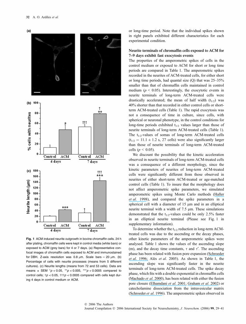
In the presence of ACM, chromaffin cells from adult bovineadrenal medulla extend one or more neurite processes(Fig. 1a). Fifty percent and 79 ± 3.0% of bovine chromaffincells exhibited neurites after 4 and 7 days under ACMtreatment, while 13 ± 5% and 53 ± 4% of the chromaffin cellshad neurites in control cultures during the same periods of time(Fig. 1b). ACM treatment also significantly enhanced neuriteelongation (Fig. 1c). The length of neurite processes reached43 ± 2 lm (n ¼ 72) and 145.6 ± 11 lm (n ¼ 53), after 4 and7 days of exposure to ACM (p < 0.0005 compared with thelength of neurites of cells in control conditions). The analysisof the distribution of the vesicular enzyme dopamine b-hydroxylase (DBH) indicated that the secretory vesicles werepreferentially distributed in the neurite terminals; the DBHimmunoreactivity ratio of neurite terminal/somawas 3.6 ± 0.4fold (n ¼ 60) for ACM-treated cells and 2.7 ± 0.2 fold (n ¼50) for cells that extend neurite in control medium.
ACM-treated cells exhibited amperometric signals
mainly in the neurite terminals
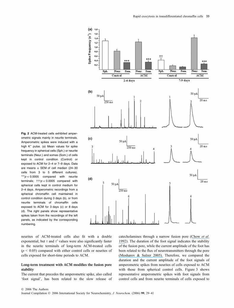
By using amperometry we examined the frequency ofexocytotic events at neurites and somas of cells cultured inthe absence and in the presence of ACM for short (2–4 days)and long (7–9 days) periods of time. Exocytosis wasstimulated by the application of a high K+ pulse. As shownin Fig. 2(a), ACM-treated cells with a neuronal phenotypeexhibited amperometric signals mainly in the neurite termi-nals, while the exocytotic events in the corresponding somaswere significantly more scarce (p < 0.0005). The frequencyof exocytotic events in the neurite terminals of cells exposedto ACM for both short- and long-time periods was similar tothat observed in cells kept in control medium for 2–4 days.On the other hand, spherical and neuron-like spherical cellsmaintained for longer periods in the control mediumexhibited a low frequency of exocytotic events (Fig. 2a).Normally, chromaffin cell cultures have a lifetime of no morethan one week, thus the scarce exocytosis of cells maintainedfor long-time periods in control medium could be aconsequence of the un-healthy condition of these cells.Conversely, the good response observed in neurite terminalsof ACM-treated cells could be a consequence of the reportedprotective properties of the astrocyte-released factors (Cuiet al. 2001).
Figure 2(b–d) show amperometric recordings obtainedfrom a spherical cell in control medium during 3 days, andfrom neurites of chromaffin cells exposed to ACM for short-
Rapid exocytosis in transdifferentiated chromaffin cells 31
� 2006 The AuthorsJournal Compilation � 2006 International Society for Neurochemistry, J. Neurochem. (2006) 99, 29–41
or long-time period. Note that the individual spikes shownin right panels exhibited different characteristics for eachexperimental condition.
Neurite terminals of chromaffin cells exposed to ACM for
7–9 days exhibit fast exocytosis events
The properties of the amperometric spikes of cells in thecontrol medium or exposed to ACM for short or long timeperiods are compared in Table 1. The amperometric spikesrecorded in the neurites of ACM-treated cells, for either shortor long time periods, had quantal size (Q) that was 25–35%smaller than that of chromaffin cells maintained in controlmedium (p < 0.05). Interestingly, the exocytotic events inneurite terminals of long-term ACM-treated cells weredrastically accelerated; the mean of half width (t1/2) was40% shorter than that recorded in either control cells or short-term ACM-treated cells (Table 1). The rapid exocytosis wasnot a consequence of time in culture, since cells, withspherical or neuronal phenotype, in the control conditions forlong-time periods exhibited t1/2 values larger than those ofneurite terminals of long-term ACM-treated cells (Table 1).The t1/2-values of somas of long-term ACM-treated cells(t1/2 ¼ 11.1 ± 1.2 s, 27 cells) were also significantly largerthan those of neurite terminals of long-term ACM-treatedcells (p < 0.05).
We discount the possibility that the kinetic accelerationobserved in neurite terminals of long-term ACM-treated cellswas a consequence of a different morphology, since thekinetic parameters of neurites of long-term ACM-treatedcells were significantly different from those observed inneurites of either short-term ACM-treated or age-matchedcontrol cells (Table 1). To insure that the morphology doesnot affect amperometric spike parameters, we simulatedamperometric spikes using Monte Carlo methods (Halleret al. 1998), and compared the spike parameters in aspherical cell with a diameter of 15 lm and in an ellipticalneurite terminal with a width of 7.5 lm. These simulationsdemonstrated that the t1/2-values could be only 2.5% fasterin an elliptical neurite terminal (Please see Fig. 1 insupplementary information).
To determine whether the t1/2 reduction in long-term ACM-treated cells was due to the ascending or the decay phases,other kinetic parameters of the amperometric spikes wereanalysed. Table 1 shows the values of the ascending slope(m), and the decay time constants, s and s¢. The ascendingphase has been related with fusion pore expansion (Schroederet al. 1996; Ales et al. 2005). As shown in Table 1, theascending slope was significantly faster in the neuriteterminals of long-term ACM-treated cells. The spike decayphase, which fits with a double exponential in chromaffin cells(Machado et al. 2000), has been related with either the fusionpore closure (Elhamdani et al. 2001; Graham et al. 2002) orcatecholamine dissociation from the intravesicular matrix(Schroeder et al. 1996). The amperometric spikes observed in
Fig. 1 ACM induced neurite outgrowth in bovine chromaffin cells. 24 h
after plating, chromaffin cells were kept in control media (white bars) or
exposed to ACM (grey bars) for 4 or 7 days. (a) Representative con-
focal images of chromaffin cells exposed to ACM and immunostained
for DBH. Z-axis resolution was 0.8 lm. Scale bars ¼ 20 lm. (b)
Percentage of cells with neurite processes (means from 5 different
cultures). (c) Neurite lengths (means from 72 and 53 cells). Data are
means ± SEM *p < 0.05, **p < 0.005, ***p < 0.0005 compared to
control cells; �p < 0.05, ���p < 0.0005 compared with cells kept dur-
ing 4 days in control medium or ACM.
32 A. O. Ardiles et al.
Journal Compilation � 2006 International Society for Neurochemistry, J. Neurochem. (2006) 99, 29–41� 2006 The Authors
neurites of ACM-treated cells also fit with a doubleexponential, but s and s¢ values were also significantly fasterin the neurite terminals of long-term ACM-treated cells(p < 0.05) compared with either control cells or neurites ofcells exposed for short-time periods to ACM.
Long-term treatment with ACM modifies the fusion pore
stability
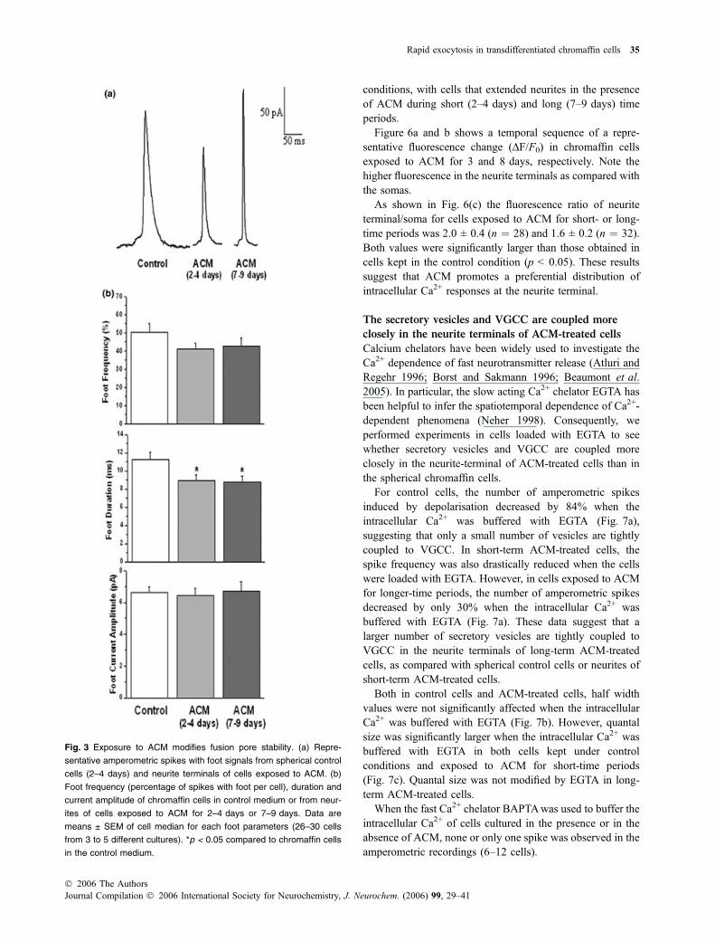
The current that precedes the amperometric spike, also called‘foot signal’, has been related to the slow release of
catecholamines through a narrow fusion pore (Chow et al.1992). The duration of the foot signal indicates the stabilityof the fusion pore, while the current amplitude of the foot hasbeen related to the flux of neurotransmitters through the pore(Mosharov & Sulzer 2005). Therefore, we compared theduration and the current amplitude of the foot signals ofamperometric spikes from neurites of cells exposed to ACMwith those from spherical control cells. Figure 3 showsrepresentative amperometric spikes with foot signals fromcontrol cells and from neurite terminals of cells exposed to
Fig. 2 ACM-treated cells exhibited amper-
ometric signals mainly in neurite terminals.
Amperometric spikes were induced with a
high K+ pulse. (a) Mean values for spike
frequency in spherical cells (Sph.) or neurite
terminals (Neur.) and somas (Som.) of cells
kept in control condition (Control) or
exposed to ACM for 2–4 or 7–9 days. Data
are means ± SEM of cell median (24–30
cells from 3 to 5 different cultures).
***p < 0.0005 compared with neurite
terminals; ���p < 0.0005 compared with
spherical cells kept in control medium for
2–4 days. Amperometric recordings from a
spherical chromaffin cell maintained in
control condition during 3 days (b), or from
neurite terminals of chromaffin cells
exposed to ACM for 3 days (c) or 8 days
(d). The right panels show representative
spikes taken from the recordings of the left
panels, as indicated by the corresponding
numbering.
Rapid exocytosis in transdifferentiated chromaffin cells 33
� 2006 The AuthorsJournal Compilation � 2006 International Society for Neurochemistry, J. Neurochem. (2006) 99, 29–41
ACM. The percentage of amperometric spikes with footsignals was similar for control chromaffin cells and neuritesof cells exposed to ACM (Fig. 3b). However, the duration ofthe foot at neurites of ACM-treated cells was significantlyshorter than in cells in control medium, indicating that thetreatment with ACM modifies the stability of the fusion pore.A short-term exposure to ACM was sufficient to produce thiseffect. On the other hand, the current amplitude of footsignals was similar for spherical cells in control medium andneurites of cells exposed to ACM (Fig. 3b).
Chromaffin cells exposed to ACM contain both large
dense-core and small clear vesicles
SCV forming clusters have been observed in neurites of ratchromaffin cells exposed for a long-time period to NGF(Doupe et al. 1985). To determine whether the smallerquantal size of ACM-treated cells was a consequence of adecrease in vesicular size, we examined ACM treated andnon-treated cells under the electron microscope.
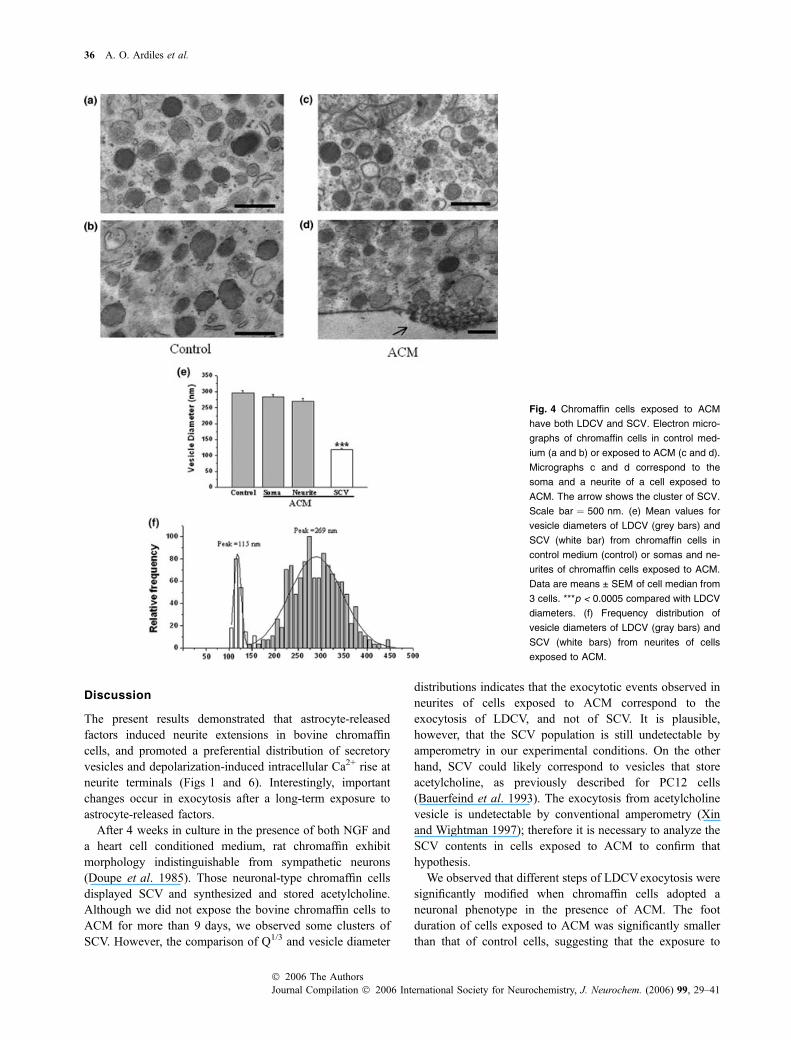
Figure 4 shows electron micrographs of chromaffin cellscultured on control medium and from cultures exposed toACM for one week. These electron micrographs reveal thepresence of LDCV in somas and neurites of ACM-treatedcells. However, the presence of SCV in neurite terminals isparticularly interesting (see arrow in Fig. 4d). The mean ofSCV diameters was approximately 40% of that of LDCVof control cells (Fig. 4e). The size of LDCV of cellsexposed to ACM was not significantly different to those ofcontrol cells (Fig. 4e). Frequency distributions of vesiclediameters from neurites of cells exposed to ACM areshown Fig. 4(f). The vesicle diameter was fitted to twoGaussian curves (R ¼ 0.98), with peaks at 269 nm and116 nm.
The rapid exocytosis events observed in neurites of cells
exposed to ACM correspond to LDCV exocytosis
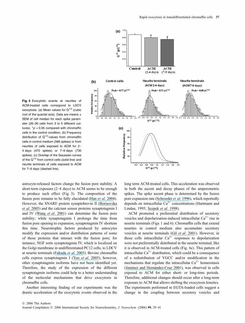
As the cubic root of the quantal size (Q1/3) has been related tothe vesicle diameter and electron micrographs of the neuriteterminals revealed the presence of both SCV and LDCV inneurites (Fig. 4), we used this parameter to investigatewhether the rapid exocytosis events correspond to SCV orLDCV.
As shown in Fig. 5(a), Q1/3 was significantly smaller forthe spikes recorded from neurites of cells exposed to ACMfor both short- and long-time periods (p < 0.05) than thosefor untreated cells. The distribution of Q1/3-values of spikesfrom control cells was fitted to a Gaussian curve (R ¼ 0.96),with a peak at 0.77 pC1/3. For neurite terminals of cellsexposed to ACM for both short- and long-time periods, thedistribution of Q1/3-values was also fitted to a Gaussian curve(R ¼ 0.97 and 0.98), with peaks at 0.71 and 0.68 pC1/3,respectively (Fig. 5b). Figure 5(c) show the overlap of theGaussian curves for the Q1/3 of spikes obtained from cellscultured under control conditions and from spikes fromneurites of long-term ACM-treated cells. A general leftwardshift to lower values is observed for cells exposed during 7–9 days to ACM. But, it is not observed an additionalpopulation corresponding to SCV, whose diameters were 2.5times smaller than that of LDCV of control cells.
Neurite terminals of cells exposed to ACM exhibit higher
intracellular Ca2+ signals than somas
We also investigated if astrocyte-released factors change thedistribution pattern of the intracellular Ca2+ responses to adepolarizing stimulus in chromaffin cells. Thus, we com-pared the distribution of depolarization-induced Ca2+ signalsin cells that extend neurites when cultured under control
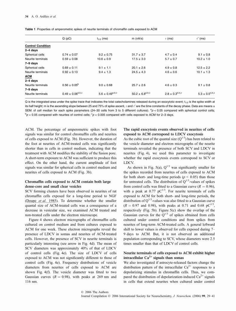
Table 1 Properties of amperometric spikes of neurite terminals of chromaffin cells exposed to ACM
Q (pC) t1/2 (ms) m (nA/s) s (ms) s¢ (ms)
Control Condition
2–4 days
Spherical cells 0.74 ± 0.07 9.2 ± 0.75 31.7 ± 3.7 4.7 ± 0.4 9.1 ± 0.8
Neurite terminals 0.59 ± 0.08 10.6 ± 0.9 17.5 ± 3.0 5.7 ± 0.7 10.2 ± 1.0
7–9 days
Spherical cells 0.69 ± 0.11 9.1 ± 1.1 20.1 ± 2.6 4.9 ± 0.8 12.5 ± 2.2
Neurite terminals 0.92 ± 0.13 9.4 ± 1.3 24.5 ± 4.3 4.6 ± 0.6 10.1 ± 1.3
ACM
2–4 days
Neurite terminals 0.56 ± 0.05b 9.0 ± 0.68 25.7 ± 2.6 4.6 ± 0.3 9.1 ± 0.8
7–9 days
Neurite terminals 0.49 ± 0.06a,b,c 5.6 ± 0.49a,b,c 50.2 ± 6.8a,b,c 2.8 ± 0.3b,b,c 5.3 ± 0.5a,b,c
Q is the integrated area under the spike trace that indicates the total catecholamines released during an exocytotic event; t1/2 is the spike width at
its half height; m is the ascending slope between 25 and 75% of spike ascent, s and s¢ are the time constants of the decay phase. Data are means ±
SEM. of cell median for each spike parameters (24–30 cells from 3 to 5 different cultures). ap < 0.05 compared with spherical control cells;bp < 0.05 compared with neurites of control cells; cp < 0.005 compared with cells exposed to ACM for 2–3 days.
34 A. O. Ardiles et al.
Journal Compilation � 2006 International Society for Neurochemistry, J. Neurochem. (2006) 99, 29–41� 2006 The Authors
conditions, with cells that extended neurites in the presenceof ACM during short (2–4 days) and long (7–9 days) timeperiods.
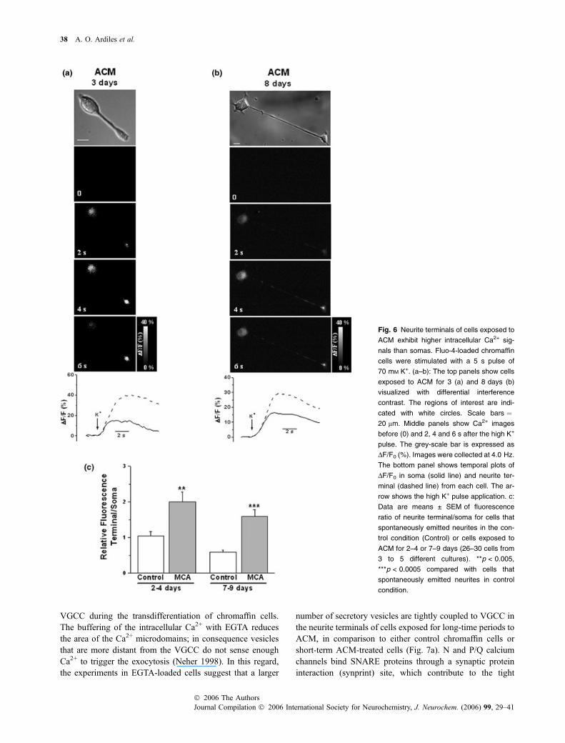
Figure 6a and b shows a temporal sequence of a repre-sentative fluorescence change (DF/F0) in chromaffin cellsexposed to ACM for 3 and 8 days, respectively. Note thehigher fluorescence in the neurite terminals as compared withthe somas.
As shown in Fig. 6(c) the fluorescence ratio of neuriteterminal/soma for cells exposed to ACM for short- or long-time periods was 2.0 ± 0.4 (n ¼ 28) and 1.6 ± 0.2 (n ¼ 32).Both values were significantly larger than those obtained incells kept in the control condition (p < 0.05). These resultssuggest that ACM promotes a preferential distribution ofintracellular Ca2+ responses at the neurite terminal.
The secretory vesicles and VGCC are coupled more
closely in the neurite terminals of ACM-treated cells
Calcium chelators have been widely used to investigate theCa2+ dependence of fast neurotransmitter release (Atluri andRegehr 1996; Borst and Sakmann 1996; Beaumont et al.2005). In particular, the slow acting Ca2+ chelator EGTA hasbeen helpful to infer the spatiotemporal dependence of Ca2+-dependent phenomena (Neher 1998). Consequently, weperformed experiments in cells loaded with EGTA to seewhether secretory vesicles and VGCC are coupled moreclosely in the neurite-terminal of ACM-treated cells than inthe spherical chromaffin cells.
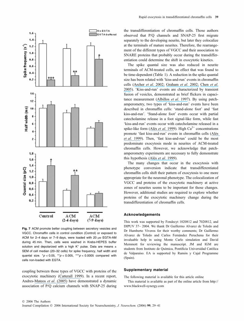
For control cells, the number of amperometric spikesinduced by depolarisation decreased by 84% when theintracellular Ca2+ was buffered with EGTA (Fig. 7a),suggesting that only a small number of vesicles are tightlycoupled to VGCC. In short-term ACM-treated cells, thespike frequency was also drastically reduced when the cellswere loaded with EGTA. However, in cells exposed to ACMfor longer-time periods, the number of amperometric spikesdecreased by only 30% when the intracellular Ca2+ wasbuffered with EGTA (Fig. 7a). These data suggest that alarger number of secretory vesicles are tightly coupled toVGCC in the neurite terminals of long-term ACM-treatedcells, as compared with spherical control cells or neurites ofshort-term ACM-treated cells.
Both in control cells and ACM-treated cells, half widthvalues were not significantly affected when the intracellularCa2+ was buffered with EGTA (Fig. 7b). However, quantalsize was significantly larger when the intracellular Ca2+ wasbuffered with EGTA in both cells kept under controlconditions and exposed to ACM for short-time periods(Fig. 7c). Quantal size was not modified by EGTA in long-term ACM-treated cells.
When the fast Ca2+ chelator BAPTAwas used to buffer theintracellular Ca2+ of cells cultured in the presence or in theabsence of ACM, none or only one spike was observed in theamperometric recordings (6–12 cells).
Fig. 3 Exposure to ACM modifies fusion pore stability. (a) Repre-
sentative amperometric spikes with foot signals from spherical control
cells (2–4 days) and neurite terminals of cells exposed to ACM. (b)
Foot frequency (percentage of spikes with foot per cell), duration and
current amplitude of chromaffin cells in control medium or from neur-
ites of cells exposed to ACM for 2–4 days or 7–9 days. Data are
means ± SEM of cell median for each foot parameters (26–30 cells
from 3 to 5 different cultures). *p < 0.05 compared to chromaffin cells
in the control medium.
Rapid exocytosis in transdifferentiated chromaffin cells 35
� 2006 The AuthorsJournal Compilation � 2006 International Society for Neurochemistry, J. Neurochem. (2006) 99, 29–41
Discussion
The present results demonstrated that astrocyte-releasedfactors induced neurite extensions in bovine chromaffincells, and promoted a preferential distribution of secretoryvesicles and depolarization-induced intracellular Ca2+ rise atneurite terminals (Figs 1 and 6). Interestingly, importantchanges occur in exocytosis after a long-term exposure toastrocyte-released factors.
After 4 weeks in culture in the presence of both NGF anda heart cell conditioned medium, rat chromaffin exhibitmorphology indistinguishable from sympathetic neurons(Doupe et al. 1985). Those neuronal-type chromaffin cellsdisplayed SCV and synthesized and stored acetylcholine.Although we did not expose the bovine chromaffin cells toACM for more than 9 days, we observed some clusters ofSCV. However, the comparison of Q1/3 and vesicle diameter
distributions indicates that the exocytotic events observed inneurites of cells exposed to ACM correspond to theexocytosis of LDCV, and not of SCV. It is plausible,however, that the SCV population is still undetectable byamperometry in our experimental conditions. On the otherhand, SCV could likely correspond to vesicles that storeacetylcholine, as previously described for PC12 cells(Bauerfeind et al. 1993). The exocytosis from acetylcholinevesicle is undetectable by conventional amperometry (Xinand Wightman 1997); therefore it is necessary to analyze theSCV contents in cells exposed to ACM to confirm thathypothesis.
We observed that different steps of LDCVexocytosis weresignificantly modified when chromaffin cells adopted aneuronal phenotype in the presence of ACM. The footduration of cells exposed to ACM was significantly smallerthan that of control cells, suggesting that the exposure to
Fig. 4 Chromaffin cells exposed to ACM
have both LDCV and SCV. Electron micro-
graphs of chromaffin cells in control med-
ium (a and b) or exposed to ACM (c and d).
Micrographs c and d correspond to the
soma and a neurite of a cell exposed to
ACM. The arrow shows the cluster of SCV.
Scale bar ¼ 500 nm. (e) Mean values for
vesicle diameters of LDCV (grey bars) and
SCV (white bar) from chromaffin cells in
control medium (control) or somas and ne-
urites of chromaffin cells exposed to ACM.
Data are means ± SEM of cell median from
3 cells. ***p < 0.0005 compared with LDCV
diameters. (f) Frequency distribution of
vesicle diameters of LDCV (gray bars) and
SCV (white bars) from neurites of cells
exposed to ACM.
36 A. O. Ardiles et al.
Journal Compilation � 2006 International Society for Neurochemistry, J. Neurochem. (2006) 99, 29–41� 2006 The Authors
astrocyte-released factors change the fusion pore stability. Ashort-term exposure (2–4 days) to ACM seems to be enoughto produce such effect (Fig. 3). The composition of thefusion pore remains to be fully elucidated (Han et al. 2004).However, the SNARE protein synaptobrevin II (Borisovskaet al. 2005) and the calcium sensor proteins synaptotagmin Iand IV (Wang et al. 2001) can determine the fusion porestability; while synaptotagmin I prolongs the time fromfusion pore opening to expansion, synaptotagmin IV shortensthis time. Neurotrophic factors produced by astrocytesmodify the expression and/or distribution patterns of someof those proteins that interact with the fusion pore; forinstance, NGF sorts synaptotagmin IV, which is localized onthe Golgi membrane in undifferentiated PC12 cells, to LDCVat neurite terminals (Fukuda et al. 2003). Bovine chromaffincells express synaptotagmin I (Yoo et al. 2005), however,other synaptotagmin isoforms have not been identified yet.Therefore, the study of the expression of the differentsynaptotagmin isoforms could help to a better understandingof the molecular mechanisms that drive exocytosis inchromaffin cells.
Another interesting finding of our experiments was thedrastic acceleration of the exocytotic events observed in the
long term ACM-treated cells. This acceleration was observedin both the ascent and decay phases of the amperometricspikes. The spike ascent phase is determined by the fusionpore expansion rate (Schroeder et al. 1996), which reportedlydepends on intracellular Ca2+ concentrations (Hartmann andLindau, 1995; Scepek et al. 1998).
ACM promoted a preferential distribution of secretoryvesicles and depolarization-induced intracellular Ca2+ rise inneurite terminals (Figs 1 and 6). Chromaffin cells that extendneurites in control medium also accumulate secretoryvesicles at neurite terminals (Gil et al. 2001). However, inthose cells intracellular Ca2+ responses to depolarizationwere not preferentially distributed in the neurite terminal, likeit is observed in ACM-treated cells (Fig. 6c). This pattern ofintracellular Ca2+ distribution, which could be a consequenceof a redistribution of VGCC and/or modification in themechanisms that regulate the intracellular Ca2+ homeostasis(Jimenez and Hernandez-Cruz 2001), was observed in cellsexposed to ACM for either short- or long-time periods.Therefore, additional changes should occur after a long-termexposure to ACM that allows shifting the exocytosis kinetics.The experiments performed in EGTA-loaded cells suggest achange in the coupling between secretory vesicles and
Fig. 5 Exocytotic events at neurites of
ACM-treated cells correspond to LDCV
exocytosis. (a) Mean values for Q1/3 (cubic
root of the quantal size). Data are means ±
SEM of cell median for each spike param-
eter (26–30 cells from 3 to 5 different cul-
tures). *p < 0.05 compared with chromaffin
cells in the control condition. (b) Frequency
distribution of Q1/3-values from chromaffin
cells in control medium (566 spikes) or from
neurites of cells exposed to ACM for 2–
4 days (470 spikes) or 7–9 days (730
spikes). (c) Overlap of the Gaussian curves
of the Q1/3 from control cells (solid line) and
neurite terminals of cells exposed to ACM
for 7–9 days (dashed line).
Rapid exocytosis in transdifferentiated chromaffin cells 37
� 2006 The AuthorsJournal Compilation � 2006 International Society for Neurochemistry, J. Neurochem. (2006) 99, 29–41
VGCC during the transdifferentiation of chromaffin cells.The buffering of the intracellular Ca2+ with EGTA reducesthe area of the Ca2+ microdomains; in consequence vesiclesthat are more distant from the VGCC do not sense enoughCa2+ to trigger the exocytosis (Neher 1998). In this regard,the experiments in EGTA-loaded cells suggest that a larger
number of secretory vesicles are tightly coupled to VGCC inthe neurite terminals of cells exposed for long-time periods toACM, in comparison to either control chromaffin cells orshort-term ACM-treated cells (Fig. 7a). N and P/Q calciumchannels bind SNARE proteins through a synaptic proteininteraction (synprint) site, which contribute to the tight
Fig. 6 Neurite terminals of cells exposed to
ACM exhibit higher intracellular Ca2+ sig-
nals than somas. Fluo-4-loaded chromaffin
cells were stimulated with a 5 s pulse of
70 mM K+. (a–b): The top panels show cells
exposed to ACM for 3 (a) and 8 days (b)
visualized with differential interference
contrast. The regions of interest are indi-
cated with white circles. Scale bars ¼20 lm. Middle panels show Ca2+ images
before (0) and 2, 4 and 6 s after the high K+
pulse. The grey-scale bar is expressed as
DF/F0 (%). Images were collected at 4.0 Hz.
The bottom panel shows temporal plots of
DF/F0 in soma (solid line) and neurite ter-
minal (dashed line) from each cell. The ar-
row shows the high K+ pulse application. c:
Data are means ± SEM of fluorescence
ratio of neurite terminal/soma for cells that
spontaneously emitted neurites in the con-
trol condition (Control) or cells exposed to
ACM for 2–4 or 7–9 days (26–30 cells from
3 to 5 different cultures). **p < 0.005,
***p < 0.0005 compared with cells that
spontaneously emitted neurites in control
condition.
38 A. O. Ardiles et al.
Journal Compilation � 2006 International Society for Neurochemistry, J. Neurochem. (2006) 99, 29–41� 2006 The Authors
coupling between those types of VGCC with proteins of theexocytotic machinery (Catterall 1999). In a recent report,Andres-Mateos et al. (2005) have demonstrated a dynamicassociation of P/Q calcium channels with SNAP-25 during
the transdifferentiation of chromaffin cells. Those authorsobserved that P/Q channels and SNAP-25 first migrateseparately to the developing neurite, but later they colocalizeat the terminals of mature neurites. Therefore, the rearrange-ment of the different types of VGCC and their association toSNARE proteins that probably occur during the transdiffer-entiation could determine the shift in exocytotic kinetics.
The spike quantal size was also reduced in neuriteterminals of ACM-treated cells, an effect that was found tobe time-dependent (Table 1). A reduction in the spike quantalsize has been related with ‘kiss-and-run’ events in chromaffincells (Archer et al. 2002; Graham et al. 2002; Chen et al.2005). ‘Kiss-and-run’ events are characterized by transientfusion of vesicles, demonstrated as brief flickers in capaci-tance measurement (Albillos et al. 1997). By using patch-amperometry, two types of ‘kiss-and-run’ events have beendescribed in chromaffin cells: ‘stand-alone foot’ and ‘fastkiss-and-run’. ‘Stand-alone foot’ events occur with partialcatecholamine release in a foot signal-like form, while fast‘kiss-and-run’ events occur with catecholamine released in aspike-like form (Ales et al. 1999). High Ca2+ concentrationspromote ‘fast kiss-and-run’ events in chromaffin cells (Aleset al. 1999). Then, ‘fast kiss-and-run’ could be the mostpredominate exocytosis mode in neurites of ACM-treatedchromaffin cells. However, we acknowledge that patch-amperometry experiments are necessary to fully demonstratethis hypothesis (Ales et al. 1999).
The many changes that occur in the exocytosis withphenotype conversion indicate that transdifferentiatedchromaffin cells shift their pattern of exocytosis to one moreappropriate for the neuronal phenotype. The colocalization ofVGCC and proteins of the exocytotic machinery at activezones of neurites seems to be important for those changes.However, additional studies are required to explore whetherproteins of the exocytotic machinery change during thetransdifferentiation of chromaffin cells.
Acknowledgements
This work was supported by Fondecyt 1020812 and 7020812, and
DIPUV 37– 2004. We thank Dr Guillermo Alvarez de Toledo and
Dr Humberto Viveros for their worthy comments; Dr Guillermo
Alvarez de Toledo and Carlos Fernandez Peruchena for their
invaluable help in using Monte Carlo simulation and David
Memmott for reviewing the manuscript. JM and IGM are
students from Instituto de Quımica, Pontificia Universidad Catolica
de Valparaıso. EA is supported by Ramon y Cajal Programme
(Spain).
Supplementary material
The following material is available for this article online
This material is available as part of the online article from http://
www.blackwell-synergy.com
Fig. 7 ACM promote better coupling between secretory vesicles and
VGCC. Chromaffin cells in control condition (Control) or exposed to
ACM for 2–4 days or 7–9 days, were loaded with 20 lM EGTA-AM
during 45 min. Then, cells were washed in Krebs-HEPES buffer
solution and depolarized with a high K+ pulse. Data are means ±
SEM of cell median (20–32 cells) for spike frequency, half width and
quantal size. *p < 0.05, **p < 0.005, ***p < 0.0005 compared with
cells non-loaded with EGTA.
Rapid exocytosis in transdifferentiated chromaffin cells 39
� 2006 The AuthorsJournal Compilation � 2006 International Society for Neurochemistry, J. Neurochem. (2006) 99, 29–41
References
Albillos A., Dernick G., Horstmann H., Almers W., Alvarez de ToledoG. and Lindau M. (1997) The exocytotic event in chromaffin cellsrevealed by patch amperometry. Nature 389, 509–512.
Ales E., Tabares L., Poyato J. M., Valero V., Lindau M. and Alvarez deToledo G. (1999) High calcium concentrations shift the modeof exocytosis to the kiss-and-run mechanism. Nat. Cell. Biol. 1,40–44.
Ales E., Neco P., Gutierrez L. M. and Alvarez de Toledo G. (2005)Regulation of the fusion pore expansion during exocitosis bymyosin II. J. Physiol. Biochem. 61, 96.
Andres-Mateos E., Renart J., Cruces J., Solis-Garrido L. M., Serantes R.,de Lucas-Cerrillo A. M., Aldea M., Garcia A. G. and Montiel C.(2005) Dynamic association of the Ca2+ channel alpha1A subunitand SNAP-25 in round or neurite-emitting chromaffin cells. Eur. J.Neurosci. 22, 2187–2198.
Archer D. A., Graham M. E. and Burgoyne R. D. (2002) Complexinregulates the closure of the fusion pore during regulated vesicleexocytosis. J. Biol. Chem. 277, 18249–18252.
Atluri P. P. and Regehr W. G. (1996) Determinants of the time course offacilitation at the granule cell to Purkinje cell synapse. J. Neurosci.16, 5661–5671.
Bauerfeind R., Regnier-Vigouroux A., Flatmark T. and Huttner W. B.(1993) Selective storage of acetylcholine, but not catecholamines,in neuroendocrine synaptic-like microvesicles of early endosomalorigin. Neuron 11, 105–121.
Beaumont V., Llobet A. and Lagnado L. (2005) Expansion of calciummicrodomains regulates fast exocytosis at a ribbon synapse. Proc.Natl Acad. Sci. USA 102, 10 700–10 705.
Borisovska M., Zhao Y., Tsytsyura Y., Glyvuk N., Takamori S., Matti U.,Rettig J., Sudhof T. and Bruns D. (2005) v-SNAREs control exo-cytosis of vesicles from priming to fusion. EMBO J. 24, 2114–2126.
Borst J. G. and Sakmann B. (1996) Calcium influx and transmitterrelease in a fast CNS synapse. Nature 383, 431–434.
Burgoyne R. D. and Morgan A. (1998) Calcium sensors in regulatedexocytosis. Cell Calcium 24, 367–376.
Catterall W. A. (1999) Interactions of presynaptic Ca2+ channels andsnare proteins in neurotransmitter release. Ann. N. Y. Acad. Sci.868, 144–159.
Chen X. K., Wang L. C., Zhou Y., Cai Q., Prakriya M., Duan K. L.,Sheng Z. H., Lingle C. and Zhou Z. (2005) Activation of GPCRsmodulates quantal size in chromaffin cells through Gbc and PKC.Nat. Neurosci. 8, 1160–1168.
Chiu A. Y., Espinosa de los Monteros A., Cole R. A., Loera S. and deVellis J. (1991) Laminin and s-laminin are produced and releasedby astrocytes, Schwann cells, and schwannomas in culture. Glia 4,11–24.
Chow R. H., von Ruden L. and Neher E. (1992) Delay in vesicle fusionrevealed by electrochemical monitoring of single secretory eventsin adrenal chromaffin cells. Nature 356, 60–63.
Cui W., Allen N. D., Skynner M., Gusterson B. and Clark A. J. (2001)Inducible ablation of astrocytes shows that these cells are requiredfor neuronal survival in the adult brain. Glia 34, 272–282.
Doupe A. J., Landis S. C. and Patterson P. H. (1985) Environmentalinfluences in the development of neural crest derivatives: gluco-corticoids, growth factors, and chromaffin cell plasticity. J. Neu-rosci. 5, 2119–2142.
Elhamdani A., Palfrey H. C. and Artalejo C. R. (2001) Quantal size isdependent on stimulation frequency and calcium entry in calfchromaffin cells. Neuron 31, 819–830.
Forander P., Brene S. and Stromberg I. (2000) Expression and regulationof CNTF receptor-alpha in the in situ and in oculo grafted adult ratadrenal medulla. Neuroreport 11, 593–597.
Forander P., Broberger C. and Stromberg I. (2001) Glial-cell-line-derived neurotrophic factor induces nerve fibre formation inprimary cultures of adrenal chromaffin cells. Cell Tissue Res.305, 43–45.
Forander P., Hoffer B. and Stromberg I. (1998) Nerve fiber formationand catecholamine content in adult rat adrenal medullary trans-plants after treatment with NGF, NT-3, NT-4/5, bFGF, CNTF, andGDNF. Cell Tissue Res. 292, 503–512.
Fukuda M., Kanno E., Ogata Y., Saegusa C., Kim T., Loh Y. P. andYamamoto A. (2003) Nerve growth factor-dependent sorting ofsynaptotagmin IV protein to mature dense-core vesicles that un-dergo calcium-dependent exocytosis in PC12 cells. J. Biol. Chem.278, 3220–3226.
Gil A., Viniegra S., Neco P. and Gutierrez L. M. (2001) Co-localizationof vesicles and P/Q Ca2+-channels explains the preferential dis-tribution of exocytotic active zones in neuritis emitted by bovinechromaffin cells. Eur. J. Cell. Biol. 80, 358–365.
Graham M. E., O’Callaghan D. W., McMahon H. T. and Burgoyne R. D.(2002) Dynamin-dependent and dynamin-independent processescontribute to the regulation of single vesicle release kinetics andquantal size. Proc. Natl Acad. Sci. USA 99, 7124–7129.
Gutierrez L. M., Gil A. and Viniegra S. (1998) Preferential localizationof exocytotic active zones in the terminals of neurite-emittingchromaffin cells. Eur. J. Cell. Biol. 76, 274–278.
Haller M., Heinemann C., Chow R. H., Heidelberger R. and Neher E.(1998) Comparison of Secretory Responses as Measured byMembrane Capacitance and by Amperometry. Biophys. J. 74,2100–2113.
Han X., Wang C. T., Bai J., Chapman E. R. and Jackson M. B. (2004)Transmembrane segments of syntaxin line the fusion pore of Ca2+-triggered exocytosis. Science 304, 289–292.
Hartmann J. and Lindau M. (1995) A novel Ca (2+)-dependent stepin exocytosis subsequent to vesicle fusion. FEBS Lett. 363, 217–220.
Islas-Suarez L., Gomez-Chavarin M., Drucker-Colin R. and Hernandez-Cruz A. (1994) Properties of the sodium current in rat chromaffincells exposed to nerve growth factor in vitro. J. Neurophysiol. 72,1938–1948.
Jimenez N. and Hernandez-Cruz A. (2001) Modifications of intracellularCa2+ signaling during nerve growth factor-induced neuronal dif-ferentiation of rat adrenal chromaffin cells. Eur. J. Neurosci. 13,1487–1500.
Kawagoe K. T., Zimmerman J. B. and Wightman R. M., (1993) Prin-ciples of voltammetry and microelectrode surface states. J. Neu-rosci. Meth. 48, 225–240.
Klingauf J. and Neher E. (1997) Modeling buffered Ca2+ diffusion nearthe membrane: implications for secretion in neuroendocrine cells.Biophys. J. 72, 674–690.
Le R. and Esquenazi S. (2002) Astrocytes mediate cerebral corticalneuronal axon and dendrite growth, in part, by release of fibroblastgrowth factor. Neurol. Res. 24, 81–92.
Livett B. G. (1984) Adrenal medullary chromaffin cells in vitro. Physiol.Rev. 64, 1103–1161.
Llinas R. R., Sugimori M. and Silver R. B. (1995) The concept ofcalcium concentration microdomains in synaptic transmission.Neuropharm. 34, 1443–1451.
Machado J. D., Segura F., Brioso M. A. and Borges R. (2000) Nitricoxide modulates a late step of exocytosis. J. Biol. Chem. 275,20 274–20 279.
Martin T. F. (2003) Tuning exocytosis for speed: fast and slow modes.Biochim. Biophys. Acta 1641, 157–165.
McCarthy K. D. and De Vellis J. (1980) Preparation of separate astroglialand oligodendroglial cell cultures from rat cerebral tissue. J. CellBiol. 85, 890–902.
40 A. O. Ardiles et al.
Journal Compilation � 2006 International Society for Neurochemistry, J. Neurochem. (2006) 99, 29–41� 2006 The Authors
Meisinger C., Hertenstein A. and Grothe C. (1996) Fibroblast growthfactor receptor 1 in the adrenal gland and PC12 cells: develop-mental expression and regulation by extrinsic molecules. Brain.Res. Mol. Brain. Res. 36, 70–78.
Mendoza I. E., Schmachtenberg O., Tonk E., Fuentealba J., Dıaz-RayaP., Lagos V. L., Garcıa A. G. and Cardenas A. M. (2003) Depo-larization-induced ERK phosphorylation depends on the cytosolicCa2+ level rather than on the Ca2+ channel subtype of chromaffincells. J. Neurochem. 86, 1477–1486.
Mosharov E. V. and Sulzer D. (2005) Analysis of exocytotic eventsrecorded by amperometry. Nat. Meth 2, 651–658.
Neher E. (1998) Vesicle pools and Ca2+ microdomains: new tools forunderstanding their roles in neurotransmitter release. Neuron 20,389–399.
Parsons T. D., Coorssen J. R., Horstmann H. and Almers W. (1995)Docked granules, the exocytic burst, and the need for ATPhydrolysis in endocrine cells. Neuron 15, 1085–1096.
Patterson P. H. (1990) Control of cell fate in a vertebrate neurogeniclineage. Cell 62, 1035–1038.
Rudge J. S., Alderson R. F., Pasnikowski E., McClain J., Ip N. Y. andLindsay R. M. (1992) Expression of Ciliary Neurotrophic Factorand the Neurotrophins-Nerve Growth Factor, Brain-Derived Neu-rotrophic Factor and Neurotrophin 3-in Cultured Rat HippocampalAstrocytes. Eur. J. Neurosci. 4, 459–471.
Sakai T. (1980) Relation between thickness and interference colors ofbiological ultrathin section. J. Electron. Microsc. (Tokyo) 29, 369–375.
Scepek S., Coorssen J. R. and Lindau M. (1998) Fusion pore expansionin horse eosinophils is modulated by Ca2+ and protein kinase C viadistinct mechanisms. EMBO J. 17, 4340–4345.
Schober A., Arumae U., Saarma M. and Unsicker K. (2000) Expressionof GFR alpha-1, GFR alpha-2, and c-Ret mRNAs in rat adrenalgland. J. Neurocytol. 29, 209–213.
Schroeder T. J., Borges R., Finnegan J. M., Pihel K., Amatore C. andWightman R.M. (1996) Temporally resolved, independent stages ofindividual exocytotic secretion events. Biophys. J. 70, 1061–1068.
Segura F., Brioso M. A., Gomez J. F., Machado J. D. and Borges R.(2000) Automatic analysis for amperometrical recordings of exo-cytosis. J. Neurosci. Meth. 103, 151–156.
Suter-Crazzolara C., Lachmund A., Arab S. F. and Unsicker K. (1996)Expression of neurotrophins and their receptors in the developingand adult rat adrenal gland. Brain Res. Mol. Brain Res. 43, 351–355.
Tischler A. S., Riseberg J. C., Hardenbrook M. A. and Cherington V.(1993) Nerve growth factor is a potent inducer of proliferation andneuronal differentiation for adult rat chromaffin cells in vitro.J. Neurosci. 13, 1533–1542.
Unsicker K., Skaper S. D. and Varon S. (1985) Developmental changesin the responses of rat chromaffin cells to neuronotrophic andneurite-promoting factors. Dev. Biol. 111, 425–433.
Wang C. T., Grishanin R., Earles C. A., Chang P. Y., Martin T. F.,Chapman E. R. and Jackson M. B. (2001) Synaptotagmin modu-lation of fusion pore kinetics in regulated exocytosis of dense-corevesicles. Science 294, 1111–1115.
Wightman R. M., Jankowski J. A., Kennedy R. T., Kawagoe K. T.,Schroeder T. J., Leszczyszyn D. J., Near J. A., Diliberto E. J. Jr.and Viveros O. H. (1991) Temporally resolved catecholaminespikes correspond to single vesicle release from individual chro-maffin cells. Proc. Natl. Acad. Sci. USA 88, 10 754–10 758.
Xin Q. and Wightman R. M. (1997) Enzyme modified amperometricsensors for choline and acetylcholine with tetrathiafulvalene tet-racyanoquinodimethane as the electron-transfer mediator. Anal.Chim. Acta 341, 43–51.
Yoo S. H., You S. H. and Huh Y. H. (2005) Presence of syntaxin 1A insecretory granules of chromaffin cells and interaction with chro-mogranins A and B. FEBS Lett. 579, 222–228.
� 2006 The AuthorsJournal Compilation � 2006 International Society for Neurochemistry, J. Neurochem. (2006) 99, 29–41
Rapid exocytosis in transdifferentiated chromaffin cells 41
Related Documents