A rapid and sensitive detection of ferritin at a nanomolar level and disruption of amyloid b fibrils using fluorescent conjugated polymer† B. Muthuraj, Sameer Hussain and Parameswar Krishnan Iyer * Enhanced levels of toxic metals, especially iron, from the labile iron pool in the brain are primarily responsible for the pathogenesis of several neurological disorders, such as Alzheimer's disease (AD). These metals are a major source for generating highly toxic reactive oxygen species (ROS), accelerating amyloid b (Ab) peptide aggregation in the brains of AD patients. Ab has high affinity for iron, resulting in its accumulation and localization in brain plaques enhancing neurotoxic H 2 O 2 , oxidative stress and free radical formation. Hence, controlling neurotoxicity would also involve regulation of the redox- active metals present, along with the Ab. Non toxic conjugated polymer (CP) poly(1,4-bis-(8-(8- hydroxyquinoline)-octyloxy)-benzene) (PHQ) binds iron containing heme and non-heme proteins, such as ferritin, at nanomolar levels with the highest known selectivity (a Stern–Volmer constant (K sv ) value of 0.84 10 7 M 1 ) in cerebrospinal fluid (CSF) and has been utilized to interact with the bound iron, including non-heme ferritin, in the Ab protofibril aggregates and diminish their accumulation. The anti- AD activity of PHQ was confirmed via in vitro control studies by doping CSF of healthy individuals (H- CSF) with Ab(1–40) with and without iron using a Thioflavin-T (ThT) binding assay test and electron microscopy analysis. This conceptually new strategy to clear the cerebral deposits using a CP allows the toxic aggregated Ab peptide fibrils present in the CSF to be successfully disrupted under physiological conditions. Introduction Numerous heme and non-heme iron proteins, along with other transition metals, such as copper, zinc etc., have been found to be responsible for several neurodegenerative disorders, such as Alzheimer's disease (AD), Parkinson's disease (PD), Hunting- ton's disease (HD), amyotrophic lateral sclerosis (ALS) and prion disease. 1 Alzheimer's disease (AD) is a prevalent neuro- degenerative disorder causing senile dementia, known to affect approximately 40 million people across the world. 2 The cogni- tive and behavioural symptoms associated with AD include the gradual loss of brain function, the inability to recollect specic events and memory loss, physical disability and ultimately leads to death. 3 In AD, the aggregates are formed by Ab peptide, which is a well-known proteolytic fragment of the amyloid precursor protein (APP). While the monomeric Ab is primarily composed of a-helical and/or unordered structures, the misfolded struc- tures are rich in b-sheet conformation. The conformational modication leads to the formation of extended b-sheets promoting homophilic interactions and consequently leading to Ab oligomer formation. Kinetic studies report that the mis- folding of monomeric Ab accelerates the formation of oligo- mers, which serve as seeds for accelerated bril growth. 4 It has been reported 5 that innocuous monomers of Ab become neurotoxic upon aggregation (oligomers). It was also reported that the toxicity of Ab, involves the self-association of mono- mers into oligomers and higher aggregated forms. 6 This was further supported by in vitro and in vivo studies illustrating that oligomeric and pre-brillar Ab assemblies are strong neuro- toxins. 7 The complex formed between the heme containing proteins and Ab peptides reduces the availability of regulatory heme, leading to a deciency of heme for the normally required biological processes. 8 Iron is also dysregulated in AD with an abnormal iron pool distribution, or an increased pool of free non-heme iron Fe 3+ /Fe 2+ and functional heme deciency that enhances the oxidative stress level in an aging brain. The degradation of heme and dysregulated free iron pools in the brain are the key biomarkers for several neurological disorders. 9 Department of Chemistry, Indian Institute of Technology Guwahati, Guwahati-781039, India. E-mail: [email protected]; Fax: +91 3612582349; Tel: +91 3612582314 † Electronic supplementary information (ESI) available: UV-Visible plots depicting the interaction of polymer PHQ with metalloproteins, non-metalloproteins and the PHQ interaction with CSF and CSF doped with Ab(1–40). The ESI also includes PL plots depicting the interaction of polymer PHQ with non-metalloproteins. A table depicting age-matched CSF samples is presented. Toxicity analysis data of PHQ and control experiment results with 8HQ are included. Two movies depicting the clearance of Ab birefringence recorded by a polarizable optical microscope and uorescence microscope are also presented. See DOI: 10.1039/c3py00680h Cite this: Polym. Chem., 2013, 4, 5096 Received 26th May 2013 Accepted 26th June 2013 DOI: 10.1039/c3py00680h www.rsc.org/polymers 5096 | Polym. Chem., 2013, 4, 5096–5107 This journal is ª The Royal Society of Chemistry 2013 Polymer Chemistry PAPER Published on 27 June 2013. Downloaded by Princeton University on 22/09/2013 09:04:59. View Article Online View Journal | View Issue

Welcome message from author
This document is posted to help you gain knowledge. Please leave a comment to let me know what you think about it! Share it to your friends and learn new things together.
Transcript

PolymerChemistry
PAPER
Publ
ishe
d on
27
June
201
3. D
ownl
oade
d by
Pri
ncet
on U
nive
rsity
on
22/0
9/20
13 0
9:04
:59.
View Article OnlineView Journal | View Issue
Department of Chemistry, Indian
Guwahati-781039, India. E-mail: pki@iitg.
3612582314
† Electronic supplementary informatiodepicting the interaction of polynon-metalloproteins and the PHQ interAb(1–40). The ESI also includes PL plotsPHQ with non-metalloproteins. A table dpresented. Toxicity analysis data of PHQ8HQ are included. Two movies depictirecorded by a polarizable optical microsalso presented. See DOI: 10.1039/c3py006
Cite this: Polym. Chem., 2013, 4, 5096
Received 26th May 2013Accepted 26th June 2013
DOI: 10.1039/c3py00680h
www.rsc.org/polymers
5096 | Polym. Chem., 2013, 4, 5096–
A rapid and sensitive detection of ferritin at ananomolar level and disruption of amyloid b fibrilsusing fluorescent conjugated polymer†
B. Muthuraj, Sameer Hussain and Parameswar Krishnan Iyer*
Enhanced levels of toxic metals, especially iron, from the labile iron pool in the brain are primarily
responsible for the pathogenesis of several neurological disorders, such as Alzheimer's disease (AD).
These metals are a major source for generating highly toxic reactive oxygen species (ROS), accelerating
amyloid b (Ab) peptide aggregation in the brains of AD patients. Ab has high affinity for iron, resulting
in its accumulation and localization in brain plaques enhancing neurotoxic H2O2, oxidative stress and
free radical formation. Hence, controlling neurotoxicity would also involve regulation of the redox-
active metals present, along with the Ab. Non toxic conjugated polymer (CP) poly(1,4-bis-(8-(8-
hydroxyquinoline)-octyloxy)-benzene) (PHQ) binds iron containing heme and non-heme proteins, such
as ferritin, at nanomolar levels with the highest known selectivity (a Stern–Volmer constant (Ksv) value of
0.84 � 107 M�1) in cerebrospinal fluid (CSF) and has been utilized to interact with the bound iron,
including non-heme ferritin, in the Ab protofibril aggregates and diminish their accumulation. The anti-
AD activity of PHQ was confirmed via in vitro control studies by doping CSF of healthy individuals (H-
CSF) with Ab(1–40) with and without iron using a Thioflavin-T (ThT) binding assay test and electron
microscopy analysis. This conceptually new strategy to clear the cerebral deposits using a CP allows the
toxic aggregated Ab peptide fibrils present in the CSF to be successfully disrupted under physiological
conditions.
Introduction
Numerous heme and non-heme iron proteins, along with othertransition metals, such as copper, zinc etc., have been found tobe responsible for several neurodegenerative disorders, such asAlzheimer's disease (AD), Parkinson's disease (PD), Hunting-ton's disease (HD), amyotrophic lateral sclerosis (ALS) andprion disease.1 Alzheimer's disease (AD) is a prevalent neuro-degenerative disorder causing senile dementia, known to affectapproximately 40 million people across the world.2 The cogni-tive and behavioural symptoms associated with AD include thegradual loss of brain function, the inability to recollect specicevents andmemory loss, physical disability and ultimately leads
Institute of Technology Guwahati,
ernet.in; Fax: +91 3612582349; Tel: +91
n (ESI) available: UV-Visible plotsmer PHQ with metalloproteins,action with CSF and CSF doped withdepicting the interaction of polymerepicting age-matched CSF samples isand control experiment results with
ng the clearance of Ab birefringencecope and uorescence microscope are80h
5107
to death.3 In AD, the aggregates are formed by Ab peptide, whichis a well-known proteolytic fragment of the amyloid precursorprotein (APP). While the monomeric Ab is primarily composedof a-helical and/or unordered structures, the misfolded struc-tures are rich in b-sheet conformation. The conformationalmodication leads to the formation of extended b-sheetspromoting homophilic interactions and consequently leadingto Ab oligomer formation. Kinetic studies report that the mis-folding of monomeric Ab accelerates the formation of oligo-mers, which serve as seeds for accelerated bril growth.4 It hasbeen reported5 that innocuous monomers of Ab becomeneurotoxic upon aggregation (oligomers). It was also reportedthat the toxicity of Ab, involves the self-association of mono-mers into oligomers and higher aggregated forms.6 This wasfurther supported by in vitro and in vivo studies illustrating thatoligomeric and pre-brillar Ab assemblies are strong neuro-toxins.7 The complex formed between the heme containingproteins and Ab peptides reduces the availability of regulatoryheme, leading to a deciency of heme for the normally requiredbiological processes.8 Iron is also dysregulated in AD with anabnormal iron pool distribution, or an increased pool of freenon-heme iron Fe3+/Fe2+ and functional heme deciency thatenhances the oxidative stress level in an aging brain. Thedegradation of heme and dysregulated free iron pools in thebrain are the key biomarkers for several neurological disorders.9
This journal is ª The Royal Society of Chemistry 2013
Paper Polymer Chemistry
Publ
ishe
d on
27
June
201
3. D
ownl
oade
d by
Pri
ncet
on U
nive
rsity
on
22/0
9/20
13 0
9:04
:59.
View Article Online
Although the mechanism of AD pathogenesis is not completelyunderstood, metal ions and the amyloid cascade are the centraltargets for the development of anti-AD molecules.10,12a Theaccumulation and imbalance of iron and other transitionmetals in the brain over the life span of an individual are alsoresponsible for the generation of reactive oxygen species (ROS)including highly neurotoxic hydrogen peroxide, oxidative stressand free radical formation and play key roles in the develop-ment of AD, PD, HD, ALS and prion disease.11 Hence, control-ling the neurotoxicity would involve the regulation of redox-active metals accumulating in the brain which in turn wouldprevent neurological disorders to be further aggravated. Despiteextensive efforts, the pathogenic mechanisms of all theseneurological disorders are yet to be ascertained and hence, notreatments or cure exist.12 Instead preventing these forms ofneurodegenerative disorders13 is considered to be an underlyingtherapeutic strategy by reducing oxidative stress and controllingthe free radical generation linked with redox active metals andtheir homeostasis.14 This strategy would also prevent damage tothe central nervous system (CNS) which is highly vulnerable toROS since the level of natural antioxidant glutathione is very lowin neurons, along with a high concentration of polyunsaturatedfatty acids in membranes, as well as the extremely high demandfor oxygen by the brain required for various metabolicactivities.15
Among the various transition metals, iron has the highestpresence in the human brain, as well as in almost all biologicalorgans and metabolic systems. Iron is essential for life and isinvolved extensively in several vital biological functions,therefore, its dysregulation not only leads to major complica-tions, but is also considered as the most potential toxin whenit is accumulated in abnormally high concentrations. Thenumber of ROS that are produced in the brain also increaseswith age due to excess iron homeostasis and can have devas-tating neurological effects since iron is capable of efficientlycatalyzing the generation of free radicals.16 Although most ofthe iron present is in the form of protein complexes, such ascytochromes (a, b and c), cytochrome oxidases, iron–sulfurcomplexes and the active sites of several enzymes, they are alsopresent mainly as a low molecular weight labile iron pool (LIP)and a few other soluble complex forms, such as ferric ATP andferric citrate.17 Labile iron, along with the heme b and non-heme metalloproteins that are present in ferritin, react readilywith peroxide and superoxide produced naturally in biologicalsystems, to form ROS and neurotoxic hydroxyl radicals.15b,c
These highly reactive neurotoxic radicals are primarilyresponsible for the neurodegradation of biomolecules18 andare conrmed to be key suspects in AD progression19 andmultiple disorders in the CNS.13b This has been proven by theanalysis of brain tissues and other brain biomarkers where aniron imbalance and overload is deposited in senile plaques ofAD brains.20 In AD, the presence of iron in the form of ferrousand ferric (heme and non-heme) are reported to enhanceb-amyloid (Ab) (40–42) deposition by forming complexes in theamyloid precursor protein (APP) that are responsible forplaque forming and progression process in AD and ROS.15,20
Thus, the involvement of heme b and non-heme iron in AD
This journal is ª The Royal Society of Chemistry 2013
patients could be targeted as a therapeutic alternative forimproving the treatment of AD and other neurodegenerativediseases such as PD and HD by specically focusing onneurotoxic metals, such as iron, which exist mainly in ferrousand ferric (Fe2+ and Fe3+ oxidation states) forms. Recently a fewmetal chelators have been used to diminish metal-mediated Abaggregation,21 however, they are reported to be highly nonspecic in biological medium. Since iron containing metal-loprotein detection and activity study is highly important forpathological screening and therapeutic development, a fewsensitive and selective reports have been established recently,which include the detection of heme and non-heme iron andtheir activity study in physiological conditions using conju-gated polymer (CP) uorescence based assays.22a–i However, thedetection of iron in ferrous and ferric forms and modulation ofthe LIP in biological medium that override the iron regulatoryprotein (IRP) loops and, thereby, contribute to the preventionof labile iron related neural degenerative disorders remainsimperative and challenging. This manuscript presents a novelmethodology using a newly synthesized neutral conjugatedpolymer for assessing and controlling the LIP in cerebrospinaluid where ROS levels play a determining role in cellularprocesses in controlling several neurodegenerative disorders. Afew previous reports6b,10,12a,21f have highlighted the applicationof metal binding ligands and hydroxyquinoline derivatives foranti-AD therapeutics. This has helped us to design PHQ anddevelop a strategy to clear the cerebral deposits using thisconjugated polymer.
Experimental sectionMaterials
All the reagents and chemicals were purchased from AldrichChemicals, Merck or Ranbaxy (India) and were used as received.Milli-Q water and HPLC grade THF were used in all the exper-iments. Solvents were degassed using three freeze thaw cycles orushed with nitrogen for at least 1 h prior to use when neces-sary. b-Amyloid (1–40), human was purchased fromGL BiochemLtd., Shanghai, China. The cerebrospinal uid (CSF) sampleswere gied by Guwahati Neurological Research Center andHospital, Guwahati, India and were obtained from patients aspart of routine care. Nonetheless, information explaining thepurpose of this study was specied at the time of samplecollection adhering to the bioethics policy of the hospital.
Instrumentation
UV-Vis absorption spectra were recorded on a Perkin ElmerLambda-25 spectrometer. Fluorescence spectra were carried outon a Varian Cary Eclipse Spectrometer. A 10 mm � 10 mmquartz cuvette was used for the solution spectra and emissionwas collected at 90� relative to the excitation beam. A Leicapolarizable optical microscope was used to study the birefrin-gence. A Nikon Eclipse 80i microscope was used to study theuorescent images. FT-IR spectra were recorded on a PerkinElmer spectrometer with samples prepared as KBr pellets. Afresh glass slide was used for every experiment. Deionized water
Polym. Chem., 2013, 4, 5096–5107 | 5097
Polymer Chemistry Paper
Publ
ishe
d on
27
June
201
3. D
ownl
oade
d by
Pri
ncet
on U
nive
rsity
on
22/0
9/20
13 0
9:04
:59.
View Article Online
was obtained from a Milli-Q system (Millipore). 1H NMR(400MHz) and 13C NMR (100MHz) spectra were obtained with aVarian-AS400NMR spectrometer. GPC was recorded with aWaters-2414 instrument (polystyrene calibration). SEM imageswere investigated by scanning electron microscopy (SEM) on aLEO 1430vp instrument operated at 8–10 kV. Field emissionscanning electron microscopy (FESEM) measurements weremade in a Carl Zeiss, SIGMA VP, instrument operated at 3 kV.
Synthesis of monomer
1,4-Bis-(8-bromooctyloxy)-benzene. Synthesis of monomer1,4-bis-(8-bromooctyloxy)-benzene and its polymer PPP-Br wascarried out using previously established procedures.22j–m
Synthesis of polymers
Poly(1,4-bis-(8-bromo-octyloxy)-benzene) (PPP-Br). Thesynthesis of polymer PPP-Br proceeded as follows. In a 100 mLthree-necked round-bottom ask equipped with a nitrogeninlet, anhydrous ferric chloride (1.48 g, 9.18 mmol) was dis-solved in 20 mL of nitrobenzene. 1,4-Bis-(8-bromo-octyloxy)-benzene (2.0 g, 4.08mmol) dissolved in 15mL nitrobenzene wasadded to the ask using a syringe. The reaction mixture wasstirred at room temperature for 36 h, followed by precipitationfrom methanol. This was stirred for 1 h, centrifuged andwashed repeatedly with methanol. The resulting polymer wasdried under reduced pressure to obtain 1.39 g (70%) as a lightbrown powder. 1H NMR (400 MHz, CDCl3): d ppm, 7.08 (s), 3.92(m), 3.36 (m) 1.82 (m), 1.68 (m), 1.37 (m), 1.2 (m). 13C NMR(100 MHz, CDCl3): d ppm, 150.2, 115.1, 67.8, 40.1, 33.6, 33.1,28.9, 28.0, 27.8, 27.2, 26.3. MW-3.48� 104, PDI-1.9 (GPC in THF,polystyrene standard).
Poly(1,4-bis-(8-(8-hydroxyquinoline)-octyloxy)-benzene) (PHQ).PPP-Br (0.1 g, 0.20 mmol) and 8-hydroxyquinoline (0.118 g, 0.816mmol) were dissolved in dry THF (15 mL) in the presence ofpotassium carbonate (197mg, 1.43mmol). Aer reuxing for 16 hthe mixture was ltered, followed by a precipitation from meth-anol. Then this was centrifuged and washed repeatedly withmethanol. The resulting polymer was dried under reduced pres-sure to obtain a yield of 78% as a light brown powder. 1H NMR(400MHz, CDCl3): d ppm, 8.88 (broad), 8.02 (broad), 7.32 (m), 7.08(s), 6.92 (broad) 4.09 (m), 3.86 (m), 1.91 (m), 1.79 (m), 1.62 (m),1.25 (m), 0.85 (m).
Preparation of metal ion stock solutions
Each inorganic metal salt stock solution was prepared at aconcentration of 10.0 � 10�3 M in 5 mLMilli-Q water. The stocksolutions were diluted to the desired concentrations with Milli-Q water when needed.
Fluorescence titration of PHQ with different metals
A solution of PHQ (6.6 � 10�6 M) was placed in a 3 mL cuvette(10.0 nm width) and then a uorescence spectrum was recor-ded. Different metal ion solutions were introduced and thechanges of the uorescence intensity were recorded at room
5098 | Polym. Chem., 2013, 4, 5096–5107
temperature each time (excitation wavelength: 332) in 4 : 1(THF–H2O).
Fluorescence intensity changes of PHQ with metalloproteinsand non-metalloproteins
The polymer PHQ (6.2 mg) stock solution was prepared at aconcentration of 1.0 � 10�3 M in 10 mL THF. This was dilutedto 6.6� 10�6 M for each titration in a 3mL cuvette. A solution ofPHQ was placed in a quartz cell (3.0 mL, 10.0 nm width) andthen a uorescence spectrum was recorded. The stock solutionsof metalloproteins and non-metalloproteins were introduced inportions and the uorescence intensity changes were recordedat pH 6.5 at room temperature (excitation wavelength: 332 nm)in 4 : 1 (THF–H2O).
Absorbance changes of PHQ with metalloproteins and non-metalloproteins
The stock solution of metalloproteins viz. ferritin (1.24 � 10�4
M), cyt c (1.0 � 10�3 M), MetHb (1.0 � 10�3 M), hemin (2.0 �10�3 M) and non-metalloproteins viz. BSA (1.0 � 10�3 M), Lyz(1.0 � 10�3 M), RNA (1.0 � 10�3 M) were prepared in MilliQwater. Aer recording the absorption of PHQ, the stock solu-tions of metalloproteins and non-metalloproteins were intro-duced in portions and the absorbance changes were recorded atroom temperature in 4 : 1 (THF–H2O).
TFA/HFIP treatment of Ab(1–40)
Ab(1–40) was disaggregated using triuoroaceticacid/1,1,1,3,3,3-hexauor-2-propanol (TFA/HFIP) by an establishedmethod.23 0.5 mg of Ab(1–40) was added to a 2.5 mL eppendorftube and dissolved in TFA to obtain a homogeneous solutionfree of aggregates. TFA was then evaporated using argon gas.Any le over TFA was further removed by adding HFIP followedby evaporation using an argon gas ow to obtain a lm likematerial. This process was repeated twice. To the eppendorftube, 2.5 mL of HEPES (10 mM, pH 7.4) was added followed bysonication and vortexing to obtain a nal concentration of 4.6�10�4 M. Fibril formation was monitored using a ThT bindingassay.
Preparation of solutions for the control study
Seven different samples of Ab(1–40) (30 mM) were preparedusing HEPES (10 mM, pH 7.4). HEPES solution was furtheradded to one of the samples to make the nal volume up to600 mL. ThT (50 mM) was added to sample II and the nalvolume was made up to 600 mL in HEPES (10 mM, pH 7.4).Incubation was carried out by keeping the sample for 60 h at37 �C in a water bath. The other samples III (ThT-50 mM, PHQ-10mM), IV (ThT-50 mM, Fe-10 mM), V (ThT-50 mM, Fe-10 mM,PHQ-10 mM), VI (ThT-50 mM, ferritin-5 mM), VII (ThT-50 mM,ferritin-5 mM, PHQ-10 mM) were also prepared in the same wayto make the nal volume of 600 mL in HEPES (10 mM, pH 7.4)and kept for incubation for 60 h at 37 �C in a water bath. Thesesamples were then used to study the changes observed inuorescence. Similarly, seven different samples of healthy CSF
This journal is ª The Royal Society of Chemistry 2013
Paper Polymer Chemistry
Publ
ishe
d on
27
June
201
3. D
ownl
oade
d by
Pri
ncet
on U
nive
rsity
on
22/0
9/20
13 0
9:04
:59.
View Article Online
(H-CSF) (30 mM) were prepared using HEPES (10 mM, pH 7.4).HEPES solution was further added to one of the sample to makethe nal volume up to 600 mL. Ab(1–40) (30 mM) and ThT (50 mM)were added to sample II, and the nal volume was made up to600 mL in HEPES (10 mM, pH 7.4). Incubation was done bykeeping the sample for 60 h at 37 �C in a water bath. The othersamples III (Ab(1–40) (30 mM), ThT-50 mM, PHQ-10 mM), IV(Ab(1–40) (30 mM), ThT-50 mM, Fe-10 mM), V (Ab(1–40) (30 mM),ThT-50 mM, Fe-10 mM, PHQ-10 mM), VI (Ab(1–40) (30 mM), ThT-50 mM, ferritin-5 mM), VII (Ab(1–40) (30 mM), ThT-50 mM, ferritin-5 mM, PHQ-10 mM) were also prepared in the same way to makethe nal volume of 600 mL in HEPES (10 mM, pH 7.4) and keptfor incubation for 60 h at 37 �C in a water bath. These sampleswere then studied for the uorescence changes.
PHQ uorescence quenching with CSF
PHQ (6.6 � 10�6 M) in THF–H2O was mixed with CSF (0 to60 mL) and the nal volume was made up to 3 mL. The emissionwas recorded at 401 nm, keeping the excitation wavelength at332 nm.
CR binding assay
A 0.2 mM stock solution of CR was prepared in water. Thesolution was ltered thrice by using 0.2 micron lters beforeuse. The CSF sample (0 to 200 mL) wasmixed with CR solution tomake the nal concentration of 10 mM in a 3 mL solution andthen incubated at room temperature for 30 min. The sameexperiment was repeated in the presence of PHQ (5.0 � 10�5 M)and incubated for 30 min before recording the absorptionspectra.
CR birefringence study
The incubated CSF–CR solution was examined under a LeicaDM 2500P microscope and changes in birefringence wereobserved aer adding PHQ.
ThT binding assay using uorescence instrument
CSF from 0 to 150 mL was incubated in 10 mMHEPES, pH 7.4 atroom temperature with ThT (5 mM) solution for 30 min. Theemission was recorded at 488 nm, keeping the excitationwavelength at 440 nm.
ThT uorescence study using microscope
The incubated CSF–ThT binding assay was examined under aNikon Eclipse 80i microscope and the changes were observedaer adding PHQ.
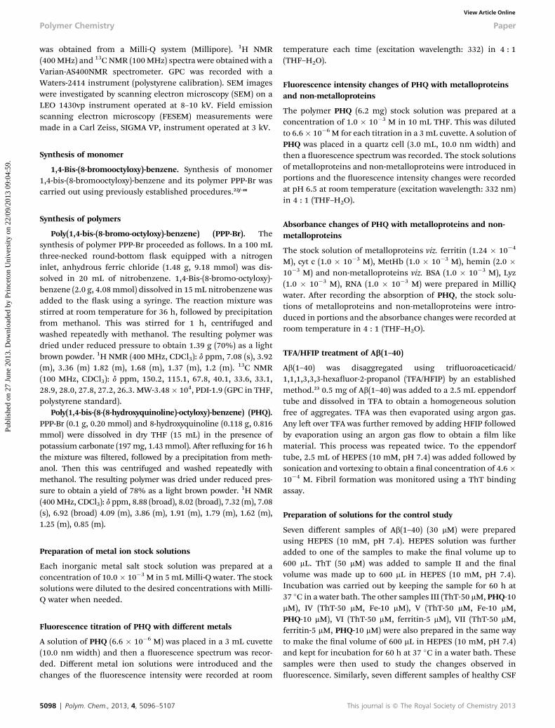
Scheme 1 Synthesis of polymer PHQ using oxidative polymerization and postpolymerization functionalization of 8-hydroxyquinoline. (a) Dibromooctane,K2CO3, dry acetone, reflux, 16 h. (b) Anhydrous FeCl3, nitrobenzene, N2 atmo-sphere, RT, 36 h. (c) 8-Hydroxyquinoline, K2CO3, dry DMF, reflux 16 h.
FT-IR spectroscopy
Room temperature FT-IR spectra were recorded by preparingpellets with KBr. The sample was prepared by spreading 2 mL ofCSF solution in the absence and presence of PHQ (1 � 10�5 M)on a glass slide and by drying under a gentle nitrogen ow.
This journal is ª The Royal Society of Chemistry 2013
Results and discussion
We report here the development of poly(1,4-bis-(8-(8-hydroxy-quinoline)-octyloxy)-benzene) (PHQ) (Scheme 1), a conjugateduorescent polymer that efficiently binds metals like Fe2+, Fe3+
and non-heme metalloprotein ferritin and heme proteins, suchas cyt c, methemoglobin and hemin, in physiological conditionsand competitive biological environments. Notably, several CPsof the type PHQ have been monitored over a long duration inin vitro studies and have been conrmed to be non toxic andable to easily penetrate the cell membrane24 (Fig. S14 of theESI†). Additionally, the highly lipophilic 8-hydroxyquinoline(8-HQ) moiety attached as a pendant to PHQ, which also readilypenetrates the cell membranes and the blood brain barrier, is awell-known non toxic compound that displays broad spectrumactivity. For these reasons, it has been used extensively as anantiseptic medicine with well known antifungal, antibacterial,antihelminthic, and antimicrobial action, and also mostimportantly, due to its property as a metal chelator25 has beenutilized in this study to bind neurotoxic metals. Due to theireasy synthesis and structural tunability, non toxic nature, cellpermeability and the wide ranging biological activity of CPs theyhave been utilized efficiently as biomarkers to study geneticalterations and proteomics.24b,26 The backbones of CPs assistelectron delocalization and exciton migration, resulting inamplied signals27 in the presence of analyte that modies thephotophysical characteristics of CP which is utilized to studytheir interaction with biological molecules or analytes ofinterest.
In this manuscript, we examined initially the interaction ofPHQ with different iron containing metalloproteins that areinvolved in neurological disorders responsible for metalhomeostasis in AD, PD and HD. The presence of 8-hydroxy-quinoline chelating groups in the polymer PHQ allows it to bindmetal ions and the p-conjugated backbone facilitates in
Polym. Chem., 2013, 4, 5096–5107 | 5099
Polymer Chemistry Paper
Publ
ishe
d on
27
June
201
3. D
ownl
oade
d by
Pri
ncet
on U
nive
rsity
on
22/0
9/20
13 0
9:04
:59.
View Article Online
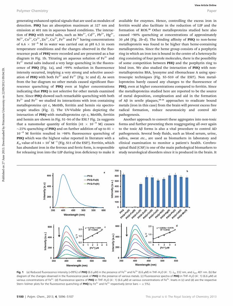
generating enhanced optical signals that are used as modules ofdetection. PHQ has an absorption maximum at 327 nm andemission at 401 nm in aqueous based conditions. The interac-tion of PHQ with metal salts, such as Mn2+, Cd2+, Pb2+, Hg2+,Ni2+, Co2+, Cr3+, Zn2+, Cu2+, Fe2+ and Fe3+ having concentrationsof 6.6 � 10�6 M in water was carried out at pH 6.5 in roomtemperature conditions and the changes observed in the uo-rescence peak of PHQ were recorded and are presented as a bardiagram in Fig. 1b. Titrating an aqueous solution of Fe2+ andFe3+ metal salts induced a very large quenching in the uores-cence of PHQ (Fig. 1a), and >99% reduction in uorescenceintensity occurred, implying a very strong and selective associ-ation of PHQ with both Fe2+ and Fe3+ (Fig. 1c and d). As seenfrom the bar diagram no other metals caused signicant uo-rescence quenching of PHQ even at higher concentrationsindicating that PHQ is not selective for other metals examinedhere. Since PHQ showed such remarkable quenching with bothFe2+ and Fe3+ we studied its interactions with iron containingmetalloproteins cyt c, MetHb, ferritin and hemin via spectro-scopic studies (Fig. 2). The UV-Visible plots depicting theinteraction of PHQ with metalloproteins cyt c, MetHb, ferritinand hemin are shown in Fig. S1–S4 of the ESI.† Fig. 2a suggeststhat a nanomolar quantity of ferritin (41 � 10�9 M) causes�25% quenching of PHQ and on further addition of up to 81 �10�8 M ferritin resulted in >98% uorescence quenching ofPHQ which was the highest value reported in literature with aKsv value of 0.84� 107 M�1 (Fig. S11 of the ESI†). Ferritin, whichhas abundant iron in the ferrous and ferric form, is responsiblefor releasing iron into the LIP during iron deciency to make it
Fig. 1 (a) Reduced fluorescence intensity (>99%) of PHQ (6.6 mM) in the presencediagram of the changes observed in the fluorescence peak of PHQ in the presencevarious concentrations of Fe2+. (d) Fluorescence spectra of PHQ in THF–H2O (4 : 1)Stern–Volmer plots for the fluorescence quenching of PHQ by Fe2+ and Fe3+ respec
5100 | Polym. Chem., 2013, 4, 5096–5107
available for enzymes. Hence, controlling the excess iron inferritin would also facilitate in the reduction of LIP and theformation of ROS.28 Other metalloproteins studied here alsocaused >90% quenching at concentrations of approximately30 mM (Fig. 2b–d). The binding affinity of PHQ to non-hememetalloprotein was found to be higher than heme-containingmetalloproteins. Since the heme group consists of a porphyrinring in which an iron ion is bound in the centre of a heterocyclicring consisting of four pyrrole molecules, there is the possibilityof some competition between PHQ and the porphyrin ring tobind iron. We also studied the interaction of PHQ with non-metalloproteins BSA, lysozyme and ribonuclease A using spec-troscopic techniques (Fig. S5–S10 of the ESI†). Non metal-loproteins barely caused any changes to the uorescence ofPHQ, even at higher concentrations compared to ferritin. Sincethe metalloproteins studied here are reported to be the sourceof metal deposition, complexation and aid in the formationof Ab in senile plaques,19–21 approaches to eradicate boundmetals (iron in this case) from the brain will prevent excess freeradical formation, reduce neurotoxicity and control ADpathogenesis.
Another approach to convert these aggregates into non-toxicforms and further preventing them reaggregating all over againto the toxic Ab forms is also a vital procedure to control ADpathogenesis. Several body uids, such as blood serum, urine,saliva, sweat etc., are used as biomarkers in laboratory andclinical examination to monitor a patient's health. Cerebro-spinal uid (CSF) is one of the main pathological biomarkers tostudy neurological disorders since it is produced in the brain. It
of Fe2+ and Fe3+ (6.6 mM) in THF–H2O (4 : 1). lex 332 nm, and lem 401 nm. (b) Barof various metals. (c) Fluorescence spectra of PHQ in THF–H2O (4 : 1) (6.6 mM) at(6.6 mM) at various concentrations of Fe3+. Insets in (c) and (d) are the respectivetively (error bars ¼ � 5%).
This journal is ª The Royal Society of Chemistry 2013
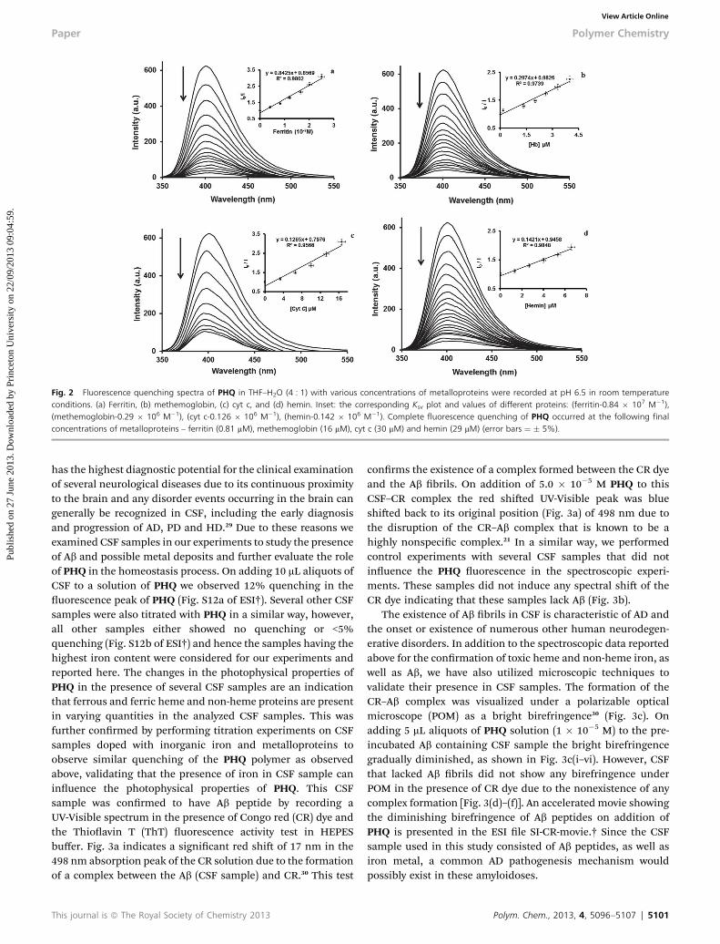
Fig. 2 Fluorescence quenching spectra of PHQ in THF–H2O (4 : 1) with various concentrations of metalloproteins were recorded at pH 6.5 in room temperatureconditions. (a) Ferritin, (b) methemoglobin, (c) cyt c, and (d) hemin. Inset: the corresponding Ksv plot and values of different proteins: (ferritin-0.84 � 107 M�1),(methemoglobin-0.29 � 106 M�1), (cyt c-0.126 � 106 M�1), (hemin-0.142 � 106 M�1). Complete fluorescence quenching of PHQ occurred at the following finalconcentrations of metalloproteins – ferritin (0.81 mM), methemoglobin (16 mM), cyt c (30 mM) and hemin (29 mM) (error bars ¼ � 5%).
Paper Polymer Chemistry
Publ
ishe
d on
27
June
201
3. D
ownl
oade
d by
Pri
ncet
on U
nive
rsity
on
22/0
9/20
13 0
9:04
:59.
View Article Online
has the highest diagnostic potential for the clinical examinationof several neurological diseases due to its continuous proximityto the brain and any disorder events occurring in the brain cangenerally be recognized in CSF, including the early diagnosisand progression of AD, PD and HD.29 Due to these reasons weexamined CSF samples in our experiments to study the presenceof Ab and possible metal deposits and further evaluate the roleof PHQ in the homeostasis process. On adding 10 mL aliquots ofCSF to a solution of PHQ we observed 12% quenching in theuorescence peak of PHQ (Fig. S12a of ESI†). Several other CSFsamples were also titrated with PHQ in a similar way, however,all other samples either showed no quenching or <5%quenching (Fig. S12b of ESI†) and hence the samples having thehighest iron content were considered for our experiments andreported here. The changes in the photophysical properties ofPHQ in the presence of several CSF samples are an indicationthat ferrous and ferric heme and non-heme proteins are presentin varying quantities in the analyzed CSF samples. This wasfurther conrmed by performing titration experiments on CSFsamples doped with inorganic iron and metalloproteins toobserve similar quenching of the PHQ polymer as observedabove, validating that the presence of iron in CSF sample caninuence the photophysical properties of PHQ. This CSFsample was conrmed to have Ab peptide by recording aUV-Visible spectrum in the presence of Congo red (CR) dye andthe Thioavin T (ThT) uorescence activity test in HEPESbuffer. Fig. 3a indicates a signicant red shi of 17 nm in the498 nm absorption peak of the CR solution due to the formationof a complex between the Ab (CSF sample) and CR.30 This test
This journal is ª The Royal Society of Chemistry 2013
conrms the existence of a complex formed between the CR dyeand the Ab brils. On addition of 5.0 � 10�5 M PHQ to thisCSF–CR complex the red shied UV-Visible peak was blueshied back to its original position (Fig. 3a) of 498 nm due tothe disruption of the CR–Ab complex that is known to be ahighly nonspecic complex.21 In a similar way, we performedcontrol experiments with several CSF samples that did notinuence the PHQ uorescence in the spectroscopic experi-ments. These samples did not induce any spectral shi of theCR dye indicating that these samples lack Ab (Fig. 3b).
The existence of Ab brils in CSF is characteristic of AD andthe onset or existence of numerous other human neurodegen-erative disorders. In addition to the spectroscopic data reportedabove for the conrmation of toxic heme and non-heme iron, aswell as Ab, we have also utilized microscopic techniques tovalidate their presence in CSF samples. The formation of theCR–Ab complex was visualized under a polarizable opticalmicroscope (POM) as a bright birefringence30 (Fig. 3c). Onadding 5 mL aliquots of PHQ solution (1 � 10�5 M) to the pre-incubated Ab containing CSF sample the bright birefringencegradually diminished, as shown in Fig. 3c(i–vi). However, CSFthat lacked Ab brils did not show any birefringence underPOM in the presence of CR dye due to the nonexistence of anycomplex formation [Fig. 3(d)–(f)]. An accelerated movie showingthe diminishing birefringence of Ab peptides on addition ofPHQ is presented in the ESI le SI-CR-movie.† Since the CSFsample used in this study consisted of Ab peptides, as well asiron metal, a common AD pathogenesis mechanism wouldpossibly exist in these amyloidoses.
Polym. Chem., 2013, 4, 5096–5107 | 5101

Fig. 3 (a) Ab aggregation was characterised by red shifting in the congo red (CR) absorption maximum from 498 nm to 515 nm and blue shifting back to 498 nm onaddition of PHQ (5.0� 10-5 M). (b) No shifting upon the addition of CSF in CR indicates a lack of Ab. (c) Birefringence was diminished upon an increasing conc. of PHQ inpre-incubated Ab containing CSF from (i–vi). (d–f) No birefringence was seen in CSF that lacked Ab.
Polymer Chemistry Paper
Publ
ishe
d on
27
June
201
3. D
ownl
oade
d by
Pri
ncet
on U
nive
rsity
on
22/0
9/20
13 0
9:04
:59.
View Article Online
From the spectroscopic experiments we conrmed that PHQcan bind with Fe2+ and Fe3+ forms, as well as heme iron, incompetitive biological environments, therefore, any ironpresent in the CSF sample examined here would bind with PHQ.
Fig. 4 (a) Fluorescence spectra (lex 440 nm, lem 488 nm) of ThT (5 mM) (pH 7.4 in HThT dye does not show any emission at 488 nm in the absence of Ab (dotted line), w488 nm (lex 440 nm) and the emission of ThT–Ab in the CSF complex is quenched upcontaining CSF with ThT and (ii–vi) disappearance of the bright green fluorescence
5102 | Polym. Chem., 2013, 4, 5096–5107
Consequently, selective binding of the toxic metal and breakingthe aggregated Ab peptides, as conrmed from the disruption ofCR–Ab complex by a non toxic compound, such as PHQ used inour experiments would facilitate the controlling of the AD
EPES) with gradual addition of pre-incubated Ab containing CSF (150 mL). (b) Thehereas the ThT–Ab in the CSF complex showed an emission peak (solid line) at lemon adding PHQ (broken line). (c)-(i) Bright green fluorescence of pre-incubated Abcolor and disruption of the Ab fibrils in CSF occurs after adding PHQ (5 mL).
This journal is ª The Royal Society of Chemistry 2013
Paper Polymer Chemistry
Publ
ishe
d on
27
June
201
3. D
ownl
oade
d by
Pri
ncet
on U
nive
rsity
on
22/0
9/20
13 0
9:04
:59.
View Article Online
pathogenesis. The presence of Ab protobrils was furtherconrmed by the addition of up to 150 mL of the CSF sample to a5 mM solution of ThT (pH 7.4 in HEPES) to observe a gradualenhancement in the uorescence intensity of ThT at 488 nmvalidating strongly the presence of Ab in the CSF sample31
(Fig. 4a).In Fig. 4b, the ThT does not show any emission at 488 nm in
the absence of Ab containing peptide sample (dotted line),whereas on addition of the CSF sample having Ab to the cuvettehaving ThTwe observe an emission peak at 488 nm (lex – 440 nm)(Fig. 4a and the solid line of Fig. 4b) indicating the formation of acomplex between the ThT–Ab as discussed above. On addition of10 mM PHQ to this cuvette the 488 nm peak was completelyquenched (broken line) indicating that the ThT–Ab complex isdisrupted in the presence of PHQ and the aggregated brils arebeing inhibited in the presence of the polymer PHQ. Further, toconrm the formation of ThT–Ab complex and its disruptionby PHQ we performed similar experiments as that done forthe birefringence study, where, a glass slide containing
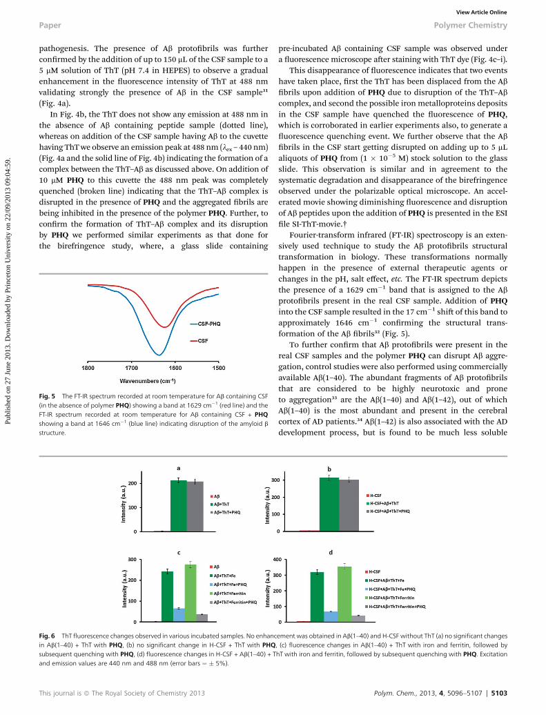
Fig. 5 The FT-IR spectrum recorded at room temperature for Ab containing CSF(in the absence of polymer PHQ) showing a band at 1629 cm�1 (red line) and theFT-IR spectrum recorded at room temperature for Ab containing CSF + PHQshowing a band at 1646 cm�1 (blue line) indicating disruption of the amyloid b
structure.
Fig. 6 ThT fluorescence changes observed in various incubated samples. No enhancin Ab(1–40) + ThT with PHQ, (b) no significant change in H-CSF + ThT with PHQsubsequent quenching with PHQ, (d) fluorescence changes in H-CSF + Ab(1–40) + Tand emission values are 440 nm and 488 nm (error bars ¼ � 5%).
This journal is ª The Royal Society of Chemistry 2013
pre-incubated Ab containing CSF sample was observed undera uorescence microscope aer staining with ThT dye (Fig. 4c–i).
This disappearance of uorescence indicates that two eventshave taken place, rst the ThT has been displaced from the Abbrils upon addition of PHQ due to disruption of the ThT–Abcomplex, and second the possible iron metalloproteins depositsin the CSF sample have quenched the uorescence of PHQ,which is corroborated in earlier experiments also, to generate auorescence quenching event. We further observe that the Abbrils in the CSF start getting disrupted on adding up to 5 mLaliquots of PHQ from (1 � 10�5 M) stock solution to the glassslide. This observation is similar and in agreement to thesystematic degradation and disappearance of the birefringenceobserved under the polarizable optical microscope. An accel-erated movie showing diminishing uorescence and disruptionof Ab peptides upon the addition of PHQ is presented in the ESIle SI-ThT-movie.†
Fourier-transform infrared (FT-IR) spectroscopy is an exten-sively used technique to study the Ab protobrils structuraltransformation in biology. These transformations normallyhappen in the presence of external therapeutic agents orchanges in the pH, salt effect, etc. The FT-IR spectrum depictsthe presence of a 1629 cm�1 band that is assigned to the Abprotobrils present in the real CSF sample. Addition of PHQinto the CSF sample resulted in the 17 cm�1 shi of this band toapproximately 1646 cm�1 conrming the structural trans-formation of the Ab brils32 (Fig. 5).
To further conrm that Ab protobrils were present in thereal CSF samples and the polymer PHQ can disrupt Ab aggre-gation, control studies were also performed using commerciallyavailable Ab(1–40). The abundant fragments of Ab protobrilsthat are considered to be highly neurotoxic and proneto aggregation33 are the Ab(1–40) and Ab(1–42), out of whichAb(1–40) is the most abundant and present in the cerebralcortex of AD patients.34 Ab(1–42) is also associated with the ADdevelopment process, but is found to be much less soluble
ement was obtained in Ab(1–40) and H-CSF without ThT (a) no significant changes, (c) fluorescence changes in Ab(1–40) + ThT with iron and ferritin, followed byhT with iron and ferritin, followed by subsequent quenching with PHQ. Excitation
Polym. Chem., 2013, 4, 5096–5107 | 5103
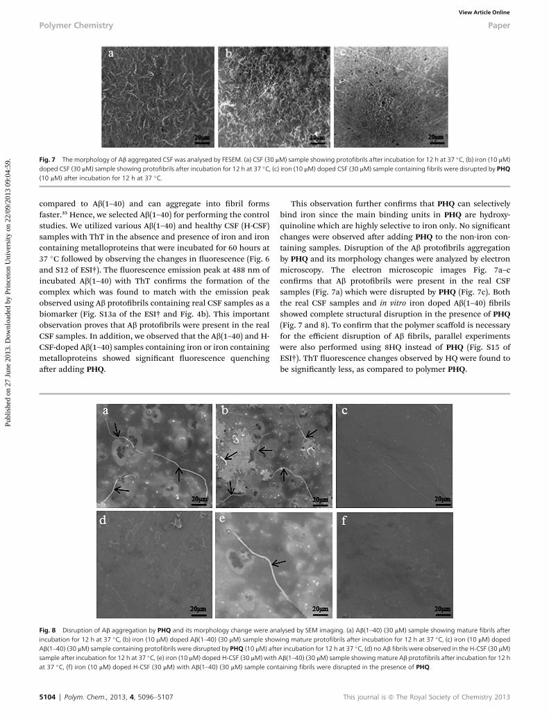
Fig. 7 The morphology of Ab aggregated CSF was analysed by FESEM. (a) CSF (30 mM) sample showing protofibrils after incubation for 12 h at 37 �C, (b) iron (10 mM)doped CSF (30 mM) sample showing protofibrils after incubation for 12 h at 37 �C, (c) iron (10 mM) doped CSF (30 mM) sample containing fibrils were disrupted by PHQ(10 mM) after incubation for 12 h at 37 �C.
Polymer Chemistry Paper
Publ
ishe
d on
27
June
201
3. D
ownl
oade
d by
Pri
ncet
on U
nive
rsity
on
22/0
9/20
13 0
9:04
:59.
View Article Online
compared to Ab(1–40) and can aggregate into bril formsfaster.35 Hence, we selected Ab(1–40) for performing the controlstudies. We utilized various Ab(1–40) and healthy CSF (H-CSF)samples with ThT in the absence and presence of iron and ironcontaining metalloproteins that were incubated for 60 hours at37 �C followed by observing the changes in uorescence (Fig. 6and S12 of ESI†). The uorescence emission peak at 488 nm ofincubated Ab(1–40) with ThT conrms the formation of thecomplex which was found to match with the emission peakobserved using Ab protobrils containing real CSF samples as abiomarker (Fig. S13a of the ESI† and Fig. 4b). This importantobservation proves that Ab protobrils were present in the realCSF samples. In addition, we observed that the Ab(1–40) and H-CSF-doped Ab(1–40) samples containing iron or iron containingmetalloproteins showed signicant uorescence quenchingaer adding PHQ.
Fig. 8 Disruption of Ab aggregation by PHQ and its morphology change were anincubation for 12 h at 37 �C, (b) iron (10 mM) doped Ab(1–40) (30 mM) sample showAb(1–40) (30 mM) sample containing protofibrils were disrupted by PHQ (10 mM) aftesample after incubation for 12 h at 37 �C, (e) iron (10 mM) doped H-CSF (30 mM)withat 37 �C, (f) iron (10 mM) doped H-CSF (30 mM) with Ab(1–40) (30 mM) sample con
5104 | Polym. Chem., 2013, 4, 5096–5107
This observation further conrms that PHQ can selectivelybind iron since the main binding units in PHQ are hydroxy-quinoline which are highly selective to iron only. No signicantchanges were observed aer adding PHQ to the non-iron con-taining samples. Disruption of the Ab protobrils aggregationby PHQ and its morphology changes were analyzed by electronmicroscopy. The electron microscopic images Fig. 7a–cconrms that Ab protobrils were present in the real CSFsamples (Fig. 7a) which were disrupted by PHQ (Fig. 7c). Boththe real CSF samples and in vitro iron doped Ab(1–40) brilsshowed complete structural disruption in the presence of PHQ(Fig. 7 and 8). To conrm that the polymer scaffold is necessaryfor the efficient disruption of Ab brils, parallel experimentswere also performed using 8HQ instead of PHQ (Fig. S15 ofESI†). ThT uorescence changes observed by HQ were found tobe signicantly less, as compared to polymer PHQ.
alysed by SEM imaging. (a) Ab(1–40) (30 mM) sample showing mature fibrils aftering mature protofibrils after incubation for 12 h at 37 �C, (c) iron (10 mM) dopedr incubation for 12 h at 37 �C, (d) no Ab fibrils were observed in the H-CSF (30 mM)Ab(1–40) (30 mM) sample showingmature Ab protofibrils after incubation for 12 htaining fibrils were disrupted in the presence of PHQ.
This journal is ª The Royal Society of Chemistry 2013
Paper Polymer Chemistry
Publ
ishe
d on
27
June
201
3. D
ownl
oade
d by
Pri
ncet
on U
nive
rsity
on
22/0
9/20
13 0
9:04
:59.
View Article Online
The pathogenesis of neurodegenerative disorders such as ADlargely occurs due to the LIP, or in other words the unwantedaccumulation and incorrect metabolism of heme and othertransition metals in the brain. In addition, due to the highaffinity of Ab peptides for iron and copper metals, their accu-mulation and increased concentration is observed in the brainplaques due to which the ROS and generation of neurotoxichydrogen peroxide are accelerated causing additional stress tothe brain. Hence, the regulation of redox-active metals presentin the brain would help in controlling the neurotoxicity andprevent damage to the brain.20 Since ferritin is responsible forsupplying iron to the LIP, the formation of ROS in biologicalsystems can be controlled by its binding and removal.16,28
Because the polymer PHQ binds heme and non-heme ironexisting in ferrous and ferric forms with very high affinity anddiminishes the Ab peptides in the CSF sample, it can signi-cantly reduce formation of neurotoxic chemicals and radicals inthe brain and disrupt the toxic polypeptides and prevent themfrom aggregating again. This strategy of removing toxic metalsand diminishing the Ab aggregates by the PHQ polymer is veryimportant and a conceptually novel strategy for the clearance ofthe cerebral deposits and control the AD pathogenesis.
Conclusions
In summary, conjugated polymer poly(1,4-bis-(8-(8-hydrox-yquinoline)-octyloxy)-benzene) (PHQ) has a unique ability tobind inorganic Fe3+ and Fe2+, heme and non-heme proteinsselectively in a competitive biological environment. Hence, PHQhas been utilized to interact with the bound iron, includingnon-heme ferritin at a nanomolar level, in the presence of Abpeptide aggregates and diminish the accumulated Ab brils. Bybinding the labile iron present in the brain, the regulation ofredox-activity is possible, thereby controlling the neurotoxicityand preventing damage to the brain. Since ferritin is respon-sible for supplying iron to the LIP, the formation of ROS inphysiological systems can be controlled by its efficient bindingand removal by PHQ. The evaluation of PHQ activity towardstoxic metal binding was performed in cerebrospinal uid (CSF)since it is a very important biomarker of AD–Ab due to itscontinuous presence and contact with the brain. Since the CSFsample studied in our experiments had both ferrous and ferricproteins along with the aggregated Ab protobrils, the patho-genesis mechanism existing in these kinds of amyloidoses isstructurally interdependent. Hence, therapeutic strategiesinvolving clearance of either the Ab accumulated in the plaquesof brain tissues or removal of metals to reduce neurotoxicity andstructurally modifying the aggregates into a non-toxic form toprevent them aggregating again are vital strategies to control ADpathogenesis. Control studies using a Thioavin-T bindingassay shows that the polymer PHQ can effectively chelate ironfrom Ab(1–40) and H-CSF-doped Ab(1–40) due to the presence ofhydroxyquinoline as a highly selective metal binding groupfor iron. Electron microscopy analysis conrms the disruptionof Ab brils by PHQ in commercial Ab(1–40), H-CSF-dopedAb(1–40) and real CSF samples. This conceptually new strategyto clear the cerebral deposits using a conjugated polymer allows
This journal is ª The Royal Society of Chemistry 2013
us to conclusively proclaim that the aggregated Ab peptidebrils present in the CSF could be successfully disrupted,modied into non toxic forms and prevented from aggregatingagain under physiological conditions. This biocompatible nontoxic conjugated polymer system also promises a valuablestrategy and novel way for developing AD therapy and may serveas an important macromolecule in improving our under-standing of the biology of the aggregated Ab peptide brils andmethods to reduce the neurotoxicity.
Acknowledgements
Financial assistance from the Department of Science andTechnology (DST), New Delhi (no. SR/S1/PC-02/2009), (no. DST/TSG/PT/2009/11) and DST-Max Planck Society, Germany (no.INT/FRG/MPG/FS/2008) is gratefully acknowledged. We thankDr Vishal Trivedi for toxicity analysis, Dr Bhubaneswar Mandalfor providing commercially available Ab(1–40) and Dr RakhiChaturvedi for uorescence microscopic studies. Authors alsothank Guwahati Neurological Research Center and Hospital,Guwahati for the gi of fresh Cerebrospinal uid samples.
Notes and references
1 L. M. Sayre, G. Perry andM. A. Smith, Curr. Opin. Chem. Biol.,1999, 3, 220.
2 D. M. Holtzman, J. C. Morris and A. M. Goate, Sci. Transl.Med., 2011, 3, 77.
3 (a) K. Blennow, M. J. De Leon and H. Zetterberg, Lancet, 2006,368, 387; (b) M. P. Mattson, Nature, 2004, 430, 631.
4 (a) C. L. Ni, H. P. Shi, H. M. Yu, Y. C. Chang and Y. R. Chen,FASEB J., 2011, 25, 1390; (b) C. Opazo, X. Huang,R. A. Cherny, R. D. Moir, A. E. Roher, A. R. White,R. Cappai, C. L. Masters, R. E. Tanzi, N. C. Inestrosa andA. I. Bush, J. Biol. Chem., 2002, 277, 40302; (c)G. D. Ciccotosto, D. Tew, C. C. Curtain, D. Smith,D. Carrington, C. L. Masters, A. I. Bush, R. A. Cherny,R. Cappai and K. J. Barnham, J. Biol. Chem., 2004, 279,42528; (d) C. Boldron, I. V. Auwera, C. Deraeve,H. Gornitzka, S. Wera, M. Pitie, F. V. Leuven andB. Meunier, ChemBioChem, 2005, 6, 1976; (e) R. A. Cherny,J. T. Legg, C. A. McLean, D. P. Fairlie, X. Huang,C. S. Atwood, K. Beyreuther, R. E. Tanzi, C. L. Masters andA. I. Bush, J. Biol. Chem., 1999, 274, 23223; (f) J. Y. Lee,J. E. Friedman, I. Angel, A. Kozak and J. Y. Koh, Neurobiol.Aging, 2004, 25, 1315.
5 C. J. Pike, A. J. Walencewicz, C. G. Glabe and C. W. Cotman,Brain Res., 1991, 563, 311.
6 (a) A. Lorenzo and B. A. Yankner, Proc. Natl. Acad. Sci. U. S. A.,1994, 91, 12243; (b) C. Deraeve, M. Pitie, H. Mazarguilb andB. Meunierz, New J. Chem., 2007, 31, 193.
7 (a) D. M. Hartley, D. M. Walsh, C. P. Ye, T. Diehl, S. Vasquez,P. M. Vassilev, D. B. Teplow and D. J. Selkoe, J. Neurosci.,1999, 19, 8876; (b) M. P. Lambert, A. K. Barlow,B. A. Chromy, C. Edwards, R. Freed, M. Liosatos,T. E. Morgan, I. Rozovsky, B. Trommer, K. L. Viola,P. Wals, C. Zhang, C. E. Finch, G. A. Kra and W. L. Klein,
Polym. Chem., 2013, 4, 5096–5107 | 5105
Polymer Chemistry Paper
Publ
ishe
d on
27
June
201
3. D
ownl
oade
d by
Pri
ncet
on U
nive
rsity
on
22/0
9/20
13 0
9:04
:59.
View Article Online
Proc. Natl. Acad. Sci. U. S. A., 1998, 95, 6448; (c) M. Townsend,G. M. Shankar, T. Mehta, D. M. Walsh and D. J. Selkoe, J.Physiol., 2006, 572, 477; (d) D. M. Walsh and D. J. Selkoe, J.Neurochem., 2007, 101, 1172; (e) W. L. Klein, G. A. Kraand C. E. Finch, Trends Neurosci., 2001, 24, 219; (f)S. Kumar and J. Walter, Aging, 2011, 3, 803.
8 H. M. Schipper, S. Cisse and E. G. Stopa, Ann. Neurol., 1995,37, 758.
9 K. P. Kepp, Chem. Rev., 2012, 112, 5193.10 (a) C. Hureau, I. Sasaki, E. Gras and P. Faller, ChemBioChem,
2010, 11, 950; (b) C. Rodriguez, M. Telpoukhovskaia andC. Orvig, Coord. Chem. Rev., 2012, 256, 2308.
11 (a) J. R. Donnor, in Metals and oxidative damage inneurological disorders, Plenum Press, New York, 1997; (b)Y. Christen, Am. J. Clin. Nutr., 2000, 71, 621S.
12 (a) L. E. Scott and C. Orvig, Chem. Rev., 2009, 109, 4885; (b)R. A. Hansen, G. Gartlehner, A. P. Webb, L. C. Morgan,C. G. Moore and D. E. Jonas, Clin. Interventions Aging,2008, 3, 211.
13 (a) M. C. Morris, Eur. J. Neurol., 2009, 16, 1; (b) S. Deweerdt,Nature, 2011, 475, S16.
14 K. Jomova, D. Vondrakova, M. Lawson and M. Valko, Mol.Cell. Biochem., 2010, 345, 91.
15 (a) A. J. L. Cooper, in The molecular and genetic basis ofneurological disease, ed. R. N. Rosenberg, S. B. Prusiner, S.DiMauro, R. L. Barch and L. M. Klunk, Butterworth-Heinemann, Boston, 1997, pp. 1242–1245; (b) G. Benzi andA. Moretti, Neurobiol. Aging, 1995, 16, 661; (c)B. J. Halliwell, J. Neurochem., 1992, 59, 1609; (d)B. Hallgren and P. J. Sourander, J. Neurochem., 1958, 3, 41;(e) P. Arosio and S. Levi, Free Radical Biol. Med., 2002, 33,457; (f) R. K. Watt, ChemBioChem, 2013, 14, 415.
16 (a) K. Yagi, N. Ishida, S. Komura, N. Ohishi, M. Kusai andM. Kohno, Biochem. Biophys. Res. Commun., 1992, 183, 945;(b) D. B. Kell, Arch. Toxicol., 2010, 84, 825; (c) D. B. Kell,BMC Medical Genomics, 2009, 2, 2.
17 (a) J. Weaver and S. Pollack, Biochem. J., 1989, 261, 787; (b)O. Kakhlon and Z. I. Cabantchik, Free Radical Biol. Med.,2002, 33, 1037.
18 B. Halliwell and J. M. C. Gutteridge, Biochem. J., 1984, 219, 1.19 (a) L. M. Sayre, D. A. Zelasko, P. L. Harris, G. Perry,
R. G. Salomon and M. A. Smith, J. Neurochem., 1997, 68,2092; (b) M. A. Smith, P. L. Harris, L. M. Sayre andG. Perry, Proc. Natl. Acad. Sci. U. S. A., 1997, 94, 9866; (c)M. A. Smith and G. Perry, J. Neurol. Sci., 1995, 134, 92.
20 (a) K. J. Thompson, S. Shoham and J. R. Connor, Brain Res.Bull., 2001, 55, 155; (b) S. Varadarajan, S. Yatin,M. B. Aksenova and D. A. Buttereld, J. Struct. Biol., 2000,130, 184; (c) Y. Christen, Am. J. Clin. Nutr., 2000, 71, 621S.
21 (a) M. P. Cuajungco, K. Y. Faget, X. Huang, R. E. Tanzi,A. I. Bush, R. E. Tanzi and A. I. Bush, Ann. N. Y. Acad. Sci.,2000, 920, 292; (b) A. I. Bush, Trends Neurosci., 2003, 26,207; (c) A. Rauk, Chem. Soc. Rev., 2009, 38, 2698; (d)R. Jakob-Roetne and H. Jacobsen, Angew. Chem., Int. Ed.,2009, 48, 3030; (e) J. J. Braymer, J.-S. Choi, A. S. DeToma,C. Wang, K. Nam, J. W. Kampf, A. Ramamoorthy andM. H. Lim, Inorg. Chem., 2011, 50, 10724; (f) C. Deraeve,
5106 | Polym. Chem., 2013, 4, 5096–5107
C. Boldron, A. Maraval, H. Mazarguil, H. Gornitzka,L. Vendier, M. Pitie and B. Meunierz, Chem.–Eur. J., 2008,14, 682; (g) G. K. Gouras and M. F. Beal, Neuron, 2001, 3, 641.
22 (a) C. Fan, K. W. Plaxco and A. J. Heeger, J. Am. Chem. Soc.,2002, 124, 5642; (b) B. S. Sandanaraj, R. Demont,S. V. Aathimanikandan, E. N. Savariar andS. Thayumanavan, J. Am. Chem. Soc., 2006, 128, 10686; (c)A. K. Dwivedi, K. M. Prasad, V. Trivedi and P. K. Iyer, ACSAppl. Mater. Interfaces, 2012, 4, 6371; (d) T. Zako,M. Sakono, T. Kobayashi, K. Sorgjerd, K. P. R. Nilsson,P. Hammarstrom, M. Lindgren and M. Maeda,ChemBioChem, 2012, 13, 358; (e) J. L. Bricks, A. Kovalchuk,C. Trieinger, M. Nofz, M. Buschel, A. I. Tolmachev,J. Daub and K. Rurack, J. Am. Chem. Soc., 2005, 127, 13522;(f) C. Qin, Y. Cheng, L. Wang, X. Jing and F. Wang,Macromolecules, 2008, 41, 7798; (g) A. K. Dwivedi, G. Saikiaand P. K. Iyer, J. Mater. Chem., 2011, 21, 2502; (h) G. Saikiaand P. K. Iyer, Macromolecules, 2011, 44, 3753; (i)A. K. Dwivedi and P. K. Iyer, Anal. Methods, 2013, 5, 2374;(j) S. Hussain, S. De and P. K. Iyer, ACS Appl. Mater.Interfaces, 2013, 5, 2234; (k) G. Saikia, A. K. Dwivedi andP. K. Iyer, Anal. Methods, 2012, 4, 3180; (l) G. Saikia,R. Singh, P. J. Sarmah, M. W. Akhtar, J. Sinha, M. Katiyarand P. K. Iyer, Macromol. Chem. Phys., 2009, 210, 2153; (m)G. S. Paul, P. J. Sarmah, P. K. Iyer and P. Agarwal,Macromol. Chem. Phys., 2008, 209, 417.
23 (a) S. Chen and R. Wetzel, Protein Sci., 2001, 10, 887; (b)P. Hortschansky, V. Schroeckh, T. Christopeit,G. Zandomeneghi and M. Fandrich, Protein Sci., 2005, 14,1753.
24 (a) N. A. A. Rahim, W. McDaniel, K. Bardon, S. Srinivasan,V. Vickerman, P. T. C. So and J. H. Moon, Adv. Mater.,2009, 21, 3492; (b) C. Zhu, L. Liu, Q. Yang, F. Lv andS. Wang, Chem. Rev., 2012, 112, 4687.
25 (a) A. Y. Shen, C. P. Chen and S. A. Roffler, Life Sci., 1999, 64,813; (b) A. Gaeta and R. C. Hider, Br. J. Pharmacol., 2005, 146,1041; (c) F. Molina-Holgado, R. C. Hider, A. Gaeta,R. Williams and P. Francis, BioMetals, 2007, 20, 639.
26 (a) D. T. McQuade, A. E. Pullen and T. M. Swager, Chem. Rev.,2000, 100, 2537; (b) K. P. R. Nilsson and O. Inganas, Nat.Mater., 2003, 2, 419.
27 T. M. Swager, Acc. Chem. Res., 1998, 31, 201.28 (a) I. D. Domenico, M. B. Vaughn, L. Li, D. Bagley, G. Musci,
D. M. Ward and J. Kaplan, EMBO J., 2006, 25, 5396; (b)J. Johnson, J. Kenealey, R. J. Hilton, D. Brosnahan,R. K. Watt and G. D. Watt, J. Inorg. Biochem., 2011, 105,202; (c) R. K. Watt, R. J. Hilton and D. M. Graff, Biochim.Biophys. Acta, Gen. Subj., 2010, 1800, 745.
29 (a) K. Blennow and H. Hampel, Lancet Neurol., 2003, 2, 605;(b) K. Blennow, H. Hampel, M. Weiner and H. Zetterberg,Nat. Rev. Neurol., 2010, 6, 131; (c) S. Peter, V. Carmen,E. Fred, L. Michael, D. Harry, D. Dave, S. Sukanto,S. Michael, W. Justine, S. Cathy, M. Robert, W. Robert,S. Dennis, L. Ivan and D. Schenk, Nature, 1992, 359, 325.
30 (a) W. E. Klunk, R. F. Jacob and R. P. Mason, Anal. Biochem.,1999, 266, 66; (b) E. P. Benditt, N. Eriksen and C. Berglund,Proc. Natl. Acad. Sci. U. S. A., 1970, 66, 1044.
This journal is ª The Royal Society of Chemistry 2013
Paper Polymer Chemistry
Publ
ishe
d on
27
June
201
3. D
ownl
oade
d by
Pri
ncet
on U
nive
rsity
on
22/0
9/20
13 0
9:04
:59.
View Article Online
31 S. G. Bolder, L. M. Sagis, P. Venema and E. van der Linden,Langmuir, 2007, 23, 4144.
32 (a) Y. Cordeiro, J. Kraineva, M. C. Suarez, A. G. Tempesta,J. W. Kelly, J. L. Silva, R. Winter and D. Foguel, Biophys. J.,2006, 91, 957; (b) G. Zandomeneghi, M. R. H. Krebs,M.G.McCammonandM.Fandrich,Protein Sci., 2004,13, 3314.
33 (a) G. Bitan, M. D. Kirkitadze, A. Lomakin, S. S. Vollers,G. B. Benedek and D. B. Teplow, Proc. Natl. Acad. Sci.
This journal is ª The Royal Society of Chemistry 2013
U. S. A., 2003, 100, 330; (b) A. Rauk, Chem. Soc. Rev., 2009,38, 2698.
34 (a) H. Mori, K. Takio, M. Ogawara and D. J. Selkoe, J. Biol.Chem., 1992, 24, 17082; (b) P. Hortschansky, V. Schroeckh,T. Christopeit, G. Zandomeneghi and M. Fandrich, ProteinSci., 2005, 14, 1753.
35 B. Caughey and P. T. Lansbury, Annu. Rev. Neurosci., 26,267.
Polym. Chem., 2013, 4, 5096–5107 | 5107
Related Documents