A Quantitative Comparison of the Hominoid Thalamus I. SPECIFIC SENSORY RELAY NUCLEI ESTE ARMSTRONG Department of Anthropology, Columbia Uniuersity, New York Ccty, New York 10027 KEY WORDS Medial lateral geniculates . Ventrobasal com- plex . Morphometrics . Evolution e Thalamus ABSTRACT Studies to date have indicated few differences in sensory per- ception among hominoids. Sensory relay nuclei in the dorsal thalamus-por- tions of the medial and lateral geniculate bodies (MGBp, LGBd) and the ven- trobasal complex (VBl-in two gibbons, one gorilla, one chimpanzee and three humans were examined for anatomical similarity by measuring and estimating the nuclear volumes, neuronal densities, numbers of neurons per nucleus, and volumes of neuronal perikarya. The absolute volumes of these nuclei were larger in the larger brains; however, with the volume of the dorsal thalamus as a standard, these sensory relay nuclei showed negative allometry. The gibbons had about half as many neurons as did the other hominoids. Although the human VB had slightly more neurons, the numbers of neurons in LGBd and MGBp did not significantly differ between the great apes and humans. The volumetric distribution of the neuronal perikarya were similar among these hominoids. Other thalamic nuclei had much more diverse numbers of neurons and relative frequencies of their neuronal perikarya. The sensory relay nuclei appear to be a group of conservative nuclei in the forebrain. These results sug- gest that as a neurological base for complex behaviors evolved in hominids, not all parts of the brain changed equally. Apes and humans appear to differ only slightly in primary sensory perception (Elder, '34; Farrer and Prim, '65; Farrer and Young, '70; Grether, '41, '42; Kellogg and Kellogg, '33; Kohler, '15; Riesen, '70; Spence, '34). Con- sequently, among extant hominoids, areas of the brain directly associated with these func- tions should be histologically similar. In the thalamus, several sensory areas can be com- pared within a limited region of the brain, thus providing information of both absolute and relative differences. In this pilot study to test for structural similarity of sensory nuclei among hominoids, measurements from the medial and lateral geniculate bodies and the ventrobasal complex in two gibbons, one gorilla, one chimpanzee, and three humans were compared. The following brief summary of information on the sensory nuclei of the dorsal thalamus is primarily based on findings in primates. When primate data are lacking, data from other specified mammals are given. Medial geniculate body The medial geniculate body (MGB), in the ventral and posterior part of the thalamus (fig. 11, is subdivided into a small pars mag- nocellularis (MGBm) and a large pars par- vocellularis (MGBp). Only the MGBp, the spe- cific auditory relay nucleus of the thalamus, was examined in this study. All MGBp neurons respond to tones, but the precise role played by MGBp is not yet known. A tonotopic organization of synapses has been detected in MGBp in the squirrel monkey (Gross et al., '74). In cats, MGBp may com- plete frequency analyses that are partially ac- complished in lower nuclei of the auditory sys- 1 Present address: Department of Anatomy, L.S.U. School of Medi- cine, 1100 Florida Avenue, Building 137, New Orleans, Louisiana 70119. AM. J. PHYS. ANTHROP. (1979) 51: 365-382. 365

Welcome message from author
This document is posted to help you gain knowledge. Please leave a comment to let me know what you think about it! Share it to your friends and learn new things together.
Transcript

A Quantitative Comparison of the Hominoid Thalamus I. SPECIFIC SENSORY RELAY NUCLEI
ESTE ARMSTRONG Department of Anthropology, Columbia Uniuersity, New York Ccty, New York 10027
KEY WORDS Medial lateral geniculates . Ventrobasal com- plex . Morphometrics . Evolution e Thalamus
ABSTRACT Studies to date have indicated few differences in sensory per- ception among hominoids. Sensory relay nuclei in the dorsal thalamus-por- tions of the medial and lateral geniculate bodies (MGBp, LGBd) and the ven- trobasal complex (VBl-in two gibbons, one gorilla, one chimpanzee and three humans were examined for anatomical similarity by measuring and estimating the nuclear volumes, neuronal densities, numbers of neurons per nucleus, and volumes of neuronal perikarya. The absolute volumes of these nuclei were larger in the larger brains; however, with the volume of the dorsal thalamus as a standard, these sensory relay nuclei showed negative allometry. The gibbons had about half as many neurons as did the other hominoids. Although the human VB had slightly more neurons, the numbers of neurons in LGBd and MGBp did not significantly differ between the great apes and humans. The volumetric distribution of the neuronal perikarya were similar among these hominoids. Other thalamic nuclei had much more diverse numbers of neurons and relative frequencies of their neuronal perikarya. The sensory relay nuclei appear to be a group of conservative nuclei in the forebrain. These results sug- gest that as a neurological base for complex behaviors evolved in hominids, not all parts of the brain changed equally.
Apes and humans appear to differ only slightly in primary sensory perception (Elder, '34; Farrer and Prim, '65; Farrer and Young, '70; Grether, '41, '42; Kellogg and Kellogg, '33; Kohler, '15; Riesen, '70; Spence, '34). Con- sequently, among extant hominoids, areas of the brain directly associated with these func- tions should be histologically similar. In the thalamus, several sensory areas can be com- pared within a limited region of the brain, thus providing information of both absolute and relative differences. In this pilot study to test for structural similarity of sensory nuclei among hominoids, measurements from the medial and lateral geniculate bodies and the ventrobasal complex in two gibbons, one gorilla, one chimpanzee, and three humans were compared.
The following brief summary of information on the sensory nuclei of the dorsal thalamus is primarily based on findings in primates. When
primate data are lacking, data from other specified mammals are given.
Medial geniculate body The medial geniculate body (MGB), in the
ventral and posterior part of the thalamus (fig. 11, is subdivided into a small pars mag- nocellularis (MGBm) and a large pars par- vocellularis (MGBp). Only the MGBp, the spe- cific auditory relay nucleus of the thalamus, was examined in this study.
All MGBp neurons respond to tones, but the precise role played by MGBp is not yet known. A tonotopic organization of synapses has been detected in MGBp in the squirrel monkey (Gross et al., '74). In cats, MGBp may com- plete frequency analyses that are partially ac- complished in lower nuclei of the auditory sys-
1 Present address: Department of Anatomy, L.S.U. School of Medi- cine, 1100 Florida Avenue, Building 137, New Orleans, Louisiana 70119.
AM. J. PHYS. ANTHROP. (1979) 51: 365-382. 365
366 ESTE ARMSTRONG
tem (Katsuki, '66). It may also be involved with discriminating sound patterns and localizing sound in space (Neff et al., '72). Neurons responsive to patterns of sinusoidal waves, to changes in stimulus frequencies, and to rising and falling frequencies are located here (Keidel, '74). Keidel hypothesized that those three electrophysiological patterns correspond to vowels, consonants, and tran- sient detectors respectively.
Lateral geniculate body The lateral geniculate body (LGB) is com-
posed of two components, LGB ventralis and LGB dorsalis. Because only the latter compo- nent was studied, LGB in this article refers to LGB dorsalis.
The LGB is surrounded by fibers and has a distinct laminar arrangement internally (figs. 1 , 2 ) . The basal two layers (numbered 1 and 21, the laminae around the hilum, are known as the pars magnocellularis (LGBm). The other layers, composed of only small cells are called pars parvocellularis (LGBp) (fig. 1). The num- ber of laminae within LGBp varies among pri- mates (Giolli and Tigges, '70; Noback and Laemle, '70). While the synapses in the parvo- and magnocellular regions are basically simi- lar (Glees, '61), LGBm may have slightly large axonal arborizations (Tigges and O'Steen, '74).
The LGB is the specific visual relay nucleus of the thalamus. First-order projections from the retinal ganglion cells synapse in LGB. The visual projections are visuotopic (Le Gros Clark, '32).
The LGB modifies some of the retinal input, but its precise functional roles are not known. Each LGB neuron appears to enhance con- tours (De Valois and Pease, '71), respond to the movement of light in all directions (Camp- bell, '74), and respond either to all wave- lengths (white light) or differentially to re- stricted wavelengths (color) (Weisel and Hubel, '66). Activity of LGB may frame or punctuate visual input and increase the ca- pacity to transmit patterns to the cortex (McIlwain, '72; Doty et al., '73). Synapses a t the LGB level may provide for a constancy of depth perception during disjunctive eye move- ments (Richards, '68).
Ventrobasal complex
The ventrobasal complex (VB) has two nu- clei, the ventroposterior lateralis (VPL) and medialis (VPM). The rostra1 region of VB is
sometimes segregated as the nucleus ven- trointermedius (VI) because i t is composed solely of large cells, (Hassler, '59; Van Buren and Borke, '72; Kanagasuntheram and Wong, '68; Andrew and Watkins, '691, but in this study it is included within VB because occa- sional areas of large cells without medium and small cells were found not only rostrally, but also in pockets throughout lateral VB. How- ever, a small-celled nuclear region medial to VPM is distinguished from the magnocellular portion of VPM by its lack of large neurons. Although the same bundle of myelinated fibers surrounds both the large- and small- celled regions, only the portion containing large neurons was analyzed in this study. A similar division was made by others (Deka- ban, '53; Hassler, '59; Shantha and Manocha, '69; Van Buren and Borke, '72). The lightly stained, small-celled region, nucleus ventro- posterior inferior (VPI), was also not exam- ined in this study (fig. 3).
The VB relays information to the cerebral cortex, primarily from mechanoreceptors (touch, pressure) as well from thermal, kinesthetic, and possibly pain receptors.
Classically, VB is seen as a link in the mam- malian medial lemniscal system and thus is concerned with spatially localized, rapidly adapting light touch that is involved in tasks requiring discriminative touching (Welker, '731. Stimulation of VB in humans produces subjective sensations of tingling, "pins and needles," or numbness in the contralateral face and body (Mark e t al., '63; Albe-Fessard, '73). These sensations are highly localized, but nonspecific in modality (Mark and Ervin, '60). Surgical lesions in VB give mediocre, if any, relief from intractable pain and lead to unpleasant sensations, ill-defined in time and space (Fazio et al., '73; Mark et al., '63). The VB may function to gauge or control the in- teraction of information arriving along sever- al convergent somatosensory pathways rather than be involved directly in conscious sensory mechanisms (Wall and Dubner, '72).
MATERIALS AND METHODS
Specimens The thalami of hominoids were studied
quantitatively in specimens from one gorilla, one chimpanzee, two gibbons, and three humans.
The gorilla's thalamus, obtained from the Department of Anatomy at Columbia Univer- sity, was from a 14-year-old male mountain
367 HOMINOID THALAMIC SENSORY RELAY NUCLEl
gorilla (Gorilla gorillal weighing 203 kg, with a brain weight of 540 gm. The body of the ani- mal, which had drowned in the moat at the Bronx Zoo, was refrigerated overnight and the brain removed within 24 hours after the time of death. The brain was then fixed by immer- sion in 10% formalin, embedded in celloidin, and cut into coronal serial sections, 30 /*m thick. Every tenth section was stained with cresyl violet (Gurr). In another series of sec- tions, myelin sheaths were stained by hema- toxylin using the Mahon methods.
The chimpanzee brain, from the compara- tive brain collection in the Laboratory of Neu- rophysiology a t the University of Wisconsin, was from a 63.5-kg male chimpanzee (Pan troglodytes), aged 10 to 12 years. The 405.5-gm brain was perfused with saline and then with formalin, hardened in celloidin, and sectioned coronally a t 40 pm. Every fifth section was stained with thionin. A myelin-stained series was also available.
Thalami from the two hemispheres of one brain, from a gibbon (Hylobatesi of unknown species and weight, were from the department of anatomy a t Thomas Jefferson Medical Col- lege. The two thalami were fixed in 10% for- malin, embedded in celloidin, and serially sec- tioned at 25 pm in the horizontal and sagittal planes. Every fifth section was stained with cresyl violet. A myelin-stained series was also available.
A third gibbon brain specimen was from the Max-Planck-Institute of Frankfurt, which had imported the presumably wild male gib- bon (Hylobates lar) from southeast Asia. The animal’s body weight was 5,930 gm. The Bouin’s-fixed brain, weighing 101.9 gm, was embedded in paraffin and serially sectioned in the transverse (coronal) plane. Every twelfth section was 20 pm thick and stained with cresyl violet. A myelin-stained series (Heiden- hain-Woelcke) was also available.
Two normal human brains came from the Yakovlev Collection a t the Armed Forces In- stitute of Pathology in Washington, D.C. One thalamus was from a man, aged 28 years, who died shortly after fracturing his neck in a fall. The second was from a 32-year-old woman who died of a thoracic aneurysm. Pathologists had found both brains normal. The brain weights were 1,890 gm (after fixation in 10% formalin) and 1,200 gm (fresh) respectively. Both brains were fixed in 10% formalin, embedded in celloidin, and sectioned 35 g m thick. The male brain was sectioned sagittally, and every
tenth section was stained with cresyl violet. A myelin stain (Loyez modification of Weigert) was also provided. These thalami were used to identify the thalamic nuclei of interest and to measure nuclear volumes,
A third human brain, used for measuring cell sizes and counting cells, was from a 113- kg, 19-year-old man who died of aspiration of vomitus. The brain, weighing 1,500 gm (fresh), was fixed in 10% formalin, embedded i n celloidin, sectioned 30 g m thick, and stained with cresyl violet. The brain could not be used for the volumetric part of the study be- cause part of the LGB had been cut.
The mean brain weight (1,545 gm) of the first two human brains approximated that of the third human brain (1,500 gm). Calcula- tions of the number of neurons per nucleus combine nuclear volume and neuronal den- sity. If neuronal density varied according to the size of the nucleus, an adequate estima- tion of the number of neurons should be found by taking the mean nuclear volume from the first two brains and combining this volume with the neuronal density from the third brain. The consistency of this methodology was tested in the anterior nucleus. The dif- ferences in estimated numbers of neurons was less than 5% when the number of neurons esti- mated for the third brain was compared with the number of neurons estimated from the combined value. So slight a difference can be attributable to individual variation.
For quick reference, each type of specimen was given a simplified code name, as listed in parenthesis: gorilla (Gorilla 9.1 ; chimpanzee (Pan t.1; gibbon specimen cut on a horizontal plane (Hy1o.-h); gibbon specimen cut on a sagittal plan (Hy1o.d ; the third gibbon speci- men, cut transversely (H. lar-t); the human male brain, cut sagittally (Homo s.-s); the human female brain, cut transversely (Homo s.-t); and the third human brain, used for cell count (Homo s . -c ) .
Nuclear volume The quantitative techniques were similar
for all specimens. The boundaries of each nu- cleus were determined a t a relatively low magnification of 15-21.6 X . The image of each section was projected by means of a flat-field optical system (with planar optics) onto a piece of paper, and the outlines of the nuclei were traced. Thalamic nuclei, unlike the cor- tex, can be observed and measured in all three spatial dimensions. The area of each outlined
368 ESTE ARMSTRONG
nucleus was determined by following the out- line with the tracer lens of a compensating polar planimeter. Two planimetric tracings of each outline were made at two arm positions. This precautionary check on the reliability of the measurements was made because of the irregularity of the nuclear shapes. The volume (V) of the nucleus is the sum of the area (A), times the section thickness (t), multiplied by the distance between tracings (d), and divided by the magnification of each axis (M?:
The measurements of the nuclei were not suf- ficiently precise to demand a more elaborate formula for estimating the volume (Bauchot, '63). The volume of a nucleus from each speci- men was calculated from the measurement of only one hemisphere. The measurements for LGB included the fibrous laminae between the cellular regions.
The thalami in this study came from dif- ferent laboratories. Different preparative techniques can produce variable amounts of tissue shrinkage (Frontera, '58; Stephan et al., '70). An assumption of this study is that shrinkage within the thalamus is approx- imately the same for all regions (Stephan et al.. '70). Comparisons that rely on ratios of the nucleus to the larger thalamus either directly or through an allometric analysis are com- parisons that control for shrinkage.
Cell size
Neuronal density and the sizes of neuronal perikarya were estimated. Every twenty-fifth section from Homo s-c and Fan t. was sampled, every twentieth from Gorilla g., every thirty- sixth from H. lar, and every fifteenth from Hylo-S and -h. Crossbars on a grid sketched on the tracings of the entire thalamic nucleus being measured marked the sampled areas of the nuclei.
Only the neuronal perikarya that had obser- vable nuclei and nucleoli were measured. Based on the premise that the neuron has an approximately elliptical shape, a frequent, but not universal condition (Van Harreveld and Schade, '621, the perikaryal volume was calcu- lated by the formula:
V = 1.04 x 116 xiab -,'%I.
V is the volume of the soma, a is the greatest length of the perikaryon passing through the nucleolus, and b is the greatest width of the
perikaryon passing through the nucleolus a t right angles to a . These axes were measured with an eyepiece micrometer under high magnification (860 XI. The data thus accu- mulated are considered to be tentative be- cause of several factors: the sample size was small; not all parts of the nucleus were sampled; and the histological processing was different for each specimen. The information is presented as an addition t o similar studies (Kraus and Gihr, '65; Blinkov and Glezer, '68) to help establish the range of variation to be expected.
Cell density Neuronal density was sampled in fields
delimited by a subdivided square reticule mounted in the eyepiece. Counting procedures followed those of Dornfield et al. ('42), Konigsmark ('701, and Welker e t al. ('64). Only those neurons with visible nucleoli were counted. When the outside edges of the reticu- le exactly bisected the nucleolus of a cell, that cell was counted only if i t was above or to the right edge of the reticule. That procedure was a means of avoiding counting the cells twice when neurons in adjacent portions of a cellu- lar region were counted. Two to four counts were made over the same microscope field.
Estimates of the total number of neurons per nucleus were then obtained by the follow- ing formula:
Nt is the estimated total number of neurons, N, is the sampled number of neurons, V, is the sampled volume, and Vt is the total volume of the nucleus.
The estimates of neuronal density were not corrected according to the size of the neuronal nucleus.' These brains were embedded in rela- tively soft material (paraffin and celloidin). Nucleoli are not frequently split in this condi- tion (Cammermeyer, '67; Jones, '37; Konigs- mark, '70).
Neuronal density is also affected by tissue shrinkage. Greater shrinkage decreases the volume of tissue and increases neuronal den- sity. If the density figures are combined with the volume of tissue the result gives the num- ber of neurons per nucleus. The number of neurons per nucleus is a ratio that can com-
'The numbers of neurons calculated with the Abercrombic correc- tion factor are not necessarily more correct. I have cited some of the references involved m this dispute. Calculations made in the gorilla and human anterior nucleus showed that the correction factor did not change the relative difference between these specimens.
HOMINOID THALAMIC SENSORY RELAY NUCLEI 369
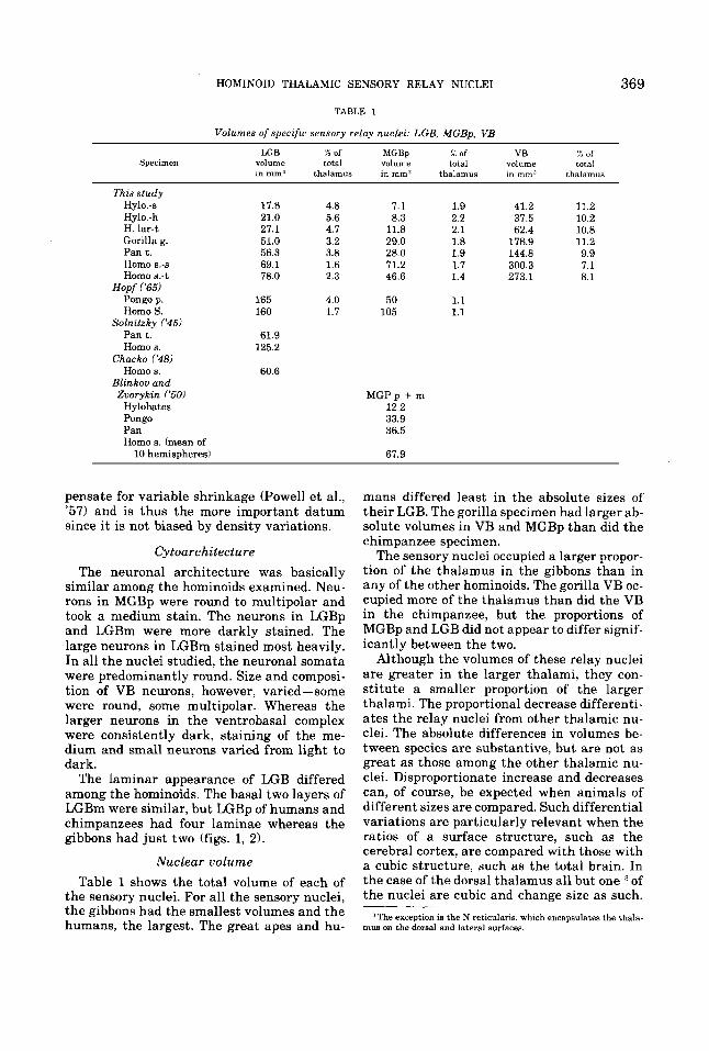
TABLE 1
Volumes of specific sensory relay nuclei: LGB, MGBp, VB
Specimen LGB x of MGBp % of VB x of
volume total volume total volume total in mmS thalamus in mm3 thalamus in mm3 thalamus
This study Hy1o.-s Hylo. - h H. lar-t Gorilla g. Pant. Homo 8.-s Homo 8.-t
Hopf ('65) Pongo p. Homo S.
Pant. Homo s.
Chacko ('481 Homo s .
Blinkow and
Hylobates Pongo Pan Homo s. (mean of
Solnitzky ('45)
Zvorykin ('50)
10 hemispheres)
17.8 21.0 27.1 51.0 56.3 69.1 78.0
165 160
61.9 125.2
60.6
4.8 5.6 4.7 3.2 3.8 1.6 2.3
4.0 1.7
7.1 8.3
11.8 29.0 28.0 71.2 46.6
50 105
MGPp + rn 12.2 33.9 36.5
67.9
1.9 41.2 11.2 2.2 37.5 10.2 2.1 62.4 10.8 1.8 178.9 11.2 1.9 144.8 9.9 1.7 300.3 7.1 1.4 273.1 8.1
1.1 1.1
pensate for variable shrinkage (Powell e t al., '57) and is thus the more important datum since it is not biased by density variations.
Cytoarchitecture The neuronal architecture was basically
similar among the hominoids examined. Neu- rons in MGBp were round to multipolar and took a medium stain. The neurons in LGBp and LGBm were more darkly stained. The large neurons in LGBm stained most heavily. In all the nuclei studied, the neuronal somata were predominantly round. Size and composi- tion of VB neurons, however, varied-some were round, some multipolar. Whereas the larger neurons in the ventrobasal complex were consistently dark, staining of the me- dium and small neurons varied from light to dark.
The laminar appearance of LGB differed among the hominoids. The basal two layers of LGBm were similar, but LGBp of humans and chimpanzees had four laminae whereas the gibbons had just two (figs. 1, 2).
Nuclear uolume Table 1 shows the total volume of each of
the sensory nuclei. For all the sensory nuclei, the gibbons had the smallest volumes and the humans, the largest. The great apes and hu-
mans differed least in the absolute sizes of their LGB. The gorilla specimen had larger ab- solute volumes in VB and MGBp than did the chimpanzee specimen.
The sensory nuclei occupied a larger propor- tion of the thalamus in the gibbons than in any of the other hominoids. The gorilla VB oc- cupied more of the thalamus than did the VB in the chimpanzee, but the proportions of MGBp and LGB did not appear to differ signif- icantly between the two.
Although the volumes of these relay nuclei are greater in the larger thalami, they con- stitute a smaller proportion of the larger thalami. The proportional decrease differenti- ates the relay nuclei from other thalamic nu- clei. The absolute differences in volumes be- tween species are substantive, but are not as great as those among the other thalamic nu- clei. Disproportionate increase and decreases can, of course, be expected when animals of different sizes are compared. Such differential variations are particularly relevant when the ratios of a surface structure, such as the cerebral cortex, are compared with those with a cubic structure, such as the total brain. In the case of the dorsal thalamus all but one of the nuclei are cubic and change size as such.
3The exception is the N reticularia, which encapsulates the thala- mu8 on the doraal and lateral surfaces.
370 ESTE ARMSTRONG
TABLE 2
Neuronal density: MGB parvocellularis
Nu. Mean No. of Specimen of neurons
fields per 0.01 mm' S.D. S.E. Minimum Maximum Median
Hy1o.-s 13 532.1 108.9 30.2 354.2 777.8 503.5 Hy1o.-h 14 479.0 142.5 38.1 222.2 676.2 518.0 H. lar-t 16 345.1 89.0 22.1 193.3 608.3 330.8
Pan t. 14 325.9 91.3 24.4 224.4 495.6 287.0 Homo s.-c 30 147.7 42.6 7.8 80.0 305.0 142.0
Gorilla g. 24 243.9 82.9 16.9 64.7 418.7 245.1
TABLE 3
Neuronal density: LGB
Specimen
LGBp Hylo.-s Hy1o.-h Gorilla g. Pan t. Homo s.-c
Hy1o.-s Hy1o.-h Gorilla g. P a n t . Homo 6.-c
LGBm
No. of
fields
20 30 28 21 31
7 19 10 11 14
Mean No. of neurons
per 0.01 mm3 S.D. S.E.
784.7 755.5 385.3 397.7 303.9
368.0 382.7 288.8 350.2 181.8
172.5 174.2 82.6 73.6 62.3
119.0 119.9 121.5 80.7 51.4
38.6 31.8 15.6 16.1 11.1
45.0 27.5 38.4 24.3 13.7
Irhnimum
458.3 291.7 217.6 264.0 175.0
243.1 250.0 170.6 236.0 120.0
Maximum
1,083.3 1,000.0
629.4 540.0 415.0
579.2 652.8 506.9 516.0 295.0
Median
781.3 809.0 370.6 405.5 313.8
366.3 343.8 253.4 346.0 170.0
TABLE 4
Neuronal density: VB
No. Mean No. of Specimens of neurons
fields per 0.01 mm' S.D. S.E. Minimum Maximum Median
Hy1o.-s 43 142.5 57.0 8.7 55.6 263.9 138.9 Hy1o.-h 30 122.1 34.8 6.4 69.4 222.2 116.3 H. lar-t 40 123.4 32.9 5.2 61.7 213.3 124.6 Gorilla g. 61 60.0 26.0 3.3 24.4 129.4 53.8 P a n t . 33 84.7 29.1 5.1 36.0 144.0 76.3 Homo 8.-c 48 58.8 22.0 3.4 25.0 115.0 57.7
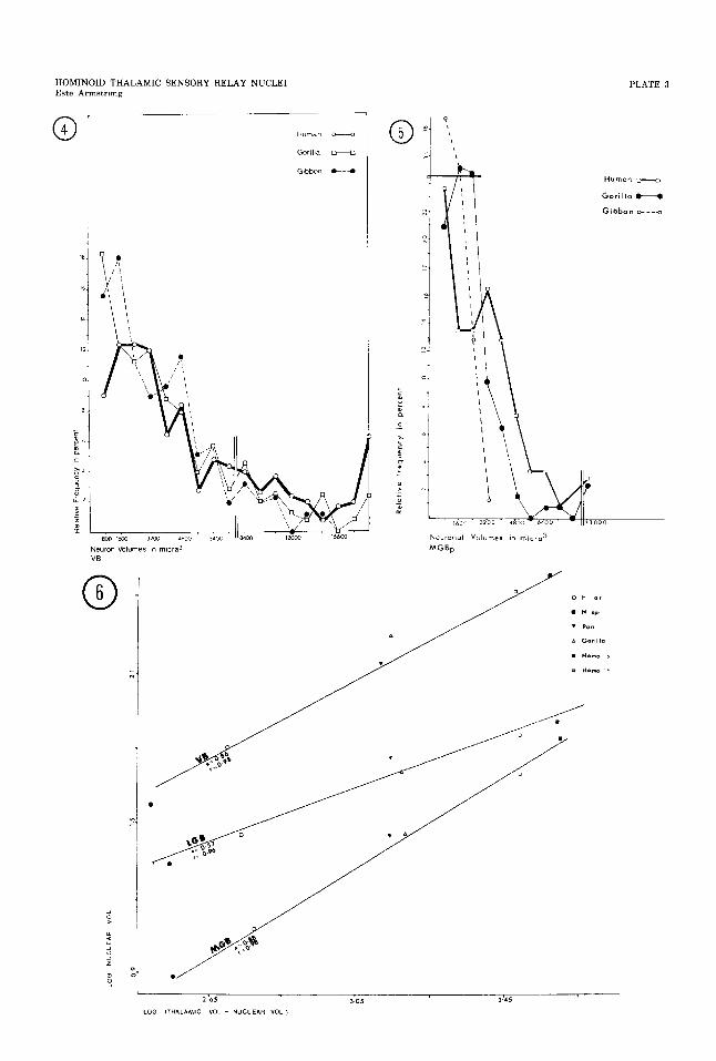
In this phylogenetic series, a log.-log. plotting of the volumes of each nucleus against the volume of the dorsal thalamus minus the nu- clear volume produces the lines of highest cor- relation (fig. 6). The relationship of the size of each nucleus to that of the total thalamic volume was determined by the allometric for- mula Log Y = cu(1og.X) + 1og.b (Gould, '661. Unlike most of the other thalamic nuclei, all the sensory nuclei have slopes that are less than one, an indication that these nuclei have experienced a differential decline in homi- noids. This allometric interpretation is sup- ported by the finding of a similar number of
neurons in the sensory nuclei of great apes and humans.
Neuronal density The neuronal density was least in the
human specimen and greatest in the gibbons (tables 2-41. The great apes had intermediate values. All of the gorilla nuclei, even in LGBm where the total volume of the nucleus was smaller than that of the chimpanzee, had less dense neuronal packing than did those of the chimpanzee (table 5).
The numbers of neurons in MGBp and LGB were similar among the great apes and hu-
HOMINOID THALAMIC SENSORY RELAY NUCLEI 371
mans (table 6). The VB in the human speci- mens had more neurons that did the VB in the great apes. The gibbons had about 50% fewer neurons in VB than did any of the other hominoids studied,
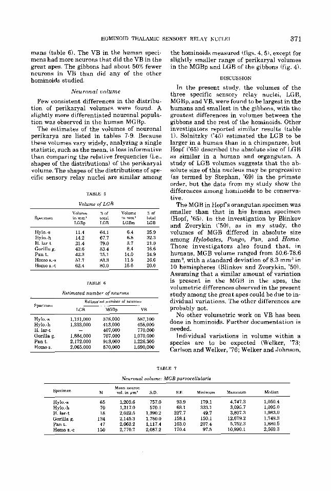
Neuronal volume Few consistent differences in the distribu-
tion of perikaryal volumes were found. A slightly more differentiated neuronal popula- tion was observed in the human MGBp.
The estimates of the volumes of neuronal perikarya are listed in tables 7-9. Because these volumes vary widely, analyzing a single statistic, such as the mean, is less informative than comparing the relative frequencies (i.e., shapes of the distributions! of the perikaryal volume. The shapes of the distributions of spe- cific sensory relay nuclei are similar among
TABLE 5
Volume of LGB
Volume % of Volume X of Specimen in mm3 total inmm3 total
LCBp LGB LGBm LGB
Hy1o.-s 11.4 64.1 6.4 35.9 Hy1o.-h 14.2 67.7 6.8 32.3 H. lar-t 21.4 79.0 5.7 21.0 Gorilla g. 42.6 83.4 8.4 16.6 Pan t. 42.3 75.1 14.0 24.9 Homo s.-s 57.7 83.3 11.5 16.6 Homo s.-t 62.4 80.0 15.6 20.0
TABLE 6
Estimated number of neurons
Estimated number of neuron8 Specimen
LGB MGBp VB
Hy1o.-s 1,131,000 378,000 587,100 Hy1o.-h 1,333,000 413,000 458,000 H. lar-t - 407,000 770,000 Gorilla g. 1,884,000 707,000 1,070,000 Pant . 2,172,000 913,000 1,226,300 Homo s. 2.065.000 870,000 1,690.000
the hominoids measured (figs. 4,5), except for slightly smaller range of perikaryal volumes in the MGBp and LGB of the gibbons (fig. 4).
DISCUSSION
In the present study. the volumes of the three specific sensory relay nuclei, LGB, MGBp, and VB, were found to be largest in the humans and smallest in the gibbons, with the greatest differences in volumes between the gibbons and the rest of the hominoids. Other investigators reported similar results (table 1). Solnitzky ('45) estimated the LGB to be larger in a human than in a chimpanzee, but Hopf ('65) described the absolute size of LGB as similar in a human and organgutan. A study of LGB volumes suggests that the ab- solute size of this nucleus may be progressive (as termed by Stephan, '69) in the primate order, but the data from my study show the differences among hominoids to be conserva- tive.
The MGB in Hopf s orangutan specimen was smaller than that in his human specimen (Hopf, '65). In the investigation by Blinkov and Zvorykin ('501, as in my study, the volumes of MGB differed in absolute size among Hylobates, Pongo, Pan, and Homo. Those investigators also found tha t , in humans, MGB volume ranged from 50.6-78.6 mm3, with a standard deviation of 8.3 mm3 in 10 hemispheres (Blinkov and Zvorykin, '50). Assuming that a similar amount of variation is present in the MGB in the apes, the volumetric differences observed in the present study among the great apes could be due to in- dividual variations. The other differences are probably not.
No other volumetric work on VB has been done in hominoids. Further documentation is needed.
Individual variations in volume within a species are to be expected (Welker, '73; Carlson and Welker, '76; Welker and Johnson,
TABLE 7
Neuronal volume: MGB parvocellulark
Specimen N vol. in @m3 S.D. S.E. Minimum Maximum Median Mean neuron
Hy10.-s 65 1,203.6 757.0 93.9 179.1 4,747.3 1,050.4 Hy1o.-h 70 1,217.0 570.1 68.1 333.1 3,095.7 1,095.0 H. lar-t 18 2,022.5 1,390.2 327.7 49.7 3,807.3 1,983.0 Gorilla g. 124 2,145.3 1,760.0 158.1 150.1 12,679.2 1,749.3 Pan t. 47 2,063.2 1,117.4 163.0 207.4 5,752.3 1,880.5 Homo 6.-c 150 2.770.7 2.087.2 170.4 97.5 10,990.1 2,569.3
372 ESTE ARMSTRONG
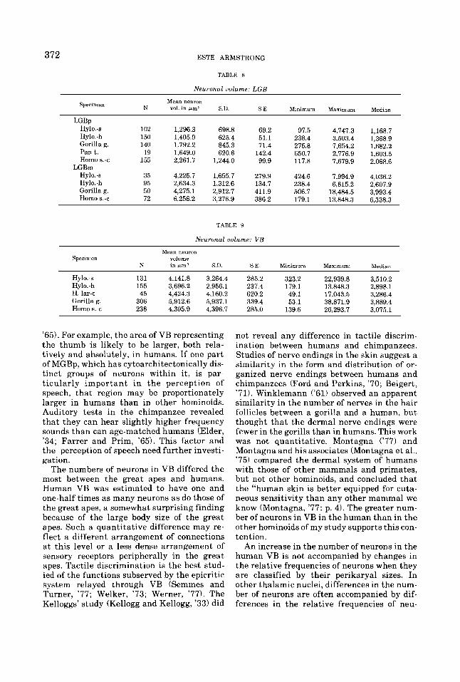
TABLE 8
Neuronal uolume: LGB
Mean neuron N vol. in pml S.U. S.E. Minimum Maximum Median
Specimen
LGBp Hy1o.-s 102 Hylo. - h 150 Gorilla g. 140 Pant. 19 Homo 9.-c 155
LGBm Hy1o.-s 35 Hylo.-h 95 Gorilla g. 50 Homo s.-c 72
1.296.3 698.8 69.2 1,405.9 625.4 51.1 1.792.2 845.3 71.4 1,649.0 620.6 142.4 2,261.7 1,244.0 99.9
4,225.7 1,655.7 279.9 2,634.3 1,312.6 134.7 4,275.1 2,912.7 411.9 6,256.2 3,276.9 386.2
TABLE 9
Neuronal uolume: VB
97.5 238.4 275.8 650.7 117.8
424.6 238.4 506.7 179.1
4,747.3 3,503.4 7,654.2 2,776.9 7,679.9
7,994.9 6,815.2
18,484.5 13,848.3
1,168.7 1,368.9 1,682.2 1,6 0 3.5 2,068.6
4,036.2 2,607.9 3,993.4 6,338.3
Mean neuron Specimen volume
K in e m ' S D SE Minimum Maxmum Median
Hylo..s 131 4.141.8 3,264.4 285.2 323.2 22,939.8 3,510.2 Hy1o.-h 155 3,696.2 2,956.1 237.4 179.1 13,848.3 2,898.1 H. lar-t 45 4,424.3 4,160.2 620.2 49.1 17,043.5 3,286.4 Gorilla g. 306 5,912.6 5,937.1 339.4 53.1 38,871.9 3,889.4 Homo s.-c 238 4.305.9 4,396.7 285.0 139.6 26,293.7 3,075.1
'65). For example, the area of VB representing the thumb is likely to be larger, both rela- tively and absolutely, in humans. If one part of MGBp, which has cytoarchitectonically dis- tinct groups of neurons within it, is par- ticularly important in the perception of speech, that region may be proportionately larger in humans than in other hominoids. Auditory tests in the chimpanzee revealed that they can hear slightly higher frequency sounds than can age-matched humans (Elder, '34; Farrer and Prim, '65). This factor and the perception of speech need further investi- gation.
The numbers of neurons in VB differed the most between the great apes and humans. Human VB was estimated to have one and one-half times as many neurons as do those of the great apes, a somewhat surprising finding because of the large body size of the great apes. Such a quantitative difference may re- flect a different arrangement of connections at this level or a less dense arrangement of sensory receptors peripherally in the great apes. Tactile discrimination is the best stud- ied of the functions subserved by the epicritic system relayed through VB (Semmes and Turner, '77; Welker, '73; Werner, '77). The Kelloggs' study (Kellogg and Kellogg, '33) did
not reveal any difference in tactile discrim- ination between humans and chimpanzees. Studies of nerve endings in the skin suggest a similarity in the form and distribution of or- ganized nerve endings between humans and chimpanzees (Ford and Perkins, '70; Beigert, '71). Winklemann ('61) observed an apparent similarity in the number of nerves in the hair follicles between a gorilla and a human, but thought that the dermal nerve endings were fewer in the gorilla than in humans. This work was not quantitative. Montagna ( '77) and Montagna and his associates (Montagna et al., '75) compared the dermal system of humans with those of other mammals and primates, but not other hominoids, and concluded that the "human skin is better equipped for cuta- neous sensitivity than any other mammal we know (Montagna, '77: p. 4). The greater num- ber of neurons in VB in the human than in the other hominoids of my study supports this con- tention.
An increase in the number of neurons in the human VB is not accompanied by changes in the relative frequencies of neurons when they are classified by their perikaryal sizes. In other thalamic nuclei, differences in the num- ber of neurons are often accompanied by dif- ferences in the relative frequencies of neu-
373 HOMINOID THALAMIC SENSORY RELAY NUCLEI
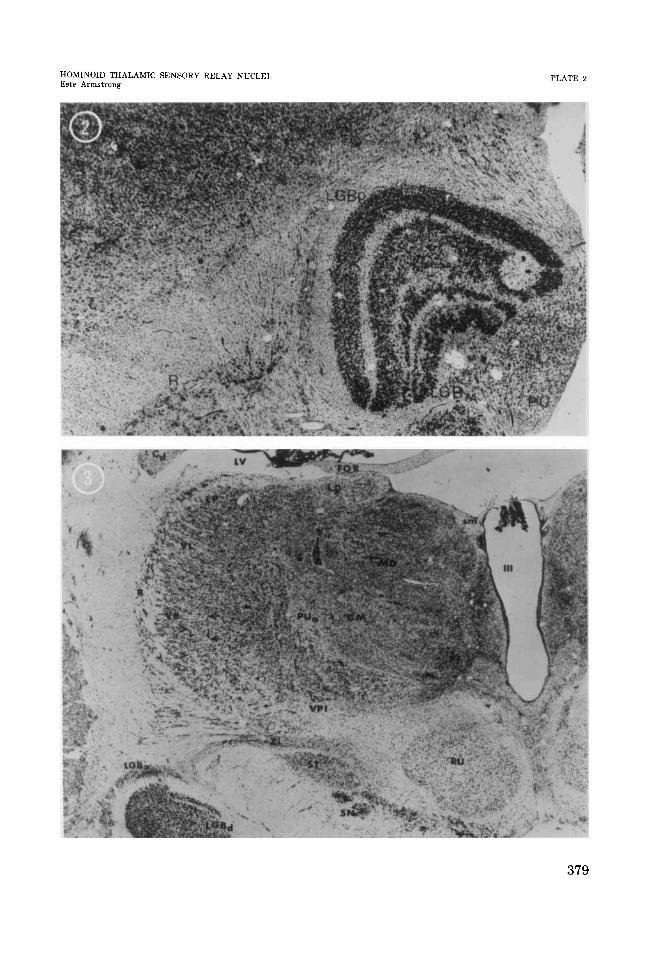
ronal volumes (Armstrong, ’76; and in prepa- ration). This lack of alteration in neuronal proportions emphasizes the conservative na- ture of the changes in VB. Furthermore, the basic organization of the neurons may be simi- lar among these hominoids as it is with other mammals. Although neither flattened nor tangential sections were available for study, coronal sections showed alignments of neu- rons suggestive of “barreloids” (Van Der Loos, ’76) in these hominoids (fig. 3). The presence of “barreloids” in large hominoid thalami sug- gests that this type of organization of nerve cells is not confined to representing vibrissae a t a thalamic level. The alignment may be important for general tactile organization or it may indicate locations for specialized tac- tile organization, e.g., in the case of homi- noids, perhaps fingertips.
The estimated number of neurons per nu- cleus depends on the volume and neuronal densities. In general, larger brains have been found to have lower neuronal densities (Bok, ’59; Shariff, ’53; Tower, ’54). This trend was noted among all the thalamic nuclei observed in this study (Armstrong, ’76). However, the degree of difference in density varied with the nucleus being measured. An estimation of the number of neurons in the sensory relay nuclei revealed that in these, and only in these, tha- lamic nuclei, the great apes and humans have approximately the same number of neurons. The gibbons have about half as many neurons (table 6). The fewer neurons in the gibbons may well reflect their smaller body size. How- ever, Beck (’671, in studying gibbon in- telligence, noted that the gibbon seems to fix- ate on the wire mesh of the cage and not the tes t material . Although this figure-and- ground problem could be related to primary sensory perception, it more likely reflects the integration of visual perceptions. The quan- titative differences in the number of neurons as well as the qualitative difference in LGB lamination pattern of gibbons should be more thoroughly explored, but until such studies are done, the fewer neurons in Hylobates are more appropriately interpreted as reflecting a smaller body size.
These data suggest that humans have fewer sensory neurons than would be expected in a nonhuman primate with the same thalamic volume. Although their brain sizes are equiva- lent (Tobias, ’711, Macaca rnulatta may have a volumetrically larger LGB than does the gib- bon. Absolute LGB volume of a macaque was
29 cc (without laminae) (Le Gros Clark, ’411, compared with 22 cc (with laminae) for the average LGB volume of the three gibbons in my study. This quantitative comparison needs verification because the specimens were pre- pared in different laboratories using different techniques. The macaque pattern of lamina- tion appears more complex than that of any of the hominoids of this study, suggesting that the macaque LGB is more differentiated than not only that of the human, as Le Gros Clark (’41) concluded, but also those of all homi- noids. That statement would be in agreement with the high placement of Cercopithecidae in Stephan’s (’69) index of progression for LGB. Hominoids may have experienced less evolu- tion in their visual nuclei than Cercopi- thecidae. The complexity of laminae in humans and macaques is probably the result of parallel evolution.
Selection pressures may have been working to stabilize the sensory nuclei during homi- noid evolution. The fact that humans and great apes appear to have the same number of neurons in their visual and auditory relay nu- clei supports behavioral studies that a show a similarity in those systems. The similarity in MGB suggests that the neurological substrate for language lies elsewhere, probably in the expanded and differentiated association re- gions of the brain (Armstrong, ’76; and in preparation). The auditory relay nuclei in the common ancestors of great apes and humans may have had sufficiently generalized func- tions to remain unchanged.
The increase in number of neurons in the human VB could be related to the importance of touching as opposed to grooming (Mon- tague, ’71) and/or to the increased develop- ment of tactile sensation necessary to support such cognitive skills as those needed in the making of tools. The fact that the human VB has more neurons than those of the other hominoids suggests some degree of differen- tial evolution within the sensory regions of the brain. If such a difference is also found in the cortex, estimates of the numbers of “ex- tra” cortical neurons (Jerison, ’73) may need slight revisions.
Although the size of the human brain is impressive, size alone cannot account for human behavior (Holloway, ’68). The largest increase in size in the cortex is thought to be in “association” regions (Holloway, ’68; Passingham, ’75). The brain is too complex to understand as a single unit. It does not have
374 ESTE ARMSTRONG
one function, nor should its evolution be seen as responsive to one selection pressure. We should not be surprised that Old World mon- keys may have a greater relative development in certain parts of their brains tha do hominoids. The successful evolution of Cer- copithecidae may have depended in part on the evolution of its visual system. The evolution of hvminids may have depended on the stabiliza- tion of the retino-geniculate visual system and other sensory systems with the concomi- tant elaboration of integration and associa- tion regions. The evolution of integrative cen- ters does not necessarily depend on the stabilization of sensory systems, but in human evolution that dependence appears to be the case.
ACKNOWLEDGMEKTS
I thank C. R. Noback, H. Stephan, N. Moskowitz, W. I. Welker and the Yakovlev col- lection for the opportunity to study some of their primate brains; R. Holloway, C. R. Noback and W. I . Welker for their technical advice; T. Hill for the excellent photographs of the chimpanzee brain; and R. Schwartz, B. Butler and Lynny Kutash for the typing of the manuscript. I appreciate the comments of the reviewers. This work was supported in part by NSF SOC. Grant 74 20149 0113.24 019.00 and NlNDS Grants 3249 and 06225 from the USPHS.
LITERATURE CITED
Albe-Fessard, D. 1973 Electrophysiological methods for the identification of the thalamic nuclei. 2. Neurol., 205: 15-28.
Andrew, J., and E. S. Watkins 1969 A Stereotaxic Atlas of the human Thalamus and Adjacent Structures: A Vari- ability Study. Williams and Wilkins, Baltimore.
Armstrong, Este 1976 A Quantitative Comparison of the Hominoid Thalamus. Unpublished Ph.D. dissertation, Columbia University.
Bauchot, R. 1963 L-architecture compark qualitative e t quantitative du diecephale des insectivores. Mam- malia, 27: Suppl. 1, 1-400.
Beck, B. B. 1967 A study of problem solving by gibbons. Behavior, 25: 95-109.
Biegert, 6. 1971 Dermatoglyphics in the chimpanzee. In: The Chimpanzee. G. Bourne, ed. Karger, Basel, Vol. 4, pp. 273-34.
Blinkov, S. M., and I, I. Glezer, 4 s . 1968 The Human Brain in Figures and Tables. Translated by Basil Haigh. Plenum Press, New York.
Blinkov, S. M., and V. P. Zvorykin 1950 Dimensions of the auditory cortex and the medial geniculate body in man and monkeys. Referred to in: The Human Brain in Fimres and Tables. S. M. Blinkov and I. I. Gleser, eds. Translated by Basil Haigh. Plenum Press, New York, 1968, pp. 225-226 and 410-412.
Bok, S. T. 1959 Histonomy of the cerebral cortex. Van Nostran Reinhold, Princeton, New Jersey. York, pp. 230-290.
Cammermeyer, J. 1967 Artifactual displacement of neu- ronal nucleoli in paraffin sections. J. Hirnforsch., 9: 209-224.
Campbell, F. W. 1974 The transmission of spatial infor- mation through the visual system. In: The Neuro- sciences: The Third Study Program. F. 0. Schmitt, ed. M.I.T. Press, Cambridge, pp. 95-103.
Carlson, M., and W. I. Welker 1976 Some morphological, physiological, and behavioral specialization in North American beaver (Castor canadensis). Brain, Behav. and Evol., 13: 302-326.
Chacko, L. W. 1948 The laminar pattern of the lateral geniculate body in the primates. J. Neurol. Neurosurg. Psychiat., 11: 211-224.
Dekaban, A. 1953 Human thalamus. An anatomical, de- velopmental and pathological study. I. J. a m p . Neur., 99: 639-683.
De Valois, R. L., and P. L. Pease 1971 Contours and con- trast: Responses of monkey lateral geniculate nucleus cells to luminance and color figures. Science, 171: 694-696.
Dornfield, E. J., D. W. Slater and H. Scheffe 1942 A method for accurate determination of volume and cell numbers in small organs. Anat. Rec., 82: 255-259.
Doty, R. W., P. D. Wilson, J. R . Bartlett and J. Pecei- Saaverdra 1973 Mesencephalic control of lateral genicu- late nucleus in primates. I. Electrophysiology. Exp. Brain Res., 18: 189-203.
Elder, J. H. 1934 Auditory acuity of the chimpanzee. J. Comp. Psychol., 17: 157-183.
Farrer, D. N., and M. M. Prim 1965 A preliminary report on auditory frequency threshold comparison of humans and pre-adolescent chimpanzees. Aeromedical Research Laboratory technical report 65-6, Holloman AFB, New Mexico.
Farrer, D. N., and F. A. Young 1970 Chimpanzee color vision, acuity, and ocular components. In: The Chim- panzee, G. Bourne, ed. Karger, Basel, Vol. 2, pp 16-25.
Fazio, C., J. Sacco and 0. Bugiani 1973 The thalarnic hemorrhage. Europ. Neurol., 9: 30-43.
Ford, D. M., and E. M. Perkins 1970 The skin of the chim- panzee. In: The Chimpanzee. G. Bourne, ed. Karger, Basel, Vol. 3, pp. 82-119.
Frontera, J. G. 1958 Evaluation of the immediate effects of some fixatives upon the measurements of the brains of macaques. J. Comp. Neur., 109: 417-438.
Giolli, R. A,, and J. Tigges 1970 The primary optic path- ways and nuclei of primates. In: The Primate Brain: Advances in Prirnatology. I. C. R. Noback and W. Montag- na, eds. Appleton-Century-Crofts, New York, pp. 29-54.
Glees, P. 1961 Terminal degeneration and transsynaptic atrophy in the lateral geniculate body of the monkey. In: The Visual System: Neurophysiology and Psychophysics. R. Jung and H. Hornhuser, eds. Springer, Berlin, pp. 104-110.
Gould, S. 1966 Allometry and size in ontogeny and phy- logeny. Biol. Rev., Cambridge Phil. SOC., 41: 587-640.
Grether, W. F. 1941 Spectral saturation curves for chim- panzees and man. J. Exp. Psychol., 28: 419-427.
1942 The magnitude of simultaneous color con- trast and simultaneous brightness contrast for chim- panzee and man. J. Exp. Psychol., 30: 69-83.
Gross, N. B., W. S. Litschitz and D. J. Anderson 1974 The tonotopic organization of the auditory thalamus of the squirrel monkey (Saimiri sciureus). Brain Res., 65: 323-332.
Hassler, R. 1959 Anatomy of the thalamus. In: Introduc- tion to Stereotaxis With an Atlas of the Human Brain. G . Schaltenbrand and P. Bailev, eds. Grune & Stratton. New
375 HOMINOID THALAMIC SENSORY RELAY NUCLEI
Holloway, R. L., Jr. 1968 The evolution of the primate brain: Some aspects of quantitative relations. Brain Res., 7: 121.172.
Hopf, A. 1965 Volumetrische Untersuchunger zur ver- gleichenden Anatomie des Thalamus. J. F. Hirnfor- schung, 8: 25-38.
Jerison, H. J. 1973 Evolution of the Brain and In- telligence. Academic Press, New York.
Jones, R . L. Split nucleoli as a source of error in nerve cell counts. Stain Technol.. 12: 91-95.
Kanagasuntheram, R., and W. C. Wong 1968 Nuclei of the diencephalon of Hylobatidae. J. Comp. Neur., 134: 265-286.
Katsucki, Y. 1966 Integrative organization in the tha- lamic and cortical auditory centers. In: The Thalamus. D. P. Purpura and M. D. Yahr, eds. Columbia University Press, New York, pp. 349-362.
Keidel, W. D. 1974 Information processing in the higher parts of the auditory pathway. In: Facts and Models in Hearing. E. Z. Wicker and E. Terhardt, eds. Springer-Ver- lag, New York, pp. 216-226.
Kellogg, W. N., and L. A. Kellogg 1933 The Age and the Child. McCraw-Hill Book Co., New York, pp. 88-118. Fascimileof the 1933 edition, Hafner Publishing Co., New York, 1967.
Kohler, W. 1915 Aus der Antropoidenstation auf Teneriffa. I. Optische Untersuchunger am Schimpansen und am Haushuhn. Abh. Preuss. Akad. Wiss., pp. 1-70.
Konigsmark, B. W. 1970 Methods for counting neurons. In: Contemporary Research Methods in Neuroanatomy. W. J. H. Nauta and S. 0. E. Ebbesson, eds. Springer-Ver- lag, New York, pp. 315-340.
Kraus, C., and M. Gihr 1965 Statischer Beitrag zur gliederung des nucleus anterior thalamic. S. F. Hirn- forsch., 8: 39-45.
Le Gros Clark, W. E. 1932 A morphological study of the la t - eral geniculate body. Br. J. Ophthalmol., 16: 264-284.
The laminar organization and cell content of the lateral geniculate body in the monkey. J. Anat., 75: 419-433.
Mark, V., and F. Ervin 1960 Stereotactic thalamotomy in the human. 11. Physiological observation in the human thalamus. Arch. Neurol., 3: 368-380.
Mark, V., F. Ervin and P. I. Yakovlev 1963 Stereotactoc thalamotomy. 111. The verification of anatomical lesion sites in the human thalamus. Arch. Neurol., 8: 528-538.
McIlwain, J. T. 1972 Nonretinal influences on the later- al geniculate nucleus. Invest. Ophthalmol., I I : 311-321.
Montagna, W. 1977 Morphology of cutaneous sensory re- ceptors. J. Invest. Dermatol., 69: 4-7.
Montagna, W., N. A. Roman and E. MacPherson 1975 A comparative study of the innervation of the facial disc of selected mammals. J. Invest. Dermatol., 64: 458-465.
Montague, A. 1971 Touching: The Human Significance of Skin. Columbia University Press, New York.
Neff, W. D., J. H. Casseday and J. L. Crawford 1972 The me- dial geniculate body and association thalamic cell group: Behavioral studies. Brain, Behav. and Evol., 6: 302-310.
1975 The visual system of primates in phy- logenetic studies. In: Phylogeny of Primates. W. P. Luckett and F. S. Szalay, eds. Plenum, New York, pp. 199-218.
Noback, C. R., and L. K. Laemle 1970 Structural and func- tional aspects of the visual pathways of primates. In: The Primate Brain: Advances in Primatology I. C. R. Noback and W. Montagna, eds. Appleton-Century-Crofts, New York, pp. 55-81.
Passingham, R. E. 1975 Changes in the size and organi-
1937
1941
Noback, C. R.
zation of the brain in man and his ancestors. Brain, Behav. and Evol., 11: 73-90.
Powell, T. P. S., R. W. Guillery and W. M. Cowan 1957 A quantitative study of the fornix-mammillothalamic sys- tem. J. Anat., 92: 419-437.
Richards, W. 1968 Spacial remapping in the primate visual system. Kybernetik.. 4: 146-153.
Riesen, A. H. 1970 Chimpanzee visual perception. In: The Chimpanzee. G. Bourne, ed. Karger, Basel, Vol. 2, pp. 1-15.
Semmes, J., and B. Turner 1977 Effects of cortical lesions on somatosensory tasks. J. Invest. Dermatol., 69: 181-189.
Shantha, T., and S. Manocha 1969 The brain of the chim- panzee (Pan troglodytes). In: The Chimpanzee. G. Bourne, ed. Karger, Basel, Vol. 1, pp. 238-305.
Shariff, G. A. 1953 Cell counts in the primate cerebral cortex. J. Cornp. Neur., 98: 381-400.
Solnitzky, 0. 1945 Volumetric and reconstruction stud- ies of the mammalian lateral geniculate nucleus. Anat. Rec., 91: 300 (Abstract).
Spence, K. W. 1934 Visual acuity and its relation to brightness in chimpanzee and man. J. Comp. Psychol.: 18: 333-361.
Stephan. H. 1969 Quantitative investigations on visual structure in primate brains. Proc. 2nd Int. Congr. Primat., 3: 34-42.
Stephan, H., R. Bauchot and 0. J. Andy 1970 Data on size of brain and of various brain parts in insectivores and pri- mates. In: The Primate Brain. C. R. Noback and W. Mon- tagna, eds. Appleton-Century-Crofts, New York, pp. 289-298.
Tigges, J., and W. K. O'Steen 1974 Termination of reti- nofugal fibers in squirrel monkeys. Brain Res., 79: 489-485.
Tobias, P. U. 1971 The Brain in Hominid Evolution. Col- umbia University Press, New York.
Tower, D. B. 1954 Structural and functional organiza- tion of the mammalian cerebral cortex. The correlationof neuron density with brain size. J. Comp. Neur., 101: 19-53.
Van Buren, J., and R. Borke 1972 Variations and Connec- tions of the Human Thalamus. Springer-Verlag, New York, Vols. I and IT.
Van Der Loos, H. 1976 Barreloids in mouse somatosenso- ry cortex. Neuroscience Lett., 2: 1-6.
van Harreveld, A,, and J. P. Schade 1962 Nerve cell de- struction by asphyxiation of the spinal cord. J. Neu- ropath. & Exper. Neurol., 21: 410-423.
Wall, P. D., and R. Dubner 1972 Somatosensory pathways. Ann. Rev. Physiol., 34: 95-110.
Welker, W. I. 1973 Principles of organization of the ven- trobasal complex in mammals. Brain, Behav. and Evol., 7: 253-336.
Welker, W. I., and J. I. Johnson 1965 Correlation between nuclear morphology and somatotopic organization in ven- trobasal complex of the racoon thalamus. J. Anat., 99: 761-790.
Welker, W. I., J. I. Johnson and B. H. Pubols, Jr. 1964 Some morphological and physiological characteristics of the somatic sensory systems in racoons. Amer. h l . , 4: 75-96.
Werner, G. 1977 Cutaneous stimulus registration and information processing in the somesthetic cortex. J. Invest. Dermatol., 69: 172-180.
Wiesel, T. H., and D. H. Hubel 1966 Spatial and chromatic interactions in the lateral geniculate body of the rhesus monkey. J. Neurophys., 29: 1115-1155.
Winklemann, R. K. 1961 Nerve endings in the akin of the gorilla. J. Comp. Neur., 116: 145-155.
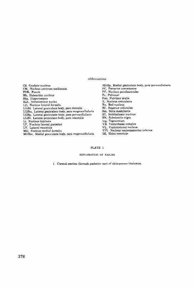
A b breuiations
Cd, Caudate nucleus CM, Nucleus centrum medianum FOR, Fornix Hb, Habenular nucleus Hip, Hippocampus ILA, Intralaminar nuclei LD, Nucleus lateral dorsalis LGBd, Lateral geniculate body, pars dorsalis LGBm, Lateral geniculate body, pars magnocellulari6 LGBp, Lateral geniculate body, pars parvocellularis LGBv, Lateral geniculate body, pars ventralis Li, Nucleus limitans LP, Nucleus lateral posterior LV, Lateral ventricle MD, Nucleus medial dorsalis MGBm, Medial geniculate body, pars magnocellularis
MGBp, Medial geniculate body, pars parvocellularis PC, Posterior commissure PF, Nucleus parafascicular Pr, Pulvinar Puo, Pulvinar oralis R, Nucleus reticularis Ru, Red nucleus SC, Superior colliculus Sm, Stria medullaris ST, Subthalamic nucleus SN, Substantia nigra teg, Tegmentum VB, Ventrobasal complex VL, Ventrolateral nucleus VPI, Nucleus ventroposterior inferior 111, Third ventricle
PLATE 1
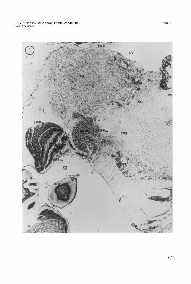
EXPLANATION OF FIGURE
1 Coronal section through posterior part of chimpanzee thalamus.
376
HOMIKOJD THALAMIC SENSORY RELAY NUCLEI EsLe Armstrong
PLATE 1
377
PLATE 2
EXPLANATlON OF FIGURES
2
3
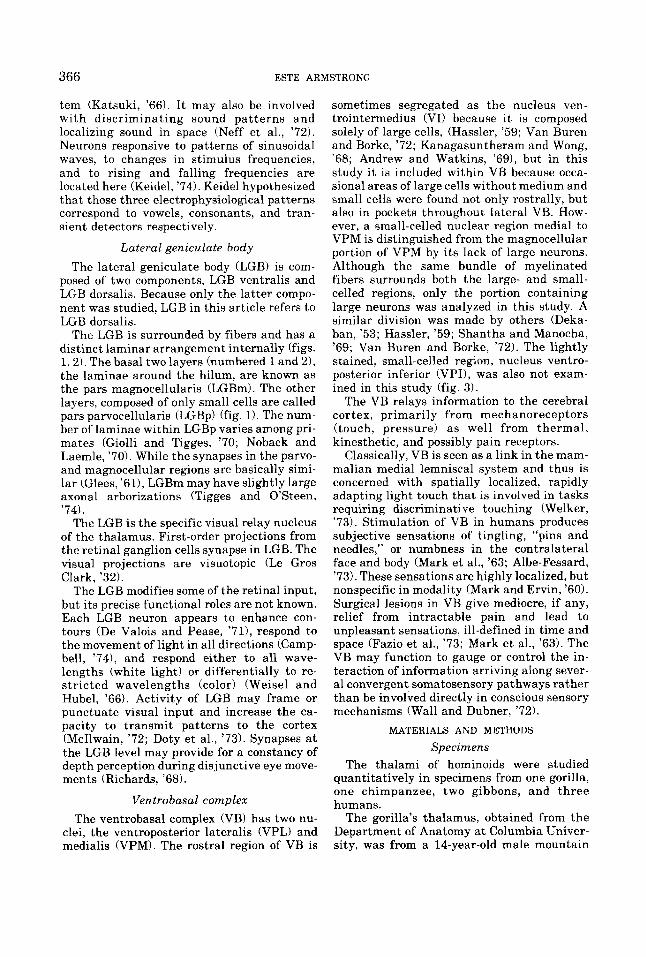
Horizontal section through gibbon lateral geniculate body
Coronal section through chimpanzee thalamus. Arrows indicate probable location of a “barreloid.” Arrowheads indicate possible “barreloid” (Van Der Loos, ’76).
378
HOMINOID THALAMIC SENSORY RELAY NUCLEI Este Armstrung
PLATE 2
379
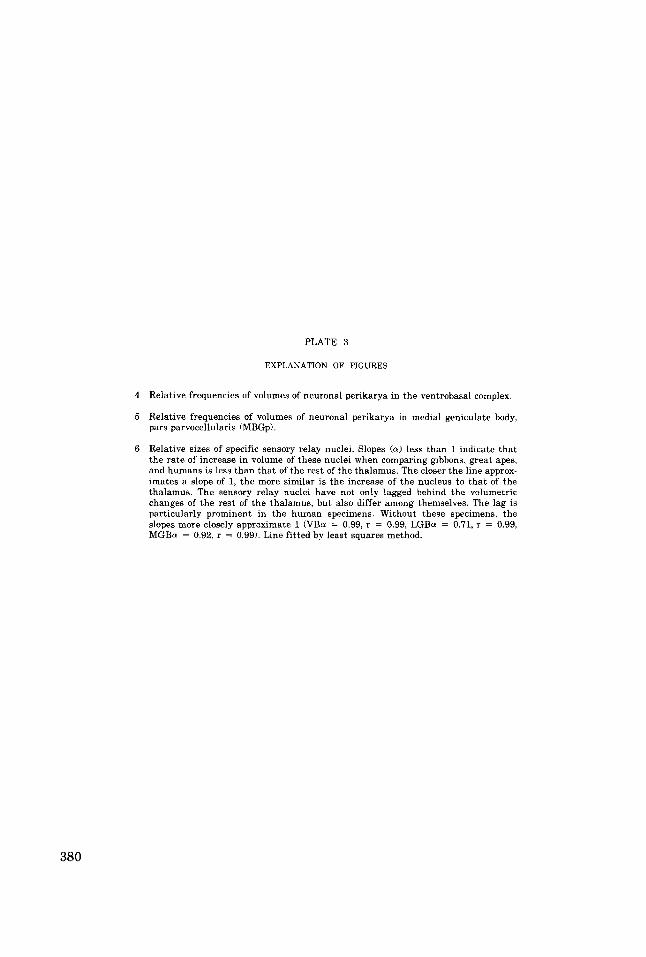
PLATE 3
EXPLANATION OF FIGURES
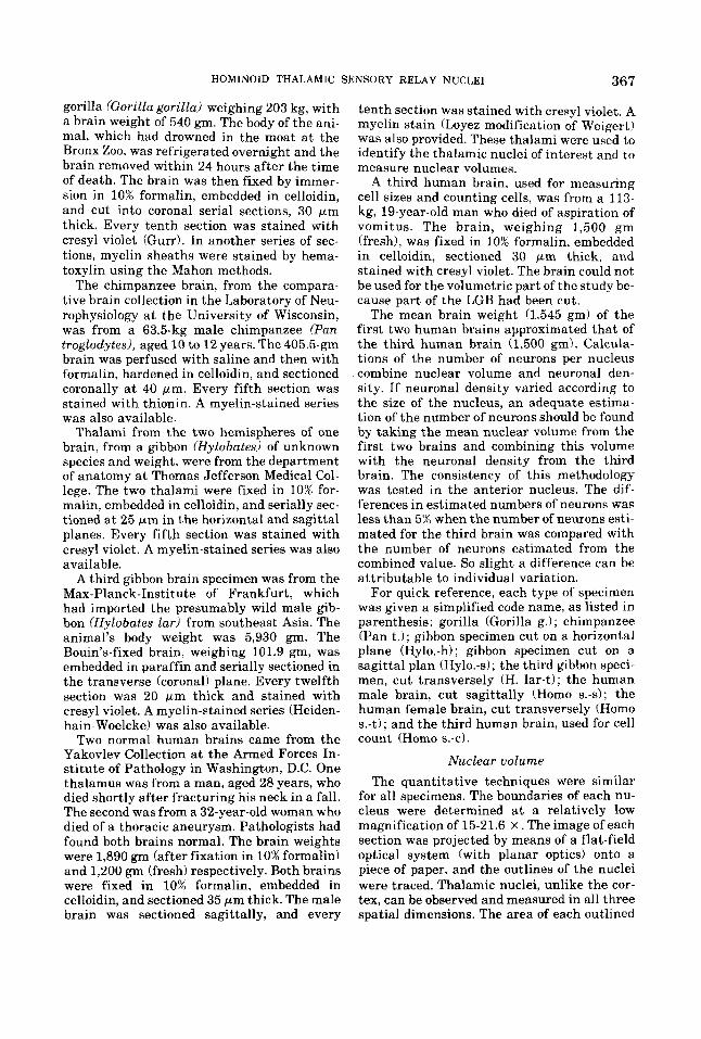
4 Relative frequencies of volumes of neuronal perikarya in the ventrobasal complex.
5 Relative frequencies of volumes of neuronal perikarya in medial geniculate body, pars parvocellularis (MBGp).
6 Relative sizes of specific sensory relay nuclei. Slopes (a) less than 1 indicate that the rate of increase in volume of these nuclei when comparing gibbons, great apes, and humans is less than that of the rest of the thalamus. The closer the line approx- imates a slope of 1, the more similar is the increase of the nucleus to that of the thalamus. The sensory relay nuclei have not only lagged behind the volumetric changes of the rest of the thalamus, but also differ among themselves. The lag is particularly prominent in the human specimens. Without these specimens, the slopes more closely approximate 1 (VBu = 0.99, r = 0.99, LGBu = 0.71, r = 0.99, MGBa = 0.92, r = 0.99). Line fitted by least squares method.
380
HOMINOID THALAMIC SENSORY RELAY NUCLEI Este Armstrong
PLATE 3
Gorilla D--O
Gibbon e-4
Neuron Volumes m rnicra' VE
Human I-
Gori 110 - Gibbon c---Q
I 1 6 s 3 05
LOG ITHALAMIC VOL - NUCLEAR VOL j
Related Documents