Cell Host & Microbe Article A Pseudomonas syringae Effector Inactivates MAPKs to Suppress PAMP-Induced Immunity in Plants Jie Zhang, 1,3 Feng Shao, 3 Yan Li, 3 Haitao Cui, 3 Linjie Chen, 3 Hongtao Li, 3 Yan Zou, 3 Chengzu Long, 3 Lefu Lan, 2 Jijie Chai, 3 She Chen, 3 Xiaoyan Tang, 2 and Jian-Min Zhou 3, * 1 National Key Laboratory of Plant Molecular Genetics, Institute of Plant Physiology and Ecology, Shanghai Institutes for Biological Sciences, Chinese Academy of Sciences, Shanghai 200032, China 2 Department of Plant Pathology, Kansas State University, Manhattan, KS 66506, USA 3 National Institute of Biological Sciences, Beijing 102206, China *Correspondence: [email protected] DOI 10.1016/j.chom.2007.03.006 SUMMARY Pathogen-associated molecular patterns (PAMPs) elicit basal defense responses in plants, and, in turn, pathogens have evolved mechanisms to overcome these PAMP-induced defenses. To suppress immunity, the phyto- pathogenic bacterium Pseudomonas syringae secretes effector proteins, the biochemical function and virulence targets of which remain largely unknown. We show that HopAI1, an ef- fector widely conserved in both plant and animal bacterial pathogens, inhibits the Arabidopsis mitogen-activated protein kinases (MAPKs) activated by exposure to PAMPs. HopAI1 inac- tivates MAPKs by removing the phosphate group from phosphothreonine through a unique phosphothreonine lyase activity, which is re- quired for HopAI1 function. The inhibition of MAPKs by HopA1 suppresses two independent downstream events, namely the reinforcement of cell wall defense and transcriptional activa- tion of PAMP response genes. The MAPKs MPK3 and MPK6 physically interact with HopAI1 indicating that they are direct targets of HopAI1. These findings uncover a mechanism by which Pseudomonas syringae overcomes host innate immunity to promote pathogenesis. INTRODUCTION Plants are known to activate defenses in response to non- specific elicitors from diverse microbes (Boller, 1995). Studies in both animal and plant innate immunity in recent years have indicated that these elicitors constitute patho- gen-associated molecular patterns (PAMPs), triggering basal defenses in the host (Nu ¨ rnberger and Lipka, 2005; Zipfel and Felix, 2005). Because PAMPs also exist in nonpathogenic microbes, they have also been referred to as microbe-associated molecular patterns (MAMPs; Ausubel, 2005). PAMP-triggered plant defenses form a for- midable layer of resistance deterring numerous potential pathogens (Kang et al., 2003; Li et al., 2005). An emerging consensus is that pathogens infecting the modern day land plants have evolved mechanisms to overcome the PAMP-induced defenses in their respective host plants (Chisholm et al., 2006). Therefore, PAMP-triggered de- fenses are at the core of understanding both plant disease resistance and bacterial pathogenicity. The best understood PAMP of plant pathogens is argu- ably flg22, a conserved peptide derived from Pseudomonas syringae flagellin (Felix et al., 1999). Flg22 is perceived by the receptor kinase FLS2 in Arabidopsis to stimulate cell wall defenses exemplified by callose deposition (Felix et al., 1999; Gomez-Gomez and Boller, 2000), ethylene production (Felix et al., 1999; Liu and Zhang, 2004), and reprogram- ming of transcriptome (Zipfel et al., 2004; Navarro et al., 2004; Thilmony et al., 2006). Immediately after the expo- sure to flg22, plants activate a rapid but transient oxidative burst (H 2 O 2 production; Felix et al., 1999) and mitogen activated protein kinases (MAPKs) MPK3 and MPK6 (Asai et al., 2002). However, it is not known if a causal relation- ship exists between MAPK activation and oxidative burst. Gram-negative bacteria use a specialized type III secre- tion system to deliver a repertoire of effector proteins into host cells to subvert host immunity, thereby promoting par- asitism (Alfano and Collmer, 2004). Several plant proteins are known to interact with P. syringae effectors (Mackey et al., 2002; Axtell and Staskawicz, 2003; Mackey et al., 2003; Shao et al., 2003; Nomura et al., 2006). However, thus far only one of these, an immunity-associated protein in Arabidopsis thaliana, is known to be targeted by the bacterial effector to mediate virulence (Nomura et al., 2006). In addition, we have only rudimentary knowledge of PAMP-signaling mechanisms, all of which has impeded our understanding of bacterial virulence mechanisms. We have shown previously that the P. syringae HopAI1 protein belongs to an effector family widely conserved in both animal and plant pathogenic bacteria (Li et al., 2005). Supporting an important role of this effector family in infection, the Salmonella typhimurium effector SpvC, a HopAI1 family member, is required for bacterial virulence in mice (Gulig and Chiodo, 1990). Expression of HopAI1 in Cell Host & Microbe 1, 175–185, May 2007 ª2007 Elsevier Inc. 175

Welcome message from author
This document is posted to help you gain knowledge. Please leave a comment to let me know what you think about it! Share it to your friends and learn new things together.
Transcript
Cell Host & Microbe
Article
A Pseudomonas syringae EffectorInactivates MAPKs to SuppressPAMP-Induced Immunity in PlantsJie Zhang,1,3 Feng Shao,3 Yan Li,3 Haitao Cui,3 Linjie Chen,3 Hongtao Li,3 Yan Zou,3 Chengzu Long,3 Lefu Lan,2
Jijie Chai,3 She Chen,3 Xiaoyan Tang,2 and Jian-Min Zhou3,*1 National Key Laboratory of Plant Molecular Genetics, Institute of Plant Physiology and Ecology, Shanghai Institutes for Biological
Sciences, Chinese Academy of Sciences, Shanghai 200032, China2 Department of Plant Pathology, Kansas State University, Manhattan, KS 66506, USA3 National Institute of Biological Sciences, Beijing 102206, China
*Correspondence: [email protected]
DOI 10.1016/j.chom.2007.03.006
SUMMARY
Pathogen-associated molecular patterns(PAMPs) elicit basal defense responses inplants, and, in turn, pathogens have evolvedmechanisms to overcome these PAMP-induceddefenses. To suppress immunity, the phyto-pathogenic bacterium Pseudomonas syringaesecretes effector proteins, the biochemicalfunction and virulence targets of which remainlargely unknown. We show that HopAI1, an ef-fector widely conserved in both plant and animalbacterial pathogens, inhibits the Arabidopsismitogen-activated protein kinases (MAPKs)activated by exposure to PAMPs. HopAI1 inac-tivates MAPKs by removing the phosphategroup from phosphothreonine through a uniquephosphothreonine lyase activity, which is re-quired for HopAI1 function. The inhibition ofMAPKs by HopA1 suppresses two independentdownstream events, namely the reinforcementof cell wall defense and transcriptional activa-tion of PAMP response genes. The MAPKsMPK3 and MPK6 physically interact withHopAI1 indicating that they are direct targetsof HopAI1. These findings uncover a mechanismby which Pseudomonas syringae overcomeshost innate immunity to promote pathogenesis.
INTRODUCTION
Plants are known to activate defenses in response to non-
specific elicitors from diverse microbes (Boller, 1995).
Studies in both animal and plant innate immunity in recent
years have indicated that these elicitors constitute patho-
gen-associated molecular patterns (PAMPs), triggering
basal defenses in the host (Nurnberger and Lipka, 2005;
Zipfel and Felix, 2005). Because PAMPs also exist in
nonpathogenic microbes, they have also been referred
to as microbe-associated molecular patterns (MAMPs;
Cel
Ausubel, 2005). PAMP-triggered plant defenses form a for-
midable layer of resistance deterring numerous potential
pathogens (Kang et al., 2003; Li et al., 2005). An emerging
consensus is that pathogens infecting the modern day
land plants have evolved mechanisms to overcome the
PAMP-induced defenses in their respective host plants
(Chisholm et al., 2006). Therefore, PAMP-triggered de-
fenses are at the core of understanding both plant disease
resistance and bacterial pathogenicity.
The best understood PAMP of plant pathogens is argu-
ablyflg22,a conserved peptide derived from Pseudomonas
syringae flagellin (Felix et al., 1999). Flg22 is perceived by
the receptor kinase FLS2 in Arabidopsis to stimulate cell
wall defenses exemplified by callose deposition (Felix et al.,
1999; Gomez-Gomez and Boller, 2000), ethylene production
(Felix et al., 1999; Liu and Zhang, 2004), and reprogram-
ming of transcriptome (Zipfel et al., 2004; Navarro et al.,
2004; Thilmony et al., 2006). Immediately after the expo-
sure to flg22, plants activate a rapid but transient oxidative
burst (H2O2 production; Felix et al., 1999) and mitogen
activated protein kinases (MAPKs) MPK3 and MPK6 (Asai
et al., 2002). However, it is not known if a causal relation-
ship exists between MAPK activation and oxidative burst.
Gram-negative bacteria use a specialized type III secre-
tion system to deliver a repertoire of effector proteins into
host cells to subvert host immunity, thereby promoting par-
asitism (Alfano and Collmer, 2004). Several plant proteins
are known to interact with P. syringae effectors (Mackey
et al., 2002; Axtell and Staskawicz, 2003; Mackey et al.,
2003; Shao et al., 2003; Nomura et al., 2006). However,
thus far only one of these, an immunity-associated protein
in Arabidopsis thaliana, is known to be targeted by the
bacterial effector to mediate virulence (Nomura et al.,
2006). In addition, we have only rudimentary knowledge
of PAMP-signaling mechanisms, all of which has impeded
our understanding of bacterial virulence mechanisms.
We have shown previously that the P. syringae HopAI1
protein belongs to an effector family widely conserved in
both animal and plant pathogenic bacteria (Li et al.,
2005). Supporting an important role of this effector family
in infection, the Salmonella typhimurium effector SpvC,
a HopAI1 family member, is required for bacterial virulence
in mice (Gulig and Chiodo, 1990). Expression of HopAI1 in
l Host & Microbe 1, 175–185, May 2007 ª2007 Elsevier Inc. 175
Cell Host & Microbe
Bacterial MAPK Inhibitor Suppresses Immunity
plants enhances disease susceptibility to P. syringae and
suppresses flg22-induced transcription of Arabidopsis
NHO1, a gene required for basal resistance to P. syringae
(Kang et al., 2003; Li et al., 2005). These results indicate
that HopAI1 targets the PAMP-mediated signaling to sup-
press host defenses. The biochemical function and host
target of HopAI1 and its family members remain unknown.
Here we show that HopAI1 directly inactivates MPK3
and MPK6 by dephosphorylation and consequently
suppresses flg22-induced gene expression, oxidative
burst, and callose deposition, resulting in increased dis-
ease susceptibility in plants. We recently found that the
Shigella effector OspF, a HopAI1 family member, cleaves
the C-OP bond at the phosphothreonine residue of
animal MAPK (Li et al., 2007). HopAI1 carries the same
activity to remove the phosphate group from phospho-
threonine of Arabidopsis MAPKs. Mutational analysis
indicates that this enzymatic activity is required for HopAI1
to suppress flg22-triggered MAPK activation and immune
responses.
RESULTS
hopAI1 Contributes to Pseudomonas syringae
pv. tomato Virulence in Plants
The hopAI1 genes carried by the two previously known
Pseudomonas strains P.s. pv. tomato DC3000 and P.s.
pv. syringae B728A appear to be inactive (Lindeberg
et al., 2006; Vinatzer et al., 2006). To determine if hopAI1
is a functional gene in any Pseudomonas strains, we
surveyed 26 strains for sequences crosshybridizing the
P.s. pv. tomato DC3000 hopAI1. All 14 P. syringae pv. to-
mato strains tested contained hopAI1 sequence (Fig-
ure S1A), suggesting that hopAI1 plays an important role
in P. syringae. pv. tomato. P.s. pv. tabaci R1152 race
0 and P.s. pv. glycinea race 0 contained clear but weaker
hybridizing sequences, suggesting the presence of ho-
mologous sequences (Figure S1A). In contrast, other
strains did not carry homologous sequences. Northern
analysis was used to determine if any of the P. syringae.
pv. tomato strains carried an active hopAI1 gene. At least
two P.s. pv. tomato strains, 0288-9 and 0488-5, actively
transcribed hopAI1 mRNA in the minimal medium that is
known to induce P. syringae type III effectors (Figure S1B).
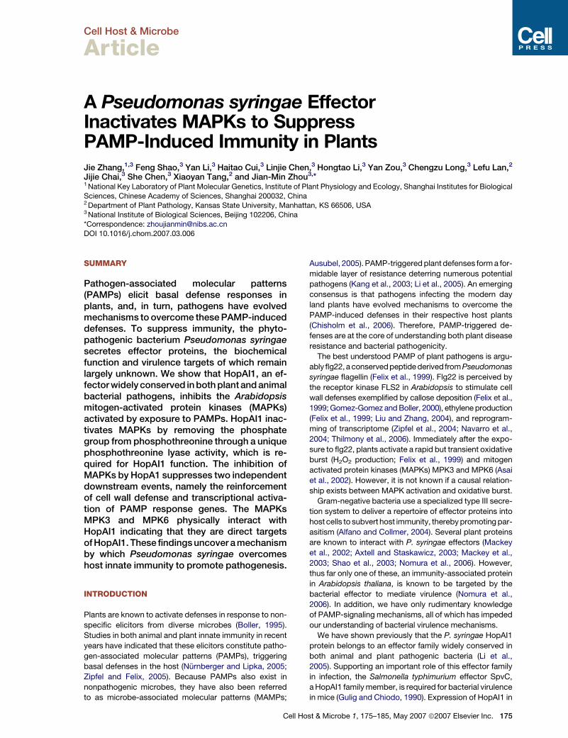
To determine if HopAI1 plays a role in bacterial viru-
lence, we generated a knockout mutant in strain 0288-9
by using homologous recombination. Because 0288-9 is
only modestly virulent on Arabidopsis but highly virulent
on tomato plants, we tested the ability of the mutant strain
to grow in tomato plants. The mutant bacteria consistently
multiplied to a lower level compared with WT bacteria
(Figure 1A). The difference is statistically significant
(Student’s t test, p value %0.01), indicating a positive
role of hopAI1 in 0288-9 virulence.
HopAI1 Suppresses Both PAMP-Induced Gene
Expression and Callose Deposition
Previously we showed that estrogen-inducible expression
of a FLAG-tagged HopAI1 transgene in plants enhanced
176 Cell Host & Microbe 1, 175–185, May 2007 ª2007 Elsevie
susceptibility to a nonpathogenic P. syringae mutant (Li
et al., 2005), suggesting a loss of PAMP-induced resis-
tance. We examined flg22-induced disease resistance in
plants expressing HopAI1. Pretreatment of flg22 protects
Arabidopsis plants from a subsequent infection by virulent
bacteria (Zipfel et al., 2004). While the flg22-pretreatment
completely inhibited the multiplication of the virulent
P. syringae strain DC3000 in the wild-type plants, the same
treatment did not protect in the HopAI1-expressing plants
(Figure 1B), indicating that HopAI1 completely suppresses
disease resistance activated by flg22.
To understand the function of HopAI1 in plants, we
sought to characterize in detail the flg22-induced gene ex-
pression and callose deposition when HopAI1 is directly
expressed in plants. Figure S2A shows that transient
expression of a FLAG-tagged HopAI1 in protoplasts
blocked the flg22-induced expression of the FRK1-LUC
reporter gene. FRK1 encodes a receptor-like kinase that
is induced rapidly in response to PAMPs (Asai et al.,
2002). The suppression is not caused by a general disrup-
tion of cellular activities, because HopAI1 does not affect
the LUC reporter gene expression under the control of
the CaMV 35S promoter (Figure S2B). We further ex-
panded this study in HopAI1 transgenic plants by
examining the expression of flg22-inducible genes includ-
ing FRK1, WRKY22, WRKY29, At1g13110, At1g30700,
At2g35930, At2g39200, At5g39580, and At5g44910
(Asai et al., 2002; http://Arabidopsis.org/info/expression/
ATGenExpress.jsp). These genes encode functionally di-
verse proteins and serve as a good survey of gene expres-
sion profiles. The expression of HopAI1 reduced flg22-
induced expression of all genes tested (Figures 1C). The
stronger suppression of flg22-induced genes by HopAI1
in protoplasts than transgenic plants might be caused
by higher-level HopAI1 expression associated with this
transient assay. The estradiol-inducible expression of
HopAI1 also blocked flg22-induced callose deposition
(Figure 1D). PAMP-induced callose deposition is required
for Arabidopsis resistance to P. syringae bacteria (Kim
et al., 2005). These analyses indicated that the plant-
expressed HopAI1 broadly suppresses flg22-induced
defenses.
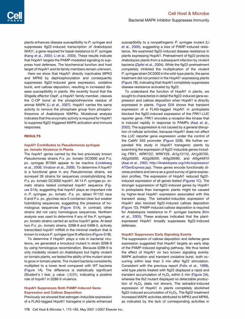
HopAI1 Suppresses Early Signaling Events
The suppression of callose deposition and defense gene
expression suggested that HopAI1 targets an early step
of the PAMP-induced signaling pathway. We thus tested
the effect of HopAI1 on two known signaling events:
MAPK activation and transient oxidative burst, both oc-
curring within less than 5 min after flg22 stimulation.
Consistent with the previous report (Felix et al., 1999),
wild-type plants treated with flg22 displayed a rapid and
transient accumulation of H2O2 within 5 min (Figure 2A),
whereas the fls2 mutant displayed no detectable produc-
tion of H2O2 (data not shown). The estradiol-induced
expression of HopAI1 in plants completely abolished
flg22-induced accumulation of H2O2. The flg22-treatment
increased MAPK activities attributed to MPK3 and MPK6,
as indicated by the lack of corresponding activities in
r Inc.
Cell Host & Microbe
Bacterial MAPK Inhibitor Suppresses Immunity
Figure 1. HopAI1 Contributes to Bacterial Virulence and Abolishes Flg22-Induced Resistance by Globally Suppressing Defenses
(A) HopAI1 is required for full virulence of P.s. pv. tomato strain 0288-9. Tomato plants were infiltrated with the WT or hopAI1� mutant bacteria
(105 CFU/ml), and leaf bacterial population was determined at the indicated times. Error bars indicate standard deviation. The experiment was
done twice with similar results.
(B) Expression of HopAI1 abolishes flg22-induced resistance to P. syringae. Wild-type (WT) and HopAI1 transgenic plants were presprayed with es-
tradiol or H2O for 12 hr, infiltrated with 1 mM flg22 or H2O for one day, and infiltrated with P. syringae DC3000 bacteria. Each data point consisted of at
least four replicates. Error bars indicate standard deviation. Two independent experiments were performed with similar results.
(C) HopAI1 suppresses flg22-induced gene expression. WT and HopAI1 transgenic plants were presprayed with estradiol for 24 hr before infiltrated
with H2O or 1 mM flg22 (flg) for the indicated times, and RNA was extracted for northern analyses using the indicated probes. A representative ethidium
bromide stain is shown for equal loading of RNA. The experiment was repeated three times with similar results.
(D) HopAI1 suppresses flg22-induced callose deposition. WT and HopAI1 transgenic plants were presprayed with estradiol or H2O, treated with
flg22, and leaves were stained with alanine blue for callose. Microscopic photographs of callose deposits were shown with number of callose
deposits indicated below each photograph. Error bars indicate standard deviation. The results are a representative of five independent
experiments.
atmpk3 and atmpk6 mutants (Figure 2B). The expression
of HopAI1 completely abolished both MPK3 and MPK6
activities (Figure 2B). The diminished MAPK activity was
not caused by a reduced MPK3 and MPK6 protein levels
as indicated by western analysis (Figure 2B). Transient
expression of HopAI1 in protoplasts also abolished flg22-
Cell H
induced MAPK activity (see below). These results are sur-
prising, as a recent paper by He et al. (2006) reported that
HopAI1 does not inhibit MAPKs. An examination of the an-
notated hopAI1 sequence suggested that the discrepancy
might be caused by an extra N-terminal sequence
(Figure S3A) present in our construct but missing in the
ost & Microbe 1, 175–185, May 2007 ª2007 Elsevier Inc. 177
Cell Host & Microbe
Bacterial MAPK Inhibitor Suppresses Immunity
annotated sequence and the construct used by He et al.
(2006). Because this sequence is highly conserved in all
the HopAI1 family members in various bacteria, the pre-
dicted amino acid sequence in the P. syringae database
is likely annotated incorrectly. To determine if the N-termi-
nal sequence is required for the activity, an N-terminal
truncated construct (DNHopAI1) lacking this sequence
was generated and expressed in protoplasts. However,
DNHopAI1 still abolished the flg22-induced MAPK activity
(Figure S3B), indicating that this N-terminal sequence is
dispensable for MAPK inhibition. In total, we conducted
8 independent assays with transgenic HopAI1 plants
and 8 assays with protoplasts expressing sequence-
Figure 2. HopAI1 Acts Very Early to Suppress PAMP
Signaling(A) HopAI1 suppresses flg22-induced oxidative burst. WT and HopAI1
transgenic plants were presprayed with estradiol or H2O. Leaf strips
were then treated with flg22, and H2O2 accumulation was measured
(expressed as relative units of luminescence). Error bars indicate stan-
dard deviation. The experiments were repeated 3 times with similar
results.
(B) HopAI1 suppresses PAMP-triggered activation of MPK3 and
MPK6. WT, HopAI1 transgenic, and the atmpk6 (m6) and atmpk3
(m3) mutant plants (carrying T-DNA insertions in respective genes)
were presprayed with H2O (�) or estradiol, treated with flg22 for 10
min, and protein was extracted for in-gel kinase assay using myelin ba-
sic protein as a substrate. Equal amounts of extracts were also sub-
jected to western blot analysis using anti-FLAG, anti-MPK3, and
anti-MPK6 antibodies. The arrow indicates the position of MPK6.
The amounts of protein loaded are indicated by the Coomassie Blue
staining of Rubisco.
178 Cell Host & Microbe 1, 175–185, May 2007 ª2007 Elsevier
confirmed full-length (3 clones) and DNHopAI1 (2 clones)
constructs. In each case, the flg22-induced MAPK activity
was abolished. We thereby conclude that HopAI1 inhibits
the MAPK activation. The suppression of oxidative burst
and inhibition of MAPK indicate that HopAI1 targets
a very early step in the PAMP-signaling pathway to block
host defenses.
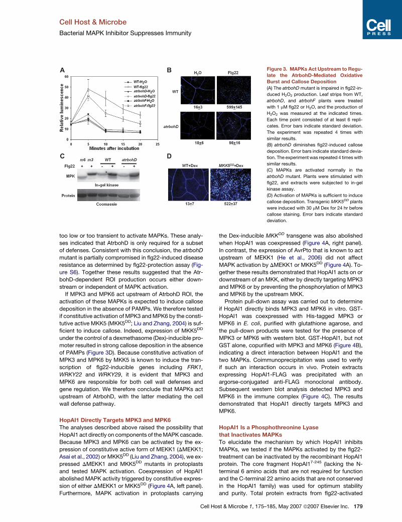
MAPKs Act Upstream of Oxidative Burst
It was proposed that ROI produced during oxidative burst
might act upstream to activate MAPKs (Kovtun et al.,
2000). It is therefore possible that HopAI1 targets a host
protein(s) required for oxidative burst that consequently
inhibits MAPK. Alternatively, MAPK may act upstream of
ROI production.
To determine if oxidative burst acts upstream of MAPK
activation, we first determined the source of ROI produc-
tion in flg22-induced tissues. In plants, NADPH oxidases,
peroxidases, and ROI scavenging systems can all contrib-
ute to the increase of ROIs in response to pathogen infec-
tion (Torres et al., 2006). Arabidopsis contains 10 genes en-
coding NADPH oxidases named respiratory burst oxidase
homologs (Atrboh; Torres and Dangl, 2005). The AtrbohD
and AtrbohF are required for ROI production in plants chal-
lenged with incompatible pathogens (Torres et al., 2002). A
recent study shows that ROIs produced by AtrbohD and
AtrbohF suppress cell death (Torres et al., 2005). Whether
these two genes play a role in PAMP-induced defenses
has not been examined. We therefore tested if they are re-
quired for ROI production in response to flg22. In contrast
to the strong accumulation of H2O2 in the wild-type plants,
the atrbohD mutant was completely impaired in this oxida-
tive burst (Figure 3A). The atrbohF mutant showed a minor
reduction in H2O2 accumulation. The results indicated that
AtrbohD is essential for flg22-induced oxidative burst,
whereas AtrbohF plays a minor role.
The flg22-induced callose deposition was examined in
the atrbohD mutant plants. In contrast to the strong
flg22-induced callose deposition in the wild-type plants,
the atrbohD mutant exhibited significantly reduced callose
deposition (approximately 16% of the wild-type; Fig-
ure 3B), suggesting that the AtrbohD-dependent ROI
production mediates callose deposition. Consistent with
this possibility, pretreatment of the wild-type plants with
diphenylene iodonium (DPI), a NADPH oxidase inhibitor,
abolished the flg22-induced callose deposition (Fig-
ure S4). Together these results demonstrated that
AtrbohD ROI regulates cell wall defense exemplified by
callose deposition.
We next determined if flg22-induced gene expression
was compromised in the atrbohD mutant by northern anal-
ysis. Interestingly, both the wild-type and atrbohD mutant
plants showed normal induction of all genes tested (Fig-
ure S5). As several of these genes are induced down-
stream of MPK3 and MPK6 (He et al., 2006), the results
suggest that AtrbohD is not required for MAPK activation.
Indeed, the flg22-induced MAPK activation was com-
pletely normal in the atrbohD mutant (Figure 3C), indicat-
ing that the AtrbohD-dependent ROI production is either
Inc.
Cell Host & Microbe
Bacterial MAPK Inhibitor Suppresses Immunity
Figure 3. MAPKs Act Upstream to Regu-
late the AtrbohD-Mediated Oxidative
Burst and Callose Deposition
(A) The atrbohD mutant is impaired in flg22-in-
duced H2O2 production. Leaf strips from WT,
atrbohD, and atrbohF plants were treated
with 1 mM flg22 or H2O, and the production of
H2O2 was measured at the indicated times.
Each time point consisted of at least 6 repli-
cates. Error bars indicate standard deviation.
The experiment was repeated 4 times with
similar results.
(B) atrbohD diminishes flg22-induced callose
deposition. Error bars indicate standard devia-
tion. The experiment was repeated 4 times with
similar results.
(C) MAPKs are activated normally in the
atrbohD mutant. Plants were stimulated with
flg22, and extracts were subjected to in-gel
kinase assay.
(D) Activation of MAPKs is sufficient to induce
callose deposition. Transgenic MKK5DD plants
were induced with 30 mM Dex for 24 hr before
callose staining. Error bars indicate standard
deviation.
too low or too transient to activate MAPKs. These analy-
ses indicated that AtrbohD is only required for a subset
of defenses. Consistent with this conclusion, the atrbohD
mutant is partially compromised in flg22-induced disease
resistance as determined by flg22-protection assay (Fig-
ure S6). Together these results suggested that the Atr-
bohD-dependent ROI production occurs either down-
stream or independent of MAPK activation.
If MPK3 and MPK6 act upstream of AtrbohD ROI, the
activation of these MAPKs is expected to induce callose
deposition in the absence of PAMPs. We therefore tested
if constitutive activation of MPK3 and MPK6 by the consti-
tutive active MKK5 (MKK5DD; Liu and Zhang, 2004) is suf-
ficient to induce callose. Indeed, expression of MKK5DD
under the control of a dexmethasome (Dex)-inducible pro-
moter resulted in strong callose deposition in the absence
of PAMPs (Figure 3D). Because constitutive activation of
MPK3 and MPK6 by MKK5 is known to induce the tran-
scription of flg22-inducible genes including FRK1,
WRKY22 and WRKY29, it is evident that MPK3 and
MPK6 are responsible for both cell wall defenses and
gene regulation. We therefore conclude that MAPKs act
upstream of AtrbohD, with the latter mediating the cell
wall defense pathway.
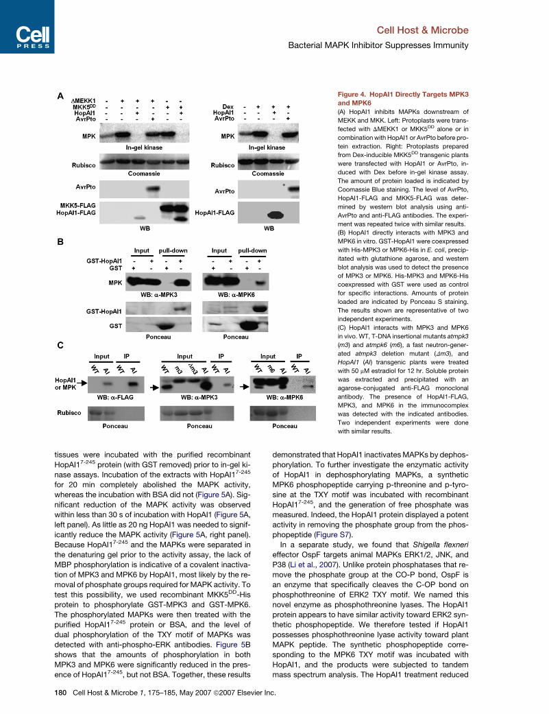
HopAI1 Directly Targets MPK3 and MPK6
The analyses described above raised the possibility that
HopAI1 act directly on components of the MAPK cascade.
Because MPK3 and MPK6 can be activated by the ex-
pression of constitutive active form of MEKK1 (DMEKK1;
Asai et al., 2002) or MKK5DD (Liu and Zhang, 2004), we ex-
pressed DMEKK1 and MKK5DD mutants in protoplasts
and tested MAPK activation. Coexpression of HopAI1
abolished MAPK activity triggered by constitutive expres-
sion of either DMEKK1 or MKK5DD (Figure 4A, left panel).
Furthermore, MAPK activation in protoplasts carrying
Cell H
the Dex-inducible MKKDD transgene was also abolished
when HopAI1 was coexpressed (Figure 4A, right panel).
In contrast, the expression of AvrPto that is known to act
upstream of MEKK1 (He et al., 2006) did not affect
MAPK activation by DMEKK1 or MKK5DD (Figure 4A). To-
gether these results demonstrated that HopAI1 acts on or
downstream of an MKK, either by directly targeting MPK3
and MPK6 or by preventing the phosphorylation of MPK3
and MPK6 by the upstream MKK.
Protein pull-down assay was carried out to determine
if HopAI1 directly binds MPK3 and MPK6 in vitro. GST-
HopAI1 was coexpressed with His-tagged MPK3 or
MPK6 in E. coli, purified with glutathione agarose, and
the pull-down products were tested for the presence of
MPK3 or MPK6 with western blot. GST-HopAI1, but not
GST alone, copurified with MPK3 and MPK6 (Figure 4B),
indicating a direct interaction between HopAI1 and the
two MAPKs. Coimmunoprecipitation was used to verify
if such an interaction occurs in vivo. Protein extracts
expressing HopAI1-FLAG was precipitated with an
argorse-conjugated anti-FLAG monoclonal antibody.
Subsequent western blot analysis detected MPK3 and
MPK6 in the immune complex (Figure 4C). The results
demonstrated that HopAI1 directly targets MPK3 and
MPK6.
HopAI1 Is a Phosphothreonine Lyase
that Inactivates MAPKs
To elucidate the mechanism by which HopAI1 inhibits
MAPKs, we tested if the MAPKs activated by the flg22-
treatment can be inactivated by the recombinant HopAI1
protein. The core fragment HopAI17-245 (lacking the N-
terminal 6 amino acids that are not required for function
and the C-terminal 22 amino acids that are not conserved
in the HopAI1 family) was used for optimum stability
and purity. Total protein extracts from flg22-activated
ost & Microbe 1, 175–185, May 2007 ª2007 Elsevier Inc. 179
Cell Host & Microbe
Bacterial MAPK Inhibitor Suppresses Immunity
Figure 4. HopAI1 Directly Targets MPK3
and MPK6
(A) HopAI1 inhibits MAPKs downstream of
MEKK and MKK. Left: Protoplasts were trans-
fected with DMEKK1 or MKK5DD alone or in
combination with HopAI1 or AvrPto before pro-
tein extraction. Right: Protoplasts prepared
from Dex-inducible MKK5DD transgenic plants
were transfected with HopAI1 or AvrPto, in-
duced with Dex before in-gel kinase assay.
The amount of protein loaded is indicated by
Coomassie Blue staining. The level of AvrPto,
HopAI1-FLAG and MKK5-FLAG was deter-
mined by western blot analysis using anti-
AvrPto and anti-FLAG antibodies. The experi-
ment was repeated twice with similar results.
(B) HopAI1 directly interacts with MPK3 and
MPK6 in vitro. GST-HopAI1 were coexpressed
with His-MPK3 or MPK6-His in E. coli, precip-
itated with glutathione agarose, and western
blot analysis was used to detect the presence
of MPK3 or MPK6. His-MPK3 and MPK6-His
coexpressed with GST were used as control
for specific interactions. Amounts of protein
loaded are indicated by Ponceau S staining.
The results shown are representative of two
independent experiments.
(C) HopAI1 interacts with MPK3 and MPK6
in vivo. WT, T-DNA insertional mutants atmpk3
(m3) and atmpk6 (m6), a fast neutron-gener-
ated atmpk3 deletion mutant (Dm3), and
HopAI1 (AI) transgenic plants were treated
with 50 mM estradiol for 12 hr. Soluble protein
was extracted and precipitated with an
agarose-conjugated anti-FLAG monoclonal
antibody. The presence of HopAI1-FLAG,
MPK3, and MPK6 in the immunocomplex
was detected with the indicated antibodies.
Two independent experiments were done
with similar results.
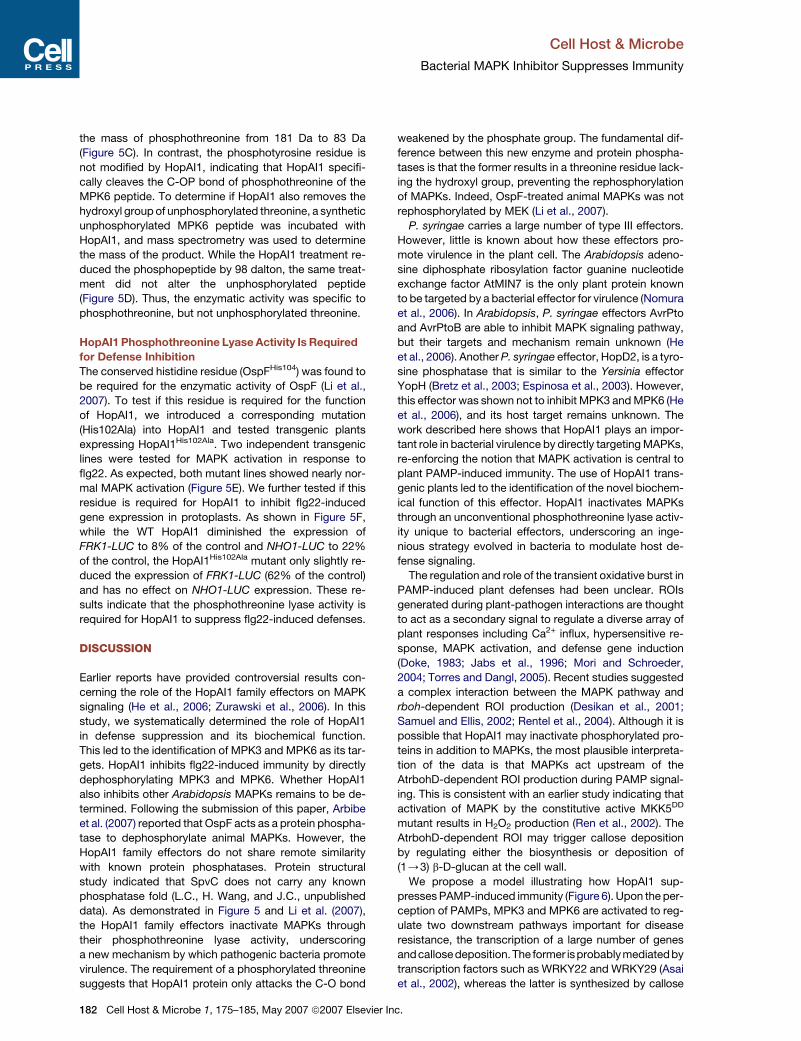
tissues were incubated with the purified recombinant
HopAI17-245 protein (with GST removed) prior to in-gel ki-
nase assays. Incubation of the extracts with HopAI17-245
for 20 min completely abolished the MAPK activity,
whereas the incubation with BSA did not (Figure 5A). Sig-
nificant reduction of the MAPK activity was observed
within less than 30 s of incubation with HopAI1 (Figure 5A,
left panel). As little as 20 ng HopAI1 was needed to signif-
icantly reduce the MAPK activity (Figure 5A, right panel).
Because HopAI17-245 and the MAPKs were separated in
the denaturing gel prior to the activity assay, the lack of
MBP phosphorylation is indicative of a covalent inactiva-
tion of MPK3 and MPK6 by HopAI1, most likely by the re-
moval of phosphate groups required for MAPK activity. To
test this possibility, we used recombinant MKK5DD-His
protein to phosphorylate GST-MPK3 and GST-MPK6.
The phosphorylated MAPKs were then treated with the
purified HopAI17-245 protein or BSA, and the level of
dual phosphorylation of the TXY motif of MAPKs was
detected with anti-phospho-ERK antibodies. Figure 5B
shows that the amounts of phosphorylation in both
MPK3 and MPK6 were significantly reduced in the pres-
ence of HopAI17-245, but not BSA. Together, these results
180 Cell Host & Microbe 1, 175–185, May 2007 ª2007 Elsevie
demonstrated that HopAI1 inactivates MAPKs by dephos-
phorylation. To further investigate the enzymatic activity
of HopAI1 in dephosphorylating MAPKs, a synthetic
MPK6 phosphopeptide carrying p-threonine and p-tyro-
sine at the TXY motif was incubated with recombinant
HopAI17-245, and the generation of free phosphate was
measured. Indeed, the HopAI1 protein displayed a potent
activity in removing the phosphate group from the phos-
phopeptide (Figure S7).
In a separate study, we found that Shigella flexneri
effector OspF targets animal MAPKs ERK1/2, JNK, and
P38 (Li et al., 2007). Unlike protein phosphatases that re-
move the phosphate group at the CO-P bond, OspF is
an enzyme that specifically cleaves the C-OP bond on
phosphothreonine of ERK2 TXY motif. We named this
novel enzyme as phosphothreonine lyases. The HopAI1
protein appears to have similar activity toward ERK2 syn-
thetic phosphopeptide. We therefore tested if HopAI1
possesses phosphothreonine lyase activity toward plant
MAPK peptide. The synthetic phosphopeptide corre-
sponding to the MPK6 TXY motif was incubated with
HopAI1, and the products were subjected to tandem
mass spectrum analysis. The HopAI1 treatment reduced
r Inc.

Cell Host & Microbe
Bacterial MAPK Inhibitor Suppresses Immunity
Figure 5. Purified HopAI1 Inactivates MPK3 and MPK6 by Dephosphorylation
(A) Left: total protein extract from flg22-induced WT plants was incubated with 10 mg purifed recombinant HopAI17-245 protein for the indicated times
before in-gel kinase assay. Right: the extract was incubated with indicated amounts of HopAI17-245 or BSA for 20 min before in-gel kinase assay.
(B) HopAI1 dephosphorylates MPK3 and MPK6 in vitro. Purified GST-MPK3 and GST-MPK6 were phosphorylated with MKK5DD-His. The reaction
was terminated by adding 10 mM EDTA before incubated with purified HopAI17-245 or BSA. Amounts of MAPK phosphorylation were detected
with anti-pERK antibodies. The dephosphorylated MAPKs were also subject to in-gel kinase assay. The amounts of GST-MPK and HopAI1 protein
were indicated by Ponceau S staining.
(C) HopAI1 is a phosphothreonine lyase. The HopAI17-245-treated synthetic MPK6 phosphopeptide (SESDFM-pT-E-pYVVTR) was analyzed by tan-
dem mass spectrometry. The fragmentation profile was indicated by the b and y ions. TD in the indicated peptide sequence denotes the modification
as a result of the treatment.
(D) The enzymatic activity of HopAI1 is specific to phosphorylated MPK6 peptide. Phosphorylated and unphosphorylated peptides were treated with
HopAI17-245, and the mass of the resulting peptides was compared with untreated peptides by mass spectrometry.
(E) HopAI1His102Ala does not inhibit MAPK activation. Two independent transgenic lines carrying FLAG-tagged HopAI1His102Ala (m-5 and m-7) in the T1
generation were induced by estradiol, treated with flg22 peptide, and MAPK activity was compared with that in WT and transgenic plants expressing
the WT HopAI1. The amounts of HopAI1-FLAG, MPK3, and MPK6 (arrow) protein were determined with the indicated antibodies. A representative
protein blot was stained with Ponceau S to show equal loading of protein.
(F) The conserved amino acid required for phosphothreonine lyase activity is necessary for HopAI1 to suppress flg22-induced gene expression. Pro-
toplasts prepared from WT plants were cotransfected with the FRK1-LUC reporter construct and WT HopAI1, HopAI1His102Ala or an empty vector.
Protoplasts prepared from NHO1-LUC transgenic plants were transfected with WT HopAI1, HopAI1His102Ala or an empty vector. The transfected pro-
toplasts were induced with flg22 for 3 hr for FRK1-LUC and 6 hr for NHO1-LUC before the LUC activity was determined.
Cell Host & Microbe 1, 175–185, May 2007 ª2007 Elsevier Inc. 181
Cell Host & Microbe
Bacterial MAPK Inhibitor Suppresses Immunity
the mass of phosphothreonine from 181 Da to 83 Da
(Figure 5C). In contrast, the phosphotyrosine residue is
not modified by HopAI1, indicating that HopAI1 specifi-
cally cleaves the C-OP bond of phosphothreonine of the
MPK6 peptide. To determine if HopAI1 also removes the
hydroxyl group of unphosphorylated threonine, a synthetic
unphosphorylated MPK6 peptide was incubated with
HopAI1, and mass spectrometry was used to determine
the mass of the product. While the HopAI1 treatment re-
duced the phosphopeptide by 98 dalton, the same treat-
ment did not alter the unphosphorylated peptide
(Figure 5D). Thus, the enzymatic activity was specific to
phosphothreonine, but not unphosphorylated threonine.
HopAI1 Phosphothreonine Lyase Activity Is Required
for Defense Inhibition
The conserved histidine residue (OspFHis104) was found to
be required for the enzymatic activity of OspF (Li et al.,
2007). To test if this residue is required for the function
of HopAI1, we introduced a corresponding mutation
(His102Ala) into HopAI1 and tested transgenic plants
expressing HopAI1His102Ala. Two independent transgenic
lines were tested for MAPK activation in response to
flg22. As expected, both mutant lines showed nearly nor-
mal MAPK activation (Figure 5E). We further tested if this
residue is required for HopAI1 to inhibit flg22-induced
gene expression in protoplasts. As shown in Figure 5F,
while the WT HopAI1 diminished the expression of
FRK1-LUC to 8% of the control and NHO1-LUC to 22%
of the control, the HopAI1His102Ala mutant only slightly re-
duced the expression of FRK1-LUC (62% of the control)
and has no effect on NHO1-LUC expression. These re-
sults indicate that the phosphothreonine lyase activity is
required for HopAI1 to suppress flg22-induced defenses.
DISCUSSION
Earlier reports have provided controversial results con-
cerning the role of the HopAI1 family effectors on MAPK
signaling (He et al., 2006; Zurawski et al., 2006). In this
study, we systematically determined the role of HopAI1
in defense suppression and its biochemical function.
This led to the identification of MPK3 and MPK6 as its tar-
gets. HopAI1 inhibits flg22-induced immunity by directly
dephosphorylating MPK3 and MPK6. Whether HopAI1
also inhibits other Arabidopsis MAPKs remains to be de-
termined. Following the submission of this paper, Arbibe
et al. (2007) reported that OspF acts as a protein phospha-
tase to dephosphorylate animal MAPKs. However, the
HopAI1 family effectors do not share remote similarity
with known protein phosphatases. Protein structural
study indicated that SpvC does not carry any known
phosphatase fold (L.C., H. Wang, and J.C., unpublished
data). As demonstrated in Figure 5 and Li et al. (2007),
the HopAI1 family effectors inactivate MAPKs through
their phosphothreonine lyase activity, underscoring
a new mechanism by which pathogenic bacteria promote
virulence. The requirement of a phosphorylated threonine
suggests that HopAI1 protein only attacks the C-O bond
182 Cell Host & Microbe 1, 175–185, May 2007 ª2007 Elsevier
weakened by the phosphate group. The fundamental dif-
ference between this new enzyme and protein phospha-
tases is that the former results in a threonine residue lack-
ing the hydroxyl group, preventing the rephosphorylation
of MAPKs. Indeed, OspF-treated animal MAPKs was not
rephosphorylated by MEK (Li et al., 2007).
P. syringae carries a large number of type III effectors.
However, little is known about how these effectors pro-
mote virulence in the plant cell. The Arabidopsis adeno-
sine diphosphate ribosylation factor guanine nucleotide
exchange factor AtMIN7 is the only plant protein known
to be targeted by a bacterial effector for virulence (Nomura
et al., 2006). In Arabidopsis, P. syringae effectors AvrPto
and AvrPtoB are able to inhibit MAPK signaling pathway,
but their targets and mechanism remain unknown (He
et al., 2006). Another P. syringae effector, HopD2, is a tyro-
sine phosphatase that is similar to the Yersinia effector
YopH (Bretz et al., 2003; Espinosa et al., 2003). However,
this effector was shown not to inhibit MPK3 and MPK6 (He
et al., 2006), and its host target remains unknown. The
work described here shows that HopAI1 plays an impor-
tant role in bacterial virulence by directly targeting MAPKs,
re-enforcing the notion that MAPK activation is central to
plant PAMP-induced immunity. The use of HopAI1 trans-
genic plants led to the identification of the novel biochem-
ical function of this effector. HopAI1 inactivates MAPKs
through an unconventional phosphothreonine lyase activ-
ity unique to bacterial effectors, underscoring an inge-
nious strategy evolved in bacteria to modulate host de-
fense signaling.
The regulation and role of the transient oxidative burst in
PAMP-induced plant defenses had been unclear. ROIs
generated during plant-pathogen interactions are thought
to act as a secondary signal to regulate a diverse array of
plant responses including Ca2+ influx, hypersensitive re-
sponse, MAPK activation, and defense gene induction
(Doke, 1983; Jabs et al., 1996; Mori and Schroeder,
2004; Torres and Dangl, 2005). Recent studies suggested
a complex interaction between the MAPK pathway and
rboh-dependent ROI production (Desikan et al., 2001;
Samuel and Ellis, 2002; Rentel et al., 2004). Although it is
possible that HopAI1 may inactivate phosphorylated pro-
teins in addition to MAPKs, the most plausible interpreta-
tion of the data is that MAPKs act upstream of the
AtrbohD-dependent ROI production during PAMP signal-
ing. This is consistent with an earlier study indicating that
activation of MAPK by the constitutive active MKK5DD
mutant results in H2O2 production (Ren et al., 2002). The
AtrbohD-dependent ROI may trigger callose deposition
by regulating either the biosynthesis or deposition of
(1/3) b-D-glucan at the cell wall.
We propose a model illustrating how HopAI1 sup-
presses PAMP-induced immunity (Figure 6). Upon the per-
ception of PAMPs, MPK3 and MPK6 are activated to reg-
ulate two downstream pathways important for disease
resistance, the transcription of a large number of genes
andcallose deposition.The former isprobably mediatedby
transcription factors such as WRKY22 and WRKY29 (Asai
et al., 2002), whereas the latter is synthesized by callose
Inc.
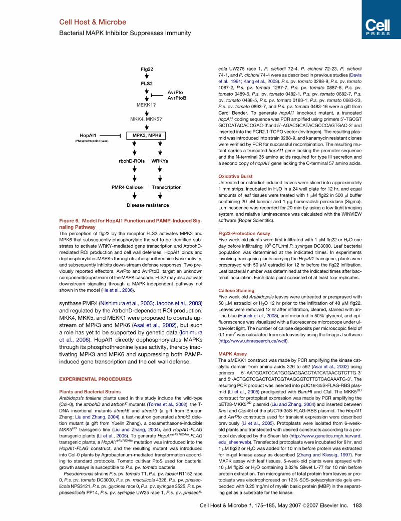
Cell Host & Microbe
Bacterial MAPK Inhibitor Suppresses Immunity
synthase PMR4 (Nishimura et al., 2003; Jacobs et al., 2003)
and regulated by the AtrbohD-dependent ROI production.
MKK4, MKK5, and MEKK1 were proposed to operate up-
stream of MPK3 and MPK6 (Asai et al., 2002), but such
a role has yet to be supported by genetic data (Ichimura
et al., 2006). HopAI1 directly dephosphorylates MAPKs
through its phosphothreonine lyase activity, thereby inac-
tivating MPK3 and MPK6 and suppressing both PAMP-
induced gene transcription and the cell wall defense.
EXPERIMENTAL PROCEDURES
Plants and Bacterial Strains
Arabidopsis thaliana plants used in this study include the wild-type
(Col-0), the atrbohD and atrbohF mutants (Torres et al., 2002), the T-
DNA insertional mutants atmpk6 and atmpk3 (a gift from Shuqun
Zhang; Liu and Zhang, 2004), a fast-neutron generated atmpk3 dele-
tion mutant (a gift from Yuelin Zhang), a dexamethasone-inducible
MKK5DD transgenic line (Liu and Zhang, 2004), and HopAI1-FLAG
transgenic plants (Li et al., 2005). To generate HopAI1His102Ala-FLAG
transgenic plants, a HopAI1His102Ala mutation was introduced into the
HopAI1-FLAG construct, and the resulting mutant was introduced
into Col-0 plants by Agrobacterium-mediated transformation accord-
ing to standard protocols. Tomato cultivar PtoS used for bacterial
growth assays is susceptible to P.s. pv. tomato bacteria.
Pseudomonas strains P.s. pv. tomato T1, P.s. pv. tabaci R1152 race
0, P.s. pv. tomato DC3000, P.s. pv. maculicola 4326, P.s. pv. phaseo-
licola NPS3121, P.s. pv. glycinea race 0, P.s. pv. syringae 3525, P.s. pv.
phaseolicola PP14, P.s. pv. syringae UW25 race 1, P.s. pv. phaseoli-
Figure 6. Model for HopAI1 Function and PAMP-Induced Sig-
naling Pathway
The perception of flg22 by the receptor FLS2 activates MPK3 and
MPK6 that subsequently phosphorylate the yet to be identified sub-
strates to activate WRKY-mediated gene transcription and AtrbohD-
mediated ROI production and cell wall defenses. HopAI1 binds and
dephosphorylates MAPKs through its phosphothreonine lyase activity,
and subsequently inhibits down-stream defense responses. Two pre-
viously reported effectors, AvrPto and AvrPtoB, target an unknown
component(s) upstream of the MAPK cascade. FLS2 may also activate
downstream signaling through a MAPK-independent pathway not
shown in the model (He et al., 2006).
Cell
cola UW275 race 1, P. cichorii 72-4, P. cichorii 72-23, P. cichorii
74-1, and P. cichorii 74-4 were as described in previous studies (Davis
et al., 1991; Kang et al., 2003). P.s. pv. tomato 0288-9, P.s. pv. tomato
1087-2, P.s. pv. tomato 1287-7, P.s. pv. tomato 0887-6, P.s. pv.
tomato 0489-5, P.s. pv. tomato 0482-1, P.s. pv. tomato 0682-7, P.s.
pv. tomato 0488-5, P.s. pv. tomato 0183-1, P.s. pv. tomato 0683-23,
P.s. pv. tomato 0893-7, and P.s. pv. tomato 0483-16 were a gift from
Carol Bender. To generate hopAI1 knockout mutant, a truncated
hopAI1 coding sequence was PCR amplified using primers 50-TGCGT
GCTCATACACCGAC-30and 50-AGACGCATACGCCCAGTGAC-30 and
inserted into the PCR2.1-TOPO vector (Invitrogen). The resulting plas-
mid was introduced into strain 0288-9, and kanamycin resistant clones
were verified by PCR for successful recombination. The resulting mu-
tant carries a truncated hopAI1 gene lacking the promoter sequence
and the N-terminal 35 amino acids required for type III secretion and
a second copy of hopAI1 gene lacking the C-terminal 57 amino acids.
Oxidative Burst
Untreated or estradiol-induced leaves were sliced into approximately
1 mm strips, incubated in H2O in a 24 well plate for 12 hr, and equal
amounts of leaf tissues were treated with 1 mM flg22 in 500 ml buffer
containing 20 mM luminol and 1 mg horseradish peroxidase (Sigma).
Luminescence was recorded for 20 min by using a low-light imaging
system, and relative luminescence was calculated with the WINVIEW
software (Roper Scientific).
Flg22-Protection Assay
Five-week-old plants were first infiltrated with 1 mM flg22 or H2O one
day before infiltrating 105 CFU/ml P. syringae DC3000. Leaf bacterial
population was determined at the indicated times. In experiments
involving transgenic plants carrying the HopAI1 transgene, plants were
presprayed with 50 mM estradiol for 12 hr before the flg22 infiltration.
Leaf bacterial number was determined at the indicated times after bac-
terial inoculation. Each data point consisted of at least four replicates.
Callose Staining
Five-week-old Arabidopsis leaves were untreated or presprayed with
50 mM estradiol or H2O 12 hr prior to the infiltration of 40 mM flg22.
Leaves were removed 12 hr after infiltration, cleared, stained with an-
iline blue (Hauck et al., 2003), and mounted in 50% glycerol, and epi-
fluorescence was visualized with a fluorescence microscope under ul-
traviolet light. The number of callose deposits per microscopic field of
0.1 mm2 was calculated from six leaves by using the Image J software
(http://www.uhnresearch.ca/wcif).
MAPK Assay
The DMEKK1 construct was made by PCR amplifying the kinase cat-
alytic domain from amino acids 326 to 592 (Asai et al., 2002) using
primers 50-AATGGATCCATGGGAGGAGCTATCATAACGTCTTG-30
and 50-ACTGGTCGACTCATGGTAAGGGTCTTCTCACAAATG-30. The
resulting PCR product was inserted into pUC19-35S-FLAG-RBS plas-
mid (Li et al., 2005) predigested with BamHI and ClaI. The MKK5DD
construct for protoplast expression was made by PCR amplifying the
pET28-MKK5DD plasmid (Liu and Zhang, 2004) and inserted between
XhoI and Csp45I of the pUC19-35S-FLAG-RBS plasmid. The HopAI1
and AvrPto constructs used for transient expression were described
previously (Li et al., 2005). Protoplasts were isolated from 6-week-
old plants and transfected with desired constructs according to a pro-
tocol developed by the Sheen lab (http://www.genetics.mgh.harvard.
edu_sheenweb). Transfected protoplasts were incubated for 6 hr, and
1 mM flg22 or H2O was added for 10 min before protein was extracted
for in-gel kinase assay as described (Zhang and Klessig, 1997). For
MAPK assay with leaf tissues, 5-week-old plants were sprayed with
10 mM flg22 or H2O containing 0.02% Silwet L-77 for 10 min before
protein extraction. Ten micrograms of total protein from leaves or pro-
toplasts was electrophoresed on 12% SDS-polyacrylamide gels em-
bedded with 0.25 mg/ml of myelin basic protein (MBP) in the separat-
ing gel as a substrate for the kinase.
Host & Microbe 1, 175–185, May 2007 ª2007 Elsevier Inc. 183
Cell Host & Microbe
Bacterial MAPK Inhibitor Suppresses Immunity
The level of HopAI1-FLAG, AvrPto, MPK3, and MPK6 proteins in the
extracts was determined by western blot analysis using anti-AvrPto
(Shan et al., 2000), anti-FLAG, anti-MPK3, or anti-MPK6 antibodies
(Sigma).
RNA Blot Analysis
Flg22-inducible genes were selected according to microarray data
deposited by T. Nurnberger (http://Arabidopsis.org/info/expression/
ATGenExpress.jsp). Gene specific primers were used to amplify
WRKY22, WRKY29, FRK1 (Asai et al., 2002), At1g13110(50-TGA
GATGCTAGTCGCTGGTG-30, 50-TGAACAAGTGGAACAAGCTC-30),
At1g30700(50-TGGAGGTTACGGTAACATG-30, 50-ACACAGGAATAC
TCTGTTCG-30), At2g35930(50-AGCAGGGATATGCAAGAATC-30, 50-
ACATCGTAAGCAACGACTC-30), At2g39200(50-TCAACCTTGACATG
CTCAAC-30, 50-AGGAGATGTGGTTAAAGGAG-30), At2g44370(50-ACT
TGAGACAAGCCTTTCTC-30, 50-ACCATCCATTGCGCAGTCAC-30),
At5g39580(50-AACCGCAGAGCAAACCCTAC-30, 50-TGTCTCTTGTG
CTGATATC-30), and At5g44910(50-ATTGAGAGACGAGAGAGTC-30,
50-ACAGTAAAGCTGGTGCAAG-30). Five-week-old plants were infil-
trated with H2O or 1 mM flg22 for the indicated hours before RNA isola-
tion. RNA blots were hybridized to radio-labeled PCR products for the
selected genes.
Expression of Recombinant Proteins in E. coli
and Protein Pull-Down Assay
Full length Arabidopsis MPK3 and MPK6 were PCR amplified from
cDNA and inserted between the BamHI and XhoI sites of pGEX-6p-1
(Pharmacia) to generate GST-MPK3, GST-MPK6, and NdeI and XhoI of
pET28b or pET30a (Novagen) to generate His-MPK3 and MPK6-His
constructs. HopAI17-245 was PCR-amplified from P. syringae DC3000
genomic DNA and inserted into pGEX-6p-1 plasmid (between BamHI
and XhoI) to generate GST- HopAI17-245. The respective recombinant
proteins were affinity purified following manufactures’ instructions.
For protein pull-down assay, His-MPK3 and MPK6-His were coex-
pressed with GST- HopAI17-245. Bacterial lysate was incubated with
glutathione agarose in a microcentrifuge tube, washed three times
with a buffer containing 25 mM Tris, PH8.0, 50 mM NaCL, and 3 mM
DTT. The bound protein was eluted with 15 mM GSH and analyzed
by using western blot.
Coimmunoprecipitation Assay
Five-weekoldplantswere sprayedwith 50mM estradiol for 12hr.Soluble
proteinwas extractedandprecipitated withanagarose-conjugated anti-
FLAG monoclonal antibody (Sigma) following manufacturer’s instruc-
tion. Western blot was used to detect the presence of HopAI1-FLAG,
MPK3, or MPK6 in the immunocomplex with a monoclonal anti-FLAG
antibody, or anti-MPK3 or anti-MPK6 antibodies (Sigma).
Reporter Assay in Protoplasts
Protoplasts were prepared from 6-week-old WT or NHO1-LUC trans-
genic plants (Li et al., 2005), transfected with the indicated plasmids,
induced with flg22, and examined for LUC activity as described (Li
et al., 2005).
MAPK Phosphorylation and Inactivation In Vitro
0.5 mg purified MKK5DD-His recombinant protein was incubated with
2 mg GST-MPK3 or GST-MPK6 in a kinase reaction buffer (Liu and
Zhang, 2004) at 22�C for 90 min before terminated by the addition of
10 mM EDTA. The phosphorylated GST-MPK3 and GST-MPK6
proteins were incubated with 5 mg purified HopAI17-245 or BSA for
10 min, boiled in SDS sample buffer before analyzed by using western
blot with anti-phospho-ERK antibodies (Cell Signaling).
Phosphothreonine Lyase Activity Assays
Synthetic MAPK phosphopeptide (SESDFM-pTE-pYVVTR; Sangon,
Shanghai) was incubated with 5 mg HopAI17-245 in a 50 ml reaction
buffer containing 10 mM HEPES (pH 7.4), 150 mM NaCl, and 1 mM
EDTA and incubated at 30�C, and the release of phosphate group
184 Cell Host & Microbe 1, 175–185, May 2007 ª2007 Elsevier
was determined by using Molybdate Dye (Promega). The specific
modification of phosphothreonine residue in the HopAI1-treated pep-
tide was determined by mass spectrometry and tandem mass spec-
trometry analyses.
Supplemental Data
The Supplemental Data include seven supplemental figures and can
be found with this article online at http://www.cellhostandmicrobe.
com/cgi/content/full/1/3/175/DC1/.
ACKNOWLEDGMENTS
The authors are grateful to Jonathan Jones for permission to use the
atrbohD and atrbohF mutants, Shuqun Zhang and Yuelin Zhang for
providing Arabidopsis mutants, Dongtao Ren for MKK5DD constructs
and transgenic seeds, and Carol Bender for Pseudomonas strains.
J.-M.Z. was supported by a grant from Chinese Ministry of Science
and Technology (2003-AA210080).
Received: October 29, 2006
Revised: January 28, 2007
Accepted: March 5, 2007
Published: May 16, 2007
REFERENCES
Alfano, J.R., and Collmer, A. (2004). Type III secretion system effector
proteins: Double agents in bacterial disease and plant defense. Annu.
Rev. Phytopathol. 42, 385–414.
Arbibe, L., Kim, D.W., Batsche, E., Pedron, T., Bogdan Mateescu, B.,
Christian Muchardt, C., Parsot, C., and Sansonetti, P.J. (2007). An
injected bacterial effector targets chromatin access for transcription
factor NF-kB to alter transcription of host genes involved in immune
responses. Nat. Immunol. 8, 47–56.
Asai, T., Tena, G., Plotnikova, J., Willmann, M.R., Chiu, W.-L., Gomez-
Gomez, L., Boller, T., Ausubel, F.M., and Sheen, J. (2002). MAP kin-
anse signaling cascade in Arabidopsis innate immunity. Nature 415,
977–983.
Ausubel, F.M. (2005). Are innate immune signaling pathways in plants
and animals conserved? Nat. Immunol. 6, 973–979.
Axtell, M.J., and Staskawicz, B.J. (2003). Initiation of RPS2-specified
disease resistance in Arabidopsis is coupled to the AvrRpt2-directed
elimination of RIN4. Cell 112, 369–377.
Boller, T. (1995). Chemoperception of microbial signals in plant cells.
Annu. Rev. Plant Physiol. Plant Mol. Biol. 46, 189–214.
Bretz, J.R., Mock, N.M., Charity, J.C., Zeyad, S., Baker, C.J., and
Hutcheson, S.W. (2003). A translocated protein tyrosine phosphatase
of Pseudomonas syringae pv. tomato DC3000 modulates plant de-
fence response to infection. Mol. Microbiol. 49, 389–400.
Chisholm, S.T., Coaker, G., Day, B., and Staskawicz, B.J. (2006). Host-
microbe interactions: Shaping the evolution of the plant immune re-
sponse. Cell 124, 803–814.
Davis, K.R., Schott, E., and Ausubel, F.M. (1991). Virulence of selected
phytopathogenic pseudomonads in Arabidopsis thaliana. Mol. Plant
Microbe Interact. 4, 477–488.
Desikan, R., Hancock, J.T., Ichimura, K., Shinozaki, K., and Neill, S.J.
(2001). Harpin induces activation of the Arabidopsis mitogenactivated
protein kinases AtMPK4 and AtMPK6. Plant Physiol. 126, 1579–1587.
Doke, N. (1983). Involvement of superoxide anion generation in the
hypersensitive response of potato tuber tissues to infection with an
incompatible race of Phytophthora infestans and to the hyphal wall
components. Physiol. Plant Pathol. 23, 345–357.
Espinosa, A., Guo, M., Tam, V.C., Fu, Z.Q., and Alfano, J.R. (2003). The
Pseudomonas syringae type III-secreted protein HopPtoD2 possesses
protein tyrosine phosphatase activity and suppresses programmed
cell death in plants. Mol. Microbiol. 49, 377–387.
Inc.
Cell Host & Microbe
Bacterial MAPK Inhibitor Suppresses Immunity
Felix, G., Duran, J.D., Volko, S., and Boller, T. (1999). Plants have a sen-
sitive perception system for the most conserved domain of bacterial
flagellin. Plant J. 18, 265–276.
Gomez-Gomez, L., and Boller, T. (2000). FLS2: An LRR receptor-like
kinase involved in the perception of the bacterial elicitor flagellin in
Arabidopsis. Mol. Cell 5, 1003–1011.
Gulig, P.A., and Chiodo, V.A. (1990). Genetic and DNA sequence anal-
ysis of the Salmonella typhimurium virulence plasmid gene encoding
the 28,000-molecular-weight protein. Infect. Immun. 58, 2651–2658.
Hauck, P., Thilmony, R., and He, S.Y. (2003). A Pseudomonas syringae
type III effector suppresses cell wall-based extracellular defense in
susceptible Arabidopsis plants. Proc. Natl. Acad. Sci. USA 100,
8577–8582.
He, P., Shan, L., Lin, N.C., Martin, G.B., Kemmerling, B., Nurnberger,
T., and Sheen, J. (2006). Specific Bacterial Suppressors of MAMP Sig-
naling Upstream of MAPKKK in Arabidopsis Innate Immunity. Cell 125,
563–575.
Ichimura, K., Casais, C., Peck, S.C., Shinozaki, K., and Shirasu, K.
(2006). MEKK1 is required for MPK4 activation and regulates tissue
specific and temperature dependent cell death in Arabidopsis.
J. Biol. Chem. 281, 36969–36976. Published online October 5, 2006.
10.1074/jbc.M605319200.
Jabs, T., Dietrich, R.A., and Dangl, J.L. (1996). Initiation of runaway cell
death in an Arabidopsis mutant by extracellular superoxide. Science
273, 1853–1856.
Jacobs, A.K., Lipka, V., Burton, R.A., Panstruga, R., Strizhov, N.,
Schulze-Lefert, P., and Fincher, G.B. (2003). An Arabidopsis callose
synthase, GSL5, is required for wound and papillary callose formation.
Plant Cell 15, 2503–2513.
Kang, L., Li, J., Zhao, T., Xiao, F., Tang, X., Thilmony, R., He, S., and
Zhou, J.M. (2003). Interplay of the Arabidopsis nonhost resistance
gene NHO1 with bacterial virulence. Proc. Natl. Acad. Sci. USA 100,
3519–3524.
Kim, M.G., da Cunha, L., McFall, A.J., Belkhadir, Y., DebRoy, S.,
Dangl, J.L., and Mackey, D. (2005). Two Pseudomonas syringae
type III effectors inhibit RIN4-regulated basal defense in Arabidopsis.
Cell 121, 749–759.
Kovtun, Y., Chiu, W.L., Tena, G., and Sheen, J. (2000). Functional anal-
ysis of oxidative stress-activated mitogen-activated protein kinase
cascade in plants. Proc. Natl. Acad. Sci. USA 97, 2940–2945.
Li, H., Xu, H., Zhou, Y., Zhang, J., Long, C., Li, S., Chen, S., Zhou, J.M.,
and Shao, F. (2007). The phosphothreonine lyase activity of a bacterial
type III effector family. Science 315, 1000–1003.
Li, X., Lin, H., Zhang, W., Zou, Y., Zhang, J., Tang, X., and Zhou, J.M.
(2005). Flagellin induces innate immunity in nonhost interactions that is
suppressed by Pseudomonas syringae effectors. Proc. Natl. Acad. Sci.
USA 102, 12990–12995.
Liu, Y., and Zhang, S. (2004). Phosphorylation of 1-aminocyclopro-
pane-1-carboxylic acid synthase by MPK6, a stress-responsive mito-
gen-activated protein kinase, induces ethylene biosynthesis in Arabi-
dopsis. Plant Cell 16, 3386–3399.
Lindeberg, M., Cartinhour, S., Myers, C.R., Schechter, L.M.,
Schneider, D.J., and Collmer, A. (2006). Closing the circle on the dis-
covery of genes encoding Hrp regulon members and Type III Secretion
System effectors in the genomes of three model Pseudomonas syrin-
gae strains. Mol. Plant Microbe Interact. 19, 1151–1158.
Mackey, D., Belkhadir, Y., Alonso, J.M., Ecker, J.R., and Dangl, J.L.
(2003). Arabidopsis RIN4 is a target of the type III virulence effector
AvrRpt2 and modulates RPS2-mediated resistance. Cell 112, 379–389.
Mackey, D., Holt, B.F., III, Wiig, A., and Dangl, J.L. (2002). RIN4 interacts
with Pseudomonas syringae type III effector molecules and is required
for RPM1-mediated resistance in Arabidopsis. Cell 108, 743–754.
Mori, I.C., and Schroeder, J.I. (2004). Reactive oxygen species activa-
tion of plant Ca2+ channels. A signaling mechanism in polar growth,
Cell
hormone transduction, stress signaling, and hypothetically mechano-
transduction. Plant Physiol. 135, 702–708.
Navarro, L., Zipfel, C., Rowland, O., Keller, I., Robatzek, S., Boller, T.,
and Jones, J.D.G. (2004). The transcriptional innate immune response
to flg22. Interplay and overlap with Avr gene-dependent defense re-
sponses and bacterial pathogenesis. Plant Physiol. 135, 1113–1128.
Nishimura, M.T., Stein, M., Hou, B.H., Vogel, J.P., Edwards, H., and
Somerville, S.C. (2003). Loss of a callose synthase results in salicylic
acid-dependent disease resistance. Science 301, 969–972.
Nomura, K., DebRoy, S., Lee, Y.H., Pumplin, N., Jones, J., and He, S.Y.
(2006). A bacterial virulence protein suppresses host immunity to
cause plant disease. Science 313, 220–223.
Nurnberger, T., and Lipka, V. (2005). Non-host resistance in plants:
New insights into an old phenomenon. Mol. Plant Pathol. 6, 335–354.
Ren, D.T., Yang, H.P., and Zhang, S.Q. (2002). Cell death mediated by
MAPK is associated with hydrogen peroxide production in Arabidop-
sis. J. Biol. Chem. 277, 559–565.
Rentel, M.C., Lecourieux, D., Ouaked, F., Usher, S.L., Petersen, L.,
Okamoto, H., Knight, H., Peck, S.C., Grierson, C.S., Hirt, H., and
Knight, M.R. (2004). OXI1 kinase is necessary for oxidative burst-
mediated signaling in Arabidopsis. Nature 427, 858–861.
Samuel, M.A., and Ellis, B.E. (2002). Double jeopardy: Both overex-
pression and suppression of a redox-activated plant mitogen activated
protein kinase render tobacco plants ozone sensitive. Plant Cell 14,
2059–2069.
Shan, L., Thara, V.K., Martin, G.B., Zhou, J.M., and Tang, X. (2000). The
pseudomonas AvrPto protein is differentially recognized by tomato
and tobacco and is localized to the plant plasma membrane. Plant
Cell 12, 2323–2338.
Shao, F., Golstein, C., Ade, J., Stoutemyer, M., Dixon, J.E., and Innes,
R.W. (2003). Cleavage of Arabidopsis PBS1 by a bacterial type III
effector. Science 301, 1230–1233.
Thilmony, R., Underwood, W., and He, S.Y. (2006). Genome-wide tran-
scriptional analysis of the Arabidopsis thaliana interaction with the
plant pathogen Pseudomonas syringae pv. tomato DC3000 and the
human pathogen Escherichia coli O157:H7. Plant J. 46, 34–53.
Torres, M.A., and Dangl, J.L. (2005). Functions of the respiratory burst
oxidase in biotic interactions, abiotic stress and development. Curr.
Opin. Plant Biol. 8, 397–403.
Torres, M.A., Dangl, J.L., and Jones, J.D. (2002). Arabidopsis
gp91phox homologues AtrbohD and AtrbohF are required for accumu-
lation of reactive oxygen intermediates in the plant defense response.
Proc. Natl. Acad. Sci. USA 99, 517–522.
Torres, M.A., Jones, J.D., and Dangl, J.L. (2005). Pathogen-induced,
NADPH oxidase-derived reactive oxygen intermediates suppress
spread of cell death in Arabidopsis thaliana. Nat Genet. 37, 1130–1134.
Torres, M.A., Jones, J.D., and Dangl, J.L. (2006). Reactive oxygen spe-
cies signaling in response to pathogens. Plant Physiol. 141, 373–378.
Vinatzer, B.A., Teitzel, G.M., Lee, M.-W., Jelenska, J., Hotton, S., Fair-
fax, K., Jenrette, J., and Greenberg, J.T. (2006). The type III effector
repertoire of Pseudomonas syringae pv. syringae B728a and its role
in survival and disease on host and non-host plants. Mol. Microbiol.
62, 26–44.
Zhang, S., and Klessig, D.F. (1997). Salicylic acid activates a 48-kD
MAP kinase in tobacco. Plant Cell 9, 809–824.
Zipfel, C., and Felix, G. (2005). Plants and animals: A different taste for
microbes? Curr. Opin. Plant Biol. 8, 353–360.
Zipfel, C., Robatzek, S., Navarro, L., Oakeley, E.J., Jones, J.D., Felix,
G., and Boller, T. (2004). Bacterial disease resistance in Arabidopsis
through flagellin perception. Nature 428, 764–767.
Zurawski, D.V., Mitsuhata, C., Mumy, K.L., McCormick, B.A., and
Maurelli, A.T. (2006). OspF and OspC1 are Shigella flexneri type III se-
cretion system effectors that are required for postinvasion aspects of
virulence. Infect. Immun. 74, 5964–5976.
Host & Microbe 1, 175–185, May 2007 ª2007 Elsevier Inc. 185
Related Documents


