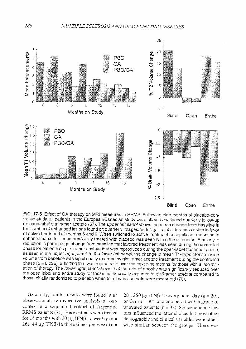
A prospective open-label study of glatiramer acetate: over a decade of continuous use in multiple sclerosis patients CC Ford 1 , KP Johnson 2 , RP Lisak 3 , HS Panitch 4 , G Shifroni 5 , JS Wolinsky 6 and The Copaxone † Study Group A decade of continuous glatiramer acetate (GA) use by relapsing remitting multiple sclerosis (RRMS) patients was evaluated in this ongoing, prospective study, and the neurological status of ‘Withdrawn’ patients was assessed at a 10-year long-term follow-up (LTFU) visit. Modified intention-to-treat (mITT, n /232) patients received ] /1 GA dose since 1991; ‘Ongoing’ patients (n /108) continued in November 2003. Of 124 patients, 50 Withdrawn patients returned for LTFU. Patients were evaluated every six months (EDSS). Mean GA exposure was 6.99, 10.1 and 4.26 years for mITT, Ongoing, and Withdrawn/LTFU patients, respectively. While on GA, mITT relapse rates declined from 1.18/year prestudy to /1 relapse/5 years; median time to ] /1 EDSS point increase was 8.8 years; mean EDSS change was 0.739 /1.66 points; 58% had stable/improved EDSS scores; and 24, 11 and 3% reached EDSS 4, 6 and 8, respectively. For Ongoing patients, EDSS increased 0.509 /1.65; 62% were stable/improved; and 24, 8 and 1% reached EDSS 4, 6 and 8, respectively. For Withdrawn patients at 10-year LTFU, EDSS increased 2.249 /1.86; 28% were stable/improved; and 68, 50 and 10% reached EDSS 4, 6 and 8, respectively. While on GA nearly all patients (mean disease duration 15 years) remained ambulatory. At LTFU, Withdrawn patients had greater disability than Ongoing patients. Multiple Sclerosis 2006; 12: 309 /320. www.multiplesclerosisjournal.com Key words: disability; disease modifying therapy; EDSS; glatiramer acetate; immunomodulator; relapse; relapsing-remitting multiple sclerosis Introduction Currently, the best therapeutic options for relap- sing remitting multiple sclerosis (RRMS) patients are the disease-modifying therapies: glatiramer acetate (GA) and the beta-interferons, subcutaneous (SC) IFNb-1b, SC IFNb-1a, and intramuscular (IM) IFNb-1a [1]. There is growing consensus that im- munomodulatory therapy should begin shortly after RRMS diagnosis, and to prevent or delay progression of disability, continuous therapy may be recommended for many years [1 /3]. However, evidence of long-term clinical efficacy, safety, and patient acceptance of immunomodulatory therapy is scarce. Indeed, the designation ‘long-term’ to describe clinical data for immunomodulators is arbitrary / three to five years [4 /6], is a relatively short interval considering the predicted treatment duration and disease course noted in MS natural history studies [2]. The ongoing US Glatiramer Acetate Trial began in 1991 and is unique in that it is the only organized, ongoing, open-label study of more than 10 years duration to prospectively evaluate 1 MIND Imaging Center, Albuquerque, NM, USA 2 Maryland Center for MS, Baltimore, MD, USA 3 Wayne State University, Detroit, MI, USA 4 University of Vermont, Burlington, VT, USA 5 Teva Pharmaceutical Industries Ltd., Petah Tiqva, Israel 6 University of Texas Health Science Center at Houston, Houston, TX, USA Author for correspondence: Kenneth P Johnson, MD, Maryland Center for Multiple Sclerosis, 11 S. Paca Street, 4th Floor, Baltimore, MD 21201, USA. E-mail: [email protected] Received 30 June 2005; accepted 22 November 2005 ARTICLE Multiple Sclerosis 2006; 12: 309 /320 – 2006 Edward Arnold (Publishers) Ltd 10.1191/135248506ms1318oa

Welcome message from author
This document is posted to help you gain knowledge. Please leave a comment to let me know what you think about it! Share it to your friends and learn new things together.
Transcript

A prospective open-label study of glatiramer acetate:over a decade of continuous use in multiple sclerosispatients
CC Ford1, KP Johnson2, RP Lisak3, HS Panitch4, G Shifroni5, JS Wolinsky6 andThe Copaxone† Study Group
A decade of continuous glatiramer acetate (GA) use by relapsing remitting multiple sclerosis (RRMS)patients was evaluated in this ongoing, prospective study, and the neurological status of‘Withdrawn’ patients was assessed at a 10-year long-term follow-up (LTFU) visit. Modifiedintention-to-treat (mITT, n�/232) patients received ]/1 GA dose since 1991; ‘Ongoing’ patients(n�/108) continued in November 2003. Of 124 patients, 50 Withdrawn patients returned for LTFU.Patients were evaluated every six months (EDSS). Mean GA exposure was 6.99, 10.1 and 4.26 yearsfor mITT, Ongoing, and Withdrawn/LTFU patients, respectively. While on GA, mITT relapse ratesdeclined from 1.18/year prestudy to �/1 relapse/5 years; median time to ]/1 EDSS point increasewas 8.8 years; mean EDSS change was 0.739/1.66 points; 58% had stable/improved EDSS scores;and 24, 11 and 3% reached EDSS 4, 6 and 8, respectively. For Ongoing patients, EDSS increased0.509/1.65; 62% were stable/improved; and 24, 8 and 1% reached EDSS 4, 6 and 8, respectively. ForWithdrawn patients at 10-year LTFU, EDSS increased 2.249/1.86; 28% were stable/improved; and68, 50 and 10% reached EDSS 4, 6 and 8, respectively. While on GA nearly all patients (mean diseaseduration 15 years) remained ambulatory. At LTFU, Withdrawn patients had greater disability thanOngoing patients. Multiple Sclerosis 2006; 12: 309�/320. www.multiplesclerosisjournal.com
Key words: disability; disease modifying therapy; EDSS; glatiramer acetate; immunomodulator;relapse; relapsing-remitting multiple sclerosis
Introduction
Currently, the best therapeutic options for relap-sing remitting multiple sclerosis (RRMS) patientsare the disease-modifying therapies: glatirameracetate (GA) and the beta-interferons, subcutaneous(SC) IFNb-1b, SC IFNb-1a, and intramuscular (IM)IFNb-1a [1]. There is growing consensus that im-munomodulatory therapy should begin shortlyafter RRMS diagnosis, and to prevent or delayprogression of disability, continuous therapy maybe recommended for many years [1�/3]. However,
evidence of long-term clinical efficacy, safety, andpatient acceptance of immunomodulatory therapyis scarce. Indeed, the designation ‘long-term’ todescribe clinical data for immunomodulators isarbitrary �/ three to five years [4�/6], is a relativelyshort interval considering the predicted treatmentduration and disease course noted in MS naturalhistory studies [2].
The ongoing US Glatiramer Acetate Trial beganin 1991 and is unique in that it is the onlyorganized, ongoing, open-label study of morethan 10 years duration to prospectively evaluate
1 MIND Imaging Center, Albuquerque, NM, USA2 Maryland Center for MS, Baltimore, MD, USA3 Wayne State University, Detroit, MI, USA4 University of Vermont, Burlington, VT, USA5 Teva Pharmaceutical Industries Ltd., Petah Tiqva, Israel6 University of Texas Health Science Center at Houston, Houston, TX, USAAuthor for correspondence: Kenneth P Johnson, MD, Maryland Center for Multiple Sclerosis, 11 S. Paca Street, 4thFloor, Baltimore, MD 21201, USA. E-mail: [email protected] 30 June 2005; accepted 22 November 2005
ARTICLE Multiple Sclerosis 2006; 12: 309�/320
– 2006 Edward Arnold (Publishers) Ltd 10.1191/135248506ms1318oa

continuous immunomodulatory therapy in RRMSpatients. Prospective clinical efficacy data werereported for continuous IFNb-1b (Betaseron†) useat four to five years, continuous IFNb-1a SC (Re-bif†) at four years, and continuous IFNb-1a IM(Avonex†) at approximately two years after initia-tion of their respective pivotal double-blind trials[4�/6]. Further data have since been collectedin non-continuous and/or retrospective, open-labelextensions of these studies. In some cases, datawere collected retrospectively after considerableintervals in which patients were not monitoredand during which they may have discontinued,switched, or added other medications to the im-munomodulatory therapy under study. AnotherMS treatment, natalizumab (Tysabri†) remainsunder investigation; reported efficacy data reflectonly two years of clinical experience and the fullserious side effect profile of this drug remainsuncertain [7].
This GA study began with a double-blind, pla-cebo-controlled phase in which 251 RRMS patientswere randomized to receive GA (20 mg) or placeboby SC injection daily [8,9]. After double-blindtreatment for a mean of 30 months, all patientswere offered active treatment as part of an ongoing,prospective, open-label study. Reported clinicalefficacy results six and eight years after randomiza-tion of GA therapy comparing differences in clin-ical outcomes between patients who received GAfrom study inception versus those in patients whobegan treatment approximately 2.5 years later (ie,patients originally randomized to placebo), demon-strate the benefits of early GA therapy comparedwith delayed therapy [10�/12].
This paper describes long-term experience withGA in all patients who received it during thedouble-blind and/or open-label phases of the study.The primary aim was to determine the long-termeffects of GA in carefully monitored patients whohad received continuous GA for a mean of 10 years.A secondary goal was to gather information onpatients who had withdrawn from the study �/ theirdisease course while in the study and why theydiscontinued. Those who agreed to return for thelong-term follow-up (LTFU) visit were evaluated fortheir neurologic status approximately 10 years afterthey had initiated GA therapy.
Methods
Patients in this study were originally enrolled inthe US Glatiramer Acetate Pivotal Trial, a double-blind, randomized, placebo-controlled studythat began in October 1991. RRMS patients whohad experienced two or more medically docu-mented relapses in the previous two years and had
EDSS scores between 0 and 5 at entry, wererandomized to receive SC GA (20 mg) or placebodaily, administered by self-injection. After double-blind treatment, all patients who had entered thestudy were given the option to continue in anopen-label extension phase. Those patients origin-ally randomized to GA continued to take the drugand those randomized to placebo switched to GA.Details of the double-blind and open-label phases ofthe study are described elsewhere [8�/12]. Themodified intention-to-treat (mITT) population inthe study reported here differs from the original ITTcohort in the pivotal trial [8], in that this analysisincludes only patients who have received at leastone dose of GA (19 patients initially randomized toplacebo in the pivotal study declined entry into theopen-label extension and are excluded from thismITT cohort).
Data cut-off for this analysis occurred in Novem-ber 2003, a mean of 10.1 years from the beginningof GA therapy for the 108 patients continuing inthe ongoing study. Because 10 years was the meantreatment duration, patients who had received GAfrom randomization had been treated for up to12 years and patients originally randomized toplacebo had been actively treated for approximatelyeight to nine years.
This organized, prospective study is ongoing.The 11 original US academic centers continue toparticipate, and the Institutional Review Boards atall centers continue to approve the study.
Study design
Ongoing study procedure
Briefly, in the open-label study, neurological statusis evaluated in the clinic every six months using theKurtzke Expanded Disability Status Scale (EDSS)[13]. Patients are examined, usually within sevendays, if they experience symptoms suggestive of arelapse (appearance or reappearance of one or moreneurologic abnormalities persisting for at least 48hours, preceded by a stable or improving neurolo-gical state of at least 30 days duration [8�/12]).Incidence, severity, and potential cause of adverseevents are recorded; serious adverse events arereported to the sponsor and to the FDA as requiredby protocol. The safety of GA therapy in thesepatients at 2 [8], 3 [9], 6 [10], and 8 [12], years hasbeen reported previously. During the open-labelphase, laboratory assessments (chemistry panel)and vital signs are documented at six-month studyvisits. In most cases, the same neurologists andstudy co-ordinators continue to assess patients ateach visit.
310 CC Ford et al.
Multiple Sclerosis 2006; 12: 309�/320 www.multiplesclerosisjournal.com
Any patient who discontinued daily GA, forwhatever reason, and/or took another immuno-modulator was withdrawn. Therefore, those whoremain in the study represent a group of RRMSpatients receiving only continuous GA monother-apy for disease modification.
Upon withdrawal, patients were classified bystudy personnel as: (1) withdrawal due to adverseevent; (2) lost-to-follow-up, which included with-drawal from the study without attending a finalvisit or providing a reason for withdrawal; (3)withdrawal due to ‘patient decision’; or (4) with-drawal for other reason(s). Because of overlap inreasons for leaving, the ‘patient decision’ and‘other’ categories were combined. The ‘patientdecision/other’ category was divided into subcate-gories based on comments patients provided attermination. Subcategories included (but were notlimited to) pregnancy, inability to adhere to studyprotocol (eg, lack of transportation or movingaway), a desire to switch or combine therapies,and perceptions of disease worsening. Patients wereassigned to a subcategory based on the consensusjudgment of three study personnel who indepen-dently reviewed patient comments provided at thefinal visit.
Long-term follow-up visit procedure
Personnel at each center made repeated attemptsto contact all study patients who had received GAand withdrawn, to invite them to return for asingle LTFU visit at approximately 10 years afterGA start. LTFU visits were conducted between Julyand December 2003. At the LTFU visit, patientsunderwent neurological evaluation by EDSS, med-ical history during the time between study discon-tinuation and LTFU was recorded, and patientswere asked what MS medications they had takenduring the period between withdrawal and theLTFU visit.
Patient cohorts
The mITT cohort (n�/232) included all patientswho had received at least one GA dose since studyinception. Data reported for the mITT cohort reflectoutcomes measured while patients were receivingGA. The mITT cohort was subdivided into thefollowing cohorts: Ongoing, which comprised pa-tients continuing in the study (and, by definition,continuing on GA) at the time of data cut-off,November 2003; Withdrawn Total, which com-prised all patients who withdrew from the studybefore November 2003; Withdrawn with LTFUcohort, which included patients who withdrew
from the study and returned for a single LTFU visit10 years after GA start; and Withdrawn withoutLTFU cohort, which included patients who with-drew from the study and could not be reached ordeclined LTFU.
Statistical methods
Baseline demographic and disease characteristicswere analysed using descriptive statistics and statis-tical inference tests (SAS† software, SAS Institute,Cary, NC, USA); comparisons among cohorts wereperformed using x2 for categorical variables and theWilcoxon non-parametric test for continuous vari-ables. Time to study withdrawal was estimatedusing Kaplan�/Meier survival analysis.
Outcome measures
The yearly relapse rate was calculated by dividingthe total number of relapses by the total numberof patients in the mITT cohort entering a giventreatment year. Accumulated disability was mea-sured by mean EDSS and mean change in EDSSfrom GA start to the last observation while onGA in all study cohorts, and at LTFU in theWithdrawn with LTFU cohort. Confirmed pro-gression of disability was defined as an increaseof ]/1.0 EDSS point sustained for at least twoclinical assessments, six months apart. Categoricalanalyses of patients’ neurological status wereperformed; patients were classified as ‘stable/im-proved’ if EDSS scores increased by 5/0.5 EDSSpoints, did not change, or decreased from onsetof treatment. Categorical analyses were repeatedwith patients stratified by entry EDSS score (0�/2or ]/2.5).
The number of patients who reached confirmedscores of EDSS 4, 6 or 8 while on GA were obtainedfor the mITT, Ongoing, and Withdrawn Totalcohorts (only patients who began GA therapywith EDSS scores lower than the endpoint wereincluded in these analyses). Additionally, Kaplan�/
Meier survival analysis was used to estimate thetime to EDSS 4, 6 and 8 while on GA.
For comparison at 10 years between Ongoingand Withdrawn with LTFU patients, numbers ofpatients who reached predefined EDSS thresholdsby the last observation for Ongoing patients and atthe single LTFU visit for Withdrawn with LTFUpatients were assessed. Efficacy comparisons at 10years were made using analysis of covariance(ANCOVA) for change from GA start in EDSS, inwhich EDSS at GA start was a covariate in themodel; x2; and when appropriate, Fisher’s Exact
Prospective study of 10 years of glatiramer acetate in RRMS 311
www.multiplesclerosisjournal.com Multiple Sclerosis 2006; 12: 309�/320

Test for categorical change in EDSS and for attain-ment of predefined EDSS thresholds.
Results
Patient characteristics
A total of 232 patients from 11 US study siteswho had received at least one dose of GA sincestudy inception comprised the mITT cohort(Figure 1). One patient discontinued after receiv-ing GA but before undergoing neurological eva-luation; therefore, the efficacy evaluable mITTcohort included 231 patients. As of November2003, 108/232 patients (47%) remained in thestudy and comprised the Ongoing cohort. Of 124(53%) patients in the Withdrawn Total cohort, 50returned for the LTFU visit (Withdrawn withLTFU cohort). In the Withdrawn without LTFUcohort (n�/74), 27 patients declined the LTFUvisit and 47 could not be reached, including fivepatients known to have died. Deaths occurred oneto six years after study withdrawal; three deathswere at least partly attributed to MS complica-tions, there was no information about one death,and one death was listed as sudden and unex-plained.
There were no differences among study cohortsin age, gender, disease duration, or annualizedrelapse rate in the two years before beginning GA(Table 1). Mean MS disease duration at GA start was8.3 years in the mITT cohort.
Patient withdrawal
The Kaplan�/Meier estimate of the median timefrom GA start to withdrawal in the mITT cohort was9.2 years. Patients who withdrew had slightlyhigher EDSS scores at GA start than those whoremained in the study (3.009/1.59 [SD] versus2.569/1.35, respectively; P�/0.03). Mean durationof GA treatment was 4.269/3.13 [SD] years (range:0.2�/11.5 years) in the Withdrawn Total cohort(Table 2). When separated into subcohorts, GAexposure was 4.479/2.95 years (range: 0.2�/10.4) inthe Withdrawn with LTFU cohort, and 4.139/3.26years (range: 0.2�/11.5) in the Withdrawn withoutLTFU cohort. There were no statistical differences indemographic or disease characteristics at GA startbetween Withdrawn patients who returned forLTFU and Withdrawn patients who did not return(Table 1).
Reasons for patient withdrawal are listed inTable 3. The most common (]/1%) adverse eventsleading to discontinuation were local injection-sitereactions (eg, erythema, pain), vasodilation, dys-pnea, and urticaria. Patients who left due to theperception that their disease was worsening werenot evaluated by objective neurological testing atthe time of withdrawal; therefore, whether indivi-dual patients had worsened by objective criteria isunknown. The mean change in EDSS score fromGA start to the last observed on-treatment EDSSvalue for all patients in the patient decision/other category (n�/87) was 1.169/1.65 [SD] andmean EDSS changes in the Withdrawn Total and
taerT-ot-noitnetnI deifidoM*trohoC )TTIm(
tsael ta deviecer ohw enoynA(232 = N )esod AG 1
trohoC gniognO801 = n
trohoC latoT nwardhtiW421 = n
UFTL htiw nwardhtiWtrohoC
05 = n
tuohtiw nwardhtiWtrohoC UFTL
*47 = n
Figure 1 Study cohorts. One patient (in the Withdrawn without LTFU cohort) withdrew after a single GA dose and before anon-treatment neurological evaluation; therefore, 231 patients comprised the efficacy evaluable mITT cohort.
312 CC Ford et al.
Multiple Sclerosis 2006; 12: 309�/320 www.multiplesclerosisjournal.com

mITT cohorts were 0.949/1.65 and 0.739/1.66, res-pectively (Table 2).
The LTFU visit for Withdrawn patients occurredat a mean of 10.0 years after GA start. These patientswere asked about other disease modifying therapiesthey had taken for MS in the interval betweenleaving the study and the LTFU visit. Of the 50Withdrawn with LTFU patients, four patients re-ported taking no medications, no data were avail-able for eight patients, and 38 reported they took avariety of agents over the interval, often in combi-nation, for varying lengths of time: 13 patients tookGA, 32 took an IFNb drug, 14 took an immunosup-pressive agent (mitoxantrone, methotrexate,azathioprine, cyclophosphamide), 10 took corticos-teroids, and five took ‘other’ drugs for MS. At thetime of the LTFU visit, 10 patients were taking GA,16 were taking an IFNb drug, four were taking animmunosuppressor agent (mitoxantrone, metho-trexate), and one was receiving intravenous immu-noglobulin (IVIg).
Efficacy
mITT cohort while on GA
Relapse rate. The medically documented annual-ized relapse rate in the mITT cohort during the twoyears before beginning GA therapy was 1.189/0.82[SD] (Table 1). While on GA, yearly relapse ratesdeclined approximately 50% to 0.619/0.85 in treat-ment year 1 and continued to decline so that intreatment year 4, patients experienced the equiva-lent of one relapse every four years. Yearly relapserates are shown in Figure 2 (after year 9, the mITTcohort comprised only patients who had receivedGA from randomization). Low relapse rates were
maintained over all subsequent years; relapse rateswere reduced by �/80% from rates at GA start toapproximately one relapse every five years.
Accumulated disability. Mean and median GAtreatment durations for the mITT, Ongoing, andWithdrawn Total cohorts, overall and according todrug assignment at randomization, are shown inTable 2. Mean GA exposure time in the Ongoingcohort was 10.19/1.32 years and in the WithdrawnTotal cohort was 4.269/3.13 years. Also shown areEDSS scores and changes in EDSS scores from GAstart to the last measurement while on GA.
The Kaplan�/Meier estimate of the median timeto confirmed (verified at two six-month clinicalvisits) progression of ]/1.0 EDSS point while on GAwas 8.82 years in the mITT cohort. Proportions ofpatients with at least one confirmed progression of1.0 EDSS point while on GA were 42% of the mITTcohort, 42% of the Ongoing cohort, and 43% of theWithdrawn Total cohort.
Categorical analysis showed 58% of patients inthe mITT cohort maintained stable/improved EDSSscores between GA start and their last on-treatmentEDSS assessment. Proportions of patients in theOngoing and Withdrawn Total cohorts with stable/improved EDSS scores while on GA were 62 and55%, respectively. When patients were stratified byentry EDSS (0�/2 and ]/2.5), proportions of patientswith stable/improved EDSS scores while on GA weresimilar in both strata in all cohorts (Table 2),indicating no difference in GA effect regardless ofdisability level at GA start. Figure 3 shows a highproportion of patients (59�/79%) in the mITT co-hort (cohort size each year shown in Figure 2) hadstable/improved EDSS scores each treatment year.
Table 4 shows the number of patients in eachcohort who reached predefined EDSS thresholds.
Table 1 Patient characteristics at GA start
mITT(n�/232)
Ongoing(n�/108)
Withdrawn total(n�/124) P valuea
Withdrawn withLTFU (n�/50)
Withdrawn withoutLTFU (n�/74) P valueb
Age (years)Mean9/SD 35.59/6.4 36.69/5.7 34.89/6.8 0.0918 35.29/6.6 34.69/7.0 0.6787Range 19.0�/48.6 22.0�/48.6 19.0�/47.9 19.0�/46.7 19.0�/47.9Female n (%) 170 (73%) 77 (71%) 93 (75%) 0.5248 33 (66%) 60 (81%) 0.0581
Disease duration (years) at GA startMean9/SD 8.39/5.1 8.49/5.1 8.19/5.2 0.8013 8.6 9/ 6.2 7.89/4.3 0.7373Median 7.23 6.85 7.49 7.34 7.66Range 0.6�/25.7 1.1�/23.9 0.6�/25.7 0.6�/25.7 0.6�/18.0
Annualized relapse rate two years before GA startMean9/SD 1.189/0.82 1.119/0.82 1.249/0.83 0.2286 1.099/0.68 1.349/0.90 0.0760Median 1.00 1.00 1.00 1.00 1.00Range 0�/5.5 0�/3.5 0�/5.5 0�/3.0 0�/5.5
aOngoing versus Withdrawn Total.bWithdrawn with LTFU versus Withdrawn without LTFU.
Prospective study of 10 years of glatiramer acetate in RRMS 313
www.multiplesclerosisjournal.com Multiple Sclerosis 2006; 12: 309�/320

Ta
ble
2G
Aexp
osu
rean
dED
SS
measu
res
wh
ileo
nG
A
Wh
ileo
nG
Am
ITT
(n�
/23
2)
On
go
ing
(n�
/10
8)
Wit
hd
raw
nT
ota
l(n
�/1
24
)
GA
exp
osu
re(y
ears
)M
ean9
/SD
6.9
99
/3.8
21
0.1
29
/1.3
24
.269
/3.1
3M
ed
ian
8.6
29
.16
3.5
5R
an
ge
0.2�/
11
.97
.9�/
11
.9
0.2�/
11
.5
GA
exp
osu
reb
ase
do
nd
rug
ass
ign
men
tat
ran
do
miz
ati
on
(years
)P
La(n
�/1
07
)G
A(n
�/1
25
)P
L(n
�/5
5)
GA
(n�
/53
)P
L(n
�/5
2)
GA
(n�
/72
)
Mean9
/SD
6.2
59
/3.3
07
.639
/4.1
38
.849
/0.2
31
1.4
49
/0.2
03
.509
/2.7
54
.819
/3.2
8M
ed
ian
8.5
09
.22
8.9
01
1.4
42
.62
4.2
6R
an
ge
0.2�/
9.2
0.2�/
11
.97
.9�/
9.2
10
.9�/
11
.90
.2�/
8.9
0.2�/
11
.5
ED
SS
sco
reat
GA
start
Mean9
/SD
2.7
99
/1.5
02
.569
/1.3
53
.009
/1.5
9M
ed
ian
2.5
02
.25
3.0
0R
an
ge
0.0�/
7.0
0.0�/
6.0
0.0�/
7.0
ED
SS
at
last
ob
serv
atio
nc
Mean9
/SD
3.5
39
/2.0
83
.069
/1.7
83
.949
/2.2
5M
ed
ian
3.0
02
.50
3.5
0R
an
ge
0.0�/
9.0
0.0�/
8.0
0.0�/
9.0
ED
SS
chan
ge
fro
mG
Ast
art
tola
sto
bse
rvati
on
b
Mean9
/SD
0.7
39
/1.6
60
.509
/1.6
50
.949
/1.6
5M
ed
ian
0.5
00
.50
0.5
0R
an
ge
�/3
.5to
5.5
�/3
.5to
5.5
�/3
.5to
5.5
Clin
ically
stab
le/i
mp
roved
ED
SS
csc
ore
wh
ileo
nG
Ab
Overa
ll1
34
/23
1(5
8%
)6
7/1
08
(62
%)
67
/12
3(5
5%
)ED
SS
0�/
2at
GA
start
58
/10
3(5
6%
)3
0/5
4(5
6%
)2
8/4
9(5
7%
)ED
SS]
/2.5
at
GA
start
76
/12
8(5
9%
)3
7/5
4(6
9%
)3
9/7
4(5
3%
)
aP
L,p
lace
bo
.bn�
/23
1in
mIT
Tco
ho
rtan
dn�
/12
3in
Wit
hd
raw
nto
tal
coh
ort
(on
ep
ati
en
th
ad
no
po
st-G
A-s
tart
ED
SS
sco
re).
cD
efi
ned
as
an
incr
ease
of5
/0.5
po
int,
no
chan
ge,
or
decr
ease
inED
SS
sco
refr
om
on
set
of
treatm
en
t.
314 CC Ford et al.
Multiple Sclerosis 2006; 12: 309�/320 www.multiplesclerosisjournal.com

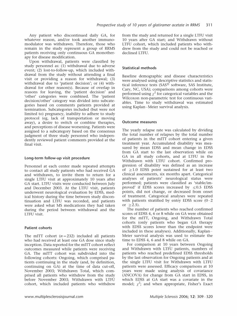
These analyses include only patients with EDSSscores at GA start below EDSS 8 (all patients), EDSS6 (n�/221), or EDSS 4 (n�/169). Inclusion criteria inthe original double-blind phase of the study waslimited to patients with EDSS 5/5, however, 10original placebo patients had an EDSS ]/6 whenthey started GA therapy in the open-label phase ofthe study. Mean EDSS scores at GA start were 2.079/
0.95 [SD], 2.649/1.34, and 2.799/1.50 in patientgroups with EDSS scores at GA start below EDSS 4, 6and 8, respectively. In the mITT cohort, fewer than25% of patients reached a score of EDSS 4, approxi-mately 11% reached EDSS 6, and 3% reached EDSS8 while on GA. Due to differences in GA exposureamong cohorts, Kaplan�/Meier survival analysis wasused to estimate the time to the predefined EDSSthresholds (mITT cohort shown in Figure 4). Thetime to 25% of patients reaching EDSS 4 was 6.58years in the mITT cohort, 9.08 years in the Ongoingcohort, and 5.47 years in the Withdrawn Totalcohort. In the Withdrawn Total cohort, median
time to EDSS 4 occurred at 9.91 years. Fewer than25% of patients in the mITT, Ongoing, and With-drawn Total cohorts reached EDSS 6 or 8 by the endof the analysis period.
Clinical comparison between Ongoing andWithdrawn patients at ten years/LTFU
Accumulated disability
The occurrence of relapse after leaving the studywas not systematically documented by prospectiveneurological assessments; therefore, these data werenot available. Neurological assessments of disabilityat LTFU for Withdrawn patients were made at amean of 10.0 years from starting GA therapy. ForOngoing patients, EDSS data reflected the lastobservation before data cut-off.
At LTFU, patients who had withdrawn showedsignificantly increased disability compared with
Table 3 Reasons for patient withdrawal
Reason for withdrawal n Patient decision/other subcategoriesa n
Total 124 Total 87Lost-to-follow-up 14 Patient perception of disease worsening 27Adverse event 23 Desire to switch or combine therapies 22Patient decision/other 87 Difficulty, inability, or unwillingness to adhere to study protocol 30
Pregnancy 8
aPatient decision/other subcategories were derived from written comments provided by Withdrawn patients at their final visit.
15
691
802
132
232
571 361941
341 831
52146
75
0
52.0
5.0
57.0
1
52.1
9 8 7 6 5 4 3 2 1 * 21 11 01
Mea
n R
elap
se R
ate
raeY †
*
Figure 2 Yearly Relapse Rate from GA Start (mITT Cohort, n=231). *Mean [SD] annualized relapse rate in the mITT cohort inthe 2 years before GA start was 1.189/0.82. $Reflects treatment duration from GA start to the year listed. Numbers of patientsevaluated in each treatment year are shown above bars; after year 9, the mITT cohort comprised only patients randomized toGA in the double-blind phase of the study (placebo/active group had a shorter duration of GA exposure).
Prospective study of 10 years of glatiramer acetate in RRMS 315
www.multiplesclerosisjournal.com Multiple Sclerosis 2006; 12: 309�/320

Ongoing patients (Table 5). The mean increase inEDSS score was 2.249/1.86 [SD] points in theWithdrawn with LTFU cohort compared with a0.509/1.65 point increase in Ongoing patients.Similarly, 62% of Ongoing patients had stable orimproved EDSS scores compared with 28% of With-drawn with LTFU patients (while receiving GA, 56%of patients in the Withdrawn with LTFU cohort hadstable/improved EDSS scores). Finally, at 10 years,24% of patients in the Ongoing cohort had reachedEDSS 4, 8% had reached EDSS 6, and 1% hadreached EDSS 8, compared with 68, 50 and 10%,respectively, in the Withdrawn with LTFU cohort.
Safety
The most commonly reported adverse events inpatients receiving long-term GA, regardless of
whether the investigator judged them to berelated to GA therapy or not, were accidentalinjury, asthenia, paresthesia, upper respiratoryand urinary tract infections, headache, and pain.Adverse events thought to be associated withGA therapy included local injection-site reactions(eg, erythema, pain, mass, edema) and symptomsassociated with an immediate post-injection reac-tion (IPIR), which may have included vasodila-tion, chest pain, palpitation, tachycardia, ordyspnea. Reporting of injection-site reactionsand symptoms associated with IPIR declinedover time. No apparent time-dependent adverseevents emerged. Moreover, no evidence of hema-tologic, hepatic, or renal dysfunction; immuno-suppression; emergence of malignancy; or deve-lopment of other autoimmune disease was ob-served.
%56%16%95%26
%66%66%96%86%07%27%57
%97
0
52
05
57
001%
of
Pat
ien
ts C
linic
ally
Sta
ble
/Imp
rove
d
1 11 01 98 7 6 5 4 3 2 1 2
*raeY
Figure 3 Yearly Percent of Patients on GA with stable/improved EDSS. Scores from GA start (mITT cohort: n�/231). *Reflecttreatment duration from GA start to the year listed (the number of patients in the mITT cohort each treatment year is shown inFigure 2). Clinically stable/improved�/an increase of 5/0.5 point, no change, or decrease in EDSS score from onset of GAtreatment.
Table 4 Number of patients reaching EDSS 4, 6 and 8 while on GA (confirmed)
Whileon GA
mITTa
n/Nb (%)Ongoingn/Nb (%)
Withdrawn Totaln/Nb (%)
Disease duration at GA startmITTa mean years9/SD
GA exposure mITTa
mean years9/SD
EDSS 4 40/169 (24%) 20/84 (24%) 20/85 (24%) 7.799/5.00 7.169/3.77EDSS 6 24/221 (11%) 8/106 (8%) 16/115 (14%) 8.109/5.01 7.119/3.81EDSS 8 6/231 (3%) 1/108 (1%) 5/123 (4%) 8.269/5.12 6.999/3.82
amITT cohorts included all patients below the EDSS threshold at GA start.bn�/number of patients who reached EDSS endpoint (confirmed); N�/number of patients who were below the EDSS threshold at GAstart.
316 CC Ford et al.
Multiple Sclerosis 2006; 12: 309�/320 www.multiplesclerosisjournal.com
Discussion
This ongoing study is the longest prospective,organized evaluation of continuous immunomodu-latory therapy in MS [4,5,14,15]. In November
2003, nine to 12 years after beginning GA therapy,47% of all patients who ever received GA remainedin the study. These data provide an opportunity toevaluate the level of neurologic disability in pa-tients who have continued to take GA for an
66521108 SSDE
42
04
42
04
22
53
91
03
71
12
11
51
0
0
6 SSDE
4 SSDE
tniopdnE gnihcaeR stneitaP fo rebmuN evitalumuC
00.0
52.0
05.0
57.0
00.1
210186420
8 SSDE
6 SSDE
4 SSDE
AG no sraeY
Su
rviv
al D
istr
ibu
tio
n F
un
ctio
n
≥
≥
≥
Figure 4 Time to confirmed EDSS 4, 6 and 8 while on GA (mITT cohort: n�/231). The mean disease duration in the mITTcohort was 8.3 years at GA Start.
Table 5 EDSS data at 10 years/LTFU
EDSS measure Ongoing (n�/108) Withdrawn with LTFUa (n�/50) P value
EDSS scoreMean9/SD 3.069/1.78 5.229/2.21 B/0.0001c
Median 2.50 6.00Range 0.0�/8.0 1.0�/9.0
EDSS change from GA startMean9/SD 0.509/1.65 2.249/1.86 B/0.0001c
Median 0.50 2.25Range �/3.5 to 5.5 3.0�/5.5
Categorical analysisClinically stable/improved 67/108 (62%) 14/50 (28%) B/0.0001d
Patients reaching EDSS 4, 6, or 8b
EDSS 4 n/N (%) 20/84 (24%) 25/37 (68%) B/0.0001d
EDSS 6 n/N (%) 8/106 (8%) 23/46 (50%) B/0.0001d
EDSS 8 n/N (%) 1/108 (1%) 5/50 (10%) B/0.0125e
aEDSS level was not confirmed at six months in Withdrawn with LTFU patients, since LTFU comprised a single visit.bn�/number of patients who reached EDSS threshold (confirmed in Ongoing patients) and N�/number of patients who were below
the EDSS threshold at GA start.cFrom ANCOVA adjusted for EDSS score at GA start.dx2 Test.eFisher’s Exact Test.
Prospective study of 10 years of glatiramer acetate in RRMS 317
www.multiplesclerosisjournal.com Multiple Sclerosis 2006; 12: 309�/320

average of 10 years and compare it with that ofpatients who withdrew from the study (and mayhave used other MS therapies) then returned forneurological evaluation 10 years after beginningGA therapy.
Results of this study are consistent with mec-hanisms of action of GA, which address both theinflammatory and neurodegenerative aspects of MSpathology within the CNS. GA-reactive Th2 cellsinduce ‘bystander’ suppression of inflammationwithin the CNS by increasing anti-inflammatorycytokine production, and facilitate neuroprotectionvia enhanced secretion of neurotrophins [16�/20].Immunological effects of GA persist in MS patientsfor at least six to nine years with continued use[18,20].
Multiple measures demonstrated the majority ofpatients who remain on long-term GA therapycontinued to do well. Relapse rates dropped byapproximately one half in the first year of treat-ment and by years nine to 12, had declined morethan 80% (Figure 2). A decline in relapse rate couldsignal transition to secondary progressive MS(SPMS). However, if this were the cause of theobserved decline in relapse rates, a commensurateincrease in disability would be expected over time.Although there was a slight trend toward increasingmean EDSS score over 10 years, the majority ofpatients in the mITT cohort (Figures 3 and 4) and inthe Ongoing cohort exhibited stable or improvedEDSS scores while on long-term GA therapy. Themean EDSS score in Ongoing patients increasedonly 0.50 points over 10 treatment years. Moreover,proportions of patients who progressed to prede-fined EDSS thresholds (Table 4) were much lowerthan what would be predicted based on MS naturalhistory data [2,21,22]. Thus, the decline in relapserate probably reflects treatment-related stabiliza-tion or improvement of the underlying diseaseprocess.
Although it would be of considerable interestto compare the long-term treatment effects ofGA with similarly collected data for other avai-lable immunomodulators, the present study is theonly prospective study with stipulated six-monthassessments of neurologic status and safety toextend beyond four years. Furthermore, it is theonly study in which patients must remain on thestudy drug and not switch or add concomitantimmunotherapy. Additional deficiencies of otherstudies include involvement of very few patients[23], potential use of multiple therapies duringunmonitored time periods [24], inclusion of differ-ent patient populations [25], or lack of informationregarding disability or relapses [23,26].
An alternative method to evaluate these resultsin context is to use MS natural history data.
Comparisons with published natural history reportsmust be made cautiously, since patient assessmentsare relatively infrequent and patient populationsare likely to be less well defined. Furthermore,disability data in some cases reflect ‘snapshot’assessments; whereas any accumulated disabilitywas confirmed in this study at two clinical exam-inations six-months apart for patients while on GAtherapy (withdrawal patient data at LTFU visit wasnot confirmed). Nevertheless, similarities betweenthis study group and reported natural historycohorts, with respect to patients’ disease durationand measures of disease progression, allow forgeneral comparisons.
Before the availability of immunomodulatorytherapies, Weinshenker et al . reported data from ageographically-based natural history cohort in Mid-dlesex County, Ont., Canada (n�/1099 RRMS pa-tients) [2]. This prospective study included yearlypatient evaluations; by 15 years after disease onset,50% of untreated patients had reached EDSS 6 and10% had reached EDSS 8. In the current study, atthe start of GA therapy the mITT cohort already hada mean disease duration since diagnosis of 8.3 yearsand mean EDSS score of 2.79. At the last observa-tion on GA (mean disease duration approximately15 years), only 11% of patients in the mITT cohorthad reached a confirmed score of EDSS 6 and 3%had reached EDSS 8. Moreover, in the Ongoingcohort, only 8% of patients reached EDSS 6 and 1%reached EDSS 8 after 10 years of continuous GAtherapy (Table 4). In another natural history cohortof RRMS patients (n�/1562) with approximately thesame disease duration (median 11.4 years fromdiagnosis), Confavreux et al . [21], reported thatalmost half (48%) of the patients had reachedEDSS 4. In the current study, only 24% of mITTpatients, with a median disease duration of 15.2years, reached a score of EDSS 4 while taking GA(Table 4).
Recently, data were reported from a population-based cohort evaluated for 10 years in OlmstedCounty, Minnesota (n�/161 patients with RRMS,SPMS, or primary progressive MS) [22]. Unlike thepresent study, patients were evaluated only twice �/
at the beginning and at the end of the 10-yearperiod. Ten-year disability outcomes for OlmstedCounty patients with disease characteristics similarto this study’s patients (RRMS and entry EDSS 0�/5)compare less favorably with 10-year outcomes inOngoing GA patients. Overall, 28% of patients inthe Olmsted County cohort had reached a score ofEDSS 6 and 7% had reached a score of EDSS 8 at 10years, compared with 8 and 1%, respectively, ofpatients in this study.
318 CC Ford et al.
Multiple Sclerosis 2006; 12: 309�/320 www.multiplesclerosisjournal.com
Ongoing versus Withdrawn patients
Long-term study data provided an opportunity toevaluate reasons for patient withdrawal and howthese patients fared during and after study partici-pation. Patients who withdrew from this study didso for multiple reasons (Table 3). Comments re-ported in the ‘patient decision/other’ categorysuggest many patients withdrew for non-MS-relatedissues, such as lack of transportation to the studysite or pregnancy, while some patients withdrewbecause they believed their disease was worsening.Although this was undoubtedly true in some cases,mean changes in EDSS scores while on GA in the‘patient decision/other’ category were not substan-tially different from those of the Withdrawn Totalor mITT cohort. In addition, many patients whowithdrew fared well before leaving the study; morethan half of all Withdrawn Total patients (55%) hadimproved or stable EDSS scores while on GA.
It is possible that withdrawal was influencedby the introduction of newly approved immuno-modulatory therapies. During the early years ofthe study, patients participating in the placebo-controlled GA trial may have wanted to try anapproved medication (Betaseron† and Avonex†
were approved in 1993 and 1995, respectively)instead of continuing to take either an experimen-tal drug or a placebo.
Findings at 10-year LTFU
Inferences regarding differences in GA treatmenteffects between Ongoing and Withdrawal withLTFU patients at a mean of 10.0 years from GAstart must also be made cautiously. At LTFU, themajority of returning patients reported havingtaken multiple medications after leaving the study,frequently switching and/or combining diseasemodifying therapies.
At 10 years after beginning GA therapy, allmeasures indicated that Withdrawn with LTFUpatients fared significantly worse than Ongoingpatients (Table 5). At LTFU, only 28% of With-drawal with LTFU patients had stable/improvedEDSS scores, whereas, while they were active inthe study (ie, taking GA for an average of 4.5 years),the proportion was twice as high (56%). Comparedwith Ongoing patients, EDSS increases were signifi-cantly higher in Withdrawn with LTFU patientsand more patients in the latter cohort reached EDSS4, 6 and 8. Thus, disability measures indicated theseWithdrawn patients worsened over time, regardlessof whether, how long, or which immunomodula-tory therapy they took after leaving the study.
The favorable safety profile of GA therapy wasmaintained over long-term use. The most common
adverse events associated with GA over the courseof the study were local injection-site reactions andIPIR, which were reported with decreasing fre-quency as treatment duration increased. No otherimmune-mediated disorders, infections, or malig-nancies have been associated with long-term GAtreatment. The willingness of RRMS patients tocontinue to self-administer daily GA SC injectionsfor more than a decade attests to the excellenttolerability of the drug.
This study is the longest ever conducted toprospectively evaluate the efficacy and safety ofcontinuous immunomodulator use in MS. Reduc-tion of relapses over 10 years of GA therapy wassustained with minimal increase (B/1 EDSS point)in confirmed disability. Patients continuing GAtherapy for 10 years had an average disease durationof more than 18 years, yet nearly all remainedambulatory.
Acknowledgements
The authors acknowledge the assistance of PippaLoupe, PhD, Teva Neuroscience, Kansas City, Mis-souri; Frederic Deutsch, BS, Teva PharmaceuticalIndustries Ltd., Petah Tiqva, Israel; Sheila Owens,BS, Medical Communication Co., Inc., Sarasota,Florida; and The Copaxone† Study Group in thedevelopment of this manuscript. The Copaxone†
Study Group includes the following investigativeteams: University of Pennsylvania Medical Center,Clyde Markowitz, MD, Amy Pruitt, MD, DorotheaPfohl, RN, BS, MSCN; University of New MexicoSchool of Medicine, Gary A Rosenberg, MD, ElidaGreinel, RN; Wayne State University School ofMedicine, Omar A Khan, MD, Deena Lisak, BS,MA, RN, Alexandros Tselis, MD, PhD, John Kam-holz, MD, PhD, Christina Caon, MSN, RN; UCLASchool of Medicine, Lawrence Myers, MD, WBaumhefner, MD, Ricki Klutch, RN; University ofMaryland School of Medicine, Christopher Bever,MD, Eleanor Katz, RN; VASLCHCS/University ofUtah, John Rose, MD, James B Burns, MD, ConnieKawai, RN; University of Rochester, Andrew Good-man, MD, Steven R Schwid, MD, Mary D Petrie, RN;Yale University School of Medicine, Jana Preinin-gerova, MD, Silva Markovic Plese, MD, GeorgeBlanco, MD; University of Southern CaliforniaSchool of Medicine, Norman Kachuck, MD; Uni-versity of Texas Health Science Center at Houston,Staley Brod, MD, PhD, J William Lindsey, MD,Myrna Koh, RN; University of Wisconsin Hospitaland Clinic, Benjamin Brooks, MD, Jennifer Parnell,BA, Kathy Roelke, RN. Teva Pharmaceuticals Indus-tries Ltd., David Ladkani, PhD, Shaul Kadosh, MS,Yafit Stark, PhD. This study was supported by grantsfrom the Federal Food and Drug Administration
Prospective study of 10 years of glatiramer acetate in RRMS 319
www.multiplesclerosisjournal.com Multiple Sclerosis 2006; 12: 309�/320
Orphan Drug Program No. FD-4000559-01, theNational Multiple Sclerosis Society No. RG 2202-A-6 and Teva Pharmaceutical Industries Ltd., PetahTiqva, Israel.
References
1. Oger J, Freedman M. Consensus statement of theCanadian MS Clinics Network on the use of diseasemodifying agents in multiple sclerosis. Can J Neurol Sci1999; 26: 274�/75.
2. Weinshenker BG. The natural history of multiplesclerosis. Neurol Clin 1995; 13: 119�/46.
3. Miller A, Cohen J, Ford C, Garmany G, Goodman A,Green B et al . National Multiple Sclerosis Society (NMSS):disease management consensus statement . National MSSociety, 2005.
4. The PRISMS (Prevention of Relapses and Disabil-ity by Interferon-B-1a Subcutaneously in MultipleSclerosis) Study Group, and the University ofBritish Columbia MS/MRI Analysis Group.PRISMS-4: long-term efficacy of interferon-b1a in relap-sing MS. Neurology 2001; 56: 1628�/36.
5. The IFNb Multiple Sclerosis Study Group and TheUniversity of British Columbia MS/MRI AnalysisGroup. Interferon beta-1b in the treatment of multiplesclerosis: final outcome of the randomized controlledtrial. Neurology 1995; 45: 1277�/85.
6. Jacobs LD, Cookfair DL, Rudick RA, Herndon RM,Richert JR, Salazar AM et al . Intramuscular interferonbeta-1a for disease progression in relapsing multiplesclerosis. The Multiple Sclerosis Collaborative ResearchGroup (MSCRG). Ann Neurol 1996; 39: 285�/94.
7. FDA Public Health Advisory. Suspended marketing ofTysabri (natalizumab). Retrieved 31 March 2005, fromhttp://www.fda.gov/cder/drug/advisory/natalizumab.htm
8. Johnson KP, Brooks BR, Cohen JA, Ford CC, Gold-stein J, Lisak RP et al . Copolymer 1 reduces relapse rateand improves disability in relapsing-remitting multiplesclerosis: results of a phase III multicenter, double-blindplacebo-controlled trial. The Copolymer 1 MultipleSclerosis Study Group. Neurology 1995; 45: 1268�/76.
9. Johnson KP, Brooks BR, Cohen JA, Ford CC, Gold-stein J, Lisak RP et al . Extended use of glatirameracetate (Copaxone) is well tolerated and maintains itsclinical effect on multiple sclerosis relapse rate and degreeof disability. Neurology 1998; 50: 701�/708.
10. Johnson KP, Brooks BR, Ford CC, Goodman A,Lisak RP, Meyers LW et al . Glatiramer acetate (Copax-one): comparison of continuous versus delayed therapyin a six-year organized multiple sclerosis trial. Mult Scler2003; 9: 585�/91.
11. Johnson KP, Brooks BR, Ford CC, Goodman A,Guarnaccia J, Lisak RP et al . Sustained clinicalbenefits of glatiramer acetate in relapsing remittingmultiple sclerosis patients observed for 6 years. Mult Scler2000; 6: 255�/66.
12. Johnson KP, Ford CC, Lisak RP, Wolinsky JS.Glatiramer acetate (Copaxone†): neurologic conse-quence of delaying glatiramer acetate therapy for multi-
ple sclerosis: 8-year data. Acta Neurol Scand 2005; 111:42�/47.
13. Kurtzke JF. Rating neurologic impairment in multiplesclerosis: an expanded disability status scale (EDSS).Neurology 1983; 33: 1444�/52.
14. Clanet M, Kappos L, Hartung H-P, Hohlfeld R. TheEuropean IFNb-1a dose-comparison study. Mult Scler2004; 10: 139�/44.
15. Kappos L, Stam Moraga M, Alsop J, The PRISMSStudy Group. Long-term tolerability of interferon Beta-1a in relapsing-remitting multiple sclerosis: 6-year safetyfollow-up of the PRISMS study. Presented at the JointMeeting of the American/European Committee for Treat-ment and Research in Multiple Sclerosis (ECTRIMS/ACTRIMS), Baltimore, MD, USA, 18�/22 September,2002: 334.
16. Yong VW. Differential mechanisms of action of inter-feron-b and glatiramer acetate in MS. Neurology 2002; 59:802�/808.
17. Gran B, Tranquill LR, Chen M, Bielekova B, ZhouW, Dhib-Jalbut S et al . Mechanisms of immunomodu-lation by glatiramer acetate. Neurology 2000; 55: 1704�/
14.18. Ragheb S, Abramczyk S, Lisak D, Lisak R. Long-
term therapy with glatiramer acetate in multiple sclero-sis: effect on T cells. Mult Scler 2001; 7: 43�/47.
19. Neuhaus O, Farina C, Wekerle H, Hohlfeld R.Mechanisms of action of glatiramer acetate in multiplesclerosis. Neurology 2001; 56: 702�/708.
20. Chen M, Conway K, Johnson KP, Martin R, Dhib-Jalbut S. Sustained immunological effects of glatirameracetate in patients with multiple sclerosis treated for over6 years. J Neurologic Sci 2002; 201: 71�/77.
21. Confavreux C, Vukusik S, Moreau T, Adeline P.Relapses and progression of disability in multiple sclero-sis. N Engl J Med 2000; 343: 1430�/38.
22. Pittock SJ, Mayr WT, McClelland RL, JorgensenNW, Weigand SD, Noseworthy JH et al . Change inMS-related disability in a population-based cohort. Neu-rology 2004; 62: 51�/59.
23. Karlik S, Kirk S, Nicolle E, Kremenchutzky M, RiceGPA. Evidence for very long-term efficacy of interferonBeta 1B in relapsing remitting MS patients. Proceedingsof the Seventeenth Annual Meeting of the EuropeanCommittee for Treatment and Research in MultipleSclerosis (ECTRIMS), Dublin, Ireland, 2001: 139.
24. Paty D, Kappos L, Stam Moraga M, Alsop J,Abdalla J. Long-term observational efficacy and safetyfollow-up of the PRISMS cohort. Proceedings of theNineteenth Annual Meeting of the European Committeefor Treatment and Research in Multiple Sclerosis (EC-TRIMS), Milan, Italy, 2003: 555.
25. Kinkel RP, Kollman C, Glassman A, Simon J,O’Connor P, Murray TJ et al . Interferon beta-1a(Avonex†) delays the onset of clinically definite MSover 5 years of treatment: results from the CHAMPIONSstudy. Presented at the Fifty-sixth Annual Meeting of theAmerican Academy of Neurology, San Francisco, CA,USA, 2004: D29.006.
26. Fisher E, Rudick RA, Simon JH, Cutter G, Baier M,Lee J-C et al . Eight-year follow-up study of brain atrophyin patients with MS. Neurology 2002; 59: 1412�/20.
320 CC Ford et al.
Multiple Sclerosis 2006; 12: 309�/320 www.multiplesclerosisjournal.com


Psychopharmacology (2005) 181: 118–125DOI 10.1007/s00213-005-2229-z
ORIGINAL INVESTIGATION
Tatyana Poltyrev . Elena Gorodetsky . Corina Bejar .Donna Schorer-Apelbaum . Marta Weinstock
Effect of chronic treatment with ladostigil (TV-3326)
on anxiogenic and depressive-like behaviour and on activity
of the hypothalamic–pituitary–adrenal axis in male and female
prenatally stressed rats
Received: 28 September 2004 / Accepted: 6 February 2005 / Published online: 14 April 2005# Springer-Verlag 2005
Abstract Objective: The aim of the study is to investigatethe effect of ladostigil, a cholinesterase and brain-selectivemonoamine oxidase (MAO) inhibitor, on anxiogenic anddepressive-like behaviour and the response of the hypotha-lamic–pituitary–adrenal axis to stress in prenatally stressed(PS)male and female rats. Methods: Ladostigil (17mg/kg/day) was administered daily for 6 weeks to control and PSrats aged 6 weeks. Behaviour was assessed in the elevatedplus maze (EPM) and forced swim tests (FST). Plasmacorticosterone (COR) was measured before, 30 and 90 minafter exposure to stress. Results: Ladostigil inhibited brainMAO-A and B by more than 60%, significantly reducedhyperanxiety of male and female PS rats in the EPM anddepressive-like behaviour in the FSTwithout affecting thatof controls and restored the delayed return to baseline ofplasma COR in PS rats after exposure to stress to that ofcontrol rats. Conclusions: A novel brain-selective MAOinhibitor, ladostigil can selectively reverse the behaviouraland neurochemical effects induced by prenatal stresswithout affecting the behaviour of controls.
Keywords Elevated plus maze . Forced swim test .Ladostigil . Monoamine oxidase inhibitor . Plasmacorticosterone . Prenatal stress
Introduction
Thompson (1957) was the first to report that subjection ofa pregnant rat to psychological stress increases emotionalbehaviour of the offspring. His findings have been con-firmed and extended in numerous studies in which it wasfound that prenatal stress impairs learning and memory(Lemaire et al. 2000; Gué et al. 2004), reduces play be-haviour (Morley-Fletcher et al. 2003b), exacerbates anx-iety in intimidating situations (Archer and Blackman 1971;Fride et al. 1986; Fride and Weinstock 1988) and inducesdepressive-like behaviour or learned helplessness morereadily than in controls (Secoli and Teixeira 1998). Whilememory deficits (Gué et al. 2004) and hyperanxiety (Frideand Weinstock 1988; Zimmerberg and Blaskey 1998) in-duced by prenatal stress are seen in rats of each gender,other behavioural and neurochemical changes are morereadily seen in female than in male rat offspring. Theseinclude the increased propensity for depressive-like be-haviour in the forced swim test (FST) (Alonso et al. 1991;Frye and Wawrzycki 2003) and the greater activationof the hypothalamic–pituitary–adrenal (HPA) axis in re-sponse to acute stress in adulthood (Weinstock et al. 1992;McCormick et al. 1995; Szuran et al. 2000). Prenatal stressalso causes a bigger reduction in hippocampal benzodiaz-epine receptors in female than in male rats (Fride et al.1985). While a reduction in the number of hippocampalcells is seen in female offspring after a single exposureof pregnant rats to stress (Schmitz et al. 2002), repeatedexposure to stress during the last week of gestation is re-quired in order to reduce hippocampal synapses in maleoffspring (Hayashi et al. 1998). This could explain why itmay be easier to detect depressive-like behaviour in pre-natally stressed (PS) female rats, since a role for hippo-campal cell loss and inhibition of neurogenesis has been
T. Poltyrev . E. Gorodetsky . C. Bejar . D. Schorer-Apelbaum .M. Weinstock (*)Department of Pharmacology,Hebrew University Hadassah School of Medicine,Ein Kerem, Jerusalem, 91120, Israele-mail: [email protected].: +972-2-6758731Fax: +972-2-6758741

posited in the aetiology of depression in human subjects(Jacobs et al. 2000). It is possible that female rats are moresensitive than males to alterations in neuronal program-ming induced by maternal stress hormones and, like wom-en, could be more prone than males to develop depressivesymptomatology (Blehar 1995).
The improvement of mood in some depressed humansubjects by antidepressant drugs has been associated with arestoration of the feedback regulation of the HPA axis in-dicated by a normalization of the dexamethasone-suppres-sion test (Holsboer and Barden 1996; Heuser et al. 1996).Furthermore, several classes of antidepressants are at leastas effective as benzodiazepines in the treatment of gener-alized anxiety disorder (Lenox-Smith and Reynolds 2003;Sheehan and Mao 2003) that may precede or accompanydepressive symptoms (Breslau et al. 1995). However, an-tidepressants generally do not improve cognitive impair-ment that is present in a significant proportion of elderlydepressed patients (Nebes et al. 2003) and may even in-crease it because of anticholinergic activity (Teri et al.1991). In an attempt to address this problem, a new drugladostigil (TV-3326, [N-propargyl-(3R)-aminoindan-5-yl]-ethyl methyl carbamate) has been developed that has cho-linesterase inhibitory activity like drugs currently used forthe treatment of dementia and also shows anxiolytic andantidepressant-like activity in rats (Weinstock et al. 2003).Ladostigil was shown to improve cognitive deficits in agedmonkeys (Buccafusco et al. 2003). It inhibits brain acetyland butyryl cholinesterase after acute administration butneeds to be given chronically in order to inhibit mono-amine oxidase (MAO) A and B selectively in the brain(Weinstock et al. 2002c).
Although many attempts have been made to demon-strate anxiolytic activity of antidepressants in rodent mod-els of hyperanxiety, most have failed to do so even whenthe drugs were administered chronically (Cassella andDavis 1985; Cole and Rodgers 1995; Van Dijken et al.1992). In all these experiments, only male rats were used,in spite of the fact that chronic anxiety states and depres-sion are more common in women than in men (Kuehner1999). In a recent study, we found that chronic treatmentwith amitriptyline selectively reduced the hyperanxiety ofPS females in the elevated plus maze (EPM test) (Poltyrevand Weinstock 2004). This treatment was only effectivein PS males after cessation of drug administration eitherbecause of gender differences in the mode of action ormetabolism of amitriptyline (Masubuchi et al. 1996) orbecause of its motor depressant effect, which was moreprominent in males. Nevertheless, the finding showed thatPS female rats are a suitable model for detecting an an-xiolytic effect of antidepressant drugs.
The aim of the present study was to determine whetherladostigil has anxiolytic and antidepressant-like activity inPS rats of both sexes. Since the antidepressant effect ofdifferent classes of drugs in humans was shown to be as-sociated with restoration of the impaired feedback regu-lation of the HPA axis, we also determined the effect ofladostigil on changes in plasma corticosterone (COR) inresponse to acute stress in the same animals.
Materials and methods
Animals
All the experiments were carried out according to theguidelines of the University Committee for InstitutionalAnimal Care, based on those of the National Institutesof Health, USA. Female pathogen-free (SPF) Sprague–Dawley rats weighing 280–300 g (Harlan, Biotec, Jeru-salem) on day 1 of pregnancy (detected by the presence ofa vaginal plug) were randomly allocated to stress (six)and control (six) groups and housed singly in the animalhouse at an ambient temperature of 22±1°C and a 12-hdiurnal light cycle (lights on at 0700 h, off at 1900 h). Foodand water were provided ad libitum, and the cages werechanged twice weekly. Control pregnant females were leftundisturbed in their home cages. From days 15 to 20 ofgestation, rats were placed three times daily for 45 minin transparent cylindrical restrainers, 8 cm in diameter and20 cm long in normal light at room temperature, 21–22°C.The mothers gave birth between the 21st and 22nd day. Inanother group of pregnant rats run at the same time, plasmaCOR was measured between 0900 and 1000 h as describedbelow in control rats and 30 min after the restraint pro-cedure. The total numbers of pups that were available forthe subsequent experiments are as follows: C males, 20; Cfemales 20; PS males, 21; PS females, 20. It was shown inprevious studies on this strain of rat that maternal restraintstress did not significantly alter birth weight (Herzog-Raalbag 2002). The pups were weaned at 3 weeks of age,distributed according to their sex and prenatal treatmentand housed in groups of four per cage at the same ambienttemperature and light cycle as shown above.
Drug administration
From the age of 6 weeks, all the rat offspring were housedtwo per cage, and ladostigil (17 mg/kg) was added to thedrinking water until the completion of all the experiments6 weeks later. This was the highest amount of the drugthat could be given in the drinking water that did not alterfood or fluid intake by the rats. In order to compute thedose of drug to be given, daily fluid intake was measuredand the rats were weighed once a week when their cageswere cleaned to avoid unnecessary handling that could in-fluence their behaviour. Previous experiments in control(C) and PS rats in our laboratory had provided fairly ac-curate information about their fluid intake and weight gainat this age (Poltyrev and Weinstock 2004). The disadvan-tage of the method is that one cannot ensure that each ratreceives the same amount of drug. This could have beenaccomplished either by housing them singly or administer-ing the drug by oral gavage or parenteral injection. How-ever, both single housing (Lopes Da Silva et al. 1996) andthe stress of repeated daily drug administration (Bodnoffet al. 1988) alone have been shown to cause hyperanxietyand could therefore have masked the difference in behav-iour between PS and C rats.
119
Behavioural tests
Behavioural experiments were carried out in PS and C ratsaged 11–12 weeks between 1500 and 1900 h in a roomadjacent to that in which the rats were housed and havingthe same conditions of temperature and humidity. All ratswere tested in the elevated plus maze (EPM) as previouslydescribed (Weinstock et al. 2002c). The experiment wasperformed under bright light to minimize the variability inthe behaviour of females in this test according to the stageof the oestrus cycle (Mora et al. 1996). Rats were tested 2days later in the FST. EPM and FSTwere performed in tenrats of each sex and prenatal treatment group given tapwater or ladostigil. Five to 6 days after the FST, six to eightrats of each group were randomly selected for measure-ment of their HPA axis response to novelty stress whilestill receiving ladostigil. Two to three of the remaininguntreated males and females and of those given ladostigilwere sacrificed, and the brains were rapidly removed formeasurement of MAO activity as previously described(Weinstock et al. 2002a).
Elevated plus maze
The EPM was made of dark wood and consisted of twoopen arms opposite each other crossed by two enclosedarms (40-cm-high walls) each 50 cm long and 10 cm wide,with a centre square of 10 cm2. The EPM stood 60 cmabove the floor. Rats were placed in the centre of the mazeand allowed to explore for 5 min. An observer, unawareof the type of treatment each rat had undergone, recordedthe amount of time spent in the open and closed arms andthe number of entries into open and closed arms. An open-arm entry was recorded when all four paws were inside theopen arm. The maze was cleaned with detergent and driedafter each rat.
Forced swim
The FST is a behavioural assay in which rats are exposedto a 15-min pretest in a cylinder of water from which theycannot escape followed 24 h later by a 5-min re-exposurethat has been shown to predict the efficacy of potentialantidepressants. Antidepressant drugs are usually injectedonce or three times between the first and second test (Borsiniand Meli 1988). Rats typically assume an immobile float-ing posture, the duration of which is significantly reducedby different types of antidepressants (Porsolt et al. 1978).More recent studies have focussed also on the individualcomponents of active behaviour, such as swimming andattempts to climb the walls, and have shown that these aresensitive to drugs that increase serotoninergic and norad-renergic transmission, respectively (Lucki 1997). It hasalso been reported that some antidepressants are more ef-fective in altering behaviour of rats in the FST if they areadministered chronically rather than acutely between the
two tests (Kitada et al. 1981; Borsini and Meli 1988). Sincewe found that it was necessary to administer ladostigil (26mg/kg/day) for at least 2 weeks in order to inhibit MAOby more than 60% (Weinstock et al. 2002a), we evaluatedthe effect of the drug on the behaviour of the rats duringboth the first and second exposures while the rats wereunder its influence. The rats were placed individually for15 min into a glass cylinder, 19 cm in diameter and 60 cmin height, containing 30 cm of water maintained at 25°C,and the duration of each of the following behaviours wasrecorded during the first 5 min: immobility (floating in thewater without active movements of forepaws), climbingor struggling (active movements with forepaws usuallydirected towards the walls) and swimming (active move-ments of fore or hindlegs across the top of the water). Therats were removed from the water, were dried gently andwere returned to their home cages. On the following day,the rats were re-exposed to the cylinder for 5 min, and theabove measurements were repeated.
Corticosterone measurement
For measurement of plasma COR in the rats, blood (50 μl)was rapidly collected after a small incision was made inthe tail at 0800 h while the rat remained in its home cage.Sixty minutes later, the rat was placed in a brightly lit openfield for 5 min. It was then returned to the home cage, andfurther blood sampleswere taken 25 and 85min later. PlasmaCOR levels were all assayed at the same time in duplicatesusing a [3H] corticosterone radioimmunoassay kit (ICNBiomedicals, Inc., California, USA). The sensitivity ofthe assay was 0.02 ng/tube, and cross-reactivity of theantiserum to other steroids was less than 0.4%. The inter-assay coefficients of variation were 6%.
Drugs
The drug used was ladostigil tartrate (TV3326, Teva Phar-maceuticals Ltd, Israel). Doses are expressed in mg/kg ofthe salt.
Statistical analysis
The EPM data were analyzed by ANOVA for factorsMaternal (stress or control), Gender and Offspring treat-ment (water or ladostigil). FST data were analyzed byANOVA for factors Experiment (first or second exposureto the FST), Gender, Maternal and Offspring treatment.The plasma COR data were analyzed by GLM for repeatedmeasures three times and factors Maternal, Gender andOffspring treatment (water or ladostigil) and also indepen-dently by GLM at three separate measures of time andusing SPSS (version 11) statistical package. Duncan’s posthoc test was used when appropriate, and a difference ofP<0.05 was considered to be statistically significant.
120

Results
Maternal plasma COR
Plasma CORmeasurements of control and stressed mothersmeasured on day 17 of gestation were 52.3±9.3 and 522±55ng/ml, respectively.
Elevated plus maze
There were no significant differences in the time spent inthe open arms of the EPM, the total number of arm entriesor entries into the open arms between naive C and PS ratsgiven water. This may have been due to the relatively highintensity of illumination in the room in which the exper-iment was performed which lowered the general activity ofthe rats. There was a significant effect of Offspring treat-ment (F1,79=7.98, P<0.01) and a Maternal × Offspring treat-ment interaction for time spent in open arms (F1,79= 8.95,P<0.005), the number of open-arm entries (F1,79= 10.11,P<0.001) and the ratio of open/total entries (F1,79=8.20,P<0.01), but no significant Gender × Maternal or Gender ×Offspring treatment interactions. Thus, under the influenceof ladostigil, PS rats of both sexes spent significantly moretime in the open arms of the maze without showing anincrease in general activity, indicating that the drug spe-cifically reduced anxiety. By contrast, ladostigil had noeffect on the behaviour of C rats in the EPM in spite of thefact that their behaviour in this test did not differ from thatof PS rats when given water (Fig. 1). Unlike amitriptyline(Poltyrev and Weinstock 2004), ladostigil did not reducethe number of entries into closed arms, indicating that itdid not inhibit general exploratory activity in the dosegiven.
Forced swim
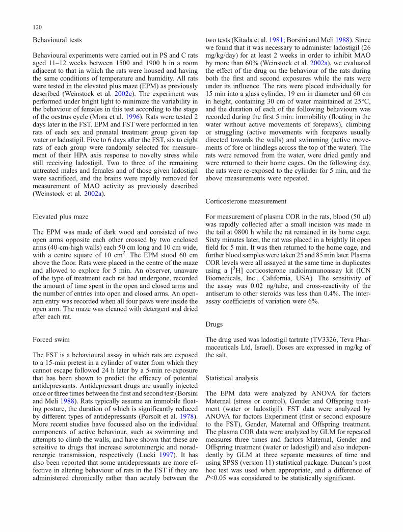
Analysis of the behaviour of untreated rats during the firstand second exposures to the FST revealed the followingmain effects. For Experiment, there were significant dif-ferences between the first and second experiments in im-mobility (F1,159=32.4, P<0.0001), climbing (F1,158=32.5,P<0.0001) and swimming (F1,159=10.30, P<0.002). Therewere also significant Gender differences in immobility(F1,159=30.5,P<0.0001), climbing (F1,159=19.2,P<0.0001)and swimming (F1,159=15.1,P<0.0001) since females spentmore time in active behaviours than males during the firstexposure to the test (Fig. 2). A significant Experiment ×Maternal treatment interaction was only found for swim-ming (F1,158=4.11, P<0.05), which was reduced more in PSthan in C rats, but there was noGender ×Maternal treatmentinteraction. Offspring treatment with ladostigil resulted insignificant main effects on immobility (F1,158=13.2,P<0.0001), climbing (F1,79=9.66, P<0.0025) and swim-ming (F1,158=5.74, P<0.02). Maternal × Offspring treat-
ment interactions were significant for all three behaviouralparadigms, immobility (F1,79=13.2, P<0.0001), climbing(F1,79=5.24, P<0.025) and swimming (F1,158=9.01, P<0.005), since ladostigil significantly affected the behaviourof PS but not that of C rats during the first and secondexposures. There were significant Experiment × Gender ×Offspring treatment interactions for immobility (F1,158=7.71, P<0.01) and swimming (F1,158=7.69, P<0.01).Although ladostigil altered all three types of behaviourof PS males in the first experiment, only the decrease inimmobility reached statistical significance. Ladostigil se-lectively increased climbing more in PS than in C females.In the second swim test, ladostigil selectively decreasedimmobility and increased swimming and climbing of PSrats but not that of controls (Fig. 2). The effect of lado-stigil was more evident in PS females than in males.
Water Ladostigil0
10
20
30
40
50
60
70
80
Tim
e in
ope
n ar
ms
(sec
)
a
Water Ladostigil0
10
20
30
40
50
Rat
io o
f Ope
n/T
otal
Ent
ries
(*10
0)
b
Cmale
PSmale
Cfemale
PSfemale
*
*
**
Fig. 1 Effect of ladostigil on behavior of PS and C rats in EPM. aTime in open arms; b ratio open/total arm entries×100. Data reflectmean and SEM. *Significantly different from water, P<0.05
121
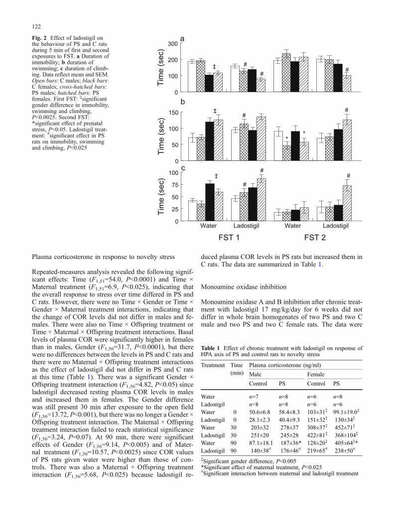
Plasma corticosterone in response to novelty stress
Repeated-measures analysis revealed the following signif-icant effects: Time (F1,51=54.0, P<0.0001) and Time ×Maternal treatment (F1,51=6.9, P<0.025), indicating thatthe overall response to stress over time differed in PS andC rats. However, there were no Time × Gender or Time ×Gender × Maternal treatment interactions, indicating thatthe change of COR levels did not differ in males and fe-males. There were also no Time × Offspring treatment orTime × Maternal × Offspring treatment interactions. Basallevels of plasma COR were significantly higher in femalesthan in males, Gender (F1,56=31.7, P<0.0001), but therewere no differences between the levels in PS and C rats andthere were no Maternal × Offspring treatment interactionsas the effect of ladostigil did not differ in PS and C ratsat this time (Table 1). There was a significant Gender ×Offspring treatment interaction (F1,56=4.82, P<0.05) sinceladostigil decreased resting plasma COR levels in malesand increased them in females. The Gender differencewas still present 30 min after exposure to the open field(F1,56=13.72, P<0.001), but there was no longer a Gender ×Offspring treatment interaction. The Maternal × Offspringtreatment interaction failed to reach statistical significance(F1,56=3.24, P=0.07). At 90 min, there were significanteffects of Gender (F1,56=9.14, P<0.005) and of Mater-nal treatment (F1,56=10.57, P<0.0025) since COR valuesof PS rats given water were higher than those of con-trols. There was also a Maternal × Offspring treatmentinteraction (F1,56=5.68, P<0.025) because ladostigil re-
duced plasma COR levels in PS rats but increased them inC rats. The data are summarized in Table 1.
Monoamine oxidase inhibition
Monoamine oxidase A and B inhibition after chronic treat-ment with ladostigil 17 mg/kg/day for 6 weeks did notdiffer in whole brain homogenates of two PS and two Cmale and two PS and two C female rats. The data were
Table 1 Effect of chronic treatment with ladostigil on response ofHPA axis of PS and control rats to novelty stress
Treatment Time(min)
Plasma corticosterone (ng/ml)
Male Female
Control PS Control PS
Water n=7 n=8 n=6 n=8Ladostigil n=8 n=8 n=6 n=6Water 0 50.6±6.8 58.4±8.3 103±31‡ 99.1±19.0‡
Ladostigil 0 28.1±2.3 40.4±9.3 151±32‡ 130±34‡
Water 30 203±32 278±37 308±37‡ 452±71‡
Ladostigil 30 251±20 245±28 422±81‡ 368±104‡
Water 90 87.1±18.1 187±36* 128±20‡ 405±64‡*Ladostigil 90 140±38# 176±46# 219±65# 238±50#
‡Significant gender difference, P<0.005*Significant effect of maternal treatment, P<0.025#Significant interaction between maternal and ladostigil treatment
0
100
200
300
Tim
e (s
ec)
a
0
50
100
150
Tim
e (s
ec)
b
FST 1 FST 2
0
25
50
75
100
Tim
e (s
ec)
c
Water Ladostigil Water Ladostigil
‡
‡
‡
**
#
##
##
#
#
#
Fig. 2 Effect of ladostigil onthe behaviour of PS and C ratsduring 5 min of first and secondexposures to FST. a Duration ofimmobility; b duration ofswimming; c duration of climb-ing. Data reflect mean and SEM.Open bars: C males; black bars:C females; cross-hatched bars:PS males; hatched bars: PSfemales. First FST: ‡significantgender difference in immobility,swimming and climbing,P<0.0025. Second FST:*significant effect of prenatalstress, P<0.05. Ladostigil treat-ment: #significant effect in PSrats on immobility, swimmingand climbing, P<0.025
122
therefore pooled, and inhibition of MAO-Awas 62.0±3.2%and MAO-B 66.8±2.9%.
Discussion
In the present study, we found that ladostigil, a novelpotential antidepressant with cholinesterase inhibitory ac-tivity, reduced hyperanxiety in both male and female PS ratsin the EPM and depressive-like behaviour in the FSTwithout affecting the behaviour of controls. This selectiveeffect of ladostigil in the EPM occurred in spite of the factthat PS rats did not show significantly greater anxiety thancontrols under the conditions of the test, probably becauseit was performed in a well-lit room in order to avoid aninfluence of different stages of the oestrus cycle on be-haviour. Under these conditions, control rats also spendlittle time in the open arms of the maze. The lack of ananxiolytic effect of ladostigil in the EPM concurs with thatof others who treated control rats chronically with anti-depressants, in contrast to diazepam, which induced a clearreduction in anxiety (Cole and Rodgers 1995; File et al.1999; Weinstock et al. 2002b). The selective sensitivity ofPS rats to the effect of ladostigil in the EPM indicates thatthere are neurochemical differences underlying the behav-iour of PS and C rats. These include, among others, a re-duction in hippocampal benzodiazepine receptors (Frideet al. 1986), alterations in glutamate and dopamine recep-tor subtypes in different forebrain regions (Berger et al.2002) and in genes associated with subunits NR1 andNR2A of glutamate NMDA receptors (Kinnunen et al.2003). Unlike amitriptyline in a previous study (Poltyrevand Weinstock 2004), ladostigil had an anxiolytic effectin PS rats of both sexes. This observation makes it un-likely that the lack of effect of amitriptyline in PS maleswas due to an innate gender difference in anxiogenic be-haviour but suggests that it resulted from a difference inthe metabolism of amitriptyline in male and female rats(Masubuchi et al. 1996) and to the greater depressioncaused by the drug in motor activity in males.
In addition to predicting antidepressant potential ofdrugs, the FST has been used to detect depressive-like be-haviour induced in rats by prenatal stress (Alonso et al.1991; Drago et al. 1999; Frye andWawrzycki 2003). Whilesome studies reported a difference in the behaviour ofPS and control males in this test (Drago et al. 1999;Morley-Fletcher et al. 2003a), others only detected a great-er degree of depressive-like activity in PS females (Alonsoet al. 1991). This may be because C males were immobilefor more than 70% of the time during the second expo-sure to the test in the latter study, making it difficult toobtain significantly higher levels in PS males. In the pres-ent study, a gender difference was seen in the behaviour ofrats in the FST. During the first exposure to the test, malerats developed an immobile posture for a greater period oftime than females. Such a gender difference has been re-ported in some (Alonso et al. 1991; Barros and Ferigolo1998) but not other rat strains (Frye and Wawrzycki 2003)and appears to vary according to the stage of the oestrous
cycle. Re-exposure to the FST increased the duration ofimmobility in females to that in males.
As in the EPM test, untreated PS rats did not differsignificantly from controls in any measure of behaviourin the FST test. Nevertheless, ladostigil significantlyaffected the behaviour of PS rats during both the first andsecond exposures, thereby confirming the difference in theneurochemical basis underlying their behaviour mentionedabove. The effect of ladostigil on swimming and climbingin PS rats resulted from a more than 60% inhibition ofbrain MAO-A and B, which had been shown to enhanceboth serotoninergic (Weinstock et al. 2002b) and norad-renergic transmission (Sagi et al. 2003). The selective ef-fect of ladostigil on the behaviour of PS rats in this test isreminiscent of that of other antidepressants, desipramineand nomifensine in the WKY rat (Tejani-Butt et al. 2003), agenetic model of depressive behaviour (Lahamame et al.1997). These drugs had no effect on the behaviour of nor-mal Wistar or Sprague–Dawley rats. The data of the pres-ent study and those of Tejani-Butt et al. (2003) serve todemonstrate the difference in the response to drugs of ratswith an anxiety-depression state induced by genetic or en-vironmental factors from that of control animals.
Hyperactivity of the HPA axis with elevation of corti-sol at rest and in response to stress is consistently foundin patients with major depression and can be explainedby impaired corticosteroid receptor signalling (Holsboer2001). HPA axis function can be normalized by clinicallyeffective treatment with antidepressants (Heuser et al.1996). A similar impairment in the regulation of the HPAaxis associated with a reduction of GR and MR receptorshas been reported for PS rats (Weinstock et al. 1992;Henry et al. 1994; McCormick et al. 1995). The presentstudy confirmed previous findings that prenatal stresscaused a greater alteration in the activity of the responseof the HPA axis to stress. This was shown by the slowerreturn of plasma COR to baseline levels after exposureto novelty stress. Although present in rats of both sexes,the effect was more marked in females which are gen-erally more reactive to stress.
Ladostigil did not significantly effect resting or peaklevels of plasma COR in C or PS rats after exposure to theopen field. However, at 90 min after stress exposure, thedifference between C and PS rats in the rate of return tocontrol levels of plasma COR was no longer seen. Thissuggests that the rate of recovery of the response of HPAaxis to stress may have been increased in PS rats byladostigil. It would be of interest to determine if this effectof the drug is related to an increase towards normal inhippocampal GR receptors, as has been shown for chronictreatment of rats and human subjects with other anti-depressants (Budziszewska 2002; Calfa et al. 2003).
In conclusion, this study showed that the EPM test canbe used to detect the potential anxiolytic effect of lado-stigil, a novel AChE and MAO inhibitor, in both male andfemale PS rats. Prenatal stress significantly disrupted theresponse of the HPA axis to stress in adult rats, resultingin an increased duration of the elevation of plasma CORin response to stress. Chronic treatment with ladostigil at
123
a dose that inhibited MAO-A and B by more than 60%selectively increased swimming and struggling behaviourof PS rats in the FST, a finding compatible with an el-evation of both brain 5HT and NA. The ability of lado-stigil to improve cognitive dysfunction because of cho-linesterase inhibition and also to decrease anxiety anddepressive symptoms may make it a potentially valuabledrug for the treatment of elderly depressive with cognitiveimpairment that do not respond to known antidepressantmedications.
Acknowledgements The authors gratefully acknowledge the fi-nancial support of Teva Pharmaceuticals Ltd and Ms. Aviva Grossfor MAO activity measurements. Dr. T. Poltyrev thanks the IsraelMinistry of Absorption for financial support.
References
Alonso SJ, Arevalo R, Afonso D, Rodriguez M (1991) Effects ofmaternal stress during pregnancy on forced swimming test onbehaviour of the offspring. Physiol Behav 50:511–517
Archer J, Blackman D (1971) Prenatal psychological stress andoffspring behavior in rats and mice. Dev Psychobiol 4:193–248
Barros HMT, Ferigolo M (1998) Ethopharmacology of imipraminein the forced swimming test: gender differences. NeurosciBiobehav Rev 22:279–287
Berger MA, Barros VG, Sarchi MI, Tarazi FI, Antonelli MC (2002)Long-term effects of prenatal stress on dopamine and glutamatereceptors in adult rat brain. Neurochem Res 27:1525–1533
Blehar MC (1995) Gender differences in risk factors for mood andanxiety disorders: implications for clinical treatment research.Psychopharmacol Bull 31:687–691
Bodnoff SR, Suranyi-Cadotte B, Aitken DH, Quirion R, Meaney MJ(1988) The effects of chronic antidepressant treatment in ananimal model of anxiety. Psychopharmacology 95:298–302
Borsini F, Meli A (1988) Is the forced swimming test a suitablemodel for revealing antidepressant activity? Psychopharmacol-ogy 94:147–160
Breslau N, Schultz L, Peterson E (1995) Sex differences in de-pression: a role for preexisting anxiety. Psychiatry Res 58:1–12
Buccafusco JJ, Terry AV Jr, Goren T, Blaugrun E (2003) Potentialcognitive actions of TV3326, a novel neuroprotective agent, asassessed in old rhesus monkeys in their performance of ver-sions of a delayed matching task. Neuroscience 119:669–678
Budziszewska B (2002) Effect of antidepressant drugs on thehypothalamic–pituitary–adrenal axis activity and glucocorticoidreceptor function. Pol J Pharmacol 54:343–349
Calfa G, Kademian S, Ceschin D, Vega G, Rabinovich GA, VolosinM (2003) Characterization and functional significance of glu-cocorticoid receptors in patients with major depression: mod-ulation by antidepressant treatment. Psychoneuroendocrinology28:687–701
Cassella JV, Davis M (1985) Fear-enhanced acoustic startle is notattenuated by acute or chronic imipramine treatment in rats.Psychopharmacology 87:278–282
Cole JC, Rodgers RJ (1995) Ethological comparison of the effects ofdiazepam and acute/chronic imipramine on the behaviour ofmice in the elevated plus-maze. Pharmacol Biochem Behav52:473–478
Drago F, Di Leo F, Giardina L (1999) Prenatal stress induces bodyweight deficit and behavioural alterations in rats: the effect ofdiazepam. Eur Neuropsychopharmacol 9:239–245
File SE, Ouagazzal AM, Gonzalez LE, Overstreet DH (1999)Chronic fluoxetine in tests of anxiety in rat lines selectivelybred for differential 5-HT1A receptor function. PharmacolBiochem Behav 62:695–701
Fride E, Weinstock M (1988) Prenatal stress increases anxiety-related behavior and alters cerebral lateralization of dopami-nergic activity. Life Sci 42:1059–1065
Fride E, Dan Y, Gavish M, Weinstock M (1985) Prenatal stressimpairs maternal behavior in a conflict situation and reduceshippocampal benzodiazepine receptors. Life Sci 36:2103–2109
Fride E, Dan Y, Feldon J, Halevy G, Weinstock M (1986) Effects ofprenatal stress on vulnerability to stress in prepubertal and adultrats. Physiol Behav 37:681–687
Frye CA, Wawrzycki J (2003) Effect of prenatal stress and gonadalhormone condition on depressive behaviors of female and malerats. Horm Behav 44:319–326
Gué M, Bravard A, Meunier J, Veyrier R, Gaillet S, Recasens M,Maurice T (2004) Sex differences in learning deficits inducedby prenatal stress in juvenile rats. Behav Brain Res 150:149–157
Hayashi A, Nagaoka M, Yamada K, Ichitani Y, Miake Y, Okado N(1998) Maternal stress induces synaptic loss and developmentaldisabilities of offspring. Int J Dev Neurosci 16:209–216
Henry C, Kabbaj M, Simon H, Le Moal M, Maccari S (1994) Pre-natal stress increases the hypothalamic–pituitary–adrenal axisresponse to stress in young and adult rats. J Endocrinol 6:341–345
Herzog-Raalbag P (2002) Effect of antidepressants on the behav-ioural abnormalities and alterations in circadian rhythm inducedby prenatal stress in rats. M.Sc. Thesis, Hebrew University,Jerusalem, Israel
Heuser I, Schweiger U, Gotthardt U, Schmider J, Lammers CH,Dettling M, Yassouridis A, Holsboer F (1996) Pituitary–adrenal-system regulation and psychopathology during ami-triptyline treatment in elderly depressed patients and normalcomparison subjects. Am J Psychiatry 153:93–99
Holsboer F (2001) Stress, hypercortisolism and corticosteroid re-ceptors in depression: implications for therapy. J Affect Disord62:77–91
Holsboer F, Barden N (1996) Antidepressants and hypothalamic–pituitary–adrenocortical regulation. Endocr Rev 17:187–205
Jacobs BL, Praag H, Gage FH (2000) Adult brain neurogenesis andpsychiatry: a novel theory of depression. Mol Psychiatry 5:262–269
Kinnunen AK, Koenig JI, Bilbe G (2003) Repeated variable prenatalstress alters pre- and postsynaptic gene expression in the ratfrontal pole. J Neurochem 86:736–748
Kitada Y, Miyauchi T, Satoh A, Satoh S (1981) Effect of anti-depressants in the rat forced swim test. Eur J Pharmacol 72:145–152
Kuehner C (1999) Gender differences in the short-term course ofunipolar depression in a follow-up sample of depressed in-patients. J Affect Disord 56:127–139
Lahamame A, del Arco C, Pazos A, Yritia M, Amario A (1997) AreWistar–Kyoto rats a genetic model of depression resistant toantidepressant drugs? Eur J Pharmacol 337:115–123
Lemaire V, Koehl M, Le Moal M, Abrous DN (2000) Prenatal stressproduces learning deficits associated with an inhibition ofneurogenesis in the hippocampus. Proc Natl Acad Sci U S A97:11032–11037
Lenox-Smith AJ, Reynolds A (2003) A double-blind, randomised,placebo controlled study of venlafaxine XL in patients withgeneralised anxiety disorder in primary care. Br J Gen Pract53:772–777
Lopes Da Silva N, Ferreira VMM, Carobrez AP, Morato GS (1996)Individual housing from rearing modifies the performance ofyoung rats on the elevated plus-maze apparatus. Physiol Behav60:391–396
Lucki I (1997) The forced swimming test as a model for core andcomponent behavioral effects of antidepressant drugs. BehavPharmacol 8:523–532
Masubuchi Y, Iwasa T, Fujita S, Suzuki T, Horie T, Narimatsu S(1996) Regioselectivity and substrate concentration-dependencyof involvement of the CYP2D subfamily in oxidative metabo-lism of amitriptyline and nortriptyline in rat liver microsomes.J Pharm Pharmacol 48:925–929
124
McCormick CM, Smythe JW, Sharma S, Meaney MJ (1995) Sex-specific effects of prenatal stress on hypothalamic–pituitary–adrenal responses to stress and brain glucocorticoid receptordensity in adult rats. Dev Brain Res 84:55–61
Mora S, Dussaubat N, Diaz-Veliz G (1996) Effects of the estrouscycle and ovarian hormones on behavioral indices of anxiety infemale rats. Psychoneuroendocrinology 21:609–620
Morley-Fletcher S, Darnaudery M, Koehl M, Casolini P, Van ReethO, Maccari S (2003a) Prenatal stress in rats predicts immobilitybehavior in the forced swim test: effects of a chronic treatmentwith tianeptine. Brain Res 989:246–251
Morley-Fletcher S, Rea M, Maccari S, Laviola G (2003b) Envi-ronmental enrichment during adolescence reverses the effectsof prenatal stress on play behaviour and HPA axis reactivity inrats. Eur J Neurosci 18:3367–3374
Nebes RD, Pollock BG, Houck PR, Butters MA, Mulsant BH,Zmuda MD, Reynolds CF (2003) Persistence of cognitive im-pairment in geriatric patients following anti-depressant treat-ment: a randomized double-blind clinical trial with nortriptylineand paroxetine. J Psychiatric Res 37:99–108
Poltyrev T, Weinstock M (2004) Gender difference in the preventionof hyperanxiety in adult prenatally-stressed rats by chronic treat-ment with amitriptyline. Psychopharmacology 171:270–276
Porsolt RD, Anton G, Blavet N, Jalfre M (1978) Behavioral despairin rats: a new model sensitive to antidepressant treatments. EurJ Pharmacol 47:379–391
Sagi Y, Weinstock M, Youdim MBH (2003) Attenuation of MPTP-induced dopaminergic neurotoxicity by TV3326, a cholines-terase-monoamine oxidase inhibitor. J Neurochem 86:290–297
Schmitz C, Rhodes ME, Bludau M, Kaplan S, Ong P, Ueffing I,Vehoff J, Korr H, Frye CA (2002) Depression: reduced numberof granule cells in the hippocampus of female, but not male,rats due to prenatal restraint stress. Mol Psychiatry 7:810–813
Secoli SR, Teixeira NA (1998) Chronic prenatal stress affects de-velopment and behavioral depression in rats. Stress 2:273–280
Sheehan DV, Mao CG (2003) Paroxetine treatment of generalizedanxiety disorder. Psychopharmacol Bull 37(Suppl 1):64–75
Szuran TF, Pliska V, Pokorny J, Welzl H (2000) Prenatal stress inrats: effects on plasma corticosterone, hippocampal glucocor-ticoid receptors, and maze performance. Physiol Behav 71:353–362
Tejani-Butt S, Kluczynski J, Paré WP (2003) Strain-dependentmodification of behavior following antidepressant treatment.Prog Neuro-Psychopharmacol Biol Psychiatry 27:7–14
Teri L, Reifler BV, Veith RC, Barnes R, White E, McLean P, RaskindM (1991) Imipramine in the treatment of depressed Alzheimer’spatients: impact on cognition. J Gerontol 46:P372–P377
Thompson WR (1957) Influence of prenatal maternal anxiety onemotionality in young rats. Science 15:698–699
Van Dijken HH, Tilders FJ, Olivier B, Mos J (1992) Effects ofanxiolytic and antidepressant drugs on long-lasting behaviouraldeficits resulting from one short stress experience in male rats.Psychopharmacology 109:395–402
Weinstock M, Matlina E, Keshet GI, Rosen H, McEwen BS (1992)Prenatal stress selectively alters the reactivity of the hypotha-lamic–pituitary adrenal system in the female rat. Brain Res 595:195–200
Weinstock M, Gorodetsky E, Wang R-H, Gross A, Weinreb O,Youdim MBH (2002a) Limited potentiation of blood pressureresponse to oral tyramine by brain-selective monoamine oxi-dase A-B inhibitor, TV-3326 in conscious rabbits. Neurophar-macology 43:999–1005
Weinstock M, Poltyrev T, Bejar C, Sagi Y, Youdim MBH (2002b)TV3326, a novel cholinesterase and MAO inhibitor for Alz-heimer’s disease with co-morbidity of Parkinson’s disease anddepression. In: Mizuno Y, Fisher A, Hanin I (eds) Mapping theprogress of Alzheimer’s and Parkinson’s disease. Kluwer Aca-demic/Plenum Publishers, New York, pp 199–204
Weinstock M, Poltyrev T, Bejar C, Youdim MBH (2002c) Effect ofTV3326, a novel monoamine–oxidase–cholinesterase inhibitor,in rat models of anxiety and depression. Psychopharmacology160:318–324
Weinstock M, Gorodetsky E, Poltyrev T, Gross A, Sagi Y, YoudimMBH (2003) A novel cholinesterase and brain-selective mono-amine oxidase inhibitor for the treatment of dementia co-morbidwith depression and Parkinson’s disease. Prog Neuro-Psycho-pharmacol Biol Psychiatry 27:555–561
Zimmerberg B, Blaskey LG (1998) Prenatal stress effects are par-tially ameliorated by prenatal administration of the neurosteroidallopregnanolone. Pharmacol Biochem Behav 59:819–827
125
POTENTIAL COGNITIVE ACTIONS OF (N-PROPARGLY-(3R)-AMINOINDAN-5-YL)-ETHYL, METHYL CARBAMATE (TV3326), A NOVELNEUROPROTECTIVE AGENT, AS ASSESSED IN OLD RHESUSMONKEYS IN THEIR PERFORMANCE OF VERSIONS OF A DELAYEDMATCHING TASK
J. J. BUCCAFUSCO,a,c* A. V. TERRY, JR.,a,b,c
T. GORENd AND E. BLAUGRUNd
aAlzheimer’s Research Center, Medical College of Georgia, Augusta,GA 30912-2300, USAbUGA College of Pharmacy, Medical College of Georgia, Augusta, GA30912-2300, USAcVeterans Administration Medical Center, Augusta, GA 30912, USAdTEVA Pharmaceutical Industries Ltd, P.O. Box 8077, Industrial ZoneKiryat Nordau, Netanya, Israel
Abstract—(N-propargyl-(3R)-aminoindan-5-yl)-ethyl, methylcarbamate (TV3326), a known neuroprotective agent exhibit-ing the properties of both an inhibitor of monoamine oxidase(brain selective) and an inhibitor of acetylcholinesterase wasadministered to seven old rhesus monkeys well trained toperform versions of a delayed matching-to-sample (DMTS)task. An increasing dose regimen of TV3326 was adminis-tered orally according to a schedule that allowed the animalsto perform the standard DMTS task and a self-titrating ver-sion of the DMTS task each week during the study. A distrac-tor version of the task was administered during two of thedoses of TV3326. Under the conditions of this experimentTV3326 failed to significantly affect accuracy on the standardDMTS task; however, the drug was very effective in improv-ing the ability of subjects to titrate to longer-duration delayintervals in the titrating version of the task. The maximaldrug-induced extension of the self-titrated delay intervalamounted to a 36.7% increase above baseline. This increasein maximum delay duration occurred without a significantchange in overall task accuracy. TV3326 also significantlyimproved task accuracy during distractor (interference) ses-sions. The compound was effective enough to return groupperformance efficiency to standard DMTS vehicle levels ofaccuracy. These results were independent of whether trialswere associated with a distractor or non-distractor delayinterval, and they were independent of delay interval. Thelack of delay selectivity in task improvement by TV3326 maynot be consistent with a selective effect on attention. TV3326was not associated with any obvious side effect or untowardreaction of the animals to the drug. Thus, TV3326 may beexpected to offer a significant positive cognitive outcome inaddition to its reported neuroprotective action. © 2003 IBRO.Published by Elsevier Science Ltd. All rights reserved.
Key words: memory, attention, MAO inhibitor, cholinesteraseinhibitor, operant task, delayed response task.
The well-known selective vulnerability of basal forebrainacetylcholine-containing neurons in Alzheimer’s diseasehas underscored the importance of this neurotransmittersystem in certain components or types of working memoryand perhaps in other behavioral and cognitive functionsaffected by the disease. Among the host of degenerativeprocesses occurring in Alzheimer’s disease, reproduciblecholinergic deficits have been consistently reported, theyappear early in the disease process, and correlate wellwith the degree of dementia (for review, Francis et al.,1999). Moreover, abnormalities in cholinergic function arefrequently reported in other degenerative conditions suchas Parkinson’s disease, diffuse Lewy body dementia andHuntington’s disease. As in Alzheimer’s disease, suchcholinergic deficits often correlate with memory declineand dementia. The use of cholinesterase inhibitors such asdonepezil and rivastigmine provide the bulk of the clinicalarmamentarium for the treatment of cognitive symptoms inthe treatment of Alzheimer’s disease. These drugs haveprovided reasonable effectiveness in mild to moderatecases, improving quality of life and a delay in symptomaticprogression of the disease, in the case of donepezil, by upto 55 weeks (Taylor, 2001). Despite their effectiveness ascognition enhancers, thus far cholinesterase inhibitorshave not been shown to significantly impact the neurode-generative process itself. Other disease-modifying ap-proaches to the treatment of Alzheimer’s disease are un-der intense investigation (see Jacobsen, 2002; Taylor etal., 2002). One of these includes the ability to offer adegree of neuroprotection, i.e. to support and possiblyrepair dystrophic neurons, and to enhance neuronal out-growth and synaptic sprouting. Whereas the use of selec-tive monoamine oxidase-B (MAO-B) inhibitors such asselegiline have been tested in Parkinson’s patients andfound to be only partially effective in slowing the progressof the disease (Parkinson Study Group, 1989), this class ofagent may offer neuroprotection in Alzheimer’s disease(Sano et al., 1997; Knoll, 2000; Pratico and Delanty, 2000).TV3326 is a compound combining properties as a CNS-selective MAO inhibitor and a cholinesterase inhibitor. Ini-tial preclinical studies with the parent compound, rasagi-line, revealed potent cytoprotective activity in vitro, that
*Correspondence to: J. J. Buccafusco, Alzheimer’s Research Center,Medical College of Georgia, 1120 15th Street, Augusta, GA 30912-2300, USA. Tel: �1-706-721-6355; fax: �1-706-721-9861.E-mail address: [email protected] (J. J. Buccafusco).Abbreviations: DMTS, delayed matching-to-sample; MAO, mono-amine oxidase; TV3326, (N-propargyl-(3R)-aminoindan-5-yl)-ethyl,methyl carbamate.
Neuroscience 119 (2003) 669–678
0306-4522/03$30.00�0.00 © 2003 IBRO. Published by Elsevier Science Ltd. All rights reserved.doi:10.1016/S0306-4522(02)00937-5
669
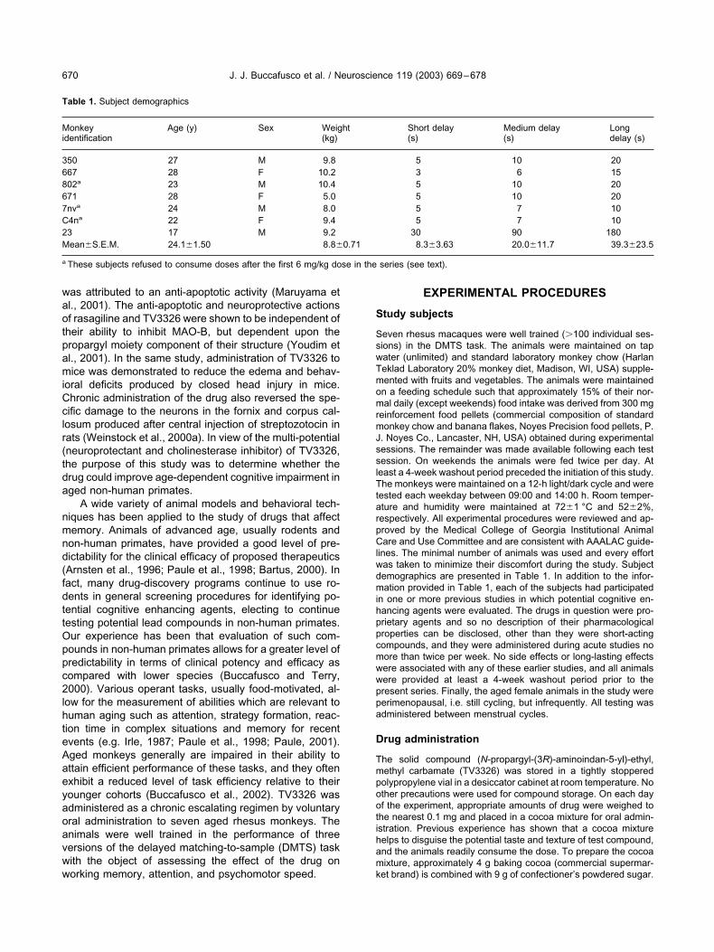
was attributed to an anti-apoptotic activity (Maruyama etal., 2001). The anti-apoptotic and neuroprotective actionsof rasagiline and TV3326 were shown to be independent oftheir ability to inhibit MAO-B, but dependent upon thepropargyl moiety component of their structure (Youdim etal., 2001). In the same study, administration of TV3326 tomice was demonstrated to reduce the edema and behav-ioral deficits produced by closed head injury in mice.Chronic administration of the drug also reversed the spe-cific damage to the neurons in the fornix and corpus cal-losum produced after central injection of streptozotocin inrats (Weinstock et al., 2000a). In view of the multi-potential(neuroprotectant and cholinesterase inhibitor) of TV3326,the purpose of this study was to determine whether thedrug could improve age-dependent cognitive impairment inaged non-human primates.
A wide variety of animal models and behavioral tech-niques has been applied to the study of drugs that affectmemory. Animals of advanced age, usually rodents andnon-human primates, have provided a good level of pre-dictability for the clinical efficacy of proposed therapeutics(Arnsten et al., 1996; Paule et al., 1998; Bartus, 2000). Infact, many drug-discovery programs continue to use ro-dents in general screening procedures for identifying po-tential cognitive enhancing agents, electing to continuetesting potential lead compounds in non-human primates.Our experience has been that evaluation of such com-pounds in non-human primates allows for a greater level ofpredictability in terms of clinical potency and efficacy ascompared with lower species (Buccafusco and Terry,2000). Various operant tasks, usually food-motivated, al-low for the measurement of abilities which are relevant tohuman aging such as attention, strategy formation, reac-tion time in complex situations and memory for recentevents (e.g. Irle, 1987; Paule et al., 1998; Paule, 2001).Aged monkeys generally are impaired in their ability toattain efficient performance of these tasks, and they oftenexhibit a reduced level of task efficiency relative to theiryounger cohorts (Buccafusco et al., 2002). TV3326 wasadministered as a chronic escalating regimen by voluntaryoral administration to seven aged rhesus monkeys. Theanimals were well trained in the performance of threeversions of the delayed matching-to-sample (DMTS) taskwith the object of assessing the effect of the drug onworking memory, attention, and psychomotor speed.
EXPERIMENTAL PROCEDURES
Study subjects
Seven rhesus macaques were well trained (�100 individual ses-sions) in the DMTS task. The animals were maintained on tapwater (unlimited) and standard laboratory monkey chow (HarlanTeklad Laboratory 20% monkey diet, Madison, WI, USA) supple-mented with fruits and vegetables. The animals were maintainedon a feeding schedule such that approximately 15% of their nor-mal daily (except weekends) food intake was derived from 300 mgreinforcement food pellets (commercial composition of standardmonkey chow and banana flakes, Noyes Precision food pellets, P.J. Noyes Co., Lancaster, NH, USA) obtained during experimentalsessions. The remainder was made available following each testsession. On weekends the animals were fed twice per day. Atleast a 4-week washout period preceded the initiation of this study.The monkeys were maintained on a 12-h light/dark cycle and weretested each weekday between 09:00 and 14:00 h. Room temper-ature and humidity were maintained at 72�1 °C and 52�2%,respectively. All experimental procedures were reviewed and ap-proved by the Medical College of Georgia Institutional AnimalCare and Use Committee and are consistent with AAALAC guide-lines. The minimal number of animals was used and every effortwas taken to minimize their discomfort during the study. Subjectdemographics are presented in Table 1. In addition to the infor-mation provided in Table 1, each of the subjects had participatedin one or more previous studies in which potential cognitive en-hancing agents were evaluated. The drugs in question were pro-prietary agents and so no description of their pharmacologicalproperties can be disclosed, other than they were short-actingcompounds, and they were administered during acute studies nomore than twice per week. No side effects or long-lasting effectswere associated with any of these earlier studies, and all animalswere provided at least a 4-week washout period prior to thepresent series. Finally, the aged female animals in the study wereperimenopausal, i.e. still cycling, but infrequently. All testing wasadministered between menstrual cycles.
Drug administration
The solid compound (N-propargyl-(3R)-aminoindan-5-yl)-ethyl,methyl carbamate (TV3326) was stored in a tightly stopperedpolypropylene vial in a desiccator cabinet at room temperature. Noother precautions were used for compound storage. On each dayof the experiment, appropriate amounts of drug were weighed tothe nearest 0.1 mg and placed in a cocoa mixture for oral admin-istration. Previous experience has shown that a cocoa mixturehelps to disguise the potential taste and texture of test compound,and the animals readily consume the dose. To prepare the cocoamixture, approximately 4 g baking cocoa (commercial supermar-ket brand) is combined with 9 g of confectioner’s powdered sugar.
Table 1. Subject demographics
Monkeyidentification
Age (y) Sex Weight(kg)
Short delay(s)
Medium delay(s)
Longdelay (s)
350 27 M 9.8 5 10 20667 28 F 10.2 3 6 15802a 23 M 10.4 5 10 20671 28 F 5.0 5 10 207nva 24 M 8.0 5 7 10C4na 22 F 9.4 5 7 1023 17 M 9.2 30 90 180Mean�S.E.M. 24.1�1.50 8.8�0.71 8.3�3.63 20.0�11.7 39.3�23.5
a These subjects refused to consume doses after the first 6 mg/kg dose in the series (see text).
J. J. Buccafusco et al. / Neuroscience 119 (2003) 669–678670
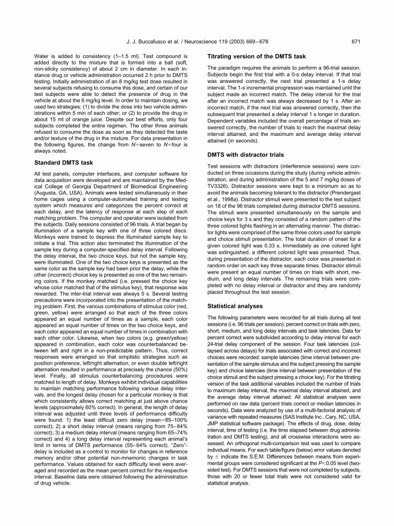
Water is added to consistency (1–1.5 ml). Test compound isadded directly to the mixture that is formed into a ball (soft,non-sticky consistency) of about 2 cm in diameter. In each in-stance drug or vehicle administration occurred 2 h prior to DMTStesting. Initially administration of an 8 mg/kg test dose resulted inseveral subjects refusing to consume this dose, and certain of ourtest subjects were able to detect the presence of drug in thevehicle at about the 6 mg/kg level. In order to maintain dosing, weused two strategies: (1) to divide the dose into two vehicle admin-istrations within 5 min of each other; or (2) to provide the drug inabout 15 ml of orange juice. Despite our best efforts, only foursubjects completed the entire regimen. The other three animalsrefused to consume the dose as soon as they detected the tasteand/or texture of the drug in the mixture. For data presentation inthe following figures, the change from N�seven to N�four isalways noted.
Standard DMTS task
All test panels, computer interfaces, and computer software fordata acquisition were developed and are maintained by the Med-ical College of Georgia Department of Biomedical Engineering(Augusta, GA, USA). Animals were tested simultaneously in theirhome cages using a computer-automated training and testingsystem which measures and categorizes the percent correct ateach delay, and the latency of response at each step of eachmatching problem. The computer and operator were isolated fromthe subjects. Daily sessions consisted of 96 trials. A trial began byillumination of a sample key with one of three colored discs.Monkeys were trained to depress the illuminated sample key toinitiate a trial. This action also terminated the illumination of thesample key during a computer-specified delay interval. Followingthe delay interval, the two choice keys, but not the sample key,were illuminated. One of the two choice keys is presented as thesame color as the sample key had been prior the delay, while theother (incorrect) choice key is presented as one of the two remain-ing colors. If the monkey matched (i.e. pressed the choice keywhose color matched that of the stimulus key), that response wasrewarded. The inter-trial interval was always 5 s. Several testingprecautions were incorporated into the presentation of the match-ing problem. First, the various combinations of stimulus color (red,green, yellow) were arranged so that each of the three colorsappeared an equal number of times as a sample, each colorappeared an equal number of times on the two choice keys, andeach color appeared an equal number of times in combination witheach other color. Likewise, when two colors (e.g. green/yellow)appeared in combination, each color was counterbalanced be-tween left and right in a non-predictable pattern. Thus, correctresponses were arranged so that simplistic strategies such asposition preference, left/right alternation, or even double left/rightalternation resulted in performance at precisely the chance (50%)level. Finally, all stimulus counterbalancing procedures werematched to length of delay. Monkeys exhibit individual capabilitiesto maintain matching performance following various delay inter-vals, and the longest delay chosen for a particular monkey is thatwhich consistently allows correct matching at just above chancelevels (approximately 60% correct). In general, the length of delayinterval was adjusted until three levels of performance difficultywere found: 1) the least difficult zero delay (mean�85–100%correct); 2) a short delay interval (means ranging from 75–84%correct); 3) a medium delay interval (means ranging from 65–74%correct) and 4) a long delay interval representing each animal’slimit in terms of DMTS performance (55–64% correct). “Zero”-delay is included as a control to monitor for changes in referencememory and/or other potential non-mnemonic changes in taskperformance. Values obtained for each difficulty level were aver-aged and recorded as the mean percent correct for the respectiveinterval. Baseline data were obtained following the administrationof drug vehicle.
Titrating version of the DMTS task
The paradigm requires the animals to perform a 96-trial session.Subjects begin the first trial with a 0-s delay interval. If that trialwas answered correctly, the next trial presented a 1-s delayinterval. The 1-s incremental progression was maintained until thesubject made an incorrect match. The delay interval for the trialafter an incorrect match was always decreased by 1 s. After anincorrect match, if the next trial was answered correctly, then thesubsequent trial presented a delay interval 1 s longer in duration.Dependent variables included the overall percentage of trials an-swered correctly, the number of trials to reach the maximal delayinterval attained, and the maximum and average delay intervalattained (in seconds).
DMTS with distractor trials
Test sessions with distractors (interference sessions) were con-ducted on three occasions during the study (during vehicle admin-istration, and during administration of the 5 and 7 mg/kg doses ofTV3326). Distractor sessions were kept to a minimum so as toavoid the animals becoming tolerant to the distractor (Prendergastet al., 1998a). Distractor stimuli were presented to the test subjecton 18 of the 96 trials completed during distractor DMTS sessions.The stimuli were presented simultaneously on the sample andchoice keys for 3 s and they consisted of a random pattern of thethree colored lights flashing in an alternating manner. The distrac-tor lights were comprised of the same three colors used for sampleand choice stimuli presentation. The total duration of onset for agiven colored light was 0.33 s. Immediately as one colored lightwas extinguished, a different colored light was presented. Thus,during presentation of the distractor, each color was presented inrandom order on each key three separate times. Distractor stimuliwere present an equal number of times on trials with short, me-dium, and long delay intervals. The remaining trials were com-pleted with no delay interval or distractor and they are randomlyplaced throughout the test session.
Statistical analyses
The following parameters were recorded for all trials during all testsessions (i.e. 96 trials per session): percent correct on trials with zero,short, medium, and long delay intervals and task latencies. Data forpercent correct were subdivided according to delay interval for each24-trial delay component of the session. Four task latencies (col-lapsed across delays) for trials associated with correct and incorrectchoices were recorded: sample latencies (time interval between pre-sentation of the sample stimulus and the subject pressing the samplekey) and choice latencies (time interval between presentation of thechoice stimuli and the subject pressing a choice key). For the titratingversion of the task additional variables included the number of trialsto maximum delay interval, the maximal delay interval attained, andthe average delay interval attained. All statistical analyses wereperformed on raw data (percent trials correct or median latencies inseconds). Data were analyzed by use of a multi-factorial analysis ofvariance with repeated measures (SAS Institute Inc., Cary, NC, USA,JMP statistical software package). The effects of drug, dose, delayinterval, time of testing (i.e. the time elapsed between drug adminis-tration and DMTS testing), and all crosswise interactions were as-sessed. An orthogonal multi-comparison test was used to compareindividual means. For each table/figure (below) error values denotedby � indicate the S.E.M. Differences between means from experi-mental groups were considered significant at the P�0.05 level (two-sided test). For DMTS sessions that were not completed by subjects,those with 20 or fewer total trials were not considered valid forstatistical analysis.
J. J. Buccafusco et al. / Neuroscience 119 (2003) 669–678 671

RESULTS
Standard DMTS
Fig. 1 presents the performance efficiencies (percentageof the trials completed that were correctly answered) ex-hibited by the subjects tested 2 h after administration of
vehicle as the first seven data points (Table 2). Increasingthe duration of delay (retention) intervals from Zero to Longwas associated with the expected decrement in perfor-mance efficiency (also shown in Table 2). Task accuracyover consecutive days was relatively stable across all de-lay intervals. Fig. 1 also shows the composite data set for
Fig. 1. Effect of daily single dose administration of TV3326 on standard DMTS task performance measured 2 h after drug administration categorizedby delay interval (note: Average indicates the mean of all completed trials in the session). Symbols are coded to indicate the dose of TV3326 that wasadministered on that experimental day: vehicle (filled circles); 1 mg/kg (open circles); 2 mg/kg (filled squares); 3 mg/kg (open squares); 4 mg/kg (filledupward triangles); 5 mg/kg (open upward triangles); 6 mg/kg (filled diamonds); 7 mg/kg (open downward triangles); washout (closed downwardtriangles). N�four with arrow indicates the experimental day where only four animals continued to consume the dose. N�seven with arrow indicatesthe washout period after drug discontinuation. Horizontal lines indicate the combined average for the first seven and the last four data points. Therewas no significant effect of drug treatment (F36,814�0.84, P�0.74), nor was there a significant interaction between treatment and delay interval(F108,814�0.55, P�0.99).
J. J. Buccafusco et al. / Neuroscience 119 (2003) 669–678672

the effects of TV3326 given by oral administration onDMTS performance efficiency categorized by delay inter-val. Despite daily administration of TV3326, there were noobvious changes in ongoing levels of task accuracy. Thiswas confirmed by statistical analysis in that there was nosignificant effect of drug treatment (F36,814�0.84,
P�0.74), nor was there a significant interaction betweentreatment and delay interval (F108,814�0.55, P�0.99).
In addition to the effect of drug treatment on taskaccuracy, aspects of working memory associated with theDMTS task, the two task latencies, also were determined.These values are presented in Table 3. There was a trend
Table 2. Average baseline performance efficiency for the seven study subjects by delay interval
Delay intervals
Average Zero Short Medium Long
73.8�1.26 85.0�1.78 79.2�1.32 66.2�1.59 64.9�2.25
Each value represents the mean�S.E.M. for 49 determinations (seven per subject). Average refers to the average level of accuracy determined fromall completed trials in a session. For the standard delayed matching-to-sample task, including all sessions together, there was a significant effect of“delay interval,” independent of treatment (F3,814�6.90, P�0.0001).
Table 3. Effect of TV3326 on median delayed matching-to-sample task latencies (in seconds)
Sample correct Choice correct Sample incorrect Choice incorrect Dose(mg/kg)
Day Average S.E.M. Average S.E.M. Average S.E.M. Average S.E.M.
1 1.29 0.23 2.29 0.51 1.66 0.47 2.71 0.53 03 1.36 0.44 2.34 0.48 1.13 0.21 2.59 0.53 05 1.09 0.20 2.17 0.46 1.26 0.20 2.59 0.42 07 0.92 0.11 2.17 0.51 0.83 0.10 2.70 0.71 08 2.22 0.97 2.58 0.62 1.78 0.56 2.75 0.57 09 1.24 0.27 1.90 0.11 1.68 0.50 2.64 0.23 0
11 1.51 0.32 2.47 0.59 1.70 0.51 2.93 0.61 014 1.20 0.19 2.13 0.50 1.26 0.24 2.77 0.80 115 1.97 0.66 2.52 0.42 1.77 0.51 2.87 0.49 116 1.37 0.24 2.23 0.46 1.20 0.19 5.29 2.50 118 1.49 0.42 2.26 0.44 1.14 0.20 2.70 0.47 121 1.36 0.15 2.19 0.47 1.47 0.36 2.60 0.58 122 1.49 0.20 2.21 0.53 2.16 0.78 2.70 0.58 123 1.52 0.27 2.37 0.45 1.33 0.16 2.95 0.68 225 1.27 0.19 2.21 0.44 1.44 0.35 2.53 0.49 228 1.10 0.12 2.11 0.46 1.16 0.16 2.59 0.61 329 1.36 0.23 2.07 0.33 1.81 0.50 2.61 0.46 330 1.33 0.28 2.03 0.33 1.57 0.49 2.59 0.50 432 1.32 0.22 2.42 0.41 1.38 0.25 2.85 0.55 435 1.73 0.29 2.33 0.55 1.70 0.38 2.63 0.47 536 1.29 0.19 2.17 0.49 1.57 0.23 2.73 0.60 538 1.24 0.20 2.07 0.47 1.40 0.22 2.49 0.53 642 1.13 0.17 2.16 0.48 1.24 0.26 2.69 0.69 643 1.43 0.24 2.60 0.70 1.53 0.43 3.03 1.03 644 0.90 0.09 2.35 0.72 0.95 0.17 2.78 0.81 646 1.20 0.08 2.58 0.68 1.15 0.26 3.03 0.93 649 1.18 0.15 2.50 0.52 1.35 0.26 3.15 0.75 650 1.07 0.06 2.93 1.03 0.90 0.09 4.17 1.71 651 1.10 0.08 2.55 0.62 1.35 0.16 2.83 0.83 653 1.10 0.18 2.97 0.98 1.00 0.13 4.13 1.50 656 1.03 0.18 2.63 0.70 0.93 0.10 3.23 1.01 657 0.90 0.05 2.80 0.91 0.90 0.05 3.53 1.28 759 1.13 0.15 2.63 0.71 0.93 0.05 3.23 1.00 763 0.94 0.14 2.06 0.49 0.89 0.06 2.66 0.68 064 1.31 0.11 2.37 0.66 1.41 0.18 3.04 0.85 065 1.33 0.28 2.73 0.60 0.95 0.08 3.18 0.83 067 1.78 0.30 1.85 0.13 2.35 0.56 2.35 0.10 0
Each value represents the mean�S.E.M. for seven subjects except for days 43–59 which were derived from only four subjects (numbers 350, 667,671, and 23). There was a trend towards a significant difference in the latency category, independent of drug treatment (F3,814�2.53, P�0.065), butthere was no significant effect related to drug treatment (F36,814�0.84, P�0.74).
J. J. Buccafusco et al. / Neuroscience 119 (2003) 669–678 673
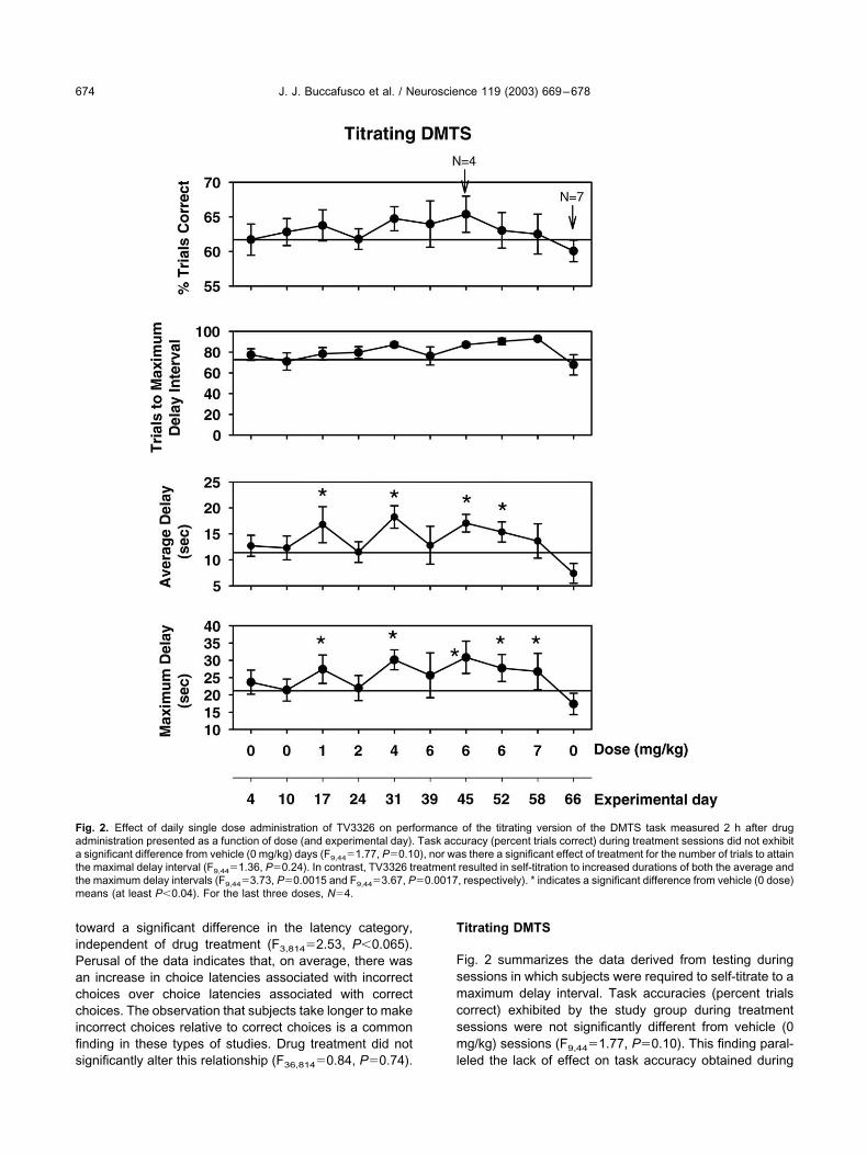
toward a significant difference in the latency category,independent of drug treatment (F3,814�2.53, P�0.065).Perusal of the data indicates that, on average, there wasan increase in choice latencies associated with incorrectchoices over choice latencies associated with correctchoices. The observation that subjects take longer to makeincorrect choices relative to correct choices is a commonfinding in these types of studies. Drug treatment did notsignificantly alter this relationship (F36,814�0.84, P�0.74).
Titrating DMTS
Fig. 2 summarizes the data derived from testing duringsessions in which subjects were required to self-titrate to amaximum delay interval. Task accuracies (percent trialscorrect) exhibited by the study group during treatmentsessions were not significantly different from vehicle (0mg/kg) sessions (F9,44�1.77, P�0.10). This finding paral-leled the lack of effect on task accuracy obtained during
Fig. 2. Effect of daily single dose administration of TV3326 on performance of the titrating version of the DMTS task measured 2 h after drugadministration presented as a function of dose (and experimental day). Task accuracy (percent trials correct) during treatment sessions did not exhibita significant difference from vehicle (0 mg/kg) days (F9,44�1.77, P�0.10), nor was there a significant effect of treatment for the number of trials to attainthe maximal delay interval (F9,44�1.36, P�0.24). In contrast, TV3326 treatment resulted in self-titration to increased durations of both the average andthe maximum delay intervals (F9,44�3.73, P�0.0015 and F9,44�3.67, P�0.0017, respectively). * indicates a significant difference from vehicle (0 dose)means (at least P�0.04). For the last three doses, N�4.
J. J. Buccafusco et al. / Neuroscience 119 (2003) 669–678674
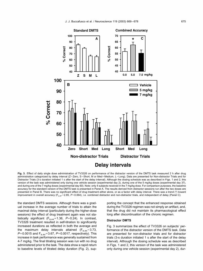
the standard DMTS sessions. Although there was a grad-ual increase in the average number of trials to attain themaximal delay interval (particularly during the higher-dosesessions) the effect of drug treatment again was not sta-tistically significant (F9,44�1.36, P�0.24). In contrast,TV3326 treatment resulted in self-titration to significantlyincreased durations as reflected in both the average andthe maximum delay intervals attained (F9,44�3.73,P�0.0015 and F9,44�3.67, P�0.0017, respectively). Thisincrease in task performance was generally sustained from4-7 mg/kg. The final titrating session was run with no drugadministered prior to the task. The data show a rapid returnto baseline levels of titrated delay duration (Fig. 2), sup-
porting the concept that the enhanced response obtainedduring the TV3326 regimen was not simply an artifact, and,that the drug did not maintain its pharmacological effectlong after discontinuation of the chronic regimen.
Distractor DMTS
Fig. 3 summarizes the effect of TV3326 on subjects’ per-formance of the distractor version of the DMTS task. Dataare presented for non-distractor trials and for distractortrials (3-s duration initiated 1 s after the start of the delayinterval). Although the dosing schedule was as describedin Figs. 1 and 2, this version of the task was administeredonly during one vehicle session (experimental day 2), dur-
Fig. 3. Effect of daily single dose administration of TV3326 on performance of the distractor version of the DMTS task measured 2 h after drugadministration categorized by delay interval (Z�Zero, S�Short, M or Med�Medium, L�Long). Data are presented for Non-distractor Trials and forDistractor Trials (3-s duration initiated 1 s after the start of the delay interval). Although the dosing schedule was as described in Figs. 1 and 2, thisversion of the task was administered only during one vehicle session (experimental day 2), during one of the 5 mg/kg doses (experimental day 37),and during one of the 7 mg/kg doses (experimental day 60). Note: only 4 subjects received in the 7 mg/kg dose. For comparison purposes, the baselineaccuracy for the standard version of the DMTS task is presented in Panel A. The results derived from distractor sessions run after the two doses arepresented in Panel B. There was no significant effect of drug treatment either alone, or as a factor with delay interval. There was a trend (*) towardimprovement in overall accuracy (F2,87�2.85, P�0.064), i.e. combined distractor and non-distractor trials, and independent of delay (Panel C).
J. J. Buccafusco et al. / Neuroscience 119 (2003) 669–678 675

ing one of the 5 mg/kg doses (experimental day 37), andduring one of the 7 mg/kg doses (experimental day 60).For these three sessions the non-distractor trials appearedto be performed as inefficiently (decrements in accuracyrelative to standard DMTS levels of performance effi-ciency) as were the 18 randomly presented distractor tri-als. We have noted carryover of the distractor-related im-pairment to non-distractor trials within the same session onother occasions, but the effect seems to be more pro-nounced in older subjects. Therefore, the results obtainedunder these conditions should be interpreted with care.Notwithstanding this cautionary note, sessions run afteradministration of TV3326, independent of dose, indepen-dent of delay interval, and independent of trial type (dis-tractor versus non-distractor) showed a trend toward asignificant level of improvement in overall accuracy(F2,87�2.85, P�0.064).
DISCUSSION
Although MAO inhibitors have been examined for theirability to protect against the development of MPTP-in-duced parkinsonian symptoms or in reversing symptoms inthese animals (e.g. Andringa and Cools, 2000; Kupsch etal., 2001), very little is known regarding the effects of thesecompounds on cognitive function in non-human primates.It is known, however, that noradrenergic neurons are nec-essary for certain frontal lobe-mediated cognitive pro-cesses. These include attention and the prevention ofdistraction in the presence of irrelevant stimuli (Robbinsand Everitt, 1995). A normal level of noradrenergic neuralactivity appears to be necessary for optimal function of theprefrontal cortex. Agonists at �2A receptors (e.g. clonidine,guanfacine) have been shown to improve prefrontal corti-cal function in non-human primates whereas antagonistsat �2 receptors (e.g. yohimbine) have been shown to im-pair function and to antagonize the positive mnemonicactions of agonists (see Mao et al., 1999). Thus, the sus-ceptibility to distracting stimuli known to occur in Alzhei-mer’s disease patients may be improved by compoundsdesigned to optimize noradrenergic activity within the pre-frontal cortex. Because of the behavioral effects of am-phetamine and its derivatives, it is unlikely that this class ofdrugs could be used as a treatment adjunct in Alzheimer’sdisease. However, the mood-elevating aspects and poten-tial cognitive enhancing effects of MAO inhibitors mayprove more appropriate in this setting. To suggest thispossibility for TV3326 are the results of behavioral studiesin rats in which chronic administration of TV3326 reversedimmobility in the forced swim test (antidepressant activity)and the drug antagonized scopolamine-induced impair-ment of spatial memory (Weinstock et al., 2000b). It isundetermined whether this potential positive effect on cog-nition in these animals was mediated through brain MAOinhibition or through cholinesterase inhibition, or by thecombination of effects. Studies in rats (Haroutunian et al.,1990) and macaques (Buccafusco et al., 1992; Terry et al.,1993) have indicated the advantage of combining the ef-
fects of central �2 adrenergic receptor stimulation(clonidine) with cholinesterase inhibition (physostigmine).
It also has not been determined whether the pharma-cological actions of TV3326 would include increased brainnoradrenergic, dopaminergic, or serotonergic activity atclinically relevant doses. However, in this study, the drugappeared to behave less like a noradrenergic agonist(Jackson and Buccafusco, 1991), and more like a dopa-minergic agonist (Prendergast et al., 1998a). This is be-cause TV3326 was more effective in improving task accu-racy in the distractor version of the DMTS task as com-pared with the standard version. The role of braindopaminergic pathways in attentional aspects of cognitionis well known. In fact, normal dopaminergic function ap-pears to be necessary for the successful performance ofmemory tasks that rely on the function of the prefrontalcortex. For example, a narrow range of the D1 selectiveagonists A77636 and SKF81297 were reported to improveperformance of a spatial working memory task in agedmonkeys (Cai and Arnsten, 1997). For MAO inhibitors, themechanism for their effects on cognition is even less ob-vious, in that they may be mediated through mechanismsother than inhibition of the MAO enzyme (Gelowitz et al.,1994; Shankaranarayana et al., 1999).
The increase in self-titrated maximal delay intervalfrom vehicle levels (22.6 s) to those obtained after admin-istration of the second of the 6 mg/kg doses (30.9 s) in thetitrating version of the DMTS task amounted to a 36.7%increase above baseline (in fact, the improvement overbaseline that was sustained by the 4–7 mg/kg doses in thetitrating version of the DMTS task averaged 25.2�4.96%of control). This increase in maximum delay duration oc-curred with a small, but non-significant, increment in thenumber of trials needed to attain the longer intervals as-sociated with drug treatment. Also, overall task accuracywas unchanged. The observation that subjects self-titratedto longer delay intervals without a significant change inoverall accuracy may indicate that the drug enhanced themotivational aspects of the task. Other behavioral ap-proaches would be needed to confirm this conjecture.Because the average titrated delay interval increased con-comitantly with the maximal delay, it is likely that once themaximal delay was attained, accuracy was not sustainedfor the next few trials, thus maintaining accuracy constant.Nevertheless, we have demonstrated that the titratingDMTS task is more sensitive to age-related task deficitsthan is the standard DMTS task. Even within the titratingDMTS the scores for session accuracy were not as wellcorrelated with age as was the number of trials to reach themaximal delay interval (Buccafusco et al., 2002). Thus weinterpret an increase in titrated delay interval (as long asaccuracy does not decrease) as a positive effect on taskperformance.
TV3326 also significantly improved task accuracy dur-ing distractor sessions. The compound was effectiveenough to return group performance efficiency to standardDMTS vehicle levels of accuracy. These results were in-dependent of whether trials were associated with a distrac-tor or non-distractor trial, and they were independent of
J. J. Buccafusco et al. / Neuroscience 119 (2003) 669–678676
delay interval. Because of the profound carryover effect ofthe distractor-impaired performance to the non-distractortrials, the positive overall effect produced by the compoundon task accuracy is difficult to interpret. Drugs like methyl-phenidate and nicotine generally affect task accuracy se-lectively during Short delay trials in the distractor task(Prendergast et al., 1998a,b) although like TV3326, nico-tine improved task accuracy for both distractor and non-distractor trials. Despite the lack of a significant delay-dependent effect of drug treatment, on average, TV3326improved accuracy most consistently during Short delaytrials (Fig. 3) both for non-distractor and distractor trials.TV3326 does appear to have at least one advantage overmethylphenidate in this task. Whereas methylphenidateexhibited clear efficacy in reversing distractor-induced dec-rements in task accuracy in young monkeys, the amphet-amine derivative was unable to reverse distractor-inducedperformance decrements by aged subjects (Prendergastet al., 1998a).
It is difficult to provide a level of comparison of themnemonic effects of TV3326 relative to other drugs testedat this center owing to the lack of effect of the drug on taskaccuracy. However, the 25% improvement over baselinethat was sustained in the titrating version of the DMTS taskis in keeping with the most effective drugs that we havetested on an acute basis for improvements in standardDMTS accuracy. We have yet to determine whether suchcomparisons based on improvement over baseline perfor-mance for different task variables are valid. Where a moredirect comparison may be made, is with regard to the mostimproved degree of accuracy associated with a delay in-terval (Short) for the non-distractor trials in the distractorversion of the DMTS task. On average, the improvementamounted to 27.5% relative to vehicle levels of perfor-mance. Notwithstanding this limitation, these data provideat least a proof of concept that development of drugs withmultiple targets and multiple pharmacological propertiesmay prove superior to either monotherapy, or to combiningdrugs with varying pharmacokinetic properties in the treat-ment of neurodegenerative diseases. TV3326 representsa new class of drug at least potentially suited to the treat-ment of Alzheimer’s disease patients who require thera-pies that will delay the progression of the disease, and whosuffer from impaired attention, impaired memory, and de-pression. The combination of the properties attributed toan adrenergic agonist and to a cholinesterase inhibitormay derive benefit from their combined cognitive enhanc-ing properties, as well as from the ability of adrenergicreceptor activation to limit the side effects of cholinester-ase inhibition (Buccafusco, 1992; Buccafusco and Terry,2000; Paule, 2001).
Acknowledgements—This work was partly supported by TEVAPharmaceutical Industries Ltd, by Prime Behavior Testing Labo-ratories, Inc., and by the Alzheimer’s Association. The authorswould like to recognize the excellent primate behavior technicalassistance provided by Ms Nancy Kille, and Ms Ritu Duhan.
REFERENCES
Andringa G, Cools (2000) The neuroprotective effects of CGP 3466Bin the best in vivo model of Parkinson’s disease, the bilaterallyMPTP-treated rhesus monkey. J Neural Trans Suppl 60:215–225.
Arnsten AF, Steere JC, Hunt RD (1996) The contribution of �2-norad-renergic mechanisms of prefrontal cortical cognitive function: po-tential significance for attention-deficit hyperactivity disorder. ArchGen Psychiatry 53:448–455.
Bartus RT (2000) On neurodegenerative diseases, models, and treat-ment strategies: lessons learned and lessons forgotten a genera-tion following the cholinergic hypothesis. Exp Neurology 163:495–529.
Buccafusco JJ (1992) Neuropharmacologic and behavioral actions ofclonidine: interactions with central neurotransmitters. Int Rev Neu-robiol 33:55–107.
Buccafusco JJ, Jackson WJ, Terry AV Jr (1992) Effects of concomitantcholinergic and adrenergic stimulation on learning and memoryperformance by primates. Life Sci 51:7–12.
Buccafusco JJ, Terry AV Jr (2000) Multiple CNS targets for elicitingbeneficial effects on memory and cognition. J Pharmacol Exp Ther295:438–446.
Buccafusco JJ, Terry AV Jr, Murdoch PB (2002) A computer assistedcognitive test battery for aged monkeys. J Mol Neurosci 19:187–193.
Cai JX, Arnsten AFT (1997) Dose-dependent effects of the dopamineD1 receptor agonists A77636 or SKF81297 on spatial workingmemory in aged monkeys. J Pharmacol Exp Ther 283:183–189.
Francis PT, Palmer AM, Snape M, Wilcock GK (1999) The cholinergichypothesis of Alzheimer’s disease: a review of progress. J NeurolNeurosurg Psychiatry 66:137–147.
Gelowitz DL, Richardson JS, Wishart TB, Yu PH, Lai CT (1994)Chronic L-deprenyl or L-amphetamine: equal cognitive enhance-ment, unequal MAO inhibition. Pharmacol Biochem Behav 47:41–45.
Haroutunian V, Kanof PD, Tsuboyama G, Davis KL (1990) Restorationof cholinomimetic activity by clonidine in cholinergic plus noradren-ergic lesioned rats. Brain Res 507:261–266.
Irle E (1987) Primate learning tasks reveal strong impairment in pa-tients with presenile dementia of the Alzheimer type. Brain Cogn6:429–449.
Jackson WJ, Buccafusco JJ (1991) Clonidine enhances delayedmatching-to-sample performance by young and aged monkeys.Pharmacol Biochem Behav 39:79–84.
Jacobsen JS (2002) Alzheimer’s disease: an overview of current andemerging therapeutic strategies. Curr Topics Med Chem 2:343–352.
Knoll J (2000) (�)Deprenyl (Selegiline): past, present and future.Neurobiology (Budapest) 8:179–199.
Kupsch A, Sautter J, Grotz ME, Breithaupt W, Schwarz J, Youdim MB,Riederer P, Gerlach M, Oertel WH (2001) Monoamine oxidase–inhibition and MPTP-induced neurotoxicity in the non-humanprimate: comparison of rasagiline (TVP 1012) with selegiline.J Neural Trans 108:985–1009.
Mao Z-M, Arnsten AFT, Li B-M (1999) Local infusion of an �-1 adren-ergic agonist into the prefrontal cortex impairs spatial working mem-ory performance in monkeys. Biol Psychiatry 46:1259–1265.
Maruyama W, Youdim MBH, Naof M (2001) Antiapoptotic properties ofrasagiline, N-proparglyamine-1(R)-aminoindan, and its optical (S)-isomer, TV1022. Ann NY Acad Sci 939:320–329.
Parkinson Study Group (1989) DATATOP: a multicenter controlledclinical trial in early Parkinson’s disease. Arch Neurol 46:1052–1060.
Paule MG, Bushnell PJ, Maurissen JPJ, Wenger GR, Buccafusco JJ,Chelonis JJ, Elliott R (1998) Symposium overview: the use ofdelayed matching-to-sample procedures in studies of short-termmemory in animals and humans. Neurotoxicol Teratol 20:493–502.
Paule MG (2001) Validation of a behavioral test battery for monkeys.
J. J. Buccafusco et al. / Neuroscience 119 (2003) 669–678 677
In: Methods of behavior analysis in neuroscience (Buccafusco JJ,ed), pp 281–294. New York: CRC Press.
Pratico D, Delanty N (2000) Oxidative injury in diseases of the centralnervous system: focus on Alzheimer’s disease. Am J Med 109:577–585.
Prendergast MA, Jackson WJ, Terry AV Jr, Kille NJ, Arneric SP,Buccafusco JJ (1998a) Age-related: differences in distractibility andresponse to methylphenidate in monkeys. Cereb Cortex 8:164–172.
Prendergast MA, Jackson WJ, Terry AV, Decker MW, Arneric SA,Buccafusco JJ (1998b) Central nicotinic receptor agonists ABT-418, ABT-089, and (�)-nicotine reduce distractibility in young-adultmonkeys. Psychopharmacology 136:50–58.
Robbins T, Everitt B (1995) Arousal systems and attention. In: Thecognitive neurosciences (Gazzangia M, ed), pp. 703–720. Cam-bridge, MA: MIT Press.
Sano M, Ernesto C, Thomas RG, Klauber MR, Schafer K, GrundmanM, Woodbury P, Growdon J, Cotman CW, Pfeiffer E, Schneider LS,Thal LJ (1997) A controlled trial of selegiline, alpha-tocopherol, orboth as treatment for Alzheimer’s disease: the Alzheimer’s DiseaseCooperative Study. N Engl J Med 336:1216–1222.
Shankaranarayana Rao BS, Lakshamana MK, Meti BL, Raju TR(1999) Chronic (�)deprenyl administration alters dendritic morphol-
ogy of layer III pyramidal neurons in the prefrontal cortex of adultBonnett monkeys. Brain Res 821:218–223.
Taylor P (2001) Anticholinesterase agents. In: Goodman and Gilman’spharmacological basis of therapeutics, 10th edition (Hardman JG,Limbird LE, Gilman AG, eds), pp 175–191. New York: McGraw-Hill.
Taylor JP, Hardy J, Fischbeck KH (2002) Toxic proteins in neurode-generative disease. Science 296:1991–1995.
Terry AV Jr, Jackson WJ, Buccafusco JJ (1993) Effects of concomitantcholinergic and adrenergic stimulation on learning and memoryperformance by young and aged monkeys. Cereb Cortex 3:304–312.
Weinstock M, Kirschbaum-Slager N, Lazarovici P, Bejar C, YoudimMBH, Shoham S (2000a) Neuroprotective effects of novel cholines-terase inhibitors derived from rasagiline as potential anti-Alzheimerdrugs. Ann NY Acad Sci 939:148–161.
Weinstock M, Bejar C, Wang RH, Poltyrev T, Gross A, Finberg JP,Youdin MB (2000b) TV3326, a novel neuroprotective drug withcholinesterase and monoamine oxidase inhibitory activities for thetreatment of Alzheimer’s disease. J Neural Trans Suppl 60:157–169.
Youdim MBH, Wadia A, Tatton W, Weinstock M (2001) The anti-Parkinson drug rasagiline and its cholinesterase inhibitor deriva-tives exert neuroprotection unrelated to MAO inhibition in cell cul-ture and in vivo. Ann NY Acad Sci 939:450–458.
(Accepted 30 October 2002)
J. J. Buccafusco et al. / Neuroscience 119 (2003) 669–678678

Rationale for considering thatpropargylamines might be
neuroprotective in Parkinson’s diseaseC. Warren Olanow, MD, FRCPC
Abstract—A neuroprotective therapy that slows or stops disease progression is the major unmet medical need inParkinson’s disease (PD). Current evidence indicates that cell death in PD occurs, at least in part, by way of a signal-mediated apoptotic process. This raises the possibility that anti-apoptotic agents might be neuroprotective in PD. Propar-gylamines have been demonstrated to be potent anti-apoptotic agents in both in vitro and in vivo studies, presumably bymaintaining glyceraldehyde-3-phosphate dehydrogenase (GAPDH) as a dimer and thereby preventing its nuclear translo-cation where it blocks upregulation of anti-apoptotic proteins. Selegiline is a monamine oxidase type B (MAO-B) inhibitorthat incorporates a propargyl ring within its molecular structure. It was shown to delay the need for symptomatic therapyin untreated PD patients in the DATATOP study, but interpretation is confounded by its symptomatic effects. Rasagilineis another MAO-B inhibitor that contains a propargyl ring and has protective effects in laboratory models. A clinical trialutilizing a delayed start design demonstrated that patients initiated on rasagiline at baseline are improved at one year incomparison to patients initiated on placebo and switched to rasagiline at 6 months even though both groups were on thesame treatment for the last 6 months of the study. These results argue against the benefit being due to a symptomaticeffect and are consistent with rasagiline having a protective effect.
NEUROLOGY 2006;66(Suppl 4):S69–S79
Parkinson’s disease (PD) is an age-related neurode-generative disorder that is characterized by degener-ation of dopaminergic neurons in the substantianigra pars compacta (SNc) coupled with intracytolas-mic proteinaceous inclusions or Lewy bodies.1 Neuro-degeneration is not restricted to dopaminergicneurons of the SNc but also occurs in norepinephrineneurons of the locus coeruleus (LC), cholinergic neu-rons of the nucleus basalis of Meynert (NBM), sero-tonin neurons of the dorsal raphe (DR), and neuronsof the dorsal motor nucleus of the vagus (DMV) andthe olfactory and peripheral autonomic systems.2
The cardinal clinical features of PD are restingtremor, rigidity, bradykinesia, and gait distur-bance with postural instability. Current therapyprimarily employs a dopamine replacement strat-egy using levodopa and dopamine agonists.3 Thisapproach provides benefits to virtually all PD pa-tients, particularly in the early stages of their dis-ease. However, long-term therapy is complicatedby the development of motor complications in themajority of patients. In addition, patients can ex-perience disability due to the emergence of freez-ing, falling, postural instability, autonomicdysfunction, sleep disturbances, mood disorders,
and dementia, which probably reflect degenerationof non-dopaminergic neurons.3,4 These problemsare not well controlled with currently availabletherapies, such that many PD patients experienceintolerable disability despite the many treatmentadvances in the disorder. The development of aneuroprotective therapy that slows, stops, or re-verses disease progression and prevents the devel-opment of clinical disability is an urgent priorityin PD. Among the many candidate agents thatmight be neuroprotective in PD,5,6 propar-gylamines are among the most promising. Propar-gylamines are molecules that incorporate apropargyl ring and typically inhibit MAO-B. Theyinclude several agents that have been studied inPD, such as selegiline, TCH346, and rasagiline.7
Many preclinical studies have demonstrated thecapacity of propargylamines to block apoptosis in invitro and in vivo models of PD independent of theircapacity to inhibit MAO-B. This review considers theexperimental data and the theoretical basis supportinga role for propargylamine-containing molecules as pu-tative neuroprotective agents in PD, and examinesclinical trials with these agents that have been per-formed in PD to date.
From the Department of Neurology, Mount Sinai School of Medicine, New York, New York.Publication of this supplement was supported by an educational grant from Teva Neuroscience and Eisai, Inc.Disclosure: The sponsor has provided the author with personal honoraria (in excess of $10,000) during his professional career.Address correspondence and reprint requests to Dr. C. Warren Olanow, Department of Neurology, Mount Sinai School of Medicine, Annenberg 14–94, OneGustave L. Levy Place, Box 1137, New York, NY 10029; e-mail: [email protected]
Neurology supplements are not peer-reviewed. Information contained in Neurology supplements represents the opinions of the authors and is notendorsed by nor does it reflect the views of the American Academy of Neurology, Editorial Board, Editor-in-Chief, or Associate Editors of Neurology.
Copyright © 2006 by AAN Enterprises, Inc. S69

Apoptosis and PD. Knowledge of the etiopatho-genesis of PD would greatly facilitate the develop-ment of a neuroprotective therapy. The preciseetiology of PD is not known, except for a small num-ber of familial cases with a genetic mutation,8 and itis possible that sporadic cases result from a complexinterplay between genetic and environmental factorsthat are not necessarily the same in all patients.Oxidative stress, mitochondrial dysfunction, inflam-mation, excitotoxicity, and protein aggregation havebeen implicated in the pathogenesis of cell death inPD.9,10 However, the role played by these factors inthe cell death process is not known, nor is it clear ifeach of these factors contributes equally to neurode-generation in all PD patients. Increasing evidencesuggests that they act as a network whereby inhibi-tion of any one factor might not be sufficient to pre-vent neuronal degeneration. A body of evidencesuggests that cell death in PD occurs, at least inpart, by way of signal-mediated apoptosis in all PDpatients.11-14 If apoptosis is common to all of the dif-ferent forms of PD, it is possible that anti-apoptoticagents might provide neuroprotective effects in eachof the different types of PD.
Apoptosis is a gradual form of cell death that ischaracterized by cell shrinkage, chromatin condensa-tion, and fragmentation of nuclear DNA with preser-vation of plasma membranes and absence of aninflammatory response.15 This contrasts with necro-sis, which is a rapid form of cell death characterizedby massive ionic fluxes across the plasma mem-brane, mitochondrial disruption with a complete lossof ATP production, disruption of subcellular or-ganelles with rupture of the plasma membrane, aninflammatory response, and relative preservation ofnuclear DNA. Although apoptosis was first describedas a counterbalance for excess cell replication in de-veloping organisms, it is now appreciated that neuro-nal apoptosis can result from a variety of toxicinsults, many of which are relevant to PD.15
A number of genes and their protein products areknown to be involved in neuronal apoptosis.16 Theseinclude the bax/bcl family (bax, bcl-2, bcl-xL), theinterleukin 1� converting enzyme (ICE) family (ice,ich-1L, and ich-1S) or caspases, and p53. Increasedexpression of p53, bax, or caspase promotes apopto-sis, whereas increased expression of bcl-2 or bcl-xLpromotes survival. In addition, the early gene c-junpromotes neuronal apoptosis, whereas activation ofERK and the PI3 kinase/AKT pathway decreasesneuronal apoptosis.17,18
Apoptosis was classically identified by the findingof “laddering” on DNA electrophoresis due to sym-metrical cleavage of nuclear DNA by endonucleases.However, this technique requires fragmented DNAfrom large numbers of cells and is useful only whenthousands of cells enter into apoptosis in a synchro-nized manner. This is not the case in neurodegenera-tive diseases such as PD, in which degeneratingnerve cells most likely enter into apoptosis in a de-synchronized manner over a prolonged period of
time. Furthermore, DNA markers of apoptosis per-sist for a relatively short period of time, probablyonly a matter of hours. The identification of apopto-sis in postmortem brain tissue of patients with PDbecame feasible with the advent of techniques thatattach a chromogen or a fluorochrome to theendonuclease-cleaved ends of nuclear DNA (e.g.,TUNEL or BODIPY/fluorescein dUTP) and by fluo-rescent DNA-binding dyes that label regions of chro-matin condensation (e.g., YOYO).
Mitochondria can play a major role in some formsof apoptosis.16,19 A fall in the mitochondrial mem-brane potential (��M) caused by a rise in cytosoliccalcium or oxidative stress is associated with open-ing of a mitochondrial megapore [also known as thepermeability transition pore (PTP)]. This results inthe free diffusion of solutes and small proteins acrossthe mitochondrial membrane, swelling and fractureof the mitochondrial membrane, and the release offactors such as cytochrome c from the mitochondrionthat signal for the initiation of apoptosis.16,19,20
Agents that maintain closure of the PTP, such asBCL-2, SOD, or cyclosporine A, preserve the ��Mand are anti-apoptotic, whereas agents that promoteopening of the pore, such as BAD and BAX are pro-apoptotic.16,21 It is now evident that ��M is reducedearly in the apoptotic process, well before evidence ofchromatin condensation and DNA fragmentation.With the use of laser confocal microscopy, these find-ings have been extended to neuronal models of apo-ptosis, and it has been confirmed that ��Mdecreases before nuclear DNA fragmentation.22 Apo-ptosis has also been implicated in other neurodegen-erative diseases, such as Alzheimer’s disease,amyotrophic lateral sclerosis, and Huntington’s dis-ease (reviewed in references 15 and 16).
A substantial body of evidence indicates that celldeath in PD occurs by way of apoptosis,11 althoughthere are reports to the contrary.23 Apoptosis has beenidentified in PD with electron microscopy,24 but mostreports have relied on the TUNEL technique to detectfragmented DNA.13,14 Based on these techniques, it hasbeen estimated that approximately 1% to 2% of SNcneurons in PD have nuclei that stain positively formarkers of apoptosis. These percentages seem high,given the short life span of nuclei with detectable DNAstrand breaks and the likelihood that nerve cell deathin PD occurs asynchronously over the course of manyyears. This has raised concerns of false-positive resultswith TUNEL approaches. However, Tatton et al.14
demonstrated that apoptotic neurons in the PD nigrastained positively for both DNA fragmentation andchromatin condensation, whereas this finding wasrarely encountered in controls. This study stronglysupports the concept that at least some SNc neuronsundergo apoptosis in PD. In further support of thisconcept, pro-apoptotic changes in BCL-2, SOD, andGAPDH, as well as increased caspase 3 and Bax im-munoreactivity, have been detected in surviving SNcneurons in PD patients.25,26 These findings support thenotion that apoptosis occurs in PD and the possibility
S70 NEUROLOGY 66(Suppl 4) May 2006
that anti-apoptotic drugs might have a neuroprotectiveeffect.
Evidence that propargylamines are neuropro-tective agents. Many agents have been demon-strated to have anti-apoptotic properties in PDmodel systems. Among the most potent and promis-ing of these are the propargylamines.7 The beststudied of these is selegiline (N-propynyl-meth-amphetamine). Selegiline is a relatively selectiveirreversible inhibitor of MAO-B. It originallyattracted attention as a possible neuroprotectivetherapy for PD based on its capacity to inhibit theMAO-B oxidation of MPTP to MPP� and thereby toblock the development of MPTP-induced parkin-sonism.27 Selegiline also has the potential to preventthe formation of reactive oxygen species derived fromthe MAO-B oxidation of dopamine, which have beenimplicated in the pathogenesis of PD.28,29 In the labo-ratory, in vitro and in vivo experiments have shownthat picomolar doses of selegiline protect dopamineand other motor neurons from many PD-related toxicevents, including peroxide, glutamate, MPTP, gluta-thione depletion, and trophic withdrawal.30,31 Inter-estingly, it has now become apparent that selegilineneuroprotection is due to an anti-apoptotic effect andoccurs independent of MAO-B inhibition.32,33 For ex-ample, selegiline protects dopamine neurons fromthe toxic effects of MPP�, the toxic byproduct ofMPTP metabolism by MAO-B. This cannot be ex-plained by inhibition of MAO-B and cannot beachieved with MAO-B inhibitors that do not includea propargyl ring.33 It is also evident that selegilinederives its protective benefit from its metabolite, des-methylselegiline (DMS). DMS provides greater pro-tective effects than selegiline at all concentrations,and P450 inhibitors, which prevent DMS formation,block selegiline-induced neuroprotection.34,35
TCH 346 (also referred to in the literature as CGP3466) is another propargylamine-containing mole-cule, but unlike selegiline it does not inhibit MAO-B.In the laboratory it has powerful neuroprotective ef-fects on dopamine and other motor neurons in bothin vitro and in vivo studies, even when employed atvery low concentrations.36-38 Importantly, TCH hasbeen shown to block MPTP toxicity and the develop-ment of parkinsonism in non-human primates, re-spectively, despite the fact that it does not inhibitMAO-B activity.39
Rasagiline [N-propargyl-(1R)-aminoindan] is anirreversible and potent MAO-B inhibitor that alsoincorporates a propargyl ring within its molecularstructure.40 It has been demonstrated to provide anti-parkinsonian benefits when used as monotherapy oras an adjunct to levodopa,41,42 and has now been ap-proved for use in many countries. Rasagiline differsfrom selegiline in that it has a closed ring structure,is metabolized to form an aminoindan, and avoidsthe amphetamine metabolites associated with selegi-line. Like other propargylamines, rasagiline hasbeen demonstrated to provide anti-apoptotic effects
in the laboratory that are independent of MAO-Binhibition.43 In vitro, rasagiline protects against avariety of toxins, including the nitric oxide donor3-morpholinosydnonimine hydrochloride (SIN-1),glutamate, 6-hydroxydopamine (6-OHDA), MPTP,�-amyloid, 1,2,3,4-tetrahydroisoquinoline, and serumand growth factor deprivation.44-51 Rasagiline hasspecifically been shown to increase survival of cul-tured fetal mesencephalic dopaminergic neurons44,45
and to protect the dopamine cell line SH-SY5Yagainst apoptotic cell death induced by thetetrahydroisoquinoline-related dopaminergic neuro-toxin N-methyl-(R)salsolinol.50,51
Rasagiline has also been shown to provide neuropro-tective effects in a variety of in vivo models. Rasagilineprotects dopamine neurons from the toxic effects of aunilateral injection of 6-OHDA52 and significantly re-duces apomorphine-induced rotational behaviors. Inthis model, pretreatment with rasagiline prevents theloss of tyrosine hydroxylase (TH)-positive dopaminergicneurons in the SNc and the loss of dopamine terminalsin the striatum by approximately 35%. This study dem-onstrates the capacity of chronic treatment with rasa-giline to protect against the behavioral and pathologicconsequences of a dopamine lesion. The drug has alsobeen reported to protect non-dopaminergic motor neu-rons. After closed head injury in the mouse, rasagilinepromotes recovery of motor function and spatial mem-ory and reduces cerebral edema.53 Rasagiline also im-proved neurologic severity score and reduced thevolume of necrotic brain tissue after middle cerebralartery occlusion in the rat.54 Benefits have also beenobserved in rats with degeneration of vasopressin-containing neurons in the paraventricular nucleus(PVN) of the hypothalamus, which experience sponta-neous hypertension.55 Rasagiline reduced PVN neuro-nal cell loss, reduced systolic blood pressure, reducedthe risk for stroke, and increased cumulative sur-vival from 56.09 � 1.77 days to 73.6 � 2.22 days(p �0.0001). Rasagiline treatment also increased sur-vival of the G93A Cu/Zn SOD transgenic mouse modelof familial ALS and extended the duration of benefitinduced by riluzole.56
In these studies, several factors suggest that rasa-giline might be a preferred neuroprotective agent incomparison with selegiline. First, in comparativestudies neuroprotection is generally greater withrasagiline than with selegiline. Second, rasagiline it-self is protective, whereas its metabolite aminoindanis less protective than the parent drug. In contrast,selegiline neuroprotection is dependent on its conver-sion to its desmethyl metabolite, and benefits arelost if metabolism is impaired. Finally, rasagiline isnot metabolized to form amphetamines, which havebeen suggested to inhibit neuroprotective effects.
A summary of the models and toxins in whichrasagiline has shown neuroprotective effects is pro-vided in table 1.
Mechanism of action of propargylamines. In-creasing evidence indicates that protection associ-
May 2006 NEUROLOGY 66(Suppl 4) S71
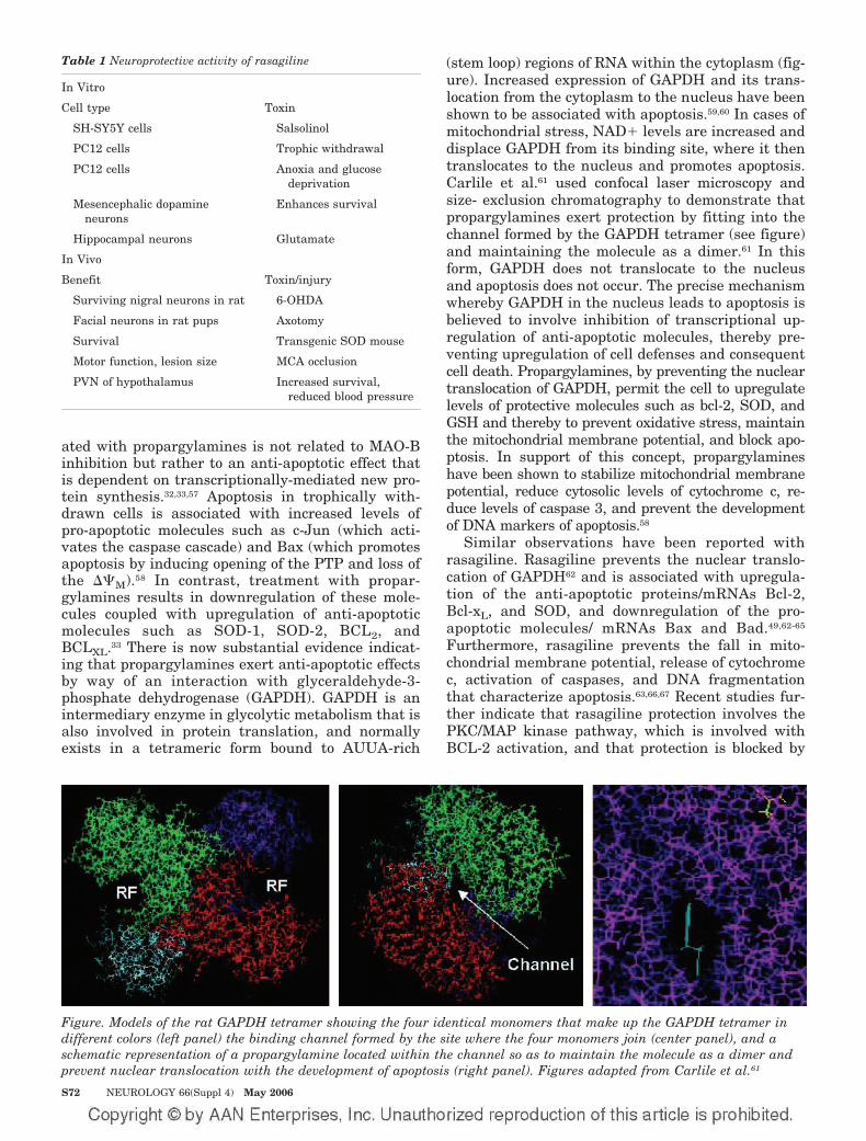
ated with propargylamines is not related to MAO-Binhibition but rather to an anti-apoptotic effect thatis dependent on transcriptionally-mediated new pro-tein synthesis.32,33,57 Apoptosis in trophically with-drawn cells is associated with increased levels ofpro-apoptotic molecules such as c-Jun (which acti-vates the caspase cascade) and Bax (which promotesapoptosis by inducing opening of the PTP and loss ofthe ��M).58 In contrast, treatment with propar-gylamines results in downregulation of these mole-cules coupled with upregulation of anti-apoptoticmolecules such as SOD-1, SOD-2, BCL2, andBCLXL.33 There is now substantial evidence indicat-ing that propargylamines exert anti-apoptotic effectsby way of an interaction with glyceraldehyde-3-phosphate dehydrogenase (GAPDH). GAPDH is anintermediary enzyme in glycolytic metabolism that isalso involved in protein translation, and normallyexists in a tetrameric form bound to AUUA-rich
(stem loop) regions of RNA within the cytoplasm (fig-ure). Increased expression of GAPDH and its trans-location from the cytoplasm to the nucleus have beenshown to be associated with apoptosis.59,60 In cases ofmitochondrial stress, NAD� levels are increased anddisplace GAPDH from its binding site, where it thentranslocates to the nucleus and promotes apoptosis.Carlile et al.61 used confocal laser microscopy andsize- exclusion chromatography to demonstrate thatpropargylamines exert protection by fitting into thechannel formed by the GAPDH tetramer (see figure)and maintaining the molecule as a dimer.61 In thisform, GAPDH does not translocate to the nucleusand apoptosis does not occur. The precise mechanismwhereby GAPDH in the nucleus leads to apoptosis isbelieved to involve inhibition of transcriptional up-regulation of anti-apoptotic molecules, thereby pre-venting upregulation of cell defenses and consequentcell death. Propargylamines, by preventing the nucleartranslocation of GAPDH, permit the cell to upregulatelevels of protective molecules such as bcl-2, SOD, andGSH and thereby to prevent oxidative stress, maintainthe mitochondrial membrane potential, and block apo-ptosis. In support of this concept, propargylamineshave been shown to stabilize mitochondrial membranepotential, reduce cytosolic levels of cytochrome c, re-duce levels of caspase 3, and prevent the developmentof DNA markers of apoptosis.58
Similar observations have been reported withrasagiline. Rasagiline prevents the nuclear translo-cation of GAPDH62 and is associated with upregula-tion of the anti-apoptotic proteins/mRNAs Bcl-2,Bcl-xL, and SOD, and downregulation of the pro-apoptotic molecules/ mRNAs Bax and Bad.49,62-65
Furthermore, rasagiline prevents the fall in mito-chondrial membrane potential, release of cytochromec, activation of caspases, and DNA fragmentationthat characterize apoptosis.63,66,67 Recent studies fur-ther indicate that rasagiline protection involves thePKC/MAP kinase pathway, which is involved withBCL-2 activation, and that protection is blocked by
Table 1 Neuroprotective activity of rasagiline
In Vitro
Cell type Toxin
SH-SY5Y cells Salsolinol
PC12 cells Trophic withdrawal
PC12 cells Anoxia and glucosedeprivation
Mesencephalic dopamineneurons
Enhances survival
Hippocampal neurons Glutamate
In Vivo
Benefit Toxin/injury
Surviving nigral neurons in rat 6-OHDA
Facial neurons in rat pups Axotomy
Survival Transgenic SOD mouse
Motor function, lesion size MCA occlusion
PVN of hypothalamus Increased survival,reduced blood pressure
Figure. Models of the rat GAPDH tetramer showing the four identical monomers that make up the GAPDH tetramer indifferent colors (left panel) the binding channel formed by the site where the four monomers join (center panel), and aschematic representation of a propargylamine located within the channel so as to maintain the molecule as a dimer andprevent nuclear translocation with the development of apoptosis (right panel). Figures adapted from Carlile et al.61
S72 NEUROLOGY 66(Suppl 4) May 2006
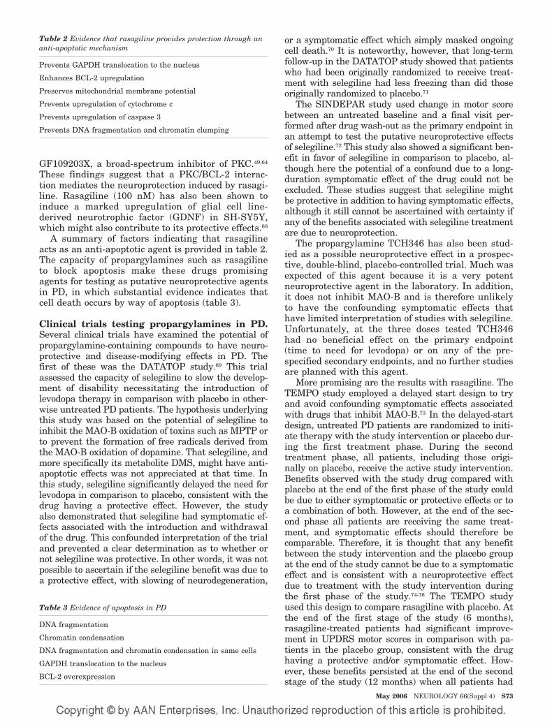
GF109203X, a broad-spectrum inhibitor of PKC.49,64
These findings suggest that a PKC/BCL-2 interac-tion mediates the neuroprotection induced by rasagi-line. Rasagiline (100 nM) has also been shown toinduce a marked upregulation of glial cell line-derived neurotrophic factor (GDNF) in SH-SY5Y,which might also contribute to its protective effects.68
A summary of factors indicating that rasagilineacts as an anti-apoptotic agent is provided in table 2.The capacity of propargylamines such as rasagilineto block apoptosis make these drugs promisingagents for testing as putative neuroprotective agentsin PD, in which substantial evidence indicates thatcell death occurs by way of apoptosis (table 3).
Clinical trials testing propargylamines in PD.Several clinical trials have examined the potential ofpropargylamine-containing compounds to have neuro-protective and disease-modifying effects in PD. Thefirst of these was the DATATOP study.69 This trialassessed the capacity of selegiline to slow the develop-ment of disability necessitating the introduction oflevodopa therapy in comparison with placebo in other-wise untreated PD patients. The hypothesis underlyingthis study was based on the potential of selegiline toinhibit the MAO-B oxidation of toxins such as MPTP orto prevent the formation of free radicals derived fromthe MAO-B oxidation of dopamine. That selegiline, andmore specifically its metabolite DMS, might have anti-apoptotic effects was not appreciated at that time. Inthis study, selegiline significantly delayed the need forlevodopa in comparison to placebo, consistent with thedrug having a protective effect. However, the studyalso demonstrated that selegiline had symptomatic ef-fects associated with the introduction and withdrawalof the drug. This confounded interpretation of the trialand prevented a clear determination as to whether ornot selegiline was protective. In other words, it was notpossible to ascertain if the selegiline benefit was due toa protective effect, with slowing of neurodegeneration,
or a symptomatic effect which simply masked ongoingcell death.70 It is noteworthy, however, that long-termfollow-up in the DATATOP study showed that patientswho had been originally randomized to receive treat-ment with selegiline had less freezing than did thoseoriginally randomized to placebo.71
The SINDEPAR study used change in motor scorebetween an untreated baseline and a final visit per-formed after drug wash-out as the primary endpoint inan attempt to test the putative neuroprotective effectsof selegiline.72 This study also showed a significant ben-efit in favor of selegiline in comparison to placebo, al-though here the potential of a confound due to a long-duration symptomatic effect of the drug could not beexcluded. These studies suggest that selegiline mightbe protective in addition to having symptomatic effects,although it still cannot be ascertained with certainty ifany of the benefits associated with selegiline treatmentare due to neuroprotection.
The propargylamine TCH346 has also been stud-ied as a possible neuroprotective effect in a prospec-tive, double-blind, placebo-controlled trial. Much wasexpected of this agent because it is a very potentneuroprotective agent in the laboratory. In addition,it does not inhibit MAO-B and is therefore unlikelyto have the confounding symptomatic effects thathave limited interpretation of studies with selegiline.Unfortunately, at the three doses tested TCH346had no beneficial effect on the primary endpoint(time to need for levodopa) or on any of the pre-specified secondary endpoints, and no further studiesare planned with this agent.
More promising are the results with rasagiline. TheTEMPO study employed a delayed start design to tryand avoid confounding symptomatic effects associatedwith drugs that inhibit MAO-B.73 In the delayed-startdesign, untreated PD patients are randomized to initi-ate therapy with the study intervention or placebo dur-ing the first treatment phase. During the secondtreatment phase, all patients, including those origi-nally on placebo, receive the active study intervention.Benefits observed with the study drug compared withplacebo at the end of the first phase of the study couldbe due to either symptomatic or protective effects or toa combination of both. However, at the end of the sec-ond phase all patients are receiving the same treat-ment, and symptomatic effects should therefore becomparable. Therefore, it is thought that any benefitbetween the study intervention and the placebo groupat the end of the study cannot be due to a symptomaticeffect and is consistent with a neuroprotective effectdue to treatment with the study intervention duringthe first phase of the study.74-76 The TEMPO studyused this design to compare rasagiline with placebo. Atthe end of the first stage of the study (6 months),rasagiline-treated patients had significant improve-ment in UPDRS motor scores in comparison with pa-tients in the placebo group, consistent with the drughaving a protective and/or symptomatic effect. How-ever, these benefits persisted at the end of the secondstage of the study (12 months) when all patients had
Table 2 Evidence that rasagiline provides protection through ananti-apoptotic mechanism
Prevents GAPDH translocation to the nucleus
Enhances BCL-2 upregulation
Preserves mitochondrial membrane potential
Prevents upregulation of cytochrome c
Prevents upregulation of caspase 3
Prevents DNA fragmentation and chromatin clumping
Table 3 Evidence of apoptosis in PD
DNA fragmentation
Chromatin condensation
DNA fragmentation and chromatin condensation in same cells
GAPDH translocation to the nucleus
BCL-2 overexpression
May 2006 NEUROLOGY 66(Suppl 4) S73
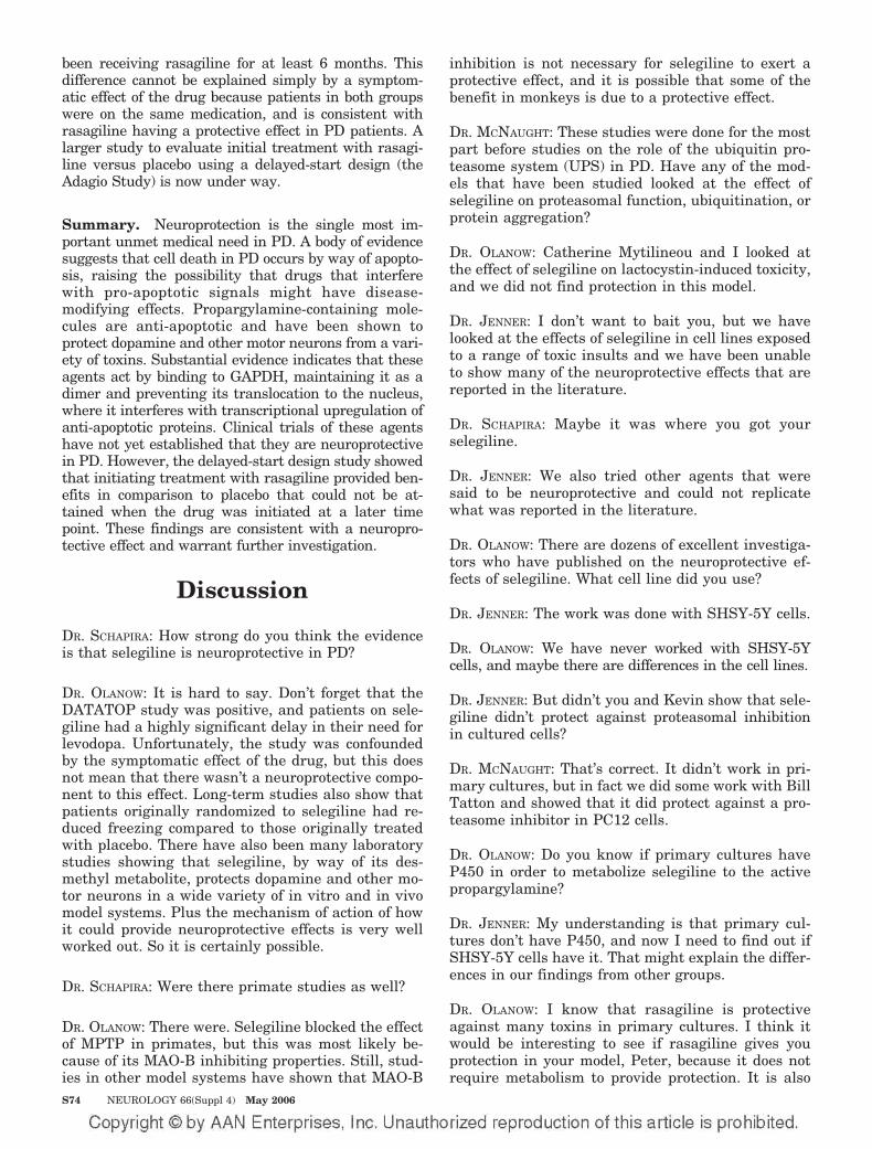
been receiving rasagiline for at least 6 months. Thisdifference cannot be explained simply by a symptom-atic effect of the drug because patients in both groupswere on the same medication, and is consistent withrasagiline having a protective effect in PD patients. Alarger study to evaluate initial treatment with rasagi-line versus placebo using a delayed-start design (theAdagio Study) is now under way.
Summary. Neuroprotection is the single most im-portant unmet medical need in PD. A body of evidencesuggests that cell death in PD occurs by way of apopto-sis, raising the possibility that drugs that interferewith pro-apoptotic signals might have disease-modifying effects. Propargylamine-containing mole-cules are anti-apoptotic and have been shown toprotect dopamine and other motor neurons from a vari-ety of toxins. Substantial evidence indicates that theseagents act by binding to GAPDH, maintaining it as adimer and preventing its translocation to the nucleus,where it interferes with transcriptional upregulation ofanti-apoptotic proteins. Clinical trials of these agentshave not yet established that they are neuroprotectivein PD. However, the delayed-start design study showedthat initiating treatment with rasagiline provided ben-efits in comparison to placebo that could not be at-tained when the drug was initiated at a later timepoint. These findings are consistent with a neuropro-tective effect and warrant further investigation.
Discussion
DR. SCHAPIRA: How strong do you think the evidenceis that selegiline is neuroprotective in PD?
DR. OLANOW: It is hard to say. Don’t forget that theDATATOP study was positive, and patients on sele-giline had a highly significant delay in their need forlevodopa. Unfortunately, the study was confoundedby the symptomatic effect of the drug, but this doesnot mean that there wasn’t a neuroprotective compo-nent to this effect. Long-term studies also show thatpatients originally randomized to selegiline had re-duced freezing compared to those originally treatedwith placebo. There have also been many laboratorystudies showing that selegiline, by way of its des-methyl metabolite, protects dopamine and other mo-tor neurons in a wide variety of in vitro and in vivomodel systems. Plus the mechanism of action of howit could provide neuroprotective effects is very wellworked out. So it is certainly possible.
DR. SCHAPIRA: Were there primate studies as well?
DR. OLANOW: There were. Selegiline blocked the effectof MPTP in primates, but this was most likely be-cause of its MAO-B inhibiting properties. Still, stud-ies in other model systems have shown that MAO-B
inhibition is not necessary for selegiline to exert aprotective effect, and it is possible that some of thebenefit in monkeys is due to a protective effect.
DR. MCNAUGHT: These studies were done for the mostpart before studies on the role of the ubiquitin pro-teasome system (UPS) in PD. Have any of the mod-els that have been studied looked at the effect ofselegiline on proteasomal function, ubiquitination, orprotein aggregation?
DR. OLANOW: Catherine Mytilineou and I looked atthe effect of selegiline on lactocystin-induced toxicity,and we did not find protection in this model.
DR. JENNER: I don’t want to bait you, but we havelooked at the effects of selegiline in cell lines exposedto a range of toxic insults and we have been unableto show many of the neuroprotective effects that arereported in the literature.
DR. SCHAPIRA: Maybe it was where you got yourselegiline.
DR. JENNER: We also tried other agents that weresaid to be neuroprotective and could not replicatewhat was reported in the literature.
DR. OLANOW: There are dozens of excellent investiga-tors who have published on the neuroprotective ef-fects of selegiline. What cell line did you use?
DR. JENNER: The work was done with SHSY-5Y cells.
DR. OLANOW: We have never worked with SHSY-5Ycells, and maybe there are differences in the cell lines.
DR. JENNER: But didn’t you and Kevin show that sele-giline didn’t protect against proteasomal inhibitionin cultured cells?
DR. MCNAUGHT: That’s correct. It didn’t work in pri-mary cultures, but in fact we did some work with BillTatton and showed that it did protect against a pro-teasome inhibitor in PC12 cells.
DR. OLANOW: Do you know if primary cultures haveP450 in order to metabolize selegiline to the activepropargylamine?
DR. JENNER: My understanding is that primary cul-tures don’t have P450, and now I need to find out ifSHSY-5Y cells have it. That might explain the differ-ences in our findings from other groups.
DR. OLANOW: I know that rasagiline is protectiveagainst many toxins in primary cultures. I think itwould be interesting to see if rasagiline gives youprotection in your model, Peter, because it does notrequire metabolism to provide protection. It is also
S74 NEUROLOGY 66(Suppl 4) May 2006

worth pointing out that the literature is confusing asto which P450 metabolizes selegiline, with conflict-ing results having been reported.
DR. MCNAUGHT: I am curious to know if there’s differ-ential expression of P450 in PC12 cells compared toprimary cultures.
DR. SIDEROWF: How much of a problem is it that pos-itive results are published and negative results tendnot to be?
DR. OLANOW: There is a bias that journals don’t liketo publish negative results, but if you were to reportthat you tried a series of agents that had previouslybeen reported to be neuroprotective and couldn’t getthem to work and the methodologies that you usedwere sound, I think you could get that work pub-lished. Have you tried to publish your results, Peter?
DR. JENNER: We haven’t published them yet because wewere so worried that we couldn’t reproduce what wasin the literature and we want to confirm them. Themore we discuss this, the more I think that results canbe system-dependent. This illustrates why it is impor-tant to discuss negative as well as positive results.
DR. KIEBURTZ: How do you block the metabolism ofselegiline?
DR. OLANOW: With P450 inhibitors, which block thedemethylation of selegiline to DMS. And interest-ingly this blocks the protection, indicating that it isthe DMS metabolite that is primarily responsible forthis effect. This is further supported by the finding ofreduced benefits with selegiline compared with DMSat the same concentration.
DR. POEWE: What about with rasagiline?
DR. OLANOW: It is different with rasagiline. Here theneuroprotective benefit is greater with rasagiline thanwith its metabolite, aminoindan, and blocking rasagi-line metabolism does not block its protective effects.
DR. HAUSER: What accounts for this difference?
DR. OLANOW: It probably has to do with the capacityof the molecule to fit into the channel formed by theGAPDH tetramers and its ability to maintainGAPDH as a dimer so that it doesn’t go to the nu-cleus and prevent the protective response that blocksapoptosis. It is likely that DMS and rasagiline fit,whereas selegiline per se does not. Bill Tattonshowed that if you employ antibodies to the proteinsthat make up the canal, you lose the protective ef-fects that are seen with propargylamines.
DR. SCHAPIRA: What was the basis for determiningthat propargylamines maintain GAPDH as a dimer?
DR. OLANOW: This was work done by Graham Carlileand Bill Tatton. They used size-exclusion chromatog-raphy and showed that GAPDH normally exists as atetramer, but when a propargylamine is added itpredominantly exists as a dimer.
DR. HAUSER: If you block the metabolism of selegilineto desmethyl selegiline, do you still get a fair amountof protection?
DR. OLANOW: No, you lose the protection.
DR. KIEBURTZ: And this has nothing to do withMAO-B inhibition?
DR. OLANOW: That’s correct. Bill Tatton showed in thetrophic withdrawal model that the selegiline benefitwas not seen with other MAO-B inhibitors. Subse-quently, Catherine Mytilineou and I reproduced theseresults. These were the original studies that led us tobelieve that the selegiline benefits were related to itspropargyl ring and based on an anti-apoptotic effect.
DR. JENNER: How did you establish that it was anti-apototic?
DR. OLANOW: First, we selected models that result inapoptosis, such as trophic withdrawal, and showedthat propargylamines prevent the development ofthe DNA markers indicative of apoptosis. Tatton andIshitani and others also showed that apoptosis wasassociated with upregulation of GAPDH and down-regulation of BCL-2, SOD-1, and SOD-2, while theaddition of a propargylamine such as DMS pre-vented the upregulation of GAPDH and induced up-regulation of BCL-2, SOD-1, and SOD-2. Thesefindings indicate that propargylamines act by inter-fering with pro-apoptotic signals.
DR. KIEBURTZ: I think I am beginning to see how thisworks. Presumably propargylamines exert anti-apoptotic effects by interacting with GAPDH andpreventing it from promoting pro-apoptotic signals inresponse to minor or inappropriate cell stresses.
DR. OLANOW: Exactly.
DR. SIDEROWF: What exactly does GAPDH do?
DR. OLANOW: It has several functions. It is wellknown to be an intermediary in glycolytic metabo-lism. It also sits on stem loops of RNA and plays animportant role in RNA translation into proteins. Andit is also involved in signaling for apoptosis underconditions of mitochondrial stress.
DR. SIDEROWF: How is it that GAPDH plays such animportant role in signaling for apoptosis?
May 2006 NEUROLOGY 66(Suppl 4) S75

DR. OLANOW: GAPDH is involved in fundamental gly-colytic metabolism and is probably a sensor of mito-chondrial function. When mitochondria are understress, this is reflected by excessive release of NAD�from mitochondria, which displaces tetramericGAPDH from stem loops where it translocates to thenucleus, inhibits the normal protective response ofthe cell, and promotes apoptosis. It is interesting tospeculate that this might occur as a way of avoidingnecrotic cell death, which is associated with inflam-mation that might have an adverse effect on neigh-boring healthy cells.
DR. POEWE: What are the differences between selegi-line and rasagiline?
DR. OLANOW: Rasagiline is similar to selegiline butthere are a few important differences. First, it is amore potent MAO-B inhibitor. Second, the ring hereis closed so that it is metabolized to form an amin-oindan and does not generate amphetamine metabo-lites. It is similar to selegiline in that it incorporatesa propargyl ring, but protection comes from the parentmolecule and not from a metabolite. Finally, it providesprotection in laboratory models and has been betterstudied as an antiparkinsonian agent in double-blindcontrolled trials. Therefore, at least in my opinion, itwarrants trial in PD as a putative neuroprotectivedrug, particularly in view of the results of the delayed-start component of the TEMPO study.
DR. POEWE: Do rasagiline and selegiline show compa-rable protection in the laboratory?
DR. OLANOW: More or less, although at the same con-centration rasagiline tends to show slightly moreprotection. The selegiline story has been nicelyworked out, and there is a nice body of work showingthat rasagiline, like other propargylamines, can pro-tect a variety of cells in vitro, including dopaminergicand non-dopaminergic cells. In vivo, you can seequite extensive protection in a variety of models, in-cluding 6-hydroxydopamine, axotomy, ALS, MCA oc-clusion, head injuries, and so on. Interestingly,Mousa Youdim did gene microarray studies beforeand after rasagiline and showed that mRNAs thatwere upregulated by rasagiline included those thathave the potential to be anti-apoptotic or protective,whereas those that were downregulated were pro-apoptotic, such as caspase-3.
DR. MCNAUGHT: You reported that buthionine sulfox-amine induces cell death. How does it do that?
DR. OLANOW: It kills cells by blocking the formation ofglutathione, but you have to deplete it by quite a lot,about 70 percent. We were surprised because theGSH depletion in PD is only about 40 percent.
DR. SCHAPIRA: Cells tend to conserve mitochondrialglutathione, which is perhaps why you need to de-plete it so much.
DR. KIEBURTZ: How does TCH346 differ from rasagi-line and selegiline?
DR. OLANOW: TCH346 is also a propargylamine, but ithas minimal if any MAO-B inhibition.
DR. JENNER: But it was supposed to have shown ex-cellent protective effects in the laboratory, is that notcorrect?
DR. OLANOW: It did. That is why the negative clinicaltrial was especially disappointing because the drugwould likely not have had a symptomatic confoundand any benefit could have been attributed to neuro-protection.
DR. JENNER: On the other hand, doesn’t the negativetrial with this agent suggest that benefits seen withdrugs such as selegiline and rasagiline are due toMAO-B inhibition rather than protection?
DR. OLANOW: I suppose that is correct, but it is alsopossible that we picked the wrong dose to test.TCH346 worked in the laboratory at concentrationsof 10–10 but had a U-shaped curve and didn’t work athigher or lower concentrations. It is also 99%protein-bound. So it was hard to pick the correctdose, and we may have gotten it wrong.
DR. STERN: What do you think is the best animal modelto test putative neuroprotective therapies for PD?
DR. OLANOW: That’s a problem. The animal models wehave such as MPTP and 6-OHDA don’t really reflectPD, and positive results in these models doesn’t neces-sarily mean you will get positive results in PD patients.The transgenic animals offer the best opportunity be-cause these animals carry the specific mutation thatcauses the human disease. However, the same muta-tion that causes PD in humans may not be harmful tothe animal model, and in fact most transgenic orknockout animals do not replicate PD behavior or pa-thology. I like the proteasome inhibition model becauseit is progressive and has dopamine and non-dopaminelesions. But many groups are having difficulty repro-ducing this model, and we can’t rely on this model untilwe resolve these issues. It is interesting that, in tissueculture, many of the agents that we think might beneuroprotective in PD are ineffective in the proteasomeinhibition model. Ideally, I would like to test a drug inPD that works in this model.
DR. KIEBURTZ: That’s a real problem, isn’t it? How do wetake a drug from the laboratory to humans? If it worksin a laboratory model, does that mean it will work inhumans? Is that a good predictor? Did we look at the
S76 NEUROLOGY 66(Suppl 4) May 2006

right models? Have we got a drug that never wouldhave worked in humans and we could have known thishad we looked at the correct models? Or would thishave worked in any system but we used the wrongdose, or perhaps we didn’t deliver it to the correct tar-get. Or perhaps we used the wrong study design?Maybe we didn’t address compensation adequately,and maybe we should have looked for longer in order totell if a drug is protecting nerve cells? Of course I don’tknow the answers to any of these questions. Given allwe know, a confounded study with selegiline and anegative trial with TCH346, are you still a fan ofpropargylamines as neuroprotective agents?
DR. OLANOW: I still am optimistic because the mecha-nism is so well worked out, because they protect atvery low concentrations that can be obtained physio-logically, and because they protect in so many differ-ent model systems. Another big question thatremains is whether you can design a trial that canshow slowing of disease progression.
DR. KIEBURTZ: It’s always hard to know whether it’s afailed drug or a failed trial. Even if it was the righttrial design, was it implemented incorrectly? Is theresomething funny about the characteristics and the be-havior of the patients who are enrolled? I think wecould learn a lot by exploring negative studies such asthe riluzole, the cephalon mixed-lineage kinase inhibi-tor, and TCH studies to see if we can learn anything.
DR. JENNER: One of the problems I have is that thesecompanies frequently go right to phase III studieswithout doing phase II dosing studies.
DR. KIEBURTZ: It’s often hard to know what dose to usein a neuroprotective study, because there is no biomar-ker to indicate activity. In which case, you might aswell look at dosing, tolerability, safety, and efficacy in apivotal phase IIb trial. In the cephalon study we wentright to a pivotal study looking at three doses andhoped that one of them would be the right dose.
DR. OLANOW: That study was another disappointmentbecause there is a good rationale for testing Jun kinaseinhibitors based on evidence that protein aggregationpromotes cell death by way of Jun kinase activation.
DR. MCNAUGHT: Let’s suppose you are successful withan anti-apoptotic drug and you reduce the number ofcells going into apoptosis. What happens to thosecells in the long run?
DR. OLANOW: That’s a very good question. We know theshort-term but not the long-term consequences of thesetherapies. We show reduced cell death with reducedmarkers of apoptosis at 24 or 48 hours, but we don’tknow what happens to the cells afterwards and
whether they behave like healthy cells. What would bebad is if these drugs converted cells from undergoingapoptosis to dying by necrosis, which could be worse forthe organism. The situation might be different, though,if there were a problem such as a complex I defectwhere the mitochondrial membrane potential was setabnormally low. Here, anti-apoptotic drugs could helpto maintain closure of the pore and reset the restingmembrane potential and eliminate the cell’s vulnera-bility. This might be the case in PD, where there isevidence at postmortem that GAPDH has translocatedto the nucleus in some cells and the cell is perhaps in apro-apoptotic state. Propargylamines might take a cellthat was getting ready to die and restore it to a morenormal function. In other words, you may be taking avulnerable cell and making it less vulnerable.
DR. SCHAPIRA: Yes, less vulnerable to die. But theissue is, is that cell able to function normally?
DR. KIEBURTZ: Tim Greenamyre took normal and HDfiberblasts and subjected them to repetitive calciumchallenges. He found that resting mitochondrialmembrane potential dropped and then recovered be-tween challenges with normal fibroblasts, while withHD fiberblasts the mitochondrial potential continuedto fall until the cells died. So he was trying to get atnot only do they die but could they recover function.
DR. OLANOW: Bill Tatton did a similar experiment inwhich he showed that fibroblasts from PD patients hada low mitochondrial membrane potential and werevery sensitive to rotenone in comparison to controls. Ifthat were to be the state of nigral neurons in PD, thenI think propargylamines might be helpful by restoringmitochondrial membrane potential in an otherwise in-tact cell that is vulnerable to undergo apoptosis whenexposed to ordinarily non-lethal stresses.
DR. HAUSER: One of my concerns is that, by the timeyou start a treatment, irreversible events may havealready occurred.
DR. OLANOW: That’s a fair concern. Venu Nair in ourgroup showed that, after exposure to hydrogen per-oxide, cultured cells very quickly express markersindicating whether or not they will undergo apopto-sis, even though DNA markers of apoptosis may notbe seen for another 24 to 48 hours. I think of propar-gylamines as a way to promote the likelihood thatthe cell will survive. As you imply, there are proba-bly many factors that determine whether a cell willultimately die or survive. And if we could, for exam-ple, through a propargylamine drug like rasagilinepreserve the mitochondrial membrane potential,that would mean that some cells might tolerate astress they otherwise might not, and a greater per-centage of cells would survive. Whether that willtranslate into a clinically meaningful effect I don’tknow. One of the jobs in the laboratory is to define
May 2006 NEUROLOGY 66(Suppl 4) S77
reasonable hypotheses, find targets, and developcandidate drugs that show protection in models. Inthe final analysis, you have to do a clinical trial toknow if it really works in PD.
DR. SIDEROWF: I was impressed when you describedthe many different systems where rasagiline exerteda protective effect. And it made me think that, inParkinson’s disease, only part of the degeneration isin the nigrostriatal dopaminergic system, but manyother extranigral neurons also degenerate, and defi-cits are not just in motor function but in cognitionand mood. Given the capacity of rasagiline to protectso many different cell types in the laboratory, doesthat make it more promising to study in PD and doesit suggest specific trial designs and endpoints thatshould be employed?
DR. KIEBURTZ: I think you make a good point, andthat trial designs that look at non-dopaminergic fea-ture as a measure of protection are potentially veryvaluable because they are less likely to be con-founded by the dopaminergic agents we use to treatPD today. I personally would feel much more com-fortable that a therapy was neuroprotective if itslowed the development of non-dopaminergic fea-tures, such as postural instability or dementia. Andeven if it turned out to be a symptomatic mechanismit would still be a boon for patients, since such ther-apies are not currently available.
DR. JENNER: Certainly, very little research has takenplace outside the nigra in PD to date, and I thinkmore information in this area could be very helpfulto clinicians designing neuroprotection trials.
DR. OLANOW: To take that one point further, if we hada good model of PD based on etiopathogenesis, posi-tive results in such a model would be very helpful indetermining that positive results in a clinical trialwere due to protection, as no current clinicalendpoint can be totally relied on as yet.
DR. KIEBURTZ: I think it’s also important for clinicaltrialists to inform basic scientists about what body ofevidence they want to see before starting a trial, sortof like a venture capitalist. We’re not going to investour time and efforts in doing a trial of that drug untilwe see this array of information. This would help inselecting a compound to study—why rasagiline, whyminocycline, why creatine, why coenzyme Q10? Well,we should have equivalent bits of information for allof them and choose the best one. The other reason isthat the basic science helps to contextualize the clin-ical findings and strengthen a possible neuroprotec-tive association.
DR. JENNER: Another thing we should be doing ismeasuring plasma levels of the drug, because itwould be very useful to know the plasma level at
which these effects are being achieved in order to tryto plan doses for clinical trials.
DR. OLANOW: One problem is whether the plasmalevel in the rat or monkey means the same thing in ahuman.
DR. JENNER: I don’t know about the story with neuro-protective drugs, but I can tell you we use plasmalevels in the common marmoset to predict doses forPD patients and find that these are a very goodguide to getting the dose right in the clinic.
References1. Forno LS. Neuropathology of Parkinson’s disease. J Neuropathol Exp
Neurol 1996;55:259–272.2. Braak H, Tredici KD, Rub U, de Vos RA, Jansen Steur EN, Braak E.
Staging of brain pathology related to sporadic Parkinson’s disease.Neurobiol Aging 2003;24:197–211.
3. Olanow CW, Watts RL, Koller WC. An algorithm (decision tree) for themanagement of Parkinson’s disease (2001): treatment guidelines. Neu-rology 2001;56(suppl 5):1–88.
4. Olanow CW. The scientific basis for the current treatment of Parkin-son’s disease. Annu Rev Med 2004;55:41–60.
5. Ravina BM, Fagan SC, Hart RG, et al. Neuroprotective agents forclinical trials in Parkinson’s disease: a systematic assessment. Neurol-ogy 2003;60:1234–1240.
6. Marsden CD, Olanow CW. Neuroprotection in Parkinson’s disease: thecauses of Parkinson’s disease are being unravelled and rational neuro-protective therapy is close to reality. Ann Neurol 1998;44:189–196.
7. Boulton AA. Symptomatic and neuroprotective properties of the ali-phatic propargylamines. Mech Ageing Dev 1999;111:201–209.
8. Schapira AHV. Etiology of Parkinson’s disease. Neurology 2006;66(Suppl 4):S10–S23.
9. Jenner P, Olanow CW. The pathogenesis of cell death in Parkinson’sdisease. Neurology 2006;66(Suppl 4):S24–S36.
10. McNaught K St.P, Jackson T, Jnobaptiste R, Kapustin A, Olanow CW.Proteasomal dysfunction in sporadic Parkinson’s disease. Neurology2006;66(Suppl 4):S37–S49.
11. Hirsch EC, Hunot S, Faucheux B, et al. Dopaminergic neurons degen-erate by apoptosis in Parkinson’s disease. Mov Disord 1999;14:383–385.
12. Agid Y. Aging, disease and nerve cell death. Bull Acad Natl Med1995;179:1193–1203.
13. Mochizuki H, Goto K, Mori H, Mizuno Y. Histochemical detection ofapoptosis in Parkinson’s disease. J Neurol Sci 1996;131:120–123.
14. Tatton NA, Maclean-Fraser A, Tatton WG, Perl DF, Olanow CW. Afluorescent double-labeling method to detect and confirm apoptoticnuclei in Parkinson’s Disease. Ann Neurol 1998;44(suppl 1):142–148.
15. Olanow CW, Tatton WG. Etiology and pathogenesis of Parkinson’sdisease. Annu Rev Neurosci 1999;22:123–144.
16. Tatton WG, Olanow CW. Apoptosis in neurodegenerative disease: therole of mitochondria. Biochim Biophys Acta 1999;1410:195–213.
17. Nair VD, Olanow CW, Sealfon SC. Activation of phosphoinositide3-kinase by D2 receptor prevents apoptosis in dopaminergic cell lines.Biochem J 2003;373:25–32.
18. Nair VD, Yuen T, Olanow CW, Sealfon S. Early single cell bifurcationof pro- and anti-apoptotic states during oxidative stress. J Biol Chem2004;25:27494–27501.
19. Susin SA, Zamzami N, Kroemer G. The cell biology of apoptosis: evi-dence for the implication of mitochondria. Apoptosis 1996;1:231–242.
20. Liu XS, Kim CN, Yang J, Jemmerson R, Wang XD. Induction of apo-ptotic program in cell-free extracts: requirement for dATP and cyto-chrome c. Cell 1996;86:147–157.
21. Susin SA, Zamzami N, Castedo M, et al. Bcl-2 inhibits the mitochon-drial release of an apoptogenic protease. J. Exp Med 1996;184:1331–1341.
22. Wadia JS, Chalmers-Redman RME, Ju WJH, et al. Mitochondrialmembrane potential and nuclear changes in apoptosis caused by tro-phic withdrawal: time course and modification by (-) deprenyl. J Neu-rosci 1996;18:932–947.
23. Jellinger KA. Cell death mechanisms in neurodegeneration. J Cell MolMed 2001;5:1–17.
24. Anglade P, Vyas S, Javoy-Agid F, et al. Apoptosis and autophagie innigral neurons of patients with Parkinson’s disease. Histol Histopathol1997;12:25–31.
25. Mogi M, Harada M, Kondo T, et al. BCL-2 protein is increased in thebrain of Parkinson patients. Neurosci Lett 1996;215:137–139.
26. Tatton NA. Increased caspase 3 and Bax immunoreactivity accompany
S78 NEUROLOGY 66(Suppl 4) May 2006

nuclear GAPDH translocation and neuronal apoptosis in Parkinson’sdisease. Exp Neurol 2000;166:29–43.
27. Cohen G, Pasik P, Cohen B, et al. Pargyline and deprenyl prevent theneurotoxicity of 1-methyl-4-phenyl-1,2,3,6-tetrahydropyridine (MPTP)in monkeys. Eur J Pharmacol 1985;106:209–210.
28. Heikkila RE, Manzino L, Duvoisin RC, Cabbat FS. Protection againstthe dopaminergic neurotoxicity of 1-methyl-4-phenyl-1,2,3,6-tetrahydropyridine (MPTP) by monoamine oxidase inhibitors. Nature1984;311:467–469.
29. Olanow CW. Oxidation reactions in Parkinson’s disease. Neurology1990;40:32–37.
30. Olanow CW, Mytilineou C, Tatton WH. Status of selegiline as a neu-roprotective agent in Parkinson’s disease. Mov Disord 1998;13(suppl):55–58.
31. Jenner P. Preclinical evidence for neuroprotection with monoamineoxidase-B inhibitors in Parkinson’s disease. Neurology 2004;63(7 suppl2):S13–22.
32. Mytilineou C, Radcliffe P, Leonardi EK, Werner P, Olanow CW.L-deprenyl protects mesencephalic dopamine neurons from glutamatereceptor-mediated toxicity. J Neurochem 1997;68:33–39.
33. Tatton WG, Chalmers-Redman RME. Modulation of gene expressionrather than monoamine oxidase inhibition: (-)deprenyl-related com-pounds in controlling neurodegeneration. Neurology 1996;47:171–183.
34. Mytilineou C, Radcliffe PM, Olanow CW. L-(-)-Desmethylselegiline, ametabolite of L-(-)-selegiline, protects mesencephalic dopamine neu-rons from excitotoxicity in vitro. J Neurochem 1997;68:434–436.
35. Mytilineou C, Leonardi EK, Radcliffe P, et al. Deprenyl and desmeth-ylselegiline protect mesencephalic neurons from toxicity induced byglutathione depletion. J Pharmacol Exp Ther 1998;284:700–706.
36. Waldmeier PC, Spooren WP, Hengerer B. CGP 3466 protects dopami-nergic neurons in lesion models of Parkinson’s disease. NaunynSchmiedebergs Arch Pharmacol 2000;362:526–537.
37. Matarredona ER, Meyer M, Seiler RW, Widmer HR. CGP 3466 in-creases survival of cultured fetal dopaminergic neurons. Restor NeurolNeurosci 2003;21:29–37.
38. Andringa G, van Oosten RV, Unger W, et al. Systemic administrationof the propargylamine CGP 3466B prevents behavioural and morpho-logical deficits in rats with 6-hydroxydopamine-induced lesions in thesubstantia nigra. Eur J Neurosci 2000;12:3033–3043.
39. Andringa G, Eshuis S, Perentes E. TCH346 prevents motor symptomsand loss of striatal FDOPA uptake in bilaterally MPTP-treated pri-mates. Neurobiol Dis 2003;14:205–217.
40. Youdim MD, Gross A, Finberg JP. Rasagiline [N-propargyl-1R(�)-aminoindan], a selective and potent inhibitor of mitochondrial mono-amine oxidase B. Br J Pharmacol 2001;132:500–506.
41. Parkinson Study Group. A randomized placebo-controlled trial of rasa-giline in levodopa-treated patients with Parkinson disease and motorfluctuations: the PRESTO study. Arch Neurol 2005;62:241–248.
42. Rascol O, Brooks DJ, Melamed E, et al. Rasagiline as an adjunct tolevodopa in patients with Parkinson’s disease and motor fluctuations(LARGO, Lasting effect in Adjunct therapy with Rasagiline GivenOnce daily, study): a randomised, double-blind, parallel-group trial.Lancet. 2005;365:947–954.
43. Weinreb O, Amit T, Bar-Am O, Chillag-Talmor O, Youdim MB. Novelneuroprotective mechanism of action of rasagiline is associated withits propargyl moiety: interaction of Bcl-2 family members with PKCpathway. Ann NY Acad Sci 2005;1053:348–355.
44. Finberg JP, Takeshima T, Johnston JM, Commissiong JW. Increasedsurvival of dopaminergic neurons by rasagiline, a monoamineoxidase-B inhibitor. Neuroreport 1998;9:703–707.
45. Goggi J, Theofilipoulos S, Riaz SS, Jauniaux E, Stern GM, BradfordHF. The neuronal survival effects of rasagiline and deprenyl on fetalhuman and rat ventral mesencephalic neurons in culture. Neuroreport2000;11:3937–3941.
46. Naoi M, Maruyama W. Future of neuroprotection in Parkinson’s dis-ease. Parkinsonism Relat Disord 2001;8:139–145.
47. Bar Am O, Amit T, Youdim M. Contrasting neuroprotective and neu-rotoxic actions of respective metabolites of anti-Parkinson drugs rasa-giline and selegiline. Neurosci Lett 2004;355:169–172.
48. Bonneh-Barkay D, Ziv N, Finberg JPM. Characterization of the neuro-protective activity of rasagiline in cerebellar granule cells. Neurophar-macology 2005;48:406–416.
49. Mandel S, Weinreb O, Amit T, Youdim MBH. Mechanism of neuropro-tection of the anti-Parkinson drug rasagiline and its derivatives. BrainRes Brain Res Rev 2005;48:379–387.
50. Maruyama W, Akao Y, Youdim MB, Naoi M. Neurotoxins induce apo-ptosis in dopamine neurons: protection by N-propargylamine-1(R)- and(S)-aminoindan, rasagiline and TV1022. J Neural Transm Suppl 2000;60:171–186.
51. Naoi M, Maruyama W, Youdim MBH, Yu P, Boulton AA. Anti-apoptotic function of propargylamine inhibitors of type-B monoamineoxidase. Inflammopharmacology 2003:11:175–181.
52. Blandini F, Armentero MT, Fancellu R, Blaugrund E, Nappi G. Neu-roprotective effects of rasagiline in a rodent model of Parkinson’s dis-ease. Exp Neurol 2004;187:455–459.
53. Huang W, Chen Y, Shohami E, Weinstock M. Neuroprotective effect ofrasagiline, a selective a monoamine oxidase-B inhibitor, against closedhead injury in the mouse. Eur J Pharmacol 1999;366:127–135.
54. Speiser Z, Mayk A, Eliash S, Cohen S. Studies with rasagiline, aMAO-B inhibitor, in experimental focal ischemia. J Neural Transm1999;106:593–606.
55. Eliash S, Speiser Z, Cohen S. Rasagiline and its (S) enantiomer in-crease survival and prevent stroke in salt-loaded stroke-prone sponta-neously hypertensive rats. J Neural Transm 2001;108:909–923.
56. Waibel S, Reuter A, Malessa S, Blaugrund E, Ludolph AC. Rasagilinealone and in combination with riluzole prolongs survival in an ALSmouse model. J Neurol 2004;251:1080–1084.
57. Tatton WG, Ju WY, Holland DP, Tai C, Kwan M. (-)-Deprenyl reducesPC12 cell apoptosis by inducing new protein synthesis. J Neurochem1994;63:1572–1575.
58. Wadia JS, Chalmers-Redman RME, Ju WJH, Carlile GW, Phillips JL,Tatton WG. Mitochondrial membrane potential and nuclear changesin apoptosis caused by trophic withdrawal: time course and modifica-tion by (-) deprenyl. J Neurosci 1998;18:932–947.
59. Sawa A, Khan AA, Hester LD, Snyder SH. Glyderaldehyde-3-phosphate dehydrogenase: nuclear translocation participates in neuro-nal and non-neuronal cell death. Proc Natl Acad Sci USA 1997;94:11669–11674.
60. Ishitani R, Kimura M, Sunaga K, Katsube N, Tanaka M, Chuang DM.An antisense oligonucleotide to glyderaldehyde-3-phosphate dehydro-genase blocks age-induced apoptosis of mature cerebro-cortical neu-rons in culture. J Pharmacol Exp Ther 1996;278:447–454.
61. Carlile GW, Chalmers-Redman RME, Tatton NA, Pong A, Borden KE,Tatton WG. Reduced apoptosis after nerve growth factor and serumwithdrawal: conversion of tetrameric glyceraldehyde-3-phosphate de-hydrogenase to a dimer. Mol Pharmacol 2000;57:2–12.
62. Maruyama W, Akao Y, Youdim MBH, Davis BA, Naoi M. Transfection-enforced Bcl-2 overexpression and an anti-Parkinson drug, rasagiline,prevent nuclear accumulation of glyceraldehyde-3-phosphate dehydro-genase induced by an endogenous dopaminergic neurotoxin,N-methyl(R)salsolinol. J Neurochem 2001;78:727–735.
63. Youdim MBH, Bar-Am O, Yogev-Falach, et al. Rasagiline: neurodegen-eration, neuroprotection, and mitochondrial permeability transition.J Neurosci Res 2005;79:172–179.
64. Weinreb O, Bar-Am O, Amit T, Chillag-Talmor O, Youdim MBH. Neu-roprotection via pro-survival protein kinase C isoforms associated withBcl-2 family members. FASEB J 2004;18:1471–1473.
65. Akao Y, Maruyama W, Yi H, Shanoto-Nagai M, Youdim M, Naoi M.An anti-Parkinson’s disease drug, N-propargyl-1(R)-aminoindan (rasa-giline), enhances expression of anti-apoptotic Bcl-2 in human dopami-nergic SH-SY5Y cells. Neurosci Lett 2002;326:105–108.
66. Akao Y, Maruyama W, Shimizu S, et al. Mitochondrial permeabilitytransition mediates apoptosis induced by N-methyl(R)salsolinol, anendogenous neurotoxin, and is inhibited by Bcl-2 and rasagiline,N-propargyl-1(R)-aminoindan. J Neurochem 2002;82:913–923.
67. Youdim MBH, Amit T, Bar-Am O, Weinstock M, Yogev-Falach. Amy-loid processing and signal transduction properties of antiParkinson-antiAlzheimer neuroprotective drugs rasagiline and TV3326. Ann NYAcad Sci 2003:993:378–386.
68. Maruyama W, Nitta A, Shamoto-Nagai M, et al. N-Propargyl-1 (R)-aminoindan, rasagiline, increases glial cell line-derived neurotrophicfactor (GDNF) in neuroblastoma SH-SY5Y cells through activation ofNF-kappaB transcription factor. Neurochem Int 2004;44:393–400.
69. Parkinson’s Study Group. Effects of tocopherol and deprenyl on theprogression of disability in early Parkinson’s disease. N Engl J Med1993;328:176–183.
70. Olanow CW, Calne D. Does selegiline monotherapy in Parkinson’sDisease act by symptomatic or protective mechanisms? Neurology1991;42:41–48.
71. Shoulson I, Oakes D, Fahn S, et al. The impact of sustained deprenyl(selegiline) in levodopa-treated Parkinson’s disease: a randomizedplacebo-controlled extension. Ann Neurol 2002;51:604–612.
72. Olanow CW, Hauser RA, Gauger L, et al. The effect of deprenyl andlevodopa on the progression of signs and symptoms in Parkinson’sdisease. Ann Neurol 1995;38:771–777.
73. The Parkinson Study Group. A controlled, randomised, delayed-startstudy of rasagiline in early Parkinson disease. Arch Neurol 2004;61:561–566.
74. Youdim MB, Weinstock M. Molecular basis of neuroprotective activi-ties of rasagiline and the anti-Alzheimer drug TV3326 [(N-propargyl-(3R)aminoindan-5-YL)-ethyl methyl carbamate]. Cell Mol Neurobiol2001;21:555–573.
75. Tatton WG, Chalmers-Redman RM, Ju WJ, et al. Propargylaminesinduce antiapoptotic new protein synthesis in serum- and nervegrowth factor (NGF)-withdrawn, NGF-differentiated PC-12 cells.J Pharmacol Exp Ther 2002;301:753–764.
76. Youdim MB, Amit T, Falach-Yogev M, Am OB, Maruyama W, Naoi M.The essentiality of Bcl-2, PKC and proteasome-ubiquitin complex acti-vations in the neuroprotective-antiapoptotic action of the anti-Parkinson drug, rasagiline. Biochem Pharmacol 2003;66:1635–1641.
May 2006 NEUROLOGY 66(Suppl 4) S79

A peptide based on the complementarity-determiningregion 1 of an autoantibody ameliorates lupusby up-regulating CD4�CD25� cells and TGF-�Amir Sharabi*, Heidy Zinger*, Maya Zborowsky*, Zev M. Sthoeger†, and Edna Mozes*‡
*Department of Immunology, Weizmann Institute of Science, Rehovot 76100, Israel; and †Department of Medicine B, Kaplan Hospital,Rehovot 76100, Israel
Communicated by Michael Sela, Weizmann Institute of Science, Rehovot, Israel, April 25, 2006 (received for review March 2, 2006)
Systemic lupus erythematosus is an autoimmune disease charac-terized by autoantibodies and systemic clinical manifestations. Apeptide, designated hCDR1, based on the complementarity-deter-mining region (CDR) 1 of an autoantibody, ameliorated the sero-logical and clinical manifestations of lupus in both spontaneousand induced murine models of lupus. The objectives of the presentstudy were to determine the mechanism(s) underlying the bene-ficial effects induced by hCDR1. Adoptive transfer of hCDR1-treatedcells to systemic lupus erythematosus-afflicted (NZB�NZW)F1 femalemice down-regulated all disease manifestations. hCDR1 treatmentup-regulated (by 30–40%) CD4�CD25� cells in association withCD45RBlow, cytotoxic T lymphocyte antigen 4, and Foxp3 expres-sion. Depletion of the CD25� cells diminished significantly thetherapeutic effects of hCDR1, whereas administration of the en-riched CD4�CD25� cell population was beneficial to the diseasedmice. Amelioration of disease manifestations was associated withdown-regulation of the pathogenic cytokines (e.g., IFN-� and IL-10)and up-regulation of the immunosuppressive cytokine TGF-�,which substantially contributed to the suppressed autoreactivity.TGF-� was secreted by CD4� cells that were affected by hCDR1-induced immunoregulatory cells. The hCDR1-induced CD4�CD25�
cells suppressed autoreactive CD4� cells, resulting in reduced ratesof activation-induced apoptosis. Thus, hCDR1 ameliorates lupusthrough the induction of CD4�CD25� cells that suppress activationof the autoreactive cells and trigger the up-regulation of TGF-�.
cytokines � Foxp3 � immunomodulating peptide � regulatory T cells �systemic lupus erythematosus
Systemic lupus erythematosus (SLE) is an autoimmune diseasecharacterized by the production of Ab against nuclear antigens
and damage to multiple organs including kidneys, CNS, joints, andskin (1). Several strains of mice that spontaneously develop anSLE-like disease were reported, of which the (NZB�NZW)F1female mice are the most widely used (2, 3). In addition, ourlaboratory has established a model of experimentally induced SLEin different susceptible strains of mice (4–6).
A peptide, designated hCDR1, based on the complementarity-determining region (CDR) 1 (7) of a human anti-DNA mAb, wasshown to ameliorate the serological and clinical manifestations inboth the spontaneous and induced models of SLE and to reduce thesecretion and expression of the pathogenic cytokines IFN-�, IL-10,IL-1�, and TNF-� (the latter in the induced model) while up-regulating the immunosuppressive cytokine TGF-� (8).
It has become increasingly evident that peripheral tolerance ismediated by suppressor T cells with a regulatory function (9, 10).The best-characterized are the CD4�CD25� cells, which constitute5–10% of the CD4� cells (11). CD4�CD25� cells are naturallyoccurring, whereas adaptive regulatory CD4�CD25� cells withsuppressive capacity may be induced in the periphery in responseto tolerogenic stimuli (11, 12). Nevertheless, several in vitro and invivo studies indicated that CD25� cells might also be generated inthe periphery (13–20). Recently it was reported that the number ofCD4�CD25� cells is diminished in patients with SLE as well as in
(NZB�NZW)F1 female mice with established lupus (21–23), thussuggesting a role for these cells in regulating the disease.
In the present study we attempted the elucidation of the mech-anism(s) underlying the ameliorating effects of treatment withhCDR1 on SLE manifestations. We demonstrated that the inhib-itory effects of hCDR1 can be adoptively transferred to mice withestablished lupus by cells originating from young, healthy(NZB�NZW)F1 female mice that were treated with hCDR1.CD4�CD25� cells were up-regulated in the hCDR1-treated cellpopulation and were found to play a crucial role in ameliorating theserological and clinical parameters of SLE. This improvement wasachieved by suppressing the activation of the CD4� cells and bytriggering the up-regulated secretion of TGF-�, which was shown toplay a key role in down-regulating SLE manifestations.
ResultsAdoptive Transfer of Spleen Cells from Mice Treated with hCDR1 to(NZB�NZW)F1 Mice with Established Disease Is Beneficial. To deter-mine whether the beneficial effects of hCDR1 can be transferred bycells of treated mice, we first performed adoptive transfer experi-ments. Thus, 2-mo-old, disease-free, (NZB�NZW)F1 mice wereinjected with hCDR1 s.c. (50 �g per mouse) for 3 alternating days.Two control groups of young mice were treated with the vehicle orwith a scrambled (control) peptide. Splenocytes (20 � 106 permouse) from the different groups were injected i.p. to respectivegroups of 8-mo-old (NZB�NZW)F1 mice with established disease.Disease severity of the recipient mice was similar in all groups asassessed by their anti-dsDNA autoantibody titers and proteinurialevels. Fig. 1 summarizes the clinical effects of the transferred cellson lupus-like manifestations at the end of a 2-wk follow-up andrepresents one experiment of five performed. As demonstrated, theproduction of dsDNA-specific autoantibodies as well as elevatedproteinuria levels and the formation of glomerular immune com-plex deposits (ICD) were significantly reduced in SLE-afflictedmice that were injected with the hCDR1-treated spleen cellscompared with recipients of cells treated with the scrambledpeptide or with the vehicle.
Treatment with hCDR1 Results in an Up-Regulation of CD4�CD25�
Cells. Because CD4�CD25� cells are the most characterized im-munoregulatory T cells and because these cells were shown to beprotective against autoimmune responses, we studied their possiblerole in the mode of action of hCDR1. For this purpose, three groupsof 2-mo-old (NZB�NZW)F1 mice were treated with hCDR1, thevehicle, or a scrambled peptide. In all experiments the magnitudeof CD4�CD25� cells ranged between 3% and 9%. Treatment with
Conflict of interest statement: M.S. serves on the Board of Directors of Teva PharmaceuticalIndustries, which supported this study.
Abbreviations: CDR, complementarity-determining region; CTLA-4, cytotoxic T lymphocyteantigen 4; FasL, Fas ligand; ICD, immune complex deposits; SLE, systemic lupus erythema-tosus; PE, phycoerythrin.
‡To whom correspondence should be addressed. E-mail: [email protected].
© 2006 by The National Academy of Sciences of the USA
8810–8815 � PNAS � June 6, 2006 � vol. 103 � no. 23 www.pnas.org�cgi�doi�10.1073�pnas.0603201103
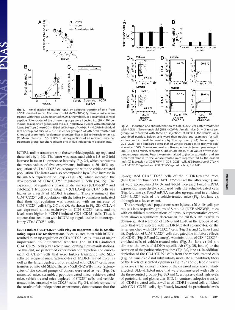
hCDR1, unlike treatment with the scrambled peptide, up-regulatedthese cells by 1–2%. The latter was associated with a 1.5- to 2-foldincrease in mean fluorescence intensity. Fig. 2A, which representsthe mean values of five experiments, indicates a 30–40% up-regulation of CD4�CD25� cells compared with the vehicle-treatedpopulation. The latter was also accompanied by a 3-fold increase inthe mRNA expression of Foxp3 (Fig. 2B), which indicated thedevelopment of CD4�CD25� regulatory T cells (24, 25). Theexpression of regulatory characteristic markers [CD45RBlow andcytotoxic T lymphocyte antigen 4 (CTLA-4)] on CD4� cells washigher as a result of hCDR1 treatment. Triple staining of theCD4�CD25� cell population with the latter two markers indicatedthat their up-regulation was associated with an increase ofCD4�CD25� cells (Fig. 2 C and D). As shown in Fig. 2D, CTLA-4was expressed almost exclusively on CD4�CD25� cells, and itslevels were higher in hCDR1-induced CD4�CD25� cells. Thus, itappears that treatment with hCDR1 up-regulates the immunoregu-latory CD4�CD25� cells.
hCDR1-Induced CD4�CD25� Cells Play an Important Role in Amelio-rating Lupus-Like Manifestations. Because treatment with hCDR1resulted in an up-regulation of CD4�CD25� cells, it was of majorimportance to determine whether the hCDR1-inducedCD4�CD25� cells play a role in ameliorating lupus manifestations.To this end, we performed experiments for depletion and enrich-ment of CD25� cells that were further transferred into SLE-afflicted recipient mice. Splenocytes of hCDR1-treated mice, aswell as the latter, depleted of or enriched with CD25� cells, weretransferred into old SLE-afflicted (NZB�NZW)F1 mice. Spleno-cytes of five control groups of donors were used as well (Fig. 3):untreated mice, scrambled peptide-treated mice, vehicle-treatedmice, vehicle-treated mice depleted of CD25� cells, and vehicle-treated mice enriched with CD25� cells. Fig. 3A, which representsthe results of six independent experiments, demonstrates that the
up-regulated CD4�CD25� cells of the hCDR1-treated mice(lane f) or enrichment of CD4�CD25� cells of the latter origin (laneh) were accompanied by 3- and 8-fold increased Foxp3 mRNAexpression, respectively, compared with the vehicle-treated cells(Fig. 3A, lane c). Foxp3 mRNA was also up-regulated in enrichedCD4�CD25� cells of the vehicle-treated mice (Fig. 3A, lane e),although to a lesser extent.
The above eight cell populations were injected (20 � 106 cells permouse) into respective groups of 8-mo-old (NZB�NZW)F1 micewith established manifestations of lupus. A representative experi-ment shows a significant decrease in the dsDNA Ab as well asdown-regulated secretion of IFN-� and IL-10 in the groups of oldmice that were injected with hCDR1-treated splenocytes, or thelatter enriched with CD4�CD25� cells (Fig. 3 B and C, lanes f andh). Depletion of CD4�CD25� cells abrogated the inhibitory effectsof hCDR1 (Fig. 3 B and C, lane g). Administration of CD4�CD25�-enriched cells of vehicle-treated mice (Fig. 3A, lane e) did notdiminish the levels of dsDNA-specific Ab (Fig. 3B, lane e) or thesecretion of the pathogenic cytokines (Fig. 3C, lane e). In addition,depletion of the CD4�CD25� cells from the vehicle-treated cells(Fig. 3A, lane d) did not substantially modulate autoantibody titersor the levels of secreted cytokines (Fig. 3 B and C, lane d versuslanes a–c). The kidney function of the diseased mice was similarlyaffected. SLE-afflicted mice that were administered with cells ofthe three control groups (Fig. 3 D and E, groups a–c) had high levelsof proteinuria and glomerular ICD. In contrast, adoptive transferof hCDR1-treated cells, as well as of hCDR1-treated cells enrichedwith CD4�CD25� cells, significantly lowered the proteinuria levels
Fig. 1. Amelioration of murine lupus by adoptive transfer of cells fromhCDR1-treated mice. Two-month-old (NZB�NZW)F1 female mice weretreated with three s.c. injections of hCDR1, the vehicle, or a scrambled controlpeptide. Splenocytes of the different groups were injected i.p. (20 � 106 permouse) to respective groups of 8-mo-old (NZB�NZW)F1 mice with establishedlupus. (A) Titers (mean OD � SD) of dsDNA-specific Ab (*, P � 0.05) in individualsera of recipient mice (n � 6–10 mice per group) 2 wk after cell transfer. (B)Kinetics of proteinuria levels (mean grams per liter � SD) in the recipient mice.(C) Mean intensity � SD of ICD of kidney sections of all recipient mice pertreatment group. Results represent one of five independent experiments.
Fig. 2. Induction and characterization of CD4�CD25� cells after treatmentwith hCDR1. Two-month-old (NZB�NZW)F1 female mice (n � 3 mice pergroup) were treated with three s.c. injections of hCDR1, the vehicle, or ascrambled peptide. Spleen cells were then pooled and examined for cell-surface and intracellular markers by flow cytometry. (A) Percentage ofCD4�CD25� cells compared with that of vehicle-treated mice that was con-sidered as 100%. Shown are results of five experiments (mean percentage �SD). (B) Foxp3 mRNA expression. Shown are mean � SD values of five inde-pendent experiments. Results were normalized to �-actin expression and arepresented relative to the vehicle-treated mice (represented by the dashedline). (C) Expression of CD45RBlow in CD4�CD25� cells. (D) Expression of CTLA-4on CD4�CD25�-gated and CD4�CD25�-gated cells. *, P � 0.05.
Sharabi et al. PNAS � June 6, 2006 � vol. 103 � no. 23 � 8811
IMM
UN
OLO
GY

and the intensity of glomerular ICD (Fig. 3 D and E, groups f andh). The efficacy of the enriched hCDR1-induced CD4�CD25� cellswas demonstrated in dose-dependent experiments. Thus, as low as106 and 105 enriched CD4�CD25� cells down-regulated protein-uria to levels observed after transfer of 20 and 10 million spleno-cytes of hCDR1-treated cells, respectively. Furthermore, the latterwas confirmed by the significant reduction of ICD determined inthe kidneys of recipients of 106 and 105 enriched CD4�CD25� cells.No significant difference could be observed in the sustained highlevels of proteinuria and ICD after the transfer of either enrichedor depleted CD4�CD25� cells originating from vehicle-treatedcells (Fig. 3 D and E, groups d and e).
hCDR1-Induced CD4�CD25� Cells Promote the Secretion of TGF-� byRecipient-Derived CD4� Cells. It was of interest to find out whetherthe up-regulated TGF-� in hCDR1-treated mice could be relatedto the hCDR1-induced CD4�CD25� cells. Fig. 4A shows the levelsof TGF-� in the supernatants of splenocytes of the donor (disease-free) mice. It can be seen that splenocytes of donor mice, which
were treated with hCDR1, secreted elevated levels of TGF-�, ascompared with those of control groups. Depletion of CD4�CD25�
cells led to diminished secretion of the latter; however, enrichmentwith CD4�CD25� cells did not result in a significant increase of thiscytokine. Nevertheless, when the treated cells were injected into oldSLE-afflicted mice, splenocytes of the recipients of the enriched(hCDR1-treated) CD4�CD25� cell population secreted the high-est levels of TGF-� (Fig. 4B). Hence, it appears that CD4�CD25�
cells originating from hCDR1-treated mice affect another subset orsubsets of cells to secrete TGF-� rather than secreting elevatedlevels of this cytokine by themselves. Nevertheless, the hCDR1-induced CD4�CD25� cells had a significantly (P � 0.05) higherexpression of both membrane-bound and intracellular TGF-� ascompared with the expression by CD4�CD25� cells of vehicle-treated mice (Fig. 4C).
To find the cell source of the elevated TGF-� levels, we deter-mined the expression of membrane-bound and intracellular TGF-�in potential producers. In comparison to the control groups,significantly higher levels of expression of both membrane-boundand intracellular TGF-� could be observed mainly in CD4� cellsfrom recipients of hCDR1-treated cells and from recipients ofhCDR1-treated cells that were enriched with CD4�CD25� cells(Fig. 4D). The expression of TGF-� in macrophages and in apo-ptotic cells was not affected in the eight groups of the recipient micedescribed. Because CD4� cells from the recipients of hCDR1-treated cells or of the enriched hCDR1-induced CD4�CD25� cellsexpressed high levels of TGF-�, it is likely that these CD4� cells alsosecreted TGF-�.
Suppression by hCDR1-Induced CD4�CD25� Cells Is Mediated byMeans of TGF-�. To further assess the suppressive efficacy ofhCDR1-induced CD4�CD25� immunoregulatory cells, we usedenriched CD4�CD25�cells from either hCDR1- or vehicle-treated
Fig. 3. Ameliorating effects of hCDR1-induced CD4�CD25� cells. Cells ofhCDR1- or vehicle-treated mice were either unmanipulated or depleted or en-richedforCD25cellsandthentransferredtoSLE-afflicted (NZB�NZW)F1 mice. (A)Donor CD4�CD25� cells and Foxp3 mRNA expression. Shown are mean � SDvalues of six independent experiments. Results are presented relative to thevehicle-treated mice (represented by the dashed line). (B) Titers (mean OD � SD)of dsDNA-specific Ab in sera (diluted 1:10 and 1:50) obtained from recipient miceof each of the eight groups described (n � 5–8 mice per group) 2 wk after the celltransfer. Results represent one of six independent experiments performed. (C)Constitutive secretion of IFN-� and IL-10. Results are relative to levels measured inthe supernatants of splenocytes from mice that were injected with the vehicle-treated cells (100% � 210 � 23 pg�ml for IFN-� and 415 � 32 pg�ml for IL-10).Shown are mean values � SD of four independent experiments. (D) Kinetics ofproteinuria levels (mean grams per liter � SD) in the recipient mice. Resultsrepresent one of six independent experiments. (E) Immunohistology of kidneysections of representative mice of each group are demonstrated. (Magnification:�400.) Also shown are the mean intensity values � SD of kidney sections of allmice per treatment group. *, P � 0.05.
Fig. 4. The status of TGF-� in CD4�CD25� and in affected CD4� cells. Levels ofsecreted TGF-� were determined in the supernatants of splenocytes of the donortreatment groups (A, n � 5 mice per group) and the different groups (B, n � 5–8mice per group) of recipient mice. (C) Staining of donor splenocytes of hCDR1-and vehicle-treated mice for the presence of membrane-bound and intracellularTGF-� in CD4�CD25�-gated cells. Dot plots are representative of one of twoexperiments performed. (D) Splenocytes of the different groups of recipient micewere stained for membrane-bound and intracellular TGF-� in CD4� cells. Per-centages of stained cells were compared with those found on cells of recipientsof vehicle-treated cells (considered as 100% and determined to be 9.5 � 0.1% formembrane-bound TGF-� and 40.0 � 4.0% for intracellular TGF-�). *, P � 0.05.
8812 � www.pnas.org�cgi�doi�10.1073�pnas.0603201103 Sharabi et al.

mice. Each of the two groups of enriched CD4�CD25� cells wascoincubated (in three different ratios) with splenocytes (designated‘‘lupus cells’’) of 8-mo-old (NZB�NZW)F1 mice with establishedlupus. Fig. 5A shows that coincubation with hCDR1-inducedCD4�CD25� cells resulted in a significant decrease of the patho-genic cytokines IFN-� and IL-10, whereas the levels of the immu-nosuppressive cytokine TGF-� were elevated (Fig. 5A). This effectwas achieved with all three concentrations of the hCDR1-inducedCD4�CD25� cells, the lowest being 1:100. Coincubation withvehicle-induced CD4�CD25� cells had no effect on the cyto-kine profile.
To determine the role of TGF-� in the inhibitory effect of thehCDR1-induced CD4�CD25� cells, lupus cells (105 cells) werecoincubated with 103 hCDR1-induced CD4�CD25� cells for 36 hwith or without anti-TGF-� neutralizing mAb (5 or 10 �g�ml) andits IgG isotype control. Fig. 5B demonstrates that both concentra-tions of anti-TGF-� mAb abrogated the ability of the hCDR1-induced CD4�CD25� cells to down-regulate IFN-� and IL-10,whereas the isotype control used did not interfere with the activityof hCDR1-induced CD4�CD25� cells. Furthermore, it can be seenin Fig. 5C that suppression of CD4� lupus cells by the hCDR1-induced CD4�CD25� cells, as indicated by a significant (P �0.0001) reduction of Fas ligand (FasL) and apoptosis (determinedby TUNEL), was also mediated by TGF-�.
hCDR1-Induced CD4�CD25� Cells Suppress the Activation of LupusCD4� Cells in Vivo. We further measured the expression of Fas andFasL (26) and the rate of apoptosis and in CD4� cells of the
different groups of recipient mice to confirm the effect ofCD4�CD25� cells on activation-induced apoptosis of the CD4�
cells of SLE-afflicted mice. Administration of hCDR1-treated cellsor the latter enriched with CD4�CD25� cells significantly down-regulated the rate of TUNEL[�] (Fig. 6A) and FasL expression(Fig. 6B) on CD4� cells in comparison with the control groups. Incontrast, when lupus-afflicted mice were injected with hCDR1-treated cells that were depleted of CD4�CD25� cells, the rate ofapoptotic CD4� cells and FasL-expressing cells was remarkablyhigh (Fig. 6). None of these changes occurred in response todepletion or enrichment with the CD4�CD25� cells of the vehicle-treated mice. These results were reproduced in three independentexperiments.
DiscussionThe main findings of this study are that amelioration of the clinicaland serological manifestations of SLE after treatment with hCDR1is, at least partially, the consequence of the induction of immuno-regulatory CD4�CD25� cells. These cells were found to suppressCD4� cells not through their deletion by apoptosis but rather bydown-regulating their state of activation and by up-regulating thesecretion of the immunosuppressive cytokine TGF-� by CD4� cellsof the recipient mice. This cascade of events is triggered directly bythe hCDR1-induced CD4�CD25� cells. Thus, this study is an invivo demonstration of the induction of CD4�CD25� immunoregu-latory cells by a CDR-based peptide that ameliorates lupus man-ifestations in association with cytokine immunomodulation.
We showed here that cells of hCDR1-treated mice could activelytransfer the inhibitory capacity of hCDR1 into mice with estab-lished lupus. The latter suggested the presence of a subpopulationof regulatory cells with suppressive activity in the splenocytes ofhCDR1-injected mice. Several types of regulatory cells of theimmune system are recognized. The CD4�CD25� regulatory Tcells are the best characterized and are known to be protectiveagainst the development of autoimmunity. We therefore studiedthe mechanistic role of CD4�CD25� regulatory T cells regardingthe ameliorative effects of hCDR1 on SLE. Indeed, treatment withhCDR1 resulted in the up-regulation of CD4�CD25�CD45RBlow
cells (Figs. 2 and 3A) with regulatory characteristics such asCTLA-4 and TGF-� and of Foxp3 mRNA, which is selectivelyexpressed in these cells (24, 25). In light of the fact that treatmentwith the scrambled control peptide, as well as with the vehicle, hadno effect on the magnitude of the CD4�CD25� cell population, andbecause a 3-fold-higher expression of Foxp3 mRNA was deter-mined exclusively in the hCDR1-treated cells, our data suggest that
Fig. 6. hCDR1-induced CD4�CD25� cells reduce activation-induced cell deathof CD4� cells. (A) Two weeks after the transfer of the various cell populations, themice (n � 5–8 mice per group) were killed, and CD4� spleen-derived cells werestained for apoptosis by using the TUNEL technique. (B) Cells from each groupwere double-stained for CD4 and FasL. Shown are representative results of oneexperiment of three performed.
Fig. 5. Suppression by hCDR1-induced CD4�CD25�
cells is mediated by means of TGF-�. Triplicate ofspleen cells (105 cells, designated lupus cells) of 8-mo-old (NZB�NZW)F1 female mice (n � 5) were coculturedwith various numbers of hCDR1-induced or vehicle-induced CD4�CD25� cells. (A) Lupus cells (105 cells)were cultured alone or together with various numbersof hCDR1-induced or vehicle-induced CD4�CD25� cellsfor 36 h. Supernatants were collected and assessed forsecreted cytokines. Lupus cells (105 cells) were cocul-tured with hCDR1-induced CD4�CD25� cells (1,000cells) for 36 h in the presence of anti-TGF-� neutraliz-ing mAb (5 or 10 �g�ml) or its IgG isotype control. (B)Effect on cytokine secretion. (C) Effect on activation-induced apoptosis.
Sharabi et al. PNAS � June 6, 2006 � vol. 103 � no. 23 � 8813
IMM
UN
OLO
GY
treatment with hCDR1 results in the peripheral generation ofCD4�CD25� immunoregulatory cells.
The relevance and importance of hCDR1-induced CD4�CD25�
cells were demonstrated in in vitro and in vivo settings. Thus, theclinical amelioration combined with the reduction of activatedCD4� lupus cells and of the pathogenic cytokines IFN-� and IL-10occurred only in the presence of hCDR1-induced CD4�CD25�
cells. This effect was also demonstrated after a 10-wk directtreatment with hCDR1 of SLE-afflicted (NZB�NZW)F1 mice(A.S. and E.M., unpublished data). In agreement, previous reportshave shown the induction of CD4�CD25� cells with regulatoryfunctions in other systems, including models of lupus (13–20). ThehCDR1-induced CD4�CD25� cells reported in the present studyare highly effective because as little as 105 enriched cells were stillprotective after transfer to SLE-afflicted recipient mice.
Antigenic specificity of CD4�CD25� regulatory cells was re-ported under both autoimmune and infectious conditions (19, 20,27–29). The specificity of hCDR1-induced CD4�CD25� cells ispresumed for several reasons. First, injection of CD4�CD25� cellsof either naıve (healthy) donors or of mice treated with a controlpeptide into mice with established lupus had no beneficial effects.Furthermore, treatment of SLE-afflicted mice with an enrichedCD4�CD25� cell population from vehicle-treated donors neitherimproved the clinical condition of the mice nor modulated theirpattern of cytokine secretion or state of cellular activation. Theseresults rule out the possibility of a quantitative replenishment in thenumber of regulatory cells as an explanation for lupus amelioration.In contrast, a small number of hCDR1-induced CD4�CD25� cellseffectively suppressed the clinical manifestations and the secretionof pathogenic cytokines. Furthermore, whereas adoptive transfer ofcells from vehicle-treated donors that were depleted of CD25� cellsdid not affect the severity of the disease, the transfer of hCDR1-treated cells that were depleted of CD4�CD25� cells resulted in amore severe kidney disease in the recipient mice, associated with anup-regulated secretion of IFN-� and IL-10 (Fig. 3 C and D).Moreover, in another model of experimental SLE, inhibition of thespecific in vitro proliferation of cells from mice immunized with ananti-DNA mAb that bears an idiotype designated 16�6Id (4–6)could be achieved only by the transfer of splenocytes from mice thatwere treated with hCDR1, but not with a dual altered peptideligand (18), which was reported to down-regulate myasthenogenicmanifestations (H.Z. and E.M., unpublished data). Collectively,these data indicate that the hCDR1-induced CD4�CD25� cellshave unique qualitative characteristics that enable them to specif-ically suppress lupus-associated manifestations.
Treatment with hCDR1 has always been associated with anup-regulation of the secretion and expression of the immunosup-pressive cytokine TGF-� (8). The latter correlated with the ame-lioration of lupus manifestations. Here we have shown that thesecretion of TGF-� depends on the presence of hCDR1-inducedCD4�CD25� cells (Fig. 4 A and B). Although CD4�CD25� cellswere reported in some studies to function independent of TGF-�(28, 29), others showed that immune suppression in vivo dependedon the presence of TGF-� (30–33). Furthermore, Thompson andPowrie (34) reported that in vivo suppression by CD4�CD25� cellsfrom TGF-��/� donor mice could still be achieved when TGF-�,clearly derived from other cell types, was present. Indeed, only inthe presence of other cell types, shown here to be CD4� cells of therecipient mice (Fig. 4D), were the levels of TGF-� elevated, asdemonstrated after the transfer of either hCDR1-treated cells orthe latter enriched with CD4�CD25� cells. Neutralization ofTGF-� abrogated the effects of hCDR1-induced CD4�CD25� cellson the secretion of cytokines and activation-induced apoptosis (Fig.5 C and D), thus supporting a central role for TGF-� in mediatingthe suppression.
It is possible that the hCDR1-induced CD4�CD25� cells, bymeans of membrane-bound or soluble forms of TGF-� and�or bymeans of engagement of CTLA-4, may raise the threshold for TCR
activation, reported to be lower in lupus cells (35). We thereforesuggest that hCDR1-induced CD4�CD25� cells cause the ‘‘silenc-ing’’ of CD4� cells as indicated by reduced expression of FasL,consequently with a reduced rate of activation-induced apoptosis(26), rather than causing the depletion of the latter by means ofapoptosis. Taken together, our results indicate a key role forCD4�CD25� cells in the mechanism of action of hCDR1, althoughother cell types and mechanisms (36, 37) may be involved as well.Based on the present report, we suggest that inhibition by thehCDR1-induced CD4�CD25� cells is mediated through TGF-�,which is secreted by other T cells that are affected by the immu-noregulatory cells. The up-regulated secretion of TGF-� and thedown-regulation of activated CD4� cells are associated with adecrease in the pathogenic cytokines IFN-� and IL-10. Eventually,the suppression of CD4� lupus cells by the hCDR1-inducedCD4�CD25� cells enables the clinical improvement of the SLE-afflicted mice.
Materials and MethodsMice. Female (NZB�NZW)F1 mice were purchased from TheJackson Laboratory. All experiments were approved by the AnimalCare and Use Committee of the Weizmann Institute of Science.
Synthetic Peptides. A peptide, GYYWSWIRQPPGKGEEWIG,designated hCDR1, based on the CDR1 of the human anti-DNAmAb that bears a major idiotype, 16�6Id (7, 38), was synthesized(solid-phase synthesis by F-moc chemistry) by Polypeptide Labo-ratories (Torrance, CA) and used in this study. A peptide contain-ing the same amino acids as hCDR1, with a scrambled order(scrambled peptide), SKGIPQYGGWPWEGWRYEI, was used asa control. hCDR1 (Edratide) is currently under clinical develop-ment for the treatment of human SLE by Teva PharmaceuticalIndustries (Netanya, Israel).
Treatment of Mice with hCDR1. Two-month-old (NZB�NZW)F1female mice were treated with s.c. injections of hCDR1 (50 �g permouse) a total of three times on alternating days. Control groupsof young mice were treated with the scrambled peptide or with thevehicle alone [Captisol, sulfobutylether �-cyclodextrin, a solventdesigned by CyDex (Lenexa, KS) to enhance the solubility andstability of drugs].
Depletion and Enrichment of CD4�CD25� Cells. Depletion and en-richment of CD25� cells were performed by using the StemSepsystem (StemCell Technologies). Briefly, splenocytes (100 � 106) ofmice treated with hCDR1 (50 �g per mouse) were incubated withanti-CD25-biotinylated mAb (clone 7D4; Southern BiotechnologyAssociates). The cells were further incubated with an anti-biotintetrameric complex (StemCell Technologies) followed by incuba-tion with magnetic beads (StemCell Technologies). The cells thatwere eluted from a column (StemCell Technologies), which wasplaced within a magnet stand, were collected. Depletion rate ofCD25� cells was �90%. Next, the column was removed from themagnet stand and washed, and the eluted cells (�80%CD4�CD25� cells) were collected.
Measurement of dsDNA-Specific Ab. Briefly, Maxisorb microtiterplates (Nunc) were coated with polyL-lysine (5 �g�ml) (Sigma)followed by coating with � phage dsDNA (5 �g�ml) (BoehringerMannheim). After the plates were blocked, the sera were added.Goat anti-mouse IgG (�-chain-specific) conjugated to horseradishperoxidase (Jackson ImmunoResearch) was added. Plates wereincubated with the substrate 2,2-azino-bis(3-ethylbenzthiazoline-6-sulfonic acid) (Sigma) and read at 405 nm with an ELISA reader.
Proteinuria. Proteinuria was measured by a standard semiquanti-tative test by using an Albustix kit (Bayer).
8814 � www.pnas.org�cgi�doi�10.1073�pnas.0603201103 Sharabi et al.

Immunohistology. For the detection of ICD, frozen cryostat kidneysections (6 �m) were incubated with FITC-conjugated goat anti-mouse IgG (�-chain-specific) (Jackson ImmunoResearch). Stain-ing was visualized by using a fluorescence microscope. The intensityof the ICDs was graded as follows: 0, no ICDs; 1, low intensity; 2,moderate intensity; 3, high intensity of immune complexes. ICDanalysis was performed by two persons blinded to whether micebelong to control or experimental groups.
Ab and Reagents. The following antibodies were used in the study:anti-CD4-phycoerythrin (PE) (clone GK1.5), anti-CD4-allophyco-cyanin (clone L3T4), anti-CD25-FITC (clone 7D4), anti-CTLA-4-PE (clone 1B8), anti-CD8-FITC (clone 53-6.7), and theirmatched isotype controls (obtained from Southern BiotechnologyAssociates). Anti-CD45RB-PE (clone 16A), anti-Fas-PE (cloneJo2), anti-FasL-PE (clone MFL3), and their matched isotypecontrols were purchased from Pharmingen. Anti-TGF-�1-PE Ab(clone TB21) was obtained from IQ Products (Groningen, TheNetherlands). Biotinylated chicken anti-TGF-�1 and anti-TGF-�1,-�2, and -�3 neutralizing mAb (clone 1D11) and its isotype controlwere purchased from R & D Systems. Streptavidin labeled with PEor FITC and anti-F 4�80 Ab (clone CI:A3-1) with its matchedisotype control were obtained from eBioscience (San Diego).Fixation and permeabilization solutions for intracellular stainingwere obtained from Serotec.
Cytokine Detection by ELISA. Splenocytes (5 � 106 cells per well)were incubated in enriched medium for 48 h and 72 h. IFN-� andIL-10 were determined in the supernatants by ELISA usingOptEIA sets (Pharmingen) according to the manufacturer’s in-structions. For the detection of TGF-�, the plates were coated witha recombinant human TGF-� sRII�Fc chimera (R & D Systems).Supernatants were added after activation of latent TGF-�1 toimmunoreactive TGF-�1, a biotinylated anti-human TGF-�1 Abwas added thereafter, and the assay was developed according to themanufacturer’s instructions (R & D Systems).
Flow Cytometry. Briefly, splenocytes (1 � 106 cells) were incubatedwith the relevant Ab and analyzed by FACS. For intracellularstaining, the cells were incubated with a fixation solution, washed,and resuspended in permeabilization solution (Serotec).
TUNEL Assay. Apoptosis, as demonstrated by fragmented DNA,was determined by using the In Situ Death Detection Kit (Roche,Indianapolis) based on TUNEL technology, according to theprotocol supplied by the manufacturer. Cells were analyzedby FACS.
In Vitro Assays. Enriched (�80%) CD4�CD25� cells obtained frommice treated with either hCDR1 or the vehicle were coincubated (indifferent ratios) for 18–48 h with splenocytes (105 cells) taken from8-mo-old (NZB�NZW)F1 female mice with established lupus.
Real-Time RT-PCR. The mRNA levels of Foxp3 were analyzed byreal-time RT-PCR by using LightCycler (Roche, Mannheim, Ger-many). Total RNA was isolated from splenocytes, and then RNAwas reverse-transcribed to prepare cDNA by using Moloney murineleukemia virus reverse transcriptase (Promega). The resultingcDNA was subjected to real-time PCR according to the manufac-turer’s instructions. Briefly, a 20-�l reaction volume contained 3mM MgCl2, LightCycler HotStart DNA SYBR Green I mix(Roche), specific primer pairs, and 5 �l of cDNA. PCR conditionswere as follows: 10 min at 95°C followed by 35–50 cycles of 15 s at95°C, 15 s at 60°C, and 15 s at 72°C. Primer sequences (forward andreverse, respectively) were used as follows: Foxp3, 5-taccacaatat-gcgaccc-3 and 5-ctcaaattcatctacggtcc-3; �-actin, 5-gacgttgacatc-cgtaaag-3. The relative expression of Foxp3 normalized to �-actinlevels was determined.
Statistical Analysis. Mann–Whitney and unpaired Student’s t testswere used for evaluating the significant differences between treatedand untreated groups. Values of P � 0.05 were consideredsignificant.
This work was supported by Teva Pharmaceutical Industries.
1. Hahn, B. H. (1993) in Dubis’ Lupus Erythomatosus, eds. Wallace, D. J. & Hahn,B. H. (Williams & Wilkins, Philadelphia), pp. 69–76.
2. Theofilopoulos, A. N. (1992) in Systemic Lupus Erythematosus, ed. Lahita R. G.(Churchill Livingston, New York), pp. 121–194.
3. Morel, L. & Wakeland, E. K. (1998) Curr. Opin. Immunol. 10, 718–725.4. Mendelovic, S., Brocke, S., Fricke, H., Shoenfeld, Y., Bakimer, R. & Mozes,
E. (1990) Immunology 69, 228–236.5. Mendlovic, S., Brocke, S., Shoenfeld, Y., Ben Bassat, M., Meshorer, A.,
Bakimer, R. & Mozes, E. (1988) Proc. Natl. Acad. Sci. USA 85, 2260–2264.6. Waisman, A., Mendlovic, S., Ruiz, J. P., Zinger, H., Meshorer, A. & Mozes, E.
(1993) Int. Immunol. 5, 1293–1300.7. Waisman, A., Shoenfeld, Y., Blank, M., Ruiz P. J. & Mozes, E. (1995) Int.
Immunol. 7, 689–696.8. Luger, D., Dayan, M., Zinger, H., Liu, J. P. & Mozes, E. (2004) J. Clin.
Immunol. 24, 579–590.9. Sakaguchi, S. (2000) Cell 101, 455–458.
10. Shevach, E. M. (2000) Annu. Rev. Immunol. 18, 423–449.11. Sakaguchi, S., Sakaguchi, N., Asano, M., Itoh, M. & Toda, M. (1995)
J. Immunol. 155, 1151–1164.12. Groux, H., O’Garra, A., Bigler, M., Rouleau, M., Antonenko, S., de Vries, J. E.
& Roncarolo, M. G. (1997) Nature 389, 737–742.13. Asano, M., Toda, M., Sakaguchi, N. & Sakaguchi, S. (1996) J. Exp. Med. 184,
387–396.14. Jonuleit, H., Schmitt, E., Schuler, G., Knop, J. & Enk, A. H. (2000) J. Exp. Med.
192, 1213–1222.15. Thorstenson, K. M. & Khoruts, A. (2001) J. Immunol. 167, 188–195.16. Zhang, X., Izikson, L., Liu, L. & Weiner, H. L. (2001) J. Immunol. 167,
4245–4253.17. Gregori, S., Casorati, M., Amuchastegui, S., Smiroldo, S., Davalli, A. M. &
Adorini, L. (2001) J. Immunol. 167, 1945–1953.18. Paas-Rozner, M., Sela, M. & Mozes, E. (2003) Proc. Natl. Acad. Sci. USA 100,
6676–6681.
19. La Cava, A., Ebling, F. M. & Hahn, B. H. (2004) J. Immunol. 173, 3542–3548.20. Kang, H.-K., Michaels, M. A., Berner, B. R. & Datta, S. K. (2005) J. Immunol.
174, 3247–3255.21. Crispin, J. C., Martinez, A. & Alcocer-Varela, J. (2003) J. Autoimmun. 21,
273–276.22. Liu, M.-F., Wang, C.-R., Fung, L.-L. & Wu, C.-R. (2004) Scand. J. Immunol.
59, 198–202.23. Wu, H. Y. & Staines, N. A. (2004) Lupus 13, 192–200.24. Hori, S., Nomura, T. & Sakaguchi, S. (2003) Science 299, 1057–1061.25. Fontenot, J. D., Gavin, M. A. & Rudensky, A. Y. (2003) Nat. Immunol. 4,
330–336.26. Green, D. R., Droin, N. & Pinkoski, M. (2003) Immunol. Rev. 193, 70–81.27. Belkaid, Y., Piccirillo, C. A., Mendez, S., Shevach, E. M. & Sacks, D. L. (2002)
Nature 420, 502–507.28. Kullberg, M. K., Jankovic, D., Gorelick, P. L., Caspar, P., Letterio, J. J.,
Cheever, A. W. & Sher, A. (2002) J. Exp. Med. 196, 505–515.29. Piccirillo, C. A., Letterio, J. J., Thornton, A. M., McHugh, R. S., Mamura, M.,
Mizuhara, H. & Shevach, E. M. (2002) J. Exp. Med. 196, 237–245.30. Parijs, L. V. & Abbas, A. K. (1998) Science 280, 243–248.31. Nakamura, K., Kitani, A. & Strober, W. (2001) J. Exp. Med. 194, 629–644.32. Chen, W. & Wahl, S. M. (2003) Cytokine Growth Factor Rev. 14, 85–89.33. Peng, Y., Laouar, Y., Li, M. O., Green Allison, E. & Flavell, R. A. (2004) Proc.
Natl. Acad. Sci. USA 101, 4572–4577.34. Thompson, C. & Powrie, F. (2004) Curr. Opin. Pharmacol. 4, 408–414.35. Tsokos, G. C., Nambiar, M. P., Tenbrock, K. & Juang, Y. T. (2003) Trends
Immunol. 24, 259–263.36. Cortesini, R., LeMaoult, J., Ciubotariu, R. & Cortesini, N. S. (2001) Immunol.
Rev. 182, 201–206.37. Mevorach, D. (2003) Clin. Rev. Allergy Immunol. 25, 49–60.38. Sthoeger, Z., Dayan, M., Tcherniack, A., Green, L., Toledo, S., Segal, R.,
Elkayam, O. & Mozes, E. (2003) Clin. Exp. Immunol. 131, 385–392.
Sharabi et al. PNAS � June 6, 2006 � vol. 103 � no. 23 � 8815
IMM
UN
OLO
GY
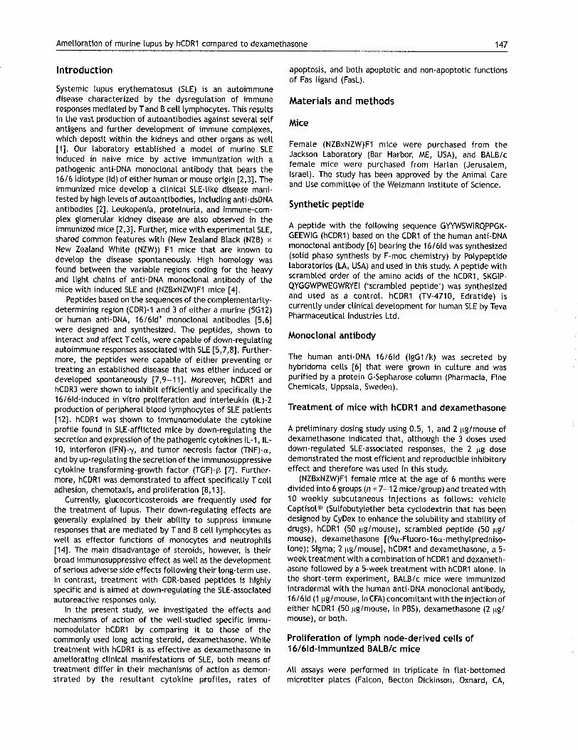
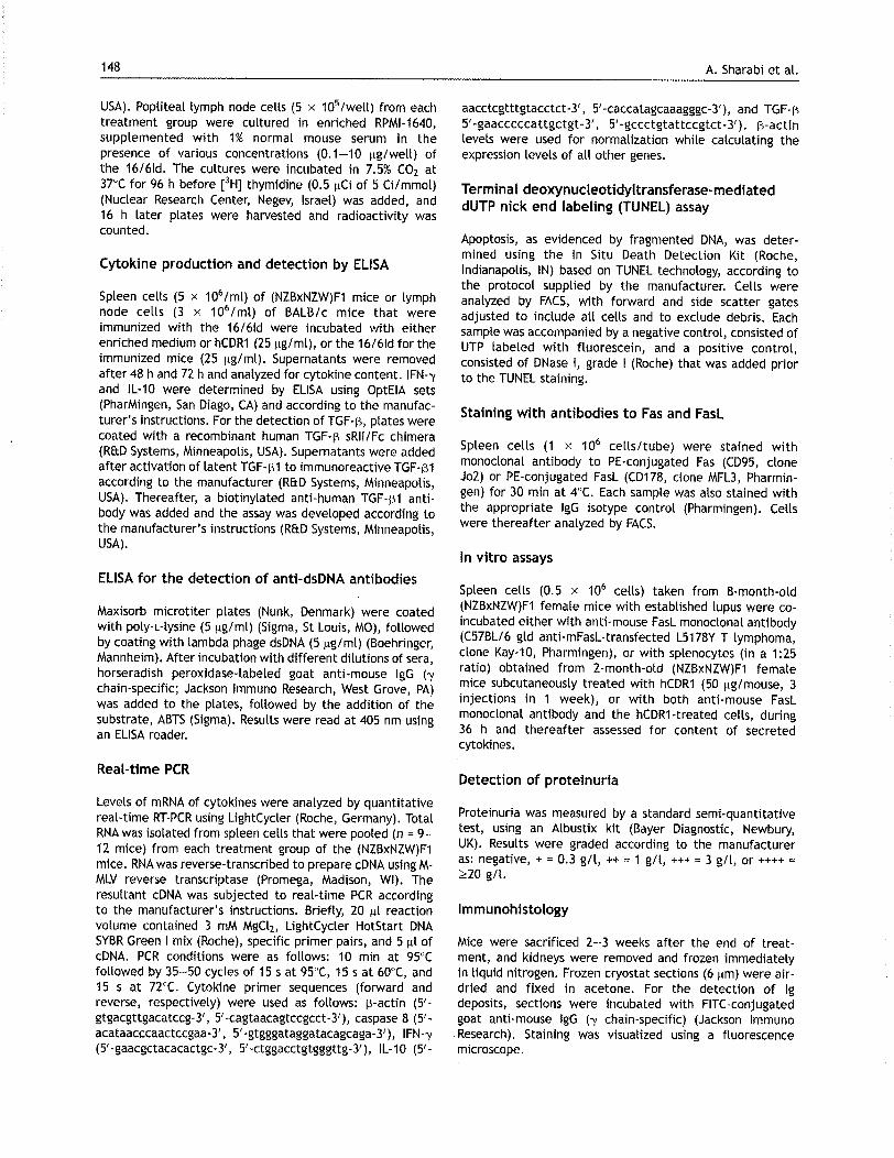


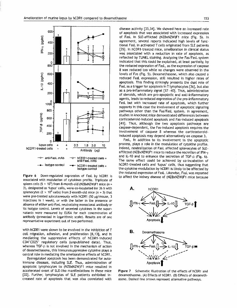

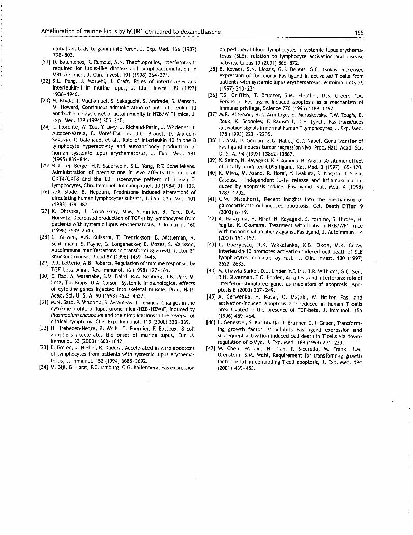
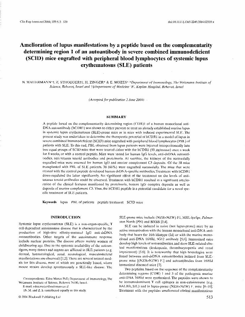

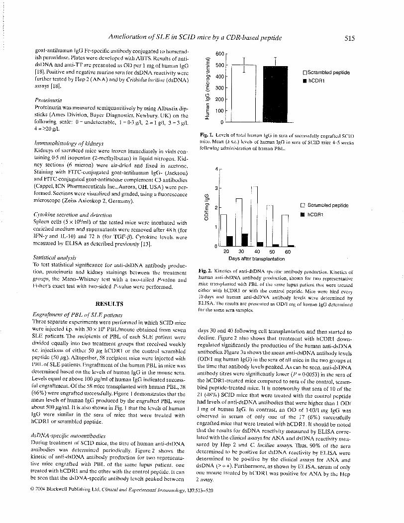

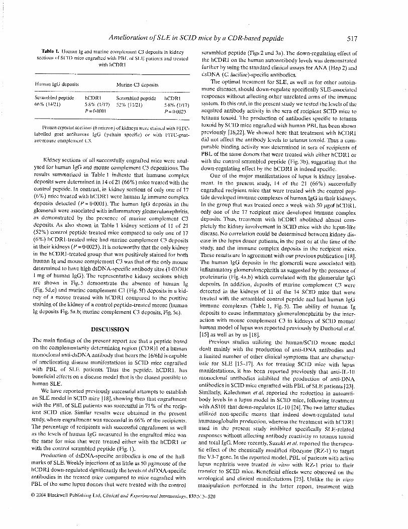

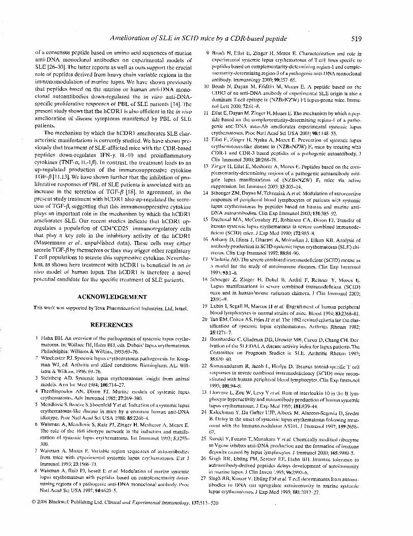
DOI: 10.1212/01.wnl.0000223353.34006.54 2006;67;20-27 Neurology
Cudkowicz B. J. Traynor, L. Bruijn, R. Conwit, F. Beal, G. O’Neill, S. C. Fagan and M. E.
Neuroprotective agents for clinical trials in ALS: A systematic assessment
This information is current as of September 12, 2006
http://www.neurology.org/cgi/content/full/67/1/20located on the World Wide Web at:
The online version of this article, along with updated information and services, is
Print ISSN: 0028-3878. Online ISSN: 1526-632X. published continuously since 1951. Copyright © 2006 by AAN Enterprises, Inc. All rights reserved. Neurology is the official journal of AAN Enterprises, Inc. A bi-monthly publication, it has been
by MICHAEL FETELL on September 12, 2006 www.neurology.orgDownloaded from
Views & Reviews
Neuroprotective agents for clinical trialsin ALS
A systematic assessmentB.J. Traynor, MD, MRCPI; L. Bruijn, PhD; R. Conwit, MD; F. Beal, MD; G. O’Neill, MD, MRCPI;
S.C. Fagan, PharmD, FCCP; and M.E. Cudkowicz, MD, MSc
Abstract—Background: Riluzole is currently the only Food and Drug Administration–approved treatment for ALS, but itseffect on survival is modest. Objective: To identify potential neuroprotective agents for testing in phase III clinical trialsand to outline which data need to be collected for each drug. Methods: The authors identified 113 compounds by invitinginput from academic clinicians and researchers and via literature review to identify agents that have been tested in ALSanimal models and in patients with ALS. The list was initially narrowed to 24 agents based on an evaluation of scientificrationale, toxicity, and efficacy in previous animal and human studies. These 24 drugs underwent more detailed pharma-cologic evaluation. Results: Twenty drugs were selected as suitable for further development as treatments for patientswith ALS. Talampanel and tamoxifen have completed early phase II trials and have demonstrated preliminary efficacy.Other agents (ceftriaxone, minocycline, ONO-2506, and IGF-1 polypeptide) are already in phase III trials involving largenumbers of patients with ALS. Remaining agents (AEOL 10150, arimoclomol, celastrol, coenzyme Q10, copaxone, IGF-1–viral delivery, memantine, NAALADase inhibitors, nimesulide, scriptaid, sodium phenylbutyrate, thalidomide, trehalose)require additional preclinical animal data, human toxicity and pharmacokinetic data including CNS penetration prior toproceeding to large scale phase III human testing. Further development of riluzole analogues should be considered.Conclusions: Several potential neuroprotective compounds, representing a wide range of mechanisms, are available andmerit further investigation in ALS.
NEUROLOGY 2006;67:20–27
Riluzole is the only Food and Drug Administration(FDA)–approved drug for ALS, but it has only a mod-est affect on survival. ALS has a median survival of3 to 5 years.1 A number of mechanisms are thoughtto initiate and propagate the neurodegenerative pro-cess in ALS, including oxidative stress, mitochon-drial dysfunction, excitotoxicity, apoptosis,inflammation, and glial activation.2 These advancesin ALS research, together with the application ofhigh throughput drug screening such as the NationalInstitute of Neurologic Disorders and Stroke Neuro-degeneration Drug Screening Consortium,3 haveyielded a large number of drug candidates that may
be neuroprotective. However, only a small number ofdrug candidates can be tested at any one time, asresources available to the ALS community are lim-ited both in terms of eligible research participantsand funds. A transparent and rational selection pro-cess is required to determine which candidate agentsshould be prioritized for clinical trial development. Asystematic approach to drug selection, rather thanpursuing the latest “hot” compound, is importantboth for ALS and for the broader issue of researchstrategy in neurodegenerative disorders.
We describe the drug identification and reviewprocesses and outline attractive neuroprotective can-didates for future ALS clinical trials. Priority wasgiven to making the drug selection process explicit,transparent, and reproducible. We also identify datato be collected for each drug prior to proceeding to
Additional material related to this article can be found on the NeurologyWeb site. Go to www.neurology.org and scroll down the Table of Con-tents for the July 11 issue to find the title link for this article.
From the Neurology Clinical Trials Unit (B.J.T., M.E.C.), Department of Neurology, Massachusetts General Hospital, Boston; The ALS Association (L.B.),Palm Harbor, FL; National Institute of Neurological Diseases and Stroke (NINDS) (R.C.), Bethesda, MD; Department of Neurology (F.B.), Weill MedicalCollege of Cornell University, New York, NY; Biogen Idec Inc. (G.O.), Cambridge, MA; and University of Georgia College of Pharmacy and Medical College ofGeorgia (S.C.F.), Augusta.Disclosure: The authors report no conflicts of interest.Received August 23, 2005. Accepted in final form March 10, 2006.Address correspondence and reprint requests to Dr. Bryan J. Traynor, SDGE, NIMH, Building 35, Room 1A110, 35 Convent Drive, Bethesda, MD20892-3720; e-mail: [email protected]
20 Copyright © 2006 by AAN Enterprises, Inc. by MICHAEL FETELL on September 12, 2006 www.neurology.orgDownloaded from

phase III efficacy testing enrolling large numbers ofpatients with ALS.
Methods. Drug selection process. We first identified a widespectrum of potential therapeutic agents and a broad range ofstrategies that could potentially slow disease progression and pro-long survival in patients with ALS. We incorporated 1) therapieswith mechanisms of action relevant to ALS pathogenesis, 2)agents that have already been tested in ALS animal models orclinical trials, and 3) medications approved or under considerationfor neurodegenerative diseases other than ALS. Input was ob-tained from academic scientists, clinicians, and patient groups tocapture the broadest range of available compounds. We alsosearched Medline to identify publications concerning agents thathad been tested in ALS animal models (table E-1 on the NeurologyWeb site at www.neurology.org) or in human trials (table E-2).
Selection of drugs for detailed pharmacologic and safety assess-ment. We identified 113 therapeutic agents as potentially benefi-cial in patients with ALS (table E-3). Each therapeuticintervention was assessed by the authors (B.J.T., L.B., R.C., F.B.,G.O., M.E.C.) according to the following criteria established at theFebruary 2004 meeting: 1) scientific rationale (i.e., an effect on apathway implicated in ALS), 2) drug safety and tolerability inhumans, 3) indication of benefit in human clinical studies, and 4)efficacy of the drug in ALS animal models. The assessment crite-ria were developed from those previously employed in evaluatingneuroprotective agents in Parkinson disease (PD) (table 1).4 Theinability of ALS animal models to predict beneficial effects inhuman trials is recognized5 and consequently data from SOD1G93A
mouse model were only one of several preclinical data points ex-amined in the drug evaluation process. Data from cell-basedscreening were included where relevant, though an in-depth re-view of drug discovery techniques is beyond the scope of thisarticle. Both published and unpublished information was consid-ered. To maintain transparency of the process, expand discussion,and further scientific rigor, we circulated the list of 113 therapeu-tic agents (table E-3) within the ALS research community.Twenty-four drugs judged to be the most promising agents wereselected for further analysis. The reasons for not selecting theremaining 89 drugs for detailed review are also listed in table E-3.
Detailed pharmacologic and safety assessment of proposedagents. A clinical pharmacologist (S.F.) with expertise in neuro-logic drugs performed a detailed pharmacokinetic and safety as-sessment of the 24 selected drugs (table E-4). The completepharmacokinetic, tolerability, and preclinical data sets were em-ployed to select neuroprotective candidates for future ALS clinicaltrials using a scorecard method (table 2). The pharmaceuticalcompany or academic scientist directly responsible for developingthat agent was contacted to obtain further data.
TCH346 was excluded from the final list based on negativeresults of phase III studies that were not available during theinitial review.6 Vitamin E was excluded based on the results oftwo negative clinical trials.7,8 Adverse side effects and unfavorablepharmacokinetics eliminated NBQX. Nimodipine was eliminateddue to insufficient scientific rationale to support its developmentas an ALS therapy. Through this process we determined that 20drugs are viable candidates to be explored in ALS clinical trials inthe future (figure and table 3).
Annual reassessments of newly published data on existing andnovel neuroprotective agents will also be provided on the ALSAssociation Web site and will be presented at the InternationalALS/MND Symposium on a yearly basis.
Description of priority agents. AEOL 10150 (AeolusScience Inc.). Oxidative damage mediated by toxic freeradicals has been implicated in the pathogenesis of ALS9
and a variety of antioxidants have been tested in patientswith ALS (table E-2). AEOL 10150 is a manganoporphyrinantioxidant that catalytically neutralizes superoxide, hy-drogen peroxide, and peroxynitrite, and inhibits lipid per-oxidation.10 Administration of AEOL 10150 to transgenicmice with the glycine 93 to alanine SOD1 mutation(SOD1G93A) commencing at symptom onset improved thesurvival interval (the time from symptom onset to death)by 196% (26.5 days).11 The compound has to be adminis-tered IV or by subcutaneous injection. A phase I singledose escalating study enrolling 30 patients with ALS isunderway to determine pharmacokinetic, optimum dose,and safety properties of this novel drug class.
Arimoclomol (Cytrx Corporation). Motor neurons havean intrinsically higher threshold for activation of the heatshock protein pathway12 and agents that upregulate thispathway may be neuroprotective. Arimoclomol is a hydrox-ylamine derivative that co-induces heat shock protein(HSP) expression,13 a powerful cytoprotective mecha-nism under acute stress conditions. Treatment with ari-moclomol prolonged the lifespan of SOD1G93A mice by22% (28 days).14 The beneficial effect was independent ofwhether the treatment was started pre-symptomaticallyor at symptom onset. Arimoclomol was well tolerated ina phase I study of healthy volunteers (http://www.cytrx.com/prDetail.cfm?pr_id� 164&showcspr�1). Safety, op-timum dose, and pharmacokinetics of arimoclomol (in-cluding ability to cross blood– brain barrier [BBB]) areunknown for patients with ALS. A multicenter, doseranging, phase II study of arimoclomol in ALS has com-menced enrollment (n � 80, www.clinicaltrials.gov,NCT00244244).
Ceftriaxone (Roche Laboratories). Glutamate-mediatedexcitotoxicity arising from repetitive firing or elevation ofintracellular calcium by calcium-permeable glutamate re-ceptors is likely to be an important contributor to motorneuronal death in ALS.1 Glutamate levels are increased inCSF of patients with sporadic ALS15 and clearance of glu-tamate from neuromuscular synapses is also diminished inpatients with ALS due to loss of the astroglial glutamatetransporter EAAT2.16 Furthermore, spinal motor neuronsare relatively reduced in intracellular calcium-bindingcomponents,17 which may account for their selectivevulnerability.2
The antiexcitatory and antioxidant properties of cepha-losporins were identified by the Neurodegeneration DrugScreening Consortium that screened 1040 FDA-approveddrugs for efficacy in in vitro models of neurodegenerativediseases.18 Cephalosporins increase EAAT2 promoter activ-
Table 1 Evaluation criteria for potential neuroprotective agentsin ALS*
Criteria Operational definition
Scientific rationale Consistency of preclinical data; crediblemechanism relevant to ALS althoughmechanism may be unknown inmany cases
Safety andtolerability
Safe and tolerable in humans in thedose and route of administrationneeded for the proposed effect.Further safety data may be requiredbefore use in ALS
Efficacy in relevantanimal
Efficacy in rodent model of ALS orother relevant models of disease
Indication ofbenefit in humanclinical studies
Evidence from previous trials that issuggestive of a neuroprotective effector epidemiologic data fulfillingcriteria for causal inference
*Developed from criteria for evaluation of neuroprotective agentsin Parkinson disease.4
July (1 of 2) 2006 NEUROLOGY 67 21 by MICHAEL FETELL on September 12, 2006 www.neurology.orgDownloaded from

ity and protect motor neurons from glutamate toxicity inculture.18 The third generation ceftriaxone was selected forhuman studies because of its superior CNS penetrationand long half-life.19 SOD1G93A transgenic mice treatedwith ceftriaxone at symptom onset lived 8% (10 days)longer than control animals.18 Ceftriaxone is generally welltolerated,20 though experience with long term IV adminis-tration (beyond 6 weeks) is limited. A combined phase IIand III study will commence enrollment in the near future.
Celastrol (generic). Neuroinflammation occurs in thebrainstem and spinal cord of patients with ALS21 andSOD1G93A mice suggesting that anti-inflammatory agentsmay be effective in treating this disease.22 Celastrol is apotent anti-inflammatory and antioxidant triterpene thatsuppresses TNF�, IL-1�, and inducible nitric oxide produc-tion23 and induces a heat shock protein response.24 Admin-istration of celastrol from 4 weeks of age improved weightloss, rotorod performance, and survival of SOD1G93A
mice.25 Further data concerning the ability of the drug tocross the BBB, toxicity, and safety in patients with ALS
Table 2 Scorecard outlining detailed pharmacologic and safety assessment of 24 neuroprotective drugs in patients with ALS
Agent Mechanism of action PK ALS mouse model Human safety Benefits in ALS
AEOL 10150 Antioxidant 0 �� 0 0
Arimoclomol HSP inducer 0 � � 0
Ceftriaxone Antioxidant and antiglutamate � � ��� 0
Celastrol Antioxidant and anti-inflammatory 0 � 0 0
CoQ10 Antioxidant and mitochondrial factor � �� �� 0
Copaxone Immunomodulatory 0 –/� ��� 0
IGF-1 Neurotrophic 0 � (wobbler) �� �/–
IGF-1-AAV Neurotrophic 0 � 0 0
Memantine Antiglutamate �� 0 ��� 0
Minocycline Antiapoptotic �� �� ��� 0
Naaladase inhibitor Antiglutamate 0 � 0 0
NBQX Antiglutamate 0 � – 0
Nimesulide Anti-inflammatory and antioxidant 0 – ��� 0
Nimodipine Ca2� channel blocker �� � ��� –
ONO-2506 Glial modulator and antiglutamate 0 0 0 0
Riluzole Antiglutamate and Na channel inactivation � �� ���� ���
Scriptaid Antiaggregation 0 0 0 0
Phenylbutyrate HDAC inhibitor 0 � ��� 0
Talampanel Antiglutamate � � �� �
Tamoxifen Protein kinase C inhibitor � � (viral model) ��� �
TCH346 Antiapoptotic � –/� �� –
Thalidomide Immunomodulatory and antiangiogenic � � ��� 0
Trehalose Antiaggregation 0 0 ��� 0
Vitamin E Antioxidant � � ��� –
Pharmacokinetics: 0 � data on ability of drug to penetrate CSF are unknown; � � drug known to penetrate CSF; �� � drug has ex-cellent CSF penetration. ALS mouse model: 0 � effect on ALS mouse model unknown to us; – � one mouse study with negative effecton survival; � � one mouse study with a positive effect on survival; �� � two mouse studies with positive effects on survival; ½ �two animal studies performed, one showed beneficial effect, the other displayed a negative effect on survival. Human safety: 0 � safetyin humans not known; � � one human trial that showed drug was relatively safe; �� � two human trials that showed drug was rela-tively safe; ��� � FDA/EMEC approved for chronic use in other disorders; ���� � FDA/EMEC approved as an ALS drug. Benefitsin ALS: 0 � unknown; – � negative trial in well-designed studies; � � positive (or trend) efficacy in phase II trials; ��� � FDA/EMEC approved as an ALS drug.
Figure. Drug identification and assessment sequence.
22 NEUROLOGY 67 July (1 of 2) 2006 by MICHAEL FETELL on September 12, 2006 www.neurology.orgDownloaded from

and optimum dose remain to be collected prior to phase IIItesting.
Coenzyme Q10 (generic). The high metabolic load ofmotor neurons and the consequent dependence of thesecells on oxidative phosphorylation may make them partic-ularly vulnerable to the loss of mitochondrial function.1
Coenzyme Q10 is an antioxidant and an essential mito-chondrial cofactor facilitating electron transfer in the re-spiratory chain.26 This commonly used nutraceutical isbeing tested in neurodegenerative conditions in which mi-tochondrial dysfunction has been implicated includingALS, Huntington disease (HD), and PD. Low dose coen-zyme Q10 prolonged median survival of SOD1G93A trans-genic mice by only 4.4% (6 days),27 but higher doses aremore effective (Flint Beal, personal communication). Dosesup to 3,000 mg per day are safe and well tolerated inpatients with ALS.28 Coenzyme Q10 is lipophilic and effec-tively crosses the BBB.29 A phase II study in ALS hascommenced enrollment (NCT00243932).
Copaxone/glatiramate (Teva Pharmaceuticals). Previ-ous clinical trials of immunosuppressant therapies failedto slow progression in ALS,30 but immunomodulation maybe more effective at preventing neuronal apoptosis.31 Cop-
axone evokes a neuroprotective T cell-mediated responseand may protect against glutamate toxicity.31 Low copynumber SOD1G93A ALS mice immunized at 60 days of agefollowed by oral dosing of copolymer-1 experienced a 24.6%(52 day) increase in lifespan and delayed disease onset,31
though these findings have not been replicated in highcopy SOD1G93A mice treated with a copaxone derivative.32
Additional preclinical data are required to determine re-producibility and optimum dosing schedule to achieveimmunomodulation.
Insulin-like growth factor 1 (IGF-1, Cephalon). Neuro-trophic factors selectively regulate survival and differenti-ation of nerve cells and maintain neuronal structuralintegrity. Of all neurotrophic factors tested in ALS to date(table E-2), only IGF-1 slowed the rate of functional declineby 26% in a North American phase III trial (n � 266).33 Incontrast, a European IGF-1 trial was negative (n � 183)34
and so a third phase III trial of this neurotrophic factor iscurrently underway (n � 330, NCT00035815) to conclu-sively determine efficacy. IGF-1 is well tolerated, thoughthe large size of the IGF-1 polypeptide may prevent BBBpenetration. There are no published reports of the effect ofIGF-1 on mutant SOD1G93A mouse survival.
IGF-1—viral delivery (Ceregene, Inc.). Adeno-associated virus engineered to contain the gene for IGF-1(AAV-IGF1) allows targeted delivery of IGF-1 to motorneurons. After IM injection, the gene vector is transportedto the neuronal cell body by retrograde axonal transportalong motor neurons.35 AAV-IGF-1 prolongs median sur-vival by 30% (37 days) when administered before diseaseonset.35 Human safety, dose schedule, and pharmacokinet-ics have not yet been established for this novel gene ther-apy, though AAV-factor IX vector has proven safe inpatients with hemophilia.36 A small phase IIa trial of AAV-IGF-1 is planned for the near future. Expansion of theviral vector production capacity will be required beforeproceeding to large scale trials.
Memantine (Forest Laboratories, Inc.). The higher ex-pression of calcium-permeable �-amino-3-hydroxy-5-methyl-4-isoxazole propionic acid (AMPA) glutamatereceptors on motor neurons may explain the selective vul-nerability of this cell type to glutamate excitotoxicity.37
Memantine (an amino adamantine derivative) is an AMPAreceptor antagonist licensed as a neuroprotective agent forAlzheimer disease (AD). The drug exhibits excellent CNSpenetration38 and is well tolerated by patients with AD,but there are no published studies of the survival effect ofin ALS animal models and additional data to support therationale for testing in patients with ALS are needed.
Minocycline (generic). The role of apoptosis in motorneuron degeneration is increasingly recognized. In SOD1-mediated ALS, motor neurons probably die through theformation of insoluble mutant SOD1 aggregates that bindto and deplete motor neurons of the antiapoptotic proteinBcl-2 allowing activation of caspases.39,40 Although apopto-sis is a late event in the degeneration of motor neurons,inhibition of programmed cell death might ameliorateALS.1 Minocycline is a second generation tetracycline anti-biotic that prevents microglial activation41 and inhibitscaspase activation.42 Four SOD1 transgenic mouse studiesshow enhanced median survival ranging between 6.4% and16%.41-44 Two small phase II studies demonstrated safetyand tolerability of minocycline in patients with ALS (n �
Table 3 Priority list for phase III ALS clinical trials
A. Suitable for phase III trials in the near future
1. Talampanel
2. Tamoxifen
B. Already in phase III trials involving large number of humansubjects
1. Ceftriaxone
2. IGF-1 polypeptide
3. Minocycline
4. ONO-2506
C. More data required prior to phase III testing
1. AEOL 10150†‡§
2. Arimoclomol*†‡§
3. Celastrol*†‡§
4. Coenzyme Q10‡§
5. Copaxone*§
6. IGF-1–viral delivery*†‡§
7. Memantine*†‡§
8. NAALADase inhibitors*†‡§
9. Nimesulide*‡§
10. Scriptaid*†‡§
11. Sodium phenylbutyrate†‡§
12. Thalidomide†‡§
13. Trehalose*‡§
D. Already Food and Drug Administration approved as ALStherapy
1. Riluzole and related benzothiazole drugs (see text forfurther details)
*Transgenic efficacy animal studies.†Human toxicology.‡CNS pharmacokinetic studies.§Dose-ranging studies.
July (1 of 2) 2006 NEUROLOGY 67 23 by MICHAEL FETELL on September 12, 2006 www.neurology.orgDownloaded from

19 and 23).45 Typical side effects include gastrointestinalupset, vertigo, and cumulative dose-dependant photosensi-tivity. A phase III efficacy trial of minocycline is currentlyenrolling 400 patients (NCT00047723). The ability of thedrug to penetrate uninflamed meninges should bedetermined.
N-acetylated alpha-linked acidic dipeptidase (NAALA-Dase, Guilford Inc.). Inhibition of glutamate carboxypep-tidase 2 (GCP2) may be neuroprotective by simultaneouslydecreasing glutamate production and inhibiting glutamaterelease.46 Median survival of SOD1G93A ALS mice was pro-longed by 15% (29 days) by administration of the GCP2inhibitor 2-(3-mecaptopropyl) pentanedioic acid. GCP2 in-hibition is attractive as a therapeutic target because theeffects only occur during excessive glutamate stimulationavoiding glutamate receptor antagonist side effects.46
NAALADase inhibitors have not yet been administered tohumans and there are no data on pharmacokinetics andtolerability.
Nimesulide (generic). The enzyme COX-2 is an attrac-tive therapeutic target because of its marked increase inALS spinal cord stimulating astrocytic glutamate re-lease.47,48 Nimesulide is a preferential COX-2 inhibitorwith additional antioxidant properties.49 Nimesulide ad-ministration decreased PG-E2 levels in the spinal cord ofSOD1G93A mice and preserved motor skill integrity.50 How-ever, the COX-2 inhibitor celecoxib failed to show benefitin a phase II/III trial.51 Furthermore, safety concerns sur-rounding long-term administration of this medication classmay limit use in patients with ALS.52
ONO-2506 (Ono Pharmaceutical Co. Ltd). Chimericmice with both normal and mutant SOD1-expressing cellsindicate that glia play a role in motor neuron degenera-tion.53 Possible mechanisms include microglial-mediatedneuroinflammation, loss of neurotrophic support,2 and di-minished clearance of glutamate from neuromuscular syn-apses by the astrocytic glutamate transporter EAAT2.16
ONO-2506 is an enantiomeric homologue of valproate thatrestores normal astrocyte functions after brain damage bypreventing reactive astrocytosis, by activating astrocyticGABAA receptors and suppressing GABA transferase.54
This agent has additional antiglutamate55 and anti-inflammatory COX-2 inhibitor properties.56 Results of acompleted phase II trial of 1,200 mg per day oral formula-tion (cereact) are pending. A phase III trial of the parentcompound valproate has commenced enrollment in Europe(n � 173, NCT00136110).
Riluzole (Sanofi-Aventis). Riluzole remains the onlyFDA-approved drug for ALS based on the 3-month im-provement in survival observed in two large clinical tri-als.57,58 Riluzole has a broad range of pharmacologic effectsincluding inhibition of glutamate release, postsynaptic glu-tamate receptor activation, and voltage-sensitive sodiumchannels inactivation. It was identified before the SOD1mouse model became available by studying toxicity of CSFfrom patients with ALS on neuronal cell cultures.59 Subse-quently, riluzole was found to have a modest effect onSOD1G93A mouse survival (prolonged median survival by11%, 14 days).60
Riluzole has been included in the final list not to sug-gest that further trials of this drug are required, butrather to emphasize the surprising lack of effort to build onthe modest success of riluzole. Related benzothiazoles have
not been tested in ALS animal models or patients. A col-laborative effort between academic scientists and industrymay rejuvenate development of this drug class.
Scriptaid (Alexis Biochemicals). Abnormal protein ag-gregates have been described in neurodegenerative dis-eases including AD, PD, and HD. Ubiquitin inclusions arepresent in motor neurons and astrocytes of patients withALS.61 It is not known if these aggregates damage or pro-tect motor neurons, though several possible toxic mecha-nisms have been proposed including aberrant chemistry,loss of normal proteins through sequestration within ag-gregates, and inhibition of mitochondria, peroxisomes, orproteosomeal function overwhelmed with indigestible, mis-folded protein.2 Agents that decrease aggregation havebeen hypothesized to be neuroprotective. Scriptaid wasidentified in a screen for small molecules that disrupt invitro aggresome formation in cultured COS cells trans-fected with mutant SOD1-GFP.62 Safety, optimum dose,and pharmacokinetic animal and human data remain to bedetermined for this drug.
Sodium phenylbutyrate (Scandinavian formulas). So-dium phenylbutyrate (NaPB) is FDA-approved for chronictreatment of hyperammonia and has been tested as atreatment of spinomuscular atrophy.63 Its potential benefitin ALS is based on its ability to inhibit histone deacetylase(HDAC) leading to increased gene transcription.64 This ar-omatic short-chain fatty acid extends median survival ofSOD1G93A ALS mice by 21.9% (27.5 days).64 NaPB has ashort half life (45 minutes), though changes in gene ex-pression induced by the drug may be more persistent. CNSdistribution of NaPB has been determined by MR spectros-copy.65 An open label, dose-escalation study enrolling 40patients with ALS is underway to determine human safety(NCT00107770).
Talampanel (8-methyl-7H-1,3-dioxolo(2,3)benzodiazepine,IVAX Corporation). Talampanel is a noncompetitive mod-ulator of AMPA glutamate receptors primarily under de-velopment as an antiepileptic agent. Talampanel has beenshown to prolong SOD1G93A mouse median survival (Jef-frey Rothstein, personal communication). ALSFRS andTQNE scores declined at a slower rate in a 9-month phaseII study of talampanel in 60 patients with ALS though thestudy was not powered to detect efficacy (Robert Pascuzzi,personal communication). The most common side effectswere ataxia and sedation. The antiepileptic properties oftalampanel indicate that the drug crosses the BBB.
Tamoxifen (Astra Zeneca). Tamoxifen may be neuro-protective in ALS because of its ability to inhibit proteinkinase C, which mediates inflammation in spinal cords ofpatients with ALS.66 Tamoxifen extended survival in a vi-rally induced ALS mouse model.67 A phase II study of 60patients with ALS prolonged survival at 10 mg, 20 mg, 30mg, and 40 mg daily doses (Ben Brooks, personal commu-nication). The drug penetrates the CNS and is generallywell tolerated. The effect of the drug on survival ofSOD1G93A mice needs to be evaluated and the results ofthe phase II study should be peer-reviewed. However, ifthese results are favorable, planning for a phase III studymay be expedited.
Thalidomide (Celgene). Angiogenic factors controllingthe growth and permeability of blood vessels have beenimplicated in the pathogenesis of ALS. Mice bearing adeletion of the vascular endothelial cell growth factor
24 NEUROLOGY 67 July (1 of 2) 2006 by MICHAEL FETELL on September 12, 2006 www.neurology.orgDownloaded from

(VEGF) gene develop an ALS-like phenotype68 and poly-morphisms of the VEGF promoter region increase the riskof ALS.69 Coding mutations of a related gene angiogeninhave also been linked to the disease.70 Thalidomide is anon-barbiturate sedative that was withdrawn from theworld market in 1961 on discovery of its teratogenic ef-fects. It has been selectively reintroduced for a variety ofconditions including progressive body weight loss relatedto advanced cancer and AIDS.71 The drug has anti-angiogenic activity and immunomodulatory properties.71
Oral thalidomide reduced TNF�, attenuated weight loss,and increased survival in SOD1G93A mice.72 Thalidomidecrosses the BBB, as indicated by its sedative effects. Pe-ripheral neuropathy has been observed in 8% of patientswith HIV and can become irreversible if thalidomide is notdiscontinued.73 Lenalidomide, a novel 4 amino-glutarimideanalogue, shows the same efficacy in animal studies with-out the neurotoxic and teratogenic effects.72,74 An open-label, phase II trial is currently recruiting patients (n �24, NCT00140452).
Trehalose (Cargill, Inc.). Trehalose is a natural disac-charide used in freeze-dried products to prevent proteindenaturation. Trehalose may prevent formation of mutantSOD1 aggregates in ALS by stabilizing mutant proteins.75
The agent has a long history of human use and the FDAhas issued a “letter of no objection” (GRAS No. GRN000045). However, there are no data on toxicity in patientswith ALS, the ability of the drug to penetrate the CNS isunknown, and its effect in transgenic ALS mouse modelremains to be evaluated.
Discussion. We identified and assessed potentialcompounds for clinical trials in patients with ALS.Academic clinicians and scientists identified 113compounds, of which 24 were selected for more de-tailed pharmacokinetic and safety analysis. Twentywere chosen as the most promising agents thatshould be studied in phase III clinical ALS trials.Two agents on the priority list (talampanel and ta-moxifen) show preliminary efficacy in phase II ALSclinical studies. Other agents (ceftriaxone, minocy-cline, ONO-2506, and IGF-1 polypeptide) are alreadyin phase III trials involving large numbers of pa-tients. Most agents on the final priority list requireadditional data (preclinical animal data, human tox-icity, and pharmacokinetic data [including CNS pen-etration]) prior to proceeding to large scale humantesting (see table 3).
While a detailed attempt was made to make thereview process explicit, qualitative judgments had tobe prepared about the relative value and weightingof different types of information. The most problem-atic issues were the evaluation of unpublished dataand the authors’ biases. Although considerable effortwas made to obtain information from investigatorsdeveloping an agent, not all data were available tothe authors. The initial screen was performed byinvestigators in the field, which may have resultedin bias toward agents studied in their laboratories.To ensure transparency all data relevant to this arti-cle were made available to the ALS communitythroughout the selection process.
Animal drug-screening studies in ALS almostexclusively utilize the mutant SOD1G93A mouse(table E-1), but the ability of this model to predictdrug efficacy in humans is ambiguous. Severaldrugs that prolong survival in animal studies havenot shown efficacy in human trials (celebrex,51,76
creatine,77,78 gabapentin,60,79 N-acetylcysteine80,81).This discrepancy may be due to intraspecies differ-ences in pharmacokinetics and the difficulty in es-tablishing dose equivalence to achieve the samebiologic activity in humans as observed in mice. Itmay also be that this mouse model of familial ALSdoes not predict drug effect in patients with spo-radic ALS and that development of alternativemodels should be prioritized.
Interpretation of animal drug screening studies iscomplicated by varying experimental designs be-tween laboratories. For example, mouse strain andsex,82 as well as environmental factors such as accessto exercise, affect survival.83 There is a need to estab-lish consensus guidelines to ensure ALS animal drugstudies are conducted in a uniform manner. Experi-mental design issues that warrant standardizationinclude the type of animal models, number of ani-mals and sex distribution required to reliably detectan effect, as well as the timing and method of drugdelivery. The selection of appropriate survival andmotor function endpoints is essential. Guidelines onthe publication of negative results and the establish-ment of an online database of ALS animal drug stud-ies should be a priority. Most importantly, guidelinesshould be established outlining the magnitude andthe reproducibility of drug effect in different labora-tories and animal models required to proceed to hu-man clinical testing.
The pharmacokinetic profile, the safety/toxicityproperties, and the most efficacious dose of the drugin humans must be adequately established prior tophase III studies. There has been a tendency forpotentially beneficial candidates to move rapidly tolarge ALS clinical trials. Although this approach hasdemonstrated that certain drugs are ineffective, ithas been unsuccessful at identifying useful thera-pies. The ability of a drug to cross the human BBB toreach its target ligand should be determined prior tostarting phase II studies. Tolerability of a dose inhealthy patients should not be taken as indicationthat the same dose will be safe in patients with ALS.The frequency of adverse events was significantlyhigher in patients with ALS receiving topiramatethan was seen in patients with epilepsy, possiblyrelated to dehydration and malnutrition in patientswith ALS.84 Dose-ranging studies are a prerequisiteto phase III studies to determine the most effectiveand safe dosage. Focusing on early clinical safety,dose-finding, and pharmacokinetic testing will in-crease a drug’s early development costs, but willmaximize its chance of success in large phase IIIefficacy studies.
Multiple pathways have been implicated in thepathogenesis of ALS.2 A medication or combination
July (1 of 2) 2006 NEUROLOGY 67 25 by MICHAEL FETELL on September 12, 2006 www.neurology.orgDownloaded from
of medications that targets more than one patho-genic pathway may slow disease progression in anadditive or synergistic fashion. Such combinationtherapy has been successful in oncology, though mul-tiple drug interactions and increased incidence ofdrug side effects should be considered. More detailedanimal toxicology studies of combination therapiesare required than for therapies given alone. Drugresistance must also be considered when a medica-tion is administered for prolonged periods (months).Upregulation of multidrug resistance proteins hasbeen reported in astrocytes and BBB of patients withneurodegenerative diseases with neuroinflammatorycomponents, such as ALS. These transporters ex-trude endogenous toxins (such as medications) fromCNS from the cells and may nullify a drug’sbioactivity.85
In this article we evaluated existing drugs fortheir potential development in ALS in an explicit,systematic, and transparent manner. The selectionprocess is intended to prioritize interventions forphase III trials in patients with ALS and to identifydata that need to be collected prior to clinical studiesinvolving large numbers of human subjects. The ALSAssociation has issued a request for applicationsbuilding on the final list published in this article.86
More candidate drugs will be identified as academicresearchers adopt high throughput screening tech-niques and study candidate neuroprotective drugs innew animal models of ALS. Thus, the need to ratio-nally select agents for clinical testing in patientswith ALS will increase and the current approach canbe extended to evaluate new therapies as theyemerge. New data on both existing and novel neuro-protective agents will be assessed annually and up-dates made available on the ALS Association Website and presented at the annual International ALS/MND Symposium.
AcknowledgmentThe authors thank Vincent Meininger, MD, Robert G. Hart, MD,and Davide Trotti, PhD, for advice on the manuscript and Sus-anna Benn, PhD, for help constructing the table of animal studies.The authors thank attendees of the ALS Association-hosted con-ference to discuss candidates for ALS clinical trials: Stanley Ap-pel, MD, Robert Brown, MD, Jill Heemskerk, PhD, Tom Maniatis,PhD, Bernard Ravina, MD, Jeff Rothstein, MD, PhD, and PaulSheehy, PhD.
References1. Rowland LP, Shneider NA. Amyotrophic lateral sclerosis. N Engl J Med
2001;344:1688–1700.2. Bruijn LI, Miller TM, Cleveland DW. Unraveling the mechanisms in-
volved in motor neuron degeneration in ALS. Annu Rev Neurosci 2004;27:723–749.
3. Heemskerk J. High throughput drug screening. Amyotroph LateralScler Other Motor Neuron Disord. 2004;5(suppl 1):19–21.
4. Ravina BM, Fagan SC, Hart RG, et al. Neuroprotective agents forclinical trials in Parkinson’s disease: a systematic assessment. Neurol-ogy 2003;60:1234–1240.
5. Rothstein JD. Of mice and men: reconciling preclinical ALS mousestudies and human clinical trials. Ann Neurol 2003;53:423–426.
6. Miller RG, Bradley W, Cudkowicz M, et al. Phase II/III controlled trialof TCH 346 in patients with amyotrophic lateral sclerosis. AmyotrophLateral Scler Other Motor Neuron Disord 2005; 6(suppl 1):13.
7. Desnuelle C, Dib M, Garrel C, Favier A. A double-blind, placebo-controlled randomized clinical trial of alpha-tocopherol (vitamin E) inthe treatment of amyotrophic lateral sclerosis. ALS Riluzole-Tocopherol
Study Group. Amyotroph Lateral Scler Other Motor Neuron Disord2001;2:9–18.
8. Graf M, Ecker D, Horowski R, et al. High dose vitamin E therapy inamyotrophic lateral sclerosis as add-on therapy to riluzole: results of aplacebo-controlled double-blind study. J Neural Transm 2005; 112:649–660
9. Cleveland DW. From Charcot to SOD1: mechanisms of selective motorneuron death in ALS. Neuron 1999;24:515–520.
10. Patel M, Day BJ. Metalloporphyrin class of therapeutic catalytic anti-oxidants. Trends Pharmacol Sci 1999; 20:359–364.
11. Crow JP, Calingasan NY, Chen J, Hill JL, Beal MF. Manganese por-phyrin given at symptom onset markedly extends survival of ALS mice.Ann Neurol 2005;58:258–265.
12. Batulan Z, Shinder GA, Minotti S, et al. High threshold for induction ofthe stress response in motor neurons is associated with failure to acti-vate HSF1. J Neurosci 2003;23:5789–5798.
13. Hargitai J, Lewis H, Boros I, et al. Bimoclomol, a heat shock proteinco-inducer, acts by the prolonged activation of heat shock factor-1. Bio-chem Biophys Res Commun 2003;307:689–695.
14. Kieran D, Kalmar B, Dick JR, et al. Treatment with arimoclomol, acoinducer of heat shock proteins, delays disease progression in ALSmice. Nat Med 2004;10:402–405.
15. Rothstein JD, Tsai G, Kuncl RW, et al. Abnormal excitatory amino acidmetabolism in amyotrophic lateral sclerosis. Ann Neurol 1990;28:18–25.
16. Rothstein JD, Van Kammen M, Levey AI, Martin LJ, Kuncl RW. Selec-tive loss of glial glutamate transporter GLT-1 in amyotrophic lateralsclerosis. Ann Neurol 1995;38:73–84.
17. Ince P, Stout N, Shaw P, et al. Parvalbumin and calbindin D-28k in thehuman motor system and in motor neuron disease. Neuropathol ApplNeurobiol 1993;19:291–299.
18. Rothstein JD, Patel S, Regan MR, et al. Beta-lactam antibiotics offerneuroprotection by increasing glutamate transporter expression. Na-ture 2005;433:73–77.
19. Nau R, Prange HW, Muth P, et al. Passage of cefotaxime and ceftriax-one into cerebrospinal fluid of patients with uninflamed meninges. An-timicrob Agents Chemother 1993;37:1518–1524.
20. Guglielmo BJ, Luber AD, Paletta D Jr., Jacobs RA. Ceftriaxone therapyfor staphylococcal osteomyelitis: a review. Clin Infect Dis 2000;30:205–207.
21. Kawamata T, Akiyama H, Yamada T, McGeer PL. Immunologic reac-tions in amyotrophic lateral sclerosis brain and spinal cord tissue. AmJ Pathol 1992;140:691–707.
22. McGeer PL, McGeer EG. Inflammatory processes in amyotrophic lat-eral sclerosis. Muscle Nerve 2002;26:459–470.
23. Allison AC, Cacabelos R, Lombardi VR, Alvarez XA, Vigo C. Celastrol, apotent antioxidant and anti-inflammatory drug, as a possible treatmentfor Alzheimer’s disease. Prog Neuropsychopharmacol Biol Psychiatry2001;25:1341–1357.
24. Westerheide SD, Bosman JD, Mbadugha BN, et al. Celastrols as induc-ers of the heat shock response and cytoprotection. J Biol Chem 2004;279:56053–56060.
25. Kipiani K, Kiaei M, Chen J, Calingasan NY, Beal MF. Celastrol blocksmotor neuron cell death and extends life in transgenic mouse model ofamyotrophic lateral sclerosis. J Neurochem 2004; 90(suppl 1):92.
26. Do TQ, Schultz JR, Clarke CF. Enhanced sensitivity of ubiquinone-deficient mutants of Saccharomyces cerevisiae to products of autoxi-dized polyunsaturated fatty acids. Proc Natl Acad Sci USA 1996;93:7534–7539.
27. Matthews RT, Yang L, Browne S, Baik M, Beal MF. Coenzyme Q10administration increases brain mitochondrial concentrations and ex-erts neuroprotective effects. Proc Natl Acad Sci USA 1998;95:8892–8897.
28. Ferrante KL, Shefner J, Zhang H, et al. Tolerance of high-dose (3,000mg/day) coenzyme Q10 in ALS. Neurology 2005;65:1834–1836.
29. Crone C, Gabriel G, Wise TN. Non-herbal nutritional supplements-thenext wave: a comprehensive review of risks and benefits for the C-Lpsychiatrist. Psychosomatics 2001;42:285–299.
30. Haverkamp LJ, Smith RG, Appel SH. Trial of immunosuppression inamyotrophic lateral sclerosis using total lymphoid irradiation. AnnNeurol 1994;36:253–254.
31. Angelov DN, Waibel S, Guntinas-Lichius O, et al. Therapeutic vaccinefor acute and chronic motor neuron diseases: implications for amyotro-phic lateral sclerosis. Proc Natl Acad Sci USA 2003;100:4790–4795.
32. Perez NB, Haenggeli C, Rothstein JD. Vaccination with COP1 deriva-tive does not alter disease progression in a SOD1 G93A transgenic ALSmouse. Abstr Soc Neurosci 2005;213.9. Abstract.
33. Lai EC, Felice KJ, Festoff BW, et al. Effect of recombinant humaninsulin-like growth factor-I on progression of ALS. A placebo-controlledstudy. The North America ALS/IGF-I Study Group Neurology 1997;49:1621–1630.
34. Borasio GD, Robberecht W, Leigh PN, et al. A placebo-controlled trial ofinsulin-like growth factor-I in amyotrophic lateral sclerosis. EuropeanALS/IGF-I Study Group. Neurology 1998;51:583–586.
35. Kaspar BK, Llado J, Sherkat N, Rothstein JD, Gage FH. Retrogradeviral delivery of IGF-1 prolongs survival in a mouse ALS model. Science2003;301:839–842.
26 NEUROLOGY 67 July (1 of 2) 2006 by MICHAEL FETELL on September 12, 2006 www.neurology.orgDownloaded from

36. Manno CS, Chew AJ, Hutchison S, et al. AAV-mediated factor IX genetransfer to skeletal muscle in patients with severe hemophilia B. Blood2003;101:2963–2972.
37. Terro F, Yardin C, Esclaire F, Ayer-Lelievre C, Hugon J. Mild kainatetoxicity produces selective motoneuron death with marked activation ofCA(2�)-permeable AMPA/kainate receptors. Brain Res 1998;809:319–324.
38. Kornhuber J, Quack G. Cerebrospinal fluid and serum concentrationsof the N-methyl-D-aspartate (NMDA) receptor antagonist memantinein man. Neurosci Lett 1995;195:137–139.
39. Pasinelli P, Belford ME, Lennon N, et al. Amyotrophic lateral sclerosis-associated SOD1 mutant proteins bind and aggregate with Bcl-2 inspinal cord mitochondria. Neuron 2004;43:19–30.
40. Pasinelli P, Houseweart MK, Brown RH Cleveland DW. Caspase-1 and-3 are sequentially activated in motor neuron death in Cu,Zn superox-ide dismutase-mediated familial amyotrophic lateral sclerosis. ProcNatl Acad Sci USA 2000;97:13901–13906.
41. Kriz J, Nguyen MD, Julien JP. Minocycline slows disease progressionin a mouse model of amyotrophic lateral sclerosis. Neurobiol Dis 2002;10:268–278.
42. Zhu S, Stavrovskaya IG, Drozda M, et al. Minocycline inhibits cyto-chrome c release and delays progression of amyotrophic lateral sclerosisin mice. Nature 2002;417:74–78.
43. Zhang W, Narayanan M, Friedlander RM. Additive neuroprotectiveeffects of minocycline with creatine in a mouse model of ALS. AnnNeurol 2003;53:267–270.
44. Van Den BL, Tilkin P, Lemmens G, Robberecht W. Minocycline delaysdisease onset and mortality in a transgenic model of ALS. Neuroreport2002;13:1067–1070.
45. Gordon PH, Moore DH, Gelinas DF, et al. Placebo-controlled phase I/IIstudies of minocycline in amyotrophic lateral sclerosis. Neurology 2004;62:1845–1847.
46. Slusher BS, Vornov JJ, Thomas AG, et al. Selective inhibition ofNAALADase, which converts NAAG to glutamate, reduces ischemicbrain injury. Nat Med 1999;5:1396–1402.
47. Almer G, Guegan C, Teismann P, et al. Increased expression of thepro-inflammatory enzyme cyclooxygenase-2 in amyotrophic lateral scle-rosis. Ann Neurol 2001;49:176–185.
48. Bezzi P, Carmignoto G, Pasti L, et al. Prostaglandins stimulatecalcium-dependent glutamate release in astrocytes. Nature 1998;391:281–285.
49. Facino RM, Carini M, Aldini G. Antioxidant activity of nimesulide andits main metabolites. Drugs 1993;46(suppl 1):15–21.
50. Pompl PN, Ho L, Bianchi M, et al. A therapeutic role forcyclooxygenase-2 inhibitors in a transgenic mouse model of amyotrophiclateral sclerosis. FASEB J 2003;17:725–727.
51. Cudkowicz ME, Shefner JM, Schoenfeld D, et al. Clinical trial of cele-coxib in subjects with amyotrophic lateral sclerosis. Amyotroph LateralScler Other Motor Neuron Disord 2004;5(suppl 2):25–26.
52. FitzGerald GA. Coxibs and cardiovascular disease. N Engl J Med 2004;351:1709–1711.
53. Clement AM, Nguyen MD, Roberts EA, et al. Wild-type nonneuronalcells extend survival of SOD1 mutant motor neurons in ALS mice.Science 2003;302:113–117.
54. Nilsson M, Hansson E, Ronnback L. Interactions between valproate,glutamate, aspartate, and GABA with respect to uptake in astroglialprimary cultures. Neurochem Res 1992;17:327–332.
55. Katsumata S, Tateishi N, Kagamiishi Y, et al. Inhibitory effect of ONO-2506 on GABAA receptor disappearance in cultured astrocytes andischemic brain. Abstr Soc Neurosci 1999;843:11. Abstract.
56. Shimoda T, Tateishi N, Shintaku K, et al. ONO-2506, a novel astrocytemodulating agent, suppresses the increase of COX-2 and iNOS mRNAexpression in cultured astrocytes and ischemic brain. Abstr Soc Neuro-sci 1998;384:13. Abstract.
57. Bensimon G, Lacomblez L, Meininger V. A controlled trial of riluzole inamyotrophic lateral sclerosis. ALS/Riluzole Study Group. N Engl J Med1994;330:585–591.
58. Lacomblez L, Bensimon G, Leigh PN, Guillet P, Meininger V. Dose-ranging study of riluzole in amyotrophic lateral sclerosis. AmyotrophicLateral Sclerosis/Riluzole Study Group II. Lancet 1996;347:1425–1431.
59. Couratier P, Sindou P, Esclaire F, Louvel E, Hugon J. Neuroprotec-tive effects of riluzole in ALS CSF toxicity. Neuroreport 1994;5:1012–1014.
60. Gurney ME, Cutting FB, Zhai P, et al. Benefit of vitamin E, riluzole,and gabapentin in a transgenic model of familial amyotrophic lateralsclerosis. Ann Neurol 1996;39:147–157.
61. Leigh PN, Anderton BH, Dodson A, Gallo JM, Swash M, Power DM.Ubiquitinated deposits in anterior horn cells in motor neurone disease.Neurosci Lett 1988; 93:197–203.
62. Corcoran LJ, Mitchison TJ, Liu Q. A novel action of histone deacetylaseinhibitors in a protein aggresome disease model. Curr Biol 2004;14:488–492.
63. Mercuri E, Bertini E, Messina S, et al. Pilot trial of phenylbutyrate inspinal muscular atrophy. Neuromuscul Disord 2004;14:130–135.
64. Ryu H, Smith K, Camelo SI, et al. Sodium phenylbutyrate prolongssurvival and regulates expression of anti-apoptotic genes in trans-genic amyotrophic lateral sclerosis mice. J Neurochem 2005;93:1087–1098.
65. Barker PB, Artemov D, Raymond GV, Horska A, Moser HW. Detectionof 4-phenylbutyrate in the human brain by in vivo Proton MR Spectros-copy. Proceedings of the ISMRM 8th Scientific Meeting and Exhibition;Denver, CO; 2000.
66. Hu JH, Zhang H, Wagey R, Krieger C, Pelech SL. Protein kinase andprotein phosphatase expression in amyotrophic lateral sclerosis spinalcord. J Neurochem 2003;85:432–442.
67. Brooks B, Sanjak M, Roelke K, et al. Phase 2B randomized dose-ranging clinical trial of tamoxifen, a selective estrogen receptor mod-ulator [SERM], in ALS: sensitivity analyses of discordance betweensurvival and functional outcomes with long-term follow-up.Amyotroph Lateral Scler Other Motor Neuron Disord 2005;6(suppl1):118.
68. Oosthuyse B, Moons L, Storkebaum E, et al. Deletion of the hypoxia-response element in the vascular endothelial growth factor promotercauses motor neuron degeneration. Nat Genet 2001;28:131–138.
69. Lambrechts D, Storkebaum E, Morimoto M, et al. VEGF is a modifier ofamyotrophic lateral sclerosis in mice and humans and protects mo-toneurons against ischemic death. Nat Genet 2003;34:383–394.
70. Greenway MJ, Andersen PM, Russ C, et al. ANG mutations segregatewith familial and ‘sporadic’ amyotrophic lateral sclerosis. Nat Genet2006 Feb 26 [Epub ahead of print].
71. Mujagic H, Chabner BA, Mujagic Z. Mechanisms of action and potentialtherapeutic uses of thalidomide. Croat Med J 2002;43:274–285.
72. Kiaei M, Petri S, Kipiani K, et al. Thalidomide and lenalidomide extendsurvival in a transgenic mouse model of amyotrophic lateral sclerosis. JNeurosci 2006;26:2467–2473.
73. Alexander LN, Wilcox CM. A prospective trial of thalidomide for thetreatment of HIV-associated idiopathic esophageal ulcers. AIDS ResHum Retroviruses 1997;13:301–304.
74. List A, Kurtin S, Roe DJ, et al. Efficacy of lenalidomide in myelodys-plastic syndromes. N Engl J Med 2005;352:549–557.
75. Romisch K. A cure for traffic jams: small molecule chaperones in theendoplasmic reticulum. Traffic 2004;5:815–820.
76. Drachman DB, Frank K, Dykes-Hoberg M, et al. Cyclooxygenase 2inhibition protects motor neurons and prolongs survival in a transgenicmouse model of ALS. Ann Neurol 2002;52:771–778.
77. Klivenyi P, Ferrante RJ, Matthews RT, et al. Neuroprotective effects ofcreatine in a transgenic animal model of amyotrophic lateral sclerosis.Nat Med 1999;5:347–350.
78. Groeneveld GJ, Veldink JH, van dT I, et al. A randomized sequentialtrial of creatine in amyotrophic lateral sclerosis. Ann Neurol 2003;53:437–445.
79. Miller RG, Moore DH, Gelinas DF, et al. Phase III randomized trial ofgabapentin in patients with amyotrophic lateral sclerosis. Neurology2001;56:843–848.
80. Andreassen OA, Dedeoglu A, Klivenyi P, Beal MF, Bush AI. N-acetyl-L-cysteine improves survival and preserves motor performance in an ani-mal model of familial amyotrophic lateral sclerosis. Neuroreport 2000;11:2491–2493.
81. Kuther G, Struppler A. Therapeutic trial with N-acetylcysteine inamyotrophic lateral sclerosis. Adv Exp Med Biol 1987;209:281–284.
82. Cudkowicz ME, Pastusza KA, Sapp PC, et al. Survival in transgenicALS mice does not vary with CNS glutathione peroxidase activity.Neurology 2002;59:729–734.
83. Kirkinezos IG, Hernandez D, Bradley WG, Moraes CT. Regular exerciseis beneficial to a mouse model of amyotrophic lateral sclerosis. AnnNeurol 2003;53:804–807.
84. Cudkowicz ME, Shefner JM, Schoenfeld DA, et al. A randomized,placebo-controlled trial of topiramate in amyotrophic lateral sclerosis.Neurology 2003;61:456–464.
85. Kirkinezos IG, Hernandez D, Bradley WG, Moraes CT. An ALS mousemodel with a permeable blood-brain barrier benefits from systemiccyclosporine A treatment. J Neurochem 2004;88:821–826.
86. ALS Association. Clinical research pilot study request for proposals.Available at: http://www.alsa.org/news/article.cfm?id�682&CFID�1785756&CFTOKEN�32724346
July (1 of 2) 2006 NEUROLOGY 67 27 by MICHAEL FETELL on September 12, 2006 www.neurology.orgDownloaded from

DOI: 10.1212/01.wnl.0000223353.34006.54 2006;67;20-27 Neurology
Cudkowicz B. J. Traynor, L. Bruijn, R. Conwit, F. Beal, G. O’Neill, S. C. Fagan and M. E.
Neuroprotective agents for clinical trials in ALS: A systematic assessment
This information is current as of September 12, 2006
& ServicesUpdated Information
http://www.neurology.org/cgi/content/full/67/1/20including high-resolution figures, can be found at:
Supplementary Material http://www.neurology.org/cgi/content/full/67/1/20/DC1
Supplementary material can be found at:
Permissions & Licensing
http://www.neurology.org/misc/Permissions.shtmlor in its entirety can be found online at: Information about reproducing this article in parts (figures, tables)
Reprints http://www.neurology.org/misc/reprints.shtml
Information about ordering reprints can be found online:
by MICHAEL FETELL on September 12, 2006 www.neurology.orgDownloaded from

DOI: 10.1212/01.wnl.0000204235.81272.e2 2006;66;1117-1119 Neurology
and H. Mitsumoto H. Weimer, P. Kaufmann, A. P. Hays, L. P. Rowland, H. E. Gendelman, S. Przedborski P. H. Gordon, C. Doorish, J. Montes, R. L. Mosley, B. Diamond, R. B. MacArthur, L.
Randomized controlled phase II trial of glatiramer acetate in ALS
This information is current as of September 12, 2006
http://www.neurology.org/cgi/content/full/66/7/1117located on the World Wide Web at:
The online version of this article, along with updated information and services, is
Print ISSN: 0028-3878. Online ISSN: 1526-632X. published continuously since 1951. Copyright © 2006 by AAN Enterprises, Inc. All rights reserved. Neurology is the official journal of AAN Enterprises, Inc. A bi-monthly publication, it has been
by MICHAEL FETELL on September 12, 2006 www.neurology.orgDownloaded from

Randomizedcontrolled phase IItrial of glatiramer
acetate in ALS
Abstract—The authors conducted a randomized controlled trial to test thesafety and immunology of glatiramer acetate in ALS. Twenty treated patientswere randomly assigned to daily or biweekly injections. Ten control patientswere selected from another trial and followed up concurrently. Injection reac-tions were the only common adverse event (p � 0.01). Treated patients showedenhanced lymphocyte proliferation (p � 0.02). The safety profile and immuneeffects support conducting larger trials of dose selection and efficacy.
NEUROLOGY 2006;66:1117–1119
P.H. Gordon, MD; C. Doorish, BA; J. Montes, PT, MA; R.L. Mosley, PhD; B. Diamond, PhD;R.B. MacArthur, PharmD; L.H. Weimer, MD; P. Kaufmann, MD, MS; A.P. Hays, MD; L.P. Rowland, MD;
H.E. Gendelman, MD; S. Przedborski, MD, PhD; and H. Mitsumoto, MD, DSci
Inflammation in ALS may be a secondary responseto neuronal injury by genetic, biochemical, or envi-ronmental insults. Inflammatory cells surround de-generating neurons,1 leading to the accumulation ofproinflammatory cytokines and free radicals thatlikely contribute to neurodegeneration.2 Modulationof inflammation may reduce cell death.
The term vaccination can be used for an interven-tion that leads to the induction of an immune re-sponse, which then slows the underlying disease.Vaccination with glatiramer acetate (GA) boosts reg-ulatory T cell–mediated immunity. GA is approvedby the US Food and Drug Administration for treat-ment of multiple sclerosis (MS)3 and delays diseaseprogression in animal models of ALS andneurodegeneration.4,5
We now report a trial designed to test the safety,tolerability, and immunogenicity of different doses ofGA in human ALS.
Methods. A detailed description of the methods (appendix E-1)and a figure (figure E-1) outlining the patient flow are available
on the Neurology Web site at www.neurology.org. In brief, weconducted a 6-month prospective, randomized controlled phase IItrial. The primary aim was to determine whether GA is safe andtolerated by patients with ALS. In vitro assays were used todetermine whether ALS patients show altered T-cell proliferationbefore or after administration of GA.
Twenty patients were randomly assigned to receive 20 mgsubcutaneous GA daily (Group 1) or 20 mg subcutaneous GAbiweekly (Group 2). Ten control patients (Group 3) were selectedfrom consecutive participants in a blinded trial of minocycline inALS. All patients signed consent forms and were evaluatedmonthly. The design provided 80% power (� � 0.05) to detect a48% difference in the rates of adverse events (AEs) and a 16%difference in T-cell responses between groups. T-cell proliferativeactivities were determined in response to GA (5 to 10 �g/mL) andphytohemagglutinin (PHA; 3 �g/mL), a mitogenic control. Stimu-lation indices were calculated as the counts per million fromtreated, stimulated samples divided by counts per million of back-ground, unstimulated samples.
Results. Thirty patients were enrolled and 10 were as-signed to each group (see figure E-1) between June andSeptember 2004. All were included in the intent-to-treatanalysis. Twenty-six patients completed the trial (9 inGroup 1, 9 in Group 2, and 8 controls). Four patients died;none withdrew from the study or discontinued medicationfor other reasons. The baseline characteristics of the pa-tients (table E-1) were well matched between groups.
Five patients in each GA treatment group had at leastone injection site reaction (p � 0.01). The reactions oc-curred multiple times in nine patients. There were noother differences in safety measures between groups, or inGA-treated patients overall compared with controls (tablesE-2 and E-3). There were six serious AEs (two in Group 1,one in Group 2, and three in Group 3; table E-4); nonewere considered to be related to drug. Four patients (one inGroup 1, one in Group 2, and two in Group 3) died ofprogressive respiratory failure due to ALS.
Systemic postinjection reactions (SPIRs) consisting ofpalpitations, dyspnea, flushing, and thoracic tightness oc-curred in three GA-treated patients (p � 0.15). Two pa-tients had multiple SPIRs. All described the event asanxiety provoking or frightening. One patient was moni-tored during a SPIR. A cardiologist reviewed four EKGstaken before, during, and after the event. There were nochanges except increased heart rate.
Baseline lymphoproliferative responses were similar inall three groups. Even though some control patients pre-sumably took active minocycline, there were no differencesfrom baseline in responses for controls under each culturecondition at any time point.
Additional material related to this article can be found on the NeurologyWeb site. Go to www.neurology.org and scroll down the Table of Con-tents for the April 11 issue to find the title link for this article.
From the Departments of Neurology (P.H.G., C.D., J.M., L.H.W., P.K.,L.P.R., S.P., H.M.), Pharmacology (R.B.M.), Pathology (A.P.H.), and Biosta-tistics at the General Clinical Research Center (B.D.), Columbia University,NY; and the Department of Pharmacology and Experimental Neuroscience,Center for Neurovirology and Neurodegenerative Disorders (R.L.M.,H.E.G.), University of Nebraska Medical Center, Omaha, NE.
Supported by GCRC Grant # 3301, NIH P01 NS11766-27A1, P01 NS43985,R21 NS049264, MDA Wings Over Wall Street, and Michael Gluck, Marshaand Alan Baer, and the Frani and Louis Blumkin Foundations, 2R37NS36126. Drs. Gordon and Mitsumoto received a single consulting honorar-ium of less than $10,000 following the trial.
Disclosure: Study was partially funded by Teva Pharmaceuticals. The au-thors report no conflicts of interest.
Received September 12, 2005. Accepted in final form December 27, 2005.
Address correspondence and reprint requests to Dr. Paul H. Gordon,Eleanor and Lou Gehrig MDA/ALS Research Center, Neurological Insti-tute, 9th Floor, 710 West 168th Street, New York, NY 10032; e-mail:[email protected]
Copyright © 2006 by AAN Enterprises, Inc. 1117 by MICHAEL FETELL on September 12, 2006 www.neurology.orgDownloaded from

T-cell proliferation changed with time in both GA-treated groups compared with controls (p � 0.02; figure);the responses diminished progressively in the treatedgroups and remained level in control patients. The find-ings were most significant in the less frequent dosinggroup (p � 0.01; see figure). Response to PHA was similarin treated and control subjects.
Autopsy, performed in one patient who died during thetrial, showed severe loss of motor neurons and Buninabodies within the surviving nerve cells typical of ALS.Inflammatory cells, consisting of CD68-positive microglia/macrophages, were present diffusely within the spinal cordincluding the corticospinal tracts, vicinity of anterior horncells, and dorsal columns. The brain exhibited numerousneurofibrillary tangles in limbic regions and sparse neu-ritic plaques in the neocortex.
Discussion. Modulation of inflammation is one po-tential means of limiting neurodegeneration in ALS.Vaccination with GA induces highly cross-reactiveanti-inflammatory Th2 cells.6 Th2 cells migrate tosites of inflammation as part of their normal surveil-lance functions, where they serve as a source of anti-
inflammatory cytokines and neurotrophic factors,thereby reducing inflammation and promoting neu-ronal survival.7 GA has prolonged life span in themurine model of ALS,4 although this isunconfirmed.8
The purpose of this trial was to test the safety andtolerability of GA over prolonged periods in subjectswith ALS and to test the impact on measures ofimmune function. The most common AE wasinjection-site reaction. There were no other differ-ences in AEs or the ability to complete the trial be-tween groups. SPIRs, reported in approximately 10%of patients with MS,9 also occurred in this trial. Themechanism is unknown, but EKG monitoring, per-formed because of concerns that the symptoms mightmimic those of cardiac ischemia, showed no evidenceof cardiotoxicity. Even though our patients describedthe events as frightening, none discontinued treat-ment because of the SPIR.
We administered GA as 20-mg daily or biweeklyinjections. Daily dosing, currently suggested fortreatment of MS,9 may induce both Th1 and Th2responses.10 Less frequent dosing regimens could fa-vor a predominantly Th2 response, theoreticallypreferable in ALS. A control group, selected fromparticipants in a blinded trial of minocycline in ALS,did not show changes in immune responses, either incomparison to pretreatment measures or over time.That is, we could detect no impact of minocycline onT-cell proliferation in control patients.
In contrast, patients with ALS mounted T-cellproliferative responses to GA. These responses dif-fered significantly from those of control patients atboth doses, indicating that immune function can bemodulated in patients with ALS in ways similar tothose in MS. The difference was more marked inpatients given infrequent doses, and resulted fromsignificant diminution with time, possibly as a resultof global reduction of T-cell populations, loss ofantigen-specific effector T cells, increases in anergicT cells, or induction of regulatory T cells.
One of our four patients who died of progressiveALS had an autopsy, which was unusual for severalreasons. First, there was a diffuse microglial re-sponse in the spinal cord involving the dorsal col-umns as well as the motor nerves. Posterior columndegeneration occurs only rarely in sporadic ALS. Onepossible explanation for the finding is that the micro-glia were more widespread in response to GA. Sec-ond, pathologic findings were consistent with theconsensus criteria for stage III or IV AD. Most casesof dementia in ALS are now attributed to frontotem-poral dementia, but this autopsy shows that findingsof AD and ALS can coexist.
The trial established the clinical and immuno-logic scientific interaction in the study of vaccina-tion in human ALS. We could measureimmunologic changes in ALS patients, setting thestage for future trials in which clinical outcomesmay be compared with immune responses. Thesmall sample size and limited duration of the trial
Figure. Immune changes: T-cell proliferation. The top fig-ure shows the monthly proliferative indices in response tostimulation with 5 �g/mL glatiramer acetate (GA), andthe bottom figure shows the monthly indices in response tostimulation with 10 �g/mL GA. qd � daily; q2wk � everyother week.
1118 NEUROLOGY 66 April (1 of 2) 2006 by MICHAEL FETELL on September 12, 2006 www.neurology.orgDownloaded from

render it premature to draw conclusions aboutefficacy. The tolerability of GA was acceptable andthe immune response was sufficiently meaningfulto support proceeding to larger trials of doseselection and efficacy.
References1. Hirano A. Cytopathology in amyotrophic lateral sclerosis. Adv Neurol
1991;56:91–101.2. Shaw P, Ince P, Falkous G, Mantel D. Oxidative damage to protein in
sporadic motor neuron disease spinal cord. Ann Neurol 1995;38:691–695.
3. Goodin DS, Frohman EM, Garmany GP, et al. Disease modifying ther-apies in multiple sclerosis: report of the Therapeutics and TechnologyAssessment Subcommittee of the American Academy of Neurology andthe MS Council for Clinical Practice Guidelines. Neurology 2002;58:169–178.
4. Angelov DN, Waibel S, Guntinas-Lichius et al. Therapeutic vaccine foracute and chronic motor neuron diseases: implications for amyotrophiclateral sclerosis. Proc Nat Acad Sci USA 2003;100:4790–4795.
5. Benner EJ, Mosley RL, Destache CJ, et al. Therapeutic immunizationprotects dopaminergic neurons in a mouse model of Parkinson’s disease.Proc Natl Acad Sci USA 2004;101:9435–9440.
6. Wiesemann E, Klatt J, Sonmez D, Blasczyk R, Heidenreich F, Windha-gen A. Glatiramer acetate (GA) induces IL-13/IL-5 secretion in naıve Tcells. J Neuroimmun 2001;119:137–144.
7. Ziemssen T, Kumpfel T, Klinkert WE, Neuhaus O, Hohlfeld R. Glati-ramer acetate-specific T-helper 1- and 2-type cell lines produce BDNF:implications for multiple sclerosis therapy. Brain 2002;125:2381–2391.
8. Haenggeli D, Perez N, Rothstein JD. Copaxone lacks efficacy in theG93A SOD1 transgenic ALS mouse: a dose-response analysis. Ann Neu-rol 2005;58:S28.
9. Johnson KP, Brooks BR, Cohen JA, et al. Copolymer 1 reduces relapserate and improves disability in relapsing-remitting multiple sclerosis:results of a phase III multicenter, double-blind, placebo-controlled trial.Neurology 1995;45:1268–1276.
10. Farina C, Wagenpfeil S, Hohlfeld R. Immunological assay for assessingthe efficacy of glatiramer acetate (Copaxone) in multiple sclerosis: apilot study. J Neurol 2002;249:1587–1592.
GET VITAL INFORMATION ON NEW MEDICARE PART DPRESCRIPTION DRUG COVERAGE
Open enrollment for Medicare Part D prescription drug coverage continues through May 15, 2006. Visit www.aan.com/partd for timely information and resources to help your patients understand this new program.
April (1 of 2) 2006 NEUROLOGY 66 1119 by MICHAEL FETELL on September 12, 2006 www.neurology.orgDownloaded from

DOI: 10.1212/01.wnl.0000204235.81272.e2 2006;66;1117-1119 Neurology
and H. Mitsumoto H. Weimer, P. Kaufmann, A. P. Hays, L. P. Rowland, H. E. Gendelman, S. Przedborski P. H. Gordon, C. Doorish, J. Montes, R. L. Mosley, B. Diamond, R. B. MacArthur, L.
Randomized controlled phase II trial of glatiramer acetate in ALS
This information is current as of September 12, 2006
& ServicesUpdated Information
http://www.neurology.org/cgi/content/full/66/7/1117including high-resolution figures, can be found at:
Supplementary Material http://www.neurology.org/cgi/content/full/66/7/1117/DC1
Supplementary material can be found at:
Subspecialty Collections
d_controlled_consort_agreementhttp://www.neurology.org/cgi/collection/clinical_trials_randomize
trials Randomized controlled (CONSORT agreement)Clinical http://www.neurology.org/cgi/collection/all_clinical_trials
All Clinical trials osis_http://www.neurology.org/cgi/collection/amyotrophic_lateral_scler
Amyotrophic lateral sclerosisfollowing collection(s): This article, along with others on similar topics, appears in the
Errata
http://www.neurology.org/cgi/content/full/67/5/920 or: next page
An erratum has been published regarding this article. Please see
Permissions & Licensing
http://www.neurology.org/misc/Permissions.shtmlor in its entirety can be found online at: Information about reproducing this article in parts (figures, tables)
Reprints http://www.neurology.org/misc/reprints.shtml
Information about ordering reprints can be found online:
by MICHAEL FETELL on September 12, 2006 www.neurology.orgDownloaded from
CorrespondenceSudden deafness from stroke
To the Editor: We would like to add to the correspondence aboutthe possibility of an auditory Anton’s syndrome in the report ofsudden bilateral deafness from stroke.1 While Anton’s eponymoussyndrome is most closely associated with denial of blindness, inone seminal article, “About the perception of focal brain lesions bypatients with cortical blindness and deafness,” two of three re-ported cases were of denial of deafness.2
We saw a patient who, like those described by Anton, did notrespond to voice or loud noises but insisted he was not deaf. Aswith Anton’s original patients, communication was accomplishedby writing down queries to which the patient would respond ver-bally. Our patient was a 72-year-old man with alcohol-associateddementia who was brought into the emergency room for odd be-havior. He confabulated reasons why he could not hear (e.g., “theradio’s on too loud,” “the fan is on”). He was cheerful and engagingand told a consistent and detailed story of his migration fromSouth Carolina to New York City decades prior, but gave vagueand confabulatory answers to questions about the recent past.
He reported drinking Heavenly Hill Bourbon 80 proof, “oneglass in the morning and one at night,” every day for decades.Oddly, the only evidence of preserved hearing was that hecoughed when he heard others cough, even when he could not seethem. One of us (J.C.M.B.) dubbed this the “Metropolitan OperaReflex.” His ear examination was normal but he was uncoopera-tive with BAERs and formal audiometry. MRI of the brain showedonly generalized atrophy and evidence of microvascular disease.
The mechanisms of contagious coughing or yawning are un-clear. We propose that our patient was cortically deaf and thepreservation of acoustic cough response was mediated by path-ways known to exist between the cochlear nucleus, the inferiorcolliculus, and descending acousticomotor pathways involved inacoustic reflexes and vocalization.3
Laura S. Boylan, Robert Staudinger, John C.M. Brust, New York, NY
Disclosure: The authors report no conflicts of interest.
Reply from the Author: I found Boylan et al.’s description oftheir patient with auditory Anton’s syndrome quite fascinating,especially because this patient, though cortically deaf, respondsmimetically to people coughing. This must involve a subcorticalloop, as the authors suggest, but may also entail the activa-tion of the precuneus or posterior cingulate regions, partsof the brain probably subserving self-reference and em-pathy, and which are known to be implicated in contagiousyawning.4,5
It is also valuable to be reminded that Anton originally de-scribed auditory as well as visual syndromes, for most textbooksrefer only to the visual form.
Oliver Sacks, MD, New York, NY
Disclosure: The author reports no conflict of interest.
Copyright © 2006 by AAN Enterprises, Inc.
References
1. Sacks OW, Naumann M, Reiners K. Sudden deafness from stroke. Neu-rology 2006;66:293.
2. Anton G. Uber die Selbstwahrnehmung der Herderkrankungendes Gehirns durch den Kranken bei Rindenblindheit und Rinden-taubheit. Archiv Psychiatrie Nervenkrankheiten 1899;32:86 –127.
3. Huffman RF, Henson OW Jr. The descending auditory pathway andacousticomotor systems: connections with the inferior colliculus. BrainRes Rev 1990;15:295–323.
4. Perreiol MP, Monaca C. One person yawning sets off everyone else.J Neurol Neurosurgery Psychiatry 2006;77:3.
5. Platek SM, Mohamed FN, Gallup GG. Contagious yawning and thebrain. Cogn Brain Res 2005;23:448–452.
Brain death worldwide: Accepted fact but no globalconsensus in diagnostic criteria
To the Editor: I recently read the article by Dr. Wijdicks, whocomprehensively reviewed brain death status and guidelinesworldwide.1 Unfortunately, Table 1A provides mistaken informa-tion about the guidelines in Taiwan due to the incorrect extractionof the data from the cited article of Hung and Chen.2 The followingshould be corrected:
1. The law regulating brain death was passed by Taiwan gov-ernment in 1987. Thus the law is present instead of “absent”as summarized in the table.
2. The number of physicians is two instead of one.3. The observation time is 12 hours instead of 6 hours.4. The law requires another 4 hours for defining brain death,
and thus a confirmatory test is mandatory.I hope Neurology can provide corrected information to the readers.
Sung-Tsang Hsieh, Taipei, Taiwan
Disclosure: The author reports no conflicts of interest.
Reply from the Author: I appreciate the comments by Dr. Hsiehand I apologize if I misread the legal document. The Chinese
version of the legal document available to me states that twophysicians are needed and in Taiwan an additional 4 hours ofobservation is needed after the diagnosis of brain death is made.This is in addition to the 12 hours of observation on the ventilatorof a patient with a structural brain lesion before the first fullbrain death examination.
In my article,1 I referred to laboratory tests as confirmatorytests and it is my understanding that laboratory tests remainoptional and not mandatory. Since my article was published in2002, I have also noticed—in conversations with physicians fromother countries—that there is sometimes confusion between whatphysicians think they should do and what the law dictates.
Eelco F.M. Wijdicks, Rochester, MN
Disclosure: The author reports no conflicts of interest.
Copyright © 2006 by AAN Enterprises, Inc.
References
1. Wijdicks EFM. Brain death worldwide: accepted fact but no global con-sensus in diagnostic criteria. Neurology 2002;58:20–25.
2. Hung TP, Chen ST. Prognosis of deeply comatose patients on ventilators.J Neurol Neurosurg Psychiatry 1995;58:75–80.
Different degrees of right-to-left shunting predictmigraine and stroke: Data from 420 patients
To the Editor: The authors of this article1 suggest only embolismas a possible explanation of their findings that the size of right-to-left shunt predicts the occurrence of migraine and of stroke.
It has recently been proposed2 that migraine may be a physio-logic response to hypoxia, as evidenced by its high incidence in awide variety of hypoxia-provoking circumstances. This hypothesiswould predict that increasing degrees of right-to-left shuntingwould, by increasing the levels of hypoxia, inflate the frequencyand intensity of migraine attacks.
September (1 of 2) 2006 NEUROLOGY 67 919 by MICHAEL FETELL on September 12, 2006 www.neurology.orgDownloaded from

It is possible that the authors’ data include pO2 levels on eachof the 420 patients reported and that a strong inverse correlationmight be found between pO2 levels and migraine frequency andintensity. If so, it is possible that oxygen inhalation by thesepatients could reduce the incidence and severity of migraine at-tacks yet not alter the occurrence of putative embolism.
Gordon J. Gilbert, St. Petersburg, FL
Disclosure: The author reports no conflicts of interest.
Reply from the Authors: We appreciate Dr Gilbert’s suggestionthat right-to-left shunts may facilitate migraine through themechanism of increasing hypoxia.1,2 It is not our policy to routinelymeasure pO2 saturation in patients undergoing transcranialDoppler (TCD) testing for right-to-left shunt unless desaturationis clinically suspected, which was not the case in the patientsincluded in the study.
Comparing TCD with arterial blood gas measurement in pa-tients without obvious pulmonary disease has yielded disappoint-ing results in terms of the correlation between blood oxygencontent and degree of the shunt,3 which means that the amount ofthe shunted blood is many times too small to induce a clinicallysignificant hypoxia.
However, both the patients of Devuyst et al. and our patientswere examined in the recumbent position,1,3 whereas right-to-left
shunt may increase to a significant extent on standing, not only inpatients with the platypnea- orthodeoxia syndrome4 but also innormal individuals.5
Dr. Gilbert’s hypothesis needs to be properly tested in prospec-tive studies aimed at assessing the variation of both pO2 andright-to-left shunt from the recumbent to the upright position.
Gian Paolo Anzola, Eustaquio Onorato, Eva Morandi, FrancescoCasilli, Brescia, Italy
Disclosure: The authors report no conflicts of interest.
Copyright © 2006 by AAN Enterprises, Inc.
References
1. Anzola GP, Morandi E, Casilli F, Onorato E. Different degrees of right-to-left shunting predict migraine and stroke: data from 420 patients.Neurology 2006;66:765–767.
2. Gilbert GJ. The purpose of migraine. Florida Med Assn Quart J 2005;Oct:26–27.
3. Devuyst G, Piechowski-Jozwiak B, Karapanayiotides T, et al. Controlledcontrast transcranial Doppler and arterial blood gas analysis to quantifyshunt through patent foramen ovale. Stroke 2004;35:859–863.
4. Cheng TO. Platypnea-orthodeoxya syndrome: etiology, differential diag-nosis, and management. Cathet Cardiovasc Interv 1999;47:64–66.
5. Telman G, Kouperberg E, Sprecher E, Yarnitsky D. The positions of thepatients in the diagnosis of patent foramen ovale by transcranial Dopp-ler. J Neuroimaging 2003;13:356–358.
Corrections
Randomized controlled phase II trial of glatiramer acetate in ALS
In the Brief Communication “Randomized controlled phase II trial of glatiramer acetate in ALS” (Neurology 2006;66:1117–1119) byP.H. Gordon, C. Doorish, J. Montes, et al., the fourth author’s name is misspelled. It should be R.L. Mosley.
This error was corrected on www.neurology.org on August 21, 2006. The publisher regrets the error.
Diagnostic performance of spectroscopic and perfusion MRI for distinction of brain tumors
In the article “Diagnostic performance of spectroscopic and perfusion MRI for distinction of brain tumors” (Neurology 2006;66:1899–1906) by M.A. Weber, S. Zoubaa, M. Schlieter, et al., on page 1901, line 9 “...the serial T2-weighted images...” should read “...theserial T2*-weighted images...” On page 1903, Table 2, an asterisk is missing at the top of the “Cho/Cr” column.
920 NEUROLOGY 67 September (1 of 2) 2006 by MICHAEL FETELL on September 12, 2006 www.neurology.orgDownloaded from

2002;58;1680-1682 NeurologyG. M. Kiesler and S. Arroyo
A. S. Chappell, J. W. Sander, M. J. Brodie, D. Chadwick, A. Lledo, D. Zhang, J. Bjerke, A crossover, add-on trial of talampanel in patients with refractory partial seizures
This information is current as of September 12, 2006
http://www.neurology.org/cgi/content/full/58/11/1680on the World Wide Web at:
The online version of this article, along with updated information and services, is located
Print ISSN: 0028-3878. Online ISSN: 1526-632X. published continuously since 1951. Copyright © 2002 by AAN Enterprises, Inc. All rights reserved. Neurology is the official journal of AAN Enterprises, Inc. A bi-monthly publication, it has been
by MICHAEL FETELL on September 12, 2006 www.neurology.orgDownloaded from
A crossover, add-ontrial of talampanel in
patients withrefractory partial
seizures
Abstract—The authors report a double-blind, placebo-controlled, crossoverstudy of talampanel in 49 patients with refractory partial seizures. Threedoses of talampanel were investigated based on differences in patients’ con-comitant antiepileptic drug usage. Talampanel showed efficacy in reducingseizure frequency (p � 0.001) with a median seizure reduction of 21%. Eightypercent of patients had fewer seizures on talampanel than on placebo. Dizzi-ness (52%) and ataxia (26%) were the only significant adverse events.
NEUROLOGY 2002;58:1680–1682
A.S. Chappell, MD; J.W. Sander, MD, PhD; M.J. Brodie, MD; D. Chadwick, DM, FRCP; A. Lledo, MD, PhD;D. Zhang, PhD; J. Bjerke, RPh, MBA; G.M. Kiesler, RPh; and S. Arroyo, MD, PhD
Talampanel (®-7Acetyl-5-(4-aminophenyl)-8,9-dihydro-8-methyl-7H-1,3-dioxolo[4,5-hour][2,3] benzodiazepine)or LY300164 (formerly known as GYKI 53773) is apotent member of a novel class of pharmacologicagents.1-3 It is an orally active, noncompetitive antag-onist of the AMPA (�-amino-3-hydroxy-5-methyl-4-isoxazeoleproprionic acid) subtype of glutamateexcitatory amino acid receptors.
Anticonvulsants that directly block glutamatetransmission may exert their antiseizure effect bylimiting neuronal hyperexcitability and preventingglutamate-driven neuronal damage. This dual mech-anism might offer an advantage in terms of efficacyover traditional antiepileptic drugs (AED) that actvia �-aminobutyric acid agonism or sodium channelblockade.
Talampanel is effective in animal models of sei-zures. It both blocks the spread of seizures and in-creases the threshold for seizure at doses lower thanthose that produce motor impairment.4 Talampanelwas well tolerated in healthy human subjects. Themaximum tolerated single dose was 100 mg, the no-effect dose 20 mg, and the half-life 6.8 hours. Drows-iness and ataxia were the most common adverseevents.5
Pharmacokinetics was explored in patients withepilepsy who were taking other, concomitant anti-convulsants. Whereas concomitant therapy with he-patic enzyme inducers such as carbamazepineincreases talampanel clearance by a factor of three,concomitant therapy with valproate reduces it.
Subjects and methods. Subjects. Patients were 18 to65 years old with a diagnosis of partial seizures with orwithout generalization according to the seizure classifica-tion of the International League Against Epilepsy.6 For the3 months before the study, patients were required to havehad at least 4 or more partial seizures per month with no4-week seizure-free period. For the 2 months prerandom-ization and for the duration of the study, no changes inAED dose could be made other than those to maintainAED serum levels within 30% of baseline trough levels.
Design and procedure. This was a multicenter, cross-over, double-blind, randomized, placebo-controlled trial toevaluate three target doses of talampanel—25, 60, or 75mg by mouth three times a day—for three groups formedbased on differences in patients’ concomitant AED usage.
Patients taking phenobarbital, phenytoin, carbamaz-epine, or primidone (the induced group) received up to 75mg by mouth three times a day. Those taking valproic acidalone (the inhibited group)received up to 25 mg by mouththree times a day. Patients taking hepatic enzyme-inducing AED and valproic acid or taking gabapentin, vi-gabatrin, topiramate, lamotrigine, diazepam, clobazam,clonazepam, or ethosuximide (the balanced group) receivedup to 60 mg by mouth three times a day. A crossoverdesign was used to reduce the number of patients exposedto a yet-unproven drug.
Study protocol and consent forms were approved by In-stitutional Review Boards at each center, and the studywas conducted in accordance with the Declaration ofHelsinki.
The study consisted of a 4-week lead-in period and two14-week treatment periods (Treatment Periods I and II)separated by a 4-week washout. At their first visit patientswere screened for eligibility, seizure frequency was docu-mented, and informed consent was obtained. After alead-in period of 4 weeks, patients who met entry criteriawere randomized to placebo or talampanel (Treatment Pe-riod I).
Patients were titrated to the maximum allowable dosebased on their concomitant AED regimen. If they could nottolerate this maximum allowable dose, they were titrateddown to the highest dose they could tolerate. During theensuing 10 weeks they remained at this dose, and efficacywas evaluated. Patients were tapered off drug (or placebo)during weeks 13 and 14. After a 4-week washout, the pro-cess was repeated during Treatment Period II.
Measurement of efficacy and safety. Patients recordeddate and time of each seizure and description of seizure
From Eli Lilly and Company (Drs. Chappell, Lledo, Zhang, Bjerke, andKiesler), Lilly Corporate Center, Indianapolis, IN; Department of Clinicaland Experimental Epilepsy (Dr. Sander), Institute of Neurology, UniversityCollege London, United Kingdom; Epilepsy Unit (Dr. Brodie), UniversityDepartment of Medicine and Therapeutics; Walton Hospital (Dr. Chad-wick), United Kingdom; and Epilepsy Unit (Dr. Arroyo), Hospital Clinic deBarcelona, Spain.This article concerns a clinical trial sponsored by Eli Lilly and Company totest the efficacy in epilepsy of talampanel. The authors were either Eli Lillyemployees (A.S.C., J.B., A.L., G.M.K., and Z.D.Z.) or trial investigators(S.A., M.J.B., D.C., and J.W.S.). Trial investigators were paid according tohow many patients they recruited into the trial.Received May 10, 2001. Accepted in final form February 16, 2002.Address correspondence and reprint requests to Dr. Amy S. Chappell, EliLilly and Company, Lilly Corporate Center, Indianapolis, IN 46285; e-mail:[email protected]
1680 Copyright © 2002 by AAN Enterprises, Inc. by MICHAEL FETELL on September 12, 2006 www.neurology.orgDownloaded from

type. Investigators classified each patient seizure event assimple partial, complex partial, or secondarily generalized.Simple partial seizures had to have a motor component tobe counted.
The primary efficacy variable was the percent reductionin average weekly seizure rate (all types total). Secondaryefficacy variables included percent of seizure-free days,clinical global impression of improvement, patient globalimpression of improvement, and changes in the BondLader Mood Rating Scale,7 the Purdue Pegboard Test,8 andthe National Hospital Seizure Severity Scale.
Statistical analysis. Primary efficacy analysis was onthe logarithmic transformation of seizure frequency, whichwas analyzed by the crossover analysis of variance.9 Theanalysis of variance included investigator, sequence, andpatient nested within investigator and sequence combina-tion, period treatment, and investigator-by-treatment in-teraction. Carryover effect was assessed using the F-test of
sequence. Additional crossover analyses were performedon secondary efficacy variables.
Results. Demographic data for the 49 randomized pa-tients are shown in table 1. Forty-two (86%) completedTreatment Period I whereas 38 (75%) completed the entiretrial.
For the three dosing groups, the mean daily talampaneldosages were 60 mg for the induced group (44 patients), 58mg for the balanced group (3 patients), and 23 mg for theinhibited group (2 patients).
There was a treatment effect in favor of talampanel(p � 0.001). Carryover effect was not significant (p �0.706). Overall median percent reduction in total seizurefrequencies was 21% with a 95% CI of 8 to 30%. The figureshows the numbers of patients experiencing the indicatedpercentage change in total seizure frequency. Thirty of 41patients who had comparable data from both periodsshowed an improvement with talampanel compared withplacebo. Among the 38 patients who completed the study,30 had fewer seizures while taking talampanel.
Analyses by frequency of seizure type yielded improve-ment for simple partial seizures (p � 0.001). Seizure fre-quency reduction was not significant for complex partialseizures and secondarily generalized seizures (p � 0.217and 0.235). There were no treatment-related differences insecondary endpoints.
Patients taking two hepatic enzyme-inducing AED (n �7) had a mean plasma level of 66 ng/mL; patients takingone hepatic enzyme-inducing AED (n � 42) had a meanplasma level of 155 ng/mL; patients taking no inducingdrugs (n � 5) had a mean plasma level of 372 ng/mL.
The most common treatment emergent adverse eventsare shown in table 2.
Discussion. Talampanel was effective in reducingseizures in this treatment-refractory population. Site1, which enrolled the most patients (n � 22), saw a
Table 1 Baseline characteristics for all randomized patients
Characteristic
Talampanel/placebo,n � 26
Placebo/talampanel,
n � 23Total,n � 49
Sex
F 6 (23) 9 (39) 15 (31)
M 20 (77) 14 (61) 34 (69)
Origin
Caucasian 26 (100) 22 (96) 48 (98)
East/Southeast Asian 0 1 (4) 1 (2)
Mean age, y 39.3 (9.1) 38.4 (11) 38.9 (10)
AED group classification
Induced 23 (89) 21 (91) 44 (90)
Balanced 2 (8) 1 (4) 3 (6)
Inhibited 1 (4) 1 (4) 2 (4)
Seizure history
Simple partial 8 (31) 10 (43) 18 (37)
Complex partial 22 (85) 19 (83) 41 (84)
Secondarily generalized 23 (88) 19 (83) 42 (86)
Seizure syndrome
Symptomatic 23 (89) 20 (87) 43 (88)
Unknown 3 (11) 3 (13) 6 (12)
Patients receiving AED
1 3 (12) 3 (13) 6 (12)
2 18 (69) 16 (70) 34 (69)
�2 5 (19) 4 (17) 9 (19)
Patients receiving
Carbamazepine 19 (73) 13 (57) 32 (65)
Lamotrigine 6 (23) 6 (26) 12 (25)
Base seizures per week
Mean 12.6 9.9 11.3
Median 4.2 3.4 3.7
Values expressed as n (%) unless otherwise indicated.
AED � antiepileptic drugs.
Table 2 Treatment emergent adverse events (TEAE) among �5%of patients receiving talampanel
TEAETalampanel,
n � 46Placebo,n � 45
Dizziness 24 (52.2) 7 (15.6)
Ataxia 12 (26.1) 1 (2.2)
Headache 6 (13.0) 5 (11.1)
Somnolence 6 (13.0) 4 (8.9)
Accidental injury 5 (10.9) 0
Asthenia 5 (10.9) 5 (11.1)
Abnormal gait 5 (10.9) 2 (4.4)
Pharyngitis 5 (10.9) 6 (13.3)
Infection 4 (8.7) 2 (4.4)
Pain 4 (8.7) 1 (2.2)
Incoordination 4 (8.7) 3 (6.7)
Nystagmus 4 (8.7) 3 (6.7)
Diplopia 4 (8.7) 2 (4.4)
Rhinitis 3 (6.5) 1 (2.2)
Values expressed as n (%).
June (1 of 2) 2002 NEUROLOGY 58 1681 by MICHAEL FETELL on September 12, 2006 www.neurology.orgDownloaded from

median reduction of 31% in seizure frequency. Twoother sites that enrolled 12 and 3 patients had me-dian reductions of 26 and 11%. A fourth site, whosepatients had the most refractory seizures, enrolled12 patients and showed 0% reduction.
Six patients taking carbamazepine required dosereductions to maintain plasma levels within 30% ofbaseline. However, efficacy could not be attributed todrug interaction because seizure reduction was thesame as overall seizure reduction—21%—in patientsnot taking carbamazepine.
Plasma levels were highly variable. No correlationwas seen between plasma levels and efficacy, possi-bly because some patients in the trial had seizures sorefractory as to be unaffected by any plasma level oftalampanel. The large impact on plasma levels oftwo inducing agents was not anticipated. In retro-spect, patients taking two inducing drugs shouldhave received higher doses of talampanel or shouldhave been excluded from the study.
Dizziness was generally mild to moderate, tran-sient, and associated with peak plasma levels.Ataxia, which had not been seen in a study of pa-tients with ALS, may have been caused by pharma-codynamic interaction with concomitant AED.
In this study, talampanel had antiepileptic activ-ity when added to standard therapies in patientswith treatment-refractory partial seizures. Talam-panel promises not only antiseizure efficacy via aunique mechanism but also the prospect of much-
needed neuroprotection in patients with seizures.Additional trials are warranted.
AcknowledgmentThe authors thank Carol Mitchell, MD, Nika Butler, and FaithWilhite for editorial assistance.
References1. Vizi ES, Mike A, Tarnawa I. 2,3-Benzodiazepines (GYKI 52466
analogs): negative allosteric modulators of AMPA receptors.CNS Drug Reviews 1996;2:91–126.
2. Bleakman D, Ballyk B, Schoepp D, et al. Activity of 2,3-benzodiazepines at native rat and recombinant human gluta-mate receptors in vitro: stereospecificity and selectivityprofiles. Neuropharmacology 1996;35:1689–1702.
3. Lodge D, Bond A, O’Neill MJ, Hicks CA, Jones MG. Stereose-lective effects of 2,3-benzodiazepines in vivo: electrophysiologyand neuroprotection studies. Neuropharmacology 1996;35:1681–1688.
4. Kallman MJ, Tizzano JP, Modlin DL, et al. Behavioral charac-terization of a noncompetitive AMPA antagonist LY300164. SocNeurosci Abstr 1995;21(Pt 1):350.
5. Jewell H, Lucas R, Schaefer H, Mant T. LY300164 initial expe-rience in healthy subjects. Clin Pharmacol Ther 1998;63:188.
6. Commission on Classification and Terminology of the Interna-tional League Against Epilepsy. Proposal for revised classifica-tion of epilepsies and epileptic syndromes. Epilepsia 1989;30:389–399.
7. Bond A, Lader M. The use of analogue scales in rating subjec-tive feelings. Br J Med Psychol 1974;47:211–218.
8. Tiffin J, Asher EJ. The Purdue pegboard: norms and studies ofreliability and validity. J Appl Psychol 1948;32:234–247.
9. Grizzle JE. The two-period change over design and its use inclinical trials. Biometrics 1965;21:467–480.
Figure. Number of patients showingimprovement. Striped bars � placebo;black bars � talampanel. TS � totalseizures; SP � simple partial; CP �complex partial; SG � secondarilygeneralized.
1682 NEUROLOGY 58 June (1 of 2) 2002 by MICHAEL FETELL on September 12, 2006 www.neurology.orgDownloaded from

2002;58;1680-1682 NeurologyG. M. Kiesler and S. Arroyo
A. S. Chappell, J. W. Sander, M. J. Brodie, D. Chadwick, A. Lledo, D. Zhang, J. Bjerke, A crossover, add-on trial of talampanel in patients with refractory partial seizures
This information is current as of September 12, 2006
& ServicesUpdated Information
http://www.neurology.org/cgi/content/full/58/11/1680including high-resolution figures, can be found at:
Subspecialty Collections
http://www.neurology.org/cgi/collection/complex_partial_seizures partial seizures
Complex http://www.neurology.org/cgi/collection/partial_seizures Partial seizures
http://www.neurology.org/cgi/collection/antiepileptic_drugs Antiepileptic drugs al_study_cohort_case_control
http://www.neurology.org/cgi/collection/clinical_trials_observation trials Observational study (Cohort, Case control)
Clinical http://www.neurology.org/cgi/collection/all_clinical_trials All Clinical trials
following collection(s): This article, along with others on similar topics, appears in the
Permissions & Licensing
http://www.neurology.org/misc/Permissions.shtmlor in its entirety can be found online at: Information about reproducing this article in parts (figures, tables)
Reprints http://www.neurology.org/misc/reprints.shtml
Information about ordering reprints can be found online:
by MICHAEL FETELL on September 12, 2006 www.neurology.orgDownloaded from
BAT monoclonal antibody immunotherapy of human metastatic
colorectal carcinoma in mice
Britta Hardya,*, Sara Morgensternb, Annat Raitera, Galina Rodionova,Ludmilla Fadaeeva, Yaron Nivc
aFelsenstein Medical Research Center, Tel Aviv University School of Medicine, Rabin Medical Center, Beilinson Campus,
Petah Tikva 49100, IsraelbDepartments of Pathology, Rabin Medical Center, Beilinson Campus, Petah Tikva 49100, Israel
cGastroenterology, Rabin Medical Center, Beilinson Campus, Petah Tikva 49100, Israel
Received 20 March 2005; accepted 14 June 2005
Abstract
BAT monoclonal antibody exhibited anti-tumor activity mediated by T and NK cells. We have evaluated the efficacy of
murine and humanized BAT for the treatment of human colorectal carcinoma liver metastases in nude mice. HM7, a human
colorectal carcinoma was injected into the spleen to colonize the liver. A single intravenous administration of both BAT
antibodies significantly reduced the number of metastases and liver weights. Histological examinations demonstrated
lymphocyte accumulation near remnant tumors and in tumor-free tissues of BAT treated mice. The efficacy of humanized BAT
in the regression of hepatic metastases in human colorectal carcinoma has potential clinical use.
q 2005 Elsevier Ireland Ltd. All rights reserved.
Keywords: Cancer immunotherapy; BAT monoclonal antibody; Mice tumor model; Metastatic colorectal carcinoma; liver metastases; Liver
histopathology
1. Introduction
Colorectal cancer (CRC) is a major cause of
cancer-associated morbidity and mortality in
developed countries [1–3]. Primary colorectal carci-
noma becomes life threatening when it metastasizes to
the liver [4–5]. Surgery alone may be curative in the
early stages but adjuvant therapy is needed at later
stages [6]. Unfortunately, in cases diagnosed in
0304-3835/$ - see front matter q 2005 Elsevier Ireland Ltd. All rights re
doi:10.1016/j.canlet.2005.06.046
* Corresponding author. Tel.: 972 3 9376782; fax: 972 3 9216979.
E-mail address: [email protected] (B. Hardy).
advanced stages, cure with either surgery or
chemotherapy is not efficient [7–8]. Immunotherapy
is used in cancer treatment to modulate the immune
system response to kill tumor cells. Several types of
immunotherapy based on different humoral and cell-
immunity factors are currently undergoing preclinical
and clinical trials. One approach involves the use of
monoclonal antibodies that selectively bind to a
specific determinant on T cells, thereby either
initiating an activation pathway or inducing an
inhibitory effect [9–11].
We have described a monoclonal antibody (mAb)
which we termed BAT, that induces regression of
Cancer Letters 229 (2005) 217–222
www.elsevier.com/locate/canlet
served.
B. Hardy et al. / Cancer Letters 229 (2005) 217–222218
murine tumors in the lungs of mice (B16 melanoma,
3LL Lewis lung carcinoma or MCA fibrosarcoma)
[12] and of human tumors (SK-28 melanoma) in lungs
of SCID mice engrafted with human lymphocytes
[13]. However, BAT differs from other agonistic
antibodies, such as anti-CD3 and anti-CTLA4, in that
its anti-tumor activity involves the stimulation of both
T cells and natural killer (NK) cells. BAT induces
proliferation of CD4CT cells and secretion of
interferon-gamma [14]. BAT mAb is directed against
a determinant on Daudi cells, a human Burkitt
lymphoma cell line which stimulates murine lympho-
cytes and human peripheral blood T cells [15]. The
murine BAT mAb has been humanized for clinical
trials. Humanized antibodies are important because
they bind to the same antigen as the original
antibodies, but are less immunogenic when injected
into humans [16]. The aim of the present study was to
investigate the effect of both humanized BAT mAb
(HuBAT) and murine BAT in the treatment of hepatic
metastases of human CRC in mice.
2. Materials and Methods
2.1. BAT antibody production
BAT is a murine mAb developed in our laboratory
as previously described [12]. Murine BAT was used at
10 mg/mouse, which was previously found to be an
optimal anti-tumor concentration in mice [12].
HuBAT mAb was prepared by Aeres Ltd., (London,
England) by recombinant DNA technology in which
complementary-determining regions (CDRs) from the
murine BAT, converts donor murine BAT immuno-
globulin into a human-like immunoglobulin, by CDR
grafting [16].
2.2. Binding of BAT mAb and HuBAT to Daudi cells.
Murine BAT mAb and HuBAT were added at
different concentrations (10, 20, 40 and 80 mg/ml) to
0.5!106 Daudi cells samples for 2 h on ice. After
washing the cells, anti-mouse or anti-human fluor-
escein (Sigma, Rehovot, Israel) were added to the
samples for 30 minutes on ice. Mean fluorescent
intensity (MFI) was evaluated by FACS analysis,
using FACSCalibur flow cytometer (Becton Dick-
inson, Esembodegem, Belgium).
2.3. Human CRC cell line HM7
HM7 is a sub-clone of the human CRC cell line
LS174T. It was selected for its high mucin synthesis
and metastatic potential [17]. The cells were obtained
as a generous gift from Professor Robert S. Breselier
(MD Andersen, USA). They were grown in DMEM
supplemented with 10% FCS, L-glutamine (2mM),
Na-pyruvate (1nMol), penicillin (100 units/ml),
streptomycin sulfate (0.1 mg/ml) and nystatin
(12.5 mg/ml). Cultures were maintained at 37 8C in a
humidified 5% CO2 incubator.
A large stock of cells was prepared to maintain the
homogeneity and tumorigeneity of the cell lines. The
stock was prepared by subcutaneous injection of 106
HM7 cells to nude mice, the resulting tumor at 0.5 cm
diameter was removed, followed by tumor disruption,
suspension and growth in vitro for about 1 week in
large bottles and then frozen.
2.4. In vivo studies
We used a previously published human CRC liver
metastatic tumor model [17]. In brief, BALB/c nude
mice were anesthetized and their spleens were
exposed. The tumor cell line, HM7 (2!106 cells in
0.250 ml PBS), was injected into the exposed spleen;
after 1 minute, spleens were removed and the
excisions closed. Mice were sacrificed 24 days after
tumor inoculation.
HM7 cells retained their ability to colonize the
liver as bulky metastatic nodules. BAT (10 mg/mouse)
in PBS was injected intravenously 12 days after tumor
inoculation. HuBAT was injected at 1 mg/mouse
(effective concentration determined in preliminary
experiments). Control mice were injected with mouse
IgG3 at 10 mg/mouse. The livers were weighed, and
the number of tumor nodules was counted.
2.5. Histological examinations
Liver specimens were fixed in 4% buffered
formalin, embedded in paraffin blocks, sectioned in
4-micron thick layers and stained with hematoxilin
and eosin. Liver sections were examined for

B. Hardy et al. / Cancer Letters 229 (2005) 217–222 219
the presence of tumor metastases; special attention
was addressed to the liver-metastasis interface in the
involved parenchyma and to the presence of lympho-
cytes and histiocytes.
3. Results
3.1. Comparative binding of BAT and HuBAT
to Daudi cells
Binding of HuBAT to that of murine BAT mAb to
Daudi cells were compared by FACS analysis. As can
be seen in Fig. 1, increasing concentrations of murine
BAT mAb or HuBAT bound Daudi cells in a similar
fluorescent intensity. The percent of mean fluor-
escence intensity (%MFI) was calculated as the
fluorescent intensity per maximal fluorescent intensity
x100, for each concentration of antibody used in this
assay.
3.2. BAT treatment of CRC liver metastases
in mice injected with human HM7 cells
Nude mice injected with human HM7 cells were
divided into 3 groups: (1) injected 12 days after tumor
inoculation with BAT mAb at a concentration of
10 mg/mouse; (2) injected with humanized BAT
Fig. 1. Comparative study of murine and HuBAT monoclonal
antibodies HuBAT or murine BAT mAbs were added in different
concentrations to Daudi cells. Percent of mean fluorescence
intensity (%MFI) was determined by FACS analysis. Increasing
concentrations of murine BAT mAb or HuBAT bind Daudi cells in
a similar fluorescent intensity to Daudi cells.
F
m
i
i
(
l
c
(HuBAT) at a concentration of 1 mg/mouse; (3)
injected with 10 mg/mouse of normal mouse IgGs
(controls). All mice were sacrificed 24 days after
inoculation and the anti-tumor effect of HuBAT was
compared to that of the BAT mAb and the control
mice (Fig. 2). Both BAT and HuBAT exhibited anti
tumor effect on hepatic metastasis, as manifested by
the reduced number of liver tumor nodules and the
reduced liver weight. Livers obtained from 11
untreated nude mice injected with human colon
carcinoma, had an average weight 3.58G1.66gr,
whereas livers obtained from mice given a single
ig. 2. Mice injected with HM7 cells and treated with BAT Nude
ice injected with human colorectal cancer HM7 cells were divided
nto 3 groups 12 days later: (1) injected with BAT (nZ8); (2)
njected with HuBAT (nZ8); (3) injected with normal mouse IgG 3
nZ11). Mice were sacrificed 24 days after tumor inoculation. The
ivers were weighed (Fig. 2a) and the number of tumor nodules was
ounted (Fig. 2b).
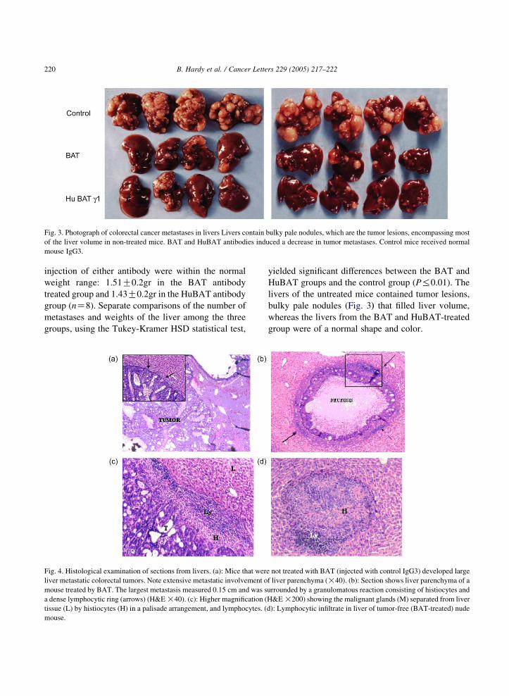
Fig. 3. Photograph of colorectal cancer metastases in livers Livers contain bulky pale nodules, which are the tumor lesions, encompassing most
of the liver volume in non-treated mice. BAT and HuBAT antibodies induced a decrease in tumor metastases. Control mice received normal
mouse IgG3.
B. Hardy et al. / Cancer Letters 229 (2005) 217–222220
injection of either antibody were within the normal
weight range: 1.51G0.2gr in the BAT antibody
treated group and 1.43G0.2gr in the HuBAT antibody
group (nZ8). Separate comparisons of the number of
metastases and weights of the liver among the three
groups, using the Tukey-Kramer HSD statistical test,
Fig. 4. Histological examination of sections from livers. (a): Mice that wer
liver metastatic colorectal tumors. Note extensive metastatic involvement o
mouse treated by BAT. The largest metastasis measured 0.15 cm and was s
a dense lymphocytic ring (arrows) (H&E !40). (c): Higher magnification (
tissue (L) by histiocytes (H) in a palisade arrangement, and lymphocytes. (
mouse.
yielded significant differences between the BAT and
HuBAT groups and the control group (P%0.01). The
livers of the untreated mice contained tumor lesions,
bulky pale nodules (Fig. 3) that filled liver volume,
whereas the livers from the BAT and HuBAT-treated
group were of a normal shape and color.
e not treated with BAT (injected with control IgG3) developed large
f liver parenchyma (!40). (b): Section shows liver parenchyma of a
urrounded by a granulomatous reaction consisting of histiocytes and
H&E !200) showing the malignant glands (M) separated from liver
d): Lymphocytic infiltrate in liver of tumor-free (BAT-treated) nude
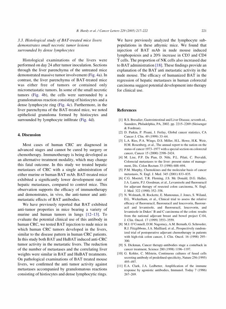
B. Hardy et al. / Cancer Letters 229 (2005) 217–222 221
3.3. Histological study of BAT-treated mice livers
demonstrates small necrotic tumor lesions
surrounded by dense lymphocytes
Histological examinations of the livers were
performed on day 24 after tumor inoculation. Sections
through the liver parenchyma of the untreated mice
demonstrated massive tumor involvement (Fig. 4a). In
contrast, the liver parenchyma of BAT-treated mice
was either free of tumors or contained only
micrometastatic tumors. In some of the small necrotic
tumors (Fig. 4b), the cells were surrounded by a
granulomatous reaction consisting of histiocytes and a
dense lymphocyte ring (Fig. 4c). Furthermore, in the
liver parenchyma of the BAT-treated mice, we noted
epithelioid granuloma formed by histiocytes and
surrounded by lymphocyte infiltrate (Fig. 4d).
4. Discussion
Most cases of human CRC are diagnosed in
advanced stages and cannot be cured by surgery or
chemotherapy. Immunotherapy is being developed as
an alternative treatment modality, which may change
this fatal outcome. In this study we treated hepatic
metastases of CRC with a single administration of
either murine or human BAT mAb. BAT-treated mice
exhibited a significantly lower development rate of
hepatic metastases, compared to control mice. This
observation supports the efficacy of immunotherapy
and demonstrates, in vivo, the anti-tumor and anti-
metastatic effects of BAT antibodies.
We have previously reported that BAT exhibited
anti-tumor properties in mice bearing a variety of
murine and human tumors in lungs [12–13]. To
evaluate the potential clinical use of this antibody in
human CRC, we tested BAT injection to nude mice in
which human CRC tumors developed in the livers,
similar to the disease pattern in human CRC patients.
In this study both BAT and HuBAT induced anti-CRC
tumor activity in the metastatic livers. The reduction
of the number of metastases and the correlating liver
weights were similar in BAT and HuBAT treatments.
On pathological examinations of BAT treated mouse
livers, we confirmed the anti tumor activity against
metastases accompanied by granulomatous reactions
consisting of histiocytes and dense lymphocytic rings.
We have previously analyzed the lymphocyte sub-
populations in these athymic mice. We found that
injection of BAT mAb in nude mouse induced
lymphopoiesis and a 20% increase in CD3 and CD4
T cells. The proportion of NK cells also increased due
to BAT administration [18]. These findings provide an
explanation of the BAT anti metastatic activity in the
nude mouse. The efficacy of humanized BAT in the
regression of hepatic metastases in human colorectal
carcinoma suggest potential development into therapy
for clinical use.
References
[1] R.S. Bresalier, Gastrointestinal and Liver Disease, seventh ed.,
Saunders, Philadelphia, PA, 2002. pp. 2215–2269 (Sleisenger
& Fordtran).
[2] D. Parkin, P. Pisani, J. Ferlay, Global cancer statistics, CA
Cancer J. Clin. 49 (1999) 33–64.
[3] L.A. Ries, P.A. Wingo, D.S. Miller, H.L. Howe, H.K. Weir,
H.M. Rosenberg, et al., The annual report to the nation on the
status of cancer 1973–1977 with a special section on colorectal
cancer, Cancer 15 (2000) 2398–2424.
[4] M. Lise, P.P. Da Pian, D. Nihi, P.L. Pilati, C. Prevaldi,
Colorectal metastases to the liver: present status of manage-
ment, Dis. Colon Rectum 33 (1990) 688–694.
[5] P.M. Murphy, Chemokines and the molecular basis of cancer
metastasis, N. Engl. J. Med. 345 (2001) 833–835.
[6] C.G. Moertel, T.R. Fleming, J.S. Mc Donald, D.G. Haller,
J.A. Laurie, P.J. Goodman, et al., Levamisole and fluorouracil
for adjuvant therapy of resected colon carcinoma, N. Engl.
J. Med. 322 (1990) 352–358.
[7] N. Wolmark, H. Rockette, E. Mamounas, J. Jones, S. Wiland,
D.L. Wickerham, et al., Clinical trial to assess the relative
efficacy of fluorouracil, fluorouracil and leucovorin, fluorour-
acil and levamisole, and fluorouracil, leucovorin, and
levamisole in Dukes’ B and C carcinoma of the colon: results
from the national adjuvant breast and bowel project C-04,
J. Clin. Oncol. 17 (1999) 3553–3559.
[8] M.J. O’Connell, D.M. Nagorney, A.M. Bernath, G. Schroeder,
R.J. Fitzgibbons, J.A. Mailliard, et al., Prospectively random-
ized trial of postoperative adjuvant chemotherapy in patients
with high-risk colon cancer, J. Clin. Oncol. 16 (1998) 295–
300.
[9] S. Dickman, Cancer therapy-antibodies stage a comeback in
cancer treatment, Science 280 (1998) 1196–1197.
[10] G. Kohler, C. Milstein, Continuous cultures of fused cells
secreting antibody of predefined specificity, Nature 256 (1995)
495–497.
[11] E.A. Clark, J.A. Ledbetter, Amplification of the immune
response by agonistic antibodies, Immunol. Today 7 (1986)
267–269.
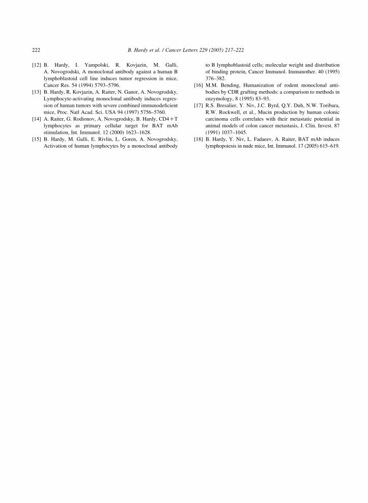
B. Hardy et al. / Cancer Letters 229 (2005) 217–222222
[12] B. Hardy, I. Yampolski, R. Kovjazin, M. Galli,
A. Novogrodski, A monoclonal antibody against a human B
lymphoblastoid cell line induces tumor regression in mice,
Cancer Res. 54 (1994) 5793–5796.
[13] B. Hardy, R. Kovjazin, A. Raiter, N. Ganor, A. Novogrodsky,
Lymphocyte-activating monoclonal antibody induces regres-
sion of human tumors with severe combined immunodeficient
mice, Proc. Natl Acad. Sci. USA 94 (1997) 5756–5760.
[14] A. Raiter, G. Rodionov, A. Novogrodsky, B. Hardy, CD4CT
lymphocytes as primary cellular target for BAT mAb
stimulation, Int. Immunol. 12 (2000) 1623–1628.
[15] B. Hardy, M. Galli, E. Rivlin, L. Goren, A. Novogrodsky,
Activation of human lymphocytes by a monoclonal antibody
to B lymphoblastoid cells; molecular weight and distribution
of binding protein, Cancer Immunol. Immunother. 40 (1995)
376–382.
[16] M.M. Bending, Humanization of rodent monoclonal anti-
bodies by CDR grafting methods: a comparison to methods in
enzymology, 8 (1995) 83–93.
[17] R.S. Bresalier, Y. Niv, J.C. Byrd, Q.Y. Duh, N.W. Toribara,
R.W. Rockwell, et al., Mucin production by human colonic
carcinoma cells correlates with their metastatic potential in
animal models of colon cancer metastasis, J. Clin. Invest. 87
(1991) 1037–1045.
[18] B. Hardy, Y. Niv, L. Fadaeev, A. Raiter, BAT mAb induces
lymphopoiesis in nude mice, Int. Immunol. 17 (2005) 615–619.
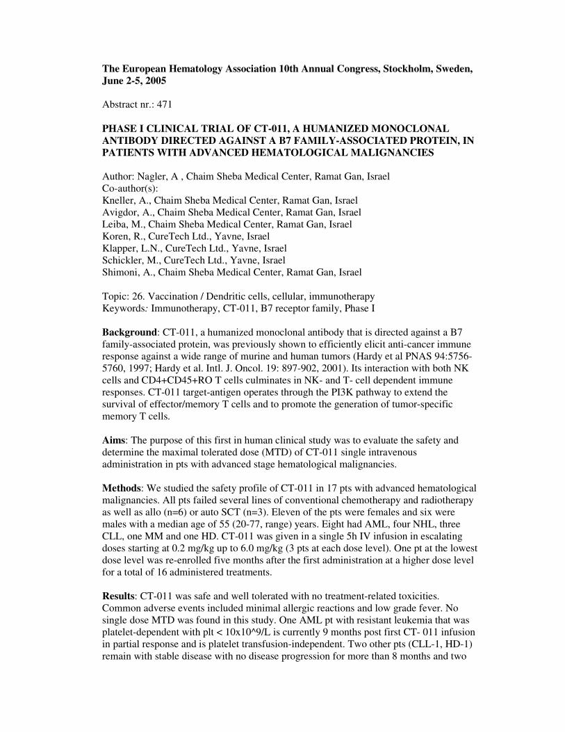
The European Hematology Association 10th Annual Congress, Stockholm, Sweden,
June 2-5, 2005
Abstract nr.: 471
PHASE I CLINICAL TRIAL OF CT-011, A HUMANIZED MONOCLONAL
ANTIBODY DIRECTED AGAINST A B7 FAMILY-ASSOCIATED PROTEIN, IN
PATIENTS WITH ADVANCED HEMATOLOGICAL MALIGNANCIES
Author: Nagler, A , Chaim Sheba Medical Center, Ramat Gan, Israel
Co-author(s):
Kneller, A., Chaim Sheba Medical Center, Ramat Gan, Israel
Avigdor, A., Chaim Sheba Medical Center, Ramat Gan, Israel
Leiba, M., Chaim Sheba Medical Center, Ramat Gan, Israel
Koren, R., CureTech Ltd., Yavne, Israel
Klapper, L.N., CureTech Ltd., Yavne, Israel
Schickler, M., CureTech Ltd., Yavne, Israel
Shimoni, A., Chaim Sheba Medical Center, Ramat Gan, Israel
Topic: 26. Vaccination / Dendritic cells, cellular, immunotherapy
Keywords: Immunotherapy, CT-011, B7 receptor family, Phase I
Background: CT-011, a humanized monoclonal antibody that is directed against a B7
family-associated protein, was previously shown to efficiently elicit anti-cancer immune
response against a wide range of murine and human tumors (Hardy et al PNAS 94:5756-
5760, 1997; Hardy et al. Intl. J. Oncol. 19: 897-902, 2001). Its interaction with both NK
cells and CD4+CD45+RO T cells culminates in NK- and T- cell dependent immune
responses. CT-011 target-antigen operates through the PI3K pathway to extend the
survival of effector/memory T cells and to promote the generation of tumor-specific
memory T cells.
Aims: The purpose of this first in human clinical study was to evaluate the safety and
determine the maximal tolerated dose (MTD) of CT-011 single intravenous
administration in pts with advanced stage hematological malignancies.
Methods: We studied the safety profile of CT-011 in 17 pts with advanced hematological
malignancies. All pts failed several lines of conventional chemotherapy and radiotherapy
as well as allo (n=6) or auto SCT (n=3). Eleven of the pts were females and six were
males with a median age of 55 (20-77, range) years. Eight had AML, four NHL, three
CLL, one MM and one HD. CT-011 was given in a single 5h IV infusion in escalating
doses starting at 0.2 mg/kg up to 6.0 mg/kg (3 pts at each dose level). One pt at the lowest
dose level was re-enrolled five months after the first administration at a higher dose level
for a total of 16 administered treatments.
Results: CT-011 was safe and well tolerated with no treatment-related toxicities.
Common adverse events included minimal allergic reactions and low grade fever. No
single dose MTD was found in this study. One AML pt with resistant leukemia that was
platelet-dependent with plt < 10x10^9/L is currently 9 months post first CT- 011 infusion
in partial response and is platelet transfusion-independent. Two other pts (CLL-1, HD-1)
remain with stable disease with no disease progression for more than 8 months and two
additional pts (NHL) exhibit minimal response to treatment . Four other pts are alive with
active disease with a median follow up of 3 (1-6) months, while seven pts died from their
advanced resistant disease. Accrual to this study as well as pts follow up continues.
Conclusions: A single administration of CT-011 is safe and well tolerated in pts with
advanced hematological malignancies. The observed anti-tumor activity may be related to
CT-011 interaction with the B7 receptor family-associated protein resulting in
enhancement of tumor-specific immune response. Future studies will evaluate the
combination of donor lymphocyte infusion and CT-011 for pts with hematological
malignancies having minimal residual disease after stem cell transplantation.


Proc. Natl. Acad. Sci. USAVol. 94, pp. 5756–5760, May 1997Immunology
A lymphocyte-activating monoclonal antibody induces regressionof human tumors in severe combined immunodeficient mice
(human melanomaytumor regressionyimmune stimulationynude and beige miceyT or NK cell depletion)
BRITTA HARDY*, RIVA KOVJAZIN, ANNAT RAITER, NIRIT GANOR, AND ABRAHAM NOVOGRODSKYFelsenstein Medical Research Center, Rabin Medical Center, Belinson Campus, Petah Tikva 49100, Israel, and Sackler School of Medicine, Tel Aviv University,Tel Aviv 69978, Israel
Communicated by Richard A. Lerner, The Scripps Research Institute, La Jolla, CA, March 25, 1997 (received for review August 7, 1996)
ABSTRACT Monoclonal antibodies were raised againstDaudi B-lymphoblastoid cell line membranes. An mAb (BAT)was selected for its ability to stimulate human and murinelymphocyte proliferation. BAT induced cytotoxicity in humanand murine lymphocytes against natural killer cell-sensitiveand -resistant tumor cell lines. A single intravenous admin-istration of BAT to mice that had been inoculated with variousmurine tumors (e.g., B16 melanoma, 3LL carcinoma, andmethylcholanthrene fibrosarcoma) resulted in striking anti-tumor effects as manifested by complete tumor regression andprolonged survival of the treated mice. BAT exhibited adiminished but significant antitumor effect in athymic nudemice, which are deficient in T lymphocytes, and in beige mice,which are deficient in NK cells. Furthermore, selective deple-tion of T or NK cells in mice reduced the response to theantitumor effect of BAT. These data indicate a dual role for Tand NK cells in mediating the antitumor activity of BAT. Wereport here on the antitumor activity of BAT mAb on humantumor xenografts in mice. BAT demonstrated an antitumoreffect in nude mice bearing human colon carcinoma (HT29)xenografts. It failed, however, to inhibit established lungmetastases in severe combined immunodeficient (SCID) micethat had been inoculated (i.v.) with SK28 human melanoma.Engraftment of human lymphocytes into SCID mice bearinghuman melanoma xenografts rendered them responsive to theantitumor effect of BAT. The efficacy of BAT in the regressionof human tumors by activation of human lymphocytes indi-cates its potential clinical use.
Monoclonal antibodies (mAbs) directed against various T celldeterminants were previously reported to induce proliferationand differentiation of T cells (1). The most remarkable amongthem is the mAb against CD3 determinant, which was able toinduce clonal proliferation, elicit mitogenic activity, and alsotrigger the cytolytic process in T lymphocytes (2–4). Addi-tional immunostimulatory mAbs were found to react with CD5(5), CD69 (6), and CD28. The latter, an antigen on the T cellthat interacts with its ligand, B7, present on antigen presentingcells including tumor cells (7–9). In vivo antitumor activity ofanti-CD3 and of anti-CD28 was previously reported (10, 11).Activation of T cells, which elicit a variety of effector functions,results from interaction of antigen with the T cell antigenreceptor and a costimulation directed to additional surfacedeterminants such as the CD28 (12).We previously reported a mAb directed against human
B-lymphoblastoid cell membranes (BAT) that stimulates hu-man lymphocytes, as manifested by enhanced murine andhuman lymphocyte proliferation and cytolytic activity against
tumor cells in vitro (13). BAT binding protein was identified asa 48- to 50-kDa monomeric protein (13). BAT was found toinduce murine tumor regression in C57BL mice that wasmediated by its immune stimulatory properties (14).In the present study, we evaluated the antitumor activity of
BAT in nude mice carrying human tumor xenografts and insevere combined immunodeficient (SCID) mice engraftedwith human lymphocytes and inoculated with human tumorcells (15, 16).
MATERIALS AND METHODS
Monoclonal Antibodies. BAT was generated and purified asdescribed (14). In brief, BALByc mice were immunized withmembranes from Daudi cells. Spleen cells were fused withmyeloma NS-O cells. BAT was selected by its ability to bindDaudi cells and by its ability to induce proliferation of humanperipheral blood mononuclear cells (PBMC). Cells weregrown in RPMI 1640 medium supplemented with fetal calfserum (10%), sodium pyruvate, glutamine, and antibiotics andincubated at 378C in a humidified atmosphere containing 5%CO2. BAT was purified on a protein G Sepharose columnaccording to manufacturer’s instructions (Pharmacia).Cell Preparations for Engraftment to SCID Mice. Human
PBMC were obtained from blood of healthy donors by FicollyHypaque density centrifugation. Cells were washed and sus-pended in PBS. Cells (53 107) were injected i.p. to each SCIDmouse to construct a human immune system in these mice.Splenocytes were obtained from either untreated C57BL
mice or from mice 24 h after injection of 100 mgymouse of antiCD3 (PharMingen) or anti-asialoGM1 (ASGM1) (WakoChemicals, Dallas). Cells (5 3 107) were injected i.p. to eachSCID mouse.Activation of Lymphocytes by Tumor Cells and BAT. Hu-
man PBL (2 3 106yml) were incubated on HT29 human coloncarcinoma cells monolayers for 1 day. Peripheral blood lym-phocyte (PBL) cells were then removed from the tumor cellmonolayer, washed twice with medium, and suspended at theinitial concentration. Splenocytes obtained from tumor-inoculated mice were suspended at 23 106yml, and BAT at 0.1mgyml was added for 3 days in vitro.Proliferation Assay of Lymphocytes upon Incubation with
BAT. PBLs were separated from PBMC by removing theadherant monocytes after 1-h incubation on plastic Petridishes. Aliquots of 2 3 106 PBL (200 ml) in culture mediumcontaining 5% human type AB serum were incubated for 3days in 96-well f lat-bottom plates with and without BAT at 0.1mgyml. [3H]Thymidine (1 mCiywell; 1 Ci5 37 GBq) was addedfor 20 h before harvesting. Cultures were harvested into glass
The publication costs of this article were defrayed in part by page chargepayment. This article must therefore be hereby marked ‘‘advertisement’’ inaccordance with 18 U.S.C. §1734 solely to indicate this fact.
Copyright q 1997 by THE NATIONAL ACADEMY OF SCIENCES OF THE USA0027-8424y97y945756-5$2.00y0PNAS is available online at http:yywww.pnas.org.
Abbreviations: SCID mice, severe combined immunodeficient mice;PBMC, peripheral blood mononuclear cells; PBL, peripheral bloodlymphocyte.*To whom reprint requests should be addressed at: FelsensteinMedical Research Center, Beilinson Campus, Petah Tikva 49100,Israel.
5756

filters and radioactivity was counted using a liquid b-scintil-lation counter.Mouse Tumor Models. Five to six female mice, obtained
from Harlan Laboratories (Jerusalem), 6–8 weeks old wereused in each group for every experiment. SCID mice weremaintained in sterile conditions at a controlled temperature.B16 murine melanoma cells (5 3 104) were injected i.v. into
C57BL wild type, nude mice (C57BL nuynu), beige mice, orSCID mice that had been engrafted with murine splenocytes.BAT was injected i.v. at 10 mgymouse 14 days later. Twenty-four days after tumor inoculation, mice were killed and lungsmetastases were counted and scored by number, size, and lungweight.HT29 human colon carcinoma cells were injected s.c. at
106ymouse at the axillary region of wild-type or nude mice.BAT (10 mgymouse) was injected i.p. at days 7, 14, and 21posttumor inoculation. Beginning at 7 days posttumor inocu-lation, the tumor size was measured daily until day 30 post-tumor inoculation, when the untreated mice died.SK-mel 28 (SK28), a human melanoma-derived cell line
(originally obtained from Sloan–Kettering Institute, NewYork) was injected i.v. into SCID mice at 5 3 105ymouse. Wefound that i.v. injection of these cells resulted in tumor lesionsin the lungs. SK28 melanoma cells were inoculated 1 dayfollowing i.p. administration of anti-ASGM1 (35 mgymouse)(Wako Chemicals). Anti-ASGM1 is a rabbit polyclonal anti-body that binds to murine NK cells and depletes these cellswhen injected i.p. into mice (17). This antibody was previouslyused to enhance engraftment of human PBMC into SCIDmice(17). Human PBMC (5 3 107ymouse) were engrafted i.p. toSCID mice. Treatment with BAT was done by a single i.v.injection (10 mgymouse) 14 days posttumor inoculation or atdifferent times as indicated in the tables. Twenty-four daysposttumor inoculation, the mice were killed, the lungs re-moved, melanoma metastases were counted, and lung weightwas determined.
RESULTS
We evaluated the antitumor activity of BAT in mice bearinghuman tumors. Initially we studied the antitumor effect ofBAT in athymic nude mice bearing murine B16 melanoma. Asdemonstrated in Table 1, BAT was found to be effective in thistumor model although to a lesser extent than in the wild-typemice. C57BL mice inoculated with murine B16 melanoma andtreated with BAT had zero or only 1 6 1 melanoma lesioncompared with the nude mice that had numerous tumormetastases. The antitumor effect of BAT in nude mice that aredeficient in T cells, suggests that non-T cells could mediate itsantitumor activity. Similar experiments using beige mice (Ta-
ble 1) which are deficient in NK cells (18), showed similarresults, namely, a reduced number of tumor lesions in the lungsof BAT-treated beige mice although not to the same extent aswas observed in the wild-type mice. In the following experi-ment, C57BL mice were depleted of T or NK cells by in vivoadministration of the appropriate antibodies. As seen in Table2, depletion of either T cells or NK cells increased the tumorresistance to BAT. However prolonged administration of theantibodies as in experiment 1 (Table 2) indicates that NKdepletion was effective in rendering mice resistant to theantitumor activity of BAT.The antitumor activity of BAT in nude mice implanted s.c.
with human colon carcinoma (HT29), was also demonstrated(Fig. 1). The growth of the tumor in these mice treated withBAT was delayed up to 24 days posttumor inoculation and washalf the size on day 46 compared with the tumor size inuntreated mice.We have investigated whether BAT is capable of inducing
human tumor regression via the stimulation of human lym-phocytes. SCID mice injected with murine or human tumorsfailed to respond to BAT treatment and died within 14 daysposttumor inoculation. We then established that engraftmentof murine splenocytes enabled BAT to induce regression ofmurine B16 melanoma in the SCID mice (Table 3). BAT wasadministered 14 days posttumor inoculation, the time at whichBAT was previously found to be most effective in inducingregression of murine tumors in wild-type mice (14). BATtreatment in the engrafted SCID mice reduced the number oflung metastases from 217 6 65 to an average of only 7 6 3.Engraftment of splenocytes fromC57BLmice, which had been
FIG. 1. BAT treatment of human colon carcinoma (HT29) growthin nude mice. Nude mice were injected (s.c.) with human HT29 cells(106ymouse) and treated with BAT (10 mgymouse). BAT was injectedi.p. at days 7, 14, and 21 posttumor inoculation.
Table 1. Antitumor activity of BAT in nude and beige micebearing B16 melanoma
BAT treatment
No. of metastases
Exp. 1 Exp. 2
2 1 2 1
Effect of BAT in nude miceC57BL .200 0 .200 1 6 1Nude .200 48 6 25 95 6 45 10 6 3
Effect of BAT in beige miceC57Bl 141 6 8 0 49 6 8 0Beige .200 47 6 39 140 6 47 101 6 44
Mice were injected (i.v.) with B16 melanoma (5 3 104) cells and 14days later with BAT (10 mgymouse). Mice were sacrificed 24 daysposttumor inoculation and the number of lungmetastases were scored.More than 200 represents confluent tumor growth in the lungs. Fivemice were used in each group in each experiment, and the results areexpressed as mean 6 SD.
Table 2. The effect of T- or NK cell-depletion in mice bearingB16 melanoma on the antitumor activity of BAT
BAT treatment
Number of metastases
Exp. 1 Exp. 2
2 1 2 1
Nondepleted 113 6 80 0.5 6 1 .200 0T cells depleted .200 56 6 9 .200 37 6 23NK cells depleted .200 .200 .200 160 6 60
C57BL mice were injected i.p. with either anti-CD3 (100 mg) oranti-asialo GM1 (35 mg) 1 day prior to B16 melanoma injection andon days 7, 14, and 21 days posttumor inoculation. Experiment 1: Miceinjected with anti-CD3 or anti-asialoGM1 on day 12 and 22 posttumorinoculation. Experiment 2: Five mice were used in each group.Animals were sacrificed on day 24 posttumor injection and thenumbers of metastases were scored. Results are expressed as themeans 6 SD.
Immunology: Hardy et al. Proc. Natl. Acad. Sci. USA 94 (1997) 5757
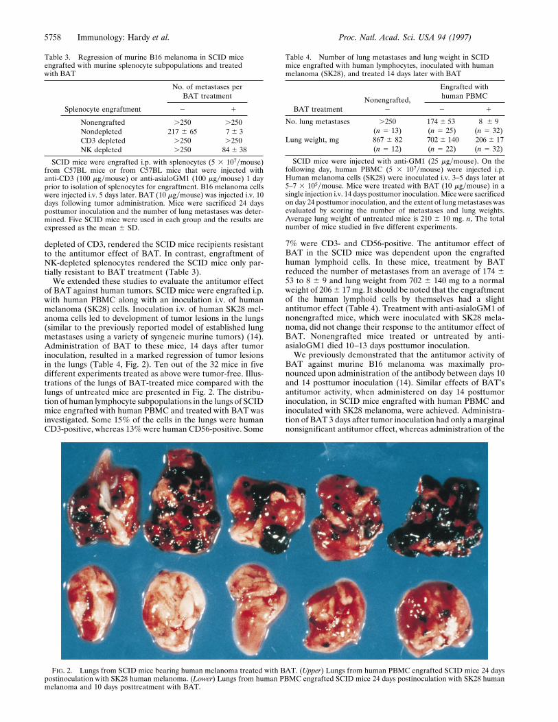
depleted of CD3, rendered the SCID mice recipients resistantto the antitumor effect of BAT. In contrast, engraftment ofNK-depleted splenocytes rendered the SCID mice only par-tially resistant to BAT treatment (Table 3).We extended these studies to evaluate the antitumor effect
of BAT against human tumors. SCID mice were engrafted i.p.with human PBMC along with an inoculation i.v. of humanmelanoma (SK28) cells. Inoculation i.v. of human SK28 mel-anoma cells led to development of tumor lesions in the lungs(similar to the previously reported model of established lungmetastases using a variety of syngeneic murine tumors) (14).Administration of BAT to these mice, 14 days after tumorinoculation, resulted in a marked regression of tumor lesionsin the lungs (Table 4, Fig. 2). Ten out of the 32 mice in fivedifferent experiments treated as above were tumor-free. Illus-trations of the lungs of BAT-treated mice compared with thelungs of untreated mice are presented in Fig. 2. The distribu-tion of human lymphocyte subpopulations in the lungs of SCIDmice engrafted with human PBMC and treated with BAT wasinvestigated. Some 15% of the cells in the lungs were humanCD3-positive, whereas 13% were human CD56-positive. Some
7% were CD3- and CD56-positive. The antitumor effect ofBAT in the SCID mice was dependent upon the engraftedhuman lymphoid cells. In these mice, treatment by BATreduced the number of metastases from an average of 174 653 to 8 6 9 and lung weight from 702 6 140 mg to a normalweight of 2066 17 mg. It should be noted that the engraftmentof the human lymphoid cells by themselves had a slightantitumor effect (Table 4). Treatment with anti-asialoGM1 ofnonengrafted mice, which were inoculated with SK28 mela-noma, did not change their response to the antitumor effect ofBAT. Nonengrafted mice treated or untreated by anti-asialoGM1 died 10–13 days posttumor inoculation.We previously demonstrated that the antitumor activity of
BAT against murine B16 melanoma was maximally pro-nounced upon administration of the antibody between days 10and 14 posttumor inoculation (14). Similar effects of BAT’santitumor activity, when administered on day 14 posttumorinoculation, in SCID mice engrafted with human PBMC andinoculated with SK28 melanoma, were achieved. Administra-tion of BAT 3 days after tumor inoculation had only a marginalnonsignificant antitumor effect, whereas administration of the
FIG. 2. Lungs from SCID mice bearing human melanoma treated with BAT. (Upper) Lungs from human PBMC engrafted SCID mice 24 dayspostinoculation with SK28 human melanoma. (Lower) Lungs from human PBMC engrafted SCID mice 24 days postinoculation with SK28 humanmelanoma and 10 days posttreatment with BAT.
Table 3. Regression of murine B16 melanoma in SCID miceengrafted with murine splenocyte subpopulations and treatedwith BAT
Splenocyte engraftment
No. of metastases perBAT treatment
2 1
Nonengrafted .250 .250Nondepleted 217 6 65 7 6 3CD3 depleted .250 .250NK depleted .250 84 6 38
SCID mice were engrafted i.p. with splenocytes (5 3 107ymouse)from C57BL mice or from C57BL mice that were injected withanti-CD3 (100 mgymouse) or anti-asialoGM1 (100 mgymouse) 1 dayprior to isolation of splenocytes for engraftment. B16 melanoma cellswere injected i.v. 5 days later. BAT (10 mgymouse) was injected i.v. 10days following tumor administration. Mice were sacrificed 24 daysposttumor inoculation and the number of lung metastases was deter-mined. Five SCID mice were used in each group and the results areexpressed as the mean 6 SD.
Table 4. Number of lung metastases and lung weight in SCIDmice engrafted with human lymphocytes, inoculated with humanmelanoma (SK28), and treated 14 days later with BAT
BAT treatmentNonengrafted,
2
Engrafted withhuman PBMC
2 1
No. lung metastases .250 174 6 53 8 6 9(n 5 13) (n 5 25) (n 5 32)
Lung weight, mg 867 6 82 702 6 140 206 6 17(n 5 12) (n 5 22) (n 5 32)
SCID mice were injected with anti-GM1 (25 mgymouse). On thefollowing day, human PBMC (5 3 107ymouse) were injected i.p.Human melanoma cells (SK28) were inoculated i.v. 3–5 days later at5–7 3 105ymouse. Mice were treated with BAT (10 mgymouse) in asingle injection i.v. 14 days posttumor inoculation.Mice were sacrificedon day 24 posttumor inoculation, and the extent of lung metastases wasevaluated by scoring the number of metastases and lung weights.Average lung weight of untreated mice is 210 6 10 mg. n, The totalnumber of mice studied in five different experiments.
5758 Immunology: Hardy et al. Proc. Natl. Acad. Sci. USA 94 (1997)

antibody after 14 days reduced metastases from confluence toan average of 10 6 17. BAT administered 5 days and 10 daysposttumor inoculation was also effective and reduced metas-tases to 89 6 7 and 35 6 12, respectively (Table 5).To study whether BAT elicited its antitumor effect as a result
of a stimulatory signal provided by the tumor cells, we con-ducted experiments in which splenocytes isolated from micebearing tumors exhibited an enhanced response in vitro tostimulation by BAT (Fig. 3). Splenocytes of C57BL miceinjected with B16 melanoma, were co-cultured in the presenceof BAT (1 mgyml) in vitro and the [3H]thymidine uptake wasmeasured. As can be seen, uptake of thymidine in splenocytesfrom mice bearing tumors for 5 days increased (26633 6 872)in the presence of BAT, compared with the splenocytes oftumor-free mice (13156 6 447). To further examine themechanism of BAT activation, we studied the stimulatoryeffect of BAT in vitro on human PBLs that were preincubatedon HT29 human colon carcinoma cell monolayers. Results of4 experiments using 4 different human PBLs are shown in Fig.4. Exposure of lymphocytes to the tumor cells in vitro led to asignificant increase in their proliferative response ranging froma 12- to 22-fold increase in [3H]thymidine uptake. Moreover,in lymphocytes that were pre-exposed to tumor cells and thencultured with BAT, thymidine uptake increased 22- to 44-fold.Lymphocytes that were incubated on allogeneic macrophagemonolayers did not acquire the enhanced sensitivity to stim-ulation by BAT (data not shown).The cell surface markers (CD3 for T cells and CD56 for NK
cells) of the lymphocytes from the different experimentalgroups described in Fig. 4 were analyzed by fluorescence cellanalyzer. The percent of CD3yCD56-positive double-labeledcells increased to 25 6 2 following preincubation on tumormonolayers and incubation with BAT. The percent of CD3yCD56 cells of the lymphocytes that were incubated on tumorcells alone was 17 6 1%, whereas it was 9 6 3% of thelymphocytes treated with BAT alone. In control untreatedgroup 6 6 1% of CD3yCD56-positive cells were detected.
DISCUSSION
The BAT mAb generated against human B-lymphoblastoidcell line was found to bind to and stimulate human T cells. Thestimulation was manifested by induction of cell proliferationand cytolytic activity to NK-resistant and NK-sensitive tumortarget cells (13). This antibody also stimulated murine spleno-cytes in vitro and was found to induce in vivo regression of avariety of murine tumors (14). Tumor regression was relatedto the immune-stimulatory properties of the antibody. Thisconclusion is strongly supported by the observation that theantitumor activity could be transferred with splenocytes frommice treated with the antibody. Furthermore, mice bearing
tumors that had been cured by treatment with BAT wererefractory to a tumor rechallenge (14).To evaluate the potential clinical use of this antibody in
human cancer, experiments were initiated in mice bearinghuman tumors. First, we investigated whether BATwould elicitan antitumor effect in nude mice, which are commonly em-ployed for studies involving human tumor xenografts (19).Nude mice are deficient in T cells, and the question was raisedwhether BAT would induce antitumor activity in these im-mune-compromised animals. As observed in Table 1, BATexhibited antitumor activity in nude mice bearing the B16melanoma. This effect, however, was incomplete as comparedwith the curing effect that was attained in wild-type (C57BL)animals bearing the same tumor. A similar incomplete anti-tumor effect of BATwas also observed in nudemice implantedsubcutaneously with human colon carcinoma (Fig. 1).The lymphocyte cell type that mediates the antitumor effect
in nude mice may involve NK cells that are present in thesemice. This possibility was further supported by the experiment
FIG. 3. Proliferation of splenocytes from B16 melanoma bearingmice in the presence and absence of BAT. Splenocytes obtained fromC57BL mice at various days postinjection with murine B16 melanomacells were cultured in the presence of BAT (1 mgyml) for 3 days (E)and without BAT for 3 days (■). [3H]Thymidine uptake was deter-mined as described.
FIG. 4. Thymidine uptake in human PBLs incubated on humanHT29 colon carcinoma cells in the presence and absence of BAT. Fourexperiments using four different human PBLs are shown. PBLs werecocultured on HT29 human colon cells. The lymphocytes were re-moved after 1 day, washed, and resuspended to 2 3 106yml. Lympho-cytes were cultured in the absence and presence of BAT at 0.1 mgymlfor 3 more days and [3H]thymidine uptake was determined as de-scribed.
Table 5. BAT treatment at different times following humanmelanoma inoculation of SCID mice engrafted withhuman lymphocytes
BAT treatment, day No. metastases Lung weight, mg
None .250 750 6 703 206 6 75 665 6 1505 89 6 7 315 6 810 35 6 12 219 6 114 10 6 17 217 6 6
BAT was administered (10 mgymouse) at different times in relationto tumor inoculation at day 0. Human PBMC were engrafted i.p. at 53 107ycells per mouse, 1 day postinjection of anti-GM1 (25 mgymouse). Tumor cells were injected i.v. 3–5 days later at 7 3 105cellsymouse. Twenty-four-day posttumor inoculation mice were sac-rificed and the number of lung metastases and weights were deter-mined. Average lung weight of untreated mice is 210 6 10 mg.
Immunology: Hardy et al. Proc. Natl. Acad. Sci. USA 94 (1997) 5759
depicted in Table 2, which indicated that continuous depletionof NK cells diminishes the antitumor effect of BAT. Howeverthe possibility that T cells could mediate the antitumor effectof BAT is indicated from experiments (Table 1) demonstratingthat BAT exhibited antitumor response in the beige mouse thatis deficient in NK cells (18). Supporting this view are exper-iments in which SCID mice bearing murine B16 melanomaengrafted with splenocytes depleted of CD3 lymphocytesfailed to respond to BAT (Table 3). In these experimentsengraftment of splenocytes which were NK-depleted renderedthe SCID mice partially responsive to BAT. Recent studieshave shown that CD3yCD56-positive lymphocytes exhibited apotent antitumor activity (20–23). We have found that CD3yCD56-positive cells are generated in vitro upon stimulation ofhuman lymphocytes by BAT and human tumor cells. Thus it ispossible that this cell type plays a role in the antitumor effectinduced by BAT. Taken together, it is possible that either NKor T cells can mediate the antitumor effect of BAT. Further-more cross talk between these sub-populations (24) may alsoplay an important role in mediating the effect of BAT.A question of utmost clinical importance was whether BAT
could induce human tumor regression mediated by activationof human lymphocytes. To address this question, we evaluatedthe effect of BAT in SCID mice engrafted with humanlymphocytes and inoculated with human SK28melanoma cells.BAT alone failed to induce tumor regression in SCID mice.Engraftment of PBL alone into SCID mice had a slightantitumor effect, which most probably is related to the geneticdisparity between the tumor and the engrafted PBLs. Themostpronounced antitumor effect was achieved when BAT wasadministered into the SCIDmice that had been engrafted withhuman PBLs. In contrast to the curative effect that wasattained in the wild strain of mice, the antitumor effect in theSCID mouse engrafted with PBL, although most pronounced,was incomplete in a few cases. This may result from theincomplete reconstitution of the immune system in the SCIDmice (25) and was also demonstrated in the experiment inSCID mice that had been inoculated with B16 melanoma andengrafted with syngeneic splenocyctes (Table 3).Our previous results in normal C57BL mice bearing murine
tumors indicated that maximal antitumor effect was obtainedupon administration of BAT late after tumor inoculation. Asimilar effect was also observed when BAT was administeredto human PBMC-engrafted SCID mice at different times afterinoculation of SK28 human melanoma. It is possible thatlymphocytes sensitization by the tumor cells led to theirenhanced response to BAT. Support for this notion wasobtained from experiments in which the stimulatory responseto BAT was assessed in vitro in splenocytes derived from tumorbearing mice and was also demonstrated in our in vitroexperiments which showed that human lymphocytes sensitizedby human tumor cells increased proliferation in the presenceBAT (Fig. 4).The nature of the two signals elicited by the tumor cells and
by BAT is not known. It is possible that the first signal elicitedby the tumor involves the presentation of a tumor antigen to
the T cell receptor. The second signal provided by BAT mayrepresent an accessory signal. The finding that BAT inducedregression of human tumors mediated by activation of humanlymphocytes points to its therapeutic potential in cancerpatients.
We thank Dr. Yehuda Shoenfeld for helpful discussions and hiscritical review of the manuscript. We thank Mrs. Sara Domintz for thepreparation of the manuscript.
1. Clark, E. A. &. Ledbetter, J. A. (1986) Immunol. Today 7,267–270.
2. Meurer, S. C., Hussey, R. E., Cantrel, D. A., Hodgdon, J. C.,Schlossman S. F., Smith, K. A. & Reinherz, E. L. (1984) Proc.Natl. Acad. Sci. USA 81, 1509–1513.
3. Van Wauwe, J. P., DeMey, J. P. & Goossens, J. G. (1980) J. Im-munol. 124, 2708–2713.
4. Jung, G., Martin D. E. & Muller-Eberhard, J. H. (1987) J. Im-munol. 139, 639–644.
5. Ledbetter, J. A., Martin P. J., Spooner, C. E., Wofsy, D., Tsu,T. T., Beatty, P. G. & Gladstone, P. (1985) J. Immunol. 135,2331–2336.
6. Moretta, A., Poggi, A., Pende D., Tripodi, G., Orengo, A. M.,Pella, N., Augugliaro, R., Bottino, C., Ciccone, E. & Moretta, L.(1991) J. Exp. Med. 174, 1393–1398.
7. Razi-Wolf, Z., Freeman, G. J., Galvin, F., Benacerraf, B., Nadler,L. & Reiser, J. (1992) Proc. Natl. Acad. Sci. USA 89, 4210–4214.
8. Norton, S. D., Zuckerman, L., Urdahl, K. B., Shefner, R., Miller,J. & Jenkins, M. K. (1992) J. Immunol. 149, 1556–1561.
9. Li, Y., McGowan, P., Hellstrom, I., Hellstrom, K. E. & Chen, L.(1994) J. Immunol. 153, 421–428.
10. Ellenhorn, J. D., Hirsch, R., Schreiber, H. & Bluestone, J. A.(1988) Science 242, 569–571.
11. Townsend, S. E. & Allison J. P. (1993) Science 259, 368–380.12. Jenkins M. K. Taylor, P. S., Norton, S. D. & Urdahl, K. B. (1991)
J. Immunol 147, 2461–2466.13. Hardy, B., Galli, M., Rivlin, E., Goren L. & Novogrodsky, A.
(1995) Cancer Immunol. Immunother. 40, 376–382.14. Hardy, B., Yampolski, I., Kovjazin, R., Galli, M. & Novogrodsky,
A. (1994) Cancer Res. 54, 5793–5796.15. Mueller, B. M. &Reisfeld R. A. (1991)Cancer Metastasis Rev. 10,
1930–200.16. Sandhu, J., Shpitz, B., Gallinger, S. & Hozumi, N. (1994)
J. Immunol. 152, 3806–3813.17. Shpitz, B., Chambers, C. A., Singhal, A. B., Hozumi, N., Fer-
nandes, B. J., Roifman, C. M., Weiner, L. M., Roder, J. C. &Gallinger, S. (1994) J. Immunol. Methods 169, 1–15.
18. Stutman, O. & Cuttito, M. J. (1981) Nature (London) 290,254–257.
19. Garofalo, A., Chirivi, R. G. S., Scanziani, E., Mayo, J. G., Vecchi,A. & Giavazzi, R. (1993) Invasion Metastasis 13, 82–91.
20. Lu, P.-H. & Negrin, R. S. (1994) J. Immunol. 153, 1687–1696.21. Schmidt-Wolf, I. G. H., Lefterova, P., Johnston, V., Huhn, D.,
Blum, K. G. & Negrin, R. S. (1994) Br. J. Haematol. 87, 453–458.22. Mehta, B. A., Schmidt-Wolf, G. H., Weissman, I. L. & Negrin,
R. S. (1995) Blood 86, 3493–3499.23. Takeda, K. & Dennert, G. (1994) Transplantation 58, 496–504.24. Kurosawa, S., Harada, M.Matsuzaki, G. Shinomiya, Y. Terao, H.
Kobayashi, N. & Nomoto, K. (1995) Immunology 85, 338–346.25. Hendrickson, E. A. (1993) Am. J. Pathol. 143, 1511–1522.
5760 Immunology: Hardy et al. Proc. Natl. Acad. Sci. USA 94 (1997)
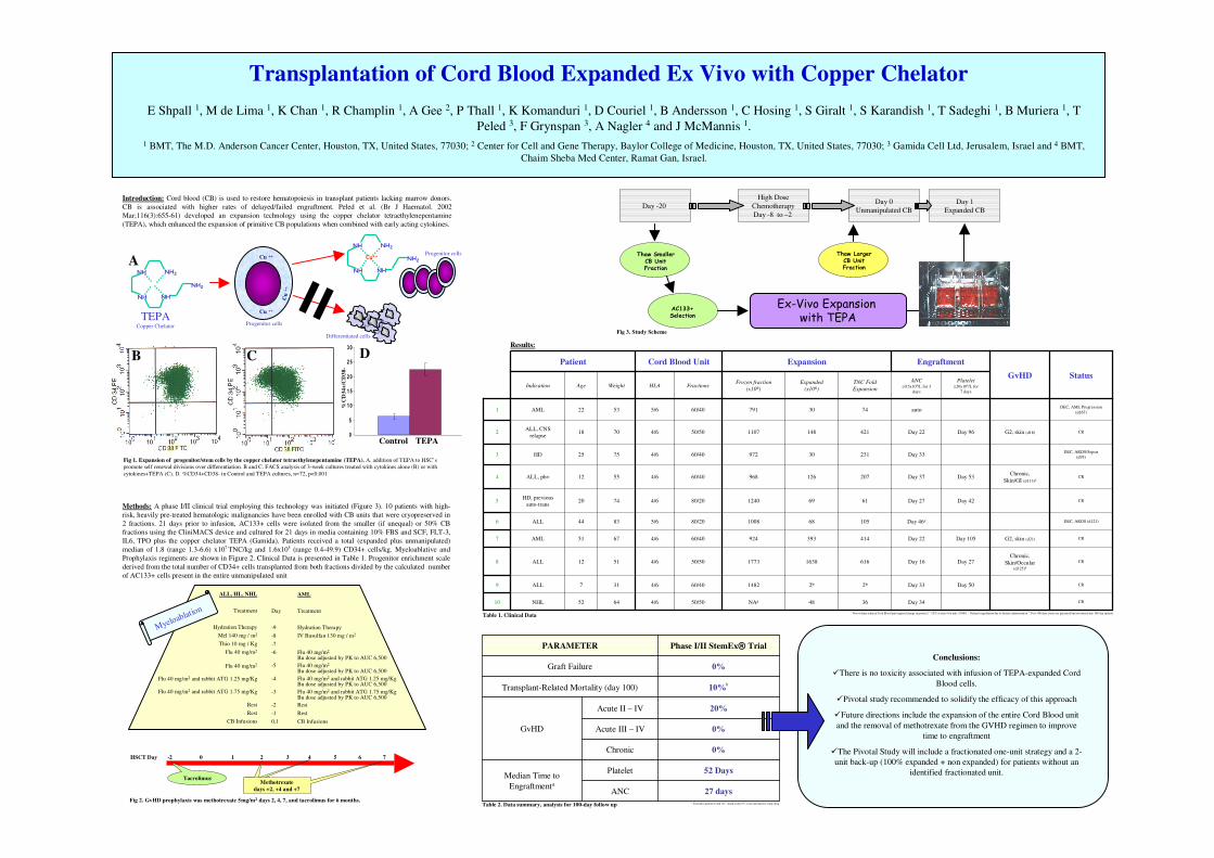
52 DaysPlatelet
0 %Graft Failure
Median Time to
Engraftmenta
27 daysANC
0 %Chronic
0 %Acute III – IV
20 %Acute II – IV
GvHD
10 %b
Transplant-Related Mortality (day 100)
Phase I/II StemEx® TrialPARAMETER
Transplantation of Cord Blood Expanded Ex Vivo with Copper Chelator
E Shpall 1, M de Lima 1, K Chan 1, R Champlin 1, A Gee 2, P Thall 1, K Komanduri 1, D Couriel 1, B Andersson 1, C Hosing 1, S Giralt 1, S Karandish 1, T Sadeghi 1, B Muriera 1, T
Peled 3, F Grynspan 3, A Nagler 4 and J McMannis 1.
1 BMT, The M.D. Anderson Cancer Center, Houston, TX, United States, 77030; 2 Center for Cell and Gene Therapy, Baylor College of Medicine, Houston, TX, United States, 77030; 3 Gamida Cell Ltd, Jerusalem, Israel and 4 BMT,
Chaim Sheba Med Center, Ramat Gan, Israel.
Introduction: Cord blood (CB) is used to restore hematopoiesis in transplant patients lacking marrow donors.
CB is associated with higher rates of delayed/failed engraftment. Peled et al. (Br J Haematol. 2002
Mar;116(3):655-61) developed an expansion technology using the copper chelator tetraethylenepentamine
(TEPA), which enhanced the expansion of primitive CB populations when combined with early acting cytokines.
Day 1
Expanded CB
Day 0
Unmanipulated CB
High Dose
Chemotherapy
Day -8 to –2
Day -20
Thaw Smaller CB Unit Fraction
AC133+ Selection
Ex-Vivo Expansion with TEPA
Thaw Larger CB Unit Fraction
Chronic,
Skin/Occular(d125)d
G2, skin (d21)
Chronic,
Skin/GI (d113)d
G2, skin (d16)
GvHD
CR
CR
CR
CR
DEC, ARDS (d121)
CR
CR
DEC, ARDS/Sepsis
(d93)
CR
DEC, AML Progression
(d167)
Status
36
2b
616
414
105
61
207
231
421
74
TNC Fold
Expansion
48
2b
1638
393
68
69
126
30
148
30
Expanded
(x106)
NAa
1482
1773
924
1008
1240
968
972
1107
791
Frozen fraction
(x106)
ExpansionCord Blood Unit EngraftmentPatient
Day 3450/504/66452NHL10
Day 50Day 3360/404/6317ALL9
Day 27Day 1650/504/65112ALL8
Day 105Day 2260/404/66751AML7
Day 46c80/205/68344ALL6
Day 42Day 2780/204/67420HD, previous
auto-trans5
Day 53Day 3760/404/65512ALL, ph+4
Day 3360/404/67525HD3
Day 96Day 2250/504/67018ALL, CNS
relapse2
auto60/405/65322AML1
Platelet ≥20x109/L for
7 days
ANC ≥0.5x10⁹/L for 3
days
FractionsHLAWeightAgeIndication
Fig 3. Study Scheme
a. Non-volume reduced Cord Blood unit required change in protocol.
b. CFU at time=0 of unit: 3/1000.
c. Delayed engraftment due to bactrim administration.
d. Post-100 days events are presented but not entered into 100-day analysis
Table 1. Clinical Data
Table 2. Data summary, analysis for 100-day follow up
Conclusions:
�There is no toxicity associated with infusion of TEPA-expanded Cord
Blood cells.
�Pivotal study recommended to solidify the efficacy of this approach
�Future directions include the expansion of the entire Cord Blood unit
and the removal of methotrexate from the GVHD regimen to improve
time to engraftment
�The Pivotal Study will include a fractionated one-unit strategy and a 2-
unit back-up (100% expanded + non expanded) for patients without an
identified fractionated unit.
HSCT Day -2 0 1 2 3 4 5 6 7
TacrolimusMethotrexate
days +2, +4 and +7
ALL, HL, NHL
Treatment
Hydration Therapy
Mel 140 mg / m2
Thio 10 mg / Kg
Flu 40 mg/m2
Flu 40 mg/m2
Flu 40 mg/m2 and rabbit ATG 1.25 mg/Kg
Flu 40 mg/m2 and rabbit ATG 1.75 mg/Kg
Rest
Rest
CB Infusions
AML
Day Treatment
-9 Hydration Therapy
-8 IV Busulfan 130 mg / m2
-7
-6 Flu 40 mg/m2
Bu dose adjusted by PK to AUC 6,500
-5 Flu 40 mg/m2
Bu dose adjusted by PK to AUC 6,500
-4 Flu 40 mg/m2 and rabbit ATG 1.25 mg/KgBu dose adjusted by PK to AUC 6,500
-3 Flu 40 mg/m2 and rabbit ATG 1.75 mg/KgBu dose adjusted by PK to AUC 6,500
-2 Rest
-1 Rest
0,1 CB Infusions
Myeloablation
Fig 2. GvHD prophylaxis was methotrexate 5mg/m2 days 2, 4, 7, and tacrolimus for 6 months.a. Excludes patients 6 and 10.
b. death at day 93, event unrelated to study drug.
0
5
10
15
20
25
30
NH
NH NH
NH2
NH2
Cu ++
Cu ++
Cu
++
NH
NH NH
NH2
NH2Cu++A
B C D
Control TEPA
Fig 1. Expansion of progenitor/stem cells by the copper chelator tetraethylenepentamine (TEPA). A. addition of TEPA to HSC’s
promote self renewal divisions over differentiation. B and C. FACS analysis of 3-week cultures treated with cytokines alone (B) or with
cytokines+TEPA (C). D. %CD34+CD38- in Control and TEPA cultures, n=72, p<0.001
TEPACopper Chelator
%C
D3
4+
/CD
38
-
Progenitor cells
Progenitor cells
Differentiated cells
Methods: A phase I/II clinical trial employing this technology was initiated (Figure 3). 10 patients with high-
risk, heavily pre-treated hematologic malignancies have been enrolled with CB units that were cryopreserved in
2 fractions. 21 days prior to infusion, AC133+ cells were isolated from the smaller (if unequal) or 50% CB
fractions using the CliniMACS device and cultured for 21 days in media containing 10% FBS and SCF, FLT-3,
IL6, TPO plus the copper chelator TEPA (Gamida). Patients received a total (expanded plus unmanipulated)
median of 1.8 (range 1.3-6.6) x107 TNC/kg and 1.6x10⁵ (range 0.4-49.9) CD34+ cells/kg. Myeloablative and
Prophylaxis regiments are shown in Figure 2. Clinical Data is presented in Table 1. Progenitor enrichment scale
derived from the total number of CD34+ cells transplanted from both fractions divided by the calculated number
of AC133+ cells present in the entire unmanipulated unit
Results:

0
d
Experimental Hematology 33 (2005) 1092–1100
Chelatable cellular copper modulates differentiationand self-renewal of cord blood–derived hematopoietic progenitor cells
Toni Peleda, Elina Glukhmana, Nira Hassona, Sophie Adia, Harel Assora, Dima Yudina,Chana Landora, Julie Mandela, Efrat Landaua, Eugenia Prusc, Arnon Naglerb, and Eitan Fibachc
aGamida Cell Ltd., Jerusalem, Israel;bChaim Sheba Medical Center, Tel-Hashomer, Israel;
cHadassah–Hebrew University Medical Center, Jerusalem, Israel
(Received 3 February 2005; revised 2 June 2005; accepted 2 June 2005)
Objectives. We have demonstrated epigenetic modulation of CD34+ cell differentiation by thehigh-affinity copper (Cu) chelator tetraethylenepentamine (TEPA). TEPA slowed down therate of CD34+ cell differentiation and increased their engraftability in SCID mice. TEPAbiological activity was attributed to its effect on cellular Cu levels as (a) treatment with TEPAresulted in reduction of cellular Cu, and (b) excess of Cu reversed TEPA’s activity andaccelerated differentiation. In the present study we further evaluated the role of cellular Cu inTEPA’s biological activity.
Methods. The effects of Cu-chloride, TEPA, TEPA/Cu mixtures at various ratios, anda synthesized, stable, TEPA-Cu complex on short- and long-term cord blood–derived CD34+
cell cultures as well as on the overall and chelatable cellular Cu were investigated.
Results. Addition of TEPA, TEPA/Cu mixtures at up to equimolar concentrations, and theTEPA-Cu complex to CD34+ cell cultures resulted in inhibition of differentiation andenhancement of long-term self-renewal. Measurement of the overall cellular Cu by atomicabsorption spectrophotometry showed 20 to 40% decrease by TEPA while the TEPA-Cumixture and the TEPA-Cu complex increased cellular Cu by 10- to 20-fold, as did CuCl2.However, measurement of the cellular pool of labile Cu showed similar reduction (50% fromthe control) by all the TEPA forms, while CuCl2 increased it. Thus, inhibition ofdifferentiation and enhancement of self-renewal of CD34+ cells was correlated with reductionin the cellular chelatable Cu content.
Conclusion. The results suggest that decreasing of the chelatable Cu pool, rather than overallCu, is the mechanism that stands behind TEPA’s biological activity. � 2005 InternationalSociety for Experimental Hematology. Published by Elsevier Inc.
IntroductionMetal ions such as iron (Fe), calcium (Ca), magnesium(Mg), and zinc (Zn) are known to play important roles inbasic cell functions such as cell survival, proliferation, anddifferentiation. Relatively little attention, however, has beendrawn to the role of copper (Cu) in key cellular functions,despite well-documented and significant clinical manifes-tations of Cu deficiency [1–3]. The symptoms of suchdeficiency involve several organ systems, yet of particularrelevance to this study is the fact that Cu deficiency is oftenassociated with hematopoietic cell differentiation arrest,
Offprint requests to: Dr. Toni Peled, Gamida-Cell, Ltd., Research and
Development, 5 Nahum Hfzadi St., Ofer Building, Jerusalem 95484,
Israel; E-mail: [email protected]
301-472X/05 $–see front matter. Copyright � 2005 International Society for
oi: 10.1016/j.exphem.2005.06.015
which results in anemia, neutropenia, and thrombocytope-nia [1–3]. These pathological manifestations are unrespon-sive to iron therapy but are rapidly reversed following Cusupplementation [1–5]. Morphological and functionalevaluation of the bone marrow (BM) of neutropenic, Cu-deficient patients demonstrates the striking absence ofmature cells (‘‘maturation arrest’’) along with the presenceof intact progenitor cells. This finding suggests that the short-age of functional circulating blood cells in these patientsis due to a block in development of the hematopoieticstem/progenitor cells (HSPCs) in a Cu-deficient micro-environment [1].
Further insight into the role of Cu in hematopoiesiscomes from studies with established cell lines. Bae andPercival [6] have demonstrated that retinoic acid–induced
Experimental Hematology. Published by Elsevier Inc.
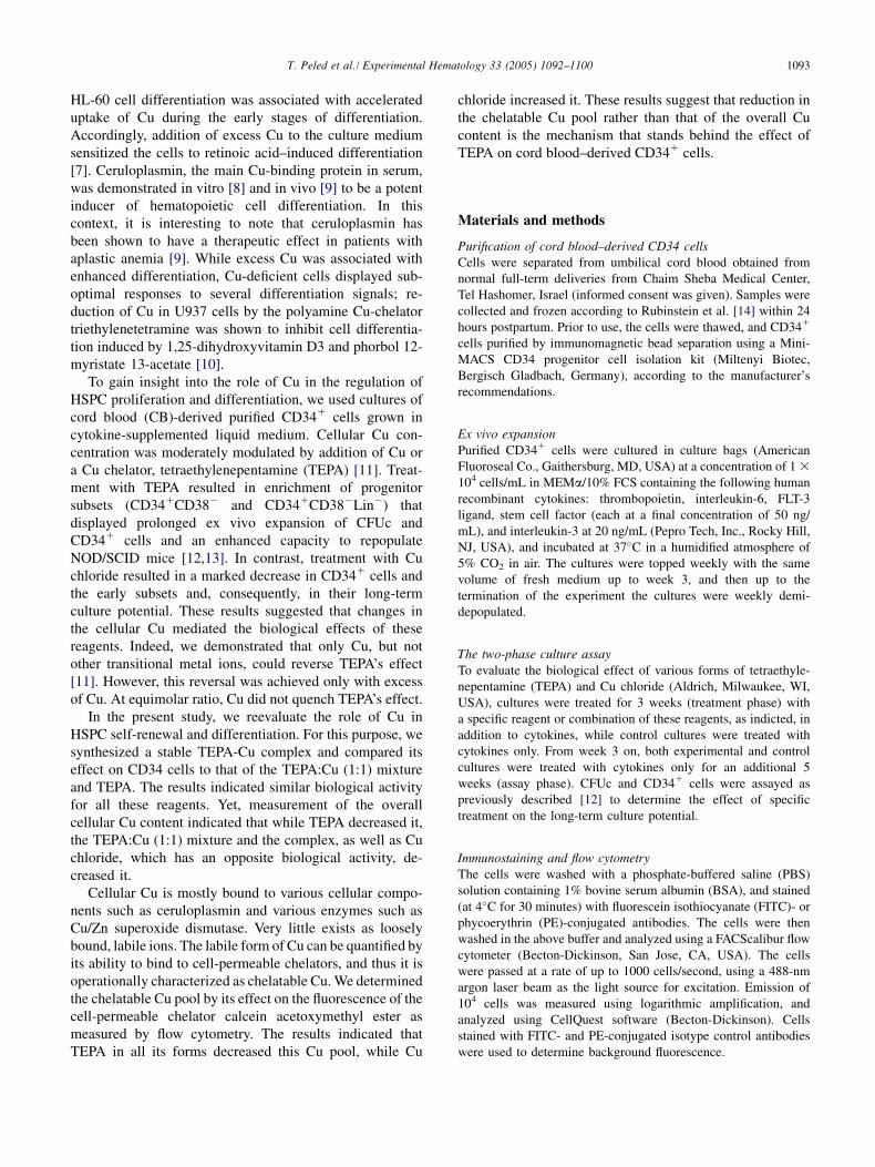
1093T. Peled et al./ Experimental Hematology 33 (2005) 1092–1100
HL-60 cell differentiation was associated with accelerateduptake of Cu during the early stages of differentiation.Accordingly, addition of excess Cu to the culture mediumsensitized the cells to retinoic acid–induced differentiation[7]. Ceruloplasmin, the main Cu-binding protein in serum,was demonstrated in vitro [8] and in vivo [9] to be a potentinducer of hematopoietic cell differentiation. In thiscontext, it is interesting to note that ceruloplasmin hasbeen shown to have a therapeutic effect in patients withaplastic anemia [9]. While excess Cu was associated withenhanced differentiation, Cu-deficient cells displayed sub-optimal responses to several differentiation signals; re-duction of Cu in U937 cells by the polyamine Cu-chelatortriethylenetetramine was shown to inhibit cell differentia-tion induced by 1,25-dihydroxyvitamin D3 and phorbol 12-myristate 13-acetate [10].
To gain insight into the role of Cu in the regulation ofHSPC proliferation and differentiation, we used cultures ofcord blood (CB)-derived purified CD341 cells grown incytokine-supplemented liquid medium. Cellular Cu con-centration was moderately modulated by addition of Cu ora Cu chelator, tetraethylenepentamine (TEPA) [11]. Treat-ment with TEPA resulted in enrichment of progenitorsubsets (CD341CD382 and CD341CD382Lin2) thatdisplayed prolonged ex vivo expansion of CFUc andCD341 cells and an enhanced capacity to repopulateNOD/SCID mice [12,13]. In contrast, treatment with Cuchloride resulted in a marked decrease in CD341 cells andthe early subsets and, consequently, in their long-termculture potential. These results suggested that changes inthe cellular Cu mediated the biological effects of thesereagents. Indeed, we demonstrated that only Cu, but notother transitional metal ions, could reverse TEPA’s effect[11]. However, this reversal was achieved only with excessof Cu. At equimolar ratio, Cu did not quench TEPA’s effect.
In the present study, we reevaluate the role of Cu inHSPC self-renewal and differentiation. For this purpose, wesynthesized a stable TEPA-Cu complex and compared itseffect on CD34 cells to that of the TEPA:Cu (1:1) mixtureand TEPA. The results indicated similar biological activityfor all these reagents. Yet, measurement of the overallcellular Cu content indicated that while TEPA decreased it,the TEPA:Cu (1:1) mixture and the complex, as well as Cuchloride, which has an opposite biological activity, de-creased it.
Cellular Cu is mostly bound to various cellular compo-nents such as ceruloplasmin and various enzymes such asCu/Zn superoxide dismutase. Very little exists as looselybound, labile ions. The labile form of Cu can be quantified byits ability to bind to cell-permeable chelators, and thus it isoperationally characterized as chelatable Cu. We determinedthe chelatable Cu pool by its effect on the fluorescence of thecell-permeable chelator calcein acetoxymethyl ester asmeasured by flow cytometry. The results indicated thatTEPA in all its forms decreased this Cu pool, while Cu
chloride increased it. These results suggest that reduction inthe chelatable Cu pool rather than that of the overall Cucontent is the mechanism that stands behind the effect ofTEPA on cord blood–derived CD341 cells.
Materials and methods
Purification of cord blood–derived CD34 cellsCells were separated from umbilical cord blood obtained fromnormal full-term deliveries from Chaim Sheba Medical Center,Tel Hashomer, Israel (informed consent was given). Samples werecollected and frozen according to Rubinstein et al. [14] within 24hours postpartum. Prior to use, the cells were thawed, and CD341
cells purified by immunomagnetic bead separation using a Mini-MACS CD34 progenitor cell isolation kit (Miltenyi Biotec,Bergisch Gladbach, Germany), according to the manufacturer’srecommendations.
Ex vivo expansionPurified CD341 cells were cultured in culture bags (AmericanFluoroseal Co., Gaithersburg, MD, USA) at a concentration of 1 3
104 cells/mL in MEMa/10% FCS containing the following humanrecombinant cytokines: thrombopoietin, interleukin-6, FLT-3ligand, stem cell factor (each at a final concentration of 50 ng/mL), and interleukin-3 at 20 ng/mL (Pepro Tech, Inc., Rocky Hill,NJ, USA), and incubated at 37�C in a humidified atmosphere of5% CO2 in air. The cultures were topped weekly with the samevolume of fresh medium up to week 3, and then up to thetermination of the experiment the cultures were weekly demi-depopulated.
The two-phase culture assayTo evaluate the biological effect of various forms of tetraethyle-nepentamine (TEPA) and Cu chloride (Aldrich, Milwaukee, WI,USA), cultures were treated for 3 weeks (treatment phase) witha specific reagent or combination of these reagents, as indicted, inaddition to cytokines, while control cultures were treated withcytokines only. From week 3 on, both experimental and controlcultures were treated with cytokines only for an additional 5weeks (assay phase). CFUc and CD341 cells were assayed aspreviously described [12] to determine the effect of specifictreatment on the long-term culture potential.
Immunostaining and flow cytometryThe cells were washed with a phosphate-buffered saline (PBS)solution containing 1% bovine serum albumin (BSA), and stained(at 4�C for 30 minutes) with fluorescein isothiocyanate (FITC)- orphycoerythrin (PE)-conjugated antibodies. The cells were thenwashed in the above buffer and analyzed using a FACScalibur flowcytometer (Becton-Dickinson, San Jose, CA, USA). The cellswere passed at a rate of up to 1000 cells/second, using a 488-nmargon laser beam as the light source for excitation. Emission of104 cells was measured using logarithmic amplification, andanalyzed using CellQuest software (Becton-Dickinson). Cellsstained with FITC- and PE-conjugated isotype control antibodieswere used to determine background fluorescence.
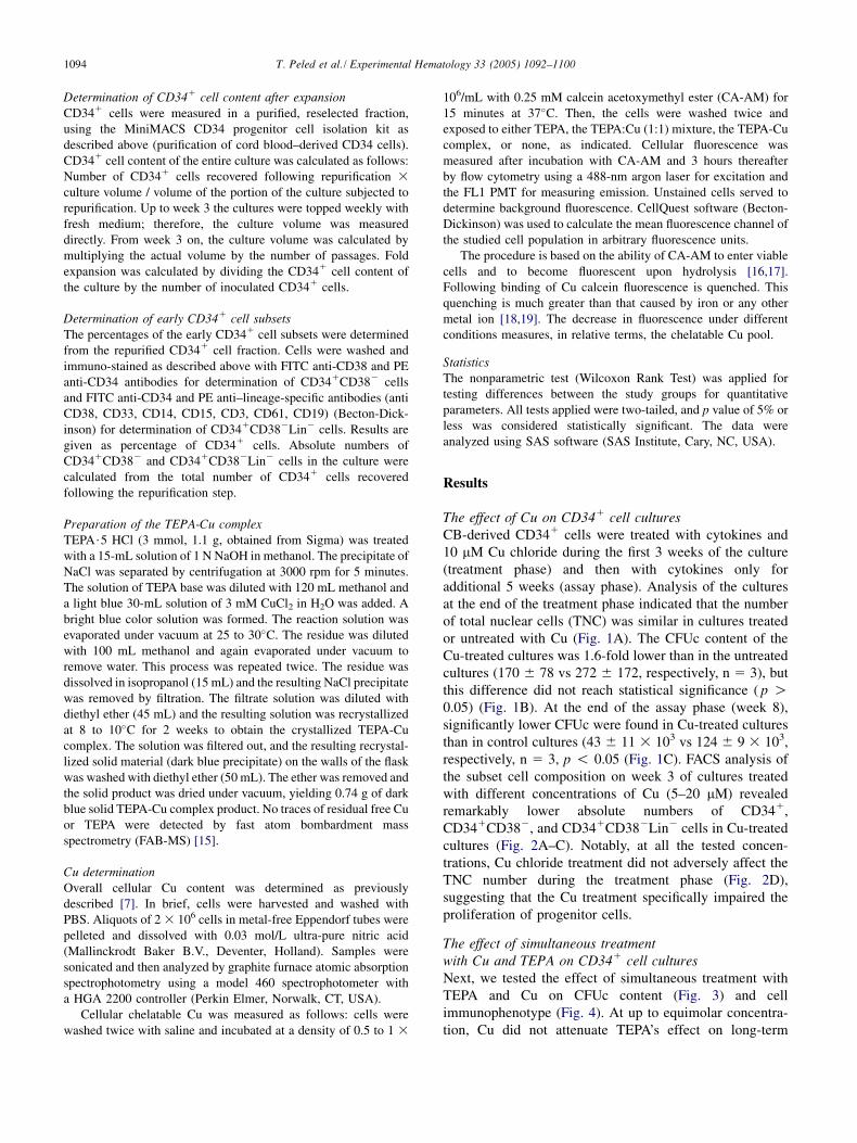
1094 T. Peled et al./ Experimental Hematology 33 (2005) 1092–1100
Determination of CD341 cell content after expansionCD341 cells were measured in a purified, reselected fraction,using the MiniMACS CD34 progenitor cell isolation kit asdescribed above (purification of cord blood–derived CD34 cells).CD341 cell content of the entire culture was calculated as follows:Number of CD341 cells recovered following repurification 3
culture volume / volume of the portion of the culture subjected torepurification. Up to week 3 the cultures were topped weekly withfresh medium; therefore, the culture volume was measureddirectly. From week 3 on, the culture volume was calculated bymultiplying the actual volume by the number of passages. Foldexpansion was calculated by dividing the CD341 cell content ofthe culture by the number of inoculated CD341 cells.
Determination of early CD341 cell subsetsThe percentages of the early CD341 cell subsets were determinedfrom the repurified CD341 cell fraction. Cells were washed andimmuno-stained as described above with FITC anti-CD38 and PEanti-CD34 antibodies for determination of CD341CD382 cellsand FITC anti-CD34 and PE anti–lineage-specific antibodies (antiCD38, CD33, CD14, CD15, CD3, CD61, CD19) (Becton-Dick-inson) for determination of CD341CD382Lin2 cells. Results aregiven as percentage of CD341 cells. Absolute numbers ofCD341CD382 and CD341CD382Lin2 cells in the culture werecalculated from the total number of CD341 cells recoveredfollowing the repurification step.
Preparation of the TEPA-Cu complexTEPA$5 HCl (3 mmol, 1.1 g, obtained from Sigma) was treatedwith a 15-mL solution of 1 N NaOH in methanol. The precipitate ofNaCl was separated by centrifugation at 3000 rpm for 5 minutes.The solution of TEPA base was diluted with 120 mL methanol anda light blue 30-mL solution of 3 mM CuCl2 in H2O was added. Abright blue color solution was formed. The reaction solution wasevaporated under vacuum at 25 to 30�C. The residue was dilutedwith 100 mL methanol and again evaporated under vacuum toremove water. This process was repeated twice. The residue wasdissolved in isopropanol (15 mL) and the resulting NaCl precipitatewas removed by filtration. The filtrate solution was diluted withdiethyl ether (45 mL) and the resulting solution was recrystallizedat 8 to 10�C for 2 weeks to obtain the crystallized TEPA-Cucomplex. The solution was filtered out, and the resulting recrystal-lized solid material (dark blue precipitate) on the walls of the flaskwas washed with diethyl ether (50 mL). The ether was removed andthe solid product was dried under vacuum, yielding 0.74 g of darkblue solid TEPA-Cu complex product. No traces of residual free Cuor TEPA were detected by fast atom bombardment massspectrometry (FAB-MS) [15].
Cu determinationOverall cellular Cu content was determined as previouslydescribed [7]. In brief, cells were harvested and washed withPBS. Aliquots of 2 3 106 cells in metal-free Eppendorf tubes werepelleted and dissolved with 0.03 mol/L ultra-pure nitric acid(Mallinckrodt Baker B.V., Deventer, Holland). Samples weresonicated and then analyzed by graphite furnace atomic absorptionspectrophotometry using a model 460 spectrophotometer witha HGA 2200 controller (Perkin Elmer, Norwalk, CT, USA).
Cellular chelatable Cu was measured as follows: cells werewashed twice with saline and incubated at a density of 0.5 to 1 3
106/mL with 0.25 mM calcein acetoxymethyl ester (CA-AM) for15 minutes at 37�C. Then, the cells were washed twice andexposed to either TEPA, the TEPA:Cu (1:1) mixture, the TEPA-Cucomplex, or none, as indicated. Cellular fluorescence wasmeasured after incubation with CA-AM and 3 hours thereafterby flow cytometry using a 488-nm argon laser for excitation andthe FL1 PMT for measuring emission. Unstained cells served todetermine background fluorescence. CellQuest software (Becton-Dickinson) was used to calculate the mean fluorescence channel ofthe studied cell population in arbitrary fluorescence units.
The procedure is based on the ability of CA-AM to enter viablecells and to become fluorescent upon hydrolysis [16,17].Following binding of Cu calcein fluorescence is quenched. Thisquenching is much greater than that caused by iron or any othermetal ion [18,19]. The decrease in fluorescence under differentconditions measures, in relative terms, the chelatable Cu pool.
StatisticsThe nonparametric test (Wilcoxon Rank Test) was applied fortesting differences between the study groups for quantitativeparameters. All tests applied were two-tailed, and p value of 5% orless was considered statistically significant. The data wereanalyzed using SAS software (SAS Institute, Cary, NC, USA).
Results
The effect of Cu on CD341 cell culturesCB-derived CD341 cells were treated with cytokines and10 mM Cu chloride during the first 3 weeks of the culture(treatment phase) and then with cytokines only foradditional 5 weeks (assay phase). Analysis of the culturesat the end of the treatment phase indicated that the numberof total nuclear cells (TNC) was similar in cultures treatedor untreated with Cu (Fig. 1A). The CFUc content of theCu-treated cultures was 1.6-fold lower than in the untreatedcultures (170 6 78 vs 272 6 172, respectively, n 5 3), butthis difference did not reach statistical significance ( p O0.05) (Fig. 1B). At the end of the assay phase (week 8),significantly lower CFUc were found in Cu-treated culturesthan in control cultures (43 6 11 3 103 vs 124 6 9 3 103,respectively, n 5 3, p ! 0.05 (Fig. 1C). FACS analysis ofthe subset cell composition on week 3 of cultures treatedwith different concentrations of Cu (5–20 mM) revealedremarkably lower absolute numbers of CD341,CD341CD382, and CD341CD382Lin2 cells in Cu-treatedcultures (Fig. 2A–C). Notably, at all the tested concen-trations, Cu chloride treatment did not adversely affect theTNC number during the treatment phase (Fig. 2D),suggesting that the Cu treatment specifically impaired theproliferation of progenitor cells.
The effect of simultaneous treatmentwith Cu and TEPA on CD341 cell culturesNext, we tested the effect of simultaneous treatment withTEPA and Cu on CFUc content (Fig. 3) and cellimmunophenotype (Fig. 4). At up to equimolar concentra-tion, Cu did not attenuate TEPA’s effect on long-term

1095T. Peled et al./ Experimental Hematology 33 (2005) 1092–1100
(8-week) CFUc. Surprisingly, even at equimolar ratio(TEPA:Cu 1:1) the CFUc content of the cultures wascomparable to that of only-TEPA-treated cultures and wassignificantly above that of control (nontreated) cultures.Only excess Cu (TEPA:Cu 1:2) overrode TEPA’s effect
Figure 2. Analysis of the cell composition of 3-week cultures treated with
Cu. CD341 cell cultures (n 5 3), grown in the presence of cytokines, were
treated for 3 weeks with or without different concentrations of Cu chloride.
Absolute numbers of CD341 (A) , CD341CD382 (B) , and
CD341CD382Lin2 cells (C), stained, analyzed, and calculated as
described in Materials and methods, are demonstrated. TNC numbers are
shown in D.
Figure 1. The effect of Cu on CD341 cell cultures. CD341 cell cultures
(n 5 3), grown in the presence of cytokines, were treated for 3 weeks with
or without 10 mM Cu chloride. The numbers of TNC (A) and CFUc (B)
were determined. From week 3 on, the cultures were grown with cytokines
alone. CFUc content was determined on week 8 (C). Cumulative numbers
are shown.
(Fig. 3B). Notably, on week 3, the CFUc (Fig. 3A) andCD341 cell content (Fig. 4C) were comparable in controland treated cultures. In sharp contrast, at this time CD341
early cell subsets (CD341CD382 and CD341Lin2) werehigher in cultures treated with either TEPA:Cu 1:1 or TEPAalone compared to control cultures (Fig. 4A,B).
The effect of a TEPA-Cu complex on CD341 cell culturesIn order to further explore the effects of TEPA and Cu, wesynthesized a stable TEPA-Cu complex. Mass-spectraanalysis of the TEPA-Cu-complex in solution (Fig. 5A)indicated the presence of two peaks of TEPA-Cu complexwith molecular mass of 252 and 287, which correspond toTEPA-Cu complex and TEPA–Cu chloride complex,respectively. The two-dimensional chemical structure ofthe ionized form is shown in Figure 5B. Peaks of free Cu(MW563) and free TEPA (MW5190) as well as otheranalogs of the TEPA-Cu complex were not detected. Asimilar pattern was observed after one month incubation at37�C, indicating the stability of the synthesized compound.
To evaluate its biological activity, CD341 cell cultureswere treated for 3 weeks with cytokines in the absence orpresence of various concentrations (15–40 mM) of theTEPA-Cu complex. Analysis of cultures on week 8demonstrated a dose-related increase in CFUc in culturespretreated with the TEPA-Cu complex at times whencontrol cultures declined (Fig. 6A). The CD341 cell
Figure 3. Effect of simultaneous treatment with TEPA and Cu at 1:1 and
1:2 molar ratios on short- and long-term CFUc expansion. CD341 cell
cultures (n 5 3), grown in the presence of cytokines with 10 mM TEPA, 10
mM TEPA 1 10 mM Cu chloride (1:1), 10 mM TEPA 1 20 mM Cu chloride
(1:2), or none. From week 3 on, the cultures were grown with cytokines
alone. CFUc were determined on weeks 3 (A) and 8 (B). Cumulative
numbers were calculated as described in Materials and methods.

1096 T. Peled et al./ Experimental Hematology 33 (2005) 1092–1100
Figure 4. Effect of simultaneous treatment with Cu and TEPA on short-term expansion of CD341 cells. CD341 cells were treated for 3 weeks with 10 mM
TEPA, 10 mM TEPA 1 10 mM Cu chloride (1:1), or cytokines alone (n 5 3). For FACS analysis of early progenitor subsets, CD341CD382 (A) and
CD341CD382Lin2 (B), purified CD341 cells were stained, analyzed, and cumulative numbers were calculated as described in Materials and methods.
Numbers of purified CD341 cells and TNC numbers are shown in C and D, respectively.
content of 8-week cultures treated with optimal concen-trations of TEPA-Cu complex (40 mM) and TEPA (10 mM)were comparable (27 6 10 3 106 and 27 6 9 3 106,respectively), and significantly above that of controlcultures (4 6 2 3 106, n 5 4, p ! 0.05) (Fig. 6B).
Phenotype analysis of 3-week cultures (Fig. 7A–C)demonstrated that the fold expansion of CD341CD382 cellsin the treated cultures was significantly (p! 0.05) above thatof control cultures (169 6 40 and 21 6 6, respectively).Similar results were obtained with CD341CD382Lin2 cells(153 6 26 and 37 6 14, respectively), while CD341 cellexpansion remained comparable to control cultures. To
determine the specificity of the TEPA-Cu complex forTEPA’s biological effects, we prepared and analyzeda TEPA-Zn complex. The preparation of this molecule wassimilar to that of the TEPA-Cu complex. Its biologicalanalysis indicated no effect on CD34 cells (data notshown).
The effect of TEPA, TEPA:Cu (1:1), TEPA-Cucomplex, and Cu chloride on cellular Cu contentDetermination of the total cellular Cu content aftertreatment with TEPA, Cu-chloride, and the TEPA-Cucomplex at various concentrations was performed by
Figure 5. Mass spectrum analysis of the TEPA-Cu complex. Mass spectrum analysis of TEPA-Cu complex maintained in solution (A) and the two-
dimensional structure of the TEPA-Cu complex (B) are shown.
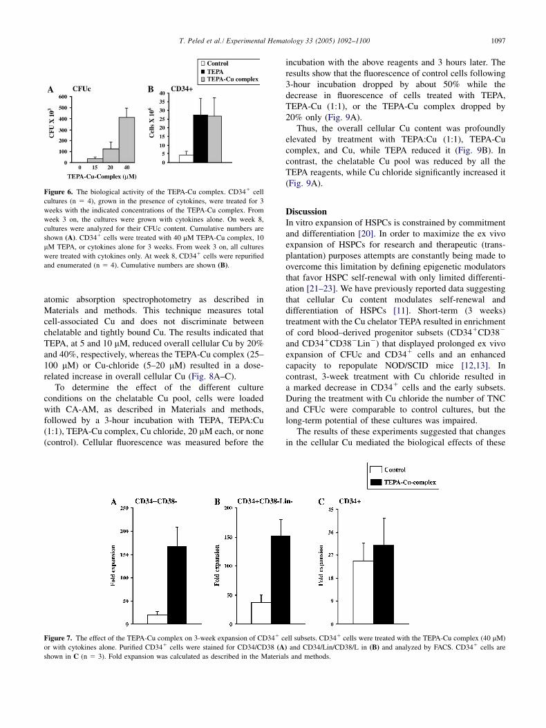
1097T. Peled et al./ Experimental Hematology 33 (2005) 1092–1100
atomic absorption spectrophotometry as described inMaterials and methods. This technique measures totalcell-associated Cu and does not discriminate betweenchelatable and tightly bound Cu. The results indicated thatTEPA, at 5 and 10 mM, reduced overall cellular Cu by 20%and 40%, respectively, whereas the TEPA-Cu complex (25–100 mM) or Cu-chloride (5–20 mM) resulted in a dose-related increase in overall cellular Cu (Fig. 8A–C).
To determine the effect of the different cultureconditions on the chelatable Cu pool, cells were loadedwith CA-AM, as described in Materials and methods,followed by a 3-hour incubation with TEPA, TEPA:Cu(1:1), TEPA-Cu complex, Cu chloride, 20 mM each, or none(control). Cellular fluorescence was measured before the
Figure 6. The biological activity of the TEPA-Cu complex. CD341 cell
cultures (n 5 4), grown in the presence of cytokines, were treated for 3
weeks with the indicated concentrations of the TEPA-Cu complex. From
week 3 on, the cultures were grown with cytokines alone. On week 8,
cultures were analyzed for their CFUc content. Cumulative numbers are
shown (A). CD341 cells were treated with 40 mM TEPA-Cu complex, 10
mM TEPA, or cytokines alone for 3 weeks. From week 3 on, all cultures
were treated with cytokines only. At week 8, CD341 cells were repurified
and enumerated (n 5 4). Cumulative numbers are shown (B).
incubation with the above reagents and 3 hours later. Theresults show that the fluorescence of control cells following3-hour incubation dropped by about 50% while thedecrease in fluorescence of cells treated with TEPA,TEPA-Cu (1:1), or the TEPA-Cu complex dropped by20% only (Fig. 9A).
Thus, the overall cellular Cu content was profoundlyelevated by treatment with TEPA:Cu (1:1), TEPA-Cucomplex, and Cu, while TEPA reduced it (Fig. 9B). Incontrast, the chelatable Cu pool was reduced by all theTEPA reagents, while Cu chloride significantly increased it(Fig. 9A).
DiscussionIn vitro expansion of HSPCs is constrained by commitmentand differentiation [20]. In order to maximize the ex vivoexpansion of HSPCs for research and therapeutic (trans-plantation) purposes attempts are constantly being made toovercome this limitation by defining epigenetic modulatorsthat favor HSPC self-renewal with only limited differenti-ation [21–23]. We have previously reported data suggestingthat cellular Cu content modulates self-renewal anddifferentiation of HSPCs [11]. Short-term (3 weeks)treatment with the Cu chelator TEPA resulted in enrichmentof cord blood–derived progenitor subsets (CD341CD382
and CD341CD382Lin2) that displayed prolonged ex vivoexpansion of CFUc and CD341 cells and an enhancedcapacity to repopulate NOD/SCID mice [12,13]. Incontrast, 3-week treatment with Cu chloride resulted ina marked decrease in CD341 cells and the early subsets.During the treatment with Cu chloride the number of TNCand CFUc were comparable to control cultures, but thelong-term potential of these cultures was impaired.
The results of these experiments suggested that changesin the cellular Cu mediated the biological effects of these
Figure 7. The effect of the TEPA-Cu complex on 3-week expansion of CD341 cell subsets. CD341 cells were treated with the TEPA-Cu complex (40 mM)
or with cytokines alone. Purified CD341 cells were stained for CD34/CD38 (A) and CD34/Lin/CD38/L in (B) and analyzed by FACS. CD341 cells are
shown in C (n 5 3). Fold expansion was calculated as described in the Materials and methods.

1098 T. Peled et al./ Experimental Hematology 33 (2005) 1092–1100
Figure 8. Measurement of overall cellular Cu content. CD341 cells were treated with the indicated concentrations of TEPA (A), the TEPA-Cu complex (B),
Cu chloride (C), or cytokines alone (control). Cellular Cu was determined by atomic absorption spectrophotometry as described in Materials and methods.
Results of the Cu content are shown as percentages of control, cytokine-treated cultures.
reagents. Indeed, we demonstrated that only Cu, but notother transitional metal ions, could reverse the TEPA’seffect [11]. However, this reversal was achieved only withexcess of Cu. Contrary to studies in other cell systems[24,10], in our experiments, at equimolar concentration(TEPA:Cu 1:1), Cu did not quench the TEPA’s effect. Thesesurprising results prompted us to reevaluate the role of Cuin self-renewal and differentiation. For this purpose, wesynthesized a stable TEPA-Cu complex at 1:1 molar ratioand evaluated its effect on CD34 cells. The results indicatedthat the complex had biological activity similar to theTEPA:Cu (1:1) mixture as well as TEPA.
We next determined the effect of these reagents on theoverall Cu content of CD34 cells using atomic absorptionspectrophotometry. The results indicated that TEPA re-duced it whereas the TEPA-Cu mixture and complex or Cuchloride increased it (Figs. 8 and 9B).
Cu is present in cells in at least two various forms: onewhich is firmly bound to compounds such as ceruloplasminand various enzymes such as Cu/Zn superoxide dismutaseand the other a more loosely bound, labile pool, possiblyinvolved in the synthesis of Cu proteins [25,26]. Underphysiological conditions the amount of the labile pool isquite small [26], but it may have a significant biologicalrole. To measure the cytosolic pool of labile Cu we utilizeda novel flow cytometry method that makes use of a cell-permeable Cu chelator: calcein. The change in calceinfluorescence under different culture conditions measuresthe amount of calcein-bound Cu that in turn reflects therelative levels of cellular chelatebale Cu. We demonstratedthat a 3-hour treatment with TEPA, TEPA:Cu (1:1), andTEPA-Cu complex of calcein preloaded cells reduced by50% the decrease in fluorescence, relative to the decrease inthe fluorescence of untreated cells (Fig. 9A), suggesting
Figure 9. The effect of TEPA, TEPA:Cu (1:1), TEPA-Cu complex, and Cu chloride on chelatable (A) and overall (B) cellular Cu. Cells were loaded with
calcein, then washed twice and exposed for 3 hours to either TEPA, the TEPA:Cu (1:1) mixture, the TEPA-Cu complex, and Cu chloride (20 mM each) or
none (control), as indicated. Cellular fluorescence was measured after incubation with calcein (T0) and 3 hours thereafter (T3) by flow cytometry as described
in Materials and methods. Percentage decrease in fluorescence during 3-hour incubation was calculated relative to the fluorescence of calcein-loaded cells
(T0) as follows: fluorescence at T3 3 100/fluorescence at T0 (A). The decrease in fluorescence represents, in relative terms, the available chelatable Cu pool.
In parallel, overall Cu content of cells treated with 20 mM of the above-mentioned reagents was measured by atomic absorption spectrophotometry as
described in Materials and methods (B).

1099T. Peled et al./ Experimental Hematology 33 (2005) 1092–1100
that all these reagents reduce the availability of chelatableCu. Thus, although the overall cellular Cu content wasprofoundly elevated by treatment with TEPA:Cu (1:1)mixture and complex, as well as Cu chloride, and TEPAreduced it, the chelatable Cu was reduced by all the TEPAreagents, while Cu chloride significantly increased it.
At least two mechanisms may account for the biologicaleffects of TEPA; the first suggests that a TEPA-Cu chelate isthe specific active intermediate responsible for TEPA’sbiological activity. Since TEPA has the strongest bindingaffinity for Cu (its stability constants for Cu, Zn, Co, Fe, Mnare 23, 15, 13, 10, 7, respectively) [27], it is expected toform a chelate, similar to the synthesized TEPA-Cu complex[25]. The chelate, as well as the complex, acts directly toaffect pathways involved in self-renewal and differentiationof CD34 cells. This mechanism does not require reductionof cellular Cu since the TEPA:Cu (1:1) mixture and theTEPA-Cu complex, which display biological activitysimilar to TEPA, have opposite effect on the overall Cucontent. This mechanism also fails to explain the effect ofCu chloride on self-renewal and CD34 cell differentiation.The second mechanism involves changes in the levels of thechelateble Cu pool. All the TEPA reagents were found toreduce this pool while Cu chloride was found to increase it.This correlates with their effect on self-renewal anddifferentiation.
Intracellular Cu was reported to regulate gene expression[28–31] and cell differentiation [32,33] by a variety ofpathways. It modifies the function of transcription factorsand the activities of Cu enzymes such as S-adenosylhomo-cysteine hydrolase and protein arginine methyltransferase1, which are involved in protein methylation. Cu chelationby TEPA deactivates these enzymes, resulting in proteinhypomethylation and inhibition of neurite differentiation[24]. Cu has also been shown to suppress the enzymehistone acetyl transferase, resulting in a decrease in overalland specific histone acetylation; Cu chelators, in contrast,had an opposite effect [34]. Reversible histone acetylation/deacetylation plays a pivotal role in transcriptionalmodulation of cell fate [35,36]. Histone deacetylaseinhibitors were reported to increase the self-renewal ofhematopoietic CD341 cells in vitro and their engraftabilityin vivo [37]. Cu may be involved in hematopoietic cellregulation by modulating cellular post-translational modi-fication activities.
The results of the present study support the notion thatreduction of the chelatable Cu pool rather than a specificTEPA-Cu chelate mediates the mechanism of TEPA’sactivity on CD341 cells. Further studies are in progressto clarify this mechanism.
AcknowledgmentsWe thank Dr. Yaron Daniely for helpful discussions during thepreparation of the manuscript.
References1. Zidar BL, Shadduck RK, Zeigler Z, Winkelstein A. Observations on
the anemia and neutropenia of human Cu deficiency. Am J Hematol.
1977;3:177–185.
2. Hirase N, Abe Y, Sadamura S, Yufu Y. Anemia and neutropenia in
a case of copper deficiency: Role of copper in normal hematopoiesis.
Acta Haematol. 1992;87:195–197.
3. Banno S, Niita M, Kikuchi M. Anemia and neutropenia in elderly
patients caused by copper deficiency for long-term eternal nutrition.
Rinsho Ketsueki. 1994;35:1276–1281.
4. Wasa M, Satani M, Tanano H. Copper deficiency with pancytopenia
during total parenteral nutrition. J Parenter Enteral Nutr. 1994;18:190–
192.
5. Gregg XT, Reddy V, Prchal JT, et al. Copper deficiency masquerading
as myelodysplastic syndrome. Blood. 2002;100:1493–1495.
6. Bae B, Percival SS. Copper uptake and intracellular distribution
during retinoic acid–induced differentiation of HL-60 cells. J Nutr
Biochem. 1994;5:457–461.
7. Bae B, Percival SS. Retinoic acid–induced HL-60 cell differenti-
ation is augmented by Cu supplementation. J Nutr. 1993;123:997–
1002.
8. Peled T, Treves AJ, Rachmilewitz EA, et al. Identification of a serum-
derived differentiation-inducing activity as the copper-binding protein
ceruloplasmin. Blood. 1998;92:618a.
9. Shimizu M. Clinical results on the use of human ceruloplasmin in
aplastic anemia. Transfusion. 1979;19:742–748.
10. Huang ZL, Failla ML, Reeves PG. Differentiation of human U937
promonocytic cells is impaired by moderate copper deficiency. Exp
Biol Med (Maywood). 2001;226:222–228.
11. Peled T, Landau E, Prus E, et al. Cellular copper content modulates
differentiation and self-renewal in cultures of cord blood–derived
CD341 cells. Br J Haematol. 2002;116:655–661.
12. Peled T, Landau E, Mandel J, et al. Linear polyamine copper
chelator tetraethylenepentamine augments long-term ex vivo expan-
sion of cord blood–derived CD341 cells and increases their
engraftment potential in NOD/SCID mice. Exp Hematol. 2004;32:
547–555.
13. Peled T, Mandel J, Goudsmid RN, et al. Pre-clinical development of
cord blood–derived progenitor cell graft expanded ex vivo with
cytokines and the polyamine copper chelator tetraethylenepentamine.
Cytotherapy. 2004;6:344–355.
14. Rubinstein P, Dobrila L, Rosenfield RE, et al. Processing and
cryopreservation of placental/umbilical cord blood for unrelated bone
marrow reconstitution. Proc Natl Acad Sci U S A. 1995;92:10119–
10122.
15. Grodzicki M, Chrzaszez M, Kachniarz E, Piszezek P, Rozploch F.
Spectral and magnetic properties of some copper (II) carboxylate
compounds with hexamethylenetetramine. Pol J Chem. 1994;68:
445–452.
16. Weston SA, Parish CR. New fluorescent dyes for lymphocyte
migration studies. Analysis by flow cytometry and fluorescence
microscopy. J Immunol Methods. 1990;133:87–97.
17. Weston SA, Parish CR. Calcein: A novel marker for lymphocytes
which enter lymph nodes. Cytometry. 1992;13:739–749.
18. Dean KE, Klein G, Renaudet O, Reymond JL. A green fluorescent
chemosensor for amino acids provides a versatile high-throughput
screening (HTS) assay for proteases. Bioorg Med Chem Lett. 2003;13:
1653–1656.
19. Luo W, Ma YM, Quinn PJ, Hider RC, Liu ZD. Design, synthesis and
properties of novel iron (III)-specific fluorescent probes. J Pharm
Pharmacol. 2004;56:529–536.
20. Von Drygalski A, Alespeiti G, Ren L, et al. Murine bone marrow cells
cultured ex vivo in the presence of multiple cytokine combinations
lose radioprotective and long-term engraftment potential. Stem Cells
Dev. 2004;13:101–111.
1100 T. Peled et al./ Experimental Hematology 33 (2005) 1092–1100
21. Zheng X, Beissert T, Kukoc-Zivojnov N, et al. g-catenin contributes to
leukemogenesis induced by AML-associated translocation products by
increasing the self-renewal of very primitive progenitor cells. Blood.
2004;103:3535–3543.
22. Amsellem S, Pflumio F, Bardinet D, et al. Ex vivo expansion of human
hematopoietic stem cells by direct delivery of the HOXB4
homeoprotein. Nat Med. 2003;9:1423–1427.
23. Willert K, Brown JD, Danenberg E, et al. Wnt proteins are lipid-
modified and can act as stem cell growth factors. Nature. 2003;423:
448–452.
24. Birkaya B, Aletta JM. NGF promotes copper accumulation required
for optimum neurite outgrowth and protein methylation. J Neurobiol.
2005;63:49–61.
25. McArdle HJ, Gross SM, Vogel HM, Ackland ML, Danks DM. The
effect of tetrathiomolybdate on the metabolism of copper by
hepatocytes and fibroblasts. Biol Trace Elem Res. 1989;22:179–188.
26. Rosenzweig AC. Copper delivery by metallochaperone proteins. Acc
Chem Res. 2001;34:119–128.
27. Smith RM, Martell AE. Critical stability constants. New York: Plenum
Press; 1975. Vol. 2.
28. Hainaut P, Rolley N, Davies M, et al. Modulation by copper of p53
conformation and sequence-specific DNA binding: role for Cu (II)/Cu
(I) redox mechanism. Oncogene. 1995;10:27–32.
29. Kudrin AV. Trace elements in regulation of NF-kb activity. J Trace
Elem Med Biol. 2000;14:129–142.
30. Vanacore RM, Eskew JD, Morales PJ, et al. Role for copper in
transient oxidation and nuclear translocation of MTF-1, but not of NF-
kb, by the heme-hemopexin transport system. Antioxid Redox Signal.
2000;2:739–752.
31. Ostrakhovitch EA, Lordnejad MR, Schliess F, et al. Copper ions
strongly activate the phosphoinositide-3-kinase/Akt pathway indepen-
dent of the generation of reactive oxygen species. Arch Biochem
Biophys. 2002 Jan 15;397:232–239.
32. Nakamura H, Nakamura K, Yodoi J. Redox regulation of cellular
activation. Annu Rev Immunol. 1997;15:351–369.
33. Iseki A, Kambe F, Okumura K, Hayakawa T, Seo H. Regulation of
thyroid follicular cell function by intracellular redox-active copper.
Endocrinology. 2000;141:4373–4382.
34. Kang J, Lin C, Chen J, et al. Copper induces histone hypoacetylation
through directly inhibiting histone acetyltransferase activity. Chem
Biol Interact. 2004;148:115–123.
35. Akashi K, He X, Chen J, et al. Transcriptional accessibility for genes
of multiple tissues and hematopoietic lineages is hierarchically
controlled during early hematopoiesis. Blood. 2003;101:383–389.
36. Lehrmann H, Pritchard LL, Harel-Bellan A. Histone acetyltransferases
and deacetylases in the control of cell proliferation and differentiation.
Adv Cancer Res. 2002;86:41–65.
37. Milhem M, Mahmud N, Lavelle D, et al. Modification of
hematopoietic stem cell fate by 5aza 2#deoxycytidine and trichostatin
A. Blood. 2004;103:4102–4110.
ASH ABSTRACT #1
CONFIDENTIAL
Please select Print from the file menu to print your Abstract.
Please print, sign and fax this Official Print-Out to Marathon Multimedia at US
507-334-0126
before August 10, 2004, 11:59 PM EDT.
46th ASH Annual Meeting
Filename: 551903
Presenting Author E Shpall
Department/Institution: Department of Blood and Marrow
Transplantation, University of Texas M.D. Anderson Cancer Center
Address: 1515 Holcombe Blvd., Unit 423
City/State/Zip/Country: Houston, TX, 77030, United States
Phone: 1-713-745-2803 Fax: 1-713-794-4902 E-mail:
Presenting author is member of the American Society of
Hematology: No
Presenting author is an Associate Member of ASH (member in
training): No
Sponsoring Member: Elizabeth Shpall, MD
Department/Institution: Department of Blood and Marrow
Transplantation, M D Anderson Cancer Center
Address: 1515 Holcombe blvd. unit 423
City/State/Zip/Country: Houston, TX, 77030, United States
Phone: 713-792-8750 Fax: 713-792-8503 E-mail:
Abstract Category: 732. Clinical Results - Allogeneic Mismatched or
Unrelated Transplantation
Presentation format preference: No preference
Publication preference: Publish in Blood if not accepted for
presentation.
Special consideration: No.
Award Category: No award
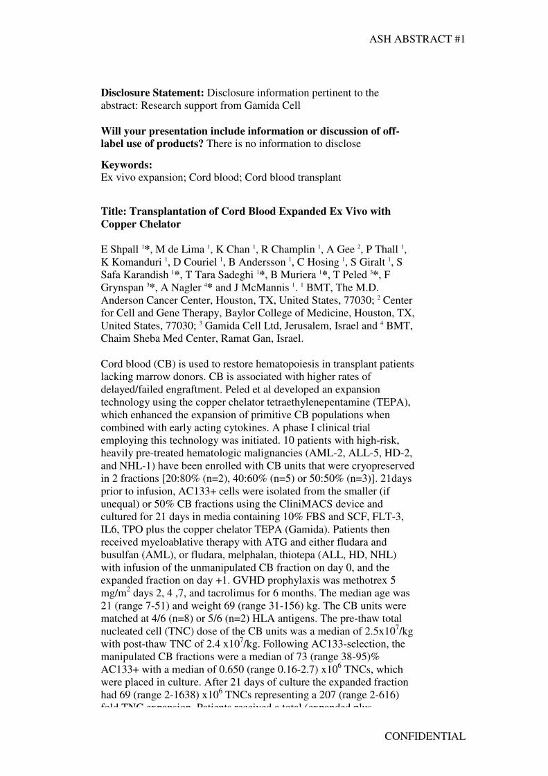
ASH ABSTRACT #1
CONFIDENTIAL
Disclosure Statement: Disclosure information pertinent to the
abstract: Research support from Gamida Cell
Will your presentation include information or discussion of off-
label use of products? There is no information to disclose
Keywords: Ex vivo expansion; Cord blood; Cord blood transplant
Title: Transplantation of Cord Blood Expanded Ex Vivo with
Copper Chelator
E Shpall 1*, M de Lima 1, K Chan 1, R Champlin 1, A Gee 2, P Thall 1,
K Komanduri 1, D Couriel 1, B Andersson 1, C Hosing 1, S Giralt 1, S
Safa Karandish 1*, T Tara Sadeghi 1*, B Muriera 1*, T Peled 3*, F
Grynspan 3*, A Nagler 4* and J McMannis 1. 1 BMT, The M.D.
Anderson Cancer Center, Houston, TX, United States, 77030; 2 Center
for Cell and Gene Therapy, Baylor College of Medicine, Houston, TX,
United States, 77030; 3 Gamida Cell Ltd, Jerusalem, Israel and 4 BMT,
Chaim Sheba Med Center, Ramat Gan, Israel.
Cord blood (CB) is used to restore hematopoiesis in transplant patients
lacking marrow donors. CB is associated with higher rates of
delayed/failed engraftment. Peled et al developed an expansion
technology using the copper chelator tetraethylenepentamine (TEPA),
which enhanced the expansion of primitive CB populations when
combined with early acting cytokines. A phase I clinical trial
employing this technology was initiated. 10 patients with high-risk,
heavily pre-treated hematologic malignancies (AML-2, ALL-5, HD-2,
and NHL-1) have been enrolled with CB units that were cryopreserved
in 2 fractions [20:80% (n=2), 40:60% (n=5) or 50:50% (n=3)]. 21days
prior to infusion, AC133+ cells were isolated from the smaller (if
unequal) or 50% CB fractions using the CliniMACS device and
cultured for 21 days in media containing 10% FBS and SCF, FLT-3,
IL6, TPO plus the copper chelator TEPA (Gamida). Patients then
received myeloablative therapy with ATG and either fludara and
busulfan (AML), or fludara, melphalan, thiotepa (ALL, HD, NHL)
with infusion of the unmanipulated CB fraction on day 0, and the
expanded fraction on day +1. GVHD prophylaxis was methotrex 5
mg/m2 days 2, 4 ,7, and tacrolimus for 6 months. The median age was
21 (range 7-51) and weight 69 (range 31-156) kg. The CB units were
matched at 4/6 (n=8) or 5/6 (n=2) HLA antigens. The pre-thaw total
nucleated cell (TNC) dose of the CB units was a median of 2.5x10⁷/kg
with post-thaw TNC of 2.4 x10⁷/kg. Following AC133-selection, the
manipulated CB fractions were a median of 73 (range 38-95)%
AC133+ with a median of 0.650 (range 0.16-2.7) x106 TNCs, which
were placed in culture. After 21 days of culture the expanded fraction
had 69 (range 2-1638) x106 TNCs representing a 207 (range 2-616)
fold TNC expansion. Patients received a total (expanded plus
ASH ABSTRACT #1
CONFIDENTIAL
fold TNC expansion. Patients received a total (expanded plus
unmanipulated) median of 1.8 (range 1.1-6.1) x107
TNC/kg and
1.6x10⁵ (range 0.4-49.9) CD34+ cells/kg. Two patients have CB
cultures in progress. Of the 8 patients transplanted, 1 had autologous
recovery with relapse of AML on day 30 and death. Of the remaining
patients, 7 were evaluable for neutrophil engraftment and 4 of them for
platelet engraftment (2 too early for platelet evaluation and 1 early
death). The median time to engraftment was 27 days for neutrophils
(range 16-46) and 48 days for platelets (range 27-96). Preliminary
analysis suggest a correlation between a shorter time to neutrophil
engraftment and total TNC/kg infused (p=0.02), and a trend for
CD34+ cells/kg infused (p=0.09). Three patients have developed grade
<2 acute skin GVHD and one had chronic extensive GVHD of the skin
and GI tract; all resolved with steroids. One patient (without GVHD)
died of a systemic viral infection on day 56, despite adequate
neutrophil recovery (not platelets). All of the remaining patients are all
alive and free of malignancy at a median follow-up of 4 (range 1-16)
months. Conclusion: There was no toxicity associated with infusion of
the TEPA-expanded CB cells. Additional data is necessary to
determine the efficacy of this approach. Future directions include the
expansion of the entire CB unit and removal of methotrex from the
GVHD regimen to improve time to neutrophil engraftment, as well as
comprehensive assessment of immune reconstitution.
Certification for Human Subjects: I certify that this study abides by
the rules of the appropriate internal review board and the tenets of the
Helsinki protocol, if human subjects were involved.
Agree
Signature of Presenting Author:
_____________________________________________________________________
__
E Shpall
Sponsoring ASH Member:
_____________________________________________________________________
__
Elizabeth Shpall
Close Window
Pre-clinical development of cord blood-derivedprogenitor cell graft expanded ex vivo with
cytokines and the polyamine copper chelatortetraethylenepentamine
T Peled1, J Mandel1, RN Goudsmid1, C Landor1, N Hasson1, D Harati1, M Austin1,
A Hasson1, E Fibach2, EJ Shpall3 and A Nagler4
1Gamida-Cell Ltd, Jerusalem, Israel, 2Hadassah�/Hebrew University Medical Center, Jerusalem, Israel, 3MD Anderson, Houston, Texas, USA,
and 4Chaim Sheba Medical Center, Tel Hashomer, Israel
Background
We have previously demonstrated that the copper chelator tetra-
ethylenepentamine (TEPA) enables preferential expansion of early
hematopoietic progenitor cells (CD34�CD38� , CD34�CD38�Lin� )
in human umbilical cord blood (CB)-derived CD34� cell cultures.
This study extends our previous findings that copper chelation can
modulate the balance between self-renewal and differentiation of
hematopoietic progenitor cells.
Methods
In the present study we established a clinically applicative protocol
for large-scale ex vivo expansion of CB-derived progenitors.
Briefly, CD133� cells, purified from CB using Miltenyi Biotec’s
(Bergisch Gladbach, Germany) CliniMACS separation device and
the anti-CD133 reagent, were cultured for 3 weeks in a clinical-
grade closed culture bag system, using the chelator-based
technology in combination with early-acting cytokines (SCF,
thrombopoietin, IL-6 and FLT-3 ligand). This protocol was
evaluated using frozen units derived from accredited cord blood
banks.
Results
Following 3 weeks of expansion under large-scale culture conditions that
were suitable for clinical manufacturing, the median output value of
CD34� cells increase by 89-fold, CD34�CD38� increase by 30-fold
and CFU cells (CFUc) by 172-fold over the input value. Transplanta-
tion into sublethally irradiated non-obese diabetic (NOD/SCID) mice
indicated that the engraftment potential of the ex vivo expanded
CD133� cells was significantly superior to that of unexpanded cells:
609/5.5% vs. 219/3.5% CD45� cells, P�/0.001, and 119/1.8% vs.
49/0.68% CD45�CD34� cells, P�/0.012, n�/32, respectively.
Discussion
Based on these large-scale experiments, the chelator-based ex vivo
expansion technology is currently being tested in a phase 1 clinical
trial in patients undergoing CB transplantation for hematological
malignancies.
Keywords
ex vivo large-scale expansion, pre-clinical development, tetraethyle-
nepentamine.
IntroductionCord blood (CB) is a valuable source of stem cells.
Transplanted CB hematopoietic stem/progenitors cells
(HPC) can treat malignant and non-malignant disorders
[1�/3]. However, the major clinical limitation of CB
transplantation is the low number of HPC in comparison
with mobilized peripheral blood or BM grafts. This
limitation may explain the slower time to engraftment
and higher rate of engraftment failure following CB
transplantation [4,5]. To overcome this limitation, ex vivo
expansion of CB progenitors with a cocktail of growth
factors has been attempted [6]. It was shown that a
Correspondence to: Professor Arnon Nagler, Director of Hematology Division, Bone Marrow Transplantation & Cord Blood Bank, Chaim Sheba
Medical, Tel Hashomer, Israel.
Cytotherapy (2004) Vol. 6, No. 4, 344�/355
– 2004 ISCT DOI: 10.1080/14653240410004916

combination of early- and late-acting cytokines, including
SCF, thrombopoietin (TPO), G-CSF and IL-3, resulted in
only a marginal-fold expansion of late (CD34�) and early
(CD34�CD38� ) progenitor cells, probably due the fact
that the late-acting cytokines drive the cultures mainly
toward accelerated differentiation [7�/9]. On the other
hand, cultures with only early-acting cytokines (SCF,
TPO, IL-6 and FLT-3 ligand) resulted in better and
prolonged expansion of both late and early progenitors
[10,11], which are important for short-term early trilineage
engraftment [12�/14].
We have previously demonstrated that short-term
(3 weeks) treatment with the polyamine copper chelator
tetraethylenepentamine (TEPA) augmented the long-term
expansion potential of CB-derived progenitor cells [15].
During the treatment period, TEPA inhibited the onset
of cytokine-driven differentiation of early progenitor
cells, resulting in a robust accumulation of CD34�CD38�
and CD34�Lin� cells, with no effect on proliferation
and differentiation of more mature committed cells
[CD34�Lin� and CFU culture (CFUc)] [16]. These
results strongly suggest that TEPA supports the self-
renewal division cycle without compromising differentia-
tion capacity of hematopoietic stem cells.
In view of these results, the TEPA-based expansion
procedure was adapted to comply with current good
manufacturing practice (cGMP) standards required for
clinical trials. In the present study we describe the
development of a clinical-scale procedure using Miltenyi
Biotec’s (Bergisch Gladbach, Germany) CliniMACS se-
paration device, and the anti-CD133 reagent for progeni-
tor cell enrichment and 3-week expansion in culture
bags, using the chelator-based technology with an
early-acting cytokine cocktail (FLT-3 ligand, IL-6, TPO,
SCF).
MethodsCB samples
Cells were obtained from neonatal umbilical cord blood
after normal full-term delivery (informed consent was
given). Samples were collected and frozen in our labora-
tories according to Rubinstein et al . [17] within 24 h
postpartum, or kindly provided by the New York Blood
Bank (New York, NY) and the Duke University Medical
Center Cord Blood Bank (Durham, NC).
Thawing procedure and CliniMACS separation of
CD133� and CD34� cells
The cells were thawed by doubling the volume of the
blood sample in 2.5% dextran (Sigma, St Louis, MO) and
1.25% HSA (Bayer Co., Elkhart, IN). Prior to centrifuging
an additional 40 mL of 10% dextran was added.
The cells were resuspended in 40 mL of 2.5% dextran/
1.25% HSA and then filled to 100 mL with PBS
(Biological Industries, Beit-HaEmek, Israel) containing
0.4% sodium citrate solution (Baxter HealthCare Co.,
Deerfield, IL) and 1% HSA. The pellet was incubated with
0.15% w/v intravenous immunoglobulin (IvIg; Omrix
Biopharmaceuticals, Nes-Ziona, Israel) for 10 min at
room temperature before centrifugation, and then resus-
pended in PBS containing 0.4% sodium citrate solution
and 1% HAS, and 0.25 mg/mL recombinant human
deoxyribonuclease (rHu-Dnase) added. Subsequently, the
cells were labeled with Miltenyi’s anti-CD133 (clone 1) or
anti-CD34 CliniMACS reagent (Miltenyi Biotec) and
separated by CliniMACS (according to the manufacturer’s
instructions). Following separation, cells were stained with
trypan blue, counted, assayed for CFUc and immunophe-
notyped to determine purity.
Purity determination of CD34�/ and CD133�/
enriched cell fractions
The cells were washed with a PBS solution containing 1%
BSA (PBS/1%BSA), and stained (at 48C for 30 min) with
FITC-conjugated anti-CD45 (Becton Dickinson, San Jose,
CA) and either PE-conjugated anti-CD34 (DAKO,
Glostrup, Denmark) or PE anti-CD133 (Miltenyi Biotec)
Ab. In addition, the percentage of cells exhibiting both the
CD133 and CD34 markers was measured by staining the
cells with FITC�/anti-CD34 (IQ Products, Groningen, the
Netherlands) and PE�/anti-CD133 (Miltenyi Biotec) Ab.
The cells were then washed in the above buffer and
analyzed using a FACScalibur† flow cytometer (Becton
Dickinson, Immunofluorometry Systems, Mountain View,
CA). The cells were passed at a rate of up to 1000 cells/
second, using a 488-nm argon laser beam as the light
source for excitation. Emission of 5000 cells was measured
using logarithmic amplification, and analyzed using the
CellQuest software (Becton Dickinson). Cells stained with
FITC- and PE-conjugated isotype control Ab were used to
determine background fluorescence.
Development of ex vivo expanded cord blood-derived cell graft 345
Cell counting
The number of total nucleated cells (TNC) was deter-
mined by diluting the cells 1:2 with trypan blue and
differentially counting viable and dead cells using a
hemocytometer under an upright microscope at 100�/
magnification.
Assay for CFUc
Cells, 1000 (CD34� or CD133�) before culture and 1500
following culture, were added per 3 mL semisolid minimal
essential alpha medium (MEMa), containing methylcellu-
lose (Sigma), 30% FCS, 1% BSA, 1�/10�5 M b-
mercaptoethanol (Sigma), 1 mM glutamine (Biological
Industries), 2 IU/mL erythropoietin (Eprex, Cilag AG Int.,
Schaffhausen, Switzerland), SCF and IL-3, both at
20 ng/mL, G-CSF and GM-CSF, both at 10 ng/mL
(Perpo Tech Inc., Rocky Hill, NJ), and 2 mm hemin
(Sigma). Following stirring, the mixture was divided into
two 35-mm dishes and incubated for 14 days at 378C in a
humidified atmosphere of 5% CO2 in air. At the end of the
incubation period, colonies (both myeloid and erythroid)
were counted under an inverted microscope at 40�/
magnification. CFUc content was calculated as the
following: number of scored colonies per two dishes�/
total cell number/1500 or 1000. The number of cells was
determined by multiplying the number of cells/mL by the
culture volume. CFUc frequency was calculated as number
of colonies divided by the number of cells seeded.
Ex vivo expansion
Purified CD34� or CD133� cells were cultured at
1�/104 cells/mL in MEMa and 10% FCS (Biological
Industries) containing the following human recombinant
cytokines: TPO, IL-6, FLT-3 ligand and SCF, each at a
final concentration of 50 ng/mL (Pepro Tech Inc.), and 5
mm TEPA (Aldrich, Milwaukee, WI). VueLife Teflon PEP
culture bags (American Fluoroseal Co., Gaithersburg, MD)
were used: 72-mL bags were used for up to 20�/104
initiating cells, and 270-mL bags were used for up to
20�/70�/104 cells. The cultures were incubated for 3
weeks (unless otherwise stated) at 378C in a humidified
atmosphere of 5% CO2 in air. Cultures were topped
weekly with the same volume of fresh medium, FCS,
cytokines and TEPA. At the termination of the experi-
ment, cells were counted following staining with trypan
blue, assayed for CFUc and immunophenotyped for
surface antigens.
Surface antigen analysis of cultured cells
The cells were washed with PBS/1%BSA and stained (at
48C for 30 min) with both FITC�/anti-CD45 and PE�/
anti-CD34 (both from DAKO) Ab for determination of
CD34� cells, with FITC�/anti-CD38 and PE�/anti-CD34
for determination of CD34�CD38� cells and with FITC�/
anti-CD45 and PE�/Ab to lineage specific antigens
(Becton Dickinson). The cells were then washed and
analyzed as described above.
Clinical grade reagents
During the development phase, the research grade ingre-
dients were replaced by clinical grade ingredients as follows.
Thawing procedure and CliniMACS separation: Gen-
tran-40, a ready-made 10% w/v dextran solution, HSA
and IvIg (all from Baxter), Dnase (Genentech Inc., San
Francisco, CA) and PBS (Hyclone, Logan, UT). CFUc
assay: MethoCultTM, a methylcellulose-based medium
(StemCell Technologies, Vancouver, Canada). Ex vivo
expansion: MEMa and FCS (gamma-irradiated defined
fetal bovine serum batch), from Hyclone.
TEPA was purchased from NovaSep (Boothwyn, PA).
The cytokines TPO, IL-6, SCF and FLT-3 ligand were
from R&D Systems (Minneapolis, MN). They are human
recombinant cytokines from non-mammalian origin (de-
rived from either Escherichia coli or Sf-21 cells). FLT-3
ligand and TPO were purified on affinity columns
containing MAb. The cytokines were filtered through a
0.2-micron membrane, packaged under aseptic conditions
and tested for endotoxin. Cytokine batches used in the
study were tested for sterility by Charles River Labora-
tories (Rockville, MD) according to the ICH guideline
Viral safety evaluation of biotechnology products derived from cell
lines of human or animal origin adopted by the ICH Steering
Committee (March 5, 1997) and the Code of Federal
Regulations (April 1, 2003).
Reselection of cultured CD34� cells
To purify the CD34� cells, cultured cells were harvested
and subjected to two cycles of immunomagnetic bead
separation, using a MiniMACS CD34 progenitor cell
isolation kit (Miltenyi Biotec) according to the manufac-
turer’s recommendations. The purity of the CD34�
population thus obtained was 90�/98%, as evaluated by
flow cytometry. The eluted cells were then counted and
dually stained with PE�/anti-CD34 and FITC�/anti-CD38
Ab for determination of the percentage of CD34�CD38�
346 T Peled et al .
cells. CD34� cell content of the entire culture was
calculated as follows: number of CD34� cells recovered
following repurification�/culture volume/volume of the
portion of the culture subjected to repurification. Fold
expansion of CD34� cells was calculated by dividing the
CD34� cell content of the culture by the number of
inoculated CD34� cells. The CD34�CD38� cell content
of the entire culture was calculated from the total CD34�
cells. Fold expansion was calculated by dividing the total
CD34�CD38� cell number following culture by the
number of inoculated cells.
Stability tests of the expanded graft
Following 3-week expansion, the cells were harvested,
washed twice with PBS/EDTA�/HSA solution, resus-
pended in transfusion solution (PBS/EDTA�/HSA buffer)
at 1�/1.5�/106 cells/mL and transferred (at least 30 mL)
into a transfusion bag (Transfer bag-Terumo, Teruflex T-
150, Tokyo, Japan). Closure clamps were used to prevent
foaming. The bags were kept in a shipping container
(Styrofoam) at 229/48C. Data loggers were put inside the
container and its inner surface fastened for temperature
monitoring. The bags were sampled to assess the number
of viable cells and CFUc at 0, 6, 10 and 24 h.
Transplantation of human CB-derived CD133�
cells into NOD/SCID mice
Each CB unit was frozen in two portions. CD133� cells
purified from the first portion were cultured for 3 weeks
with TEPA, as described above. The second portion of
each unit was kept frozen until the day of transplantation
(non-cultured cells). Mice were transplanted either with
the progeny of 5�/104 cultured CD133� cells or with
10�/106 non-cultured mononuclear cells. Control mice
were injected with medium only.
NOD/SCID mice, aged 10�/11 weeks, bred and main-
tained at the Department of Immunology, the Weizmann
Institute of Science, Rehovot, Israel, were injected intra-
venously with the above cells 1 day after they had been
irradiated at 375 cGy.
The mice were killed 4 weeks post transplantation; BM
was collected from both femurs and tibiae. The BM cells
were washed in PBS/1%BSA and stained (at 48C for 30 min)
with PE-conjugated Ab to human CD45 (DAKO) and
FITC-conjugated Ab to human CD34 (IQ Products),
CD41, CD61, glycophorin A (DAKO), CD14, CD15,
CD33 and CD19 (Becton Dickinson). Following incubation,
the suspension was treated with FACS lysing solution
(Becton Dickinson) to remove red blood cells, washed in
PBS/1%BSA and analyzed by flow cytometry, as described
above.
Calculations
Ex vivo expansion of TNC, CD34, CD34�CD38� cells
and CFUc are reported either as total numbers (number of
cells per mL multiplied by the final culture volume) or as
fold-expansion (total numbers divided by initial seeding
cell number).
Statistics
The following statistical tests were used. The non-para-
metric test (Wilcoxon rank test) was applied for testing
differences between the study groups for quantitative
parameters. The data was analyzed using the SAS software
(SAS version 8.2; SAS Institute Inc., Cary, NC).
ResultsProgenitor cell purification
As a first step toward large-scale experiments and a clinical
trial, we replaced the research-grade CD34-based separa-
tion device with the clinical grade CD133-based device.
For this purpose, we compared the anti-CD34 and anti-
CD133 CliniMACS reagents, using the CliniMACS
separation device, with respect to the yield (number of
cells) (Figure 1a) and purity (percent of CD34� or
CD133� cells) (Figure 1b). The results indicated no
statistically significant difference. Most (�/90%) of the
cells in the enriched populations were double positive for
both CD133 and CD34. A representative FACS analysis is
shown in Figure 1c. A comparison of the fractions with
respect to their CFUc frequency also produced similar
results, 0.29/0.1% and 0.129/0.07%, respectively.
Clinical-scale ex vivo expansion
To optimize the duration of the expansion procedure, we
compared 2- vs. 3-week cultures. Cultures were initiated
with 1�/104 cells/mL purified by the CliniMACS utiliz-
ing the anti-CD133 reagent. The cells were grown in
290-mL culture bags (initial culture volume/bag, 25 mL)
in alpha medium supplemented with FCS, a combination
of four cytokines (SCF, TPO, IL-6 and FLT-3 ligand,
50 ng/mL each) and 5 mm TEPA. The cultures were
topped up weekly with an equal volume of fresh medium.
Development of ex vivo expanded cord blood-derived cell graft 347

Figure 2 shows that the cumulative numbers (�/106) of
TNC, CD34� and CD34�CD38� cells were significantly
higher following 3-week expansion compared with 2
weeks: 849/7 vs. 349/3, 229/2 vs. 119/2, and 5.49/1 vs.
2.69/0.4, respectively. Only limited expansion was ob-
served following the first week of culturing (data not
shown).
We then compared the expansion potential of cells
purified from the same CB unit with either the anti-
CD133� or the anti-CD34� reagents. The cultures
(n�/4) were initiated with 2.5�/105 cells and grown for
3 weeks. The yield of TNC was 10659/124�/105 and
7609/75�/105 (P�/0.19), CFUc 819/9 and 839/11�/ 105
(P�/0.66), CD34� cells 439/7�/105 and 399/9�/105
Figure 1. Comparison between anti-CD133 and anti-CD34 CliniMACS enrichment reagents. Frozen CB units (n�/6) were thawed, divided
into two equal portions and enriched for progenitor cells using anti-CD133 or anti-CD34� reagents and CliniMACS separation device. Cell yield
(a) was determined by counting the number of viable cells in the positive fraction. Purity (b) was determined by FACS analysis of double stained
cells with PE�/anti-CD45 and either anti-CD34 or FITC�/anti-CD133 Ab. A representative FACS analysis dot-plot of CD133-enriched cells is
shown in (c). The eluted cells were double stained with isotype controls (left panel) or with both PE�/anti-CD133 and FITC�/anti-CD34 Ab
(right panel). The percentage of cells in each quadrant is indicated.
348 T Peled et al .
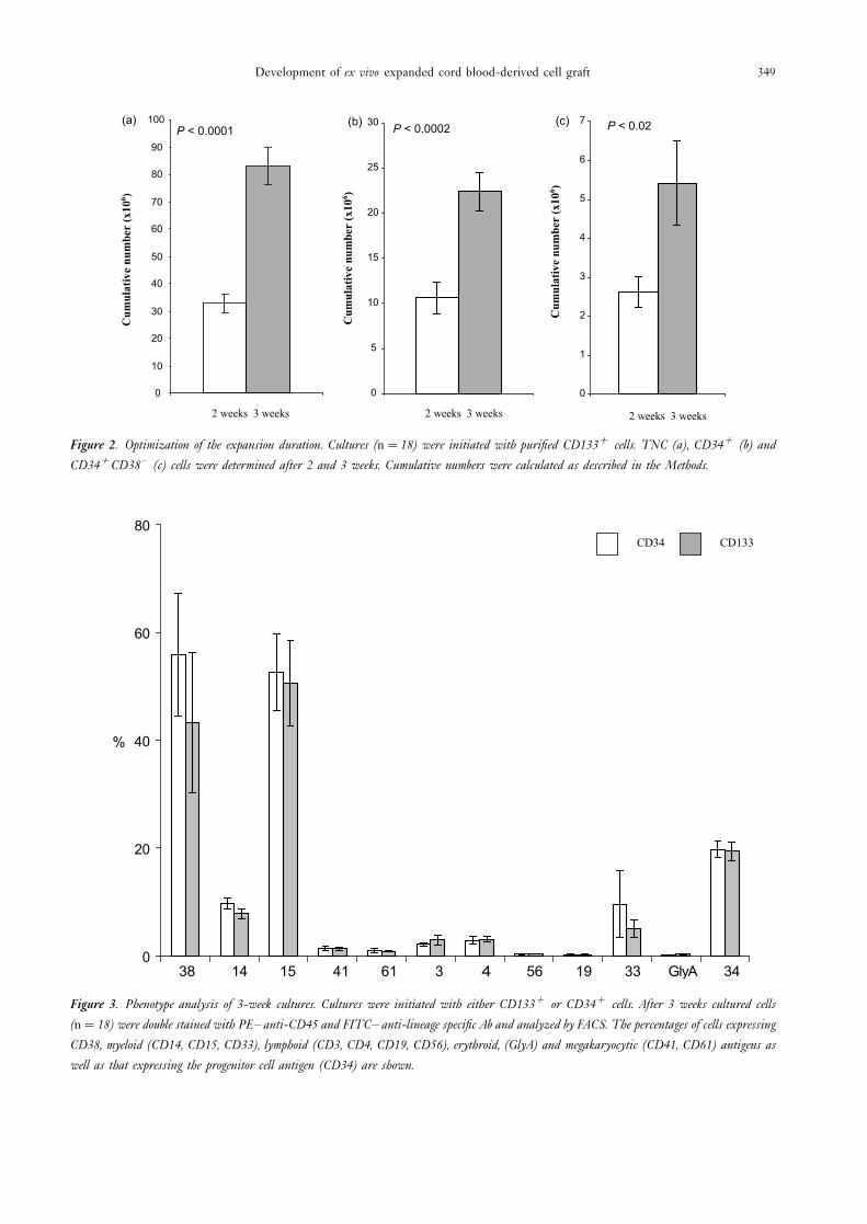
Figure 2. Optimization of the expansion duration. Cultures (n�/18) were initiated with purified CD133� cells. TNC (a), CD34� (b) and
CD34�CD38� (c) cells were determined after 2 and 3 weeks. Cumulative numbers were calculated as described in the Methods.
Figure 3. Phenotype analysis of 3-week cultures. Cultures were initiated with either CD133� or CD34� cells. After 3 weeks cultured cells
(n�/18) were double stained with PE�/anti-CD45 and FITC�/anti-lineage specific Ab and analyzed by FACS. The percentages of cells expressing
CD38, myeloid (CD14, CD15, CD33), lymphoid (CD3, CD4, CD19, CD56), erythroid, (GlyA) and megakaryocytic (CD41, CD61) antigens as
well as that expressing the progenitor cell antigen (CD34) are shown.
Development of ex vivo expanded cord blood-derived cell graft 349

(P�/0.89), CD34�CD38� cells 129/3.6�/105 and 5.69/
1.3�/105 (P�/0.11), in cultures initiated with CD133�
cells and with CD34� cells, respectively. In this set of
experiments, CD34� and CD34�CD38� cells were deter-
mined following affinity reselection of CD34� cells, as
described in the Methods. Additional immunophenotyping
indicated similar proportions of cells expressing myeloid,
lymphoid or megakaryocytic phenotype in cultures in-
itiated either with CD34� or CD133� cells (Figure 3).
Evaluation of the expansion procedure
Based on the above-described experiments, we carried out
a large-scale evaluation of the following expansion proce-
dure. A 20% portion of a CB unit was thawed and
progenitor cells were enriched by the CliniMACS anti-
CD133 procedure. The purified cells were grown for 3
weeks in culture bags with cGMP components, including
cytokines and TEPA. Of the frozen CB units studied, 18
were derived from accredited CB banks (Netcord Dussel-
dorf, Germany, and COBLT) and 4 units from the
Gamida-Cell research-grade CB bank (Jerusalem, Israel).
The results showed that the yield of viable cells was in the
range of 17�/35�/104 and the purity 58�/97%. The
percentages of CD34� and CD34�CD38� cells following
3-week expansion are shown in Figure 4.
The input numbers of CD34�, CD34�CD38� and
CFU cells as well as the output numbers following 3 weeks
expansion are shown in Table 1. The median output value
of CD34� cells increased by 89-fold, CD34�CD38�
increased by 30-fold and CFUc by 172-fold over the input
values.
We then determined the expansion efficacy with regard
to the number of cells of different subtypes available for
transplantation. For this purpose, the number of cells in the
expanded product (derived from 20% of the CB unit) and
the number of cells in the non-manipulated 80% portion,
were combined and compared with the number of cells in
the whole (100%) non-manipulated unit (Table 2).
Statistical analysis of the data demonstrated that the
numbers of CFUc, CD34� and CD34�CD38� cells in a
graft also containing expanded cells were significantly
higher than in a non-manipulated graft (P B/0.025),
whereas the total nuclear cell numbers were comparable
(P�/0.4).
Stability of the expanded graft
To prepare the expanded cell product for transplantation,
the cells are resuspended in infusion buffer and transferred
into a transfusion bag. Since there may be a delay of several
hours between the completion of the manufacturing
process and the infusion of the cells into the patient, we
conducted a 24-h stability study as described in the
Methods. Cell samples were taken immediately after
inoculation of the cells into the transfusion bags, and 6,
10 and 24 h thereafter. The results shown in Table 3
demonstrate that the numbers of viable cells and CFUc
during the 24 h were statistically comparable.
NOD/SCID engraftment potential of the
expanded graft
The marrow-repopulating ability of the expanded cells in
NOD/SCID mice was compared with that of non-
Figure 4. Large-scale evaluation of the 3-week expansion procedure. Cultures were initiated with CD133�-enriched cells following separation on
CliniMACS of 22 CB units [four research-grade units (squares) and 18 clinical-grade units (circles)]. After 3 weeks, the cultured cells were double
stained with PE�/anti-CD45 and FITC�/anti-CD34 (a) or FITC�/anti-CD34 and PE�/anti-CD38 (b). Percentages of CD34� and
CD34�CD38� cells were determined by FACS analysis.
350 T Peled et al .
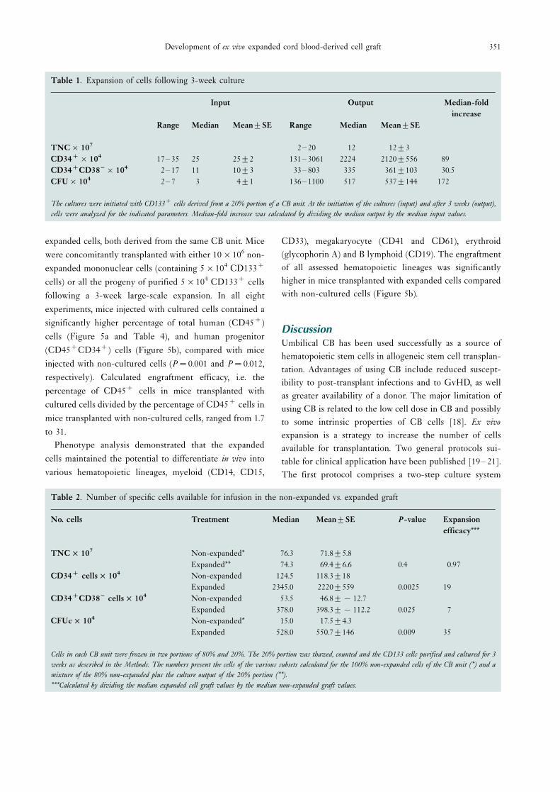
expanded cells, both derived from the same CB unit. Mice
were concomitantly transplanted with either 10�/106 non-
expanded mononuclear cells (containing 5�/104 CD133�
cells) or all the progeny of purified 5�/104 CD133� cells
following a 3-week large-scale expansion. In all eight
experiments, mice injected with cultured cells contained a
significantly higher percentage of total human (CD45�)
cells (Figure 5a and Table 4), and human progenitor
(CD45�CD34�) cells (Figure 5b), compared with mice
injected with non-cultured cells (P�/0.001 and P�/0.012,
respectively). Calculated engraftment efficacy, i.e. the
percentage of CD45� cells in mice transplanted with
cultured cells divided by the percentage of CD45� cells in
mice transplanted with non-cultured cells, ranged from 1.7
to 31.
Phenotype analysis demonstrated that the expanded
cells maintained the potential to differentiate in vivo into
various hematopoietic lineages, myeloid (CD14, CD15,
CD33), megakaryocyte (CD41 and CD61), erythroid
(glycophorin A) and B lymphoid (CD19). The engraftment
of all assessed hematopoietic lineages was significantly
higher in mice transplanted with expanded cells compared
with non-cultured cells (Figure 5b).
DiscussionUmbilical CB has been used successfully as a source of
hematopoietic stem cells in allogeneic stem cell transplan-
tation. Advantages of using CB include reduced suscept-
ibility to post-transplant infections and to GvHD, as well
as greater availability of a donor. The major limitation of
using CB is related to the low cell dose in CB and possibly
to some intrinsic properties of CB cells [18]. Ex vivo
expansion is a strategy to increase the number of cells
available for transplantation. Two general protocols sui-
table for clinical application have been published [19�/21].
The first protocol comprises a two-step culture system
Table 1. Expansion of cells following 3-week culture
Input Output Median-fold
increase
Range Median Mean9/SE Range Median Mean9/SE
TNC�/107 2�/20 12 129/3
CD34��/104 17�/35 25 259/2 131�/3061 2224 21209/556 89
CD34�CD38 � �/104 2�/17 11 109/3 33�/803 335 3619/103 30.5
CFU�/104 2�/7 3 49/1 136�/1100 517 5379/144 172
The cultures were initiated with CD133� cells derived from a 20% portion of a CB unit. At the initiation of the cultures (input) and after 3 weeks (output),
cells were analyzed for the indicated parameters. Median-fold increase was calculated by dividing the median output by the median input values.
Table 2. Number of specific cells available for infusion in the non-expanded vs. expanded graft
No. cells Treatment Median Mean9/SE P-value Expansion
efficacy***
TNC�/107 Non-expanded* 76.3 71.89/5.8
Expanded** 74.3 69.49/6.6 0.4 0.97
CD34� cells�/104 Non-expanded 124.5 118.39/18
Expanded 2345.0 22209/559 0.0025 19
CD34�CD38 � cells�/104 Non-expanded 53.5 46.89/�/12.7
Expanded 378.0 398.39/�/112.2 0.025 7
CFUc�/104 Non-expanded* 15.0 17.59/4.3
Expanded 528.0 550.79/146 0.009 35
Cells in each CB unit were frozen in two portions of 80% and 20%. The 20% portion was thawed, counted and the CD133 cells purified and cultured for 3
weeks as described in the Methods. The numbers present the cells of the various subsets calculated for the 100% non-expanded cells of the CB unit (*) and a
mixture of the 80% non-expanded plus the culture output of the 20% portion (**).
***Calculated by dividing the median expanded cell graft values by the median non-expanded graft values.
Development of ex vivo expanded cord blood-derived cell graft 351
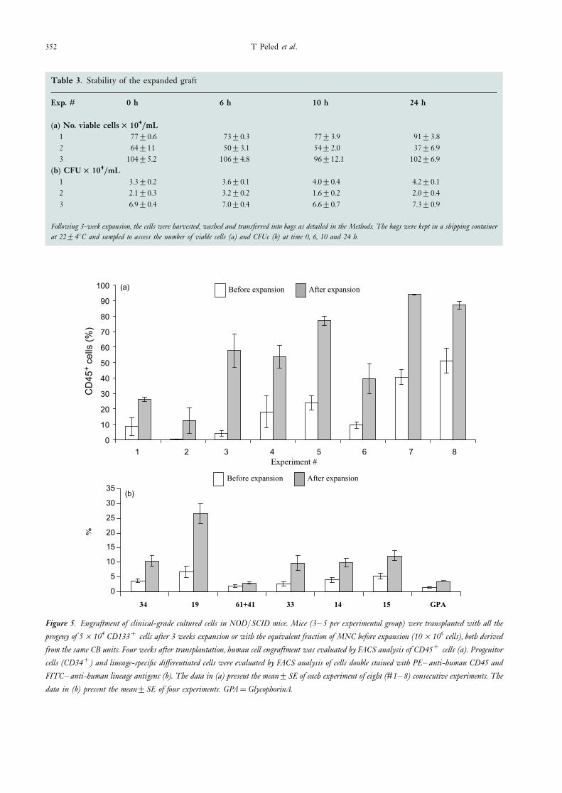
Table 3. Stability of the expanded graft
Exp. # 0 h 6 h 10 h 24 h
(a) No. viable cells�/104/mL
1 779/0.6 739/0.3 779/3.9 919/3.8
2 649/11 509/3.1 549/2.0 379/6.9
3 1049/5.2 1069/4.8 969/12.1 1029/6.9
(b) CFU�/104/mL
1 3.39/0.2 3.69/0.1 4.09/0.4 4.29/0.1
2 2.19/0.3 3.29/0.2 1.69/0.2 2.09/0.4
3 6.99/0.4 7.09/0.4 6.69/0.7 7.39/0.9
Following 3-week expansion, the cells were harvested, washed and transferred into bags as detailed in the Methods. The bags were kept in a shipping container
at 229/48C and sampled to assess the number of viable cells (a) and CFUc (b) at time 0, 6, 10 and 24 h.
Figure 5. Engraftment of clinical-grade cultured cells in NOD/SCID mice. Mice (3�/5 per experimental group) were transplanted with all the
progeny of 5�/104 CD133� cells after 3 weeks expansion or with the equivalent fraction of MNC before expansion (10�/106 cells), both derived
from the same CB units. Four weeks after transplantation, human cell engraftment was evaluated by FACS analysis of CD45� cells (a). Progenitor
cells (CD34�) and lineage-specific differentiated cells were evaluated by FACS analysis of cells double stained with PE�/anti-human CD45 and
FITC�/anti-human lineage antigens (b). The data in (a) present the mean9/SE of each experiment of eight (#1�/8) consecutive experiments. The
data in (b) present the mean9/SE of four experiments. GPA�/GlycophorinA.
352 T Peled et al .

initiated with CB-derived CD34� cells. The cells were
cultured in Teflon culture bags supplemented with defined
medium containing SCF, G-CSF and TPO [9]. The
second protocol comprises an automated continuous
perfusion culture device developed by Aastrom Bios-
ciences [21]. In this system cultures are initiated with
CB-derived mononuclear cells and expanded for 12 days
in media supplemented with FBS, horse serum, PIXY321,
FLT-3 ligand and erythropoietin [21]. The duration of
both clinical applicative protocols is 12�/14 days and in
both protocols the cultures are supplemented with a
mixture of early- and late-acting cytokines. These condi-
tions accelerate cell proliferation and differentiation,
resulting in increased expansion of TNC and further
maturation of myeloid precursors. Clinical studies per-
formed using these protocols demonstrated that clinical-
scale expansion of CB is feasible, and that administration
of these cells is well tolerated [19�/21].
Our current study presents an applicable protocol for
clinical-scale ex vivo expansion of CB-derived CD133�
progenitor cells in the presence of early-acting cytokines
and the copper chelator TEPA. As opposed to the
previously reported expansion protocols, it appears that
the present culture conditions enable better expansion of
less differentiated cell subsets.
Due to regulatory and proprietary concerns, we utilized
the Miltenyi Biotec’s CliniMACS apparatus [22] and the
anti-CD133 CliniMACS reagent, both of them clinically
approved, replacing the research-grade MiniMACS CD34
progenitor cell isolation kit. The CD133 antigen is a novel
marker for stem/progenitor cells. Phenotypic and func-
tional studies indicated that the CD133-enriched popula-
tion could serve as an alternative to CD34� cell selection
and engraftment purposes [23�/25]. Furthermore,
CD133� selected cells have already been used in clinical
transplantation settings without any safety impairment
[26]. In our study, we demonstrated similar yield and
purity, crucial parameters for successful transplantation
[22], using the CliniMACS instrument in combination
with either the anti-CD34 or anti-CD133 reagent.
The duration of most expansion protocols for clinical
application is between 10 and 14 days [19�/21]. We
evaluated the feasibility of the chelator technology to
extend the expansion in order to maximize the number of
early and late progenitor cells. To this end, we compared 2
vs. 3-week cultures and found that the latter was superior
with respect to the yields of TNC, CD34� and
Tab
le4
.E
ngr
aftm
ent
ofh
um
ance
lls
inS
CID
mic
eB
M
Ex
p.
#%
CD
45�
hu
man
cell
sin
mo
use
BM
4w
eek
saf
ter
tran
spla
nta
tio
n
No
n-c
ult
ure
dce
lls
Cu
ltu
red
cell
s
Mo
use
1M
ou
se2
Mo
use
3M
ou
se4
Mo
use
5A
vera
geS
EM
ou
se1
Mo
use
2M
ou
se3
Mo
use
4M
ou
se5
Ave
rage
SE
120
.03.
62.
1� /
�/8.
65.
728
.923
.326
.3�/
�/26
.21.
6
20.
20.
60.
4� /
�/0.
40.
128
.95.
13.
1�/
�/12
.48.
3
37.
52.
91.
9� /
�/4.
11.
763
.536
.873
.0�/
�/57
.810
.8
437
.62.
414
.2� /
�/18
.110
.461
.060
.939
.0�/
�/53
.67.
3
58.
726
.719
.033
.731
.623
.94.
678
.269
.483
.876
.4D
77.0
3.0
69.
011
.714
.510
.22.
59.
62.
060
.314
.437
.545
.9D
39.5
9.6
746
.128
.449
.338
.6D
40.6
4.7
94.6
94.3
93.2
93.8
D94
.00.
3
843
.267
.532
.361
.1D
51.0
8.1
77.5
86.6
91.8
87.7
91.6
87.0
2.6
The
tabl
ede
mon
stra
tes
the
raw
data
ofth
eex
peri
men
tspr
esen
ted
inF
igur
e5a
.
D�
/de
ad;�/�
/n
otdo
ne.
Development of ex vivo expanded cord blood-derived cell graft 353
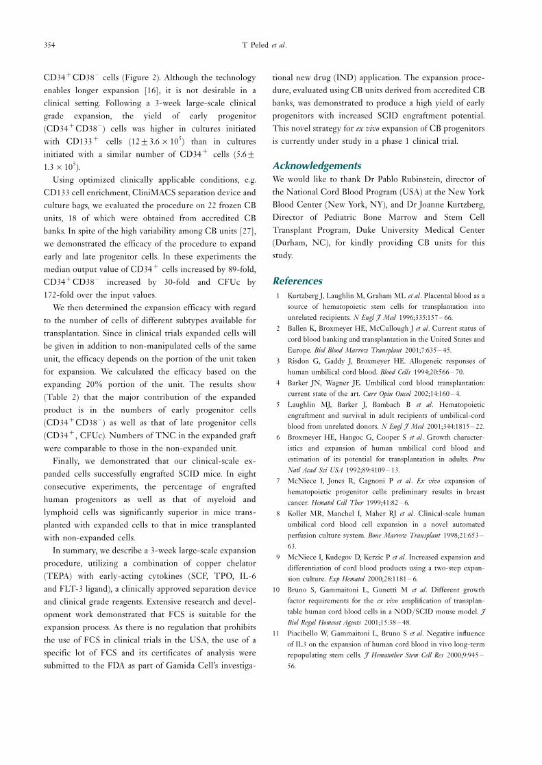
CD34�CD38� cells (Figure 2). Although the technology
enables longer expansion [16], it is not desirable in a
clinical setting. Following a 3-week large-scale clinical
grade expansion, the yield of early progenitor
(CD34�CD38� ) cells was higher in cultures initiated
with CD133� cells (129/3.6�/105) than in cultures
initiated with a similar number of CD34� cells (5.69/
1.3�/105).
Using optimized clinically applicable conditions, e.g.
CD133 cell enrichment, CliniMACS separation device and
culture bags, we evaluated the procedure on 22 frozen CB
units, 18 of which were obtained from accredited CB
banks. In spite of the high variability among CB units [27],
we demonstrated the efficacy of the procedure to expand
early and late progenitor cells. In these experiments the
median output value of CD34� cells increased by 89-fold,
CD34�CD38� increased by 30-fold and CFUc by
172-fold over the input values.
We then determined the expansion efficacy with regard
to the number of cells of different subtypes available for
transplantation. Since in clinical trials expanded cells will
be given in addition to non-manipulated cells of the same
unit, the efficacy depends on the portion of the unit taken
for expansion. We calculated the efficacy based on the
expanding 20% portion of the unit. The results show
(Table 2) that the major contribution of the expanded
product is in the numbers of early progenitor cells
(CD34�CD38� ) as well as that of late progenitor cells
(CD34�, CFUc). Numbers of TNC in the expanded graft
were comparable to those in the non-expanded unit.
Finally, we demonstrated that our clinical-scale ex-
panded cells successfully engrafted SCID mice. In eight
consecutive experiments, the percentage of engrafted
human progenitors as well as that of myeloid and
lymphoid cells was significantly superior in mice trans-
planted with expanded cells to that in mice transplanted
with non-expanded cells.
In summary, we describe a 3-week large-scale expansion
procedure, utilizing a combination of copper chelator
(TEPA) with early-acting cytokines (SCF, TPO, IL-6
and FLT-3 ligand), a clinically approved separation device
and clinical grade reagents. Extensive research and devel-
opment work demonstrated that FCS is suitable for the
expansion process. As there is no regulation that prohibits
the use of FCS in clinical trials in the USA, the use of a
specific lot of FCS and its certificates of analysis were
submitted to the FDA as part of Gamida Cell’s investiga-
tional new drug (IND) application. The expansion proce-
dure, evaluated using CB units derived from accredited CB
banks, was demonstrated to produce a high yield of early
progenitors with increased SCID engraftment potential.
This novel strategy for ex vivo expansion of CB progenitors
is currently under study in a phase 1 clinical trial.
AcknowledgementsWe would like to thank Dr Pablo Rubinstein, director of
the National Cord Blood Program (USA) at the New York
Blood Center (New York, NY), and Dr Joanne Kurtzberg,
Director of Pediatric Bone Marrow and Stem Cell
Transplant Program, Duke University Medical Center
(Durham, NC), for kindly providing CB units for this
study.
References
1 Kurtzberg J, Laughlin M, Graham ML et al . Placental blood as a
source of hematopoietic stem cells for transplantation into
unrelated recipients. N Engl J Med 1996;335:157�/66.
2 Ballen K, Broxmeyer HE, McCullough J et al . Current status of
cord blood banking and transplantation in the United States and
Europe. Biol Blood Marrow Transplant 2001;7:635�/45.
3 Risdon G, Gaddy J, Broxmeyer HE. Allogeneic responses of
human umbilical cord blood. Blood Cells 1994;20:566�/70.
4 Barker JN, Wagner JE. Umbilical cord blood transplantation:
current state of the art. Curr Opin Oncol 2002;14:160�/4.
5 Laughlin MJ, Barker J, Bambach B et al . Hematopoietic
engraftment and survival in adult recipients of umbilical-cord
blood from unrelated donors. N Engl J Med 2001;344:1815�/22.
6 Broxmeyer HE, Hangoc G, Cooper S et al . Growth character-
istics and expansion of human umbilical cord blood and
estimation of its potential for transplantation in adults. Proc
Natl Acad Sci USA 1992;89:4109�/13.
7 McNiece I, Jones R, Cagnoni P et al . Ex vivo expansion of
hematopoietic progenitor cells: preliminary results in breast
cancer. Hematol Cell Ther 1999;41:82�/6.
8 Koller MR, Manchel I, Maher RJ et al . Clinical-scale human
umbilical cord blood cell expansion in a novel automated
perfusion culture system. Bone Marrow Transplant 1998;21:653�/
63.
9 McNiece I, Kudegov D, Kerzic P et al . Increased expansion and
differentiation of cord blood products using a two-step expan-
sion culture. Exp Hematol 2000;28:1181�/6.
10 Bruno S, Gammaitoni L, Gunetti M et al . Different growth
factor requirements for the ex vivo amplification of transplan-
table human cord blood cells in a NOD/SCID mouse model. J
Biol Regul Homeost Agents 2001;15:38�/48.
11 Piacibello W, Gammaitoni L, Bruno S et al . Negative influence
of IL3 on the expansion of human cord blood in vivo long-term
repopulating stem cells. J Hematother Stem Cell Res 2000;9:945�/
56.
354 T Peled et al .
12 Henon P, Sovalat H, Becker M et al . Primordial role of CD34�/
38- cells in early and late trilineage haemopoietic engraftment
after autologous blood cell transplantation. Br J Haematol
1998;103:568�/81.
13 Henon PH, Sovalat H, Bourderont D. Importance of CD34�
cell subsets in autologous PBSC transplantation: the mulhouse
experience using CD34�. J Biol Regul Homeost Agents
2001;15:62�/7.
14 Ishikawa F, Livingston AG, Minamiguchi H et al . Human cord
blood long-term engrafting cells are CD34�CD38 � . Leukemia
2003;17:960�/4.
15 Peled T, Landau E, Prus E et al . Cellular copper content
modulates differentiation and self-renewal in cultures of cord
blood-derived CD34� cells. Br J Haematol 2002;116:655�/61.
16 Peled T, Landau E, Mandel J et al . Linear polyamine copper
chelator tetraethylenepentamine augments long-term ex vivo
expansion of cord-blood-derived CD34(�/) cells and increases
their engraftment potential in NOD/SCID mice. Exp Hematol
2004;32:547�/55.
17 Rubinstein P, Dobrila L, Rosenfield RE et al . Processing and
cryopreservation of placental/umbilical cord blood for unrelated
bone marrow reconstitution. Proc Natl Acad Sci USA
1995;92:10119�/22.
18 Lewis ID. Clinical and experimental use of umbilical cord blood.
Intern Med J 2002;32:601�/9.
19 Astori G, Malangone W, Adami V et al . A novel protocol that
allows short-term stem cell expansion of both committed and
pluripotent hematopoietic progenitor cells suitable for clinical
use. Blood Cells Mol Dis 2001;27:715�/24, 725�/7.
20 Shpall EJ, Quinones R, Giller R et al . Transplantation of ex vivo
expanded cord blood. Biol Blood Marrow Transplant 2002;8:368�/
76.
21 Jaroscak J, Goltry K, Smith A et al . Augmentation of umbilical
cord blood (UCB) transplantation with ex vivo -expanded UCB
cells: results of a phase 1 trial using the AastromReplicell
System. Blood 2003;101:5061�/7.
22 McNiece IK, Stoney GB, Keren BP et al . CD34�cell selection
from frozen cord blood products using the Isolex 300i and
CliniMACS CD34 selection device. J Hematother 1998;7:457�/61.
23 de Wynter EA, Buck D, Hart C et al . CD34�CD133� cells
isolated from cord blood are highly enriched in long-term
culture-initiating cells, NOD/SCID-repopulating cells and
dendritic cell progenitors. Stem Cells 1998;16:387�/96.
24 Yin AH, Miraglia S, Zanjani ED et al . CD133, a novel marker for
human hematopoietic stem and progenitor cells. Blood
1997;90:5002�/12.
25 Pasino M, Lanza T, Marotta F et al . Flow cytometric and
functional characterization of CD133� cells from human
umbilical cord blood. Br J Haematol 2000;108:793�/800.
26 Koehl U, Esser R, Zimmermann S et al . Clinical scale
purification of progenitor cells by CD133� selection: from
laboratory experience to the first transplantation of a pediatric
patient with relapsed leukemia. Blood 2001;98:851a.
27 Encabo A, Mateu E, Carbonell-Uberos F et al . CD34�CD38 � is
a good predictive marker of cloning ability and expansion
potential of CD34� cord blood cells. Transfusion 2003;43:
383�/9.
Development of ex vivo expanded cord blood-derived cell graft 355
International Journal of Pharmaceutics 312 (2006) 15–23
Cutaneous gene expression of plasmid DNA in excised humanskin following delivery via microchannels created
by radio frequency ablation
James Birchall a,∗, Sion Coulman a, Alexander Anstey b, Chris Gateley b,Helen Sweetland c, Amikam Gershonowitz d, Lewis Neville d, Galit Levin d
a Gene Delivery Research Group, Welsh School of Pharmacy, Cardiff University, Cardiff CF10 3XF, UKb Gwent Healthcare NHS Trust, Royal Gwent Hospital, Cardiff Road, Newport, South Wales NP20 2UB, UKc School of Medicine, Cardiff University & University Hospital of Wales, Heath Park, Cardiff CF14 4XN, UK
d TransPharma Medical Ltd., 2 Yodfat Street, Northern Industrial Zone, Lod 71291, Israel
Received 8 September 2005; received in revised form 5 December 2005; accepted 5 December 2005Available online 15 February 2006
A
ptTntDtta©
K
1
edow‘ceaa
(
0d
bstract
The skin is a valuable organ for the development and exploitation of gene medicines. Delivering genes to skin is restricted however by thehysico-chemical properties of DNA and the stratum corneum (SC) barrier. In this study, we demonstrate the utility of an innovative technologyhat creates transient microconduits in human skin, allowing DNA delivery and resultant gene expression within the epidermis and dermis layers.he radio frequency (RF)-generated microchannels were of sufficient morphology and depth to permit the epidermal delivery of 100 nm diameteranoparticles. Model fluorescent nanoparticles were used to confirm the capacity of the channels for augmenting diffusion of macromoleculeshrough the SC. An ex vivo human organ culture model was used to establish the gene expression efficiency of a �-galactosidase reporter plasmidNA applied to ViaDermTM treated skin. Skin treated with ViaDermTM using 50 �m electrode arrays promoted intense levels of gene expression in
he viable epidermis. The intensity and extent of gene expression was superior when ViaDermTM was used following a prior surface application ofhe DNA formulation. In conclusion, the RF-microchannel generator (ViaDermTM) creates microchannels amenable for delivery of nanoparticlesnd gene therapy vectors to the viable region of skin.
2006 Elsevier B.V. All rights reserved.
eywords: Radiofrequency-microchannels; Radiofrequency ablation; Plasmid DNA; Skin; Gene therapy
. Introduction
The ability to target genes directly to the skin provides a strat-gy for the treatment of certain localised heritable genetic skiniseases (Greenhalgh et al., 1994; Ehrlich et al., 1995), vari-us forms of malignancies (Hart and Vile, 1994) and cutaneousounds (Byrnes et al., 2004; Lee et al., 2004). Furthermore,
genetic immunisation’ via the skin provides a method of vac-inating patients by introducing DNA into cells, leading toxpression of foreign antigen and the subsequent induction ofn immune response (Fynan et al., 1993; Raz et al., 1994; Shi etl., 1999). Intra-cutaneous DNA vaccines utilise the highly com-
∗ Corresponding author. Tel.: +44 29 20875815; fax: +44 29 20874149.E-mail addresses: [email protected], [email protected]
J. Birchall).
petent antigen-presenting capabilities of epidermal Langerhanscells in eliciting a systemic immune response, leading to moreproficient and cost-efficient vaccination compared with conven-tional vaccines (Lin et al., 2000). As the immune response isinduced by a single gene rather than an entire organism, thisapproach is also considered to be safer than using live attenu-ated vaccines (Durrant, 1997).
The challenge of delivering genes to the viable region ofskin is a product of the physico-chemical properties of the largehydrophilic DNA molecule, with or without an additional car-rier vehicle, and the significant barrier properties of cutaneoustissue. Superficially the skin is regarded as a valuable organ forthe development and clinical administration of gene medicinesas it is readily accessed, well characterized and easily monitored(Hengge et al., 1996). However, if cutaneous gene therapy is totranslate from the laboratory to clinical practice then approaches
378-5173/$ – see front matter © 2006 Elsevier B.V. All rights reserved.oi:10.1016/j.ijpharm.2005.12.036

16 J. Birchall et al. / International Journal of Pharmaceutics 312 (2006) 15–23
must be developed to efficiently and reproducibly transport thedelivered transgene to the target cell population. The primaryrole of the skin however, is to serve as a physical barrier to theinvasion of foreign material. In humans, the epidermis, whichconstitutes the uppermost layer of the skin, is approximately50–150 �m thick with the non-viable SC layer, approximately15–20 �m in thickness, representing the principal barrier to pen-etration and permeation of substances through the skin (Birchall,2004). Therefore, in order to deliver therapeutic compounds tothe epidermis, the underlying dermis or the systemic circulation,delivery strategies must overcome the physical barrier createdby the nature of the tightly packed dead cells of the SC. Tra-ditional transdermal formulation strategies aim to enhance thedelivery of small therapeutic molecules, less than 500 molecularweight, across the SC by paracellular, transcellular or intracellu-lar routes. However, in order to deliver DNA and proteins, moreinnovative and radical methods of drug delivery are required. Todate, the physico-chemical methods employed to promote ther-apeutic drug or gene transfer to the skin include the use of directDNA injection (Hengge et al., 1995, 1996; Chesnoy and Huang,2002) chemical enhancers (Barry, 1987; Pillai and Panchagnula,2003), iontophoresis (Green, 1996; Preat and Dujardin, 2001),biolistic particle bombardment (Cheng et al., 1993; Heiser, 1994;Udvardi et al., 1999), electroporation (Prausnitz et al., 1993;Dujardin et al., 2001; Zhang et al., 2002), sonophoresis (Lavonand Kost, 2004), laser ablation (Nelson et al., 1991), microseed-it1
casVwd(ttcoorim
ntecusCdpUt
microchannels by cell ablation at specific locations (Levin etal., 2005).
The purpose of the present study using the ViaDermTM
technology was two-fold. Firstly, to extensively characterizeViaDermTM-generated microchannels within ex vivo humanskin. Secondly, to assess the feasibility of ViaDermTM in sup-porting the transdermal delivery of a mammalian expressionplasmid with subsequent reporter expression within the targetregion of the skin.
2. Materials and methods
2.1. Materials
The 7.2 kb pCMV� plasmid construct containing the �-galactosidase reporter gene and the pEGFP-N1 (4.7 kb) plas-mid containing the green fluorescent protein reporter gene werepropagated and purified as detailed previously (Birchall et al.,1999). Fluorescein isothiocyante (FITC)-labelled polystyrenenanospheres (L-1280) were obtained from Sigma Chemicals(Poole, UK). OCT embedding medium and Histobond® micro-scope slides were from RA Lamb Ltd. (Eastbourne, UK).One percent aqueous eosin solution and Harris’ haematoxylinsolution were from BDH Laboratory Supplies (Dorset, UK).One percent aqueous toludine blue solution was from TAABLaboratories Equipment Ltd. (Berkshire, UK). Cell culturepUEpwoF
2
tidwtiwb
mIBt(t(nefi3
ng (Eriksson et al., 1998), skin tattooing (Bins et al., 2005) andhe recent use of microfabricated microneedles (Henry et al.,998; McAllister et al., 2000, 2003; Chabri et al., 2004).
Recently, we have developed an innovative technology,oined ViaDermTM, which creates transient microchannelscross the SC thereby enabling a more direct and controlled pas-age of molecules to the underlying viable epidermis and dermis.iaDermTM has an intimately spaced array of microelectrodeshich are placed against the surface of skin to individually con-uct an applied alternating electrical current at radio frequencyRF). Application of this rf electrical current (100–500 kHz) tohe tissue elicits a vibration in motion of ions with localized fric-ional heating of tissue resulting in a rapid obliteration of cellslose to the energy source. The intimate and orderly spacingf the microelectrodes therefore drives the orderly generationf functional microchannels. The passage of the electric cur-ent through cells in the upper skin strata generates localisedonic vibrations, heating, evaporation and cell ablation to create
icrochannels.Previously, we have reported that RF-generated microchan-
els reside in the epidermis and dermis and are amenable tohe effective transdermal delivery of small molecules (Sintovt al., 2003) and proteins (Levin et al., 2005) into the systemicirculation. Furthermore, the microchannels did not impinge onnderlying blood vessels and nerve endings thus minimizingkin trauma, bleeding and neural sensations (Sintov et al., 2003).learly, the use of electricity for augmenting transcutaneousrug delivery also applies to some of the other aforementionedhysical delivery methods, e.g. iontophoresis, electroporation.nlike these examples however, the technology described in
his study leads to the creation of an orderly array of defined
lastics were obtained from Corning-Costar (High Wycombe,K). MEM (EAGLES) 25 mM HEPES, Dulbecco’s Modifiedagle’s Medium (DMEM 25 mM HEPES), foetal bovine serum,enicillin-streptomycin solution and trypsin-EDTA solution 1×ere obtained from In-Vitrogen Corporation, Paisley, UK. Allther reagents were of analytical grade and purchased fromisher Scientific UK (Loughborough, UK).
.2. ViaDermTM treatment of human skin
Full-thickness human breast skin was obtained from mastec-omy or breast reduction with ethical committee approval andnformed patient consent. Skin was collected from a variety ofonors ranging from 45 to 65 years of age. Matched samplesere used for each individual experiment. To maintain struc-
ural and cellular viability the skin tissue was transported once in MEM (EAGLES) 25 mM HEPES growth media and usedithin 3 h of excision. All excess adipose tissue was removedy blunt dissection.
The components and operating conditions of the RF-icrochannel generator (ViaDermTM, TransPharma Medical,
srael) have been described previously (Sintov et al., 2003).riefly the ViaDermTM device comprises an electronic con-
roller unit and a disposable array of stainless steel electrodes100 or 50 �m in length) at a density of 100 electrodes/cm2 in aotal area of 1.4 cm2. Thus, application of an RF-activated array1.2 cm × 1.2 cm) resulted in the generation of 144 microchan-els over the 1.4 cm2 area. Studies were performed using thelectrodes at device parameter settings resulting in one, two orve bursts of 700 �s burst length at an applied voltage of 290 or30 V and an RF frequency of 100 kHz. Control experiments
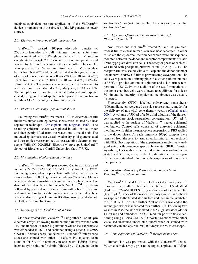
J. Birchall et al. / International Journal of Pharmaceutics 312 (2006) 15–23 17
involved equivalent pressure application of the ViaDermTM
device to human skin in the absence of the RF-generating powersource.
2.3. Electron microscopy of full thickness skin
ViaDermTM treated (100 �m electrode, density of200 microchannels/cm2) full thickness human skin sam-ples were fixed with 2.5% glutaraldehyde in 0.1 M sodiumcacodylate buffer (pH 7.4) for 60 min at room temperature andwashed for 10 min (2 × 5 min) in the same buffer. The sampleswere post-fixed in 1% osmium tetroxide in 0.1 M cacodylatebuffer for 1 h at 4 ◦C and then dehydrated with a graded seriesof ethanol concentrations as follows (70% for 10 min at 4 ◦C,100% for 10 min at 4 ◦C, 100% for 10 min at 4 ◦C, 100% for10 min at 4 ◦C). The samples were subsequently transferred toa critical point drier (Samdri 780, Maryland, USA) for 12 h.The samples were mounted on metal stubs and gold sputtercoated, using an Edward sputter coater, prior to examination ina Philips XL-20 scanning electron microscope.
2.4. Electron microscopy of epidermal sheets
Following ViaDermTM treatment (100 �m electrode) of fullthickness human skin, epidermal sheets were isolated by a heatsramasS
2
iFsldfawK
2
ePwCssh
solution for 5 s or (iii) toludine blue: 1% aqueous toludine bluesolution for 5 min.
2.7. Diffusion of fluorescent nanoparticles throughRF-microchannelsTM
Non-treated and ViaDermTM treated (50 and 100 �m elec-trodes) full thickness human skin was heat separated in orderto isolate the epidermal membranes which were subsequentlymounted between the donor and receptor compartments of staticFranz-type glass diffusion cells. The receptor phase of each cellwas filled with phosphate buffered saline (PBS; pH 7.4). Thereceptor arm was sealed with a foil cap and the donor chamberoccluded with NESCO® film to prevent sample evaporation. Thecells were placed on a stirring plate in a water-bath maintainedat 37 ◦C, to provide continuous agitation and a skin surface tem-perature of 32 ◦C. Prior to addition of the test formulations tothe donor chamber, cells were allowed to equilibrate for at least30 min and the integrity of epidermal membranes was visuallyinspected.
Fluorescently (FITC) labelled polystyrene nanospheres(100 nm diameter) were used as a size-representative model forthe delivery of non-viral gene therapy vectors (Chabri et al.,2004). A volume of 500 �l of a 50 �l/ml dilution of the fluores-cent nanosphere stock suspension, concentration 4.5710 �l−1,was applied to the surface of ViaDermTM treated epidermalmmtrwyAafn
2V
a((wfsw1tvh
2
5
eparation technique (Christophers and Kligman, 1963). Theesulting epidermal sheets were placed in cold distilled waternd then gently lifted from the water onto a metal stub. Theounted epidermal sheet was allowed to dry, gold sputter coated
nd the samples were examined using a scanning electron micro-cope (Philips Xl-200 SEM) (Electron Microscopy Unit, Cardiffchool of Biosciences, Cardiff University, Cardiff, UK).
.5. Visualisation of microchannels en face
ViaDermTM treated (100 �m electrode) skin was incubatedn media (MEM (EAGLES), 25 mM HEPES) for 24 h at 37 ◦C.ollowing two washes in phosphate buffered saline (PBS) thekin was fixed in 0.5% gluataraldehyde for 2 h on ice. Methy-ene blue staining involved a 5 min surface application of fiverops of methylene blue solution on the ViaDermTM treated skinollowed by removal of excessive stain with a brief PBS rinsend an ethanol surface swab. Tissue stained with methylene blueas visualised using an Olympus BX50 microscope and a SchottL1500 electronic light source.
.6. Histology of ViaDermTM treated tissue
Skin was treated with ViaDermTM using either 50 or 100 �mlectrode arrays. Following treatment the skin was washed withBS and fixed for 4 h in 0.5% glutaraldehyde on ice. Fixed tissueas embedded in OCT and sectioned using a Leica CM3050Sryostat. Sections were collected on Histobond® microscope
lides and stained with either—(i) eosin: 1% aqueous eosinolution for 5 s, (ii) haematoxylin and eosin (H&E): Harris’aematoxylin solution for 5 min followed by 1% aqueous eosin
embranes. Control cells consisted of untreated epidermalembrane with either the nanosphere suspension or PBS applied
o the donor phase. At each timepoint 200 �l samples wereemoved from the receptor arm at regular intervals and replacedith PBS. On completion of the experiment, samples were anal-sed using a fluorescence spectrophotometer (BMG Fluostar,ylesbury, UK) with excitation and emission wavelengths sett 485 and 520 nm, respectively. A calibration curve was per-ormed using standard dilutions of the suspension of fluorescentanoparticles.
.8. Localised delivery of fluorescent nanoparticles iniaDermTM treated human skin
ViaDermTM treated (100 �m electrode) skin was placed insix-well cell culture plate and maintained in 1.5 ml MEM
EAGLES) 25 mM HEPES. Fifty microliters of a concentrated4.5710 �l−1) stock of fluorescent red polystyrene nanospheresas applied to the treated skin surface and the sample incubated
or 6 h at 37 ◦C. At 6 h a further 2 ml of media was added theubmerged skin was incubated for a further 18 h. Following twoashes in PBS the skin was fixed in 0.5% gluataraldehyde forh on ice and embedded in OCT medium prior to tissue sec-
ioning using a Leica CM3050S Cryostat. Sections were eitherisualised unstained under blue fluorescence or stained withaematoxylin and eosin (H&E) (Olympus BX50 microscope).
.9. Gene expression in ViaDermTM treated human skin
Human skin was pre-treated with the ViaDermTM device,0 �m electrode arrays, prior to the topical application of 50 �l
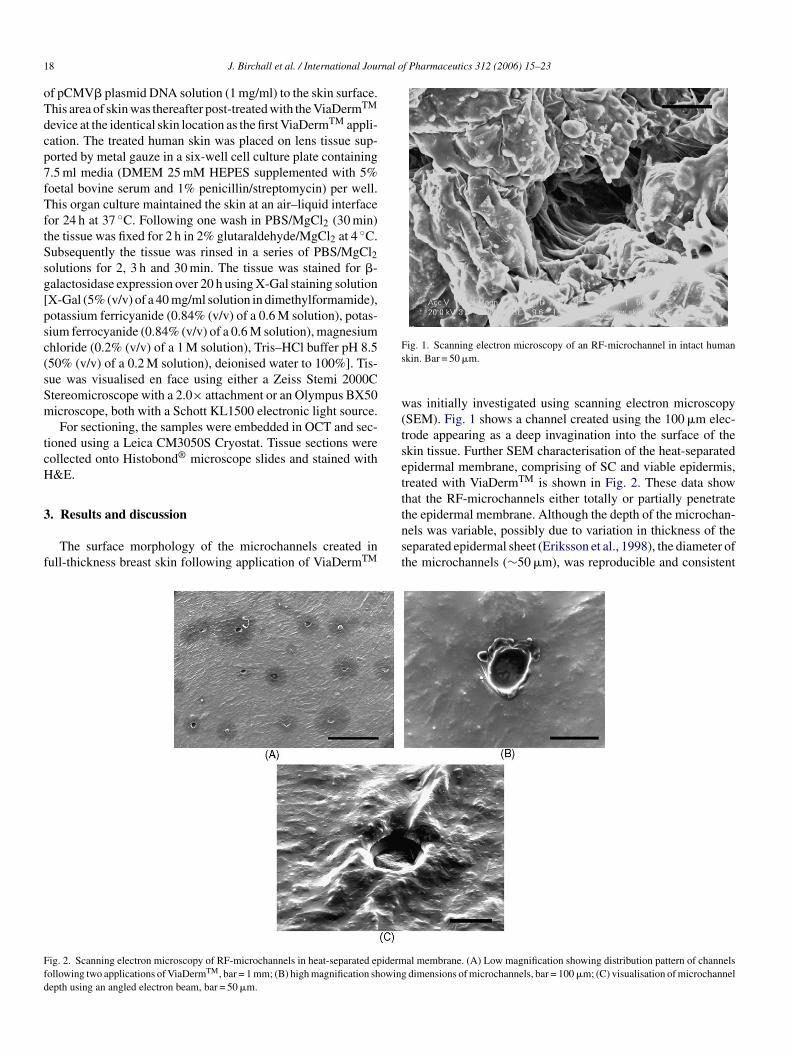
18 J. Birchall et al. / International Journal of Pharmaceutics 312 (2006) 15–23
of pCMV� plasmid DNA solution (1 mg/ml) to the skin surface.This area of skin was thereafter post-treated with the ViaDermTM
device at the identical skin location as the first ViaDermTM appli-cation. The treated human skin was placed on lens tissue sup-ported by metal gauze in a six-well cell culture plate containing7.5 ml media (DMEM 25 mM HEPES supplemented with 5%foetal bovine serum and 1% penicillin/streptomycin) per well.This organ culture maintained the skin at an air–liquid interfacefor 24 h at 37 ◦C. Following one wash in PBS/MgCl2 (30 min)the tissue was fixed for 2 h in 2% glutaraldehyde/MgCl2 at 4 ◦C.Subsequently the tissue was rinsed in a series of PBS/MgCl2solutions for 2, 3 h and 30 min. The tissue was stained for �-galactosidase expression over 20 h using X-Gal staining solution[X-Gal (5% (v/v) of a 40 mg/ml solution in dimethylformamide),potassium ferricyanide (0.84% (v/v) of a 0.6 M solution), potas-sium ferrocyanide (0.84% (v/v) of a 0.6 M solution), magnesiumchloride (0.2% (v/v) of a 1 M solution), Tris–HCl buffer pH 8.5(50% (v/v) of a 0.2 M solution), deionised water to 100%]. Tis-sue was visualised en face using either a Zeiss Stemi 2000CStereomicroscope with a 2.0× attachment or an Olympus BX50microscope, both with a Schott KL1500 electronic light source.
For sectioning, the samples were embedded in OCT and sec-tioned using a Leica CM3050S Cryostat. Tissue sections werecollected onto Histobond® microscope slides and stained withH&E.
3
f
Fig. 1. Scanning electron microscopy of an RF-microchannel in intact humanskin. Bar = 50 �m.
was initially investigated using scanning electron microscopy(SEM). Fig. 1 shows a channel created using the 100 �m elec-trode appearing as a deep invagination into the surface of theskin tissue. Further SEM characterisation of the heat-separatedepidermal membrane, comprising of SC and viable epidermis,treated with ViaDermTM is shown in Fig. 2. These data showthat the RF-microchannels either totally or partially penetratethe epidermal membrane. Although the depth of the microchan-nels was variable, possibly due to variation in thickness of theseparated epidermal sheet (Eriksson et al., 1998), the diameter ofthe microchannels (∼50 �m), was reproducible and consistent
F idermfd
. Results and discussion
The surface morphology of the microchannels created inull-thickness breast skin following application of ViaDermTM
ig. 2. Scanning electron microscopy of RF-microchannels in heat-separated epTM
ollowing two applications of ViaDerm , bar = 1 mm; (B) high magnification showingepth using an angled electron beam, bar = 50 �m.
al membrane. (A) Low magnification showing distribution pattern of channelsdimensions of microchannels, bar = 100 �m; (C) visualisation of microchannel
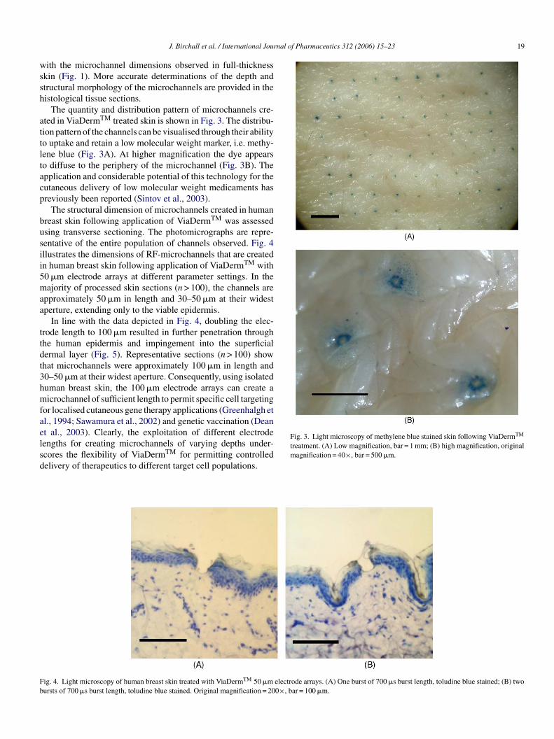
J. Birchall et al. / International Journal of Pharmaceutics 312 (2006) 15–23 19
with the microchannel dimensions observed in full-thicknessskin (Fig. 1). More accurate determinations of the depth andstructural morphology of the microchannels are provided in thehistological tissue sections.
The quantity and distribution pattern of microchannels cre-ated in ViaDermTM treated skin is shown in Fig. 3. The distribu-tion pattern of the channels can be visualised through their abilityto uptake and retain a low molecular weight marker, i.e. methy-lene blue (Fig. 3A). At higher magnification the dye appearsto diffuse to the periphery of the microchannel (Fig. 3B). Theapplication and considerable potential of this technology for thecutaneous delivery of low molecular weight medicaments haspreviously been reported (Sintov et al., 2003).
The structural dimension of microchannels created in humanbreast skin following application of ViaDermTM was assessedusing transverse sectioning. The photomicrographs are repre-sentative of the entire population of channels observed. Fig. 4illustrates the dimensions of RF-microchannels that are createdin human breast skin following application of ViaDermTM with50 �m electrode arrays at different parameter settings. In themajority of processed skin sections (n > 100), the channels areapproximately 50 �m in length and 30–50 �m at their widestaperture, extending only to the viable epidermis.
In line with the data depicted in Fig. 4, doubling the elec-trode length to 100 �m resulted in further penetration throughthe human epidermis and impingement into the superficialdt3hmfaelsd
Fig. 3. Light microscopy of methylene blue stained skin following ViaDermTM
treatment. (A) Low magnification, bar = 1 mm; (B) high magnification, originalmagnification = 40×, bar = 500 �m.
F electrode arrays. (A) One burst of 700 �s burst length, toludine blue stained; (B) twob 0×, bar = 100 �m.
ermal layer (Fig. 5). Representative sections (n > 100) showhat microchannels were approximately 100 �m in length and0–50 �m at their widest aperture. Consequently, using isolateduman breast skin, the 100 �m electrode arrays can create aicrochannel of sufficient length to permit specific cell targeting
or localised cutaneous gene therapy applications (Greenhalgh etl., 1994; Sawamura et al., 2002) and genetic vaccination (Deant al., 2003). Clearly, the exploitation of different electrodeengths for creating microchannels of varying depths under-cores the flexibility of ViaDermTM for permitting controlledelivery of therapeutics to different target cell populations.
ig. 4. Light microscopy of human breast skin treated with ViaDermTM 50 �mursts of 700 �s burst length, toludine blue stained. Original magnification = 20

20 J. Birchall et al. / International Journal of Pharmaceutics 312 (2006) 15–23
Fig. 5. Light microscopy of human breast skin treated with ViaDermTM 100 �m electrode arrays. (A) One burst of 700 �s burst length, toludine blue stained; (B)three bursts of 700 �s burst length, toludine blue stained. Original magnification = 200×, bar = 100 �m.
Previously, from ex vivo studies employing a permeationmethodology, we have demonstrated the total inability of theViaDermTM device to generate microchannels when discon-nected from a power source as evidenced by both negativevisualization and lack of drug permeation (Sintov et al., 2003).Such findings were totally substantiated in follow up in vivostudies whereby application of drugs at a ViaDermTM treatedskin site in the absence of a power supply resulted in no trans-dermal drug delivery as compared to robust drug deliveries with
a functional power supply (Sintov et al., 2003; Levin et al.,2005). In our histological studies, and subsequent gene deliv-ery experiments, we confirm the previously published ex vivoand in vivo observations (Sintov et al., 2003; Levin et al., 2005)of the total absence of microchannels on the surface skin follow-ing the placement of the ViaDermTM device disconnected froma functional power source.
Following confirmation of the ability of ViaDermTM to cre-ate microchannels in human skin, further experiments were
F(tt
ig. 6. Light (A) and fluorescent (B) photomicrographs of RF-microchannels contaC) diffusion of fluorescent nanoparticles through ViaDermTM treated epidermal memhe receptor phase of Franz cells over a 12 h period. (©) Untreated skin—PBS donor preated skin—topical nanoparticles, (�) 100 �m array ViaDermTM treated skin—top
ining fluorescent nanoparticles. Original magnification = 100×, bar = 100 �m;branes. Data presented as percentage of topical nanoparticle dose detected in
hase, (×) Untreated skin—topical nanoparticles, (�) 50 �m array ViaDermTM
ical nanoparticles (N = 3 ± S.D.).
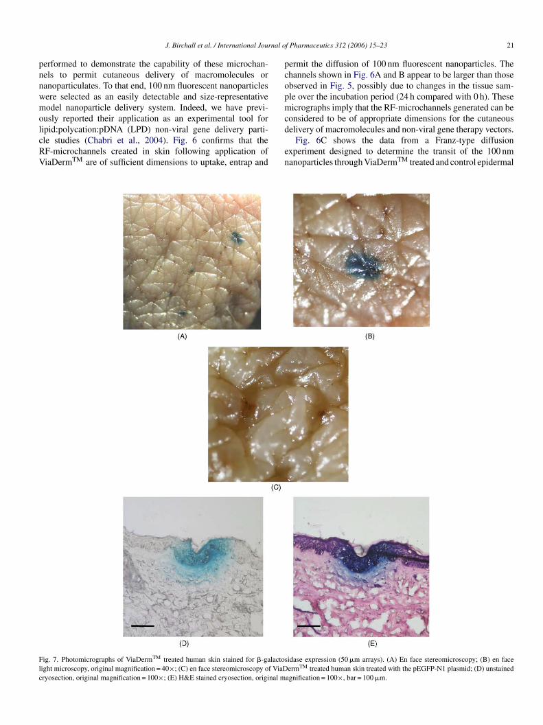
J. Birchall et al. / International Journal of Pharmaceutics 312 (2006) 15–23 21
performed to demonstrate the capability of these microchan-nels to permit cutaneous delivery of macromolecules ornanoparticulates. To that end, 100 nm fluorescent nanoparticleswere selected as an easily detectable and size-representativemodel nanoparticle delivery system. Indeed, we have previ-ously reported their application as an experimental tool forlipid:polycation:pDNA (LPD) non-viral gene delivery parti-cle studies (Chabri et al., 2004). Fig. 6 confirms that theRF-microchannels created in skin following application ofViaDermTM are of sufficient dimensions to uptake, entrap and
permit the diffusion of 100 nm fluorescent nanoparticles. Thechannels shown in Fig. 6A and B appear to be larger than thoseobserved in Fig. 5, possibly due to changes in the tissue sam-ple over the incubation period (24 h compared with 0 h). Thesemicrographs imply that the RF-microchannels generated can beconsidered to be of appropriate dimensions for the cutaneousdelivery of macromolecules and non-viral gene therapy vectors.
Fig. 6C shows the data from a Franz-type diffusionexperiment designed to determine the transit of the 100 nmnanoparticles through ViaDermTM treated and control epidermal
Flc
ig. 7. Photomicrographs of ViaDermTM treated human skin stained for �-galactosight microscopy, original magnification = 40×; (C) en face stereomicroscopy of ViaDryosection, original magnification = 100×; (E) H&E stained cryosection, original m
idase expression (50 �m arrays). (A) En face stereomicroscopy; (B) en faceermTM treated human skin treated with the pEGFP-N1 plasmid; (D) unstained
agnification = 100×, bar = 100 �m.
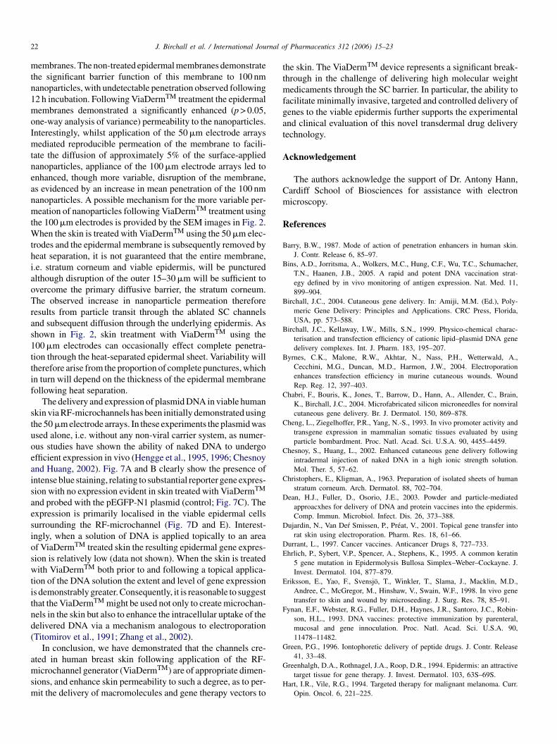
22 J. Birchall et al. / International Journal of Pharmaceutics 312 (2006) 15–23
membranes. The non-treated epidermal membranes demonstratethe significant barrier function of this membrane to 100 nmnanoparticles, with undetectable penetration observed following12 h incubation. Following ViaDermTM treatment the epidermalmembranes demonstrated a significantly enhanced (p > 0.05,one-way analysis of variance) permeability to the nanoparticles.Interestingly, whilst application of the 50 �m electrode arraysmediated reproducible permeation of the membrane to facili-tate the diffusion of approximately 5% of the surface-appliednanoparticles, appliance of the 100 �m electrode arrays led toenhanced, though more variable, disruption of the membrane,as evidenced by an increase in mean penetration of the 100 nmnanoparticles. A possible mechanism for the more variable per-meation of nanoparticles following ViaDermTM treatment usingthe 100 �m electrodes is provided by the SEM images in Fig. 2.When the skin is treated with ViaDermTM using the 50 �m elec-trodes and the epidermal membrane is subsequently removed byheat separation, it is not guaranteed that the entire membrane,i.e. stratum corneum and viable epidermis, will be puncturedalthough disruption of the outer 15–30 �m will be sufficient toovercome the primary diffusive barrier, the stratum corneum.The observed increase in nanoparticle permeation thereforeresults from particle transit through the ablated SC channelsand subsequent diffusion through the underlying epidermis. Asshown in Fig. 2, skin treatment with ViaDermTM using the100 �m electrodes can occasionally effect complete penetra-ttif
stuoeaisaesioswtitnd(
amsm
the skin. The ViaDermTM device represents a significant break-through in the challenge of delivering high molecular weightmedicaments through the SC barrier. In particular, the ability tofacilitate minimally invasive, targeted and controlled delivery ofgenes to the viable epidermis further supports the experimentaland clinical evaluation of this novel transdermal drug deliverytechnology.
Acknowledgement
The authors acknowledge the support of Dr. Antony Hann,Cardiff School of Biosciences for assistance with electronmicroscopy.
References
Barry, B.W., 1987. Mode of action of penetration enhancers in human skin.J. Contr. Release 6, 85–97.
Bins, A.D., Jorritsma, A., Wolkers, M.C., Hung, C.F., Wu, T.C., Schumacher,T.N., Haanen, J.B., 2005. A rapid and potent DNA vaccination strat-egy defined by in vivo monitoring of antigen expression. Nat. Med. 11,899–904.
Birchall, J.C., 2004. Cutaneous gene delivery. In: Amiji, M.M. (Ed.), Poly-meric Gene Delivery: Principles and Applications. CRC Press, Florida,USA, pp. 573–588.
Birchall, J.C., Kellaway, I.W., Mills, S.N., 1999. Physico-chemical charac-terisation and transfection efficiency of cationic lipid–plasmid DNA gene
B
C
C
C
C
D
D
DE
E
F
G
G
H
ion through the heat-separated epidermal sheet. Variability willherefore arise from the proportion of complete punctures, whichn turn will depend on the thickness of the epidermal membraneollowing heat separation.
The delivery and expression of plasmid DNA in viable humankin via RF-microchannels has been initially demonstrated usinghe 50 �m electrode arrays. In these experiments the plasmid wassed alone, i.e. without any non-viral carrier system, as numer-us studies have shown the ability of naked DNA to undergofficient expression in vivo (Hengge et al., 1995, 1996; Chesnoynd Huang, 2002). Fig. 7A and B clearly show the presence ofntense blue staining, relating to substantial reporter gene expres-ion with no expression evident in skin treated with ViaDermTM
nd probed with the pEGFP-N1 plasmid (control; Fig. 7C). Thexpression is primarily localised in the viable epidermal cellsurrounding the RF-microchannel (Fig. 7D and E). Interest-ngly, when a solution of DNA is applied topically to an areaf ViaDermTM treated skin the resulting epidermal gene expres-ion is relatively low (data not shown). When the skin is treatedith ViaDermTM both prior to and following a topical applica-
ion of the DNA solution the extent and level of gene expressions demonstrably greater. Consequently, it is reasonable to suggesthat the ViaDermTM might be used not only to create microchan-els in the skin but also to enhance the intracellular uptake of theelivered DNA via a mechanism analogous to electroporationTitomirov et al., 1991; Zhang et al., 2002).
In conclusion, we have demonstrated that the channels cre-ted in human breast skin following application of the RF-icrochannel generator (ViaDermTM) are of appropriate dimen-
ions, and enhance skin permeability to such a degree, as to per-it the delivery of macromolecules and gene therapy vectors to
delivery complexes. Int. J. Pharm. 183, 195–207.yrnes, C.K., Malone, R.W., Akhtar, N., Nass, P.H., Wetterwald, A.,
Cecchini, M.G., Duncan, M.D., Harmon, J.W., 2004. Electroporationenhances transfection efficiency in murine cutaneous wounds. WoundRep. Reg. 12, 397–403.
habri, F., Bouris, K., Jones, T., Barrow, D., Hann, A., Allender, C., Brain,K., Birchall, J.C., 2004. Microfabricated silicon microneedles for nonviralcutaneous gene delivery. Br. J. Dermatol. 150, 869–878.
heng, L., Ziegelhoffer, P.R., Yang, N.-S., 1993. In vivo promoter activity andtransgene expression in mammalian somatic tissues evaluated by usingparticle bombardment. Proc. Natl. Acad. Sci. U.S.A. 90, 4455–4459.
hesnoy, S., Huang, L., 2002. Enhanced cutaneous gene delivery followingintradermal injection of naked DNA in a high ionic strength solution.Mol. Ther. 5, 57–62.
hristophers, E., Kligman, A., 1963. Preparation of isolated sheets of humanstratum corneum. Arch. Dermatol. 88, 702–704.
ean, H.J., Fuller, D., Osorio, J.E., 2003. Powder and particle-mediatedapproacxhes for delivery of DNA and protein vaccines into the epidermis.Comp. Immun. Microbiol. Infect. Dis. 26, 373–388.
ujardin, N., Van Der Smissen, P., Preat, V., 2001. Topical gene transfer intorat skin using electroporation. Pharm. Res. 18, 61–66.
urrant, L., 1997. Cancer vaccines. Anticancer Drugs 8, 727–733.hrlich, P., Sybert, V.P., Spencer, A., Stephens, K., 1995. A common keratin
5 gene mutation in Epidermolysis Bullosa Simplex–Weber–Cockayne. J.Invest. Dermatol. 104, 877–879.
riksson, E., Yao, F., Svensjo, T., Winkler, T., Slama, J., Macklin, M.D.,Andree, C., McGregor, M., Hinshaw, V., Swain, W.F., 1998. In vivo genetransfer to skin and wound by microseeding. J. Surg. Res. 78, 85–91.
ynan, E.F., Webster, R.G., Fuller, D.H., Haynes, J.R., Santoro, J.C., Robin-son, H.L., 1993. DNA vaccines: protective immunization by parenteral,mucosal and gene innoculation. Proc. Natl. Acad. Sci. U.S.A. 90,11478–11482.
reen, P.G., 1996. Iontophoretic delivery of peptide drugs. J. Contr. Release41, 33–48.
reenhalgh, D.A., Rothnagel, J.A., Roop, D.R., 1994. Epidermis: an attractivetarget tissue for gene therapy. J. Invest. Dermatol. 103, 63S–69S.
art, I.R., Vile, R.G., 1994. Targeted therapy for malignant melanoma. Curr.Opin. Oncol. 6, 221–225.

J. Birchall et al. / International Journal of Pharmaceutics 312 (2006) 15–23 23
Heiser, W.C., 1994. Gene transfer into mammalian cells by particle bom-bardment. Anal. Biochem. 217, 185–196.
Hengge, U.R., Chan, E.F., Foster, R.A., Walker, P.S., Vogel, J.C., 1995.Cytokine gene expression in epidermis with biological effects followinginjection of naked DNA. Nat. Genet. 10, 161–166.
Hengge, U.R., Walker, P.S., Vogel, J.C., 1996. Expression of naked DNA inhuman, pig and mouse skin. J. Clin. Invest. 97, 2911–2916.
Henry, S., McAllister, D.V., Allen, M.G., Prausnitz, M.R., 1998. Microfab-ricated microneedles: a novel approach to transdermal drug delivery. J.Pharm. Sci. 87, 922–925.
Lavon, I., Kost, J., 2004. Ultrasound and transdermal drug delivery. DrugDiscovery Today 9, 670–676.
Lee, P-Y., Chesnoy, S., Huang, L., 2004. Electroporatic delivery of TFG-�1 gene works synergistically with electric therapy to enhance diabeticwound healing in db/db mice. J. Invest. Dermatol. 123, 791–798.
Levin, G., Gershonowitz, A., Sacks, H., Stern, M., Sherman, A., Rudaev,S., Zivin, I., Phillip, M., 2005. Transdermal delivery of human growthhormone through RF-microchannels. Pharm. Res. 22, 550–555.
Lin, M.T.S., Pulkkinen, L., Uitto, J., 2000. Cutaneous gene therapy: principlesand prospects. New Emerg. Therap. 18, 177–188.
McAllister, D.V., Allen, M.G., Prausnitz, M.R., 2000. Microfabricatedmicroneedles for gene and drug delivery. Annu. Rev. Biomed. Eng. 2,289–313.
McAllister, D.V., Wang, P.M., Davis, S.P., Park, J.H., Canatella, P.J.,Allen, M.G., Prausnitz, M.R., 2003. Microfabricated needles for trans-dermal delivery of macromolecules and nanoparticles: fabrication meth-ods and transport studies. Proc. Natl. Acad. Sci. U.S.A. 100, 13755–13760.
Nelson, J.S., McCullough, J.L., Glenn, T.C., Wright, W.H., Liaw, L.H.,Jacques, S.L., 1991. Mid-infrared laser ablation of stratum corneumenhances in vitro percutaneous transport of drugs. J. Invest. Dermatol.
Pillai, O., Panchagnula, R., 2003. Transdermal delivery of insulin from plox-amer gel: ex vivo and in vivo skin permeation studies in rat usingiontophoresis and chemical enhancers. J. Contr. Release 89, 127–140.
Prausnitz, M.R., Bose, V.G., Langer, R.S., Weaver, J.C., 1993. Electroporationof mammalian skin: a mechanism to enhance transdermal drug delivery.Proc. Natl. Acad. Sci. U.S.A. 90, 10504–10508.
Preat, V., Dujardin, N., 2001. Topical delivery of nucleic acids in the skin.Pharmascience 11, 57–68.
Raz, E., Carson, D.A., Parker, S.E., Parr, T.B., Abai, A.M., Aichinger, G.,Gromkowski, S.H., Singh, M., Lew, D., Yankauckas, M.A., Baird, S.M.,Rhodes, G.H., 1994. Intradermal gene immunization: The possible roleof DNA uptake in the induction of cellular immunity to viruses. Proc.Natl. Acad. Sci. U.S.A. 91, 9519–9523.
Sawamura, D., Yasukawa, K., Kodama, K., Yokota, K., Sato-Matsumura,K.C., Toshihiro, T., Shimizu, H., 2002. The majority of keratinocytesincorporate intradermally injected plasmid DNA regardless of size butonly a small proportion of cells can express the gene product. J. Invest.Dermatol. 118, 967–971.
Shi, Z., Curiel, D.T., Tang, D.C., 1999. DNA-based non-invasive vaccinationonto the skin. Vaccine 17, 2136–2141.
Sintov, A.C., Krymberk, I., Daniel, D., Hannan, T., Sohn, Z., Levin, G., 2003.Radiofrequency-driven skin microchanneling as a new way for electricallyassisted transdermal delivery of hydrophilic drugs. J. Contr. Release 89,311–320.
Titomirov, A.V., Sukharev, S., Kistanova, E., 1991. In vivo electroporationand stable transformation of skin cells of newborn mice by plasmid DNA.Biochim. Biophys. Acta 1088, 131–134.
Udvardi, A., Kufferath, I., Grutsch, H., Zatloukal, K., Volc-Platzer, B., 1999.Uptake of exogenous DNA via the skin. J. Mol. Med. 77, 744–750.
Zhang, L., Nolan, E., Kreitschitz, S., Rabussay, D.P., 2002. Enhanced deliv-ery of naked DNA to the skin by non-invasive in vivo electroporation.
97, 874–879.
Biochim. Biophys. Acta 1572, 1–9.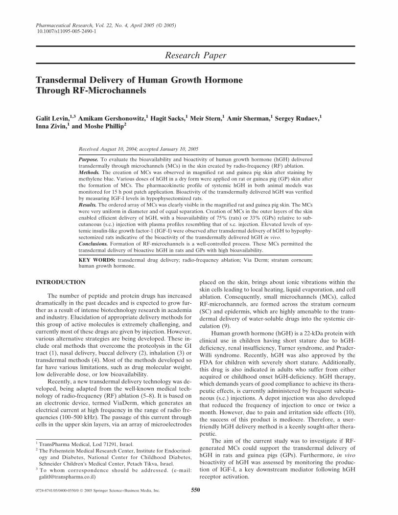
Research Paper
Transdermal Delivery of Human Growth HormoneThrough RF-Microchannels
Galit Levin,1,3 Amikam Gershonowitz,1 Hagit Sacks,1 Meir Stern,1 Amir Sherman,1 Sergey Rudaev,1
Inna Zivin,1 and Moshe Phillip2
Received August 10, 2004; accepted January 10, 2005
Purpose. To evaluate the bioavailability and bioactivity of human growth hormone (hGH) deliveredtransdermally through microchannels (MCs) in the skin created by radio-frequency (RF) ablation.Methods. The creation of MCs was observed in magnified rat and guinea pig skin after staining bymethylene blue. Various doses of hGH in a dry form were applied on rat or guinea pig (GP) skin afterthe formation of MCs. The pharmacokinetic profile of systemic hGH in both animal models wasmonitored for 15 h post patch application. Bioactivity of the transdermally delivered hGH was verifiedby measuring IGF-I levels in hypophysectomized rats.Results. The ordered array of MCs was clearly visible in the magnified rat and guinea pig skin. The MCswere very uniform in diameter and of equal separation. Creation of MCs in the outer layers of the skinenabled efficient delivery of hGH, with a bioavailability of 75% (rats) or 33% (GPs) relative to sub-cutaneous (s.c.) injection with plasma profiles resembling that of s.c. injection. Elevated levels of sys-temic insulin-like growth factor-1 (IGF-I) were observed after transdermal delivery of hGH to hypophy-sectomized rats indicative of the bioactivity of the transdermally delivered hGH in vivo.Conclusions. Formation of RF-microchannels is a well-controlled process. These MCs permitted thetransdermal delivery of bioactive hGH in rats and GPs with high bioavailability.
KEY WORDS: transdermal drug delivery; radio-frequency ablation; Via Derm; stratum corneum;human growth hormone.
INTRODUCTION
The number of peptide and protein drugs has increaseddramatically in the past decades and is expected to grow fur-ther as a result of intense biotechnology research in academiaand industry. Elucidation of appropriate delivery methods forthis group of active molecules is extremely challenging, andcurrently most of these drugs are given by injection. However,various alternative strategies are being developed. These in-clude oral methods that overcome the proteolysis in the GItract (1), nasal delivery, buccal delivery (2), inhalation (3) ortransdermal methods (4). Most of the methods developed sofar have various limitations, such as drug molecular weight,low deliverable dose, or low bioavailability.
Recently, a new transdermal delivery technology was de-veloped, being adapted from the well-known medical tech-nology of radio-frequency (RF) ablation (5–8). It is based onan electronic device, termed ViaDerm, which generates anelectrical current at high frequency in the range of radio fre-quencies (100–500 kHz). The passage of this current throughcells in the upper skin layers, via an array of microelectrodes
placed on the skin, brings about ionic vibrations within theskin cells leading to local heating, liquid evaporation, and cellablation. Consequently, small microchannels (MCs), calledRF-microchannels, are formed across the stratum corneum(SC) and epidermis, which are highly amenable to the trans-dermal delivery of water-soluble drugs into the systemic cir-culation (9).
Human growth hormone (hGH) is a 22-kDa protein withclinical use in children having short stature due to hGH-deficiency, renal insufficiency, Turner syndrome, and Prader-Willi syndrome. Recently, hGH was also approved by theFDA for children with severely short stature. Additionally,this drug is also indicated in adults who suffer from eitheracquired or childhood onset hGH-deficiency. hGH therapy,which demands years of good compliance to achieve its thera-peutic effects, is currently administered by frequent subcuta-neous (s.c.) injections. A depot injection was also developedthat reduced the frequency of injection to once or twice amonth. However, due to pain and irritation side effects (10),the success of this product is mediocre. Therefore, a user-friendly hGH delivery method is a keenly sought-after thera-peutic.
The aim of the current study was to investigate if RF-generated MCs could support the transdermal delivery ofhGH in rats and guinea pigs (GPs). Furthermore, in vivobioactivity of hGH was assessed by monitoring the produc-tion of IGF-I, a key downstream mediator following hGHreceptor activation.
1 TransPharma Medical, Lod 71291, Israel.2 The Felsenstein Medical Research Center, Institute for Endocrinol-
ogy and Diabetes, National Center for Childhood Diabetes,Schneider Children’s Medical Center, Petach Tikva, Israel.
3 To whom correspondence should be addressed. (e-mail:[email protected])
Pharmaceutical Research, Vol. 22, No. 4, April 2005 (© 2005)10.1007/s11095-005-2490-1
5500724-8741/05/0400-0550/0 © 2005 Springer Science+Business Media, Inc.
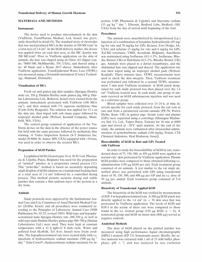
MATERIALS AND METHODS
Instruments
The device used to produce microchannels in the skin(ViaDerm, TransPharma Medical, Lod, Israel) was previ-ously described in detail (9). The standard array of electrodesthat was used produced MCs in the density of 100 MCs/cm2 ina total area of 1.4 cm2. In the hGH delivery studies, the devicewas applied twice on each skin area, so the MC density was200 MCs/cm2. Prior to ViaDerm application on the skin ofanimals, the hair was clipped using an Oster A5 clipper (cat.no. 78005-500, McMinnville, TN, USA), and shaved using ano. 40 blade and a Braun 3615 shaver. Immediately afterViaDerm application, TransEpidermal Water Loss (TEWL)was measured using a Dermalab instrument (Cortex Technol-ogy, Hadsund, Denmark).
Visualization of MCs
Fresh rat and guinea pig skin samples (Sprague-Dawleymale rat, 350 g; Dunkin Hartley male guinea pig, 600 g; Har-lan Laboratories Ltd., Rehovot, Israel) were excised from theanimals, immediately pretreated with ViaDerm (100 MCs/cm2), and then stained with 1% aqueous methylene blue(Carlo Erba Reagenti). The solution was applied for 15 s onthe skin site, then wiped with soft tissue paper followed byisopropyl alcohol pads (Webcol, Kendall Company, Mans-field, MA, USA).
The control group consisted of application of the Via-Derm device on the skin in the absence of the power sourcebut held with the same pressure followed by methylene bluestaining. A Video Inspection System (S-T Industries Inc.model 20-8600, St. James, MN, USA) equipped with ×10 lens,was used in order to observe the created MCs.
Preparation of hGH Patches
Lyophilized hGH (Genotropin 16 or 36 IU/vial, Pharma-cia & Upjohn, Puurs, Belgium) was used for the preparationof “printed” patches, in a proprietary owned process (11).This “print-like” method is based on accurately depositingsmall droplets of hGH solution on a transdermal backing linerat a total area of 1.4 cm2 followed by a controlled dryingprocess. This method permits accurate dosing and stablepatches that contain a thin uniform layer of the protein in adry form.
Animals
Study protocols were approved by the Institutional Ani-mal Care and Use Committee of Assaf Harofeh Medical Cen-ter (Zriffin, Israel), and all procedures were conducted ac-cording to the Principles of Labotarory Animal Care (NIHPublication No. 85-23, revised 1985). Wild-type and hypophy-sectomized male Sprague-Dawley rats, 200–350 g, as well aswild-type male Dunkin Hartley guinea pigs 500–700 g (HarlanLaboratories Ltd.) were used. They were kept at constanttemperature with a 12 h light:12 h dark cycle. Water andpelleted food (Koffolk, Tel Aviv, Israel) were freely avail-able. The hypophysectomized rats were treated daily with s.c.injections of hydrocortisone sodium succinate (500 �g kg−1
day−1 Solu-Cortef�, (hydrocortisone sodium succinate for in-
jection, USP, Pharmacia & Upjohn) and thyroxine sodium(15 �g kg−1 day−1, Eltroxin, Bedford Labs, Bedford, OH,USA) from the day of arrival until beginning of the trial.
Procedures
The animals were anaesthetized by intraperitoneal (i.p.)injection of a combination of ketamine hydrochloride (85 mg/kg for rats and 70 mg/kg for GPs; Ketaset, Fort Dodge, IA,USA) and xylazine (3 mg/kg for rats and 6 mg/kg for GPs,Xyl-M2 veterinary, VMD, Arendonk, Belgium). Anesthesiawas maintained using Isoflurane (0.5–1.5%, Isoflurane, Rho-dia, Bristol, UK) or Halothane (0.5–2%, Rhodia, Bristol, UK)gas. Animals were placed in a dorsal recumbancy, and theabdominal hair was clipped and shaved. The application sitewas then wiped using an isopropyl alcohol pads (Webcol,Kendall). Thirty minutes later, TEWL measurements wereused to check the skin integrity. Then, ViaDerm treatmentwas performed and followed by a second TEWL measure-ment 5 min post ViaDerm treatment. A hGH patch desig-nated for each study protocol was then placed over the 1.4cm2 ViaDerm treated area. In each study, one group of ani-mals received an hGH subcutaneous injection and was usedas a reference group.
Blood samples were collected over 15–24 h, at time in-tervals specific for each study protocol, from the tail vein inrats and from a preinserted carotid cannula (PE-50, PortexHythe, Kent, UK) in guinea pigs. Serum (rats) and plasma(GPs) were separated using a centrifuge (Hsiangtai Machin-ery Ind. Co. Ltd., Taipei Hsien, Taiwan) for 10 min at 6000rpm and stored at −20°C until analysis. At the end of thestudy, the animals were euthanized after intracardial admin-istration of pentobarbitone sodium (140 mg/kg, Pental, CTSChemical Industries, Hod Hasharon, Israel).
Bioavailability of hGH in Rats and GPs Treatedwith ViaDerm
In order to study the bioavailability of hGH in rats, trans-dermal doses of 75, 150, 300, or 450 �g hGH were applied onnormal rats’ skin pretreated by ViaDerm application. PlasmahGH profiles were compared to those obtained following s.c.administration (150 �g hGH per rat). Each treatment groupconsisted of six animals. A test similar to the rat study de-scribed above was performed with GPs using transdermaldoses of 50, 150, 300, and 400 �g per GP and an s.c. dose of50 �g per animal. Each treatment group consisted of 5–6animals.
Bioactivity of Transdermal Applied hGH
The bioactivity of the hGH was verified by measurementof IGF-I in hypophysectomized rats. A 200 �g hGH patch wasdirectly applied to the 1.4 cm2 (n � 9) skin area that waspretreated by ViaDerm application. The levels of hGH andIGF-I in the serum of these rats were compared to thosefound in the s.c. treated group (150 �g hGH, n � 6). Anontreated group and hGH on intact skin (800 �g) served asnegative controls.
Analytical Methods
The dose of hGH placed on the printed patches wasmeasured using high performance liquid chromatography(HPLC) analysis (EP 5.0, Somatropin assay). Briefly, the ac-tive material was extracted with 1 ml of 25 mM buffer phos-phate, pH � 7, and was analyzed by size exclusion
Transdermal Delivery of Human Growth Hormone 551
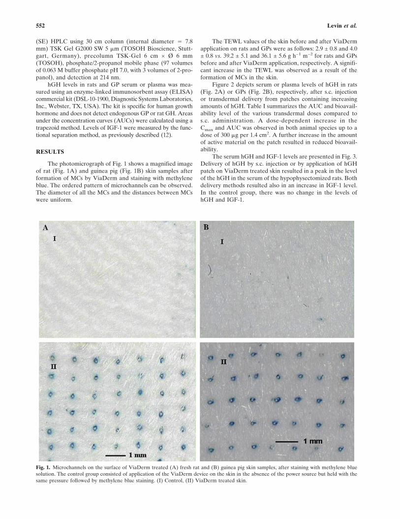
(SE) HPLC using 30 cm column (internal diameter � 7.8mm) TSK Gel G2000 SW 5 �m (TOSOH Bioscience, Stutt-gart, Germany), precolumn TSK-Gel 6 cm × Ø 6 mm(TOSOH), phosphate/2-propanol mobile phase (97 volumesof 0.063 M buffer phosphate pH 7.0, with 3 volumes of 2-pro-panol), and detection at 214 nm.
hGH levels in rats and GP serum or plasma was mea-sured using an enzyme-linked immunosorbent assay (ELISA)commercial kit (DSL-10-1900, Diagnostic Systems Laboratories,Inc., Webster, TX, USA). The kit is specific for human growthhormone and does not detect endogenous GP or rat GH. Areasunder the concentration curves (AUCs) were calculated using atrapezoid method. Levels of IGF-1 were measured by the func-tional separation method, as previously described (12).
RESULTS
The photomicrograph of Fig. 1 shows a magnified imageof rat (Fig. 1A) and guinea pig (Fig. 1B) skin samples afterformation of MCs by ViaDerm and staining with methyleneblue. The ordered pattern of microchannels can be observed.The diameter of all the MCs and the distances between MCswere uniform.
The TEWL values of the skin before and after ViaDermapplication on rats and GPs were as follows: 2.9 ± 0.8 and 4.0± 0.8 vs. 39.2 ± 5.1 and 36.1 ± 5.6 g h−1 m−2 for rats and GPsbefore and after ViaDerm application, respectively. A signifi-cant increase in the TEWL was observed as a result of theformation of MCs in the skin.
Figure 2 depicts serum or plasma levels of hGH in rats(Fig. 2A) or GPs (Fig. 2B), respectively, after s.c. injectionor transdermal delivery from patches containing increasingamounts of hGH. Table I summarizes the AUC and bioavail-ability level of the various transdermal doses compared tos.c. administration. A dose-dependent increase in theCmax and AUC was observed in both animal species up to adose of 300 �g per 1.4 cm2. A further increase in the amountof active material on the patch resulted in reduced bioavail-ability.
The serum hGH and IGF-1 levels are presented in Fig. 3.Delivery of hGH by s.c. injection or by application of hGHpatch on ViaDerm treated skin resulted in a peak in the levelof the hGH in the serum of the hypophysectomized rats. Bothdelivery methods resulted also in an increase in IGF-1 level.In the control group, there was no change in the levels ofhGH and IGF-1.
Fig. 1. Microchannels on the surface of ViaDerm treated (A) fresh rat and (B) guinea pig skin samples, after staining with methylene bluesolution. The control group consisted of application of the ViaDerm device on the skin in the absence of the power source but held with thesame pressure followed by methylene blue staining. (I) Control, (II) ViaDerm treated skin.
Levin et al.552

DISCUSSION
The orderly pattern of RF-generated MCs in terms oftheir diameter and separated distances (Fig. 1) lays credenceas to the reproducibility of the ViaDerm in creating MCs.Because methylene blue coloration of the MCs was a veryrapid process, it would demonstrate the hydrophilic nature ofthe MCs. Indeed, previous studies showing the presence of
extracellular fluid in MCs from porcine skin would supportthe theory of the hydrophilic nature of the MCs (9).
The notion of using electricity for enhancement of trans-dermal drug delivery is not exclusive to the RF-microchanneltechnology. Iontophoresis uses an electrical field in order todrive ionized drug molecules across the SC barrier (13). Inelectroporation, short electrical pulses are used to create tran-sient aqueous pores in the SC (14). Neither of these methodscreates an orderly array of pores or MCs, by ablation of cellsin specific locations, as presented in this study. Moreover, theViaDerm device is user-friendly and minimally invasive. Ahuman device safety study with 20 subjects was successfullycompleted with the electric parameters tested in this study. Itwas found that the ViaDerm device produced only slight ir-ritation responses (minimal erythema and no edema) of atransient nature and the pain levels recorded were within theacceptable range for clinical use (in preparation).
It is important to note that the area covered with MCs isvery small compared to the total skin area. MCs were createdin a density of 200 MCs per cm2 and less than 1% of the totaltreated area consists of MCs. Nevertheless, these MCs arehighly amenable to the transdermal delivery of hGH, as non-ViaDerm treated skin is totally impermeable to hGH due toits large molecular size and hydrophilic nature (see also Fig.3A, hGH on intact skin).
It is known that breaching the SC integrity is accompa-nied by an elevation in TEWL (15). Therefore, the formation
Table I. Mean AUC and Relative Bioavailability Values in Rats andGuinea Pigs
Mode of delivery
Dosemicrograms
(mcg)AUC
(ng.hr/ml.)Bioavailability
(% of s.c.)
Ratss.c. 150 489 100Transdermal 75 184 75.3Transdermal 150 376 76.9Transdermal 300 727 74.3Transdermal 450 884 60.3Guinea pigss.c. 50 176 100Transdermal 50 57 32.4Transdermal 150 175 33.1Transdermal 300 362 34.3Transdermal 400 404 28.7
Fig. 2. hGH levels (ng/ml) in serum or plasma after application ofincreasing doses of transdermal hGH on 1.4 cm2 ViaDerm treatedarea and s.c. injection of hGH: (A) Serum levels in rats. (B) Plasmalevels in GP. Each data point represents the mean ± SEM of 5–6animals.
Fig. 3. Serum levels (ng/ml, mean ± SEM) of (A) hGH and (B) IGF-Iafter application of 200 �g hGH on 1.4 cm2 ViaDerm treated area (n� 9), s.c. injection of 150 �g hGH (n � 7), or no treatment (n � 9)(control). Serum levels of hGH after application of 800 �g hGH onintact skin served as negative control (A).
Transdermal Delivery of Human Growth Hormone 553
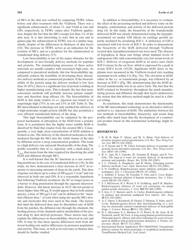
of MCs in the skin was verified by comparing TEWL valuesbefore and after treatment with the ViaDerm. There was asignificant enhancement, of about 13- and 9-fold in rats andGPs, respectively, in TEWL values after ViaDerm applica-tion, despite the fact that the MCs occupy less than 1% of theskin area. It is also interesting to note that in rats and inguinea pigs the enhancement in TEWL was of a similar mag-nitude, despite of the differences in thickness of skin layers(16). The increase in TEWL serves as an indication for thecreation of MCs, and as a predictor for the enhancement intransdermal drug delivery (17).
Low bioavailability is one of the major obstacles for thedevelopment of user-friendly delivery methods for peptidesand proteins. The manufacturing processes of these activematerials are usually complex with associated high costs. Ascompared to parenteral methods, this low bioavailability sig-nificantly reduces the feasibility of developing these alterna-tive delivery methods as commercial products. If the bioavail-ability of the protein using the delivery method is low (lessthan 10–20%), there is a significant loss of protein resulting inhigher manufacturing costs. This is despite the fact that moreconvenient methods will probably increase patient compli-ance and therefore drug efficacy (4). The bioavailability ofhGH in this study relative to s.c. injection was found to besurprisingly high (75% in rats and 33% in GP; Table I). TheRF-microchannel technology not only enabled the delivery ofa high-molecular-weight protein (hGH) but also permitted avery efficient transdermal delivery of the drug.
This high bioavailability can be explained by the pro-posed mechanism of absorption of the hGH from a powderform. It is postulated that the highly water soluble hGH isdissolved by fluid that exudes from the created MCs. Conse-quently, a very high, local concentration of hGH solution isformed in situ. The delivery of the dissolved molecules is thenmediated through the MCs into the viable tissues of the skinby diffusion across a steep concentration gradient. This leadsto a high delivery rate and peak blood profile of the drug. Theprofile resembles that of s.c. injection, with a small delay inTmax that stems from the time required for dissolving the solidhGH and diffusion through MCs.
It is well-known that the SC functions as a rate control-ling membrane in the case of transdermal delivery (18). In thisstudy, we have demonstrated a clear increase in AUC in re-sponse to increasing amounts of drug on the patch. This doseresponse was linear up to a dose of 300 �g per 1.4 cm2 and wasobserved in both rats and GPs. It is a reasonable hypothesisthat following ViaDerm treatment, the SC no longer poses asa barrier to drug penetration through the aqueous microcon-duits. However, this linear increase in AUC did not persist atdoses higher than 300 �g. It would appear that in both animalspecies, a dose of 300 �g/1.4 cm2 can be defined as the “maxi-mal efficient dose,” at least when using the specific MCs den-sity and electrodes that were used in this study. The factorsthat limit the delivered dose may be dissolution rate of hGHfrom the patches, the diffusion rate through the channels, thehealing process of the channels and/or metabolism of the pro-tein drug by skin derived proteases. These factors may alsoexplain the differences in bioavailability observed in rats andGPs. It may be that these species differences stem from dif-ferent healing rate and/or differences in proteases populationand activity. This issue, as well as its relevance to human skin,should be further studied.
In addition to bioavailability, it is necessary to evaluatethe effect of the processing method and delivery route on theintegrity, conformation, and activity of the delivered proteindrug (4). In this study, the bioactivity of the transdermallydelivered hGH was clearly demonstrated using the hypophy-sectomized rat model. GH effects on cartilage growth arepartly mediated by circulating IGF-1. A deficiency of GH isassociated with low levels of IGF-1. In order to demonstratethe bioactivity of the hGH delivered through ViaDermtreated skin, hypophysectomized rats were used. The absenceof hypophysa in these rats brings about minimal levels ofendogenous GH, with concomitantly very low serum levels ofIGF-1. Delivery of exogenous hGH in an active state elicitsIGF-I release by the rat liver, which is expressed by a peak inserum IGF-I levels (19,20). Significant hGH doses in theplasma were measured in the ViaDerm treated rats reachingmaximum levels within 4 h (Fig. 3A). The elevation in hGH,either in the s.c. or transdermal groups, was followed by anincrease in IGF-1 (Fig. 3B), demonstrating that the hGH de-livered transdermally was in an active form. The fact that thehGH retained its bioactivity throughout the patch manufac-turing process and diffusion through skin layers underscoresthe notion that this delivery method might be used in a clini-cal setting.
In conclusion, this study demonstrates the functionalityof the RF-microchannel technology as an alternative deliverymethod to s.c. injection of hGH. The similarities between thetwo methods in bioavailability, bioactivity, and serum drugprofile offer much hope that the development of a commer-cial product based on this transdermal technology might befeasible.
REFERENCES
1. R. R. B. Shah, F. Ahsan, and M. A. Khan. Oral delivery ofproteins: progress and prognostication. Crit. Rev. Ther. Drug Car-rier Syst. 19:135–169 (2002).
2. A. P. Sayani and Y. W. Chien. Systemic delivery of peptides andproteins across absorptive mucosae. Crit. Rev. Ther. Drug CarrierSyst. 13:85–184 (1996).
3. R. U. Agu, M. I. Ugwoke, M. Armand, R. Kinget, and N. Ver-beke. The lung as a route for systemic delivery of therapeuticproteins and peptides. Respir. Res. 2:198–209 (2001).
4. J. L. Cleland, A. Daugherty, and R. Mrsny. Emerging proteindelivery methods. Curr. Opin. Biotechnol. 12:212–219 (2001).
5. S. N. Goldberg. Radiofrequency tumor ablation: principles andtechniques. Eur. J. Ultrasound 13:129–147 (2001).
6. L. Solbiati, T. Ierace, M. Tonolini, V. Osti, and L. Cova. Radio-frequency thermal ablation of hepatic metastases. Eur. J. Ultra-sound 13:149–158 (2001).
7. F. J. McGovern, B. J. Wood, S. N. Goldberg, and P. R. Mueller.Radiofrequency ablation of renal cell carcinoma via imageguided needle electrodes. J. Urol. 161:599–600 (1999).
8. F. Izzo, C. C. Barnett, and S. A. Curley. Radiofrequency ablationof primary and metastatic malignant liver tumors. Adv. Surg.35:225–250 (2001).
9. A. C. Sintov, I. Krymberk, D. Daniel, T. Hannan, Z. Sohn, and G.Levin. Radiofrequency-driven skin microchanneling as a newway for electrically assisted transdermal delivery of hydrophilicdrugs. J. Control. Release 89:311–320 (2003).
10. B. L. Silverman, S. L. Blethen, E. O. Reiter, K. M. Attie, R. B.Neuwirth, and K. M. Ford. A long-acting human growth hormone(Nutropin depot): efficacy and safety following two years of treat-ment in children with growth hormone deficiency. J. Pediatr. En-docrinol. Metab. 15:715–722 (2002).
11. International Patent Application WO 2004/039428. Transdermaldelivery system for dried particulate or lyophilized medications,TransPharma Medical Ltd., Lod, Israel.
Levin et al.554
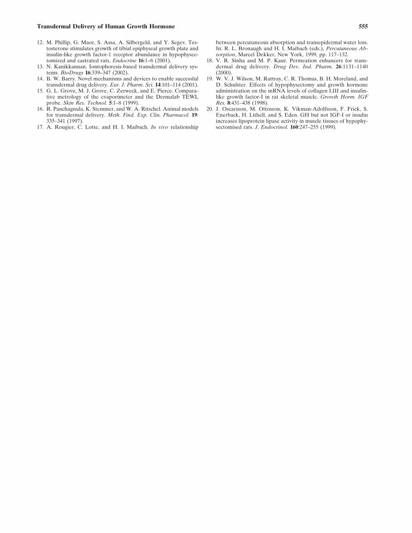
12. M. Phillip, G. Maor, S. Assa, A. Silbergeld, and Y. Segev. Tes-tosterone stimulates growth of tibial epiphyseal growth plate andinsulin-like growth factor-1 receptor abundance in hypophysec-tomized and castrated rats. Endocrine 16:1–6 (2001).
13. N. Kanikkannan. Iontophoresis-based transdermal delivery sys-tems. BioDrugs 16:339–347 (2002).
14. B. W. Barry. Novel mechanisms and devices to enable successfultransdermal drug delivery. Eur. J. Pharm. Sci. 14:101–114 (2001).
15. G. L. Grove, M. J. Grove, C. Zerweck, and E. Pierce. Compara-tive metrology of the evaporimeter and the Dermalab TEWLprobe. Skin Res. Technol. 5:1–8 (1999).
16. R. Panchagnula, K. Stemmer, and W. A. Ritschel. Animal modelsfor transdermal delivery. Meth. Find. Exp. Clin. Pharmacol. 19:335–341 (1997).
17. A. Rougier, C. Lotte, and H. I. Maibach. In vivo relationship
between percutaneous absorption and transepidermal water loss.In: R. L. Bronaugh and H. I. Maibach (eds.), Percutaneous Ab-sorption, Marcel Dekker, New York, 1999, pp. 117–132.
18. V. R. Sinha and M. P. Kaur. Permeation enhancers for trans-dermal drug delivery. Drug Dev. Ind. Pharm. 26:1131–1140(2000).
19. W. V. J. Wilson, M. Rattray, C. R. Thomas, B. H. Moreland, andD. Schulster. Effects of hypophysectomy and growth hormoneadministration on the mRNA levels of collagen I,III and insulin-like growth factor-I in rat skeletal muscle. Growth Horm. IGFRes. 8:431–438 (1998).
20. J. Oscarsson, M. Ottosson, K. Vikman-Adolfsson, F. Frick, S.Enerback, H. Lithell, and S. Eden. GH but not IGF-I or insulinincreases lipoprotein lipase activity in muscle tissues of hypophy-sectomised rats. J. Endocrinol. 160:247–255 (1999).
Transdermal Delivery of Human Growth Hormone 555

Journal of Controlled Release 89 (2003) 311–320www.elsevier.com/ locate/ jconrel
R adiofrequency-driven skin microchanneling as a new way forelectrically assisted transdermal delivery of hydrophilic drugs
a , a b b b*Amnon C. Sintov , Igor Krymberk , Dorit Daniel , Talli Hannan , Ze’ev Sohn ,bGalit Levin
aThe Institutes for Applied Research, Ben Gurion University of the Negev, P.O. Box 653, Beer Sheva 84105,IsraelbTransPharma Ltd., 3a Geron St., P.O. Box 222, Yehud, Israel
Received 27 November 2002; accepted 14 February 2003
Abstract
The aim of this study was to increase the skin penetration of two drugs, granisetron hydrochloride and diclofenac sodium,using a microelectronic device based on an ablation of outer layers of skin using radiofrequency high-voltage currents. Theseradiofrequency currents created an array of microchannels across the stratum corneum deep into the epidermis. Thepercutaneous penetration studies were first performed in vitro using excised full thickness porcine ear skin. An array of 100
2microelectrodes/cm was used in these studies. The skin permeability of both molecules was significantly enhanced afterpretreatment with the radiofrequency microelectrodes, as compared to the delivery through the untreated control skin. Steady
2 2state fluxes of 41.6mg/cm /h (r50.997) and 23.0mg/cm /h (r50.989) were obtained for granisetron and diclofenac,respectively. The enhanced transdermal delivery was also demonstrated in vivo in rats. It was shown that diclofenac plasmalevels in the pretreated rats reached plateau levels of 1.2260.32mg/ml after 3 h to 1.4760.33mg/ml after 6 h, as comparedto 0.1660.04 mg/ml levels obtained after 6 h in untreated rats. Similarly, application of granisetron patches (3% incrosslinked hydrogel) onto rats’ abdominal skin pretreated with radiofrequency electrodes resulted in an averaged peakplasma level of 239.3643.7 ng/ml after 12 h, which was about 30 times higher than the plasma levels obtained by 24-hpassive diffusion of the applied drug. The results emphasize, therefore, that the new transdermal technology is suitable fortherapeutic delivery of poorly penetrating molecules. 2003 Elsevier Science B.V. All rights reserved.
Keywords: Radiofrequency-microchannels; Radiofrequency ablation; Granisetron; Diclofenac; Transdermal delivery; Skin permeation
1 . Introduction ministration of drugs and other substances is re-markably restricted. Passive penetration of the SC is
The outmost dermal layer, the stratum corneum particularly difficult for hydrophilic and charged(SC), forms an effective barrier to the permeation of molecules. Consequently, transdermal delivery ofexternal chemicals; therefore, the transdermal ad- drugs has been the subject of intensive research. In
addition to the vehicle formulations and the chemicalenhancers [1,2], physical methods such as micronee-*Corresponding author. Tel.:1972-8-647-2709; fax:1972-8-dles [3,4], iontophoresis and electroosmosis [5–7],647-2960.
E-mail address: [email protected](A.C. Sintov). electroporation [8–13], and ultrasound [14] have
0168-3659/03/$ – see front matter 2003 Elsevier Science B.V. All rights reserved.doi:10.1016/S0168-3659(03)00123-8

312 A.C. Sintov et al. / Journal of Controlled Release 89 (2003) 311–320
been investigated. Synergistic interactions between The present paper is the first report describing thischemical and physical enhancers as well as syner- novel method in facilitating the transport of hydro-gism between two physical means of drug enhance- philic drugs, granisetron hydrochloride (pK 59.4,a
ment were also studied [15–17]. MW5348.9) and diclofenac sodium (pK 54.0,a
Radiofrequency (RF) thermal ablation is a well- MW5318.1), through the SC barrier. We haveknown and effective technology for electrosurgery chosen these drugs because the transdermal adminis-and ablation of malignant tissues. The method in- tration of charged and polar molecules is difficultvolves placing of a thin, needle-like electrode direct- due to the intrinsic lipophilicity of the SC. Thely into the tumor. During the application of RF method of using RF energy is based on creating anenergy, a frequency alternating current moves from array of small microchannels across the SC into thethe tip of the electrode into the tissue surrounding viable epidermis by microablating skin cells. Thethat electrode. As the ions within the tissue attempt high frequency electrical current conducted throughto follow the change in the direction of the alternat- the aqueous medium of the stratum corneum gener-ing current, their movements result in frictional ates heat that brings about an instant removal of cellsheating of the tissue, producing coagulative necrosis beneath the electrode. Due to the high velocity (1 msand cell ablation. Operations using radiofrequencies per electrode), it is postulated that only heat conduc-are considered safe and convenient during the tion results in the creation of microchannels, andsurgery, since cessation of neuromuscular stimula- other mechanisms such as electrochemical reactionstion occurs at approximately 100 kHz. That is, while do not take place. Skin electroporation, which isthe target tissue absorbs the heat energy released operated by low duty cycle, high intensity electric-during the electrosurgery, the applied high-frequency field pulsing, is also believed to create transientcurrent does not affect the proximal muscles [18– aqueous microchannels [8–13]. The creation of21]. This technology has been adapted as an optional transient aqueous microchannels by RF energy hasphysical enhancer of drug transport across the skin. been evidenced for the first time in the presentIts potential in the creation of aqueous microchannels report. The operating principle of RF-microchannel-in the outer layer of the skin was studied. ing formation are shown in Scheme 1. As illustrated,
Scheme 1. Schematic presentation of RF-microchannels.

A.C. Sintov et al. / Journal of Controlled Release 89 (2003) 311–320 313
a closely spaced array of tiny electrodes is placed cess, such as a peak current, final current, voltage,against the skin while an alternating current at radio time and energy. The array is made of a polycarbo-frequency is applied to each of the microelectrodes. nate body and stainless steel electrodes of length 100This forms RF-microchannels on the very outer layer mm and diameter 40mm. It has 140 electrodesof the skin through ablation of cells. The microchan- spaced 1 mm from each other in a square matrixnels are designed to penetrate only the outer layers of arrangement. In this prototype, each electrode isthe skin where there are no blood vessels or nerve individually operated. The electrodes are designed toendings, resulting in minimal skin trauma and neural create micro-channels that are 40mm wide and 70sensation. mm deep. The operation of the RF-microchannel
generator is simple and easy. The user holds thecontroller in his hand and presses the array against
2 . Materials and methods the test site on the skin. When a minimum pressure isplaced on the skin the RF-generator is activated and
2 .1. Instruments and materials the treatment begins automatically. Within seconds(typically less than a millisecond per burst per
The RF-microchannels generator (ViaDerm�, electrode) the array completes its work. The densityTranspharma Ltd, Israel) is illustrated in Scheme 2 of the microelectrode array used in these studies was
2[22]. ViaDerm� generator is made of two primary 100 or 200 microelectrodes/cm . Each electrodecomponents: a reusable electronic controller in a size received a multiple number of bursts of RF energy assimilar to a phone handset, and a disposable array programmed. The animal studies (rats) were donethat can snap onto the end of the controller. The with the following conditions: applied voltage: 200controller can communicate with a computer and be or 250 V; RF frequency: 100 kHz; burst length: 1reprogrammed to change any of the critical parame- ms; number of bursts: 5; time between bursts: 15 ms.ters used. The controller is also able to measure and Excised porcine skin for in vitro testing and his-download electrical inputs during the ablation pro- tological observation was treated with an applied
Scheme 2. The handset system and the microelectrodes array (ViaDerm�, Transpharma Ltd.) as designed to facilitate the transdermal drugdelivery by transmission of radio frequency currents through the electrodes into the skin, and by the creation of microchannels.
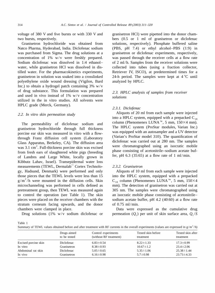
314 A.C. Sintov et al. / Journal of Controlled Release 89 (2003) 311–320
voltage of 380 V and five bursts or with 330 V and granisetron HCl) were pipetted into the donor cham-two bursts, respectively. bers (0.5 or 1 ml of granisetron or diclofenac
Granisetron hydrochloride was obtained from solutions, respectively). Phosphate buffered salineNatco Pharma, Hyderabad, India. Diclofenac sodium (PBS, pH 7.4) or ethyl alcohol–PBS (1:9) inwas purchased from Sigma. The drug solutions at a granisetron or diclofenac experiments, respectively,concentration of 1% w/v were freshly prepared. was passed through the receiver cells at a flow rateSodium diclofenac was dissolved in 1:4 ethanol– of 2 ml /h. Samples from the receiver solutions werewater, while granisetron HCl was dissolved in dis- collected into tubes (using a fraction collector,tilled water. For the pharmacokinetics experiments, Retriever IV, ISCO), at predetermined times for agranisetron in solution was soaked into a crosslinked 24-h period. The samples were kept at 48C untilpolyethylene oxide wound dressing (Vigilon, Bard analyzed by HPLC.Inc.) to obtain a hydrogel patch containing 3% w/vof drug substance. This formulation was prepared 2 .3. HPLC analysis of samples from receiverand used in vivo instead of 1% w/v concentration solutionsutilized in the in vitro studies. All solvents wereHPLC grade (Merck, Germany).
2 .3.1. DiclofenacAliquots of 20 ml from each sample were injected2 .2. In vitro skin permeation study
into a HPLC system, equipped with a prepacked C18
column (Phenomenex LUNA�, 5 mm, 15034 mm).The permeability of diclofenac sodium andThe HPLC system (ProStar modules, Varian Inc.)granisetron hydrochloride through full thicknesswas equipped with an autosampler and a UV detectorporcine ear skin was measured in vitro with a flow-(Varian’s ProStar model 310). The quantification ofthrough Franz diffusion cell system (Laboratorydiclofenac was carried out at 280 nm. The samplesGlass Apparatus, Berkeley, CA). The diffusion area
2 were chromatographed using an isocratic mobilewas 3.1 cm . Full-thickness porcine skin was excisedphase consisting of acetonitrile–sodium acetate buf-from fresh ears of slaughtered white pigs (breedingfer, pH 6.3 (35:65) at a flow rate of 1 ml /min.of Landres and Large White, locally grown in
Kibbutz Lahav, Israel). Transepidermal water lossmeasurements (TEWL, Dermalab Cortex Technolo- 2 .3.2. Granisetron
gy, Hadsund, Denmark) were performed and only Aliquots of 10 ml from each sample were injectedthose pieces that the TEWL levels were less than 15 into the HPLC system, equipped with a prepacked
2g/m /h were mounted in the diffusion cells. Skin C column (Phenomenex LUNA�, 5 mm, 1503418
microchanneling was performed in cells defined as mm). The detection of granisetron was carried out atpretreatment group, then TEWL was measured again 305 nm. The samples were chromatographed usingto control the operation (see Table 1). The skin an isocratic mobile phase consisting of acetonitrile–pieces were placed on the receiver chambers with the sodium acetate buffer, pH 4.2 (40:60) at a flow ratestratum corneum facing upwards, and the donor of 0.75 ml /min.chambers were clamped in place. Data were expressed as the cumulative drug
Drug solutions (1% w/v sodium diclofenac or permeation (Q ) per unit of skin surface area,Q /St t
Table 12Summary of TEWL values obtained before and after treatment with RF currents in the overall experiments (values are expressed in g/m /h)
Drugs aimed Control experiments Tested skin before Tested skin afterto be tested (without RF treatment) treatment treatment
Excised porcine skin Diclofenac 6.8360.54 8.2261.33 17.360.99In vitro Granisetron 8.3860.93 10.6761.2 23.462.06Abdominal rat skin Diclofenac 5.0560.65 5.3561.06 25.3861.44In vivo Granisetron 6.1660.98 5.760.98 23.7364.33
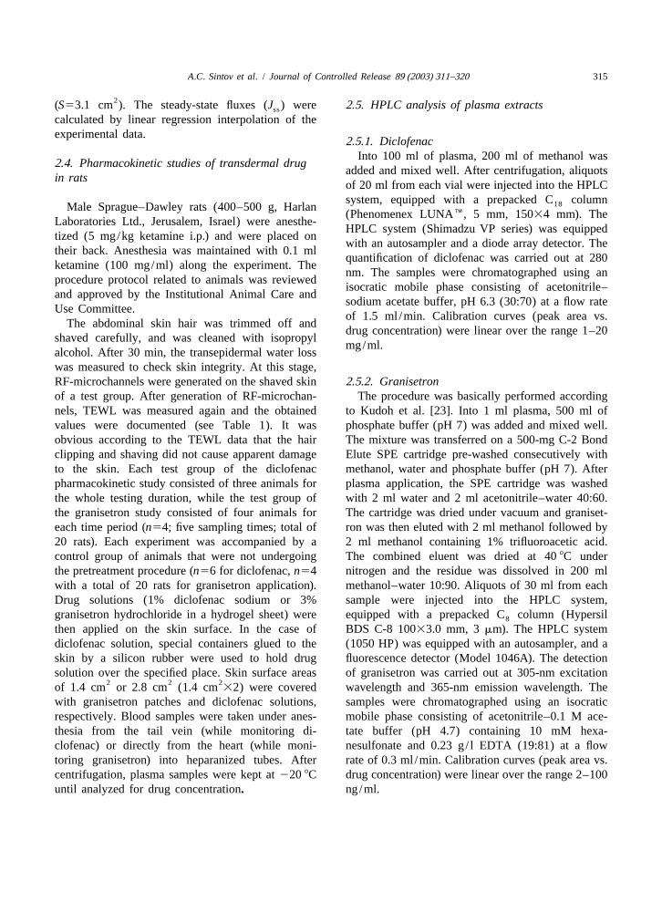
A.C. Sintov et al. / Journal of Controlled Release 89 (2003) 311–320 315
2(S53.1 cm ). The steady-state fluxes (J ) were 2 .5. HPLC analysis of plasma extractsss
calculated by linear regression interpolation of theexperimental data.
2 .5.1. DiclofenacInto 100 ml of plasma, 200 ml of methanol was
2 .4. Pharmacokinetic studies of transdermal drugadded and mixed well. After centrifugation, aliquots
in ratsof 20 ml from each vial were injected into the HPLCsystem, equipped with a prepacked C column18Male Sprague–Dawley rats (400–500 g, Harlan(Phenomenex LUNA�, 5 mm, 15034 mm). The
Laboratories Ltd., Jerusalem, Israel) were anesthe-HPLC system (Shimadzu VP series) was equipped
tized (5 mg/kg ketamine i.p.) and were placed onwith an autosampler and a diode array detector. The
their back. Anesthesia was maintained with 0.1 mlquantification of diclofenac was carried out at 280
ketamine (100 mg/ml) along the experiment. Thenm. The samples were chromatographed using an
procedure protocol related to animals was reviewedisocratic mobile phase consisting of acetonitrile–
and approved by the Institutional Animal Care andsodium acetate buffer, pH 6.3 (30:70) at a flow rate
Use Committee.of 1.5 ml /min. Calibration curves (peak area vs.
The abdominal skin hair was trimmed off anddrug concentration) were linear over the range 1–20
shaved carefully, and was cleaned with isopropylmg/ml.
alcohol. After 30 min, the transepidermal water losswas measured to check skin integrity. At this stage,RF-microchannels were generated on the shaved skin2 .5.2. Granisetronof a test group. After generation of RF-microchan- The procedure was basically performed accordingnels, TEWL was measured again and the obtained to Kudoh et al. [23]. Into 1 ml plasma, 500 ml ofvalues were documented (see Table 1). It was phosphate buffer (pH 7) was added and mixed well.obvious according to the TEWL data that the hair The mixture was transferred on a 500-mg C-2 Bondclipping and shaving did not cause apparent damage Elute SPE cartridge pre-washed consecutively withto the skin. Each test group of the diclofenac methanol, water and phosphate buffer (pH 7). Afterpharmacokinetic study consisted of three animals for plasma application, the SPE cartridge was washedthe whole testing duration, while the test group of with 2 ml water and 2 ml acetonitrile–water 40:60.the granisetron study consisted of four animals for The cartridge was dried under vacuum and graniset-each time period (n54; five sampling times; total of ron was then eluted with 2 ml methanol followed by20 rats). Each experiment was accompanied by a 2 ml methanol containing 1% trifluoroacetic acid.control group of animals that were not undergoing The combined eluent was dried at 408C underthe pretreatment procedure (n56 for diclofenac,n54 nitrogen and the residue was dissolved in 200 mlwith a total of 20 rats for granisetron application). methanol–water 10:90. Aliquots of 30 ml from eachDrug solutions (1% diclofenac sodium or 3% sample were injected into the HPLC system,granisetron hydrochloride in a hydrogel sheet) were equipped with a prepacked C column (Hypersil8
then applied on the skin surface. In the case of BDS C-8 10033.0 mm, 3mm). The HPLC systemdiclofenac solution, special containers glued to the (1050 HP) was equipped with an autosampler, and askin by a silicon rubber were used to hold drug fluorescence detector (Model 1046A). The detectionsolution over the specified place. Skin surface areas of granisetron was carried out at 305-nm excitation
2 2 2of 1.4 cm or 2.8 cm (1.4 cm32) were covered wavelength and 365-nm emission wavelength. Thewith granisetron patches and diclofenac solutions, samples were chromatographed using an isocraticrespectively. Blood samples were taken under anes- mobile phase consisting of acetonitrile–0.1 M ace-thesia from the tail vein (while monitoring di- tate buffer (pH 4.7) containing 10 mM hexa-clofenac) or directly from the heart (while moni- nesulfonate and 0.23 g/ l EDTA (19:81) at a flowtoring granisetron) into heparanized tubes. After rate of 0.3 ml /min. Calibration curves (peak area vs.centrifugation, plasma samples were kept at220 8C drug concentration) were linear over the range 2–100until analyzed for drug concentration. ng/ml.

316 A.C. Sintov et al. / Journal of Controlled Release 89 (2003) 311–320
2 .6. Histology epidermis — 36.9mm vs. 49.5mm, respectively [26]— indicating that RF-microchannels would go into
RF-microchannels were created at a density of 200 the human epidermis without perturbing the dermis.2channels /cm on the dorsal skin of a pig. Skin The permeability of diclofenac sodium and
biopsies were taken immediately after RF-mi- granisetron hydrochloride through excised porcinecrochannels creation, by a biopsy punch, and pre- ear skin was significantly enhanced after pretreat-served in 4% buffered formaldehyde solution. The ment with RF energy, as compared to the deliverysamples were embedded in paraffin wax, cut to a through the untreated control skin (Figs. 2–3). Afterthickness of 4–5mm, stained with hematoxylin and lag times of 3 and 9 h, pseudo steady state fluxes of
2 2eosin (HandE) and examined microscopically. 41.6mg/cm /h (r50.997) and 23.0mg/cm /h (r50.989) were obtained for granisetron and diclofenac,respectively. These results were compared to fluxes
3 . Results obtained after 24-h penetration through untreated2intact skin (passive delivery) — 5.9mg/cm /h (r5
2The photomicrograph of Fig. 1 shows the mi- 0.983) and 6.0mg/cm /h (r50.988) for granisetroncrochannel produced by RF energy of 330 V applied and diclofenac, respectively. The concentration ofvoltage (100 kHz; two bursts). The tissue around the the drugs in the donor was 10 mg/ml; therefore, themicrochannel showed normal epidermal and dermal ‘apparent permeability coefficients’ (J divided byss
structure, with no pathological changes. The mi- donor concentration) after RF-microchanneling were24 24crochannel produced at these conditions measures 41.63310 and 22.98310 cm/h for granisetron
about 30mm in diameter and 70mm in depth from and diclofenac respectively (Table 2). The valuesthe epidermal surface into the superficial dermis. The obtained for granisetron and diclofenac were 7.1 andporcine epidermis is usually thinner than human 3.8 times, respectively, higher than the coefficients
2Fig. 1. Photomicrograph of cross-section of porcine skin treated with RF currents (density5200 electrodes/cm ) of 200 V applied voltage(100 kHz; five bursts) showing a localized microchannel intruding the epidermis into the superficial dermis (70mm in length),hematoxylin–eosin staining, 4003.

A.C. Sintov et al. / Journal of Controlled Release 89 (2003) 311–320 317
obtained after a passive diffusion through the porcineskin. As shown in Fig. 2, by multiplying the densityof the electrodes array from 100 to 200 electrodes/
2cm , the in vitro percutaneous penetration of di-clofenac changed slightly (apparent permeability
24coefficient526.3310 cm/h compared to 23.032410 cm/h), although it was not statistically signifi-
cant.The enhanced transdermal delivery was also dem-
onstrated in vivo in rats. It was shown that di-clofenac plasma levels in the pre-treated rats reachedplateau levels of 1.2260.32 mg/ml after 3 h to1.4760.33 mg/ml after 6 h, as compared to0.1660.04 mg/ml levels obtained after 6 h inFig. 2. In vitro percutaneous penetration of diclofenac sodium
from 1% hydroalcoholic solution after skin was pretreated with untreated rats (Fig. 4). This enhancement wasRF currents of 200 V applied voltage (100 kHz; five bursts), using achieved by using an array consisting of 1002an array of 100 microelectrodes/cm (triangles) and 200 2electrode/cm and an applied voltage of 250 V.2microelectrodes/cm (circles). Passive diffusion through untreated
When a power of 200 V was applied, the drugskin is also illustrated (squares). The in vitro testing was per-plasma levels were significantly reduced (P.0.05),formed on porcine ear skin in Franz diffusion cells (n56).
rising to only 0.93mg/ml after 6 h. Fig. 5 shows thepharmacokinetics profiles of granisetron in rats for24 h after dermal application of patches containing3% granisetron. A group of animals that were pre-
2treated with an array of 100 electrodes/cm deliver-ing an RF energy of 250 V applied voltage wascompared with the untreated control group (n54).The plasma levels of granisetron in the pretreatedgroup reached a peak after 12 h with an averagedvalue of 239.3643.7 ng/ml. This enhanced con-
Fig. 3. In vitro percutaneous penetration of granisetron hydrochlo-ride from 1% aqueous solution after skin was pretreated with RFcurrents of 200 V applied voltage (100 kHz; five bursts), using an
2array of 100 microelectrodes/cm (squares). Passive diffusionthrough untreated skin is also illustrated (diamonds). The in vitrotesting was performed on porcine ear skin in Franz diffusion cells(n56).
Table 2Comparison of the apparent permeability coefficients obtained forgranisetron and diclofenac transport through porcine skin
Permeability coefficient (cm/h)Fig. 4. Plasma levels (mg/ml) of diclofenac in rats after applica-
2Granisetron Diclofenac tion of 1% hydroalcoholic solution on 2.8 cm skin surface area.The diamond-shaped symbols represent skin pretreatment with RF24 24Intact skin 5.86310 6.05310currents of 250 V applied voltage (100 kHz; five bursts) (n53),a 24 24RF -treated skin 41.63310 22.98310while the circle-shaped symbols represent skin pretreatment with
a 2RF-microchannels were created using 100 electrodes/cm . RF currents of 200 V applied voltage (n53). The pharmacokineticApplied voltage: 200 or 250 V; RF frequency: 100 kHz; and profile of passive diffusion through untreated skin is representednumber of bursts: 5. by the square-shaped symbols (n56).
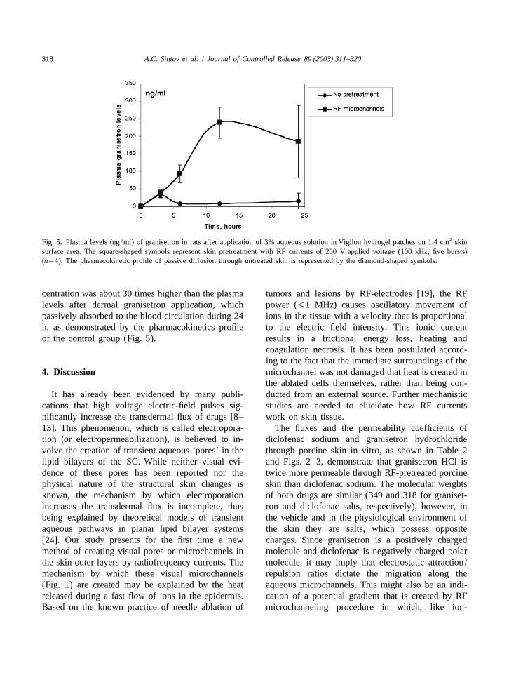
318 A.C. Sintov et al. / Journal of Controlled Release 89 (2003) 311–320
2Fig. 5. Plasma levels (ng/ml) of granisetron in rats after application of 3% aqueous solution in Vigilon hydrogel patches on 1.4 cm skinsurface area. The square-shaped symbols represent skin pretreatment with RF currents of 200 V applied voltage (100 kHz; five bursts)(n54). The pharmacokinetic profile of passive diffusion through untreated skin is represented by the diamond-shaped symbols.
centration was about 30 times higher than the plasma tumors and lesions by RF-electrodes [19], the RFlevels after dermal granisetron application, which power (,1 MHz) causes oscillatory movement ofpassively absorbed to the blood circulation during 24 ions in the tissue with a velocity that is proportionalh, as demonstrated by the pharmacokinetics profile to the electric field intensity. This ionic currentof the control group (Fig. 5). results in a frictional energy loss, heating and
coagulation necrosis. It has been postulated accord-ing to the fact that the immediate surroundings of the
4 . Discussion microchannel was not damaged that heat is created inthe ablated cells themselves, rather than being con-
It has already been evidenced by many publi- ducted from an external source. Further mechanisticcations that high voltage electric-field pulses sig- studies are needed to elucidate how RF currentsnificantly increase the transdermal flux of drugs [8– work on skin tissue.13]. This phenomenon, which is called electropora- The fluxes and the permeability coefficients oftion (or electropermeabilization), is believed to in- diclofenac sodium and granisetron hydrochloridevolve the creation of transient aqueous ‘pores’ in the through porcine skin in vitro, as shown in Table 2lipid bilayers of the SC. While neither visual evi- and Figs. 2–3, demonstrate that granisetron HCl isdence of these pores has been reported nor the twice more permeable through RF-pretreated porcinephysical nature of the structural skin changes is skin than diclofenac sodium. The molecular weightsknown, the mechanism by which electroporation of both drugs are similar (349 and 318 for graniset-increases the transdermal flux is incomplete, thus ron and diclofenac salts, respectively), however, inbeing explained by theoretical models of transient the vehicle and in the physiological environment ofaqueous pathways in planar lipid bilayer systems the skin they are salts, which possess opposite[24]. Our study presents for the first time a new charges. Since granisetron is a positively chargedmethod of creating visual pores or microchannels in molecule and diclofenac is negatively charged polarthe skin outer layers by radiofrequency currents. The molecule, it may imply that electrostatic attraction/mechanism by which these visual microchannels repulsion ratios dictate the migration along the(Fig. 1) are created may be explained by the heat aqueous microchannels. This might also be an indi-released during a fast flow of ions in the epidermis. cation of a potential gradient that is created by RFBased on the known practice of needle ablation of microchanneling procedure in which, like ion-
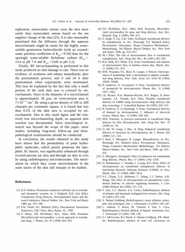
A.C. Sintov et al. / Journal of Controlled Release 89 (2003) 311–320 319
[4] D.V. McAllister, M.G. Allen, M.R. Prausnitz, Microfabri-tophoresis, monovalent cations cross the skin morecated microneedles for gene and drug delivery, Ann. Rev.easily than monovalent anions based on the netBiomed. Eng. 2 (2000) 298–313.
negative charge of the skin [25]. It is also reasonably [5] P. Singh, P. Liu, S.M. Dinh, Facilitated transdermal deliverypostulated that the diffusion through the aqueous by iontophoresis, in: R.L. Bronaugh, H.I. Maibach (Eds.),
Percutaneous Absorption, Drugs–Cosmetics–Mechanisms–microchannels might be easier for the highly water-Methodology, 3rd Edition, Marcel Dekker, Inc., New Yorksoluble granisetron hydrochloride (with an octanol /and Basel, 1999, pp. 633–657.
water partition coefficient ofK 50.28) than for theow [6] M.J. Pikal, The role of electroosmotic flow in transdermalsparingly water-soluble diclofenac sodium (K 5 iontophoresis, Adv. Drug Del. Rev. 46 (2001) 281–305.ow
[7] B.D. Bath, H.S. White, E.R. Scott, Visualization and analysis13.4 at pH 7.4 andK 51545 at pH 5.2).owof electroosmotic flow in hairless mouse skin, Pharm. Res.Finally, RF microchanneling as performed in this17 (2000) 471–475.study produced no skin damage in rats. There was no
[8] M.R. Prausnitz, V.G. Bose, R. Langer, J. Weaver, Electropo-evidence of erythema and edema immediately after ration of mammalian skin: a mechanism to enhance transder-the pretreatment process, and 6 and 24 h after mal drug delivery, Proc. Natl. Acad. Sci. USA 90 (1993)
10504–10508.pretreatment when experiments were terminated.[9] R. Vanbever, N. Lecouturier, V. Preat, Transdermal deliveryThis may be explained by the fact that only a small
of metoprolol by electroporation, Pharm. Res. 11 (1994)portion of the total skin area is covered by mi- 1657–1662.crochannels. Since in the diameter of a microchannel [10] J.E. Riviere, N.A. Monteiro-Riviere, R.A. Rogers, D. Bom-is approximately 30mm, its cross-section area is mannan, J.A. Tamada, R.O. Potts, Pulsatile transdermal
26 2 delivery of LHRH using electroporation: drug delivery and7310 cm . By using a given density of 100 or 200skin toxicology, J. Controlled Release 36 (1995) 229–233.channels per centimeter square, it is found that less
[11] R. Vanbever, E. Le Boulenge, V. Preat, Transdermal deliverythan 0.2% of the skin area is occupied by mi- of fentanyl by electroporation. I. Influence of electricalcrochannels. Due to this small figure and the rela- factors, Pharm. Res. 13 (1996) 559–565.tively low microchanneling depth, no apparent skin [12] M.R. Prausnitz, A practical assessment of transdermal drug
delivery by skin electroporation, Adv. Drug Del. Rev. 35reaction was documented. Since toxicological re-(1999) 61–76.search was beyond the scope of this paper, more
[13] Q. Hu, W. Liang, J. Bao, Q. Ping, Enhanced transdermalstudies including long-term follow-up and histo- delivery of tetracaine by electroporation, Int. J. Pharm. 202pathological examinations should be conducted. (2000) 121–124.
In conclusion, the results obtained in this study [14] J. Kost, S. Mitragotri, R. Langer, Phonophoresis, in: R.L.Bronaugh, H.I. Maibach (Eds.), Percutaneous Absorption,have shown that the permeability of polar hydro-Drugs–Cosmetics–Mechanisms–Methodology, 3rd Edition,philic molecules, which poorly penetrate the lipo-Marcel Dekker, Inc., New York and Basel, 1999, pp. 615–
philic SC barrier, was significantly enhanced through 631.excised porcine ear skin and through rat skin in vivo [15] S. Mitragotri, Synergistic effect of enhancers for transdermalby using radiofrequency microelectrodes. The mech- drug delivery, Pharm. Res. 17 (2000) 1354–1359.
[16] D. Bommannan, J. Tamada, L. Leung, R.O. Potts, Effect ofanism by which they create microchannels in theelectroporation on transdermal iontophoretic delivery ofouter layers of the skin still remains to be studied.luteinizing hormone releasing hormone (LHRH) in vitro,Pharm. Res. 11 (1994) 1809–1814.
[17] S-L. Chang, G.A. Hofmann, L. Zhang, L.J. Deftos, A.K.Banga, The effect of electroporation on iontophoretic trans-
R eferences dermal delivery of calcium regulating hormones, J. Con-trolled Release 66 (2000) 127–133.
[1] K.A. Walters, Penetration enhancers and their use in transder- [18] F. Izzo, C.C. Barnett, S.A. Curley, Radiofrequency ablationmal therapeutic systems, in: J. Hadgraft, R.H. Guy (Eds.), of primary and metastatic malignant liver tumors, Adv. Surg.Transdermal Drug Delivery, Developmental Issues and Re- 35 (2001) 225–250.search Initiatives, Marcel Dekker, Inc., New York and Basel, [19] S. Nahum Goldberg, Radiofrequency tumor ablation: princi-1989, pp. 197–246. ples and techniques, Eur. J. Ultrasound 13 (2001) 129–147.
[20] L. Solbiati, T. Ierace, M. Tonolini, V. Osti, L. Cova,[2] E.W. Smith, H.I. Maibach (Eds.), Percutaneous PenetrationRadiofrequency thermal ablation of hepatic metastases, Eur.Enhancers, CRC Press, Boca Raton, FL, 1995.J. Ultrasound 13 (2001) 149–158.[3] S. Henry, D.V. McAllister, M.G. Allen, M.R. Prausnitz,
[21] F.J. McGovern, B.J. Wood, S. Nahum Goldberg, P.R. Muel-Microfabricated microneedles: a novel approach to transder-ler, Radiofrequency ablation of renal cell carcinoma viamal drug, J. Pharm. Sci. 87 (1998) 922–925.

320 A.C. Sintov et al. / Journal of Controlled Release 89 (2003) 311–320
image guided needle electrodes, J. Urol. 161 (1999) 599– [24] J.C. Weaver, Y.A. Chizmadzhev, Theory of electroporation: a600. review, Bioelectrochem. Bioenerg. 41 (1996) 135–160.
[22] Z. Avrahami, Transdermal Drug Delivery and Analyte [25] R.R. Burnette, B. Ongpipattanakul, Characterization of theExtraction, US Patent No. 6,148,232, (2000). preselective properties of excised human skin during ion-
[23] S. Kudoh, T. Sato, H. Okada, H. Kumakura, H. Nakamura, tophoresis, J. Pharm. Sci. 76 (1987) 765–773.Simultaneous determination of granisetron and 7-hydroxy- [26] R. Panchagnula, K. Stemmer, W.A. Ritschel, Animal modelsgranisetron in human plasma by high-performance liquid for transdermal drug delivery, Meth. Find. Exp. Clin. Phar-chromatography with fluorescence detection, J. Chromatogr. macol. 19 (1997) 335–341.B 660 (1994) 205–210.
Related Documents


![Lupron (leuprolide acetate) Injection, Solution Rx only ... · Lupron (leuprolide acetate) Injection, Solution [Abbott Laboratories] Rx only . DESCRIPTION . Leuprolide acetate is](https://static.cupdf.com/doc/110x72/5bd5671d09d3f2733e8b8a35/lupron-leuprolide-acetate-injection-solution-rx-only-lupron-leuprolide.jpg)