REVIEW Open Access A proposal for a CT driven classification of left colon acute diverticulitis Massimo Sartelli 1* , Frederick A Moore 2 , Luca Ansaloni 3 , Salomone Di Saverio 4 , Federico Coccolini 3 , Ewen A Griffiths 5 , Raul Coimbra 6 , Ferdinando Agresta 7 , Boris Sakakushev 8 , Carlos A Ordoñez 9 , Fikri M Abu-Zidan 10 , Aleksandar Karamarkovic 11 , Goran Augustin 12 , David Costa Navarro 13 , Jan Ulrych 14 , Zaza Demetrashvili 15 , Renato B Melo 16 , Sanjay Marwah 17 , Sanoop K Zachariah 18 , Imtiaz Wani 19 , Vishal G Shelat 20 , Jae Il Kim 21 , Michael McFarlane 22 , Tadaja Pintar 23 , Miran Rems 24 , Miklosh Bala 25 , Offir Ben-Ishay 26 , Carlos Augusto Gomes 27 , Mario Paulo Faro 28 , Gerson Alves Pereira Júnior 29 , Marco Catani 30 , Gianluca Baiocchi 31 , Roberto Bini 32 , Gabriele Anania 33 , Ionut Negoi 34 , Zurabs Kecbaja 35 , Abdelkarim H Omari 36 , Yunfeng Cui 37 , Jakub Kenig 38 , Norio Sato 39 , Andras Vereczkei 40 , Matej Skrovina 41 , Koray Das 42 , Giovanni Bellanova 43 , Isidoro Di Carlo 44 , Helmut A Segovia Lohse 45 , Victor Kong 46 , Kenneth Y Kok 47 , Damien Massalou 48 , Dmitry Smirnov 49 , Mahir Gachabayov 50 , Georgios Gkiokas 51 , Athanasios Marinis 52 , Charalampos Spyropoulos 53 , Ioannis Nikolopoulos 54 , Konstantinos Bouliaris 55 , Jaan Tepp 56 , Varut Lohsiriwat 57 , Elif Çolak 58 , Arda Isik 59 , Daniel Rios-Cruz 60 , Rodolfo Soto 61 , Ashraf Abbas 62 , Cristian Tranà 63 , Emanuele Caproli 64 , Darija Soldatenkova 35 , Francesco Corcione 65 , Diego Piazza 66 and Fausto Catena 67 Abstract Computed tomography (CT) imaging is the most appropriate diagnostic tool to confirm suspected left colonic diverticulitis. However, the utility of CT imaging goes beyond accurate diagnosis of diverticulitis; the grade of severity on CT imaging may drive treatment planning of patients presenting with acute diverticulitis. The appropriate management of left colon acute diverticulitis remains still debated because of the vast spectrum of clinical presentations and different approaches to treatment proposed. The authors present a new simple classification system based on both CT scan results driving decisions making management of acute diverticulitis that may be universally accepted for day to day practice. CT imaging in the diagnosis of acute diverticulitis Diverticular disease is common in western countries, however the prevalence of colonic diverticulosis is in- creasing throughout the world, probably because of changes in lifestyle [1]. Diverticulitis is the most usual complications of diverticulosis, affecting 15-25% of pa- tients [2]. Acute diverticulitis encompasses a variety of conditions, ranging from localized diverticular inflam- mation to fecal peritonitis. It has been usually divided in to uncomplicated and complicated basing on the exten- sion of infection process to the peritoneum. Acute un- complicated diverticulitis is now successfully treated in most patients by conservative management [3,4]. In recent years the disease natural history and the in- dividual physiology of local and/or systemic inflamma- tory response have been understood and minimally invasive strategies, have been applied to selected patients with more severe disease [5] despite the lack of well con- ducted randomized clinical trials [6]. As a consequence the choice of the surgical strategy has been often guided by the surgeon’ s personal preference [7] rather than by available published medical literature. An accurate as- sessment of patients with acute diverticulitis by both clinical signs and radiological features is necessary to identify the best treatment for each patient before the treatment. Over the last three decades, acute complicated diver- ticulitis has been divided traditionally into four stages * Correspondence: [email protected] 1 Department of Surgery, Macerata Hospital, Macerata, Italy Full list of author information is available at the end of the article WORLD JOURNAL OF EMERGENCY SURGERY © 2015 Sartelli et al.; licensee BioMed Central. This is an Open Access article distributed under the terms of the Creative Commons Attribution License (http://creativecommons.org/licenses/by/4.0), which permits unrestricted use, distribution, and reproduction in any medium, provided the original work is properly credited. The Creative Commons Public Domain Dedication waiver (http://creativecommons.org/publicdomain/zero/1.0/) applies to the data made available in this article, unless otherwise stated. Sartelli et al. World Journal of Emergency Surgery 2015, 10:3 http://www.wjes.org/content/10/1/3

Welcome message from author
This document is posted to help you gain knowledge. Please leave a comment to let me know what you think about it! Share it to your friends and learn new things together.
Transcript

WORLD JOURNAL OF EMERGENCY SURGERY
Sartelli et al. World Journal of Emergency Surgery 2015, 10:3http://www.wjes.org/content/10/1/3
REVIEW Open Access
A proposal for a CT driven classification of leftcolon acute diverticulitisMassimo Sartelli1*, Frederick A Moore2, Luca Ansaloni3, Salomone Di Saverio4, Federico Coccolini3,Ewen A Griffiths5, Raul Coimbra6, Ferdinando Agresta7, Boris Sakakushev8, Carlos A Ordoñez9,Fikri M Abu-Zidan10, Aleksandar Karamarkovic11, Goran Augustin12, David Costa Navarro13, Jan Ulrych14,Zaza Demetrashvili15, Renato B Melo16, Sanjay Marwah17, Sanoop K Zachariah18, Imtiaz Wani19, Vishal G Shelat20,Jae Il Kim21, Michael McFarlane22, Tadaja Pintar23, Miran Rems24, Miklosh Bala25, Offir Ben-Ishay26,Carlos Augusto Gomes27, Mario Paulo Faro28, Gerson Alves Pereira Júnior29, Marco Catani30, Gianluca Baiocchi31,Roberto Bini32, Gabriele Anania33, Ionut Negoi34, Zurabs Kecbaja35, Abdelkarim H Omari36, Yunfeng Cui37,Jakub Kenig38, Norio Sato39, Andras Vereczkei40, Matej Skrovina41, Koray Das42, Giovanni Bellanova43,Isidoro Di Carlo44, Helmut A Segovia Lohse45, Victor Kong46, Kenneth Y Kok47, Damien Massalou48,Dmitry Smirnov49, Mahir Gachabayov50, Georgios Gkiokas51, Athanasios Marinis52, Charalampos Spyropoulos53,Ioannis Nikolopoulos54, Konstantinos Bouliaris55, Jaan Tepp56, Varut Lohsiriwat57, Elif Çolak58, Arda Isik59,Daniel Rios-Cruz60, Rodolfo Soto61, Ashraf Abbas62, Cristian Tranà63, Emanuele Caproli64, Darija Soldatenkova35,Francesco Corcione65, Diego Piazza66 and Fausto Catena67
Abstract
Computed tomography (CT) imaging is the most appropriate diagnostic tool to confirm suspected left colonicdiverticulitis. However, the utility of CT imaging goes beyond accurate diagnosis of diverticulitis; the grade ofseverity on CT imaging may drive treatment planning of patients presenting with acute diverticulitis.The appropriate management of left colon acute diverticulitis remains still debated because of the vast spectrumof clinical presentations and different approaches to treatment proposed. The authors present a new simpleclassification system based on both CT scan results driving decisions making management of acute diverticulitisthat may be universally accepted for day to day practice.
CT imaging in the diagnosis of acute diverticulitisDiverticular disease is common in western countries,however the prevalence of colonic diverticulosis is in-creasing throughout the world, probably because ofchanges in lifestyle [1]. Diverticulitis is the most usualcomplications of diverticulosis, affecting 15-25% of pa-tients [2]. Acute diverticulitis encompasses a variety ofconditions, ranging from localized diverticular inflam-mation to fecal peritonitis. It has been usually divided into uncomplicated and complicated basing on the exten-sion of infection process to the peritoneum. Acute un-complicated diverticulitis is now successfully treated inmost patients by conservative management [3,4].
* Correspondence: [email protected] of Surgery, Macerata Hospital, Macerata, ItalyFull list of author information is available at the end of the article
© 2015 Sartelli et al.; licensee BioMed Central.Commons Attribution License (http://creativecreproduction in any medium, provided the orDedication waiver (http://creativecommons.orunless otherwise stated.
In recent years the disease natural history and the in-dividual physiology of local and/or systemic inflamma-tory response have been understood and minimallyinvasive strategies, have been applied to selected patientswith more severe disease [5] despite the lack of well con-ducted randomized clinical trials [6]. As a consequencethe choice of the surgical strategy has been often guidedby the surgeon’s personal preference [7] rather than byavailable published medical literature. An accurate as-sessment of patients with acute diverticulitis by bothclinical signs and radiological features is necessary toidentify the best treatment for each patient before thetreatment.Over the last three decades, acute complicated diver-
ticulitis has been divided traditionally into four stages
This is an Open Access article distributed under the terms of the Creativeommons.org/licenses/by/4.0), which permits unrestricted use, distribution, andiginal work is properly credited. The Creative Commons Public Domaing/publicdomain/zero/1.0/) applies to the data made available in this article,
Sartelli et al. World Journal of Emergency Surgery 2015, 10:3 Page 2 of 11http://www.wjes.org/content/10/1/3
according to the Hinchey classification. The Hincheyclassification can only be accurately applied in patientswho are operated on [8]. Therefore, there is a need todevelop radiological staging systems to help in themanagement of all patients, especially as a significantproportion are managed without surgery or with radio-logical drainage.However, in recent years the emergency manage-
ment of acute sigmoid diverticulitis has evolveddramatically. Computed tomography imaging hasbecome by now the gold standard in the diagnosisand staging of patients with acute diverticulitis. CTimaging with intravenous contrast has excellent sen-sitivity and specificity, reported as high as 98% and99% [9,10]. To improve image quality it should beperformed with intravenous contrast medium injec-tion (when acute renal failure is not present) and atleast two phases (without contrast and portal ven-ous phase). The utility of CT imaging goes beyondaccurate diagnosis of diverticulitis; the grade of se-verity on CT may drive treatment planning of pa-tients with acute diverticulitis [11-18]. CT is nowthe most useful tool to diagnose acute diverticulitisand associated to the clinical conditions and thephysiological reserve of the patients it can object-ively grade its severity into mild-moderate and se-vere diverticulitis [11].Abdominal ultrasound is an alternative imaging
modality that may be useful in patients with relativecontraindications to CT scanning (pregnancy, renalinsufficiency, and contrast allergy). CT is thereforesuperior to US, especially in the detection of freeair and deeply located or small fluid collections andcan drive the clinicians in the management plan[19]. Limitations of ultrasound include operator-dependency, poor assessment in obese patients andit may not be practical in patients with abdominaltenderness because the transducer probe requires com-pression. Some authors recommend a step-up approachwith CT performed after an inconclusive or negativeUS [20,21].CT scan of the abdomen and pelvis is the most appro-
priate imaging modality in the assessment of suspectedacute diverticulitis.CT scan should be performed in all patients with
suspected complicated acute diverticulitis. In theseforms CT is superior to US, especially in the detec-tion of free air and deeply located or small fluidcollections.
A proposal for a new classificationThe appropriate management of left colon acute diverticu-litis remains still debated because of the vast spectrum of
clinical presentations and the different approaches totreatment proposed. Some trials are in progress and willfurther define the appropriate management of compli-cated diverticulitis [22].Based on the surgical findings of abscesses and peri-
tonitis, Hinchey et al. classically classified the severity ofacute diverticulitis into four levels [8]. In order to correl-ate the classification with the therapeutic approach mod-ifications of Hinchey original classification weredeveloped [23,24]. The authors present a new simpleclassification system based on both CT scan results thatmay drive clinicians in management of acute diverticu-litis and that may be universally accepted for day to daypractice. The new classification divides acute diverticu-litis into 2 groups: uncomplicated and complicated.In the event of an uncomplicated case of acute diver-
ticulitis, the infection only involves the colon and doesnot extend to the peritoneum.In the event of complicated IAI, the infectious process
proceeds beyond the colon. Complicated acute diverticu-litis is divided into 4 stages, based on the extension ofthe infectious process.
Uncomplicated diverticulitisDiverticula, thickening of the wall, increased density ofthe pericolic fatComplicated diverticulitis1 A Pericolic air bubbles or little pericolic fluidwithout abscess1 B Abscess ≤ 4 cm2 A Abscess > 4 cm2 B Distant air (>5 cm from inflamed bowel segment)3 Diffuse fluid without distant free air (no hole incolon)4 Diffuse fluid with distant free air (persistent hole incolon)
The new classification is a starting point to stratify pa-tients in well-defined stages. The definitive treatment foreach stage can vary according to the clinical conditionand functional reserves of the patient. The authorspresent the possible treatment strategies for each stagebasing on both the clinical conditions of the patient andthe presence of severe/multiple comorbidities (Figure 1).
Uncomplicated acute diverticulitisUncomplicated diverticulitis is a confined inflammatoryprocess. CT findings include diverticula, thickening ofthe wall and, increased density of the pericolic fat(Figure 2). Patients with uncomplicated diverticulitisusually have an indolent course with a low incidence ofsubsequent complications. Complicated recurrence afteran uncomplicated episode of diverticulitis is rare (<5%)
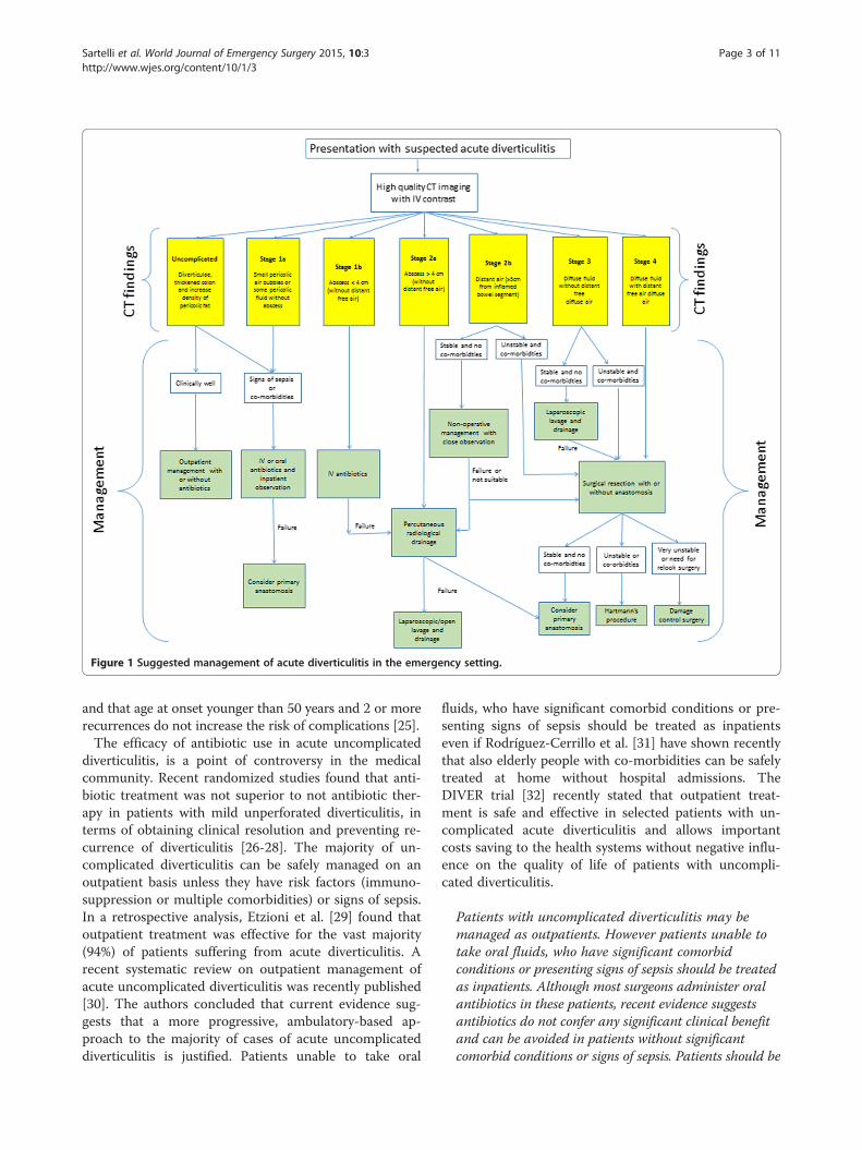
Figure 1 Suggested management of acute diverticulitis in the emergency setting.
Sartelli et al. World Journal of Emergency Surgery 2015, 10:3 Page 3 of 11http://www.wjes.org/content/10/1/3
and that age at onset younger than 50 years and 2 or morerecurrences do not increase the risk of complications [25].The efficacy of antibiotic use in acute uncomplicated
diverticulitis, is a point of controversy in the medicalcommunity. Recent randomized studies found that anti-biotic treatment was not superior to not antibiotic ther-apy in patients with mild unperforated diverticulitis, interms of obtaining clinical resolution and preventing re-currence of diverticulitis [26-28]. The majority of un-complicated diverticulitis can be safely managed on anoutpatient basis unless they have risk factors (immuno-suppression or multiple comorbidities) or signs of sepsis.In a retrospective analysis, Etzioni et al. [29] found thatoutpatient treatment was effective for the vast majority(94%) of patients suffering from acute diverticulitis. Arecent systematic review on outpatient management ofacute uncomplicated diverticulitis was recently published[30]. The authors concluded that current evidence sug-gests that a more progressive, ambulatory-based ap-proach to the majority of cases of acute uncomplicateddiverticulitis is justified. Patients unable to take oral
fluids, who have significant comorbid conditions or pre-senting signs of sepsis should be treated as inpatientseven if Rodríguez-Cerrillo et al. [31] have shown recentlythat also elderly people with co-morbidities can be safelytreated at home without hospital admissions. TheDIVER trial [32] recently stated that outpatient treat-ment is safe and effective in selected patients with un-complicated acute diverticulitis and allows importantcosts saving to the health systems without negative influ-ence on the quality of life of patients with uncompli-cated diverticulitis.
Patients with uncomplicated diverticulitis may bemanaged as outpatients. However patients unable totake oral fluids, who have significant comorbidconditions or presenting signs of sepsis should be treatedas inpatients. Although most surgeons administer oralantibiotics in these patients, recent evidence suggestsantibiotics do not confer any significant clinical benefitand can be avoided in patients without significantcomorbid conditions or signs of sepsis. Patients should be

Figure 2 Slightly thickened sigmoid diverticular disease, withoutabscess or perforation.
Sartelli et al. World Journal of Emergency Surgery 2015, 10:3 Page 4 of 11http://www.wjes.org/content/10/1/3
clinical monitored as outpatients to assess for resolutionof the inflammatory processes.
Stage 1 AStage 1 A diverticulitis is a confined inflammatory processthat may include a microperforation but excludes anabscess, and/or peritonitis. CT findings include perico-lic air in the form of air bubbles or little pericolic fluidwithout abscess. Pericolic air is defined as air bubblesor air collection within 5 cm of the inflamed bowel seg-ment without distant air (Figure 3). Patients have leftiliac fossa pain, initial localized tenderness and thereare usually signs of sepsis. Small locules of pericolic airis a feature of a (micro) perforation of a diverticulum.Stage 1 A diverticulitis may progress to more compli-cated clinical forms if it is not treated promptly. Broad-spectrum antibiotics are therefore indicated in stage 1 Apatients. Antibiotics should be always given to the patientswith pericolic air or small fluid collection. Various
Figure 3 Diverticular disease, colonic wall thickening, fatstranding and pericolic fluid and air bubbles.
antimicrobial regimens may be used in the treatment ofacute diverticulitis in order to ensure complete coverageagainst Gram-positive, Gram-negative, and aerobic–anaer-obic bacterial strains [33]. Antimicrobial regimens withbeta-lactamase inhibiting antibiotics such as amoxicil-lin/clavulanic acid or ciprofloxacin plus metronidazoleare appropriate for community acquired acute diverticu-litis, although high rates of resistance to quinolones havereported in many countries. In critically ill patients or inimmunocompromised patients broader-spectrum regi-mens should be used. An appropriate antimicrobial ther-apy given for an appropriate duration has minimal impacton the emergence of antimicrobial resistance.Patients with CT findings of pericolic air and signs of
sepsis and initial tenderness should require bowel rest,intravenous fluid hydration and empirical intravenousantimicrobial therapy. In these patients to optimize anti-microbial therapy and minimize hospital stay, antimicro-bial therapy may be started initially intravenously andswitched to oral therapy as soon as clinical conditionsimprove.
Patients with pericolic air or small fluid collectionshould be managed by antimicrobial therapy. Patientswith signs of sepsis and initial tenderness shoulddemand bowel rest, intravenous fluid hydration andempirical intravenous antimicrobial therapy. In thesepatients intravenous antimicrobial therapy may beswitched to oral therapy as soon as clinical conditionsimprove.
Clinical findings should be sufficient to monitorresolution of the acute episode. In patients who havepersistent or recurrent clinical evidence of intra-abdominal infection after 4-6 days of therapy, new CTimaging should be undertaken.
Stage 1 BAlthough the clinical presentation of acute left diverticu-litis with associated abscess formation has increased inrecent years, the therapeutic strategies for these patientsare still debated. Deciding which patients with diverticu-lar abscesses require percutaneous drainage rather thanmedical management therefore remains controversial[3]. Diverticular abscesses may be initially treated byintravenous antibiotics alone and/or percutaneous drain-age, depending on the size of the abscess rather than thelocation (pelvic versus pericolic) [33-35]. The size of4 cm (local severe diverticulitis) may be a reasonablelimit between antimicrobial versus percutaneous drain-age in management of diverticular abscesses. It is gener-ally observed that abscesses with a size of up to 4 cmseem to respond better to intravenous antibiotics alone
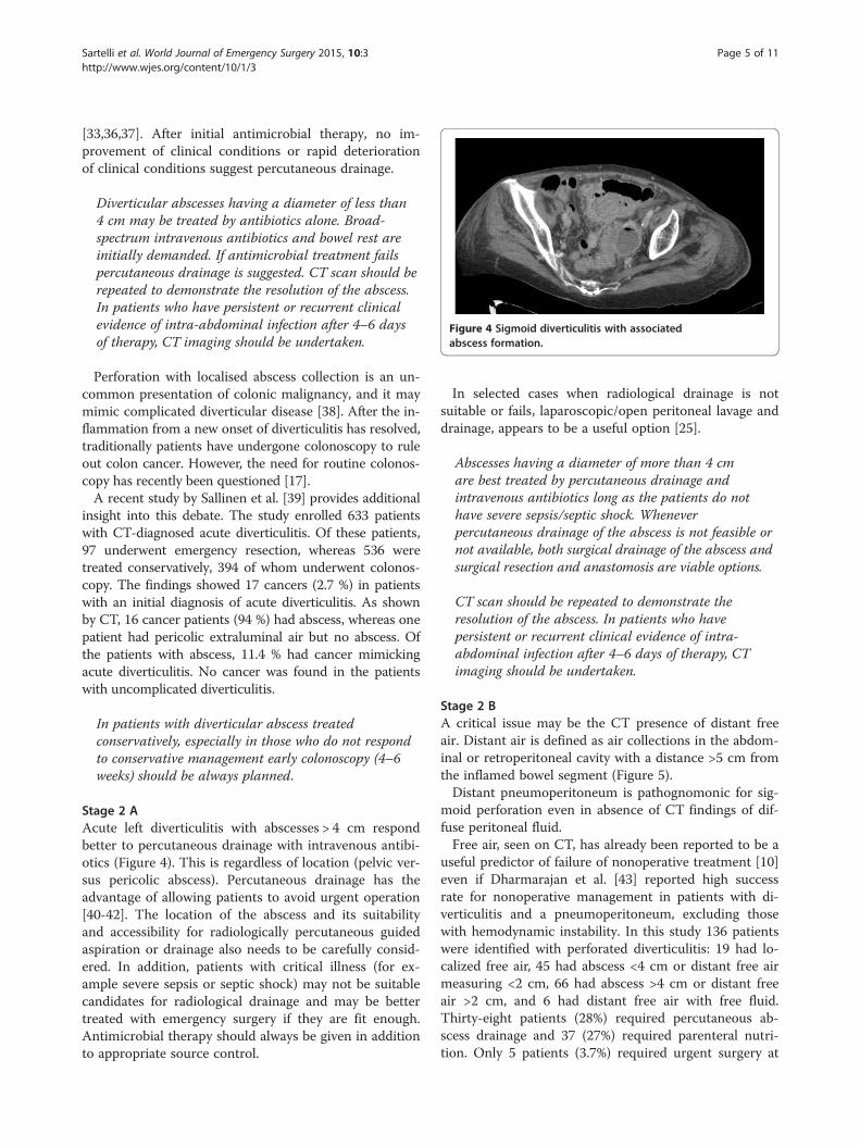
Figure 4 Sigmoid diverticulitis with associatedabscess formation.
Sartelli et al. World Journal of Emergency Surgery 2015, 10:3 Page 5 of 11http://www.wjes.org/content/10/1/3
[33,36,37]. After initial antimicrobial therapy, no im-provement of clinical conditions or rapid deteriorationof clinical conditions suggest percutaneous drainage.
Diverticular abscesses having a diameter of less than4 cm may be treated by antibiotics alone. Broad-spectrum intravenous antibiotics and bowel rest areinitially demanded. If antimicrobial treatment failspercutaneous drainage is suggested. CT scan should berepeated to demonstrate the resolution of the abscess.In patients who have persistent or recurrent clinicalevidence of intra-abdominal infection after 4–6 daysof therapy, CT imaging should be undertaken.
Perforation with localised abscess collection is an un-common presentation of colonic malignancy, and it maymimic complicated diverticular disease [38]. After the in-flammation from a new onset of diverticulitis has resolved,traditionally patients have undergone colonoscopy to ruleout colon cancer. However, the need for routine colonos-copy has recently been questioned [17].A recent study by Sallinen et al. [39] provides additional
insight into this debate. The study enrolled 633 patientswith CT-diagnosed acute diverticulitis. Of these patients,97 underwent emergency resection, whereas 536 weretreated conservatively, 394 of whom underwent colonos-copy. The findings showed 17 cancers (2.7 %) in patientswith an initial diagnosis of acute diverticulitis. As shownby CT, 16 cancer patients (94 %) had abscess, whereas onepatient had pericolic extraluminal air but no abscess. Ofthe patients with abscess, 11.4 % had cancer mimickingacute diverticulitis. No cancer was found in the patientswith uncomplicated diverticulitis.
In patients with diverticular abscess treatedconservatively, especially in those who do not respondto conservative management early colonoscopy (4–6weeks) should be always planned.
Stage 2 AAcute left diverticulitis with abscesses > 4 cm respondbetter to percutaneous drainage with intravenous antibi-otics (Figure 4). This is regardless of location (pelvic ver-sus pericolic abscess). Percutaneous drainage has theadvantage of allowing patients to avoid urgent operation[40-42]. The location of the abscess and its suitabilityand accessibility for radiologically percutaneous guidedaspiration or drainage also needs to be carefully consid-ered. In addition, patients with critical illness (for ex-ample severe sepsis or septic shock) may not be suitablecandidates for radiological drainage and may be bettertreated with emergency surgery if they are fit enough.Antimicrobial therapy should always be given in additionto appropriate source control.
In selected cases when radiological drainage is notsuitable or fails, laparoscopic/open peritoneal lavage anddrainage, appears to be a useful option [25].
Abscesses having a diameter of more than 4 cmare best treated by percutaneous drainage andintravenous antibiotics long as the patients do nothave severe sepsis/septic shock. Wheneverpercutaneous drainage of the abscess is not feasible ornot available, both surgical drainage of the abscess andsurgical resection and anastomosis are viable options.
CT scan should be repeated to demonstrate theresolution of the abscess. In patients who havepersistent or recurrent clinical evidence of intra-abdominal infection after 4–6 days of therapy, CTimaging should be undertaken.
Stage 2 BA critical issue may be the CT presence of distant freeair. Distant air is defined as air collections in the abdom-inal or retroperitoneal cavity with a distance >5 cm fromthe inflamed bowel segment (Figure 5).Distant pneumoperitoneum is pathognomonic for sig-
moid perforation even in absence of CT findings of dif-fuse peritoneal fluid.Free air, seen on CT, has already been reported to be a
useful predictor of failure of nonoperative treatment [10]even if Dharmarajan et al. [43] reported high successrate for nonoperative management in patients with di-verticulitis and a pneumoperitoneum, excluding thosewith hemodynamic instability. In this study 136 patientswere identified with perforated diverticulitis: 19 had lo-calized free air, 45 had abscess <4 cm or distant free airmeasuring <2 cm, 66 had abscess >4 cm or distant freeair >2 cm, and 6 had distant free air with free fluid.Thirty-eight patients (28%) required percutaneous ab-scess drainage and 37 (27%) required parenteral nutri-tion. Only 5 patients (3.7%) required urgent surgery at

Figure 5 Distant retroperitoneal free gas by perforateddiverticular disease.
Sartelli et al. World Journal of Emergency Surgery 2015, 10:3 Page 6 of 11http://www.wjes.org/content/10/1/3
the time of admission, and 7 (5%) required urgent sur-gery for failed non-operative management.An interesting retrospective cohort analysis was re-
cently published to evaluate the safety and effectivenessof non-operative treatment of acute diverticulitis withextraluminal air [44].A total of 132 patients underwent non-operative treat-
ment and 48 patients were primarily operated on. Pa-tients treated non-operatively were divided into 3groups. Patients with pericolic air without abscess had a99% success rate with 0% mortality. Patients with distantintraperitoneal air had a 62% success rate.Whereas small series have demonstrated successful
initial non-operative management of patients with acutecomplicated diverticulitis with perforation, even in theface of pneumoperitoneum, this strategy is reserved forhighly selected stable patients without diffuse peritonealfindings with the goal of converting an emergent or ur-gent situation to one where an elective, single-stage op-eration can be performed.
Patients with distant air (>5 cm from inflamed bowelsegment) may be treated by conservative treatment inselected cases.
However it is associated with failure and maynecessitate surgical operation. Careful clinicalmonitoring is mandatory. A CT scan should berepeated early on the basis of the clinical evolution.
Figure 6 Pelvic free fluid in patient with diffuse fluid and nodistant air.
Surgical resection and anastomosis with or withoutstoma is suggested in stable patients without multipleco-morbidities. Hartmann resection is suggested inunstable patients or in patients with multipleco-morbidities.
Stage 3In recent decades, all cases of diffuse peritonitis havebeen treated by colonic resection.Hartmann’s procedure has been the treatment of
choice for decades, but in recent literature, a few inter-esting alternatives have emerged [45].The restoration of intestinal continuity following
Hartmann’s resection can be difficult and many patientscannot undergo the surgery due to medical co-morbidities;therefore, many of these patients remain with permanentstoma [46].Primary colonic anastomosis, with or without defunc-
tioning stoma, may be a safe alternative even in thepresence of diffuse peritonitis, Several authors have con-sider a primary anastomosis an appropriate option indiffuse peritonitis, with or without defunctioning stomawith no difference in mortality or surgical site infectionrate [47-56].The first randomized trial of Hartmann’s procedure vs.
primary anastomosis with ileostomy in patients with dif-fuse disease was published by Oberkofler et al. in 2012.It reported no difference in initial mortality, but a reduc-tion in length of stay, lower costs, fewer serious compli-cations and greater stoma reversal rates in the primaryanastomosis group [56].We divided diffuse peritonitis in two stages. Stage 3 in-
cludes diffuse fluid without CT findings of perforation.In this stage CT does not reveal any evidence of distantfree air (Figure 6). Fluid should be visualised in at leasttwo distant abdominal quadrants.A more conservative approach of laparoscopic peritoneal
lavage and drainage has been described as an alternative tocolonic resection by Myers et al. [57]. It can potentiallyavoid a stoma in the patients with diffuse peritonitis.Pus is aspirated typically by laparoscopic access followed
by abdominal lavage and the accurate placement of ab-dominal drains which remain for many days after theprocedure [58].
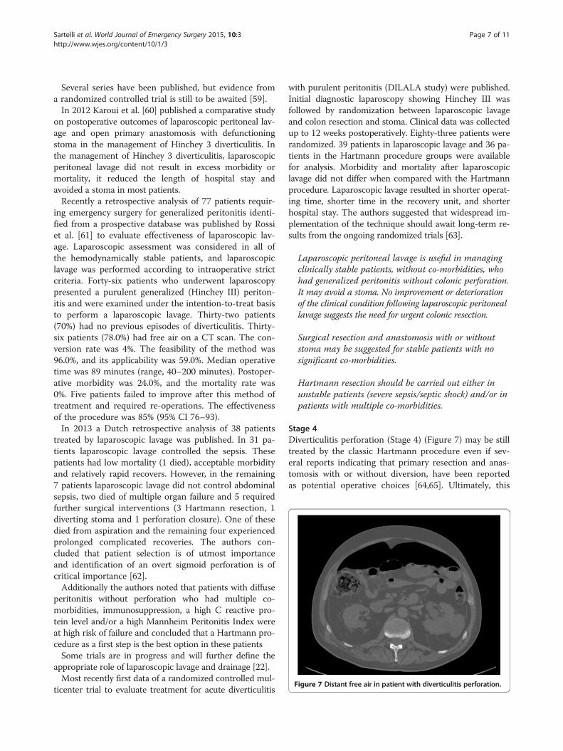
Figure 7 Distant free air in patient with diverticulitis perforation.
Sartelli et al. World Journal of Emergency Surgery 2015, 10:3 Page 7 of 11http://www.wjes.org/content/10/1/3
Several series have been published, but evidence froma randomized controlled trial is still to be awaited [59].In 2012 Karoui et al. [60] published a comparative study
on postoperative outcomes of laparoscopic peritoneal lav-age and open primary anastomosis with defunctioningstoma in the management of Hinchey 3 diverticulitis. Inthe management of Hinchey 3 diverticulitis, laparoscopicperitoneal lavage did not result in excess morbidity ormortality, it reduced the length of hospital stay andavoided a stoma in most patients.Recently a retrospective analysis of 77 patients requir-
ing emergency surgery for generalized peritonitis identi-fied from a prospective database was published by Rossiet al. [61] to evaluate effectiveness of laparoscopic lav-age. Laparoscopic assessment was considered in all ofthe hemodynamically stable patients, and laparoscopiclavage was performed according to intraoperative strictcriteria. Forty-six patients who underwent laparoscopypresented a purulent generalized (Hinchey III) periton-itis and were examined under the intention-to-treat basisto perform a laparoscopic lavage. Thirty-two patients(70%) had no previous episodes of diverticulitis. Thirty-six patients (78.0%) had free air on a CT scan. The con-version rate was 4%. The feasibility of the method was96.0%, and its applicability was 59.0%. Median operativetime was 89 minutes (range, 40–200 minutes). Postoper-ative morbidity was 24.0%, and the mortality rate was0%. Five patients failed to improve after this method oftreatment and required re-operations. The effectivenessof the procedure was 85% (95% CI 76–93).In 2013 a Dutch retrospective analysis of 38 patients
treated by laparoscopic lavage was published. In 31 pa-tients laparoscopic lavage controlled the sepsis. Thesepatients had low mortality (1 died), acceptable morbidityand relatively rapid recovers. However, in the remaining7 patients laparoscopic lavage did not control abdominalsepsis, two died of multiple organ failure and 5 requiredfurther surgical interventions (3 Hartmann resection, 1diverting stoma and 1 perforation closure). One of thesedied from aspiration and the remaining four experiencedprolonged complicated recoveries. The authors con-cluded that patient selection is of utmost importanceand identification of an overt sigmoid perforation is ofcritical importance [62].Additionally the authors noted that patients with diffuse
peritonitis without perforation who had multiple co-morbidities, immunosuppression, a high C reactive pro-tein level and/or a high Mannheim Peritonitis Index wereat high risk of failure and concluded that a Hartmann pro-cedure as a first step is the best option in these patientsSome trials are in progress and will further define the
appropriate role of laparoscopic lavage and drainage [22].Most recently first data of a randomized controlled mul-
ticenter trial to evaluate treatment for acute diverticulitis
with purulent peritonitis (DILALA study) were published.Initial diagnostic laparoscopy showing Hinchey III wasfollowed by randomization between laparoscopic lavageand colon resection and stoma. Clinical data was collectedup to 12 weeks postoperatively. Eighty-three patients wererandomized. 39 patients in laparoscopic lavage and 36 pa-tients in the Hartmann procedure groups were availablefor analysis. Morbidity and mortality after laparoscopiclavage did not differ when compared with the Hartmannprocedure. Laparoscopic lavage resulted in shorter operat-ing time, shorter time in the recovery unit, and shorterhospital stay. The authors suggested that widespread im-plementation of the technique should await long-term re-sults from the ongoing randomized trials [63].
Laparoscopic peritoneal lavage is useful in managingclinically stable patients, without co-morbidities, whohad generalized peritonitis without colonic perforation.It may avoid a stoma. No improvement or deteriorationof the clinical condition following laparoscopic peritoneallavage suggests the need for urgent colonic resection.
Surgical resection and anastomosis with or withoutstoma may be suggested for stable patients with nosignificant co-morbidities.
Hartmann resection should be carried out either inunstable patients (severe sepsis/septic shock) and/or inpatients with multiple co-morbidities.
Stage 4Diverticulitis perforation (Stage 4) (Figure 7) may be stilltreated by the classic Hartmann procedure even if sev-eral reports indicating that primary resection and anas-tomosis with or without diversion, have been reportedas potential operative choices [64,65]. Ultimately, this

Sartelli et al. World Journal of Emergency Surgery 2015, 10:3 Page 8 of 11http://www.wjes.org/content/10/1/3
decision on the surgical strategy is left to the judgment ofthe surgeon, taking into account the clinical status of thepatient including comorbidities, health of the remainingintestine, and extent of peritoneal contamination.
Hartmann resection is still useful in managingdiffuse peritonitis with signs of diverticular diffuseperforations, however in clinically stable patients withno co-morbidities primary resection with anastomosiswith or without and diversion stoma may beperformed.
In specific cases of diverticulitis with perforation inunstable patients the ‘damage control surgery’ has be-come a valuable technique.The term damage control surgery (DCS) for trauma
patients was introduced in the 1990s. It was defined asinitial control of haemorrhage and contamination, allow-ing for resuscitation to normal physiology in the inten-sive care unit and subsequent definitive re-exploration[66]. This concept can be equally applied to abdominalsepsis and the management of diverticular disease per-foration in selected patients [67]. However, it shouldonly be used in those rare instances, where the severephysiological compromise of the patient due to advancedgeneralised peritonitis preclude safe definitive manage-ment. Its overuse may potentially lead to increased mor-bidity due to the effects of multiple laparotomies andthe sequalae of an open abdomen. A variety of tempor-ary abdominal closure options have been described andinclude the classical Bogota bag, sandwich techniques,more modern and commercial Vacuum Assisted Closure(VAC) techniques. These techniques are advised in pa-tients who require relook surgery or who are at risk ofdeveloping abdominal compartment syndrome [68].The damage control surgery in generalized peritonitis
can be performed in different ways [69-71]. In somesense, the Hartmann’s resection is a very good damagecontrol procedure. In critically ill patients the operationcan be staged. For example, the bowel can be resectedand if the patient is too unwell for stoma formation, de-finitive intervention can be performed in 24–48 hoursafter appropriate resuscitation and stabilisation on theICU. A retrospective study by Ordonez et al. [72] sug-gested that a deferred primary anastomosis (DPA) in pa-tients with secondary peritonitis managed with stagedlaparotomies may allow eventual restoration of intestinalcontinuity during the same hospital stay. Among 112 pa-tients there were 34 patients subjected to DPA and 78 todiversion. Fistulas/leakages developed in three patients(8.8%) with DPA and four patients (5.1%) with diversion(p = 0.359). ARDS was present in 6 patients (17.6%) withDPA and 24 patients (30.8%) with diversion (p = 0.149).There were 30 patients (88.2%) with DPA and 65
patients (83.3%) with diversion discharged alive (p =0.51). There were not statistical significant differencesbetween groups among survivors regarding hospitallength of stay, ICU length of stay, and days on mechan-ical ventilation.In a prospective study 51 patients with perforated di-
verticulitis (stage III/IV) [71] were initially managed withlimited resection, lavage and temporary abdominal clos-ure by a vacuum-assisted closure device followed bysecond, reconstructive operation 24–48 hours later.Hospital mortality rate was 9.8%; 35 (76%) of patientswere discharged with reconstructed colon, and 93% ofpatients live without a stoma at follow-up. By damagecontrol concept, an acceptable hospital mortality rateand a high rate of bowel reconstruction at second lookwere achieved in patients with perforated diverticulitisand generalized peritonitis.
Damage control surgery may be a useful strategy inclinically unstable patients with perforateddiverticulitis (severe sepsis/septic shock).
ConclusionsAcute diverticulitis should be managed according to itsseverity. Management options include conservative man-agement with antibiotic treatment, abscess drainage,laparoscopy, and open surgery. Although the manage-ment strategy depends on more factors such as peritonitisdiffusion, clinical conditions and physiological reserve ofthe patient, this new simple classification system based onCT scan results, may drive decisions making in non opera-tive and operative management of acute diverticulitis. andcan help making critical decisions in patients having acutediverticulitis. A prospective study should be designed tovalidate it.
Competing interestsThe authors declare that they have no competing interests.
Authors’ contributionsMS wrote the manuscript. All authors reviewed and approved the finalmanuscript.
Author details1Department of Surgery, Macerata Hospital, Macerata, Italy. 2Department ofSurgery, University of Florida, Gainesville, FL, USA. 3General Surgery I, PapaGiovanni XXIII Hospital, Bergamo, Italy. 4Trauma Surgery Unit, MaggioreHospital, Bologna, Italy. 5Department of Surgery, Queen Elizabeth Hospital,Birminham, UK. 6Department of Surgery, UC San Diego Health System, SanDiego, USA. 7Department of Surgery, Ospedale Civile, ULSS19 del Veneto,Adria, (RO), Italy. 8First Clinic of General Surgery, University Hospital StGeorge, Plovdiv, Bulgaria. 9Department of Surgery, Fundación Valle del Lili,Hospital Universitario del Valle, Universidad del Valle, Cali, Colombia.10Department of Surgery, Faculty of Medicine and Health Sciences, UAEUniversity, Al-Ain, United Arab Emirates. 11Surgery 3 Unit, University Clinic forEmergency Surgery, Belgrade, Serbia. 12Department of Surgery, UniversityHospital Center, Zagreb, Croatia. 13General and Digestive Tract Surgery,Alicante University General Hospital, Alicante, Spain. 141st SurgicalDepartment of First Faculty of Medicine, General University Hospital, Prague

Sartelli et al. World Journal of Emergency Surgery 2015, 10:3 Page 9 of 11http://www.wjes.org/content/10/1/3
Charles University, Prague, Czech Republic. 15Department of General Surgery,Kipshidze Central University Hospital, Tbilisi, Georgia. 16Department ofGeneral Surgery, Centro Hospitalar São João, Faculdade de Medicina daUniversidade do Porto, Porto, Portugal. 17Department of Surgery, Pt BDSPost-graduate Institute of Medical Sciences, Rohtak, India. 18Department ofSurgery, Mosc Medical College, Kolenchery, Cochin, India. 19Department ofSurgery, Sheri-Kashmir Institute of Medical Sciences, Srinagar, India.20Department of General Surgery, Tan Tock Seng Hospital, Tan Tock Seng,Singapore. 21Department of Surgery, Ilsan Paik Hospital, Inje UniversityCollege of Medicine, Goyang, Republic of Korea. 22Department of Surgery,Radiology, Anaesthetics and Intensive Care University Hospital of the WestIndies, Kingston, Jamaica. 23Department of Abdominal Surgery, UmcLjubljana, Ljubljana, Slovenia. 24Surgical Department, General HospitalJesenice, Jesenice, Slovenia. 25Department of General Surgery, HadassahMedical Center, Jerusalem, Israel. 26Department of General Surgery, RambamHealth Care Campus, Haifa, Israel. 27Federal University of Juiz de Fora (UFJF)AND Faculdade de Ciências Médicas e da Saúde de Juiz de Fora (SUPREMA),Juiz de Fora, MG, Brazil. 28Department of General Surgery, Trauma andEmergency Surgery Division, ABC Medical School, Santo André, SP, Brazil.29Emergency Surgery and trauma Unit, Department of Surgery, Ribeirão,Preto, Brazil. 30DEA, Umberto I University Hospital, Rome, Italy. 31Clinical andExperimental Sciences, Brescia Ospedali Civili, Brescia, Italy. 32General andEmergency Surgery SG Bosco Hospital, Turin, Italy. 33Department of Surgery,Arcispedale S. Anna, Medical University of Ferrara, Ferrara, Italy. 34Universityof Medicine and Pharmacy Carol Davila Bucharest, Emergency Hospital ofBucharest, Bucharest, Romania. 35General and Emergency SurgeryDepartment, Riga East University Hospital “Gailezers”, Riga, Latvia.36Department of General Surgery, King Abdalla University Hospital, Irbid,Jordan. 37Department of Surgery, Tianjin Nankai Hospital, Nankai ClinicalSchool of Medicine, Tianjin Medical University, Tianjin, China. 383rdDepartment of Generał Surgery, Narutowicz Hospital, Krakow, Połand.39Department of Primary Care & Emergency Medicine, Kyoto UniversityGraduate School of Medicine, Kyoto, Japan. 40Department of Surgery,Medical School University of Pécs, Pécs, Hungary. 41Department of SurgeryHospital and Oncological Centre Novy Jicin, Novy Jicin, Czech Republic.42Department of General Surgery, Numune Training and Research Hospital,Adana, Turkey. 432nd Surgical Unit, Santa Chiara Hospital, Trento, Italy.44Department of Surgery, Hamad General Hospital, Doha, Qatar. 45II Cátedrade Clínica Quirúrgica, Hospital de Clínicas, Asuncion, Paraguay. 46Departmentof Surgery, Edendale Hospital, Pietermaritzburg, South Africa. 47Departmentof Surgery, Ripas Hospital, Bandar Seri Begawan, Brunei. 48Department ofSurgery, University Hospital of Nice, University of Nice Sophia-Antipolis,Sophia-Antipolis, France. 49Department of Surgical Diseases, South Ural StateMedical University, Chelyabinsk City, Russian Federation. 50Department ofSurgery, Clinical Hospital of Emergency Medicine, Vladimir City, RussianFederation. 512nd Department of Surgery, Aretaieio University Hospital,Athens, Greece. 52First Department of Surgery, Tzanion General Hospital,Piraeus, Greece. 533rd Department of Surgery, Iaso General Hospital, Athens,Greece. 54Department of General Surgery, Lewisham & Greenwich NHS Trust,London, UK. 55Department of General Surgery, University Hospital of Larissa,Larissa, Greece. 56Department of General Surgery, North Estonia MedicalCenter, Tallinn, Estonia. 57Department of Surgery, Faculty of Medicine SirirajHospital Mahidol University, Bangkok, Thailand. 58Department of Surgery,Samsun Education and Research Hospital, Samsun, Turkey. 59Department ofSurgery, Mengucek Gazi Training Research Hospital, Erzincan, Turkey.60Department of Surgery, Hospital de Alta Especialidad de Veracruz, Veracruz,Mexico. 61Department of Emergency Surgery and Critical Care, CentroMedico Imbanaco, Cali, Colombia. 62Emergency Surgery, Faculty of Medicine,Mansoura University, Mansoura, Egypt. 63Department of Emergency Medicineand Surgery, Macerata Hospital, Macerata, Italy. 64Surgical Clinic, AnconaUniversity Hospital, Ancona, Italy. 65Department of Laparoscopic and RoboticSurgery, Colli-Monaldi Hospital, Naples, Italy. 66Division of Surgery, VittorioEmanuele Hospital, Catania, Italy. 67Emergency Department, MaggioreUniversity Hospital, Parma, Italy.
Received: 26 November 2014 Accepted: 30 December 2014Published: 19 February 2015
References1. Weizman AV, Nguyen GC. Diverticular disease: epidemiology and
management. Can J Gastroenterol. 2011;25:385–9.
2. Laméris W, Van Randen A, Van Gulik TM, Bush OR, Winkelhagen J, Bossuyt PM,et al. A clinical decision rule to establish the diagnosis of acute diverticulitis atthe emergency department. Dis Colon Rectum. 2010;53:896–904.
3. Ricciardi R, Baxter NN, Read TE, Marcello PW, Hall J, Roberts PL. Is thedecline in the surgical treatment for diverticulitis associated with anincrease in complicated diverticulitis? Dis Colon rectum. 2009;52:1558–63.
4. Alonso S, Pera M, Parès D, Pascual M, Gil MJ, Courtier R, et al. Outpatienttreatment of patients with uncomplicated acute diverticulitis. Colorectal Dis.2010;12:278–82.
5. McDermott F, Collins D, Heeney A, Winter D. Minimally invasive and surgicalmanagement strategies tailored to the severity of acute diverticulitis. Br JSurg. 2014;101:90–9.
6. Morris AM, Regenbogen SE, Hardiman KM, Hendren S. Sigmoid diverticulitis:a systematic review. JAMA. 2014;311:287–97.
7. Andeweg CS, Mulder IM, Felt-Bersma RJ, Verbon A, van der Wilt GJ, vanGoor H, et al. Guidelines of diagnostics and treatment of acute left-sidedcolonic diverticulitis. Dig Surg. 2013;30:278–92.
8. Hinchey EJ, Schaal PH, Richards MB. Treatment of perforated diverticulardisease of the colon. Adv Surg. 1978;12:85–109.
9. Laméris W, Van Randen A, Bossuyt PMM, Bipat S, Bossuyt PM, BoermeesterMA, et al. Graded compression ultrasonography and computed tomographyin acute colonic diverticulitis: meta-analysis of test accuracy. Eur Radiol.2008;18:2498–511.
10. Ambrosetti P, Jenny A, Becker C, Terrier TF, Morel P. Acute left colonicdiverticulitis–compared performance of computed tomography and water-soluble contrast enema: prospective evaluation of 420 patients. Dis ColonRectum. 2000;43:1363–7.
11. Ambrosetti P. Acute diverticulitis of the left colon: value of the initial CT andtiming of elective colectomy. J Gastrointest Surg. 2008;12:1318–20.
12. Ambrosetti P, Becker C, Terrier F. Colonic diverticulitis: impact of imaging onsurgical management: a prospective study of 542 patients. Eur Radiol.2002;12:1145–9.
13. Kaiser AM, Jiang JK, Lake JP. The management of complicated diverticulitisand the role of computed tomography. Am J Gastroent. 2005;100:910–7.
14. Kohler L, Sauerland S, Neugebauer E. Diagnosis and treatment ofdiverticular disease: results of a consensus development conference. Thescientific committee of the European association for endoscopic surgery.Surg Endosc. 1999;13:430–6.
15. Sher ME, Agachan F, Bortul M. Laparoscopic surgery for diverticulitis. SurgEndosc. 1997;11:264–7.
16. Wasvary H, Turfah F, Kadro O. Same hospitalization resection for acutediverticulitis. Am Surg. 1999;65:632–5.
17. Moore FA, Catena F, Moore EE, Leppaniemi A, Peitzmann AB. Positionpaper: management of perforated sigmoid diverticulitis. World J EmergSurg. 2013;8:55.
18. Klarenbeek BR, De Korte N, van der Peet DL, Cuesta MA. Review of currentclassifications for diverticular disease and a translation into clinical practice.Int J Colorectal Dis. 2012;27:207–14.
19. Puylaert JB. Ultrasound of colon diverticulitis. Dig Dis. 2012;30:56–9.20. Andeweg CS, Wegdam JA, Groenewoud J, van der Wilt GJ, van Goor H,
Bleichrodt RP. Toward an evidence-based step-up approach in diagnosingdiverticulitis. Scand J Gastroenterol. 2014;49:775–84.
21. Mazzei MA, Cioffi Squitieri N, Guerrini S, Stabile Ianora AA, Cagini L, MacariniL, et al. Sigmoid diverticulitis: US findings. Crit Ultrasound J. 2013;5:5.
22. Swank HA, Vermeulen J, Lange JF, Mulder IM, van der Hoeven JA, StassenLP, et al. The ladies trial: laparoscopic peritoneal lavage or resection forpurulent peritonitis and Hartmann's procedure or resection with primaryanastomosis for purulent or faecal peritonitis in perforated diverticulitis(NTR2037). BMC Surg. 2010;10:29.
23. Hansen O, Graupe F, Stock W. Prognostic factors in perforating diverticulitisof the large intestine. Chirurg. 1998;69:443–9.
24. Köhler L, Sauerland S, Neugebauer E. Diagnosis and treatment ofdiverticular disease: results of a consensus development conference. TheScientific Committee of the European Association for Endoscopic Surgery.Surg Endosc. 1999;13:430–6.
25. Regenbogen SE, Hardiman KM, Hendren S, Morris AM. Surgery forDiverticulitis in the 21st Century: a systematic review. JAMA Surg.2014;149:292–303.
26. de Korte N, Kuyvenhoven JP, van der Peet DL, Felt-Bersma RJ, Cuesta MA,Stockmann HB. Mild colonic diverticulitis can be treated without antibiotics.A case–control study. Colorectal Dis. 2012;14:325–30.

Sartelli et al. World Journal of Emergency Surgery 2015, 10:3 Page 10 of 11http://www.wjes.org/content/10/1/3
27. Chabok A, Påhlman L, Hjern F, Haapaniemi S, Smedh K, AVOD Study Group.Randomized clinical trial of antibiotics in acute uncomplicated diverticulitis.Br J Surg. 2012;99:532–9.
28. Shabanzadeh DM, Wille-Jørgensen P. Antibiotics for uncomplicateddiverticulitis. Cochrane Database Syst Rev. 2012;11, CD009092.
29. Etzioni DA, Chiu VY, Cannom RR, Burchette RJ, Haigh PI, Abbas MA.Outpatient treatment of acute diverticulitis: rates and predictors of failure.Dis Colon Rectum. 2010;53:861–5.
30. Jackson JD, Hammond T. Systematic review: outpatient management ofacute uncomplicated diverticulitis. Int J Colorectal Dis. 2014;29:775–81.
31. Rodrìguez-Cerrillo M, Poza-Montoro A, Fernandez-Diaz E, Matesanz-DavidM, Inurrieta RA. Treatment of elderly patients with uncomplicateddiverticulitis, even with comorbidity, at home. Eur J Intern Med.2013;24:430–2.
32. Biondo S, Golda T, Kreisler E, Espin E, Vallribera F, Oteiza F, et al.Outpatient versus hospitalization management for uncomplicateddiverticulitis: a prospective, multicenter randomized clinical trial (DIVER Trial).Ann Surg. 2014;259:38–44.
33. Sartelli M, Viale P, Catena F, Ansaloni L, Moore E, Malangoni M, et al. 2013WSES guidelines for management of intra-abdominal infections. World JEmerg Surg. 2013;8:3.
34. Kumar RR, Kim JT, Haukoos JS, Macias LH, Dixon MR, Stamos MJ, et al.Factors affecting the successful management of intra-abdominal abscesseswith antibiotics and the need for percutaneous drainage. Dis Colon Rectum.2006;49:183–9.
35. Brandt D, Gervaz P, Durmishi Y, Platon A, Morel P, Poletti PA.Percutaneous CT scan-guided drainage versus antibiotherapy alone forHinchey II diverticulitis: a case–control study. Dis Colon Rectum.2006;49:1533–8.
36. Andersen JC, Bundgaard L, Elbrønd H, Laurberg S, Walker LR, Støvring J,et al. Danish national guidelines for treatment of diverticular disease. DanMed J. 2012;59:C4453.
37. Feingold D, Steele SR, Lee S, Kaiser A, Boushey R, Buie WD, et al. Practiceparameters for the treatment of sigmoid diverticulitis. Dis Colon Rectum.2014;57:284–94.
38. Yeo ES, Ng KH, Eu KW. Perforated colorectal cancer: an importantdifferential diagnosis in all presumed diverticular abscesses. Ann Acad MedSingapore. 2011;40(8):375–8.
39. Sallinen V, Mentula P, Leppäniemi A. Risk of colon cancer after computedtomography-diagnosed acute diverticulitis: is routine colonoscopynecessary? Surg Endosc. 2014;28(3):961–6.
40. Durmishi Y, Gervaz P, Brandt D, Bucher P, Platon A, Morel P, et al. Resultsfrom percutaneous drainage of Hinchey stage II diverticulitis guided bycomputed tomography scan. Surg Endosc. 2006;20:1129–33.
41. Siewert B, Tye G, Kruskal J, Sosna J, Opelka F. Impact of CT-guided drainagein the treatment of diverticular abscess: size matters. AJR Am J Roentgenol.2006;186:680–6.
42. Ambrosetti P, Chautems R, Soravia C, Peiris-Waser N, Terrier F.Long-term outcome of mesocolic and pelvic diverticular abscessesof the left colon: a prospective study of 73 cases. Dis Colon Rectum.2005;48:787–91.
43. Dharmarajan S, Hunt SR, Birnbaum EH, Fleshman JW, Mutch MG. Theefficacy of nonoperative management of acute complicated diverticulitis.Dis Colon Rectum. 2011;54:663–71.
44. Sallinen VJ, Mentula PJ, Leppäniemi AK. Nonoperative management ofperforated diverticulitis with extraluminal air is safe and effective in selectedpatients. Dis Colon Rectum. 2014;57:875–81.
45. Abbas S. Resection and primary anastomosis in acute complicateddiverticulitis, a systematic review of the literature. Int J Colorectal Dis.2007;22:351–7.
46. Fleming FJ, Gillen P. Reversal of Hartmann's procedure followingacute diverticulitis: is timing everything? Int J Colorectal Dis.2009;24:1219–25.
47. Herzog T, Janot M, Belyaev O, Sülberg D, Chromik AM, Bergmann U, et al.Complicated sigmoid diverticulitis–Hartmann's procedure or primaryanastomosis? Acta Chir Belg. 2011;111:378–83.
48. Myers E, Winter DC. Adieu to Henri Hartmann? Colorectal Dis.2010;12:849–50.
49. Trenti L, Biondo S, Golda T, Monica M, Kreisler E, Fraccalvieri D, et al.Generalized peritonitis due to perforated diverticulitis: Hartmann'sprocedure or primary anastomosis? Int J Colorectal Dis. 2011;26:377–84.
50. Biondo S, Jaurrieta E, Martí Ragué J, Ramos E, Deiros M, Moreno P, et al.Role of resection and primary anastomosis of the left colon in the presenceof peritonitis. Br J Surg. 2000;87:1580–4.
51. Salem L, Flum DR. Primary anastomosis or Hartmann’s procedure forpatients with diverticular peritonitis? A systematic review. Dis Colon Rectum.2004;47:1953–64.
52. Toro A, Mannino M, Reale G, Cappello G, Di Carlo I. Primary anastomosis vsHartmann procedure in acute complicated diverticulitis. Evolution over thelast twenty years. Chirurgia. 2012;107:598–604.
53. Kreis ME, Mueller MH, Thasler WH. Hartmann's Procedure or primaryanastomosis? Dig Dis. 2012;30:83–5.
54. Masoomi H, Stamos MJ, Carmichael JC, Nguyen B, Buchberg B, Mills S. Doesprimary anastomosis with diversion have Any advantages over Hartmann'sprocedure in acute diverticulitis? Dig Surg. 2012;29:315–20.
55. Tadlock M, Karamanos E, Skiada D, Inaba K, Talving P, Senagore A, et al.Emergency surgery for acute diverticulitis: which operation? A NationalSurgical Quality Improvement Program study. J Trauma Acute Care Surg.2013;74:1385–91.
56. Oberkofler CE, Rickenbacher A, Raptis DA, Lehmann K, Villiger P, Buchli C,et al. A multicenter randomized clinical trial of primary anastomosis orHartmann’s procedure for perforated left colonic diverticulitis with purulentor fecal peritonitis. Ann Surg. 2012;256:819–26.
57. Myers E, Hurley M, O’Sullivan GC, Kavanagh D, Wilson I, Winter DC.Laparoscopic peritoneal lavage for generalized peritonitis due to perforateddiverticulitis. Br J Surg. 2008;95:97–101.
58. Afshar S, Kurer MA. Laparoscopic peritoneal lavage for perforated sigmoiddiverticulitis. Colorectal Dis. 2012;14:135–42.
59. Agresta F, Ansaloni L, Baiocchi GL, Bergamini C, Campanile FC, Carlucci M,et al. Laparoscopic approach to acute abdomen from theConsensusDevelopment Conference of the Società Italiana di ChirurgiaEndoscopica e nuove tecnologie (SICE), Associazione Chirurghi OspedalieriItaliani (ACOI), Società Italiana di Chirurgia (SIC), Società Italiana di Chirurgiad'Urgenza e del Trauma (SICUT), Società Italiana di Chirurgia nell'OspedalitàPrivata (SICOP), and the European Association for Endoscopic Surgery(EAES). Surg Endosc. 2012;26:2134–64.
60. Karoui M, Champault A, Pautrat K, Valleur P, Cherqui D, Champault G.Laparoscopic peritoneal lavage or primary anastomosis with defunctioningstoma for Hinchey 3 complicated diverticulitis: results of a comparativestudy. Dis Colon Rectum. 2009;52:609–15.
61. Rossi GL, Mentz R, Bertone S, Ojea Quintana G, Bilbao S, Im VM, et al.Laparoscopic Peritoneal Lavage for Hinchey III Diverticulitis: is it as effectiveas it is applicable? Dis Colon Rectum. 2014;57:1384–90.
62. Swank HA, Mulder IM, Hoofwijk AG, Nienhuijs SW, Lange JF, Bemelman WA,et al. Early experience with laparoscopic lavage for perforated diverticulitis.Br J Surg. 2013;8:704–10.
63. Angenete E, Thornell A, Burcharth J, Pommergaard HC, Skullman S, BisgaardT, et al. Laparoscopic Lavage Is Feasible and Safe for the Treatment ofPerforated Diverticulitis With Purulent Peritonitis: The First Results From theRandomized Controlled Trial DILALA. Ann Surg. 2014 [Epub ahead of print].
64. Zingg U, Pasternak I, Dietrich M, Seifert B, Oertli D, Metzger U.Primary anastomosis vs Hartmann's procedure in patients undergoingemergency left colectomy for perforated diverticulitis. Colorectal Dis.2010;12:54–60.
65. Jiménez M, Costa D. Resection and primary anastomosis without divertingIleostomy for left colon emergencies: is it a safe procedure? World J Surg.2012;36:1148–53.
66. Rotondo MF, Mc Gonigol MD, Schwab CW, Kauder DR, Hanson CW.Damage control: an approach for improved survival in exsanguinatingpenetrating abdominal injury. J Trauma. 1993;35:375–83.
67. Weber DG, Bendinelli C, Balogh ZJ. Damage control surgery for abdominalemergencies. Br J Surg. 2014;101:109–18.
68. Atema JJ, Gans SL, Boermeester MA. Systematic review and meta-analysisof the open abdomen and temporary abdominal closure techniques innon-trauma patients. World J Surg. 2014 [Epub ahead of print].
69. Sartelli M, Catena F, Di Saverio S, Ansaloni L, Malangoni M, Moore EE, et al.Current concept of abdominal sepsis: WSES position paper. World J EmergSurg. 2014;9:22.
70. Perathoner A, Klaus A, Mühlmann G, Oberwalder M, Margreiter R,Kafka-Ritsch R. Damage control with abdominal vacuum therapy (VAC) tomanage perforated diverticulitis with advanced generalized peritonitis – aproof of concept. Int J Colorectal Dis. 2010;25:767–74.

Sartelli et al. World Journal of Emergency Surgery 2015, 10:3 Page 11 of 11http://www.wjes.org/content/10/1/3
71. Kafka-Ritsch R, Birkfellner F, Perathoner A, Raab H, Nehoda H, Pratschke J,et al. Damage control surgery with abdominal vacuum and delayed bowelreconstruction in patients with perforated diverticulitis Hinchey III/IV. JGastrointest Surg. 2012;16:1915–22.
72. Ordoñez C, Sanchez A, Pineda J, Badiel M, Mesa R, Cardona U, et al.Deferred primary anastomosis versus diversion in patients with severesecondary peritonitis managed with staged laparotomies. World J Surg.2010;34:169–76.
doi:10.1186/1749-7922-10-3Cite this article as: Sartelli et al.: A proposal for a CT driven classificationof left colon acute diverticulitis. World Journal of Emergency Surgery2015 10:3.
Submit your next manuscript to BioMed Centraland take full advantage of:
• Convenient online submission
• Thorough peer review
• No space constraints or color figure charges
• Immediate publication on acceptance
• Inclusion in PubMed, CAS, Scopus and Google Scholar
• Research which is freely available for redistribution
Submit your manuscript at www.biomedcentral.com/submit
Related Documents