This is an electronic reprint of the original article. This reprint may differ from the original in pagination and typographic detail. Powered by TCPDF (www.tcpdf.org) This material is protected by copyright and other intellectual property rights, and duplication or sale of all or part of any of the repository collections is not permitted, except that material may be duplicated by you for your research use or educational purposes in electronic or print form. You must obtain permission for any other use. Electronic or print copies may not be offered, whether for sale or otherwise to anyone who is not an authorised user. Lin, Hai; Li, Wei Ping; Carlson, Synnöve A privileged working memory state and potential top-down modulation for faces, not scenes Published in: FRONTIERS IN HUMAN NEUROSCIENCE DOI: 10.3389/fnhum.2019.00002 Published: 01/02/2019 Document Version Publisher's PDF, also known as Version of record Published under the following license: CC BY Please cite the original version: Lin, H., Li, W. P., & Carlson, S. (2019). A privileged working memory state and potential top-down modulation for faces, not scenes. FRONTIERS IN HUMAN NEUROSCIENCE, 13, [2]. https://doi.org/10.3389/fnhum.2019.00002

Welcome message from author
This document is posted to help you gain knowledge. Please leave a comment to let me know what you think about it! Share it to your friends and learn new things together.
Transcript

This is an electronic reprint of the original article.This reprint may differ from the original in pagination and typographic detail.
Powered by TCPDF (www.tcpdf.org)
This material is protected by copyright and other intellectual property rights, and duplication or sale of all or part of any of the repository collections is not permitted, except that material may be duplicated by you for your research use or educational purposes in electronic or print form. You must obtain permission for any other use. Electronic or print copies may not be offered, whether for sale or otherwise to anyone who is not an authorised user.
Lin, Hai; Li, Wei Ping; Carlson, SynnöveA privileged working memory state and potential top-down modulation for faces, not scenes
Published in:FRONTIERS IN HUMAN NEUROSCIENCE
DOI:10.3389/fnhum.2019.00002
Published: 01/02/2019
Document VersionPublisher's PDF, also known as Version of record
Published under the following license:CC BY
Please cite the original version:Lin, H., Li, W. P., & Carlson, S. (2019). A privileged working memory state and potential top-down modulation forfaces, not scenes. FRONTIERS IN HUMAN NEUROSCIENCE, 13, [2].https://doi.org/10.3389/fnhum.2019.00002

fnhum-13-00002 January 24, 2019 Time: 16:34 # 1
ORIGINAL RESEARCHpublished: 28 January 2019
doi: 10.3389/fnhum.2019.00002
Edited by:Seiki Konishi,
Juntendo University, Japan
Reviewed by:Weizhen Xie,
National Institutes of Health (NIH),United States
Junichi Chikazoe,National Institute for Physiological
Sciences (NIPS), Japan
*Correspondence:Wei-ping Li
[email protected]öve Carlson
Received: 17 July 2018Accepted: 04 January 2019Published: 28 January 2019
Citation:Lin H, Li W-p and Carlson S
(2019) A Privileged Working MemoryState and Potential Top-Down
Modulation for Faces, Not Scenes.Front. Hum. Neurosci. 13:2.
doi: 10.3389/fnhum.2019.00002
A Privileged Working Memory Stateand Potential Top-Down Modulationfor Faces, Not ScenesHai Lin1,2,3, Wei-ping Li1,2* and Synnöve Carlson3,4*
1 Zhongshan School of Medicine, Sun Yat-sen University, Guangzhou, China, 2 Department of Neurosurgery, ShenzhenSecond People’s Hospital, The First Affiliated Hospital of Shenzhen University, Shenzhen, China, 3 Departmentof Neuroscience and Biomedical Engineering, Advanced Magnetic Imaging Centre, Aalto NeuroImaging, Aalto UniversitySchool of Science, Espoo, Finland, 4 Neuroscience Unit, Department of Physiology, Faculty of Medicine, Universityof Helsinki, Helsinki, Finland
Top-down modulation is engaged during multiple stages of working memory (WM),including expectation, encoding, and maintenance. During WM maintenance period,an “incidental cue” can bring one of the two items into a privileged state and make theprivileged item be recalled with higher precision, despite being irrelevant to which one tobe probed as the target. With regard to the different representational states of WM, it’sunclear whether there is top-down modulation on earth sensory cortical areas. Here, Weused this behavioral paradigm of “incidental cue” and event-related fMRI to investigatewhether there were a privileged WM state and top-down modulation for complexstimuli including faces and natural scenes. We found that faces, not scenes, couldenter into the privileged state with improved accuracy and response time of WM task.Meanwhile, cue-driven baseline activity shifts in fusiform face area (FFA) were identifiedby univariate analysis in the recognition of privileged faces, compared to that of non-privileged ones. In addition, the functional connectivity between FFA and right inferiorfrontal junction (IFJ), middle frontal gyrus (MFG), inferior frontal gyrus, right intraparietalsulcus (IPS), right precuneus and supplementary motor area was significantly enhanced,corresponding to the improved WM performance. Moreover, FFA connectivity with IFJand IPS could predict WM improvements. These findings indicated that privileged WMstate and potential top-down modulation existed for faces, but not scenes, during WMmaintenance period.
Keywords: top-down modulation, face recognition, FFA, functional connectivity, working memory
INTRODUCTION
Working memory (WM) is a cognitive system of temporarily holding information available forprocessing with a limited capacity (Baddeley, 2003). When several items are maintained in WMsimultaneously, they can be kept in different representational states (Larocque et al., 2014). If oneitem is more relevant to the WM task or more likely to be probed than others, it can be brought intoa privileged state and be easier to be retrieved (Pertzov et al., 2013). By the introduction of “retro-cue” during the WM maintenance period, different representational states can be manipulatedfor items in WM (Lepsien et al., 2011; Berryhill et al., 2012). Specifically, a retro-cue will giveparticipants a knowledge or expectation about which items to be relevant to the subsequent probedtarget. And then the cued item will be recalled with higher precision than other uncued items.Interestingly, no matter whether the retro-cue is valid or not, the benefit always exists for the
Frontiers in Human Neuroscience | www.frontiersin.org 1 January 2019 | Volume 13 | Article 2

fnhum-13-00002 January 24, 2019 Time: 16:34 # 2
Lin et al. Privileged WM State for Faces
cued item (Gunseli et al., 2015). The neural underpinningsof the retro-cue effect have been investigated in some fMRIstudies (Lepsien and Nobre, 2007; Nelissen et al., 2013). In anevent-related fMRI study, a retro-cue informed participants thecategory information of the probed target in a WM task toremember from two categories of faces and scenes (Lepsien andNobre, 2007). The improved recall precision of the cued item wasaccompanied with the increased activity in the category-specificbrain region involved in object recognition: fusiform face area(FFA) for faces and parahippocampal place area (PPA) for scenes.
Another tool of manipulating different representational statesin WM is presenting items in series, with the last item naturallygetting into the privileged state, which is known as the “recencyeffect” (Allen et al., 2014). The last item is recalled withhigher accuracy and shorter response time than all previousitems. The recency effect is volatile and susceptible to someattention interference such as presenting irrelevant information(Manohar and Husain, 2016). And its magnitude is dependenton the number of items in all. By fMRI, an increased activityin the inferior temporal cortex was found in the recognition ofthe last item compared to that of previous items in a words-remembered task (Nee and Jonides, 2008). In addition, Oztekinet al. (2009) further identified a decreased activity in hippocampalalong with prioritized memory of the last item. These studiessuggested differences in both behavioral WM states and neuralrepresentations due to the recency effect.
Similar to the invalid retro-cue, an “incidental cue” couldbring one of the two items into a privileged state and makethe privileged item be recalled with higher precision, despitebeing irrelevant to which one to be probed as the target (Zokaeiet al., 2014). In a WM experiment by Zokaei et al. (2013)participants were required to remember the motion directionsof two groups of dots in two different colors, simultaneouslyappearing above and below a fixation cross. The incidentalcue is the colored fixation cross during maintenance period,the color of which was the same to one group of dots. AndParticipants were required to answer whether the cued groupof dots was above or below the fixation cross right after theappearance of the incidental cue. Although the incidental cue wascompletely irrelevant to which group of dots to be probed, thedirection of the cued group was recalled with higher precisioncompared to that of the other group. Furthermore, by TMSapplied to motion sensitive area MT+ after the incidental cueduring maintenance period, the benefit of the cued group wasimpaired along with the improvement in the retrieval of theuncued group, which provided causal evidence about differentrepresentational states in WM. This finding is a bit similar tothe phenomenon or experience in our life where the memory inthe natural visual world can be incidentally enhanced by someassociated information. However, with the effect of incidentalcue proved on motion direction as a low-level feature of object,it’s unclear whether the same effect would exist for high-levelcomplex objects such as faces and natural scenes.
Different representational states in WM are accompanied withsensory cortical activity biasing, which is mediated via top-downcontrol (Gazzaley and Nobre, 2012). Top-down modulation onearly sensory brain areas, from prefrontal and parietal control
regions, influences WM performance during both stimulus-present and stimulus-absent stages of WM tasks, to focus ourcognitive resources on goal-relevant information (Gazzaley andNobre, 2012). During WM encoding period, cortical controlregions involved in top-down modulation were investigated infMRI studies (Gazzaley et al., 2007; Chadick and Gazzaley,2011). Functional connectivity between left middle frontal gyrus(MFG) and a scene-selective visual region was enhanced whenscenes were remembered compared to that when scenes wereignored in an object delayed-response task (Gazzaley et al.,2007). In addition, the strength of this coupling correlatedwith the magnitude of activated enhancement for relevantstimuli and suppression of irrelevant ones in the scene-selectivevisual region, which suggested that top-down modulationworked via functional couplings. Similarly, another fMRI studyrevealed visual cortical areas that selectively processing relevantinformation were functionally connected with the frontal-parietalnetwork including intraparietal sulcus (IPS), inferior frontaljunction (IFJ) and MFG, while those processing irrelevantinformation were coupled with the default network (Chadick andGazzaley, 2011). Interestingly, the degree of couplings betweenvisual cortices and default network regions predicted the WMperformance. During WM maintenance period, the mechanismsof top-down modulation are similar to that during perception,but possibly with additional regulatory functions (Kuo et al.,2011, 2012). By fMRI, a common set of frontal and parietalcontrol areas are involved in mediating sensory cortical activityfor different representational states. In a feature delayed-responsetask, functional connectivity between frontal and posterior visualareas increased after the effective retro-cue and had a relationshipwith WM performance (Kuo et al., 2011). Besides, a particularbrain area, in ventrolateral prefrontal cortex and around inferiorfrontal gyrus (IFG) and IFJ, has been implicated in regulatingthe dynamic of neural representations during WM maintenanceperiod (Kuo et al., 2012). A TMS-fMRI study provided causalevidence for the effect of this area on regulating the level ofactivity of representations in posterior brain areas to guideperception and action (Higo et al., 2011).
With regard to the effect of incidental cue during WMmaintenance period, we supposed top-down modulation played arole in mediating the activity of early sensory areas, which wouldbe investigated in our study. We added an incidental cue in a WMtask for two categories of complex objects including faces andnatural scenes, to study the privileged WM state and underlyingtop-down modulation.
MATERIALS AND METHODS
ParticipantsEighteen right-handed volunteers (mean age, 27.4 ± 6.6 years;eight females) were recruited from universities with pays. Thisstudy was approved by the local ethics committee of our instituteand informed consent was obtained from all participants. Allparticipants had normal or corrected-to normal vision and werescreened to make sure they had no history of neurologicalor psychiatric diseases and were not taking any psychotropic
Frontiers in Human Neuroscience | www.frontiersin.org 2 January 2019 | Volume 13 | Article 2

fnhum-13-00002 January 24, 2019 Time: 16:34 # 3
Lin et al. Privileged WM State for Faces
FIGURE 1 | Experimental stimuli and paradigm. (A) The stimuli (a face and ascene) and mask presented as an example. Stimuli consisted of grayscaleimages of 50 neutral faces (half male and half female) on a gray backgroundand 50 natural scenes (3.2◦ horizontal and 4.5◦ vertical visual angles,respectively). (B) The procedure of WM task. Before the WM task (task 2),participants were required to complete a cue-responding task (task 1). Thecue type (arrows pointing to right or left) and category of the probed target(face or scene) were counterbalanced across trials.
medications. All participants were naive regarding the purposeof the study.
StimuliStimuli consisted of grayscale images of 50 neutral faces(half male and half female) on a gray background and 50natural scenes (3.2◦ horizontal and 4.5◦ vertical visual angles,respectively; Figure 1A). Using MATLAB (MathWorks, Natick,MA, United States), all stimuli were grayscale filtered andGaussian band-pass filtered for spatial frequency with a centerspatial frequency of 0.5 cycles/pixel and a Gaussian functionsigma value of 0.2 cycles/pixel. The face stimuli were edited sothat the main features fit inside an oval window, with the outlinesof the stimuli (the edges of the faces) not visible. All images wereadjusted to have the same luminous flux.
WM Task With an Incidental CueAn event-related fMRI experiment was performed with thebehavioral paradigm of incidental cue. The procedure wasdisplayed in Figure 1B. Each trial started with a fixation cross(500 ms), followed by two pictures (one is a face and the other
is a scene; 300 ms) and masks (100 ms) on both sides of thefixation cross. The mask consisted of small grids with randomgray values and had the same size with stimuli (Figure 1A). Thelocations of face and scene pictures (left or right) were randomacross trials. After a 300 ms unfilled delay, two arrows pointingto either the right or the left appeared above and below thefixation cross, which served as the incidental cue. If the arrowspointed to the left, participants were required to recall and answerwhether the previous left picture is a face or a scene with akey press. And if the arrows pointed to the right, participantsmade a response to the previous right picture, correspondingly(Task 1). After a 4400 ms unfilled delay, a face or scene picture(same or different to the previous one) was randomly chosento appear at the center for 300 ms, regarded as the WM target.Participants were instructed to make a response about whetherthe target is same or different to the previous picture of thesame category, as accurately and soon as possible (Task 2).The target was followed by a 6400 ms intertrial interval duringwhich a blank screen was presented. The category of the picturethat the arrows pointed to was irrelevant to the category ofthe target. That is to say, the incidental cue was uninformativeabout which category of the picture would be probed. Therewere 30 trials per block and 6 blocks, separated by a 2-minbreak.
Eye Movement RecordingsTo ensure that participants’ eyes were fixating on the fixationcross when the two pictures of a face and a scene appeared onboth sides of the fixation cross, eye movements were recordedat sampling rate of 1000 Hz with an MRI compatible Eyelink100 eye-tracker (SR Research, Ottawa, Canada) during scanning.A 9-point calibration and validation were performed before eachblock of the WM task. The criteria for saccade onset wereconsidered an eye movement velocity of 30◦/s and an accelerationof 4000◦/s2. The trials in which participants didn’t keep thefixation within the 2◦× 2◦ region centered on the fixation crossduring the appearance of the two pictures would be discarded,to prevent participants from spontaneously making saccade toone of two pictures with better perception of one picture thanthe other one.
Localizer TaskAn independent functional localizer task was performed toidentify the face-selective region of FFA and the scene-selectiveregion of PPA for each participant. The localizer scan consistedof 2 blocks of fixation (rest), viewing faces and viewingscenes (task), respectively, with the duration of 20 s foreach block. Each task block contained 20 stimuli, with thestimulus duration of 300 ms and the inter-stimulus intervalof 500 ms. During the task block, participants were instructedto pay attention to the stimuli and press the button whenthey noticed a stimulus appearing twice non-intermittently (aone-back task). The order of different kinds of blocks wascounterbalanced within and across scans. Participants performedthree localizer scans for a total of 6 blocks of each type, lasting for7 min.
Frontiers in Human Neuroscience | www.frontiersin.org 3 January 2019 | Volume 13 | Article 2

fnhum-13-00002 January 24, 2019 Time: 16:34 # 4
Lin et al. Privileged WM State for Faces
fMRI Data Acquisition and PreprocessingScanning was performed using a 3T Siemens MAGNETOMSkyra MRI system (Erlangen, Germany) with a whole headcoil. A high-resolution 3D T1-weighted MRI scan was acquiredusing a magnetization-prepared rapid gradient-echo sequence.Functional images were obtained using a gradient-echo planarimaging sequence (TR 2500 ms, TE 30 ms, flip angle 75◦, FOV220 mm, matrix size 64 × 64, in plane resolution 3.5 × 3.5 mm).Each functional volume consisted of 45 axial slices of 3.4 mmwith no inter-slice gap and covered the whole cerebrum andcerebellum.
Preprocessing of the imaging data as performed in FSL,consisted of brain extraction, slice timing correction, motioncorrection, and spatial smoothing (6 mm FWHM Gaussiankernel). Differently, the functional data of the localizer taskremained in subject-specific space for the definition of ROIs (FFAand PPA). The functional images of the WM task were registeredto the individual’s structural scan and the MNI152 standard spacetemplate with a 2 mm resolution using FMRIB’s Linear Image
Registration Tool (FLIRT). Low frequencies (cutoff 128 s) wereremoved from the functional data of two tasks by a high-passfilter.
Analysis of Event-Related fMRIExperimentThe WM task was a 2× 2 experimental design, with two factors ofcue type (face or scene) and target category (face or scene). Therewere four conditions of trials (Fcue_Ftarget denoting the cue typeof face and target category of face, Scue_Ftarget, Fcue_Starget,and Scue_Starget). For the analysis of behavioral data, WMperformance was evaluated by accuracy and response time (RT)of those trials in which participants made correct answers in Task1. Statistical significance of behavioral differences was separatelyassessed on accuracy and RT, using repeated-measures ANOVAsand paired two-tailed t-tests.
For individual analyses of fMRI data, cue-related activity wasidentified by convolving a vector of maintenance period (fromthe onset of the cue to the onset of the probe stimulus) with
FIGURE 2 | Behavioral performance. (A,B) Comparisons of WM accuracy and RT between Fcue_Ftarget and Scue_Ftarget. By paired two-tailed t-tests, if the targetwas a face, the accuracy in Fcue_Ftarget was significantly higher and RT was significantly shorter, compared to the corresponding values in Scue_Ftarget (p = 0.003and 0.005, respectively). (C,D) Comparisons of WM accuracy and RT between Fcue_Starget and Scue_Starget. If the target was a scene, there were no significantdifferences of accuracy or RT between groups of different cue types (both p-values > 0.1). The symbols (∗) and (n.s.) indicate being and not being significant,respectively.
Frontiers in Human Neuroscience | www.frontiersin.org 4 January 2019 | Volume 13 | Article 2
fnhum-13-00002 January 24, 2019 Time: 16:34 # 5
Lin et al. Privileged WM State for Faces
the canonical synthetic hemodynamic response function (HRF)and its temporal derivative. The general linear model (GLM)as performed in FSL, was used to model the effects includingmain effect of two factors, interaction effect and pairwise effects(Fcue_Ftarget > Scue_Ftarget and Scue_Starget > Fcue_Starget).Motion parameters were included in the GLM to account formotion-related variance. Group analyses were conducted onMontreal Neurological Institute (MNI) normalized data, usingrandom effects model to assess each effect. Statistical thresholdwas set at Z > 2.3 and p < 0.05, FDR corrected at cluster level.
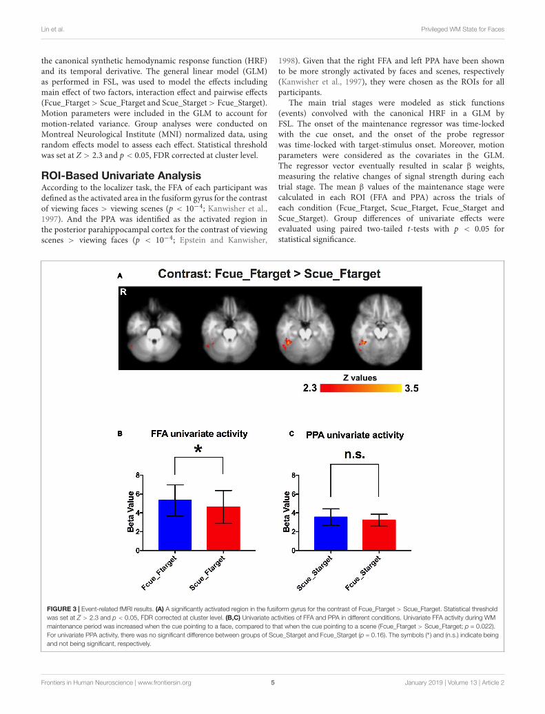
ROI-Based Univariate AnalysisAccording to the localizer task, the FFA of each participant wasdefined as the activated area in the fusiform gyrus for the contrastof viewing faces > viewing scenes (p < 10−4; Kanwisher et al.,1997). And the PPA was identified as the activated region inthe posterior parahippocampal cortex for the contrast of viewingscenes > viewing faces (p < 10−4; Epstein and Kanwisher,
1998). Given that the right FFA and left PPA have been shownto be more strongly activated by faces and scenes, respectively(Kanwisher et al., 1997), they were chosen as the ROIs for allparticipants.
The main trial stages were modeled as stick functions(events) convolved with the canonical HRF in a GLM byFSL. The onset of the maintenance regressor was time-lockedwith the cue onset, and the onset of the probe regressorwas time-locked with target-stimulus onset. Moreover, motionparameters were considered as the covariates in the GLM.The regressor vector eventually resulted in scalar β weights,measuring the relative changes of signal strength during eachtrial stage. The mean β values of the maintenance stage werecalculated in each ROI (FFA and PPA) across the trials ofeach condition (Fcue_Ftarget, Scue_Ftarget, Fcue_Starget andScue_Starget). Group differences of univariate effects wereevaluated using paired two-tailed t-tests with p < 0.05 forstatistical significance.
FIGURE 3 | Event-related fMRI results. (A) A significantly activated region in the fusiform gyrus for the contrast of Fcue_Ftarget > Scue_Ftarget. Statistical thresholdwas set at Z > 2.3 and p < 0.05, FDR corrected at cluster level. (B,C) Univariate activities of FFA and PPA in different conditions. Univariate FFA activity during WMmaintenance period was increased when the cue pointing to a face, compared to that when the cue pointing to a scene (Fcue_Ftarget > Scue_Ftarget; p = 0.022).For univariate PPA activity, there was no significant difference between groups of Scue_Starget and Fcue_Starget (p = 0.16). The symbols (∗) and (n.s.) indicate beingand not being significant, respectively.
Frontiers in Human Neuroscience | www.frontiersin.org 5 January 2019 | Volume 13 | Article 2

fnhum-13-00002 January 24, 2019 Time: 16:34 # 6
Lin et al. Privileged WM State for Faces
Functional ConnectivityROI-based functional connectivity maps of the whole brain wereestimated for each participant, as described previously usinga β-series correlation analysis approach (Gazzaley et al., 2004;Rissman et al., 2004). Mean β values of each ROI (FFA, PPA)were correlated with every brain voxel in subject’s native space foreach participant and each condition (Fcue_Ftarget, Scue_Ftarget,Fcue_Starget and Scue_Starget). Single-participant functionalconnectivity maps were then normalized to the standardizedMNI space and spatially smoothed (6 mm FWHM Gaussiankernel) for group analysis. Non-parametric permutation testswere performed to estimate whole-brain contrast maps betweenthe conditions of Fcue_Ftarget and Scue_Ftarget for the ROIof FFA, and between the conditions of Scue_Starget andFcue_Starget for the ROI of PPA. Statistical threshold was set atp < 0.01, FDR corrected at cluster level.
Neurobehavioral Correlation AnalysisCorrelation analysis was performed between functionalconnectivity differences (Fcue_Ftarget – Scue_Ftarget) basedon the ROI of FFA and the WM performance improvements(Fcue_Ftarget – Scue_Ftarget; for accuracy and RT, respectively),and between functional connectivity differences (Scue_Starget –Fcue_Starget) based on the ROI of PPA and the WMperformance improvements (Scue_Starget – Fcue_Starget).Statistical threshold of neurobehavioral correlations was set atp < 0.05, after Bonferroni correction for multiple comparisons.Furthermore, the Pearson–Filon statistic based on Fisher’s r-to-Ztransformation (ZPF) was used to compare these two kinds ofneurobehavioral correlations based on FFA and PPA, respectively(Raghunathan et al., 1996).
RESULTS
Behavioral PerformanceFor the two factors of cue type (face or scene) and target category(face or scene) in WM task, a 2 × 2 repeated-measures ANOVAwas separately performed on WM accuracy and RT. There wasstatistical significance of interaction effect and main effect oftwo factors for WM accuracy [F(1,17) = 9.81, p < 0.005 forinteraction effect; F(1,17) = 4.71, p < 0.05 for main effect of cuetype; F(1,17) = 5.78, p < 0.05 for main effect of target category].And there was statistical significance of interaction effect andmain effect of target category for RT [F(1,17) = 16.9, p < 0.001for interaction effect; F(1,17) = 2.66, p > 0.1 for main effect ofcue type; F(1,17) = 28.36, p < 0.001 for main effect of targetcategory]. By paired two-tailed t-tests, if the target was a face,the accuracy in Fcue_Ftarget was significantly higher and RT wassignificantly shorter, compared to the corresponding values inScue_Ftarget (p = 0.003 and 0.005, respectively; Figures 2A,B).However, if the target was a scene, there were no significantdifferences of accuracy or RT between groups of different cuetypes (both p-values > 0.1; Figures 2C,D). Therefore, only faceswere recalled with improved WM performance due to the effectof incidental cue, which indicated that the incidental cue could
bring faces into a privileged WM state during WM maintenanceperiod, but not scenes.
fMRI ResultsTo investigate neural underpinnings of the incidental cue’seffect, the conventional 2-stage random effects model wasperformed for fMRI analysis. A significantly activated regionin the fusiform gyrus was identified for the contrast ofFcue_Ftarget > Scue_Ftarget (Figure 3A). And this region wascompleted covered by the significant activations for interactioneffect (Supplementary Figure S1). Moreover, no activationwas observed in the posterior parahippocampal cortex for thecontrast of Scue_Starget > Fcue_Starget, which was consistentwith the behavioral results. To confirm whether the significantlyactivated region in the fusiform gyrus was in FFA, a ROI-based univariate analysis was performed to investigate cue-drivenbaseline activity shifts in FFA. As we expected, univariate FFAactivity during WM maintenance period was increased whenthe cue pointing to a face, compared to that when the cuepointing to a scene (Fcue_Ftarget > Scue_Ftarget; p = 0.022;Figure 3B). The similar analysis, based on PPA, failed to identifya significant difference between groups of Scue_Starget andFcue_Starget (Scue_Starget > Fcue_Starget; p = 0.16; Figure 3C).These results indicate that neural representations of the cue effectare dependent on the category of stimuli, comparable with theWM performance discrepancy.
Function Connectivity ResultsGiven that cue-driven memory benefits and baseline activityshifts were found only on faces and FFA, respectively, theROI-based functional connectivity analysis focused on the trialsin which the target was a face. FFA connectivity maps ofthe whole brain were estimated by the β-series correlationmethod for each participant. A non-parametric analysis was usedto compare FFA connectivity maps during WM maintenanceperiod of different cue types. The FFA connectivity withright IFJ, MFG, IFG, right IPS, supplementary motor area(SMA) and right precuneus were significantly increased inFcue_Ftarget, in contrast to that in Scue_Ftarget (Figure 4 andTable 1), which suggested these frontal and parietal regionsmight be engaged in potential top-down modulation of FFAactivity.
Neurobehavioral CorrelationsThe incidental cue brought faces into a privileged state andresulted in a benefit on WM performance. To investigatewhether cue-driven FFA connectivity changes were associatedwith the behavioral benefit, correlation analysis was conductedbetween differences of FFA connectivity with those frontaland parietal regions (Fcue_Ftarget – Scue_Ftarget), and theWM improvements (Fcue_Ftarget – Scue_Ftarget; for accuracyand RT, respectively). Significant correlations were revealedbetween accuracy increase and connectivity differences of FFA-IFJ (r = 0.74; p = 0.003; Figure 5A), and between RTdecrease and connectivity difference of FFA-IPS (r = −0.63;p = 0.035; Figure 5B), after Bonferroni correction for multiplecomparisons. All the results of neurobehavioral correlations
Frontiers in Human Neuroscience | www.frontiersin.org 6 January 2019 | Volume 13 | Article 2

fnhum-13-00002 January 24, 2019 Time: 16:34 # 7
Lin et al. Privileged WM State for Faces
FIGURE 4 | Comparisons of FFA functional connectivity for the contrast of Fcue_Ftarget > Scue_Ftarget. The FFA connectivity with right IFJ (A), right MFG (B), rightIFG (C), right IPS (D), SMA (E), and right precuneus (F) were significantly increased in Fcue_Ftarget, in contrast to that in Scue_Ftarget. Statistical threshold was setat p < 0.01, FDR corrected at cluster level. Stereotaxic MNI coordinated and mean p-values for significant regions are shown in Table 1. The symbol (∗) indicatesbeing significant.
were shown in Supplementary Figure S2. What’s more, by thePearson-Filon statistic, the correlation between accuracy increaseand connectivity differences of FFA-IFJ was significantly higherthan the correlation between accuracy increase and connectivitydifferences of PPA-IFJ (ZPF = 2.01, p = 0.022, one-tailed), and the
same result was obtained for the correlation between RT decreaseand connectivity difference of FFA-IPS (ZPF = 1.84, p = 0.033,one-tailed). These results suggested that top-down modulationmight be involved in cued-driven WM benefits for faces, but notscenes.
Frontiers in Human Neuroscience | www.frontiersin.org 7 January 2019 | Volume 13 | Article 2

fnhum-13-00002 January 24, 2019 Time: 16:34 # 8
Lin et al. Privileged WM State for Faces
TABLE 1 | FFA connectivity comparisons (contrast: Fcue_Ftarget > Scue_Ftarget).
Brain region No. voxels Mean p-value MNI_X MNI_Y MNI_Z
R inferior frontalgyrus
78 0.004 38 26 6
L inferior frontalgyrus
43 0.005 −32 24 8
R middle frontalgyrus
216 0.004 −34 42 12
L middle frontalgyrus
84 0.004 36 42 20
R inferior frontaljunction
233 0.003 44 14 24
R intraparietalsulcus
56 0.004 42 −28 46
R superior parietallobule
38 0.007 34 −36 44
R precuneus 82 0.006 10 −46 58
Supplementarymotor area
115 0.004 0 −4 56
L, Left; R, Right; MNI_X, Coordinate X in MNI space; MNI_Y, Coordinate Y in MNIspace; MNI_Z, Coordinate Z in MNI space.
DISCUSSION
By the behavioral paradigm of incidental cue, our study providesinitial evidence that there is a privileged WM state on complexobjects of faces, but not scenes during WM maintenance period.fMRI analysis revealed an activated brain region in FFA underlyingmemory benefits of faces, which was potentially modulated bya frontoparietal network of regions (right IFJ, MFG, IFG, rightIPS, right precuneus and SMA) via their functional couplingswith FFA. Furthermore, FFA connectivity with IFJ and IPS couldpredict cue-driven improvements of WM performance.
The effect of the incidental cue during WM maintenanceperiod was first found in orientation-discriminating WM task,which provided direct evidence for the existence of at least twodifferent representational states of WM (Zokaei et al., 2013,2014). The incidental cue, different from the valid retro-cue, wascompletely uninformative about which object or which categoryof objects would be probed. For complex objects such as faces andnatural scenes, the incidental cue behaved variously for differentcategories of stimuli. In this study, we revealed that the incidentalcue could bring faces into a privileged WM state, but notscenes. The similar phenomenon was found in the effect of thecategory-predictive cue during WM expectation period, whichonly worked for faces, not scenes (Bollinger et al., 2010). Oneexplanation in the paper was that the diversity of scenes featuresmakes it difficult to generate a robust template and faces are morestereotyped than scenes. We provided another explanation thatfaces, as the most important and salient visual stimuli a humanencounters, are a special kind of objects, which are thought tobe processed and represented in a holistic manner (Tanaka andFarah, 2003). In our situation, the holistic representational stateof faces in WM possibly made them easy to be prioritized by theincidental cue, just as the basic feature of orientation. And ourresults indicate that the incidental cue during WM maintenanceperiod enhances memory of both low-level elementary featuresand high-level complex faces.
In current study, we could measure the WM performance ofboth cued and uncued stimuli with the behavioral paradigm ofincidental cue. In addition, we could distinguish and determinecategory-selective neural underpinnings of the incidental cue’seffect for the contrast of cued > uncued stimuli, using twocategories of stimuli (faces and scenes) in the event-relatedfMRI experiment. As we expected, we found consistent resultsin behavioral performance and fMRI analysis. There were
FIGURE 5 | Neurobehavioral correlations. Significant correlations were revealed between WM accuracy increase and connectivity differences of FFA-IFJ (r = 0.74;p = 0.003; A), and between WM RT decrease and connectivity difference of FFA-IPS (r = –0.63; p = 0.035; B), after Bonferroni correction for multiple comparisons.
Frontiers in Human Neuroscience | www.frontiersin.org 8 January 2019 | Volume 13 | Article 2

fnhum-13-00002 January 24, 2019 Time: 16:34 # 9
Lin et al. Privileged WM State for Faces
significant differences of WM accuracy and RT, between cuedfaces and uncued faces. Meanwhile, A significantly activatedregion in the fusiform gyrus was revealed for the contrast ofcued > uncued faces, and cue-driven baseline activity shifts wereidentified in the face-selective region of FFA. However, all thesecorresponding results failed to be found on scenes and the scene-selective region of PPA. Besides, the neurobehavioral correlationsbased on FFA were significantly higher than those based on PPA.Thus, as the complex objects, faces and natural scenes are not onlyprocessed in different brain regions, but also possibly stored inWM of different patterns.
Different representational states of faces in WM wereaccompanied with sensory activity biasing in FFA, which waspotentially mediated via top-down control from a frontoparietalnetwork. Our study proved that FFA connectivity with right IFJ,MFG, IFG, right IPS, SMA and right precuneus were significantlyincreased for cued faces during WM maintenance period, incontrast to that of uncued faces. What’s more, FFA connectivitywith IFJ and IPS could predict WM improvements. The top-down modulation involved with a frontoparietal network ofregions was studied not only during WM maintenance period,but also in other WM periods (Gazzaley and Nobre, 2012).During WM expectation period, Bollinger et al. (2010) found theeffect of predictive category cueing for faces in an object delayed-response task and the degree of functional connectivity betweenFFA and brain regions of prefrontal and parietal cortex (rightIFJ, MFG, IFG, and IPS) correlated with the magnitude of pre-stimulus activity modulation in FFA. Particularly, IFJ, definedby Brass and von Cramon (2004), is located at the intersectionof the inferior frontal sulcus and precentral sulcus. It’s provedto be a functionally discrete region and a key node connectingthe dorsal and the ventral attention networks (Szczepanski et al.,2010). During WM encoding period, a fMRI study revealed visualcortical areas that selectively processing relevant informationwere functionally connected with the frontal-parietal networkincluding IPS, IFJ and MFG, while those processing irrelevantinformation were coupled with the default network (Chadick andGazzaley, 2011). Interestingly, the degree of coupling betweenvisual cortices and default network regions predicted the WMperformance. During WM maintenance period, Kuo et al. (2018)found that the retro-cues modulated the strength of functionalconnectivity between the frontoparietal and early visual areasin favor of the most relevant information. Previous studies andour findings indicated top-down modulation might play animportant part in multiple stages of representations supportingWM performance.
About neural circuit mechanisms of WM, recentneurophysiological studies revealed stable population codingwithin a specific subspace coexisting with heterogeneous neuraldynamics in prefrontal cortex during WM maintenance period(Murray et al., 2017). Our finding about the coexistenceof at least two different representational states of WMwas possibly explained by neural population coding withindifferent subspaces. However, it’s unclear whether the incidentalcue, similar to an uninformative retro-cue, has an effect tostrengthen the WM representation of cued item or inhibit theWM representation of uncued item to protect the selectedrepresentation from interference (Souza and Oberauer, 2016).
Bays and Taylor (2018) presented a population coding modelsuggesting retro-cue can’t increase the total information storedabout a stimulus and protects items from time-based decayinstead, which are supportive for the latter explanation. In thisstudy, for different categories of complex objects, the privilegedWM state was only found for faces due to the incidental cue,not natural scenes, which suggested different patterns of neuralpopulation coding for faces and scenes, with different responsesto the incidental cue. Thus, this kind of behavioral discrepancycould have some implications for neural circuit mechanisms andproper computational models of WM.
CONCLUSION
In conclusion, under the effect of the incidental cue, theprivileged WM state and potential top-down modulation existedfor faces, but not scenes, during WM maintenance period.
ETHICS STATEMENT
This study was carried out in accordance with therecommendations of the ethics committee in University ofHelsinki with written informed consent from all subjects. Allsubjects gave written informed consent in accordance with theDeclaration of Helsinki. The protocol was approved by the ethicscommittee in University of Helsinki.
AUTHOR CONTRIBUTIONS
HL and SC contributed conception and design of the study. HLorganized the database, performed the statistical analysis, andwrote the first draft of the manuscript. All authors contributed tomanuscript revision, read, and approved the submitted version.
FUNDING
This work was supported by the National Natural ScienceFoundation of China (81772685), the International CooperationResearch Projects of Shenzhen Science and Technology Program(GJHZ20160301163419476), the Basic Research Projects(subject arrangement) of Shenzhen Science and TechnologyProgram (JCYJ20170413173149177), the Academy of Finland(Grant #259752 and #273147), the Finnish Graduate Schoolof Neuroscience, National Science Foundation of China(Chinese-Finnish International Collaboration, Project-NeuroNos. 30621130076 and 30530270).
SUPPLEMENTARY MATERIAL
The Supplementary Material for this article can be foundonline at: https://www.frontiersin.org/articles/10.3389/fnhum.2019.00002/full#supplementary-material
FIGURE S1 | The cue × target interaction effect.
FIGURE S2 | All Neurobehavioral Correlations.
Frontiers in Human Neuroscience | www.frontiersin.org 9 January 2019 | Volume 13 | Article 2

fnhum-13-00002 January 24, 2019 Time: 16:34 # 10
Lin et al. Privileged WM State for Faces
REFERENCESAllen, R. J., Baddeley, A. D., and Hitch, G. J. (2014). Evidence for two attentional
components in visual working memory. J. Exp. Psychol. Learn. Mem. Cogn. 40,1499–1509. doi: 10.1037/xlm0000002
Baddeley, A. (2003). Working memory: looking back and looking forward. Nat.Rev. Neurosci. 4, 829–839. doi: 10.1038/nrn1201
Bays, P. M., and Taylor, R. (2018). A neural model of retrospective attention invisual working memory. Cogn. Psychol. 100, 43–52. doi: 10.1016/j.cogpsych.2017.12.001
Berryhill, M. E., Richmond, L. L., Shay, C. S., and Olson, I. R. (2012). Shiftingattention among working memory representations: testing cue type, awareness,and strategic control. Q. J. Exp. Psychol. 65, 426–438. doi: 10.1080/17470218.2011.604786
Bollinger, J., Rubens, M. T., Zanto, T. P., and Gazzaley, A. (2010). Expectation-driven changes in cortical functional connectivity influence working memoryand long-term memory performance. J. Neurosci. 30, 14399–14410. doi: 10.1523/JNEUROSCI.1547-10.2010
Brass, M., and von Cramon, D. Y. (2004). Decomposing components of taskpreparation with functional magnetic resonance imaging. J. Cogn. Neurosci. 16,609–620. doi: 10.1162/089892904323057335
Chadick, J. Z., and Gazzaley, A. (2011). Differential coupling of visual cortex withdefault or frontal-parietal network based on goals. Nat. Neurosci. 14, 830–832.doi: 10.1038/nn.2823
Epstein, R., and Kanwisher, N. (1998). A cortical representation of the local visualenvironment. Nature 392, 598–601. doi: 10.1038/33402
Gazzaley, A., and Nobre, A. C. (2012). Top-down modulation: bridging selectiveattention and working memory. Trends Cogn. Sci. 16, 129–135. doi: 10.1016/j.tics.2011.11.014
Gazzaley, A., Rissman, J., Cooney, J., Rutman, A., Seibert, T., Clapp, W.,et al. (2007). Functional interactions between prefrontal and visualassociation cortex contribute to top-down modulation of visualprocessing. Cereb. Cortex 17(Suppl. 1), i125–i135. doi: 10.1093/cercor/bhm113
Gazzaley, A., Rissman, J., and D’esposito, M. (2004). Functional connectivityduring working memory maintenance. Cogn. Affect. Behav. Neurosci. 4, 580–599. doi: 10.3758/CABN.4.4.580
Gunseli, E., Van, M. D., Meeter, M., and Olivers, C. N. (2015). The reliabilityof retro-cues determines the fate of noncued visual working memoryrepresentations. Psychon. Bull. Rev. 22, 1474–1474. doi: 10.3758/s13423-015-0914-4
Higo, T., Mars, R. B., Boorman, E. D., Buch, E. R., and Rushworth, M. F. (2011).Distributed and causal influence of frontal operculum in task control. Proc.Natl. Acad. Sci. U.S.A. 108, 4230–4235. doi: 10.1073/pnas.1013361108
Kanwisher, N., McDermott, J., and Chun, M. M. (1997). The fusiform face area: amodule in human extrastriate cortex specialized for face perception. J. Neurosci.17, 4302–4311. doi: 10.1523/JNEUROSCI.17-11-04302.1997
Kuo, B. C., Lin, S. H., and Yeh, Y. Y. (2018). Functional interplay of top-downattention with affective codes during visual short-term memory maintenance.Cortex 103, 55–70. doi: 10.1016/j.cortex.2018.02.003
Kuo, B. C., Stokes, M. G., and Nobre, A. C. (2012). Attention modulatesmaintenance of representations in visual short-term memory. J. Cogn. Neurosci.24, 51–60. doi: 10.1162/jocn_a_00087
Kuo, B. C., Yeh, Y. Y., Chen, A. J. W., and D’esposito, M. (2011).Functional connectivity during top-down modulation of visual short-termmemory representations. Neuropsychologia 49, 1589–1596. doi: 10.1016/j.neuropsychologia.2010.12.043
Larocque, J. J., Lewis-Peacock, J. A., and Postle, B. R. (2014). Multiple neural statesof representation in short-term memory? It’s a matter of attention. Front. Hum.Neurosci. 8:5. doi: 10.3389/fnhum.2014.00005
Lepsien, J., and Nobre, A. C. (2007). Attentional modulation of objectrepresentations in working memory. Cereb. Cortex 17, 2072–2083. doi: 10.1093/cercor/bhl116
Lepsien, J., Thornton, I., and Nobre, A. C. (2011). Modulation of working-memorymaintenance by directed attention. Neuropsychologia 49, 1569–1577. doi: 10.1016/j.neuropsychologia.2011.03.011
Manohar, S. G., and Husain, M. (2016). Working memory for sequences oftemporal durations reveals a volatile single-item store. Front. Psychol. 7:1655.doi: 10.3389/fpsyg.2016.01655
Murray, J. D., Bernacchia, A., Roy, N. A., Constantinidis, C., Romo, R.,and Wang, X. J. (2017). Stable population coding for working memorycoexists with heterogeneous neural dynamics in prefrontal cortex.Proc. Natl. Acad. Sci. U.S.A. 114, 394–399. doi: 10.1073/pnas.1619449114
Nee, D. E., and Jonides, J. (2008). Neural correlates of access to short-term memory.Proc. Natl. Acad. Sci. U.S.A. 105, 14228–14233. doi: 10.1073/pnas.0802081105
Nelissen, N., Stokes, M., Nobre, A. C., and Rushworth, M. F. (2013). Frontaland parietal cortical interactions with distributed visual representations duringselective attention and action selection. J. Neurosci. 33, 16443–16458. doi: 10.1523/JNEUROSCI.2625-13.2013
Oztekin, I., McElree, B., Staresina, B. P., and Davachi, L. (2009). Working memoryretrieval: contributions of the left prefrontal cortex, the left posterior parietalcortex and the hippocampus. J. Cogn. Neurosci. 21, 581–593. doi: 10.1162/jocn.2008.21016
Pertzov, Y., Bays, P. M., Joseph, S., and Husain, M. (2013). Rapid forgettingprevented by retrospective attention cues. J. Exp. Psychol. Human 39, 1224–1231. doi: 10.1037/a0030947
Raghunathan, T. E., Rosenthal, R., and Rubin, D. B. (1996). Comparing correlatedbut nonoverlapping correlations. Psychol. Methods 1, 178–183. doi: 10.1037/1082-989X.1.2.178
Rissman, J., Gazzaley, A., and D’Esposito, M. (2004). Measuring functionalconnectivity during distinct stages of a cognitive task. Neuroimage 23, 752–763.doi: 10.1016/j.neuroimage.2004.06.035
Souza, A. S., and Oberauer, K. (2016). In search of thefocus of attention in working memory: 13 years of theretro-cue effect. Atten. Percept. Psychophys. 78, 1839–1860.doi: 10.3758/s13414-016-1108-5
Szczepanski, S. M., Konen, C. S., and Kastner, S. (2010). Mechanisms of spatialattention control in frontal and parietal cortex. J. Neurosci. 30, 148–160. doi:10.1523/JNEUROSCI.3862-09.2010
Tanaka, J. W., and Farah, M. J. (2003). “The holistic representation of faces,” inPerception Of Faces, Objects, and Scenes: Analytic and Holistic Processes. Oxford:Oxford University Press, 53–74.
Zokaei, N., Manohar, S., Husain, M., and Feredoes, E. (2013). Causal evidence for aprivileged working memory state in early visual cortex. J. Neurosci. 34, 158–162.doi: 10.1523/JNEUROSCI.2899-13.2014
Zokaei, N., Ning, S., Manohar, S., Feredoes, E., and Husain, M. (2014). Flexibilityof representational states in working memory. Front. Hum. Neurosci. 8:853.doi: 10.3389/fnhum.2014.00853
Conflict of Interest Statement: The authors declare that the research wasconducted in the absence of any commercial or financial relationships that couldbe construed as a potential conflict of interest.
Copyright © 2019 Lin, Li and Carlson. This is an open-access article distributedunder the terms of the Creative Commons Attribution License (CC BY). The use,distribution or reproduction in other forums is permitted, provided the originalauthor(s) and the copyright owner(s) are credited and that the original publicationin this journal is cited, in accordance with accepted academic practice. No use,distribution or reproduction is permitted which does not comply with these terms.
Frontiers in Human Neuroscience | www.frontiersin.org 10 January 2019 | Volume 13 | Article 2
Related Documents