Copyright © 2010 The Korean Society of Electrolyte Metabolism A Practical Approach to Genetic Hypokalemia Received: March 18, 2010. Accepted: April 7, 2010 Corresponding author: Shih-Hua Lin, M.D. Division of Nephrology, Department of Medicine, Tri-Service General Hospital, No. 325, Section 2, Cheng- Kung Road, Neihu 114, Taipei, Taiwan Tel: +886-2-87927213, Fax: +886-2-87927134 E-mail: [email protected] Mutations in genes encoding ion channels, transporters, exchangers, and pumps in human tissues have been increasingly reported to cause hypokalemia. Assessment of history and blood pressure as well as the K + excretion rate and blood acid-base status can help differentiate between acquired and inherited causes of hypokalemia. Familial periodic paralysis, Andersen’s syndrome, congenital chloride-losing diarrhea, and cystic fibrosis are genetic causes of hypokalemia with low urine K + excretion. With respect to a high rate of K + excretion associated with faster Na + disorders (mineralocorticoid excess states), glucoricoid-remediable aldosteronism and congenital adrenal hyperplasia due to either 11 β-hydroxylase and 17 α-hydroxylase deficiencies in the adrenal gland, and Liddle’s syndrome and apparent mineralocorticoid excess in the kidney form the genetic causes. Among slow Cl¯ disorders (normal blood pressure, low extracellular fluid volume), Bartter’s and Gitelman’s syndrome are most common with hypochloremic metabolic alkalosis. Renal tubular acidosis caused by mutations in the basolateral Na + /HCO 3 ¯ cotransporter (NBC1) in the proximal tubules, apical H + -ATPase pump, and basolateral Cl ¯ /HCO 3 ¯ exchanger (anion exchanger 1, AE1) in the distal tubules and carbonic anhydroase II in both are genetic causes with hyperchloremic metabolic acidosis. Further work on genetic causes of hypokalemia will not only provide a much better understanding of the underlying mechanisms, but also set the stage for development of novel therapies in the future. Key Words: acid-base equilibrium; aldosterone; blood pressure; genes; hypokalemia; mutation; renin; urine electrolyte Review Shih-Hua Lin, M.D. 1 Sung-Sen Yang, M.D. 1 Tom Chau, M.D. 2 1 Division of Nephrology, Department of Medicine, Tri-Service General Hospital, National Defense Medical Center, Taipei, Taiwan, Republic of China 2 Department of Medicine, Providence St. Vincent Medical Center, Portland, Oregon, USA Introdution Potassium (K + ), a major intracellular cation, is critically important in maintaining the osmotic equilibrium of the cell and electrical gradient across the cell membrane. Most of the cellular K + in the body resides in the intracellular fluid (ICF) (~50 mEq/kg, 98%), largely in the skeletal muscles (3,000 mEq), red blood cells (300 mEq), and liver (200 mEq). In contrast, the total K + content in the extracellular fluid (ECF) is very low (<1 mEq/kg, approximately 2%) and similar to the daily dietary intake and renal excretion of K + . Plasma K + concentration is a function of total body K + and the distribution between intracellular and extracellular stores. Hypokalemia, defined as plasma K + <3.5 mEq/L, is the most common electrolyte abnormality encountered in clinical practice and usually arises from redistribution of K + from ECF to ICF stores and/or total body K + depletion. The most significant effects of hypokalemia are cardiovascular, neuromuscular, renal, and metabolic, thus associated with higher morbidity and mortality 1) . Prompt diagnosis with appropriate management of hypokalemia avoids Electrolytes Blood Press 8:38-50, 2010 • doi: 10.5049/EBP.2010.8.1.38 ISSN 1738-5997 (Print) • ISSN 2092-9935 (Online)

Welcome message from author
This document is posted to help you gain knowledge. Please leave a comment to let me know what you think about it! Share it to your friends and learn new things together.
Transcript
Copyright © 2010 The Korean Society of Electrolyte Metabolism
A Practical Approach to Genetic Hypokalemia
Received: March 18, 2010. Accepted: April 7, 2010 Corresponding author: Shih-Hua Lin, M.D.
Division of Nephrology, Department of Medicine,
Tri-Service General Hospital, No. 325, Section 2, Cheng-
Kung Road, Neihu 114, Taipei, Taiwan
Tel: +886-2-87927213, Fax: +886-2-87927134
E-mail: [email protected]
Mutations in genes encoding ion channels, transporters, exchangers, and pumps in human tissues have been increasingly reported to cause hypokalemia. Assessment of history and blood pressure as well as the K+ excretion rate and blood acid-base status can help differentiate between acquired and inherited causes of hypokalemia. Familial periodic paralysis, Andersen’s syndrome, congenital chloride-losing diarrhea, and cystic fibrosis are genetic causes of hypokalemia with low urine K+ excretion. With respect to a high rate of K+ excretion associated with faster Na+ disorders (mineralocorticoid excess states), glucoricoid-remediable aldosteronism and congenital adrenal hyperplasia due to either 11β-hydroxylase and 17α-hydroxylase deficiencies in the adrenal gland, and Liddle’s syndrome and apparent mineralocorticoid excess in the kidney form the genetic causes. Among slow Cl¯ disorders (normal blood pressure, low extracellular fluid volume), Bartter’s and Gitelman’s syndrome are most common with hypochloremic metabolic alkalosis. Renal tubular acidosis caused by mutations in the basolateral Na+/HCO3¯ cotransporter (NBC1) in the proximal tubules, apical H+-ATPase pump, and basolateral Cl̄ /HCO3¯ exchanger (anion exchanger 1, AE1) in the distal tubules and carbonic anhydroase II in both are genetic causes with hyperchloremic metabolic acidosis. Further work on genetic causes of hypokalemia will not only provide a much better understanding of the underlying mechanisms, but also set the stage for development of novel therapies in the future.
Key Words: acid-base equilibrium; aldosterone; blood pressure; genes; hypokalemia; mutation; renin; urine electrolyte
Review
Shih-Hua Lin, M.D.1 Sung-Sen Yang, M.D.1
Tom Chau, M.D.2
1Division of Nephrology, Department of Medicine, Tri-Service General Hospital, National Defense Medical Center, Taipei, Taiwan, Republic of China2Department of Medicine, Providence St. Vincent Medical Center, Portland, Oregon, USA
Introdution
Potassium (K+), a major intracellular cation, is critically
important in maintaining the osmotic equilibrium of the
cell and electrical gradient across the cell membrane. Most
of the cellular K+ in the body resides in the intracellular
fluid (ICF) (~50 mEq/kg, 98%), largely in the skeletal
muscles (3,000 mEq), red blood cells (300 mEq), and
liver (200 mEq). In contrast, the total K+ content in
the extracellular fluid (ECF) is very low (<1 mEq/kg,
approximately 2%) and similar to the daily dietary intake
and renal excretion of K+. Plasma K+ concentration is a
function of total body K+ and the distribution between
intracellular and extracellular stores. Hypokalemia,
defined as plasma K+<3.5 mEq/L, is the most common
electrolyte abnormality encountered in clinical practice
and usually arises from redistribution of K+ from ECF
to ICF stores and/or total body K+ depletion. The most
significant effects of hypokalemia are cardiovascular,
neuromuscular, renal, and metabolic, thus associated
with higher morbidity and mortality1). Prompt diagnosis
with appropriate management of hypokalemia avoids
Electrolytes Blood Press 8:38-50, 2010 • doi: 10.5049/EBP.2010.8.1.38
ISSN 1738-5997 (Print) • ISSN 2092-9935 (Online)
Electrolytes Blood Press 8:38-50, 2010 • doi: 10.5049/EBP.2010.8.1.38 39
Copyright © 2010 The Korean Society of Electrolyte Metabolism
unnecessary testing and potential dire complications. The
treatment of hypokalemia involves weighing the degree
and timing of hypokalemia with clinical manifestations,
underlying causes, associated conditions, and risks during
therapy.
With the unprecedented progress in molecular and
genetic analysis, many previously confusing phenotypic
features of the inherited disorders can now be understood2).
Of note, although hypokalemia in these genetic disorders
may be the foremost finding, one should understand
that hypokalemia per se is not a specific disease but an
associated finding in a large number of different diseases3).
An accurate diagnosis will help determine the molecular
defect if the basis for hypokalemia is a genetic disorder. In
this paper, our approach to hypokalemia is introduced and
the genetic causes of hypokalemia are discussed to provide
some insights in the field.
K+ homeostasis
K+ concentration in the ICF is close to 35-fold greater
than in the ECF and daily K+ intake is approximately
equal to the amount of K+ in the ECF. Maintaining
normal serum K+ concentration in the ECF requires tight
regulation of the distribution of K+ between the ICF and
ECF (internal K+ balance) and renal K+ excretion (external
balance)4). Derangements in either the internal balance (e.g.
K+ shift) or external balance (e.g. K+ wasting) can result in
hypokalemia.
1. Regulation of K+ between ICF and ECF
1) Driving force
The force driving K+ shift into cells is the more negative
voltage in cells. This is created by the Na+,K+-ATPase .
Na+,K+-ATPase extrudes three sodium ions (Na+) for every
two K+ ions that enter cells, thus producing a net export of
positively charged ions. The main hormones that increase
the activity of Na+,K+-ATPase include β2-adrenergic
agonists, insulin, and thyroid hormone5) (Fig. 1). Increases
in the concentration of Na+ inside cells can also activate the
Na+,K+-ATPase. Insulin stimulates a membrane Na+/H+
exchanger (NHE), thus helping to prevent hyperkalemia
when K+ is ingested in carbohydrate-rich food. Metabolic
alkalosis also affects the NHE and causes K+ shift into
cells.
2) K+ channels
K+ channels are a diverse and ubiquitous family of
,
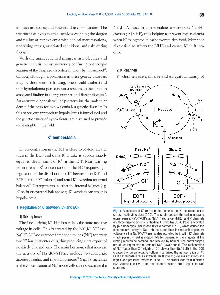
Fig. 1. Regulation of K+ redistribution in cells and K+ secretion in the cortical collecting duct (CCD). The circle depicts the cell membrane (upper panel). Na+,K+-ATPase, Na+/H+ exchanger (NHE), and K+ channels are three major elements controlling K+ shift. Na+,K+-ATPase is activated by β2-adrenergics, insulin and thyroid hormone. NHE, which causes the electroneutral entry of Na+ into cells and thus the net exit of positive voltage via the Na+,K+-ATPase, is also activated by insulin. K+ channels which permit K+ exit is responsible for generating the majority of the resting membrane potential and blocked by barium. The barrel shaped structures represent the terminal CCD (lower panel). The reabsorption of Na+ faster than Cl¯ (right) or Cl¯ slower than Na+ (left) in the CCD creates the lumen negative voltage that drives the net secretion of K+. Fast Na+ disorders cause extracellular fluid (ECF) volume expansion and high blood pressure, whereas, slow Cl¯ disorders lead to diminished ECF volume and low to normal blood pressure. ENaC, epithelial Na+ channels.
40 SH Lin et al. • A Practical Approach to Genetic Hypokalemia
Copyright © 2010 The Korean Society of Electrolyte Metabolism
membrane-spanning proteins that selectively conduct K+
ions across the cell membrane along its electrochemical
gradient in both excitable and non-excitable cells6).
There are several different types of K+ channels. These
K+ channels share the common features of a water-filled
permeation pore allowing K+ ions to flow across the
cell membrane, a selectivity filter that specifies K+ as the
permeant ion species, and a gating mechanism that serves
to switch between open and closed channel conformations.
Because K+ ions do not reach diffusion equilibrium,
control of the open-probability of K+ channel regulates
the transmembrane voltage. K+ channels are inhibited by
barium and other drugs6). Closure or opening may not
only alter the membrane potential, but also acutely affect
the plasma K+ concentration.
2. Renal handling of K+
Most of the filtered K+ is reabsorbed by the proximal
tubule and the loop of Henle. The final amount of K+
excreted in the urine is primarily controlled by the late
distal convoluted tubule (DCT), the connecting tubule, and
the cortical collecting duct (CCD)7). Two factors influence
the rate of K+ excretion: the flow rate in the terminal CCD
and the net secretion of K+ by principal cells in the CCD
which raise the luminal concentration of K+ ([K+]CCD)
(Fig. 1). There is an interplay between the magnitudes of
the flow rate in the CCD and the [K+]CCD which permits
the kidney to excrete all the K+ that is ingested in a steady
state8).
1) The flow rate in the CCD
The flow rate in the CCD is directly proportional to the
rate of excretion of osmoles and can be expressed as the
urine osmole excretion rate divided by plasma osmolality
(unrine osmolality × volume/plasma oslmolality) because
urine osmolality in the terminal CCD is equal to the
plasma osmolality when anti-diuretic hormone (ADH)
is present8). While the rate of flow in the CCD is high
during a water diuresis, the rate of K+ excretion need not
be elevated because ADH must be present to have high
rates of K+ secretion. The flow rate in the CCD can also be
indirectly represented by urine osmolality/creatinine.
2) [K+]CCD
K+ secretion in the CCD depends on the K+ channel
conductance of the apical membrane (primarily by
renal outer medullary K+ Channel, ROMK) driven by
the electrogenic reabsorption of Na+ (a lumen-negative
transepithelial voltage) via epithelial Na+ channels (ENaC)9).
Blood aldosterone and luminal bicarbonate (HCO3 )̄ are
two major factors enhancing K+ secretion in the CCD10).
Aldosterone action leads to an increase in the activity of
the ENaC and an alkaline luminal pH appears to exert a
decrease in the apparent permeability of chloride (Cl̄ ) and/
or an increase in open probability of ROMK. There are
two ways to generate more negativity in the lumen of the
CCD and thus drive K+ secretion. When Na+ is reabsorbed
faster than Cl¯ (fast Na+ disorders) or Cl¯ is reabsorbed
slower than Na+ (slow Cl̄ disorders) (Fig. 1), the lumen of
the CCD becomes more negatively charged (electrogenic)
and drives K+ secretion via the ROMK channel. The [K+] in
the CCD can be estimated by calculating the transtubular
K+ gradient [TTKG=(urine/plasma [K+])/(urine/plasma
osmolality)]. A TTKG greater than 3 in the presence of
hypokalemia indicates fast Na+ or slow Cl̄ disorders11).
Aproach to the genetic causes of hypokalemia
Several acquired extrarenal and renal disorders can
have the same findings of hypokalemia as genetic causes.
An understanding of the genetic causes of hypokalemia
will help determine the molecular defect and avoid
inappropriate and cumbersome genetic testing. A
detailed history and measurement of blood pressure
(BP), K+ excretion rate, and blood acid-base status can
help discriminate between the diverse acquired and
genetic causes of hypokalemia. Historical clues such as
prior hypokalemia, associated organ abnormalities in
family members, use of drugs affecting hypokalemia—
diuretics, licorice, and aminoglycosides—onset of age,

Electrolytes Blood Press 8:38-50, 2010 • doi: 10.5049/EBP.2010.8.1.38 41
Copyright © 2010 The Korean Society of Electrolyte Metabolism
and nephrolithiasis or nephrocalcinosis must be carefully
obtained1). The finding of concomitant hypertension
and hypokalemia without the use of diuretics suggest the
presence of a mineralocorticoid excess state (MES). A
major branch point in hypokalemic pathophysiology is the
renal K+ excretion response during hypokalemia. The 24-
hour urine and/or spot urine have been used to assess renal
K+ excretion rate. We prefer a spot (timed) urine collection
prior to therapy as a fast and practical alternative. Four
spot urine indices of renal response to hypokalemia have
been used: fractional excretion of K+, TTKG, urine K+/
creatinine ratio and urine osmolality/creatinine1, 12). A 24-
hour urine, if collected properly, can provide additional
information such as how much K+ is needed to replace
the K+ deficit and the state of K+ balance from calculation
of K+ input and output. Because K+ deficits usually
accompany HCO3¯ or Cl¯ loss regardless of the route of
K+ loss (gastrointestinal, sweat and renal), either metabolic
acidosis or alkalosis is common13). In contrast, conditions
of intracellular shift usually exhibit relatively normal acid-
base balance. Accordingly, the simultaneous assessment of
blood acid-base state is also very crucial in patients with
hypokalemia. Therefore, the etiology of hypokalemia can
simply be divided into two groups: those with a low K+
excretion rate and those with a high K+ excretion rate.
1. Disorders with a low urine K+ excretion rate
Disorders in the gastrointestinal tract, excessive sweating
and conditions with increased shift of extracellular K+
into cells can lead to a low urine K+ excretion. Generally,
an increased shift of extracellular K+ into cells can occur
acutely in a number of highly stressful conditions associated
with increased catecholamines or hyperinsulinemia via the
activation of membrane Na+,K+-ATPase or NHE activity.
Barium intoxication and chloroquine overdose cause
hypokalemia via the direct closure of cellular membrane
K+ channels. One should separate the disorders with
hypokalemic periodic paralysis (HPP) due to acute shift
of K+ into cells from non-HPP, where there is a large total
body deficit of K+14). Within the HPP subgroups, the most
common are thyrotoxic periodic paralysis (TPP) and
sporadic or idiopathic periodic paralysis (SPP) in Asia and
familial periodic paralysis (FPP) in Western countries15).
1) Genetic hypokalemia due to increased K+ shift
(1) Familial hypokalemia periodic paralysis (FPP)
It is an autosomal dominant disorder, which is accom-
panied by muscle weakness or paralysis and occurs more
frequently in males, with incomplete penetrance and later
onset observed in affected females16). Some cases may
present as sporadic because of the incomplete penetrance
of the disease, mostly in women. Attacks can be induced
by rest after exercise, carbohydrate-rich meals, exposure
to cold, or the administration of glucose or insulin or
glucocorticoid. Acetazolamide, a carbonic anhydrase
inhibitor, can reduce the frequency of attacks in FPP with
unclear mechanisms. The molecular lesions affect ion
channel genes encoding the dihydropyridine-sensitive
voltage-gated Ca2+ channel α1-subunit (CACNA1S)
(FPP type I) and tetrodotoxin-sensitive voltage-gated
Na+ channel α-subunit (SCN4A) (FPP type II) of skeletal
muscle17). The Na+ channel α-subunit shares primary and
secondary structure with the Ca2+ channel α-subunit
including segments spanning the membrane (S1-S6). The
S4 segment contains a high density of positively charged
amino acids, with every third one being lysine or arginine,
and acts as the voltage sensor in voltage-dependent channel
activation. To date, five point mutations affecting arginine
substitutions in segment S4 of domains II, III and IV
(R528G/H, R900S, R1239G/H) of CACNA1S and eleven
point mutations in segment S4 of domains I, II and III of
SCN4A (R222W, R669H, R672G,/H/C/S, R675G/Q/
W, R1132Q, R1135H) have been found in patients with
FPP18). Why mutations in segment S4 of CACNA1S and
SCN4A cause episodic hypokalemia remains unclear.
(2) Andersen-Tawil syndrome
It is an autosmal-dominant channelopathy resulting
in episodic attacks of muscle weakness (mainly acute
42 SH Lin et al. • A Practical Approach to Genetic Hypokalemia
Copyright © 2010 The Korean Society of Electrolyte Metabolism
hypokalemia, but can be normo- or hyperkalemia), cardiac
arrhythmia (ventricular arrhythmias and QT prolongation)
and distinctive physical features19). Mutations in the gene
(KCNJ2) encoding a pore-forming subunit of the inward
rectifier K+ channel protein, Kir2.1, which is expressed
in skeletal muscles and heart, lead to this syndrome19).
The majority of patients with this syndrome have
missense mutations that inhibit channel function through
a dominant negative effect. Like FPP, some cases may
present as sporadic because of the incomplete penetrance
of the disease or have de novo mutation. Furthermore, a
wide range of phenotypic severity exists, which can make
the correct diagnosis difficult. In contrast to FPP, where
cardiac arrhythmias provoked by severe hypokalemia
during attacks normalize upon recovery, persistent
electrocardiogram abnormalities between attacks is likely
in this syndrome20).
2) Genetic hypokalemia where the defect is in the intes tinal
tract
Congenital chloride-losing diarrhea (CLD) is a rare
autosomal recessive disorder characterized by watery
diarrhea, hypokalemia and hypochloremic metabolic
alkalosis with high fecal content of Cl ̄(>90 mEq/L)21). It
is caused by an inactivating mutation in the downregulated
in adenoma (DRA) gene encoding a Cl /̄OH¯ (HCO3 )̄
exchanger expressed in the apical membranes of the colon
and ileum with functional similarities with the anion
exchange proteins22) Like acquired intestinal inflammation
with reduced DRA expression, mutated DRA causes
the reabsorption of Cl¯ and secretion of HCO3¯ to fall.
Proton-pump inhibition of gastric chloride secretion is
helpful in diminishing the delivery of Cl¯ to the colon
and in lessening the degree of hypokalemia and metabolic
alkalosis in CLD.
3) Genetic hypokalemia where the defect is in exocrine
glands
Cystic fibrosis (CF) is an exocrine disease affecting
multiple organ systems. The defect in the cystic fibrosis
transmembrane regulator (CFTR), acting primarily as a
Cl¯ channel, is associated with CF23). Hypokalemia is not
uncommon in patients with CF, especially in tropical or
subtropical areas. Defective chloride reabsorption by the
dysfunctional CFTR in the sweat ducts of CF patients is
responsible for excessive Cl¯ and Na+ loss in sweat. ECF
volume depletion with secondary hyperaldosteronism not
only causes Na+ reabsorption and K+ secretion in the CCD,
but may also augment K+ secretion in sweat ducts and
thereby contribute to the hypokalemia.
2. Disorders with a high urine K+ excretion rate
Hypokalemia due to renal K+ wasting is chronic and
usually related to disorders with either increased flow rate
to the CCD or increased K+ in the CCD (fast Na+ or slow
Cl̄ ).
1) Increased urine flow rate to CCD
Flow rate to the CCD is enhanced when osmole excre-
tion rate is increased. Increased osmole excretion rate can
be caused by increased excretion of electrolytes (diuretics
or tubular defects) or non-electrolytes (mannitol, glucose,
urea).
2) Increased [K+] in the CCD
(1) Fast Na+ disorders
Because Na+ reabsorption in the CCD is augmented,
ECF volume is usually expanded and thus BP is high,
as in states of mineralocorticoid excess1). In this setting,
measurement of plasma renin activity, aldosterone and
cortisol concentration help to narrow the differential
diagnosis of fast Na+ disorders (Fig. 2). Plasma renin
activity and aldosterone levels are high in patients with
renin-secreting tumor, renal vascular stenosis, malignant
hypertension, and pheochromocytoma. High plasma
aldosterone levels but low plasma renin activity indicates
primary hyperaldosteronism which can be caused by
bilateral adrenal hyperplasia, adrenal adenoma/carcinoma,
and glucocorticoid-remediable aldosteronism (GRA). In
Electrolytes Blood Press 8:38-50, 2010 • doi: 10.5049/EBP.2010.8.1.38 43
Copyright © 2010 The Korean Society of Electrolyte Metabolism
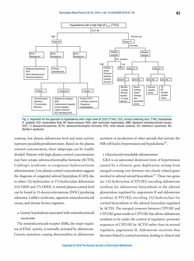
contrast, low plasma aldosterone level and renin activity
represent pseudohyperaldosteronism. Based on the plasma
cortisol concentration, three subgroups can be readily
divided. Patients with high plasma cortisol concentrations
may have ectopic adrenocorticotrophic hormone (ACTH),
Cushing’s syndrome or exogenous hydrocortisone
administration. Low plasma cortisol concentration suggests
the diagnosis of congenital adrenal hyperplasia (CAH) due
to either 11β-hydroxylase or 17α-hydroxylase deficiencies
(11β-OHD and 17α-OHD). A normal plasma cortisol level
can be found in 11-deoxycorticosterone (DOC) producing
adenoma, Liddle’s syndrome, apparent mineralocorticoid
excess, and chronic licorice ingestion.
a. Genetic hypokalemia associated with mineralocorticoid
excess state
The mineralocorticoid receptor (MR), the major regula-
tor of ENaC activity, is normally activated by aldosterone.
Genetic mutations causing abnormalities in aldosterone
secretion or production of other steroids that activate the
MR will lead to hypertension and hypokalemia24).
i. Glucorticoid-remediable aldosteronism
GRA is an autosomal dominant form of hypertension
caused by a chimeric gene duplication arising from
unequal crossing over between two closely related genes
involved in adrenal steroid biosynthesis25). These two genes
are 11β-hydroxylase (CYP11B1) encoding aldosterone
synthase for aldosterone biosynthesis in the adrenal
glomerulosa regulated by angiotensin II and aldosterone
synthase (CYP11B2) encoding 11β-hydroxylase for
cortisol biosynthesis in the adrenal fasciculata regulated
by ACTH. The unequal crossover between CYP11B1 and
CYP11B2 genes results in CYP11B2 that allows aldosterone
synthesis to be under the control of regulatory promoter
sequences of CYP11B1 by ACTH rather than its normal
regulator, angiotensin II. Aldosterone secretion thus
becomes linked to cortisol secretion, leading to clinical and
Fig. 2. Algorithm for the approach to hypokalemia with a high urine [K+]CCD (TTKG). CCD, cortical collecting duct; TTKG, transtubular K+ gradient; ECF, extracellular fluid; BP, blood pressure; RVH, right ventricular hypertrophy; AME, Apparent mineralocorticoid excess; DOC, 11-deoxycorticosterone; ACTH, adrenocorticotrophic hormone; RTA, renal tubular acidosis; GS, Gitelman’s syndrome; BS, Bartter’s syndrome
Hypokalemia with a high High [K+]CCD (TTKG)
ECF, BP
High
Faster Na+ Slower Clˉ
Normal~Low
Renin ↑Aldosterone ↑
Renin ↓Aldosterone ↑
Renin ↓Aldosterone ↓
· Malignant hypertension· RVH· Renin-secreting tumor· Pheochromocytoma
Primaryaldosteronism
Cortisol ↓ Cortisol N Cortisol ↑
· 11β-hydroxylase deficiency· 17α-hydorylase deficiency
· Aldosterone analogue· AME· Carbenoxolone ingestion· Licorice ingestion· Liddle’s syndrome· DOC-secreting tumor
· Ectopic ACTH· Cushing's syndrome· Exogenous hydrocortisone
Acid-base
Metablicacidosis
NH4+ excretion
High Low· Toluene abuse· Profound diarrhea· Ureteral diversion
· RTA Urine CIˉ ↓
Urine Na+ ↓ Urine Na+ ↑
· Remote vomiting· Remote diuretics· Clˉ-lsong diarrhea
· Recent vomiting· Gastric drainage· Non- absorbable anions
· Diarrhea· Laxative abuse
· GS· BS· Diuretics· Cisplatin· Others
Urine Na+ ↓ Urine Na+ ↑
Urine CIˉ ↑
Metablicalkalosis
44 SH Lin et al. • A Practical Approach to Genetic Hypokalemia
Copyright © 2010 The Korean Society of Electrolyte Metabolism
laboratory features of primary aldosteronism. Exogenous
glucocorticoids diminish the ACTH-regulated chimeric
genes, in turn suppressing the secretion of aldosterone,
leading to reversal of the features of GRA.
ii. CAH due to 11β-OHD and 17α-OHD
Two forms of CAH have hypertension and hypokalemia
accompanying a characteristic phenotype with abnormal
androgen production and sexual differentiation26). They
are 11β-OHD and 17α-OHD caused by CYP11B1 and
CYP17 mutations, respectively. Increased ACTH levels in
11β-OHD leads to elevation of 11-deoxycortisol (compound
S) and DOC, causing MES. Cortisol precursors are
diverted through the 17,20-lyase pathway and this results
in hyperandrogenemia, causing acne, hyperpigmentation,
and an enlarged phallus in males and prenatal virilization
of genitalia in the female newborn27). 17α-OHD has both
defective 17α-hydroxylase and 17,20-lyase activities of
the 17α-hydroxylase essential for the synthesis of cortisol
and gonadal hormones. Lack of 17,20-lyase activity in
17α-OHD prevents androgen synthesis and results in under-
virilization in males and failure of spontaneous pubertal
development in females. Steroid therapy ameliorates the
hypertension and hypokalemia as well as the associated
signs and symptoms.
iii. Liddle’s syndrome
It is characterized by autosomal dominant transmission
of early onset hypertension associated with hypokalemia,
metabolic alkalosis, suppressed plasma renin activity, and
extremely low plasma aldosterone levels28). This disease
is caused by mutations in either the β or the γ subunit of
ENaC that delete or alter their cytoplasmic C termini.
The mutated ENaC are not internalized (clathrin-coated
pits pathway) or degraded (Nedd4 pathway), and instead
remain in an activated form on the cell surface. The
channel is amiloride and triamterene sensitive provided
their concentrations are high enough in luminal fluid,
explaining the efficacy of these K+-sparing diuretics in the
syndrome.
iv. Apparent mineralocorticoid excess (AME)
AME is a rare but potentially fatal autosomal re-
cessive form of hypertension and hypokalemic me-
tabolic alkalosis associated with hyporeninemia and
hypoaldosteronemia and an abnormal ratio of urinary
metabolites of cortisol with a high tetrahydrocortisol:tet
rahydrocortisone (THF:THE) ratio29). AME is caused by
mutations in the gene (HSD11B2) encoding renal-specific
11β-hydroxysteroid dehydrogenase type 2 (11β-HSD2).
11β-HSD2 is responsible for converting cortisol to
cortisone in the principal cells of distal tubules and crucial
for protecting the MR from being occupied by cortisol.
The hallmark features in AME resemble those in licorice
ingestion because licorice contains glycyrrhetinic acid,
which inhibits 11β-HSD230).
(2) Slow Cl̄ disorders
Because Cl̄ reabsorption in the CCD is diminished, ECF
volume is usually contracted and thus BP is relatively low to
normal. Because extracellular K+ loss is often accompanied
by ECF HCO3¯ or Cl¯ loss, slow Cl¯ disorders can have
either hyperchloremic metabolic acidosis or hypochloremic
metabolic alkalosis.
a. Hypochloremic metabolic alkalosis
Metabolic alkalosis is diagnostically useful in patients
with severe KCl depletion. An assessment of urine Na+
and Cl¯ may reveal the basis for renal tubular electrolyte
disorders and distinguish it from non-renal Na+ loss
(Fig. 2). Low excretion of Na+ and Cl¯ indicate remote
vomiting, remote or yesterday’s diuretics, Cl¯-losing
diarrhea (e.g. congenital chloridorrhea), or excessive
sweating. Low urine Na,+ but high Cl¯ excretion, in the
presence of hypokalemia and metabolic alkalosis suggests
laxative abuse or some chronic diarrhea states with chronic
stimulation of renal NH4+ excretion. Conditions of high
Na+ excretion, but low Cl ,̄ suggests the presence of an
anion that is not reabsorbed. If the urine is alkaline (pH
>7), vomiting and/or ingestion of bases are the likely
causes. If the urine is not alkaline, intake or generation
Electrolytes Blood Press 8:38-50, 2010 • doi: 10.5049/EBP.2010.8.1.38 45
Copyright © 2010 The Korean Society of Electrolyte Metabolism
of anions that are poorly reabsorbed by the kidney is
likely31). A high urine Na+ and Cl¯ excretion is indicative
of the recent use of diuretics, intrinsic renal disease, or
lack of signaling to stimulate NaCl reabsorption. Inherited
or acquired renal tubular disorders such as Gitelman’s
syndrome (GS), Bartter’s syndrome (BS), and related drug-
induced toxic disorders, such as from aminoglycosides,
cisplatin, diuretics, and foscarnet, are often the diagnoses32).
The evaluation of urine divalent Mg2+ and Ca2+ excretion
helps localize the exact tubular defect. High urine Ca2+
and Mg2+ excretion is universally present in lesions of the
loop of Henle (LOH) whereas low urine Ca2+ and high
Mg2+ excretion is invariably found in lesions of the DCT33).
i. Bartter’s syndrome (BS) and Gitelman’s syndrome (GS)
BS and GS are autosomal recessive renal tubular disor-
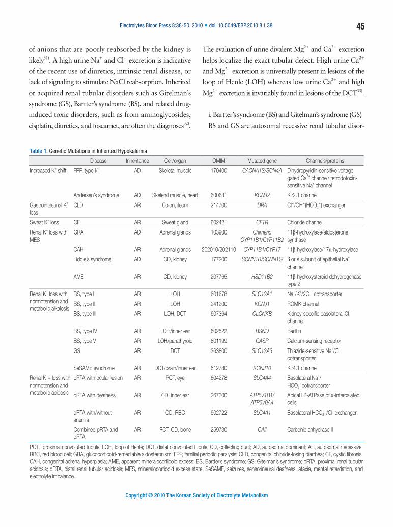
Table 1. Genetic Mutations in Inherited Hypokalemia
Disease Inheritance Cell/organ OMIM Mutated gene Channels/proteins
Increased K+ shift FPP, type I/II AD Skeletal muscle 170400 CACNA1S/SCN4A Dihydropyridin-sensitive voltage gated Ca2+ channel/ tetrodotoxin-sensitive Na+ channel
Andersen’s syndrome AD Skeletal muscle, heart 600681 KCNJ2 Kir2.1 channel
Gastrointestinal K+ loss
CLD AR Colon, ileum 214700 DRA Cl¯/OH¯(HCO3¯) exchanger
Sweat K+ loss CF AR Sweat gland 602421 CFTR Chloride channel
Renal K+ loss with MES
GRA AD Adrenal glands 103900 Chimeric CYP11B1/CYP11B2
11β-hydroxylase/aldosterone synthase
CAH AR Adrenal glands 202010/202110 CYP11B1/CYP17 11β-hydroxylase/17α-hydroxylase
Liddle’s syndrome AD CD, kidney 177200 SCNN1B/SCNN1G β or γ subunit of epithelial Na+ channel
AME AR CD, kidney 207765 HSD11B2 11β-hydroxysteroid dehydrogenase type 2
Renal K+ loss with normotension and metabolic alkalosis
BS, type I AR LOH 601678 SLC12A1 Na+/K+/2Cl¯ cotransporter
BS, type II AR LOH 241200 KCNJ1 ROMK channel
BS, type III AR LOH, DCT 607364 CLCNKB Kidney-specific basolateral Cl¯ channel
BS, type IV AR LOH/inner ear 602522 BSND Barttin
BS, type V AR LOH/parathyroid 601199 CASR Calcium-sensing receptor
GS AR DCT 263800 SLC12A3 Thiazide-sensitive Na+/Cl¯ cotransporter
SeSAME syndrome AR DCT/brain/inner ear 612780 KCNJ10 Kir4.1 channel
Renal K++ loss with normotension and metabolic acidosis
pRTA with ocular lesion AR PCT, eye 604278 SLC4A4 Basolateral Na+/HCO3¯cotransporter
dRTA with deafness AR CD, inner ear 267300 ATP6V1B1/ATP6V0A4
Apical H+-ATPase of α-intercalated cells
dRTA with/without anemia
AR CD, RBC 602722 SLC4A1 Basolateral HCO3¯/Cl¯exchanger
Combined pRTA and dRTA
AR PCT, CD, bone 259730 CAII Carbonic anhydrase II
PCT, proximal convoluted tubule; LOH, loop of Henle; DCT, distal convoluted tubule; CD, collecting duct; AD, autosomal dominant; AR, autosomal r ecessive; RBC, red blood cell; GRA, glucocorticoid-remediable aldosteronism; FPP, familial periodic paralysis; CLD, congenital chloride-losing diarrhea; CF, cystic fibrosis; CAH, congenital adrenal hyperplasia; AME, apparent mineralocorticoid excess; BS, Bartter’s syndrome; GS, Gitelman’s syndrome; pRTA, proximal renal tubular acidosis; dRTA, distal renal tubular acidosis; MES, mineralocorticoid excess state; SeSAME, seizures, sensorineural deafness, ataxia, mental retardation, and electrolyte imbalance.
46 SH Lin et al. • A Practical Approach to Genetic Hypokalemia
Copyright © 2010 The Korean Society of Electrolyte Metabolism
ders characterized by chronic hypokalemia with renal
K+ wasting, metabolic alkalosis, renal salt wasting with
low to normal BP and secondary hyperreninemia and
hyperaldosteronism. BS results from defective reabsorption
of NaCl in the LOH whereas GS is secondary to defective
reabsorption of NaCl in the DCT. At the molecular
level, GS is mostly due to inactivating mutations in the
SLC12A3 gene, which encodes the thiazide-sensitive Na+/
Cl¯ cotransporter (NCC) on the apical membrane of the
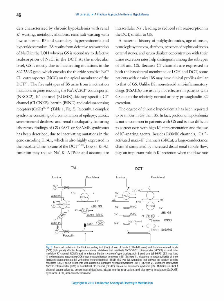
DCT34). The five subtypes of BS arise from inactivation
mutations in genes encoding the Na+/K+/2Cl̄ cotransporter
(NKCC2), K+ channel (ROMK), kidney-specific Cl ̄
channel (CLCNKB), barttin (BSND) and calcium-sensing
receptors (CaSR)35, 36) (Table 1, Fig. 3). Recently, a complex
syndrome consisting of a combination of epilepsy, ataxia,
sensorineural deafness and renal tubulopathy featuring
laboratory findings of GS (EAST or SeSAME syndrome)
has been described, due to inactivating mutations in the
gene encoding Kir4.1, which is also highly expressed in
the basolateral membrane of the DCT37, 38). Loss of Kir4.1
function may reduce Na+,K+-ATPase and accumulate
intracellular Na+, leading to reduced salt reabsorption in
the DCT, similar to GS.
A maternal history of polyhydramnios, age of onset,
neurologic symptoms, deafness, presence of nephrocalcinosis
or renal stones, and serum divalent concentration with their
urine excretion rates help distinguish among the subtypes
of BS and GS. Because Cl- channels are expressed in
both the basolateral membrane of LOH and DCT, some
patients with classical BS may have clinical profiles similar
to that of GS. Unlike BS, non-steroid anti-inflammatory
drugs (NSAIDs) are usually not effective in patients with
GS due to the relatively normal urinary prostaglandin E2
excretion.
The degree of chronic hypokalemia has been reported
to be milder in GS than BS. In fact, profound hypokalemia
is not uncommon in patients with GS and is also difficult
to correct even with high K+ supplementation and the use
of K+-sparing agents. Besides ROMK channels, Ca2+-
activated maxi-K+ channels (BKCa), a large-conductance
channel stimulated by increased distal renal tubule flow,
play an important role in K+ secretion when the flow rate
Fig. 3. Transport proteins in the thick ascending limb (TAL) of loop of Henle (LOH) (left panel) and distal convoluted tubule (DCT) (right panel) affected by gene mutations. Mutations that inactivate Na+/K+/2Cl¯ cotransporter (NKCC2) or renal outer medullary K+ channel (ROMK) lead to antenatal Bartter syndrome/hyperprostaglandin E syndrome (aBS/HPS) (BS type I and II) and mutations inactivating ClCKb cause classic Bartter syndrome (cBS) (BS type III). Mutations in barttin (chloride channel βsubunit) cause antenatal BS with sensorineural deafness (BSND) (BS type IV). Mutations that activate the calcium-sensing receptors (CaSR) occur in patients with autosomal dominant hypoparathyroidism (ADH) (BS type V). Mutations inactivating Na+/Clˉ cotransporter (NCC) or basolateral Clˉ channel (ClC-Kb) can cause Gitelman's syndrome (GS). Mutations in Kir4.1 channel cause seizures, sensorineural deafness, ataxia, mental retardation, and electrolyte imbalance (SeSAME) syndrome. ADH, anti-diuretic hormone
Electrolytes Blood Press 8:38-50, 2010 • doi: 10.5049/EBP.2010.8.1.38 47
Copyright © 2010 The Korean Society of Electrolyte Metabolism
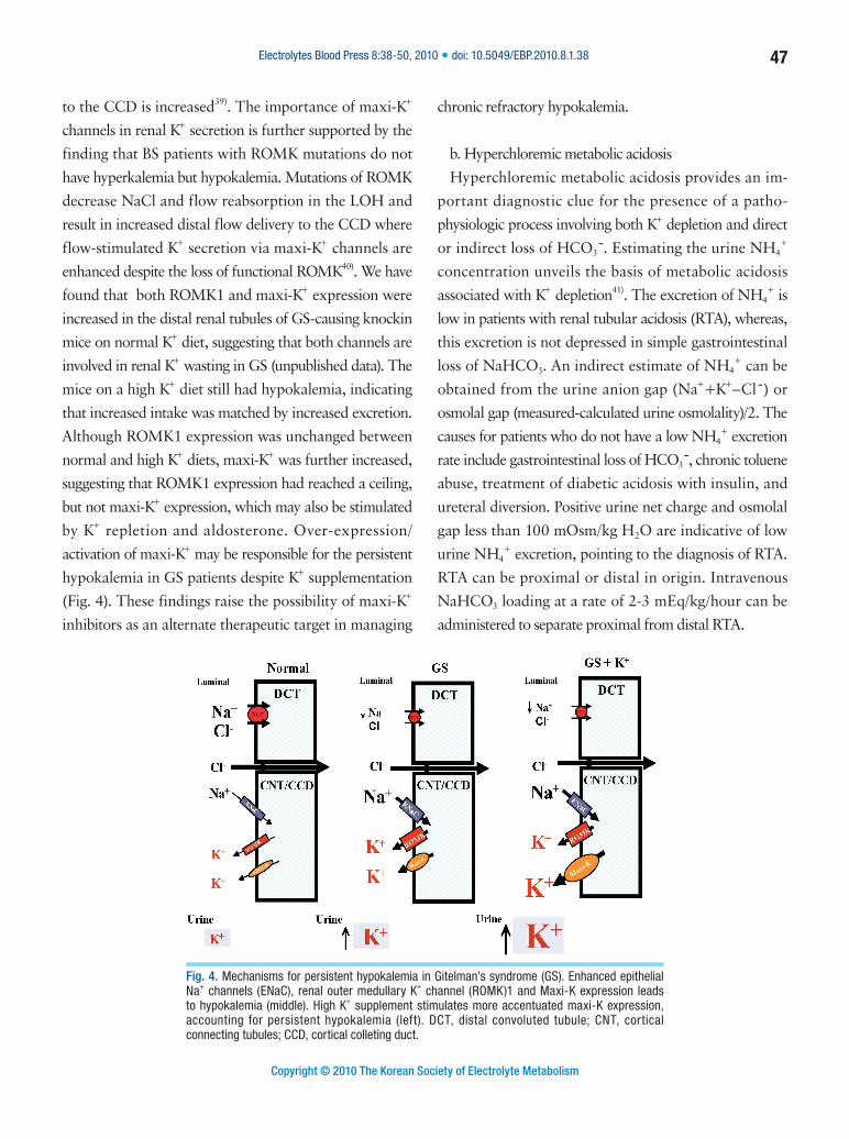
to the CCD is increased39). The importance of maxi-K+
channels in renal K+ secretion is further supported by the
finding that BS patients with ROMK mutations do not
have hyperkalemia but hypokalemia. Mutations of ROMK
decrease NaCl and flow reabsorption in the LOH and
result in increased distal flow delivery to the CCD where
flow-stimulated K+ secretion via maxi-K+ channels are
enhanced despite the loss of functional ROMK40). We have
found that both ROMK1 and maxi-K+ expression were
increased in the distal renal tubules of GS-causing knockin
mice on normal K+ diet, suggesting that both channels are
involved in renal K+ wasting in GS (unpublished data). The
mice on a high K+ diet still had hypokalemia, indicating
that increased intake was matched by increased excretion.
Although ROMK1 expression was unchanged between
normal and high K+ diets, maxi-K+ was further increased,
suggesting that ROMK1 expression had reached a ceiling,
but not maxi-K+ expression, which may also be stimulated
by K+ repletion and aldosterone. Over-expression/
activation of maxi-K+ may be responsible for the persistent
hypokalemia in GS patients despite K+ supplementation
(Fig. 4). These findings raise the possibility of maxi-K+
inhibitors as an alternate therapeutic target in managing
chronic refractory hypokalemia.
b. Hyperchloremic metabolic acidosis
Hyperchloremic metabolic acidosis provides an im-
portant diagnostic clue for the presence of a patho-
physiologic process involving both K+ depletion and direct
or indirect loss of HCO3 .̄ Estimating the urine NH4+
concentration unveils the basis of metabolic acidosis
associated with K+ depletion41). The excretion of NH4+ is
low in patients with renal tubular acidosis (RTA), whereas,
this excretion is not depressed in simple gastrointestinal
loss of NaHCO3. An indirect estimate of NH4+ can be
obtained from the urine anion gap (Na++K+–Cl¯) or
osmolal gap (measured-calculated urine osmolality)/2. The
causes for patients who do not have a low NH4+ excretion
rate include gastrointestinal loss of HCO3 ,̄ chronic toluene
abuse, treatment of diabetic acidosis with insulin, and
ureteral diversion. Positive urine net charge and osmolal
gap less than 100 mOsm/kg H2O are indicative of low
urine NH4+ excretion, pointing to the diagnosis of RTA.
RTA can be proximal or distal in origin. Intravenous
NaHCO3 loading at a rate of 2-3 mEq/kg/hour can be
administered to separate proximal from distal RTA.
Fig. 4. Mechanisms for persistent hypokalemia in Gitelman's syndrome (GS). Enhanced epithelial Na+ channels (ENaC), renal outer medullary K+ channel (ROMK)1 and Maxi-K expression leads to hypokalemia (middle). High K+ supplement stimulates more accentuated maxi-K expression, accounting for persistent hypokalemia (left). DCT, distal convoluted tubule; CNT, cortical connecting tubules; CCD, cortical colleting duct.
48 SH Lin et al. • A Practical Approach to Genetic Hypokalemia
Copyright © 2010 The Korean Society of Electrolyte Metabolism
i. Inherited isolated proximal RTA
Most of the filtered HCO3 ̄is reabsorbed in the proximal
convoluted tubules (PCT) as a result of H+ secretion via
the electroneutral Na+/H+ exchanger 3 (NHE3) in the
luminal membrane and HCO3¯ exit via the electrogenic
Na+/HCO3¯ cotransporter (NBC1) in the basolateral
membrane. Defects in NHE3 or NBC1 can theoretically
reduce the reabsorption of filtered HCO3¯ and cause
proximal RTA (pRTA). Mutations in the NHE3 gene have
not been identified in patients with proximal RTA to date.
On the other hand, mutations in the NBC1 gene (SLC4A4)
have been reported in patients with autosomal recessive
isolated proximal RTA42). Because NBC1 is also expressed
in the ocular tissues and brain, patients with inactivating
mutations in NBC1 have ocular abnormalities (glaucoma,
band keratopathy, and cataracts) as well as calcifications of
the basal ganglia. Of note, hypokalemia is almost always
present and can be very severe when a high dose of oral
NaHCO3 is given.
ii. Inherited distal RTA
Apical H+-ATPase pump and basolateral Cl /̄HCO3 ̄
exchanger (anion exchanger 1, AE1) in the late distal
tubule (predominantly in α-intercalated cells) participate
in the excretion of NH4+, which adds new HCO3¯ to the
body. Genes encoding two H+-ATPase subunits specific
to intercalated cells have been identified as ATP6V1B1
and ATP6V0A43). Because ATP6V1B1 is also expressed in
endolymphatic sac epithelia, mutations in ATP6V1B1 cause
distal RTA with sensorineural hearing loss, whereas, distal
RTA with preserved hearing is caused by mutations in
ATP6V0A4. AE1 (SLC4A1) is also present in erythrocytes
and inactivating mutation in AE1 causes distal RTA and
possible anemia44). Hypokalemia in hereditary distal RTA is
often severe. First, low distal H+ secretion reduces luminal
HCO3¯ reabsorption and this leads to bicarbonaturia.
Second, the mistargeting of AE1 due to the second
mutation in AE1 leads to HCO3 secretion. Reminiscent of
alkalinized intercalated cells in Sjogren’s syndrome with
distal RTA, high luminal HCO3¯ may lead to augmented
K+ secretion and hypokalemia.
iii. Inherited proximal and distal RTA
Carbonic anhydrase II (CAII) is known to be expressed
in both proximal and distal nephron segments and
participates in H+ secretion in both the proximal and distal
nephron; CAII is also expressed in brain and osteoclasts.
Mutations in CAII can lead to an autosomal recessive
proximal and distal RTA with osteopetrosis and cerebral
calcification45). The bone thinning effect of the coexistent
metabolic acidosis in CAII deficiency may protect the
osteopetrotic bones from the excessive thickening
generated by osteoclast dysfunction.
Therapeutic strategy in genetic hypokalemia
Genetically engineered mice are often used as animal
models of human diseases and are vital tools in investigating
molecular pathogenesis of disease and developing and
testing novel therapies. It is worthy to emphasize that
knock-out mice typically represent null mutations and
could be used to study only the effects of the loss of a
gene, not a specific mutation46). Knockin strategies, where
homologous recombination in embryonic stem (ES) cells is
used to replace the endogenous gene with a mutant variant
without any other disruption of the gene, can be used to
address the effects of specific sequence changes on gene
and protein function47). Therefore, knockin mice accurately
represent human diseases caused by specific mutations,
such as missense, nonsense, or deletion mutations. More
importantly, mutation-specific approaches according
to the different classes of mutations are currently under
investigation to provide a new alternative therapy in
genetic disorders. We believe that the application of these
diseases-causing knock-in mice may address more specific
questions regarding the pathogenesis and new directions
in rescue therapies; for instance, rational application of
aminoglycosides or premature termination codon (PTC)
124 for nonsense mutations and pharmacologic chaperones
for missense mutations48).

Electrolytes Blood Press 8:38-50, 2010 • doi: 10.5049/EBP.2010.8.1.38 49
Copyright © 2010 The Korean Society of Electrolyte Metabolism
Conclusion
Apart from a detailed history and careful physical
examination, the measurement of urine K+ excretion rate
by spot and/or 24 hour urine, and assessment of blood acid-
base status helps discriminate the causes of hypokalemia.
Once the clinical diagnosis suggests a likely molecular basis
for the disorder, genetic analysis may prove the diagnosis.
Genetic causes of hypokalemia are shown in Table 1.
Creation and investigation of disease-causing knockin mice
as a model of genetic hypokalemia will not only provide a
much better understanding of the underlying mechanisms;
but also set the stage for the development of novel therapies
in the future.
Acknowledgements
This work was supported by a grant from the Research
Fund of Tri-Service General Hospital (TSGH-C-98-81)
and from the Teh-Tzer Study Group for the Human
Medical Research Foundation.
References
1) Lin SH, Halperin ML: Hypokalemia: a practical approach to diagnosis and its genetic basis. Curr Med Chem 14:1551-1565, 2007
2) Scheinman SJ, Guay-Woodford LM, Thakker RV, Warnock DG: Genetic disorders of renal electrolyte transport. N Engl J Med 340:1177-1187, 1999
3) Gennari FJ: Hypokalemia. N Engl J Med 339:451-458, 1998
4) Sterns RH, Cox M, Feig PU, Singer I: Internal potassium balance and the control of the plasma potassium concentration. Medicine 60:339-354, 1981
5) Clausen T: Clinical and therapeutic significance of the Na+, K+ pump. Clin Sci 95:3-17, 1998
6) Shieh CC, Coghlan M, Sullivan JP, Gopalakrishnan M: Potassium channels: molecular defects, diseases, and therapeutic opportunities. Pharmacol Rev 52:557-594, 2000
7) Giebisch G, Krapf R, Wagner C: Renal and extrarenal regulation of potassium. Kidney Int 72:397-410, 2007
8) Halperin ML, Kamel KS: Potassium. Lancet 352:135-140, 1998
9) Hebert SC, Desir G, Giebisch G, Wang W: Molecular diversity and regulation of renal potassium channels. Physiol Rev 85:319-371, 2005
10) Lin SH, Cheema-Dhadli S, Gowrishankar M, Marliss EB, Kamel KS, Halperin ML: Control of excretion of potassium: lessons from studies during prolonged total fasting in human subjects. Am J Physiol 273:F796-800, 1997
11) Kamel KS, Quaggin S, Scheich A, Halperin ML: Disorders of potassium homeostasis: an approach based on pathophysiology. Am J Kidney Dis 24:597-613, 1994
12) Lin SH, Lin YF, Chen DT, Chu P, Hsu CW, Halperin ML: Laboratory tests to determine the cause of hypokalemia and paralysis. Arch Intern Med 164:1561-1566, 2004
13) Alazami M, Lin SH, Cheng CJ, Davids MR, Halperin ML: Unusual causes of hypokalaemia and paralysis. QJM 99:181-192, 2006
14) Lin SH, Lin YF, Halperin ML: Hypokalaemia and paralysis. QJM 94:133-139, 2001
15) Lin SH: Thyrotoxic periodic paralysis. Mayo Clin Proc 80:99-105, 2005
16) Lin SH, Hsu YD, Cheng NL, Kao MC: Skeletal muscle dihydropyridine-sensitive calcium channel (CACNA1S) gene mutations in chinese patients with hypokalemic periodic paralysis. Am J Med Sci 329:66-70, 2005
17) Venance SL, Cannon SC, Fialho D, et al.: The primary periodic paralyses: diagnosis, pathogenesis and treatment. Brain 129:8-17, 2006
18) Matthews E, Labrum R, Sweeney MG, et al.: Voltage sensor charge loss accounts for most cases of hypokalemic periodic paralysis. Neurology 72:1544-1547, 2009
19) Andelfinger G, Tapper AR, Welch RC, Vanoye CG, George AL, Jr., Benson DW: KCNJ2 mutation results in Andersen syndrome with sex-specific cardiac and skeletal muscle phenotypes. Am J Hum Genet 71:663-668, 2002
20) Fontaine B, Fournier E, Sternberg D, Vicart S, Tabti N: Hypokalemic periodic paralysis: a model for a clinical and research approach to a rare disorder. Neurotherapeutics 4:225-232, 2007
21) Holmberg C, Perheentupa J, Launiala K: Colonic electrolyte transport in health and in congenital chloride diarrhea. J Clin Invest 56:302-310, 1975
22) Hoglund P, Haila S, Socha J, et al.: Mutations of the Down-regulated in adenoma (DRA) gene cause congenital chloride diarrhoea. Nat Genet 14:316-319, 1996
23) Bates CM, Baum M, Quigley R: Cystic fibrosis presenting with hypokalemia and metabolic alkalosis in a previously healthy adolescent. J Am Soc Nephrol 8:352-355, 1997
24) Lifton RP, Gharavi AG, Geller DS: Molecular mechanisms of
50 SH Lin et al. • A Practical Approach to Genetic Hypokalemia
Copyright © 2010 The Korean Society of Electrolyte Metabolism
human hypertension. Cell 104:545-556, 2001
25) Lifton RP, Dluhy RG, Powers M, et al.: A chimaeric 11 beta-hydroxylase/aldosterone synthase gene causes glucocorticoid-remediable aldosteronism and human hypertension. Nature 355:262-265, 1992
26) New MI: Diagnosis and management of congenital adrenal hyperplasia. Annu Rev Med 49:311-328, 1998
27) Zachmann M: Defects in steroidogenic enzymes. Discrepancies between clinical steroid research and molecular biology results. J Steroid Biochem Mol Biol 53:159-164, 1995
28) Palmer BF, Alpern RJ: Liddle's syndrome. Am J Med 104:301-309, 1998
29) Mune T, Rogerson FM, Nikkilä H, Agarwal AK, White PC: Human hypertension caused by mutations in the kidney isozyme of 11 beta-hydroxysteroid dehydrogenase. Nat Genet 10:394-399, 1995
30) Lin SH, Yang SS, Chau T, Halperin ML: An unusual cause of hypokalemic paralysis: chronic licorice ingestion. Am J Med Sci 325:153-156, 2003
31) Kamel KS, Ethier J, Levin A, Halperin ML: Hypokalemia in the "beautiful people". Am J Med 88:534-536, 1990
32) Chou CL, Chen YH, Chau T, Lin SH: Acquired Bartter-like syndrome associated with gentamicin administration. Am J Med Sci 329:144-149, 2005
33) Bettinelli A, Bianchetti MG, Girardin E, et al.: Use of calcium excretion values to distinguish two forms of primary renal tubular hypokalemic alkalosis: Bartter and Gitelman syndromes. J Pediatr 120:38-43, 1992
34) Lin SH, Shiang JC, Huang CC, Yang SS, Hsu YJ, Cheng CJ: Phenotype and genotype analysis in Chinese patients with Gitelman's syndrome. J Clin Endocrinol Metab 90:2500-2507, 2005
35) Peters M, Jeck N, Reinalter S, et al.: Clinical presentation of genetically defined patients with hypokalemic salt-losing tubulopathies. Am J Med 112:183-190, 2002
36) Gamba G: Molecular physiology and pathophysiology of electroneutral cation-chloride cotransporters. Physiol Rev 85:423-493, 2005
37) Bockenhauer D, Feather S, Stanescu HC, et al.: Epilepsy, ataxia, sensorineural deafness, tubulopathy, and KCNJ10 mutations. N Engl J Med 360:1960-1970, 2009
38) Scholl UI, Choi M, Liu T, et al.: Seizures, sensorineural deafness, ataxia, mental retardation, and electrolyte imbalance (SeSAME syndrome) caused by mutations in KCNJ10. Proc Natl Acad Sci U S A 106:5842-5847, 2009
39) Pluznick JL, Sansom SC: BK channels in the kidney: role in K(+) secretion and localization of molecular components. Am J Physiol Renal Physiol 291:F517-529, 2006
40) Bailey MA, Cantone A, Yan Q, et al.: Maxi-K channels contribute to urinary potassium excretion in the ROMK-deficient mouse model of Type II Bartter's syndrome and in adaptation to a high-K diet. Kidney Int 70:51-59, 2006
41) Kamel KS, Briceno LF, Sanchez MI, et al.: A new classification for renal defects in net acid excretion. Am J Kidney Dis 29:136-146, 1997
42) Igarashi T, Inatomi J, Sekine T, et al.: Mutations in SLC4A4 cause permanent isolated proximal renal tubular acidosis with ocular abnormalities. Nat Genet 23:264-266, 1999
43) Karet FE: Inherited distal renal tubular acidosis. J Am Soc Nephrol 13:2178-2184, 2002
44) Wrong O, Bruce LJ, Unwin RJ, Toye AM, Tanner MJ: Band 3 mutations, distal renal tubular acidosis, and Southeast Asian ovalocytosis. Kidney Int 62:10-19, 2002
45) Borthwick KJ, Kandemir N, Topaloglu R, et al.: A phenocopy of CAII deficiency: a novel genetic explanation for inherited infantile osteopetrosis with distal renal tubular acidosis. J Med Genet 40:115-121, 2003
46) Manis JP: Knock out, knock in, knock down--genetically manipulated mice and the Nobel Prize. N Engl J Med 357:2426-2429, 2007
47) Kohan DE: Progress in gene targeting: using mutant mice to study renal function and disease. Kidney Int 74:427-437, 2008
48) Le Roy F, Charton K, Lorson CL, Richard I: RNA-targeting approaches for neuromuscular diseases. Trends Mol Med 15:580-591, 2009
Related Documents


