THE JOURNAL OF ALTERNATIVE AND COMPLEMENTARY MEDICINE Volume 8, Number 4, 2002, pp. 411–419 © Mary Ann Liebert, Inc. A Pilot Study of Functional Magnetic Resonance Imaging of the Brain During Manual and Electroacupuncture Stimulation of Acupuncture Point (LI-4 Hegu) in Normal Subjects Reveals Differential Brain Activation Between Methods JIAN KONG, M.S., Lic.Ac., 1,2 LIN MA, M.D., Ph.D., 3 RANDY L. GOLLUB, M.D., Ph.D., 2 JINGHAN WEI, Ph.D., 4 XUIZHEN YANG, M.D., 1 DEJUN LI, Ph.D., 3 XUCHU WENG, Ph.D., 4 FUCANG JIA, Ph.D., 4 CHUNMAO WANG, Ph.D., 4 FULI LI, Ph.D., 5 RUIWU LI, M.D., 1 and DING ZHUANG, M.D. 1 ABSTRACT Objectives: To characterize the brain activation patterns evoked by manual and elec- troacupuncture on normal human subjects. Design: We used functional magnetic resonance imaging (fMRI) to investigate the brain re- gions involved in electroacupuncture and manual acupuncture needle stimulation. A block de- sign was adopted for the study. Each functional run consists of 5 minutes, starting with 1-minute baseline and two 1-minute stimulation, the interval between the two stimuli was 1 minute. Four functional runs were performed on each subject, two runs for electroacupuncture and two runs for manual acupuncture. The order of the two modalities was randomized among subjects. Dur- ing the experiment, acupuncture needle manipulation was performed at Large Intestine 4 (LI4, Hegu) on the left hand. For each subject, before scanning started, the needle was inserted per- pendicular to the skin surface to a depth of approximately 1.0 cm. Electroacupuncture stimula- tion was delivered using a continuous rectangular wave form (pulse width 30 ms) at a frequency of 3 Hz. For manual acupuncture, the needle was rotated manually clockwise and counter- clockwise at a rate of about 180 times per minute (3 Hz). Subjects: Eleven right-handed, normal, healthy volunteer adults, 6 male and 5 female, ages 21–64 participated in the experiment. Results: Results showed that electroacupuncture mainly produced fMRI signal increases in precentral gyrus, postcentral gyrus/inferior parietal lobule, and putamen/insula; in contrast, manual needle manipulation produced prominent decreases of fMRI signals in posterior cingu- late, superior temporal gyrus, putamen/insula. Conclusion: These results indicate that different brain networks are involved during manual and electroacupuncture stimulation. It suggests that different brain mechanisms may be recruited during manual and electroacupuncture. ORIGINAL PAPERS 411 1 Institute of Acupuncture and Moxibustion, China Academy of Traditional Chinese Medicine, Beijing, People’s Re- public of China. 2 Department of Psychiatry, Massachusetts General Hospital East, Charlestown, MA, and Harvard Medical School, Boston, MA. 3 Department of Radiology, PLA General Hospital, Beijing, People’s Republic of China. 4 Institute of Psychology, Chinese Academy of Science, Beijing, People’s Republic of China. 5 Department of Physics, Capital Moral University, Beijing, People’s Republic of China.

Welcome message from author
This document is posted to help you gain knowledge. Please leave a comment to let me know what you think about it! Share it to your friends and learn new things together.
Transcript
THE JOURNAL OF ALTERNATIVE AND COMPLEMENTARY MEDICINEVolume 8, Number 4, 2002, pp. 411–419© Mary Ann Liebert, Inc.
A Pilot Study of Functional Magnetic ResonanceImaging of the Brain During Manual and
Electroacupuncture Stimulation of Acupuncture Point(LI-4 Hegu) in Normal Subjects Reveals Differential
Brain Activation Between Methods
JIAN KONG, M.S., Lic.Ac.,1,2 LIN MA, M.D., Ph.D.,3 RANDY L. GOLLUB, M.D., Ph.D.,2
JINGHAN WEI, Ph.D.,4 XUIZHEN YANG, M.D.,1 DEJUN LI, Ph.D.,3
XUCHU WENG, Ph.D.,4 FUCANG JIA, Ph.D.,4 CHUNMAO WANG, Ph.D.,4
FULI LI, Ph.D.,5 RUIWU LI, M.D.,1 and DING ZHUANG, M.D.1
ABSTRACT
Objectives: To characterize the brain activation patterns evoked by manual and elec-troacupuncture on normal human subjects.
Design: We used functional magnetic resonance imaging (fMRI) to investigate the brain re-gions involved in electroacupuncture and manual acupuncture needle stimulation. A block de-sign was adopted for the study. Each functional run consists of 5 minutes, starting with 1-minutebaseline and two 1-minute stimulation, the interval between the two stimuli was 1 minute. Fourfunctional runs were performed on each subject, two runs for electroacupuncture and two runsfor manual acupuncture. The order of the two modalities was randomized among subjects. Dur-ing the experiment, acupuncture needle manipulation was performed at Large Intestine 4 (LI4,Hegu) on the left hand. For each subject, before scanning started, the needle was inserted per-pendicular to the skin surface to a depth of approximately 1.0 cm. Electroacupuncture stimula-tion was delivered using a continuous rectangular wave form (pulse width 30 ms) at a frequencyof 3 Hz. For manual acupuncture, the needle was rotated manually clockwise and counter-clockwise at a rate of about 180 times per minute (3 Hz).
Subjects: Eleven right-handed, normal, healthy volunteer adults, 6 male and 5 female, ages21–64 participated in the experiment.
Results: Results showed that electroacupuncture mainly produced fMRI signal increases inprecentral gyrus, postcentral gyrus/inferior parietal lobule, and putamen/insula; in contrast,manual needle manipulation produced prominent decreases of fMRI signals in posterior cingu-late, superior temporal gyrus, putamen/insula.
Conclusion: These results indicate that different brain networks are involved during manualand electroacupuncture stimulation. It suggests that different brain mechanisms may be recruitedduring manual and electroacupuncture.
ORIGINAL PAPERS
411
1Institute of Acupuncture and Moxibustion, China Academy of Traditional Chinese Medicine, Beijing, People’s Re-public of China.
2Department of Psychiatry, Massachusetts General Hospital East, Charlestown, MA, and Harvard Medical School,Boston, MA.
3Department of Radiology, PLA General Hospital, Beijing, People’s Republic of China.4Institute of Psychology, Chinese Academy of Science, Beijing, People’s Republic of China.5Department of Physics, Capital Moral University, Beijing, People’s Republic of China.
et al., 1999). These studies indicate that fMRI,a noninvasive imaging method that can detectthe rapid changes in neural activity in discretebrain regions that accompany acupuncturestimulation in human subjects, can providevaluable information about the scientific basisof acupuncture.
No fMRI studies comparing the patterns ofbrain activation induced by the two commonlyused clinical acupuncture treatment modali-ties, manual and electroacupuncture, havebeen reported. Investigation of the pattern ofregional brain activation produced by manualacupuncture and electroacupuncture stimula-tion will not only provide more informationabout the biologic basis of acupuncture, butmay also be helpful in explaining the differenteffects of the acupuncture modalities on thecentral nervous system. Ultimately, the infor-mation gained by imaging studies may help refine acupuncture treatment protocols toachieve maximal clinical efficacy. In this ex-periment, we utilized fMRI using both manualacupuncture and electroacupuncture at LI4 inthe same subjects, to investigate the brain net-works that are involved in these two modali-ties of acupuncture. The acupoint LI4 was se-lected because it is the most frequently usedacupoint in Chinese acupuncture, especially foranalgesia and sedation, and because there areprevious fMRI studies using this point, facili-tating comparison of results with those of otherresearch groups (Hui et al., 2000; Wu et al.,1999).
MATERIALS AND METHODS
Subjects
The Academic and Ethical Committee in theInstitute of Acupuncture, China Academy ofTraditional Chinese Medicine, approved theimaging study, which was performed after ob-taining informed consent. Eleven (11) right-handed normal, healthy volunteer adults, 6male and 5 female, ages 21–64 (32 6 16.1) par-ticipated. None had a history of psychiatric orneurologic disorders or head trauma with lossof consciousness, nor intake of tranquilizingdrugs in the previous 3 days. All subjects had
KONG ET AL.412
INTRODUCTION
Acupuncture, an ancient therapeutic tech-nique, is emerging as an important modal-
ity of complementary medicine in Westerncountries (Diehl et al., 1997; Eisenberg et al.,1993, 1998). Acupuncture treatments for post-operative and chemotherapy-induced nauseaand vomiting and for postoperative dental painare promising, and acupuncture can be a ben-eficial adjunct or alternative treatment for drugaddiction, stroke rehabilitation, asthma andchronic pain (NIH, 1998). In ancient times,acupuncturists used manual manipulationmethods to achieve clinical benefits. Today,electroacupuncture is becoming more andmore popular. Both acupuncture modalities arebelieved to have clinical efficacy, however,many research studies suggest that they workthrough different neural mechanisms. For in-stance, the two modalities have significantlydifferent effects on electroencephologram(EEG), salivary flow rate, and neurotransmitterrelease in the central nervous system (Bucin-skaite et al., 1994; Dawidson et al., 1997; Saletuet al., 1975).
Recently, functional magnetic resonanceimaging (fMRI) has been used to investigate the neurobiologic mechanism underlyingacupuncture needle manipulation (Cho et al.,1998; Hui et al., 2000; Wu et al., 1999). In thesestudies, researchers investigated correlationsbetween brain activities as reflected by fMRIsignal changes and acupuncture stimuli. Forexample, in one study, it has been reported thatmanual acupuncture needle manipulation for2 minutes at Large Intestine 4 (LI4) producedfew fMRI signal increases, but widespread signal decreases in the nucleus accumbens,amygdala, hippocampus, parahippocampus,hypothalamus, ventral tegmental area, anteriorcingulate gyrus (BA24), caudate, putamen,temporal pole, and insula in all subjects whoexperienced acupuncture sensation (Hui et al.,2000). The authors interpret this result as evi-dence that acupuncture needle manipulationcan modulate the activity of the limbic systemand subcortical structures. Two other reportsalso found that manual acupuncture needlemanipulation can produce corresponding tem-poral fMRI signal changes (Cho et al., 1998; Wu
received acupuncture before and had someknowledge of acupuncture (they were eitherstudents in acupuncture school or acupunctureresearch scientists). Thus, it was easy for themto detect different acupuncture sensations.
Experimental protocol
Each subject was put into the scanner and in-structed to close his or her eyes and relaxthroughout the imaging session. Four MRI scanruns were performed, two runs for elec-troacupuncture and two for manual acupunc-ture. The order of the two modalities was ran-domized among subjects.
Acupuncture was performed at acupoint LI4on the left hand only. Intermittent acupuncturestimulation was delivered using a sterile dis-posable #32 stainless steel needle (0.26 mm indiameter). The frequency of stimulation wasmatched as closely as possible across subjects.For manual acupuncture, the needle was ro-tated manually clockwise and counterclock-wise at a rate of about 180 times per minute (3cycles per second, 3 Hz). An electroacupunc-ture instrument (ZYZ-20GZ1, Institute ofAcupuncture and Moxibustion, China Acad-emy of Traditional Chinese Medicine, Beijing)was used to administer electroacupuncturewith continuous rectangular wave form (pulsewidth, 30 ms) at a frequency of 3 Hz.
For each subject, before scanning started, theneedle was inserted perpendicularly to the skinsurface to a depth of about 1.0 cm. A superfi-cial electrode (a transcutaneous nerve stimula-tion [TENS] pad, 2 cm in diameter) was appliedon the palm surface opposite to the needle tipwhen electroacupuncture was applied. Toavoid pain during acupuncture, the needle wasadjusted and electrical current intensity foreach subject tested before scanning and thenrecorded for use during scanning. To matchtypical clinical administration paradigms, theintensity for manual acupuncture was chosento elicit a moderate degree of the uniqueacupuncture sensation called de qi. The inten-sity for electroacupuncture was also set tocause a moderate de qi sensation, which wasgenerally accompanied by mild muscle move-ment in the index finger. Despite our efforts tomatch stimulation intensity, the sensations re-
ported by subjects were different for the twomodalities. No quantitative measures wereused to compare the two acupuncture modali-ties because of the inherent difficulties in ex-actly matching stimulation.
A block design was adopted for each func-tional run. We adopted a 5-minute scan time foreach functional run. This started with 1 minuteof rest, followed by 1 minute of acupuncturestimulation, then 1 minute of rest, followed by1 minute of acupuncture stimulation, then 1minute of rest again. The acupuncture stimula-tion was either manual or electroacupunctureduring any functional run. The interval be-tween two functional runs was approximately3 minutes for the same acupuncture modalityand at least 5 minutes between differentacupuncture modalities. Acupuncture was per-formed by the same acupuncturist on all sub-jects. After scanning was completed, subjectswere questioned as to the sensation that theyexperienced when acupuncture stimuli wereapplied during each run.
Apparatus and scanning procedures
fMRI scanning was conducted on a 1.5 TGeneral Electric (GE Medical Systems, Mil-waukee, WI) Signa scanner at PLA GeneralHospital (Beijing) using a three-axis local gra-dient coil with an insertable transmit-receiveradiofrequency coil optimized for whole-brain echo-planar imaging (EPI). Functionalimaging used a gradient-echo EPI sequencewith the following parameters: 40-ms echotime, 4-second repetition time, 24-cm field ofview, 64 3 64 pixel matrix, and 3.75 3 3.75mm in plane and 7 mm through plane. Thir-teen (13) contiguous axial slice locations wereimaged encompassing the whole brain downto the level of the pons. Seventy-five (75) se-quential images were collected at each of theslice locations. High-resolution, T1-weightedanatomic reference images were obtained asa set of 60 contiguous sagittal slices using athree-dimensional fast-spoiled gradient-echosequence (FSPGR, GE Medical Systems, Mil-waukee, WI).
For each scan, the room lights were dimmedand the subjects instructed to close their eyes.Padding was placed behind the subject’s neck
fMRI STUDY ON MANUAL AND EAP 413

and around the head as needed to relax the cer-vical spine and to pack the space between thehead and the inner surface of the coil. Each 75-image EPI series began with two baseline im-ages (8 seconds) to allow the MRI signal toreach equilibrium, followed by 73 images onwhich analysis was performed.
Data analysis
All data processing was conducted with thesoftware package AFNI version 2.31b (Cox,1996). Motion correction was first applied tothe functional data. One subject whose fMRItime series had perceptible, residual headmovements based on cinematic viewing wasexcluded from further analysis. Then, the tworuns for each modality of acupuncture wereconcatenated to produce a single long func-tional run. Next, for each concatenated func-tional run (one for manual acupuncture andone for electroacupuncture) of each subject, thetime points during acupuncture manipula-tions (n 5 60) were compared, on a voxel-by-voxel basis, against all other time points (n 586). This procedure generated statistical para-metric maps (SPMs) of t deviates reflecting dif-ferences between acupuncture manipulationand baseline at each voxel location for eachsubject.
To perform the group analysis, individualanatomic (SPGR) scans and SPMs were thentransformed into the standard stereotaxic spaceof Talairach and Tournoux (1988). Anothersubject was excluded from analysis becauseanatomic data were not available. To compen-sate for normal variation in anatomy acrosssubjects, functional images were blurred usinga 5-mm root mean square Gaussian filter tocompensate for intersubject variability inanatomic and functional anatomy. A t test wasthen applied to the group detect the effects ofmanual acupuncture and electroacupunctureversus baseline on a voxel-by-voxel basis as de-scribed above for the individual fMRI data. At test was also applied to detect voxel by voxel difference between manual and electro-acupuncture. In the group average data, a tvalue of {3.15} (p , 0.014) was selected as thecutoff for statistical significance. Only clustersthat consisted of more than five continuous
voxels were counted as activation. In the indi-vidual data, a p value of ,0.01 was selected asthe cutoff for statistical significance. Only clus-ters that consisted of more than three continu-ous voxels were counted as activation.
One individual three-dimensional SPGRdata was chosen as a “standard brain” foranatomic reference. Anatomic labels for acti-vated areas were derived by interactive three-dimensional inspection of stereotaxically regis-tered functional and anatomical data usingMCW-AFNI software and by reference to theatlas of Talairach and Tournoux (1988). Acti-vation areas were given anatomic labels onlywhen the borders of the area followed bordersof a gyral or sulcal structure in the Talairachand Tournoux atlas and the label was sup-ported by three-dimensional inspection of theindividual Talairach anatomic data.
RESULTS
Subjective effects
Subjects were questioned as to the type andintensity of de qi sensation they experiencedduring the acupuncture scans. De qi is a uniquesensation of soreness, numbness, tingling, full-ness, heaviness, and dull ache that develops atthe site of acupuncture and may spread somedistance from the acupoint during needle ma-nipulation (Stux and Pomeranz, 1997). Becauseall subjects had experienced acupuncture be-fore and had some knowledge of acupuncture,it was easy for them to describe the sensationswhen acupuncture stimuli were applied. Dur-ing manual acupuncture, all nine subjectswhose fMRI data were used for analysis de-veloped the de qi sensation around the acupointduring needle manipulation, and two subjectsexperienced de qi sensation spreading to themiddle of the forearm arm. The sensation wasmainly soreness and distension, three subjectsexperienced slight pain during manipulationaccompanying de qi acupuncture sensation.During electroacupuncture, all nine subjects re-ported experiencing a tingling and numbnessspreading to index fingers or palms, one sub-ject experienced a tingling spreading to middleof forearm, and two subjects experienced slight
KONG ET AL.414
pain during stimulation accompanying the deqi sensation.
fMRI results
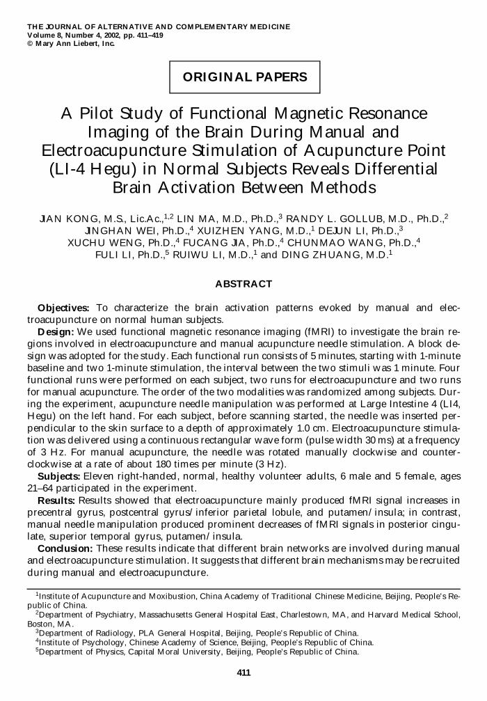
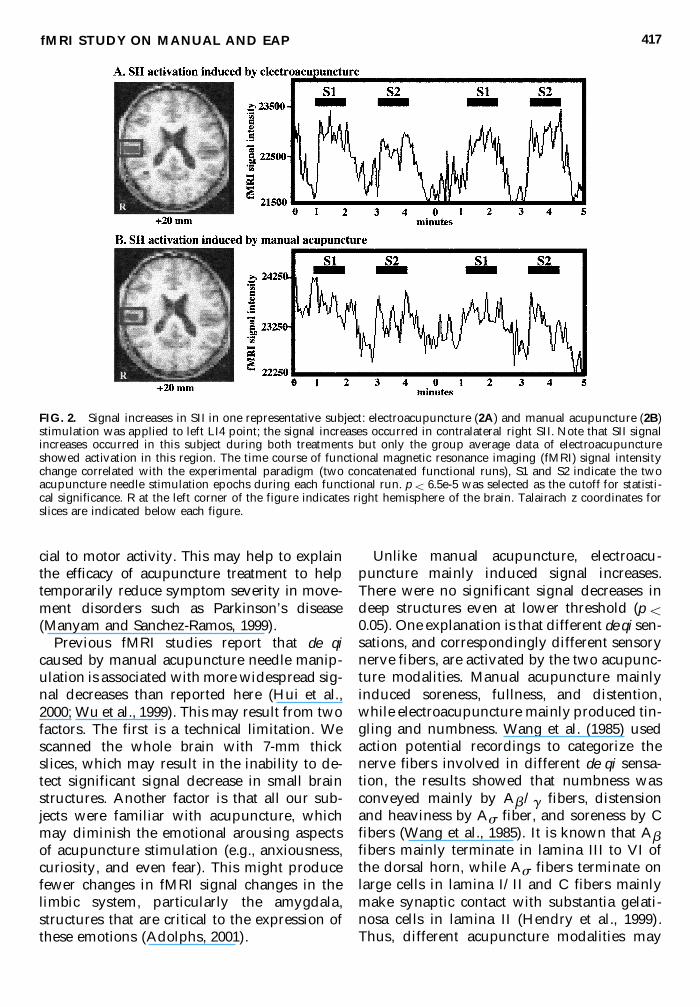
Figure 1A shows the summary statisticalmap from the group average data during elec-troacupuncture at several axial levels overlaidon the corresponding gray scale anatomic im-age. Electroacupuncture elicited fMRI signalincreases in the contralateral precentral gyrus(M1 region that represents the hand), but notin the corresponding primary somatosensorycortex (SI) region. In addition, activation wasfound on secondary somatosensory cortex (SII)in the area of the central/parietal operculum.Ipisilateral activation was found in the puta-men/insula. The only cluster that showed a sig-nal decrease during electroacupuncture wasthe right precuneus. Table 1 provides furtherdetails of these significant signal changes.
Unlike electroacupuncture, manual acupunc-ture induced no fMRI signal increase in groupaverage data but only signal decreases (Fig.1B). These regions of fMRI signal decrease in-cluded superior temporal gyrus and puta-men/insula contralateral to the site of acupunc-ture; posterior cingulate, superior temporalgyrus and lentiform/insula ipsilateral to thesite of acupuncture. Additional details are pre-sented in Table 2.
Direct comparison between the twoacupuncture modalities indicated that elec-troacupuncture resulted in a statisticallygreater fMRI signal increase than manualacupuncture in precentral gyrus (Talairach co-ordinates were as follows [right to left (mm),anterior to posterior (mm), inferior to superior(mm)]) [35.5, 12.3, 60.6], postcentral gyrus [52.7,15.9, 13.8], and insular cortex [36.3, 11.3, 18.6]on right side and superior frontal gyrus [211.8,29.1, 53.5] on the left side.
fMRI STUDY ON MANUAL AND EAP 415
FIG. 1. Functional magnetic resonance imaging (fMRI) signal changes during electroacupuncture stimulation andsignal decreases during manual acupuncture manipulation: T statistical maps of signal increases in right precentralgyrus (GPrC), right postcentral/inferior parietal lobule (GpoC/LPi), left putaman/insula (Pu/INS) and signal de-crease in right precuneus (PCu (figure 1A) induced by electroacupuncture; and signal decreases in left posterior cin-gulate (GC), right superior temporal gyrus (GTs), left putaman/insula (Pu/INS), left superior temporal gyrus andright lentiform/insula (NL/INS) (figure 1B) induced by manual acupuncture. p , 0.014 was selected as the cutoff forstatistical significance. R at the left corner of the figure indicates right hemisphere of the brain. Talairach z coordi-nates for slices are indicated below each figure.
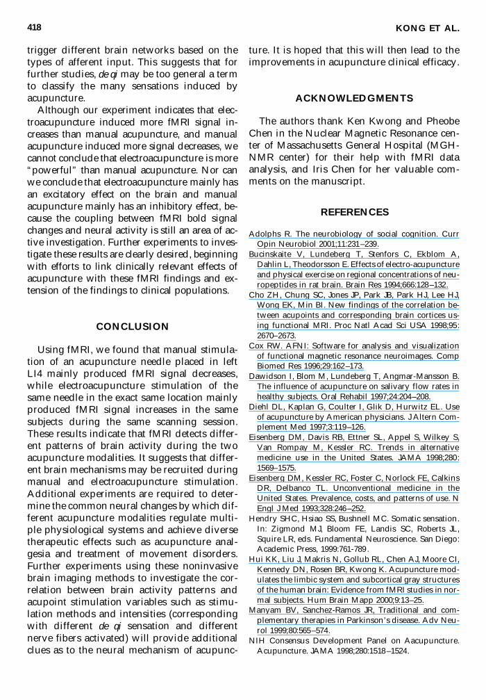
The individual analysis of somatosensorycortex indicated that seven of nine subjectsshowed signal increases in SII contralateral tothe site of acupuncture administration duringelectroacupuncture. Only three of nine subjectsshowed signal increases in contralateral SIIduring manual acupuncture. Images from arepresentative subject are shown in Figure 2.Figure 2A shows t test statistical map overlaidon the gray scale high-resolution scan at thelevel of the secondary somatosensory cortexand the time course of one representative voxelin this activated cluster elicited by elec-troacupuncture. Figure 2B shows the samebrain slice with the statistical map and the timecourse of one representative voxel in this acti-vated cluster elicited by manual acupuncture.
Both modalities of acupuncture were deliv-ered in exactly the same temporal sequence tothe left hand of all subjects, yet elec-troacupuncture induced more consistent andgreater magnitude of fMRI signal increases inSII than manual acupuncture.
DISCUSSION
In this study, we used fMRI to investigate thebrain regions involved in electroacupuncture
and manual acupuncture applied to LI4 acu-point of the left hand. Results showed that elec-troacupuncture mainly produced fMRI signalincreases, while manual needle manipulationproduced prominent decreases of fMRI signals.When electroacupuncture was administered,the index finger often moved and a tingling sen-sation commonly spread to the index finger orthe palm. This may explain the electro-acupuncture-induced signal increases in thehand region of precentral gyrus in every subject.
Of nine subjects whose fMRI data were usedfor analysis, four subjects received electro-acupuncture first and five subjects receivedmanual acupuncture first. There was no obvi-ous condition order effect.
This study provides information that can beused to formulate specific testable hypotheses re-garding the mechanism of clinical acupunctureefficacy. For instance, in both modalities, fMRIsignal changes were detected within insula. It iswell known from many published reports thatinsula is consistently activated during the ad-ministration of pain (Peyron et al., 2000). Wenoted no correlation between this insular activa-tion & subjective reports of slight pain accom-panying the deqi sensation. Another region thatis activated by both acupuncture modalities isthe putamen. The putamen is known to be cru-
KONG ET AL.416
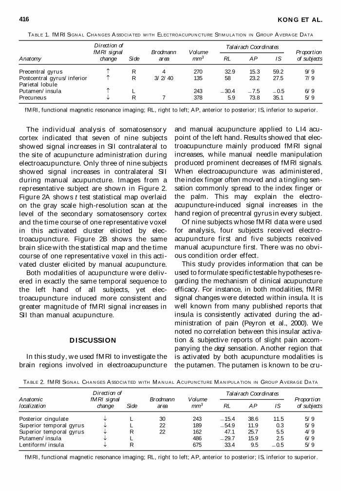
TABLE 1. fMRI SIGNAL CHANGES ASSOCIATED WITH ELECTROACUPUNCTURE STIMULATION IN GROUP AVERAGE DATA
fMRI signal Brodmann Volume ProportionAnatomy change Side area mm3 RL AP IS of subjects
Precentral gyrus R 4 270 32.9 15.3 59.2 9/9Postcentral gyrus/inferior R 3/2/40 135 58 23.2 27.5 7/9Parietal lobulePutamen/insula L 243 230.4 27.5 20.5 6/9Precuneus ¯ R 7 378 5.9 73.8 35.1 5/9
fMRI, functional magnetic resonance imaging; RL, right to left; AP, anterior to posterior; IS, inferior to superior.
Talairach Coordinates
TABLE 2. fMRI SIGNAL CHANGES ASSOCIATED WITH MANUAL ACUPUNCTURE MANIPULATION IN GROUP AVERAGE DATA
Anatomic fMRI signal Brodmann Volume Proportionlocalization change Side area mm3 RL AP IS of subjects
Posterior cingulate ¯ L 30 243 215.4 38.6 11.5 5/9Superior temporal gyrus ¯ L 22 189 254.9 11.9 0.3 5/9Superior temporal gyrus ¯ R 22 162 47.1 25.7 5.5 4/9Putamen/insula ¯ L 486 229.7 15.9 2.5 6/9Lentiform/insula ¯ R 675 33.4 9.5 20.5 5/9
fMRI, functional magnetic resonance imaging; RL, right to left; AP, anterior to posterior; IS, inferior to superior.
Talairach CoordinatesDirection of
Direction of
cial to motor activity. This may help to explainthe efficacy of acupuncture treatment to helptemporarily reduce symptom severity in move-ment disorders such as Parkinson’s disease(Manyam and Sanchez-Ramos, 1999).
Previous fMRI studies report that de qicaused by manual acupuncture needle manip-ulation is associated with more widespread sig-nal decreases than reported here (Hui et al.,2000; Wu et al., 1999). This may result from twofactors. The first is a technical limitation. Wescanned the whole brain with 7-mm thickslices, which may result in the inability to de-tect significant signal decrease in small brainstructures. Another factor is that all our sub-jects were familiar with acupuncture, whichmay diminish the emotional arousing aspectsof acupuncture stimulation (e.g., anxiousness,curiosity, and even fear). This might producefewer changes in fMRI signal changes in thelimbic system, particularly the amygdala,structures that are critical to the expression ofthese emotions (Adolphs, 2001).
Unlike manual acupuncture, electroacu-puncture mainly induced signal increases.There were no significant signal decreases indeep structures even at lower threshold (p ,0.05). One explanation is that different de qi sen-sations, and correspondingly different sensorynerve fibers, are activated by the two acupunc-ture modalities. Manual acupuncture mainlyinduced soreness, fullness, and distention,while electroacupuncture mainly produced tin-gling and numbness. Wang et al. (1985) usedaction potential recordings to categorize thenerve fibers involved in different de qi sensa-tion, the results showed that numbness wasconveyed mainly by Ab/g fibers, distensionand heaviness by As fiber, and soreness by Cfibers (Wang et al., 1985). It is known that Abfibers mainly terminate in lamina III to VI ofthe dorsal horn, while As fibers terminate onlarge cells in lamina I/II and C fibers mainlymake synaptic contact with substantia gelati-nosa cells in lamina II (Hendry et al., 1999).Thus, different acupuncture modalities may
fMRI STUDY ON MANUAL AND EAP 417
FIG. 2. Signal increases in SII in one representative subject: electroacupuncture (2A) and manual acupuncture (2B)stimulation was applied to left LI4 point; the signal increases occurred in contralateral right SII. Note that SII signalincreases occurred in this subject during both treatments but only the group average data of electroacupunctureshowed activation in this region. The time course of functional magnetic resonance imaging (fMRI) signal intensitychange correlated with the experimental paradigm (two concatenated functional runs), S1 and S2 indicate the twoacupuncture needle stimulation epochs during each functional run. p , 6.5e-5 was selected as the cutoff for statisti-cal significance. R at the left corner of the figure indicates right hemisphere of the brain. Talairach z coordinates forslices are indicated below each figure.
trigger different brain networks based on thetypes of afferent input. This suggests that forfurther studies, de qi may be too general a termto classify the many sensations induced byacupuncture.
Although our experiment indicates that elec-troacupuncture induced more fMRI signal in-creases than manual acupuncture, and manualacupuncture induced more signal decreases, wecannot conclude that electroacupuncture is more“powerful” than manual acupuncture. Nor canwe conclude that electroacupuncture mainly hasan excitatory effect on the brain and manualacupuncture mainly has an inhibitory effect, be-cause the coupling between fMRI bold signalchanges and neural activity is still an area of ac-tive investigation. Further experiments to inves-tigate these results are clearly desired, beginningwith efforts to link clinically relevant effects ofacupuncture with these fMRI findings and ex-tension of the findings to clinical populations.
CONCLUSION
Using fMRI, we found that manual stimula-tion of an acupuncture needle placed in left LI4 mainly produced fMRI signal decreases,while electroacupuncture stimulation of thesame needle in the exact same location mainlyproduced fMRI signal increases in the samesubjects during the same scanning session.These results indicate that fMRI detects differ-ent patterns of brain activity during the twoacupuncture modalities. It suggests that differ-ent brain mechanisms may be recruited duringmanual and electroacupuncture stimulation.Additional experiments are required to deter-mine the common neural changes by which dif-ferent acupuncture modalities regulate multi-ple physiological systems and achieve diversetherapeutic effects such as acupuncture anal-gesia and treatment of movement disorders.Further experiments using these noninvasivebrain imaging methods to investigate the cor-relation between brain activity patterns andacupoint stimulation variables such as stimu-lation methods and intensities (correspondingwith different de qi sensation and differentnerve fibers activated) will provide additionalclues as to the neural mechanism of acupunc-
ture. It is hoped that this will then lead to theimprovements in acupuncture clinical efficacy.
ACKNOWLEDGMENTS
The authors thank Ken Kwong and PheobeChen in the Nuclear Magnetic Resonance cen-ter of Massachusetts General Hospital (MGH-NMR center) for their help with fMRI dataanalysis, and Iris Chen for her valuable com-ments on the manuscript.
REFERENCES
Adolphs R. The neurobiology of social cognition. CurrOpin Neurobiol 2001;11:231–239.
Bucinskaite V, Lundeberg T, Stenfors C, Ekblom A,Dahlin L, Theodorsson E. Effects of electro-acupunctureand physical exercise on regional concentrations of neu-ropeptides in rat brain. Brain Res 1994;666:128–132.
Cho ZH, Chung SC, Jones JP, Park JB, Park HJ, Lee HJ,Wong EK, Min BI. New findings of the correlation be-tween acupoints and corresponding brain cortices us-ing functional MRI. Proc Natl Acad Sci USA 1998;95:2670–2673.
Cox RW. AFNI: Software for analysis and visualizationof functional magnetic resonance neuroimages. CompBiomed Res 1996;29:162–173.
Dawidson I, Blom M, Lundeberg T, Angmar-Mansson B.The influence of acupuncture on salivary flow rates inhealthy subjects. Oral Rehabil 1997;24:204–208.
Diehl DL, Kaplan G, Coulter I, Glik D, Hurwitz EL. Useof acupuncture by American physicians. J Altern Com-plement Med 1997;3:119–126.
Eisenberg DM, Davis RB, Ettner SL, Appel S, Wilkey S,Van Rompay M, Kessler RC. Trends in alternative medicine use in the United States. JAMA 1998;280:1569–1575.
Eisenberg DM, Kessler RC, Foster C, Norlock FE, CalkinsDR, Delbanco TL. Unconventional medicine in theUnited States. Prevalence, costs, and patterns of use. NEngl J Med 1993;328:246–252.
Hendry SHC, Hsiao SS, Bushnell MC. Somatic sensation.In: Zigmond MJ, Bloom FE, Landis SC, Roberts JL,Squire LR, eds. Fundamental Neuroscience. San Diego:Academic Press, 1999:761-789 .
Hui KK, Liu J, Makris N, Gollub RL, Chen AJ, Moore CI,Kennedy DN, Rosen BR, Kwong K. Acupuncture mod-ulates the limbic system and subcortical gray structuresof the human brain: Evidence from fMRI studies in nor-mal subjects. Hum Brain Mapp 2000;9:13–25.
Manyam BV, Sanchez-Ramos JR, Traditional and com-plementary therapies in Parkinson’s disease. Adv Neu-rol 1999;80:565–574.
NIH Consensus Development Panel on Aacupuncture.Acupuncture. JAMA 1998;280:1518–1524.
KONG ET AL.418

Peyron R, Laurent B, Garcia-Larrea L. Functional imag-ing of brain responses to pain. A review and meta-analysis. Neurophysiol Clin 2000;30:263–288.
Saletu B, Saletu M, Brown M, Stern J, Sletten I, Ulett G.Hypno-analgesia and acupuncture analgesia: A neuro-physiological reality? Neuropsychobiology 1975;1:218–242.
Stux G, Pomeranz B. Basics of Acupuncture. Berlin:Springer-Verlag, 1997:204.
Talairach J, Tourneaux P. Co-Planar Stereotaxic Atlas ofthe Human Brain. New York: Thieme Medical, 1988.
Wang K, Yao S, Xian Y, Hou Z. A study on the receptivefield of acupoints and the relationship between char-acteristics of needling sensation and groups of afferentfibres. Scientia Sinica (B) 1985;28:963–971.
Wu, MT, Hsieh JC, Xiong J, Yang CF, Pan HB, Chen YC,Tsai G, Rosen BR, Kwong KK. Central nervous path-way for acupuncture stimulation: localization of pro-cessing with functional MR imaging of the brain—Pre-liminary experience. Radiology 1999;212:133–141.
Address reprint requests to:Jian Kong, M.S., Lic.Ac.
Massachusetts General Hospital East149 13th Street, Room 9109
Charlestown, MA 02129
E-mail: [email protected]
fMRI STUDY ON MANUAL AND EAP 419

Related Documents


