S. Laureys et al. (Eds.) Progress in Brain Research, Vol. 177 ISSN 0079-6123 Copyright r 2009 Elsevier B.V. All rights reserved CHAPTER 14 A perturbational approach for evaluating the brain’s capacity for consciousness Marcello Massimini 1, , Melanie Boly 2 , Adenauer Casali 1 , Mario Rosanova 1 and Giulio Tononi 3 1 Department of Clinical Sciences, University of Milan, Milan, Italy 2 Coma Science Group, Cyclotron Research Center and Neurology Department, University of Liege and CHU Hospital, Belgium 3 Department of Psychiatry, University of Wisconsin, WI, USA Abstract: How do we evaluate a brain’s capacity to sustain conscious experience if the subject does not manifest purposeful behaviour and does not respond to questions and commands? What should we measure in this case? An emerging idea in theoretical neuroscience is that what really matters for consciousness in the brain is not activity levels, access to sensory inputs or neural synchronization per se, but rather the ability of different areas of the thalamocortical system to interact causally with each other to form an integrated whole. In particular, the information integration theory of consciousness (IITC) argues that consciousness is integrated information and that the brain should be able to generate consciousness to the extent that it has a large repertoire of available states (information), yet it cannot be decomposed into a collection of causally independent subsystems (integration). To evaluate the ability to integrate information among distributed cortical regions, it may not be sufficient to observe the brain in action. Instead, it is useful to employ a perturbational approach and examine to what extent different regions of the thalamocortical system can interact causally (integration) and produce specific responses (information). Thanks to a recently developed technique, transcranial magnetic stimulation and high- density electroencephalography (TMS/hd-EEG), one can record the immediate reaction of the entire thalamocortical system to controlled perturbations of different cortical areas. In this chapter, using sleep as a model of unconsciousness, we show that TMS/hd-EEG can detect clear-cut changes in the ability of the thalamocortical system to integrate information when the level of consciousness fluctuates across the sleep–wake cycle. Based on these results, we discuss the potential applications of this novel technique to evaluate objectively the brain’s capacity for consciousness at the bedside of brain-injured patients. Keywords: coma; consciousness; transcranial magnetic stimulation; electroencephalography; information; integration Evaluating a subject’s level of consciousness The bedside evaluation of patients affected by Corresponding author. disorders of consciousness (DOC) relies on Tel.: +39 33 39 92 57 91; Fax: +39 02 48002084; E-mail: [email protected] repeated behavioural observation by trained DOI: 10.1016/S0079-6123(09)17714-2 201

Welcome message from author
This document is posted to help you gain knowledge. Please leave a comment to let me know what you think about it! Share it to your friends and learn new things together.
Transcript

S. Laureys et al. (Eds.)Progress in Brain Research, Vol. 177 ISSN 0079-6123 Copyright r 2009 Elsevier B.V. All rights reserved
CHAPTER 14
A perturbational approach for evaluating the brain’s capacity for consciousness
Marcello Massimini1,�, Melanie Boly2, Adenauer Casali1, Mario Rosanova1 andGiulio Tononi3
1Department of Clinical Sciences, University of Milan, Milan, Italy2Coma Science Group, Cyclotron Research Center and Neurology Department, University of Liege and
CHU Hospital, Belgium3Department of Psychiatry, University of Wisconsin, WI, USA
Abstract: How do we evaluate a brain’s capacity to sustain conscious experience if the subject does not manifest purposeful behaviour and does not respond to questions and commands? What should we measure in this case? An emerging idea in theoretical neuroscience is that what really matters for consciousness in the brain is not activity levels, access to sensory inputs or neural synchronization per se, but rather the ability of different areas of the thalamocortical system to interact causally with each other to form an integrated whole. In particular, the information integration theory of consciousness (IITC) argues that consciousness is integrated information and that the brain should be able to generate consciousness to the extent that it has a large repertoire of available states (information), yet it cannot be decomposed into a collection of causally independent subsystems (integration). To evaluate the ability to integrate information among distributed cortical regions, it may not be sufficient to observe the brain in action. Instead, it is useful to employ a perturbational approach and examine to what extent different regions of the thalamocortical system can interact causally (integration) and produce specific responses (information). Thanks to a recently developed technique, transcranial magnetic stimulation and high-density electroencephalography (TMS/hd-EEG), one can record the immediate reaction of the entire thalamocortical system to controlled perturbations of different cortical areas. In this chapter, using sleep as a model of unconsciousness, we show that TMS/hd-EEG can detect clear-cut changes in the ability of the thalamocortical system to integrate information when the level of consciousness fluctuates across the sleep–wake cycle. Based on these results, we discuss the potential applications of this novel technique to evaluate objectively the brain’s capacity for consciousness at the bedside of brain-injured patients.
Keywords: coma; consciousness; transcranial magnetic stimulation; electroencephalography; information; integration
Evaluating a subject’s level of consciousness
The bedside evaluation of patients affected by �Corresponding author. disorders of consciousness (DOC) relies onTel.: +39 33 39 92 57 91; Fax: +39 02 48002084;
E-mail: [email protected] repeated behavioural observation by trained
DOI: 10.1016/S0079-6123(09)17714-2 201

202
personnel. During the examination, spontaneous and elicited behaviour in response to multisensory stimulation is recorded in accordance with specific scales (Giacino et al., 2004; Gill-Thwaites and Munday, 2004; Kalmar and Giacino, 2005; Shiel et al., 2000). Regardless of the scale employed, the examiner typically looks for (1) evidence of awareness of the self or of the environment, (2) evidence of sustained, reproducible, purposeful or voluntary response to tactile, auditory or noxious stimuli and (3) evidence of language comprehension and expression (Laureys et al., 2004). If none of these three defining behavioural features can be detected during careful and repeated evaluations, the subject is considered unconscious (Royal College of Physicians, 1994), while patients who show non-reflexive behaviour but are unable to communicate their thoughts and feelings are ascribed to a recently defined clinical entity, the minimally conscious state (Giacino et al., 2002). Thus, according to the clinical definition of consciousness, subjects are conscious if they can signal that this is the case. However, since in patients with severe brain injury motor responsiveness is often impaired, it may also happen that a subject is aware but unable to move or speak (Schnakers et al., 2009). Therefore, while detecting the presence of voluntary behaviour at the bedside is sufficient to infer that a subject is aware, its absence does not necessarily imply unconsciousness (Boly et al., 2007; Laureys et al., 2004; Monti et al., 2009).
Recently, the development of new neuroimaging protocols has made it possible to probe for signs of awareness even when subjects are completely unable to move (Boly et al., 2007; Owen and Coleman, 2008). For example, in a recent study (Owen and Coleman, 2008), a clinically vegetative, seemingly unresponsive patient was put in the scanner and asked to imagine playing tennis or navigating through her own apartment. Remarkably, the patient showed fMRI patterns of brain activation that were consistent and specific for the requested cognitive task, just like healthy subjects. This paradigmatic case demonstrates that the patient’s ability to wilfully enter specific neural states upon request can be used to detect the presence of awareness
even when motor outputs are absent. However, there still may be cases, such as in aphasia, akinetic mutism, catatonic depression or diffuse dopaminergic lesions, where a patient, although aware, may not be able to understand or be willing to respond (Boly et al., 2007). Moreover, because of frequent movement artefacts and because of possible alterations of the normal coupling of hemodynamics and neuronal firing (Rossini et al., 2004), acquiring and interpreting fMRI data is especially difficult in DOC patients (Giacino et al., 2006). Hence, the absence of volitional brain activity in the scanner, just like the absence of purposeful movements during a clinical examination, does not necessarily imply the absence of awareness.
The behavioural approach and the neuroimaging paradigm represent two different levels at which a communication can be established with a DOC patient (Owen et al., 2005). If an overt behaviour fails to signal consciousness, it is still possible to dig deeper by looking for purposeful neural activations. Both methods leave no doubts in case of a positive result: if the subjects respond, they are actually aware. Instead, a negative result leaves an open question.
Evaluating a brain’s capacity for consciousness
In this chapter, we propose an additional level at which consciousness can be studied even when no communication whatsoever (behavioural or neural) can be established with the subject. This paradigm does not aim at probing the subject in order to elicit wilfull behaviours or neural activations; rather, it involves probing directly the subject’s brain to gauge core properties that are theoretically relevant for consciousness. This option requires (1) starting from a theory that suggests which properties are fundamental for a physical system to give rise to conscious experience and (2) identifying and implementing a practical measuring method to weigh up these properties in a real brain. Here, we start with the information integration theory of consciousness (IITC) (Tononi, 2004, 2005, 2008), a theory that argues that consciousness is integrated

203
information and that a physical system should be able to generate consciousness to the extent that it can enter any of a large number of available states (information), yet it cannot be decomposed into a collection of causally independent subsystems (integration). Then, we devise a practical method to gauge the brain’s capacity to integrate information. To do this we employ a combination of transcranial magnetic stimulation and electroencephalography (TMS/hd-EEG), a technique that allows stimulating directly different subsets of cortical neurons and recording the immediate reaction of the rest of the brain. Based on measurements performed in sleeping subjects (Massimini et al., 2005, 2007), we argue that this method represents an effective way to appreciate, at a general level, to what extent different regions of the thalamocortical system can interact globally (integration) to produce specific responses (infor-mation). Thus, instead of asking the subjects to wilfully perform different motor or cognitive tasks, we directly ‘‘ask’’ (with TMS) their thalamocortical system to enter different neural states and we assess (with hd-EEG) to what extent these states are integrated and specific. While this approach is not meant to tell whether a subject is actually conscious or not, it may represent a principled way to objectively weigh a brain’s capacity for conscious experience.
Theoretical guidelines: the integrated informationtheory of consciousness
The IITC takes its start from phenomenology and, by making a critical use of thought experiments, argues that subjective experience is integrated information. Therefore, according to the IITC, any physical system will have subjective experience to the extent that it is capable of integrating information. In this view, experience, i.e. information integration, is a fundamental quantity that is, in principle, measurable, just as mass or energy is. Information and integration are, on the other hand, the very essence of subjective experience. Classically, information is the reduction of uncertainty among alternatives: when a coin falls on one of its two sides, it provides 1 bit of
information, whereas a die falling on one of six faces provides B2.6 bits. But then having any conscious experience, even one of pure darkness, must be extraordinarily informative, because it rules out countless other experiences instead (think of all the frames of every possible movie). In other words, having any experience is like throwing a die with a trillion faces and identifying which number came up. On the other hand, every experience is an integrated whole that cannot be subdivided into independent components. For example, with an intact brain you cannot experience the left half of the visual field independently of the right half, or visual shapes independently of their colour. In other words, the die of experience is a single one; throwing multiple dice and combining the numbers will not help.
If the capacity for consciousness corresponds to the capacity to integrate information, then a physical system should be able to generate consciousness to the extent that it can discriminate among a large number of available states (infor-mation), yet it cannot be decomposed into a collection of causally independent subsystems (integration). How can one identify such an integrated system, and how can one measure its repertoire of available states? To measure the repertoire of different states that are available to a system, one can use the entropy function, but this way of measuring information is completely insensitive to whether the information is integrated. Thus, measuring entropy would not allow us to distinguish between one million photodiodes with a repertoire of two states each, and a single integrated system with a repertoire of 21,000,000
states. To measure information integration, it is essential to know whether a set of elements constitutes a causally integrated system, or they can be broken down into a number of independent or quasi-independent subsets among which no information can be integrated.
Indeed, the theory claims that the level of consciousness of a physical system is related to the repertoire of different states (information) that can be discriminated by the system as a whole (integration). Thus, a measure of integrated information, called phi (F), has been proposed in order to quantify the information generated

204
when a system discriminates one particular state of its repertoire, above and beyond the information generated independently by its parts (Balduzzi and Tononi, 2008; Tononi, 2004).
As demonstrated through computer simulations, information integration is optimized (F is highest) if the elements of a complex are connected in such a way that they are both functionally specialized (connection patterns are different for different elements) and functionally integrated (all elements can be reached from all other elements of the network). If functional specialization is lost by replacing the heterogeneous connectivity with a homogeneous one, or if functional integration is lost by rearranging the connections to form small modules, the value of Fdecreases considerably (Tononi and Sporns, 2003).
According to the IITC, this is exactly why, among many structures of the brain, the thalamocortical system is so special for consciousness: it is naturally organized in a way that appears to emphasize at once both functional specialization and functional integration. Thus, it comprises a large number of elements that are functionally specialized, becoming activated in different circumstances (Bartels and Zeki, 2005). This is true at multiple spatial scales, from different cortical systems dealing with vision, audition, etc., to different cortical areas dealing with shape, colour, motion, etc., to different groups of neurons responding to different directions of motion. On the other hand, the specialized elements of the thalamocortical system are integrated through an extended network of intra- and inter-areal connections that permit rapid and effective interactions within and between areas (Engel et al., 2001).
But then, the theory also explicitly predicts that the fading of consciousness should be associated with either a reduction of integration within thalamocortical circuits (e.g. they could break down into causally independent modules) or a reduction in information (the repertoire of available states might shrink), or both. This specific prediction is however difficult to test in humans, since, in practice, F can only be measured rigorously for small, simulated systems. In the
next section, we try to identify an empirical method to approximate a measure of the capacity for integrated information in a human brain.
Employing TMS/hd-EEG to evaluatethalamocortical integration and informationcapacity
Different methods have been proposed in order to infer on a subject’s level of consciousness solely based on brain activity. Some of these methods, such as spectral analysis (Berthomier et al., 2007) and the proprietary ‘‘bispectral index’’ (Myles et al., 2004), seem to correlate empirically with consciousness but have no clear theoretical foundation. Other measures, such as neural complexity (Tononi et al., 1994) and causal density (Seth, 2005), are theoretically motivated (Seth et al., 2008) but have not yet been tested empirically. More or less explicitly, all these measures attempt to capture the coexistence of functional integration and functional differentiation in spontaneous (mainly hd-EEG) brain signals. Yet, to dependably appreciate the brain’s capacity for consciousness (defined as integrated information), one should go beyond spontaneous activity levels or patterns of temporal correlation among distant neuronal groups (functional connectivity). First, this is because the repertoire of available states is, by definition, potential and, thus, not necessarily observable. Second, because it is difficult to say whether a system is actually integrated or not by just observing the spontaneous activity it generates. For example, observing time-varying, complex correlations among retinal neurons that are responding to a rich visual scene may lead one to the conclusion that the retina is both functionally specialized and functionally integrated. However, such complex spa-tial-temporal correlations do not imply that the retina per se has a capacity for consciousness. In fact, it is enough to perturb a few retinal elements and to record from the rest of the cells to realize that, to a large extent, the retina is actually composed of segregated modules that do not interact with each other. Indeed, the ability to integrate information can only be demonstrated

205
from a causal perspective; one must employ a perturbational approach (effective connectivity) and examine to what extent subsets of neurons can interact causally as a whole (integration) to produce responses that are specific for that particular perturbation (information). Moreover, one should probe causal interactions by directly stimulating the cerebral cortex to avoid possible subcortical filtering or gating. Finally, since causal interactions among thalamocortical neurons develop on a sub-second time scale (just as phenomenal consciousness does), it is very important to record the neural effects of the perturbation with the appropriate temporal resolution.
Thus, in practice, one should find a way to stimulate different subsets of cortical neurons and measure, with good spatial-temporal resolution, the effects produced by these perturbations in the rest of the thalamocortical system. Today, this measurement can be performed non-invasively in
humans, thanks to the development of a novel electrophysiological technique, based on the combination of navigated TMS and high-density electroencephalography (Ilmoniemi et al., 1997) (Fig. 1). With TMS, the cerebral cortex is stimulated directly by generating a brief but strong magnetic pulse (o1 ms, 2 T) through a coil applied to the surface of the scalp. The rapid change in magnetic field strength induces a current flow in the tissue, which results in the activation of underlying neuronal population. The synchronous volley of action potential thus initiated propagates along the available connection pathways and can produce activations in target cortical regions. By integrating TMS with MR-guided infra-red navigation systems, it is also possible to render the perturbation controllable and reproducible, in most cortical regions. Finally, using multi-channel EEG amplifiers that are compatible with TMS (Virtanen et al., 1999) one
Fig. 1. TMS/hd-EEG setup. In this example, a subject is sitting on an ergonomic chair while TMS is targeted to the occipital cortex. The red arrows indicate, from left to right, the three fundamental elements that compose the set-up: (1) a cap for high-density (60 channels) hd-EEG recordings that is connected to a TMS-compatible amplifier; (2) a focal figure-of-eight stimulating coil (TMS), held in place by a mechanical arm; (3) the display of the navigated brain stimulation system (NBS). This system employs an infra-red camera (not visible in this picture) to navigate TMS on a 3D reconstruction of the subject’s MRI. The location and the intensity of the electric field induced by TMS are estimated and displayed in real time. To prevent the subject from perceiving the click associated with the coil’s discharge, noise masking is played through inserted earplugs. Please see online version of this article for full color figure.

206
can record, starting just a few milliseconds after the pulse, the impact of the perturbation on the stimulated target and in distant cortical areas. Indeed, the integrated use of neuro-navigation systems, TMS and multichannel TMS-compatible hd-EEG amplifiers together constitute a new brain scanning method in which stimulation is navigated into any desired brain target and the concurrently recorded scalp potentials are processed into source images of the TMS-evoked neuronal activation (Komssi and Kahkonen, 2006).
It is worth highlighting some of the specific advantages that TMS/hd-EEG may offer as a tool to probe the brain of DOC patients:
1. TMS-evoked activations are intrinsically causal (Paus, 2005). Thus, unlike methods based on temporal correlations, TMS/hd-EEG immediately captures the fundamental mechanism that underlies integration, i.e. the ability of different elements of a system to affect each other.
2. TMS/hd-EEG bypasses sensory pathways and subcortical structures to probe directly the thalamocortical system. Therefore, unlike peripherally evoked potentials and evoked motor activations, TMS/hd-EEG does not depend on the integrity of sensory and motor systems and can access any patient (deafferentated or paralysed). Moreover, with TMS one can stimulate most cortical areas (including associative cortices) employing several different parameters (intensity, angle, current direction), thus probing a vast repertoire of possible responses, above and beyond observable ongoing brain states.
3. TMS-evoked potentials can be recorded with millisecond resolution, a time scale that is adequate to capture effective synaptic interactions among neurons.
4. TMS/hd-EEG does not require the subject to be involved in a task and the observed activations are not affected either by the willingness of the patient to participate or by his effort and performance. Hence, this approach is well suited to assess the objective
capacity of thalamocortical circuits independently on behaviour.
5. TMS/hd-EEG can be made portable in order to overcome the logistical and economic hurdles that may separate severely brain-injured patients from advanced imaging facilities.
Thus, at least in principle, TMS/hd-EEG may represent an appropriate tool to approximate a theoretical measure of consciousness at the patient’s bedside. However, the question whether this technique may actually detect changes in the brain’s capacity to integrate information can only be answered experimentally. For example, one should demonstrate that TMS-evoked activations are widespread (integration) and specific (infor-mation) in a conscious brain but that they become either local (revealing a loss of integration) or stereotypical (revealing a loss of information) when the same brain becomes unconscious. In the next section, we describe the results of experiments where TMS/hd-EEG was used to understand what changes in human thalamocortical circuits when consciousness fades upon falling asleep.
TMS/hd-EEG detects changes in the brain’scapacity for integrated information during sleep
Sleep is the only time when healthy humans regularly lose consciousness. Subjects awakened during slow-wave sleep early in the night may report short, thought-like fragments of experience, or often nothing at all (Hobson et al., 2000). Sleep also exposes several interesting paradoxes about the relationships between consciousness and the brain. For instance, it was thought that the fading of consciousness during sleep was due to the brain shutting down. However, while metabolic rates decrease in some cortical areas, thalamocortical neurons remain active during slow-wave sleep also, with mean firing rates comparable to those of quiet wakefulness (Steriade et al., 2001). It was also hypothesized that sensory inputs are blocked during sleep and that they are necessary to sustain conscious
207
experience. However, we now know that, even during deep sleep, sensory signals continue to reach the cerebral cortex (Kakigi et al., 2003) where they are processed subconsciously (Portas et al., 2000). Gamma activity and synchrony have been viewed as possible correlates of consciousness and they were found to be low in slow-wave sleep (Cantero et al., 2004). However, they may be equally low in REM sleep, when subjective experience is usually vivid, and they can be high in anaesthesia (Vanderwolf, 2000). On the other hand, intracranial recordings show that gamma activity (Destexhe et al., 2007) and gamma-coherence (Bullock et al., 1995) persist during slow-wave sleep. Interestingly, similar paradoxes, where neural activity levels, access to sensory information and the degree of neural synchrony do not correlate with the level of consciousness, can be found in other conditions such as anaesthesia, epilepsy and DOC patients (Tononi and Laureys, 2008). In this sense, sleep represents a general model to learn what really matters for consciousness.
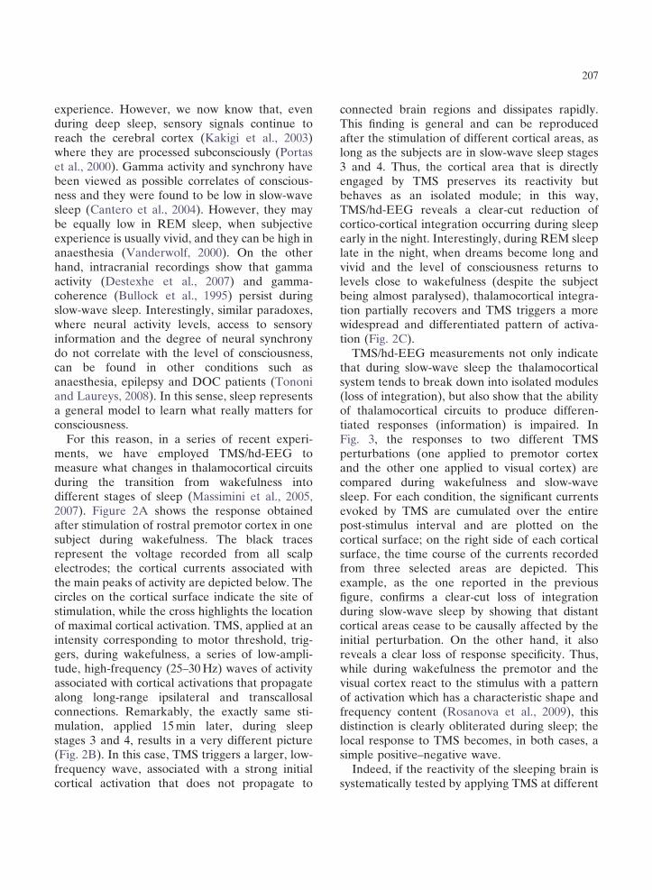
For this reason, in a series of recent experiments, we have employed TMS/hd-EEG to measure what changes in thalamocortical circuits during the transition from wakefulness into different stages of sleep (Massimini et al., 2005, 2007). Figure 2A shows the response obtained after stimulation of rostral premotor cortex in one subject during wakefulness. The black traces represent the voltage recorded from all scalp electrodes; the cortical currents associated with the main peaks of activity are depicted below. The circles on the cortical surface indicate the site of stimulation, while the cross highlights the location of maximal cortical activation. TMS, applied at an intensity corresponding to motor threshold, triggers, during wakefulness, a series of low-amplitude, high-frequency (25–30 Hz) waves of activity associated with cortical activations that propagate along long-range ipsilateral and transcallosal connections. Remarkably, the exactly same stimulation, applied 15 min later, during sleep stages 3 and 4, results in a very different picture (Fig. 2B). In this case, TMS triggers a larger, low-frequency wave, associated with a strong initial cortical activation that does not propagate to
connected brain regions and dissipates rapidly. This finding is general and can be reproduced after the stimulation of different cortical areas, as long as the subjects are in slow-wave sleep stages 3 and 4. Thus, the cortical area that is directly engaged by TMS preserves its reactivity but behaves as an isolated module; in this way, TMS/hd-EEG reveals a clear-cut reduction of cortico-cortical integration occurring during sleep early in the night. Interestingly, during REM sleep late in the night, when dreams become long and vivid and the level of consciousness returns to levels close to wakefulness (despite the subject being almost paralysed), thalamocortical integration partially recovers and TMS triggers a more widespread and differentiated pattern of activation (Fig. 2C).
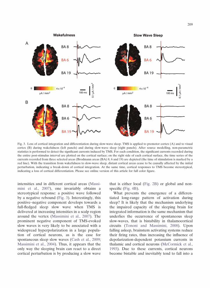
TMS/hd-EEG measurements not only indicate that during slow-wave sleep the thalamocortical system tends to break down into isolated modules (loss of integration), but also show that the ability of thalamocortical circuits to produce differentiated responses (information) is impaired. In Fig. 3, the responses to two different TMS perturbations (one applied to premotor cortex and the other one applied to visual cortex) are compared during wakefulness and slow-wave sleep. For each condition, the significant currents evoked by TMS are cumulated over the entire post-stimulus interval and are plotted on the cortical surface; on the right side of each cortical surface, the time course of the currents recorded from three selected areas are depicted. This example, as the one reported in the previous figure, confirms a clear-cut loss of integration during slow-wave sleep by showing that distant cortical areas cease to be causally affected by the initial perturbation. On the other hand, it also reveals a clear loss of response specificity. Thus, while during wakefulness the premotor and the visual cortex react to the stimulus with a pattern of activation which has a characteristic shape and frequency content (Rosanova et al., 2009), this distinction is clearly obliterated during sleep; the local response to TMS becomes, in both cases, a simple positive–negative wave.
Indeed, if the reactivity of the sleeping brain is systematically tested by applying TMS at different

Fig. 2. Cortical responses to TMS across the sleep–wake cycle. hd-EEG voltages and current densities are shown from a representative subject in whom the premotor cortex was stimulated with transcranial magnetic stimulation (TMS) (black arrow). (A) During waking, stimulation evokes hd-EEG responses first near the stimulation site (circle; the cross is the site of maximum evoked current) and then, in sequence, at other cortical locations, producing a long-range pattern of activation. (B) During slow-wave sleep, the stimulus-evoked response remains local, indicating a loss of cortical integration. At the same time, the response recorded from the electrode located under the stimulator (thick red trace) becomes a positive wave followed by a negative rebound. (C) During REM sleep, effective connectivity among distant cortical areas recovers, indicating a significant resurgence of cortical integration (adapted with permission from Massimini et al., 2007). Please see online version of this article for full color figure.
209
Fig. 3. Loss of cortical integration and differentiation during slow-wave sleep. TMS is applied to premotor cortex (A) and to visual cortex (B) during wakefulness (left panels) and during slow-wave sleep (right panels). After source modelling, non-parametric statistics is performed to detect the significant currents induced by TMS. For each condition, the significant currents recorded during the entire post-stimulus interval are plotted on the cortical surface; on the right side of each cortical surface, the time series of the currents recorded from three selected areas (Brodmann areas (BA) 8, 6 and 19) are depicted (the time of stimulation is marked by a red line). With the transition from wakefulness to slow-wave sleep, distant cortical areas cease to be causally affected by the initial perturbation, indicating a break-down of cortical integration. At the same time, cortical responses to TMS become stereotypical, indicating a loss of cortical differentiation. Please see online version of this article for full color figure.
intensities and in different cortical areas (Massimini et al., 2007), one invariably obtains a stereotypical response: a positive wave followed by a negative rebound (Fig. 3). Interestingly, this positive–negative component develops towards a full-fledged sleep slow wave when TMS is delivered at increasing intensities in a scalp region around the vertex (Massimini et al., 2007). The prominent negative component of TMS-evoked slow waves is very likely to be associated with a widespread hyperpolarization in a large population of cortical neurons, as is the case for spontaneous sleep slow waves (Cash et al., 2009; Massimini et al., 2004). Thus, it appears that the only way the sleeping brain can react to a direct cortical perturbation is by producing a slow wave
that is either local (Fig. 2B) or global and nonspecific (Fig. 4B).
What prevents the emergence of a differentiated long-range pattern of activation during sleep? It is likely that the mechanism underlying the impaired capacity of the sleeping brain for integrated information is the same mechanism that underlies the occurrence of spontaneous sleep slow-waves, that is bistability in thalamocortical circuits (Tononi and Massimini, 2008). Upon falling asleep, brainstem activating systems reduce their firing rates, thus increasing the influence of depolarization-dependent potassium currents in thalamic and cortical neurons (McCormick et al., 1993). Due to these currents, cortical neurons become bistable and inevitably tend to fall into a

210
Fig. 4. The sleeping brain reacts to TMS by producing stereotypical responses that resemble spontaneous sleep slow-waves. (A) TMS is delivered at four midline sites along the posterior–anterior axis of the cortex (posterior parietal, sensorimotor, supplementary motor, rostral premotor). The brain response to TMS is probed, at each site, at two intensities (MO is maximum stimulator output). The average responses to 15 TMS trials recorded from all channels (referenced to the mastoid) are shown for each intensity and each cortical site. At all cortical sites, TMS evokes a positive component followed by a negative rebound that develops towards a full-fledged sleep slow-wave when stimulation is delivered at increasing intensities in a scalp region closer to the sensory-motor cortex. (B) In this region, TMS triggers a large negative deflection associated with long-lasting currents that spread like an oil-spot to the surrounding cortex, starting from a fixed local maximum. In this particular case, the brain’s reaction to TMS becomes global but remains stereotypical and non-specific (adapted with permission from Massimini et al., 2007).
silent, hyperpolarized state (down-state) after a occurring spontaneously or induced by a stimulus period of activation (up-state). This bistability (like TMS), eventually triggers a stereotypical provides the mechanism for the slow oscillations of down-state that, in turn, prevents the emergence sleep where large populations of cortical neurons of specific, long-range patterns of activation. spontaneously alternate between up- and down- Altogether, TMS/hd-EEG measurements sugstates (Hill and Tononi, 2005). At the same time, gest that, during slow-wave sleep, the thalamodue to bistability, any local activation, whether cortical system, despite being active and reactive,

211
either breaks down in causally independent modules (producing a local down-state) or bursts into an explosive and non-specific response (producing a global down-state and a full-fledged hd-EEG slow-wave). In no case, during slow-wave sleep, does TMS result in a balanced, long-range, differentiated pattern of activation. The TMS/hd-EEG perturbational approach also suggests that intrinsic bistability in thalamocortical networks, the key mechanism responsible for the occurrence of the spontaneous slow oscillations of sleep, may be the reason why information integration is impaired in early NREM sleep (Massimini et al., 2009). While sleep and the associated bistability are physiological and reversible processes, pathological processes may similarly result in a modification of the brain’s ability to integrate information and this modification may be similarly detected by TMS/hd-EEG. In the next section, we discuss the possible applications of TMS/hd-EEG at the bedside of DOC patients.
TMS/EEG in DOC patients: some predictions
Given the variety of brain lesions and conditions that are associated to DOC (Laureys et al., 2004, 2009), it is very difficult to predict what kind of results TMS/hd-EEG might give in individual DOC patients. However, an informed guess can be adopted at least in some specific cases. For instance, it is conceivable that TMS-evoked activations similar to the ones described during slow-wave sleep may also be found in patients that are in a coma caused by a lesion in the ascending reticular activating system. In these cases, one could predict that, due to bistability, TMS should trigger a stereotypically local, or global, slow wave, provided that thalamocortical circuits are fundamentally intact. Similarly, due to pathological bistability in cortical circuits (Hahn and Durand, 2001), large and stereotypical responses would be expected in patients that are in a status epilepticus. On the other hand, TMS should results in mostly local responses in cases where connectivity is generally impaired, such as in patients with diffuse axonal injury (Graham et al., 2005). What would happen, instead, when a coma
patient opens her/his eyes, shows only reflexive behaviour (Schiff et al., 1999) and enters the vegetative state (Laureys and Boly, 2008)? In principle, the recovery of arousal, if not paralleled by recovery of awareness, should not be associated with significant changes in the ability of thalamocortical circuits to integrate information. In this sense, TMS-evoked activation is not expected to show relevant changes during the transition from coma to the vegetative state. Very different is the condition of locked-in patients (Plum and Posner, 1972) who awaken from their coma fully conscious (Schnakers et al., 2008) but completely paralysed, except for the ability to gaze upward; in this case, TMS should trigger more widespread and differentiated patterns of activation, just as it does during normal wakefulness or at most upon entering REM sleep, when subjects are conscious but almost paralysed.
The most important challenge for any objective measure of consciousness is proving itself capable of detecting a potential for residual cognition when no communication whatsoever can be established with the patient. This task is difficult by definition, since there is no behavioural reference to assess the subject’s actual level of consciousness. Nevertheless, some strategies could be adopted to practically validate TMS/hd-EEG measures as a dependable marker of the brain’s capacity for consciousness. First, one should demonstrate that using TMS/hd-EEG it is possible to identify significant differences between vegetative and minimally conscious patients concerning their brain’s capacity for integrated information. A positive result in such a population study would indicate that TMS/hd-EEG is sensitive enough to objectify minimal changes in the brain’s capacity for awareness. Second, one should demonstrate that the longitudinal TMS/hd-EEG measurements can predict the individual patient’s outcome. For instance, it would be relevant to observe TMS-evoked cortical responses that progressively become more global and specific in the brain of intensive care patients shortly before they regain consciousness at the clinical level. Then, TMS/ hd-EEG may be employed as a diagnostic/ prognostic tool to evaluate covert consciousness and to foster evidence-based neuro-rehabilitation.
212
Future perspectives
We attempted at identifying an objective marker of consciousness that is theoretically grounded and practically measurable. The core message of this chapter is that using by TMS/hd-EEG it is possible to detect clear-cut changes in the capacity of human thalamocortical circuits to integrate information, a theoretical requirement to generate conscious experience, when the level of consciousness fluctuates across the sleep–wake cycle. The implication of this finding is that TMS/ hd-EEG may be similarly employed to evaluate the brain’s capacity for consciousness at the bedside of non-communicative patients. Clearly, before applying this technique to DOC patients, further steps need to be taken.
First, TMS/hd-EEG normative data have to be defined. Thus, several cortical areas must be systematically perturbed in healthy subjects in order to determine the specificity and the reproducibility of cortical responses to TMS. A similar assessment has been recently performed on a limited set of cortical areas (superior occipital lobule, precuneus and premotor) and has revealed patterns of TMS-evoked cortical activation that are specific for the stimulated site and reproducible across subjects (Rosanova et al., 2009). This database needs to be further extended including more cortical areas and subjects.
Second, a standard analysis procedure must be developed in order to extract from TMS/hd-EEG data synthetic indices that capture the brain’s capacity for integration and differentiation. In fact, the results presented in this chapter are suggestive, but only qualitative. Different algorithms can be devised in order to quantify TMS/hd-EEG data in a way that is theoretically relevant. For instance, the extent of the brain area that is significantly engaged by TMS (Fig. 3) provides a simple measure of integration. Indeed, since TMS-evoked activations are intrinsically causal (Paus, 2005), it is warranted that the elements within this area are interacting effectively with each other and that the observed patterns of activations are not random. At this point, one could simply use measures related to entropy, or to algorithmic complexity, to
summarize in one number the spatial–temporal differentiation of the deterministic activation produced by this integrated network. This number will be low for modular network, because, in this case, activity remains local, and will be equally low for networks with widespread homogeneous connectivity, because, all elements will respond in the same way. Only networks that are integrated and differentiated at the same time are likely to react to TMS with a response characterized by a high complexity value.
Third, the technique and the appropriate analysis procedure must be tested in conditions where consciousness is graded and abolished in a controlled fashion, such as during anaesthesia (Alkire et al., 2008). Specifically, it would be important to apply TMS/hd-EEG measures of integrated information while consciousness is altered using different anaesthetics (such as midazolam, propofol or ketamine) that act through diverse mechanisms and that are associated with variable patterns of spontaneous EEG activity. Certainly, a reliable marker should only correlate with the level of consciousness, whether this has been altered by physiological sleep, by one anaesthetic or another, or by a pathological process.
Acknowledgements
This work was supported by European grant STREP LSHM-CT-2205-51818 to Marcello Massimini. Melanie Boly is Research Fellow at the Belgian National Fund for Scientific Research (FNRS). We thank Andrea Soddu, Silvia Casarotto and Fabio Ferrarelli for their help and comments.
References
Alkire, M. T., Hudetz, A. G., & Tononi, G. (2008). Consciousness and anesthesia. Science, 322, 876–880.
Balduzzi, D., & Tononi, G. (2008). Integrated information in discrete dynamical systems: Motivation and theoretical framework. PLoS Computational Biology, 4, e1000091.
Bartels, A., & Zeki, S. (2005). The chronoarchitecture of the cerebral cortex. Philosophical Transactions of the Royal
213
Society of London. Series B, Biological Sciences, 360, 733–750.
Berthomier, C., Drouot, X., Herman-Stoıca, M., Berthomier, P., Prado, J., Bokar-Thire, D., et al. (2007). Automatic analysis of single-channel sleep EEG: Validation in healthy individuals. Sleep, 30, 1587–1595.
Boly, M., Coleman, M. R., Davis, M. H., Hampshire, A., Bor, D., Moonen, G., et al. (2007). When thoughts become action: An fMRI paradigm to study volitional brain activity in non-communicative brain injured patients. Neuroimage, 36, 979–992.
Bullock, T. H., McClune, M. C., Achimowicz, J. Z., Iragui-Madoz, V. J., Duckrow, R. B., & Spencer, S. S. (1995). Temporal fluctuations in coherence of brain waves. Proceed-ings of the National Academy of Sciences of the United Statesof America, 92, 11568–11572.
Cantero, J. L., Atienza, M., Madsen, J. R., & Stickgold, R. (2004). Gamma EEG dynamics in neocortex and hippocampus during human wakefulness and sleep. Neuroimage, 22, 1271–1280.
Cash, S. S., Halgren, E., Dehghani, N., Rossetti, A. O., Thesen, T., Wang, C., et al. (2009). The human K-complex represents an isolated cortical down-state. Science, 324, 1084–1087.
Destexhe, A., Hughes, S. W., Rudolph, M., & Crunelli, V. (2007). Are corticothalamic ‘up’ states fragments of wakefulness? Trends in Neurosciences, 30, 334–342.
Engel, A. K., Fries, P., & Singer, W. (2001). Dynamic predictions: Oscillations and synchrony in top-down processing. Nature Review, Neurosciences, 2, 704–716.
Giacino, J. T., Ashwal, S., Childs, N., Cranford, R., Jennett, B., Katz, D. I., et al. (2002). The minimally conscious state: Definition and diagnostic criteria. Neurology, 58, 349–353.
Giacino, J. T., Kalmar, K., & Whyte, J. (2004). The JFK coma recovery scale-revised: Measurement characteristics and diagnostic utility. Archives of Physical Medicine andRehabilitation, 85, 2020–2029.
Giacino, J. T., Hirsch, J., Schiff, N., & Laureys, S. (2006). Functional neuroimaging applications for assessment and rehabilitation planning in patients with disorders of consciousness. Archives of Physical Medicine and Rehabilitation, 87, S67–S76.
Gill-Thwaites, H., & Munday, R. (2004). The Sensory Modality Assessment and Rehabilitation Technique (SMART): A valid and reliable assessment for vegetative state and minimally conscious state patients. Brain Injury, 18, 1255–1269.
Graham, D. I., Adams, J. H., Murray, L. S., & Jennett, B. (2005). Neuropathology of the vegetative state after head injury. Neuropsychological Rehabilitation, 15, 198–213.
Hahn, P. J., & Durand, D. M. (2001). Bistability dynamics in simulations of neural activity in high-extracellular-potassium conditions. Journal of Computational Neuroscience, 11, 5–18.
Hill, S., & Tononi, G. (2005). Modeling sleep and wakefulness in the thalamocortical system. Journal of Neurophysiology, 93, 1671–1698.
Hobson, J. A., Pace-Schott, E. F., & Stickgold, R. (2000). Dreaming and the brain: Toward a cognitive neuroscience
of conscious states. The Behavioral and Brain Sciences, 23, 793–842. discussion 904–1121
Ilmoniemi, R. J., Virtanen, J., Ruohonen, J., Karhu, J., Aronen, H. J., Naatanen, R., et al. (1997). Neuronal responses to magnetic stimulation reveal cortical reactivity and connectivity. Neuroreport, 8, 3537–3540.
Kakigi, R., Naka, D., Okusa, T., Wang, X., Inui, K., Qiu, Y., et al. (2003). Sensory perception during sleep in humans: A magnetoencephalograhic study. Sleep Medicine, 4, 493–507.
Kalmar, K., & Giacino, J. T. (2005). The JFK coma recovery scale - revised. Neuropsychological Rehabilitation, 15, 454–460.
Komssi, S., & Kahkonen, S. (2006). The novelty value of the combined use of electroencephalography and transcranial magnetic stimulation for neuroscience research. BrainResearch Reviews, 52, 183–192.
Laureys, S., & Boly, M. (2008). The changing spectrum of coma. Nature Clinical Practice Neurology, 4, 544–546.
Laureys, S., Owen, A. M., & Schiff, N. D. (2004). Brain function in coma, vegetative state, and related disorders. Lancet Neurology, 3, 537–546.
Laureys, S., Boly, M., Moonen, G., & Maquet, P. (2009). Coma. In L. Squire (Ed.), Encyclopedia of neuroscienceVol. 2, (pp. 1133–1142). Amsterdam: Elsevier.
Massimini, M., Huber, R., Ferrarelli, F., Hill, S., & Tononi, G. (2004). The sleep slow oscillation as a traveling wave. The Journal of Neuroscience, 24, 6862–6870.
Massimini, M., Ferrarelli, F., Huber, R., Esser, S. K., Singh, H., & Tononi, G. (2005). Breakdown of cortical effective connectivity during sleep. Science, 309, 2228–2232.
Massimini, M., Ferrarelli, F., Esser, S. K., Riedner, B. A., Huber, R., Murphy, M., et al. (2007). Triggering sleep slow waves by transcranial magnetic stimulation. Proceedingsof the National Academy of Sciences of the United States ofAmerica, 104, 8496–8501.
Massimini, M., Tononi, G., & Huber, R. (2009). Slow waves, synaptic plasticity and information processing: Insights from transcranial magnetic stimulation and high-density EEG experiments. The European Journal of Neuroscience, 29, 1761–1770.
McCormick, D. A., Wang, Z., & Huguenard, J. (1993). Neurotransmitter control of neocortical neuronal activity and excitability. Cerebral Cortex, 3, 387–398.
Monti, M. M., Coleman, M. R., & Owen, A. M. (2009). Neuroimaging and the vegetative state: Resolving the behavioral assessment dilemma? Annals of New YorkAcademy of Sciences, 1157, 81–89.
Myles, P. S., Leslie, K., McNeil, J., Forbes, A., & Chan, M. T. (2004). Bispectral index monitoring to prevent awareness during anaesthesia: The B-Aware randomised controlled trial. Lancet, 363, 1757–1763.
Owen, A. M., Coleman, M. R., Boly, M., Davis, M. H., Laureys, S., & Pickard, J. D. (2008). Detecting awareness in the vegetative state. Annals of New York Academy ofSciences, 1129, 130–138.
Owen, A. M., Coleman, M. R., Menon, D. K., Berry, E. L., Johnsrude, I. S., Rodd, J. M., Davis, M. H., et al. (2005). Using a hierarchical approach to investigate residual

214
auditory cognition in persistent vegetative state. Progress inBrain Research, 150, 457–471.
Paus, T. (2005). Inferring causality in brain images: A perturbation approach. Philosophical Transactions of theRoyal Society of London: Series B, Biological Sciences, 360, 1109–1114.
Plum, F., & Posner, J. B. (1972). The diagnosis of stupor and coma. Contemporary Neurology Series, 10, 1–286.
Portas, C. M., Krakow, K., Allen, P., Josephs, O., Armony, J. L., & Frith, C. D. (2000). Auditory processing across the sleep-wake cycle: Simultaneous EEG and fMRI monitoring in humans. Neuron, 28, 991–999.
Rosanova, M., Casali, A., Bellina, V., Resta, F., Mariotti, M., & Massimini, M. (2009). Natural frequencies of human corticothalamic circuits. Journal of Neuroscience, 29(24), 7679–7685.
Rossini, P. M., Altamura, C., Ferretti, A., Vernieri, F., Zappasodi, F., Caulo, M., et al. (2004). Does cerebrovascular disease affect the coupling between neuronal activity and local haemodynamics? Brain, 127, 99–110.
Royal College of Physicians. (1994). Medical aspects of the persistent vegetative state (1). The multi-society task force on PVS. The New England Journal Medicine, 330, 1499–1508.
Schiff, N., Ribary, U., Plum, F., & Llinas, R. (1999). Words without mind. Journal Cognitive Neuroscience, 11, 650–656.
Schnakers, C., Majerus, S., Goldman, S., Boly, M., Van Eeckhout, P., Gay, S., et al. (2008). Cognitive function in the locked-in syndrome. Journal of Neurology, 255, 323–330.
Schnakers, C., Perrin, F., Schabus, M., Hustinx, R., Majerus, S., Moonen, G., et al. (2009). Detecting consciousness in a total locked-in syndrome: An active event-related paradigm. Neurocase, 1–7.
Seth, A. K. (2005). Causal connectivity of evolved neural networks during behavior. Network, 16, 35–54.
Seth, A. K., Dienes, Z., Cleeremans, A., Overgaard, M., & Pessoa, L. (2008). Measuring consciousness: Relating
behavioural and neurophysiological approaches. Trends inCognitive Sciences, 12, 314–321.
Shiel, A., Horn, S. A., Wilson, B. A., Watson, M. J., Campbell, M. J., & McLellan, D. L. (2000). The Wessex Head Injury Matrix (WHIM) main scale: A preliminary report on a scale to assess and monitor patient recovery after severe head injury. Clinical Rehabilitation, 14, 408–416.
Steriade, M., Timodeev, I., & Grenier, F. (2001). Natural waking and sleep states: A view from inside neocortical neurons. Journal of Neurophysiology, 85, 1969–1985.
Tononi, G. (2004). An information integration theory of consciousness. BMC Neuroscience, 5, 42.
Tononi, G. (2005). Consciousness, information integration, and the brain. Progress in Brain Research, 150, 109–126.
Tononi, G. (2008). Consciousness as integrated information: A provisional manifesto. The Biological Bulletin, 215, 216–242.
Tononi, G., & Laureys, S. (2008). The neurology of consciousness: An overview. In S. Laureys and G. Tononi (Eds.), The Neurology of Consciousness (pp. 375–411). Amsterdam: Elsevier.
Tononi, G., & Massimini, M. (2008). Why does consciousness fade in early sleep? Annals of New York Academy ofSciences, 1129, 330–334.
Tononi, G., & Sporns, O. (2003). Measuring information integration. BMC Neuroscience, 4, 31.
Tononi, G., Sporns, O., & Edelman, G. M. (1994). A measure for brain complexity: Relating functional segregation and integration in the nervous system. Proceedings of theNational Academy of Science of the United States of America, 91, 5033–5037.
Vanderwolf, C. H. (2000). Are neocortical gamma waves related to consciousness? Brain Research, 855, 217–224.
Virtanen, J., Ruohonen, J., Naatanen, R., & Ilmoniemi, R. J. (1999). Instrumentation for the measurement of electric brain responses to transcranial magnetic stimulation. Med-ical & Biological Engineering & Computing, 37, 322–326.
Related Documents