A peptide inhibitor of vascular adhesion protein-1 (VAP-1) blocks leukocyte–endothelium interactions under shear stress Gennady G. Yegutkin 1 , Tiina Salminen 2 , Kaisa Koskinen 1 , Christian Kurtis 3 , Michael J. McPherson 3 , Sirpa Jalkanen 1 and Marko Salmi 1 1 MediCity Research Laboratory, Turku University and National Public Health Institute, Turku, Finland 2 Department of Biochemistry, Åbo Academi, Turku, Finland 3 School of Biochemistry and Microbiology, University of Leeds, Leeds, GB Vascular adhesion protein-1 (VAP-1) is an endothelial adhesion molecule mediating leukocyte interactions with blood vessels during leukocyte extravasation. Molecularly VAP-1 is a cell-surface-expressed ecto-enzyme belonging to the group of semicarbazide-sensitive amine oxidases (SSAO; EC 2.4.6.3), which deaminate primary amines. Here we asked whether peptides displaying a suitable free amine group could be a substrate or inhibitor of SSAO and thus regulate VAP-1-mediated leukocyte adhesion. On the basis of a molecular model of VAP-1, we designed synthetic peptides that fit to the substrate channel of VAP-1. One of these lysine-containing peptides effectively inhibits VAP-1-dependent lymphocyte rolling and firm adhesion to primary endothelial cells under physiologically relevant shear conditions. The same peptide inhibits the SSAO activity of endothelial and recombinant VAP-1 in a selective and long-lasting manner. We also show that all enzymatically active VAP-1 is displayed on the cell surface. Our results suggest that, in addition to soluble amines, specific cell-surface-bound molecules containing free NH 2 groups in a suitable posi- tion may modulate the enzymatic activity of SSAO. Moreover, the inhibitory peptide dimin- ishes leukocyte interactions with endothelial cells under conditions of shear, and thus it may be useful to treat inflammatory conditions. Supporting Information for this article is available at http://www.wiley-vch.de/contents/ jc – 2040/2004/24932 – s.html or from the author. Key words: Endothelial cells / Adhesion molecules / Peptides / Cell trafficking Received 23/1/04 Revised 5/5/04 Accepted 12/5/04 [DOI 10.1002/eji.200424932] Abbreviations: CHO: Chinese hamster ovary DAO: Diamine oxidase ECAO: Escherichia-coli-derived amine oxidase MAO: Monoamine oxidase RAO: Retina-specific amine oxidase SSAO: Semicarbazide-sensitive amine oxi- dase VAP-1: Vascular adhesion protein-1 1 Introduction Copper-containing semicarbazide-sensitive amine oxi- dases (SSAO) catalyze the oxidative deamination of pri- mary amines following the general reaction: –R–CH 2 –NH 2 +H 2 O+O 2 –R–CHO+NH 3 +H 2 O 2 [1, 2]. Vascular adhesion protein-1 (VAP-1) (also known as AOC3) is widely expressed in humans in endothelial, smooth muscle and adipose cells [3, 4]. It plays a role in glucose transport [5, 6] and in leukocyte trafficking [7] in an enzyme-dependent manner. However, the identity of physiological, soluble substrates or inhibitors for this enzyme still remains controversial [8]. Leukocyte extravasation from the blood via a multistep adhesion cascade is crucial for the normal function of immune surveillance as well as in mounting adequate inflammatory responses [9]. Endothelial VAP-1 mediates leukocyte rolling, firm adhesion and transmigration in multiple in vitro and in vivo binding assays [3] and the SSAO activity of VAP-1 plays a crucial role in this multi- step adhesion cascade [7, 10, 11]. Using a molecular model of VAP-1, we designed specific peptides that fit within the groove on the surface of VAP- 1 and can present free NH 2 groups via the narrow substrate-channel into the catalytically active site of VAP-1. We show that such polypeptides inhibit the enzy- matic activity of VAP-1 and, importantly, diminish 2276 G. G. Yegutkin et al. Eur. J. Immunol. 2004. 34: 2276–2285 © 2004 WILEY-VCH Verlag GmbH & Co. KGaA, Weinheim www.eji.de

Welcome message from author
This document is posted to help you gain knowledge. Please leave a comment to let me know what you think about it! Share it to your friends and learn new things together.
Transcript

A peptide inhibitor of vascular adhesion protein-1
(VAP-1) blocks leukocyte–endothelium interactions
under shear stress
Gennady G. Yegutkin1, Tiina Salminen2, Kaisa Koskinen1, Christian Kurtis3, Michael J.
McPherson3, Sirpa Jalkanen1 and Marko Salmi1
1 MediCity Research Laboratory, Turku University and National Public Health Institute, Turku,Finland
2 Department of Biochemistry, Åbo Academi, Turku, Finland3 School of Biochemistry and Microbiology, University of Leeds, Leeds, GB
Vascular adhesion protein-1 (VAP-1) is an endothelial adhesion molecule mediatingleukocyte interactions with blood vessels during leukocyte extravasation. Molecularly VAP-1is a cell-surface-expressed ecto-enzyme belonging to the group of semicarbazide-sensitiveamine oxidases (SSAO; EC 2.4.6.3), which deaminate primary amines. Here we askedwhether peptides displaying a suitable free amine group could be a substrate or inhibitor ofSSAO and thus regulate VAP-1-mediated leukocyte adhesion. On the basis of a molecularmodel of VAP-1, we designed synthetic peptides that fit to the substrate channel of VAP-1.One of these lysine-containing peptides effectively inhibits VAP-1-dependent lymphocyterolling and firm adhesion to primary endothelial cells under physiologically relevant shearconditions. The same peptide inhibits the SSAO activity of endothelial and recombinantVAP-1 in a selective and long-lasting manner. We also show that all enzymatically activeVAP-1 is displayed on the cell surface. Our results suggest that, in addition to solubleamines, specific cell-surface-bound molecules containing free NH2 groups in a suitable posi-tion may modulate the enzymatic activity of SSAO. Moreover, the inhibitory peptide dimin-ishes leukocyte interactions with endothelial cells under conditions of shear, and thus it maybe useful to treat inflammatory conditions.
Supporting Information for this article is available at http://www.wiley-vch.de/contents/jc–2040/2004/24932–s.html or from the author.
Key words: Endothelial cells / Adhesion molecules / Peptides / Cell trafficking
Received 23/1/04Revised 5/5/04Accepted 12/5/04
[DOI 10.1002/eji.200424932]
Abbreviations: CHO: Chinese hamster ovary DAO:Diamine oxidase ECAO: Escherichia-coli-derived amineoxidase MAO: Monoamine oxidase RAO: Retina-specificamine oxidase SSAO: Semicarbazide-sensitive amine oxi-dase VAP-1: Vascular adhesion protein-1
1 Introduction
Copper-containing semicarbazide-sensitive amine oxi-dases (SSAO) catalyze the oxidative deamination of pri-mary amines following the general reaction:–R–CH2–NH2+H2O+O2 1 –R–CHO+NH3+H2O2 [1, 2].Vascular adhesion protein-1 (VAP-1) (also known asAOC3) is widely expressed in humans in endothelial,smooth muscle and adipose cells [3, 4]. It plays a role inglucose transport [5, 6] and in leukocyte trafficking [7] in
an enzyme-dependent manner. However, the identity ofphysiological, soluble substrates or inhibitors for thisenzyme still remains controversial [8].
Leukocyte extravasation from the blood via a multistepadhesion cascade is crucial for the normal function ofimmune surveillance as well as in mounting adequateinflammatory responses [9]. Endothelial VAP-1 mediatesleukocyte rolling, firm adhesion and transmigration inmultiple in vitro and in vivo binding assays [3] and theSSAO activity of VAP-1 plays a crucial role in this multi-step adhesion cascade [7, 10, 11].
Using a molecular model of VAP-1, we designed specificpeptides that fit within the groove on the surface of VAP-1 and can present free NH2 groups via the narrowsubstrate-channel into the catalytically active site ofVAP-1. We show that such polypeptides inhibit the enzy-matic activity of VAP-1 and, importantly, diminish
2276 G. G. Yegutkin et al. Eur. J. Immunol. 2004. 34: 2276–2285
© 2004 WILEY-VCH Verlag GmbH & Co. KGaA, Weinheim www.eji.de
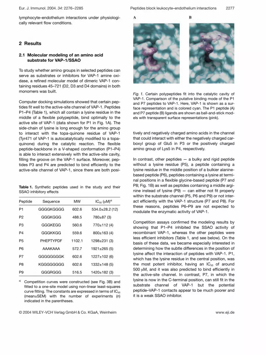
Fig. 1. Certain polypeptides fit into the catalytic cavity ofVAP-1. Comparison of the putative binding mode of the P1and P7 peptides to VAP-1. Here, VAP-1 is shown as a sur-face representation and is colored cyan. The P1 peptide (A)and P7 peptide (B) ligands are shown as ball-and-stick mod-els with transparent surface representations (pink).
lymphocyte–endothelium interactions under physiologi-cally relevant flow conditions.
2 Results
2.1 Molecular modeling of an amino acid
substrate for VAP-1/SSAO
To study whether amino groups in selected peptides canserve as substrates or inhibitors for VAP-1 amine oxi-dase, a refined molecular model of dimeric VAP-1 con-taining residues 45–721 (D2, D3 and D4 domains) in bothmonomers was built.
Computer docking simulations showed that certain pep-tides fit well to the active-site channel of VAP-1. PeptidesP1–P4 (Table 1), which all contain a lysine residue in themiddle of a flexible polypeptide, bind optimally to theactive site of VAP-1 (data shown for P1 in Fig. 1A). Theside-chain of lysine is long enough for the amino groupto interact with the topa-quinone residue of VAP-1(Tyr471 of VAP-1 is autocatalytically modified to a topa-quinone) during the catalytic reaction. The flexiblepeptide-backbone in a V-shaped conformation (P1–P4)is able to interact extensively with the active-site cavity,filling the groove on the VAP-1 surface. Moreover, pep-tides P3 and P4 are predicted to bind efficiently to theactive-site channel of VAP-1, since there are both posi-
Table 1. Synthetic peptides used in the study and theirSSAO inhibitory effects
Peptide Sequence MW IC50 ( ? M)a)
P1 GGGGKGGGG 602.6 534.0±28.2 (12)
P2 GGGKGGG 488.5 780±87 (3)
P3 GGGKEGG 560.6 770±112 (4)
P4 GGGKKGG 559.6 800±163 (4)
P5 PHEPTYPDF 1102.1 1298±231 (3)
P6 AAAKAAA 572.7 1921±265 (5)
P7 GGGGGGGGK 602.6 1227±102 (6)
P8 KGGGGGGGG 602.6 1332±148 (5)
P9 GGGRGGG 516.5 1420±182 (3)
a) Competition curves were constructed (see Fig. 3B) andfitted to a one-site model using non-linear least-squarescurve fitting. The constants are expressed in terms of IC50
(mean±SEM) with the number of experiments (n)indicated in the parentheses.
tively and negatively charged amino acids in the channelthat could interact with either the negatively charged car-boxyl group of Glu5 in P3 or the positively chargedamino group of Lys5 in P4, respectively.
In contrast, other peptides — a bulky and rigid peptidewithout a lysine residue (P5), a peptide containing alysine residue in the middle position of a bulkier alanine-based peptide (P6), peptides containing a lysine at termi-nal positions in a flexible glycine-based peptide (P7 andP8; Fig. 1B) as well as peptides containing a middle argi-nine instead of lysine (P9) — can either not fit properlywithin the substrate channel (P5, P6 and P9) or not inter-act efficiently with the VAP-1 structure (P7 and P8). Forthese reasons, peptides P6–P9 are not expected tomodulate the enzymatic activity of VAP-1.
Competition assays confirmed the modeling results byshowing that P1–P4 inhibited the SSAO activity ofrecombinant VAP-1, whereas the other peptides wereless efficient inhibitors (Table 1, and see below). On thebasis of these data, we became especially interested indetermining how the subtle differences in the position oflysine affect the interaction of peptides with VAP-1. P1,which has the lysine residue in the central position, wasthe most potent inhibitor, having an IC50 of around500 ? M, and it was also predicted to bind efficiently inthe active-site channel. In contrast, P7, in which thelysine is now in the C-terminal position, can still fit in thesubstrate channel of VAP-1 but the potentialpeptide–VAP-1 contacts appear to be much poorer andit is a weak SSAO inhibitor.
Eur. J. Immunol. 2004. 34: 2276–2285 Peptides block leukocyte–endothelium interactions 2277
© 2004 WILEY-VCH Verlag GmbH & Co. KGaA, Weinheim www.eji.de

Fig. 2. Peptide P1 inhibits lymphocyte interactions with andSSAO activity in rabbit endothelial cells. (A) Lymphocytesroll and adhere on VAP-1+ endothelial cells under definedlaminar shear (0.8 dyn/cm2). Two video frames were takensequentially with a 9-s interval. Open arrows and numbersindicate two lymphocytes (1 and 2) rolling on a confluentmonolayer of endothelial cells. The broken arrows in theright panel show the original position of the given lympho-cyte 9 s before. Other cells (two are indicated by filledarrows) remain stably adherent. The direction of flow is fromtop to bottom. Bar, 10 ? m. Rolling and firmly bound cells canbe differentiated much better in real-time videos (see thevideo in the Supporting Informaion). The numbers of rolling(B) and firmly bound (C) lymphocytes were determined afterpretreatment of endothelial monolayers with buffer (control)and with 500 ? M P1 or P7 peptides. The absolute numbersof rolling and adherent lymphocytes in control-treated capil-laries were 9.5±0.7 and 52±9 cells/mm2, respectively, andthese values were defined as 100%. *p X 0.05 as comparedwith controls (mean±SEM, n=5). (D) Cultured rabbit endo-thelial cells were preincubated with buffer (contr) or with500 ? M P1, P5 or P7 peptides for 30 min at 37°C. The cata-lytic reaction was started by addition of 25 ? M benzylamineand the amount of the H2O2 formed was detected. The ordi-nate shows SSAO activity expressed as pmol H2O2 formedper well per hour. *p X 0.05 as compared with controls(mean±SEM, n=4 or 5).
2.2 P1 peptide inhibits lymphocyte rolling and
adhesion on VAP-1+ endothelial cells and
inhibits the enzymatic activity of VAP-1
Flow-chamber assays were performed to study the pos-sible biological effect of P1 peptide on VAP-1-mediatedleukocyte extravasation. Freshly isolated PBL rolled onand stably bound to cultured endothelial cells underphysiologically relevant shear flow (Fig. 2A; and thevideo in the Supporting Information). The numbers ofboth rolling (Fig. 2B) and adherent (Fig. 2C) lymphocyteswere significantly decreased (30–40% inhibition) afterpretreatment of endothelial cells with 500 ? M of peptideP1. Importantly, the homologous peptide P7 containinglysine at the C terminus did not affect PBL trafficking atthe equimolar concentration. At a higher concentrationof the control P7 peptide (1 mM) the absolute numbersof leukocytes interacting with endothelial cells were notsignificantly different from those of untreated samples.Moreover, when compared with cells treated with 1 mMcontrol peptide P7, the number of rolling and firmlyadherent cells in the presence of 1 mM P1 was dimin-ished by 28% and 29%, respectively (n=2 or 3). Thus,the lysine-containing peptide P1 at 0.5 mM blocks VAP-1-dependent rolling and adhesion under physiologicallyrelevant laminar shear stress, whereas a control peptide,even at a two-fold higher concentration, is without anyeffect. It should be noted that pretreatment of theseendothelial cells with chemical SSAO inhibitors (semi-carbazide and hydroxylamine) or with anti-VAP-1 mAbinhibits rolling and firm binding to the same extent [7] asthe P1 peptide does.
To analyze whether the effect of P1 on the leukocyteadhesion cascade takes place via modulation of theSSAO activity of VAP-1, we measured the effect of thepeptide on SSAO activity in rabbit endothelial cells. Incu-bation of P1 alone (or of any other peptides tested here)with VAP-1 did not result in production of H2O2 (data notshown). However, the rate of benzylamine oxidation byrabbit endothelial cells was significantly decreased in thepresence of 500 ? M P1 (Fig. 2D). This suggests that P1peptide is not utilized as an SSAO substrate but it actsas an efficient SSAO inhibitor. The control peptide P7only showed a minor, statistically non-significant inhibi-tion at the same concentration, and the bulky P5 peptide— not expected to fit to the enzymatic groove of VAP-1— had no inhibitory effect (Fig. 2D). When the P1 peptidewas preincubated with the rabbit endothelial cells for20 min, and then washed away, the SSAO activity wasstill suppressed after 30 min and 1 h (data not shown).These results suggest that preincubation of the endothe-lial cells with the P1 peptide in the capillary-flow assayscauses SSAO inhibition for the whole duration of theassay, even when the unbound peptide is flushed away
during the perfusion of the lymphocytes into the capil-lary. These data show that P1 peptide is a selective andlong-lasting inhibitor of VAP-1/SSAO activity.
2278 G. G. Yegutkin et al. Eur. J. Immunol. 2004. 34: 2276–2285
© 2004 WILEY-VCH Verlag GmbH & Co. KGaA, Weinheim www.eji.de

Fig. 3. Peptide P1 inhibits enzymatic activity of VAP-1, but not other SSAO. Cultured CHO-VAP-1 transfectants (A), lysates ofRAO-transfected HEK293 cells (B), purified DAO (C) and purified ECAO (D) were left untreated (–) or treated with P1 or P7(500 ? M). After addition of the indicated preferred substrates, the amount of H2O2 produced was measured using either fluoro-metric (A, B, D) or spectrophotometric (C) assays. Bz, benzylamine; g -PEA, g -phenylethylamine; putresc, putrescine. *p X 0.05 ascompared with controls (mean±SEM, n G 3).
2.3 Peptide P1 inhibits VAP-1 activity but not
many other amine oxidases
To further elucidate the inhibitory mechanisms of pep-tides, we took advantage of stably transfected Chinesehamster ovary (CHO) cells that express human VAP-1[12]. The substrate specificity of recombinant VAP-1 inCHO cells is identical to that of native human VAP-1([12]; and G. G. Yegutkin, unpublished results), andmethylamine and benzylamine are also the two preferredsubstrates for VAP-1 in rabbit endothelial cells [7]. It isimportant to note that the SSAO substrates cannot bind,penetrate or be transported into the cell to any signifi-cant extent (data not shown).
As in the case of rabbit endothelial cells, the rate of ben-zylamine oxidation by CHO-VAP-1 cells was inhibited inthe presence of P1, whereas the control peptide P7 didnot significantly affect the SSAO activity (Fig. 3A). Evenbetter inhibitory effects of P1 were observed when stableVAP-1 transfectants in an endothelial cell host (Ax cellsderived from rat high endothelial venules) were used asthe source of enzyme in similar sets of experiments(500 ? M P1 causes 60–75% inhibition when comparedwith controls). Thus, modulation of amine oxidase activ-ity by P1 peptide is common for VAP-1/SSAO moleculesin different species and cellular backgrounds and is thuspresumably mediated via a similar mechanism.
The selectivity of P1 was tested using various amine oxi-dases and their preferred substrates in enzyme assays.The enzymatic activity of retina-specific amine oxidase(RAO) [13] and diamine oxidase (DAO) [14] — two othermolecularly defined human SSAO — was not affected byP1 or P7 (Fig. 3B and C). Moreover, P1 had no significanteffect on Escherichia-coli-derived amine oxidase(ECAO), an SSAO that is 27% identical with VAP-1(Fig. 3D). P1 peptide also did not significantly block the
distantly related FAD-containing mitochondrial mono-amine oxidase (MAO)-A and MAO-B in experiments withhuman liver lysates (when using tryptamine as the pre-ferred substrate, MAO-derived H2O2 production was63.7±3.5 and 56.3±3.5 nmol/mg protein/h in theabsence or presence of 500 ? M peptide P1, respec-tively). Thus, among different human SSAO, P1 appearsto be a rather selective inhibitor for VAP-1.
2.4 P1 peptide competitively inhibits SSAO
activity of purified recombinant human VAP-1
To study the P1–VAP-1 interactions in isolation from acellular background, purified recombinant VAP-1 (purityG 98%) was used in enzyme assays. Its catalytic activity
was inhibited in a concentration-dependent mannerwhen incubated with 100 ? M benzylamine in the pres-ence of various peptides (for clarity only data for P1, P6and P7 are shown in Fig. 4A). The competition analysesrevealed that P1–P4 showed efficient inhibition of SSAO,with P1 being the most potent (Table 1).
Kinetic analyses of SSAO activity were performed byincubation of recombinant VAP-1/SSAO in the absenceor presence of certain peptides at a 500- ? M concentra-tion (Fig. 4B). Statistical analysis revealed a significantdecrease of the SSAO affinity in the presence of P1(manifested as the 2-fold increase of the apparent Km
value) without any changes of the maximal velocity, Vmax,thus indicating that these inhibitory effects are competi-tive in nature. Use of the equimolar concentrations of P7or P8 peptides did not affect the enzyme kinetics(Fig. 4C).
Eur. J. Immunol. 2004. 34: 2276–2285 Peptides block leukocyte–endothelium interactions 2279
© 2004 WILEY-VCH Verlag GmbH & Co. KGaA, Weinheim www.eji.de

Fig. 4. Peptide P1 causes competitive inhibition of purifiedVAP-1. (A) Purified recombinant VAP-1 ( ˚ 300 ng protein)was incubated with 100 ? M benzylamine in the presence ofincreasing concentrations of various peptides. ControlSSAO activity measured in the absence of peptides wasdefined as 100% and the graph shows mean±SEM from atleast five independent experiments. (B) Rate of benzylamineoxidation by pure VAP-1/SSAO versus substrate concentra-tion plot. Purified VAP-1 was incubated with the increasingconcentrations of benzylamine in the absence (contr) orpresence of 500 ? M P1 peptide. (C) Purified VAP-1 wasincubated with various benzylamine concentrations in theabsence or presence of 500 ? M of various peptides (asshown in panel B) and Vmax and Km values were calculatedusing a nonlinear curve-fitting program. Data representmean±SEM of five independent experiments. *p X 0.05 ascompared with controls.
Fig. 5. Only surface-expressed VAP-1 is enzymaticallyactive. (A) VAP-1 is expressed both in the cytosol and on thecell surface. CHO-VAP-1 transfectants grown on coverslipswere left untreated (left panel) or permeabilized with a sapo-nin treatment (right panel), stained for VAP-1 and analyzedusing confocal microscopy. Bar, 20 ? m. (B, C) Untreated orsurface-biotinylated CHO-VAP transfectants were lysed forimmunoblotting and enzyme assays. Biotinylated lysate wasseparated into the surface protein fraction (precipitation withstreptavidin beads) and the cytosolic fraction (the proteinsnot bound to streptavidin beads). VAP-1 migrates at180 kDa. neg.co, negative control; STR-HRP, streptavidin-conjugated horseradish peroxidase. A representative assayout of three independent experiments is shown. (C) Amplex-red assays show that VAP-1 in the cytosolic fraction is enzy-matically inactive. The mean±SEM of H2O2 production(duplicate wells) in a representative experiment is shown(out of three independent experiments).
2.5 Only surface-expressed VAP-1 is
enzymatically active
VAP-1 is expressed both on the luminal surface andintracellularly in vivo [3]. Similarly, confocal micrographsfrom non-permeabilized and permeabilized (by saponin)CHO-VAP-1 transfectants clearly revealed cytoplasmicand surface pools of VAP-1/SSAO (Fig. 5A). No specificstaining was seen when the VAP-1 transfectants werestained with a negative control mAb or when the mock-transfectants were stained with anti-VAP-1 mAb (datanot shown).
Finally, we asked whether VAP-1 at both subcellularlocations contributes to the SSAO activity. Biotinylated
2280 G. G. Yegutkin et al. Eur. J. Immunol. 2004. 34: 2276–2285
© 2004 WILEY-VCH Verlag GmbH & Co. KGaA, Weinheim www.eji.de
surface proteins from CHO-VAP-1 transfectants wereprecipitated using streptavidin-coupled beads, and dif-ferent fractions were analyzed for the presence of VAP-1protein and SSAO activity. VAP-1 was detectable byimmunoblotting in the original whole-cell lysate, in thebiotinylated whole-cell lysate, in the biotinylated surface-protein fraction and also in the cytosolic fraction (afterdepletion of the surface proteins) (Fig. 5B). Thus, immu-noreactive VAP-1 of approximately the same mw is pres-ent both on the cell surface and in the cytoplasm andbiotinylation does not impair its detection.
A similar level of SSAO activity was also seen in theuntreated and biotinylated total cell lysate, indicatingthat the biotinylation procedure does not affect theSSAO activity of VAP-1 (Fig. 5C). In a notable contrast,absolutely no SSAO activity was present in the cytosolicfraction devoid of biotinylated surface proteins. Thesurface-protein fraction was not amenable for measure-ment of SSAO activity, since binding of VAP-1 to all kindsof sepharose beads drastically diminishes its enzymaticactivity (our unpublished results). Radiochemical mea-surements of SSAO activity in intact and sonicatedCHO-VAP-1 cells confirmed that there is no increase inthe rate of [14C]benzylamine oxidation after sonication ofCHO-VAP-1 cells, i.e. when the substrate is made avail-able for potential oxidation by the intracellular enzymepool (normally the substrate cannot penetrate into thecell; data not shown). Thus, all the enzymatically activeVAP-1/SSAO is expressed on the cell surface, and isaccessible to P1 and other peptides.
3 Discussion
We show here that the VAP-1 activity from varioussources can be inhibited by certain polypeptides that fitinto the active-site cavity of this enzyme and present freeamino groups to the catalytic center of VAP-1/SSAO.Most notably, the peptide-mediated inhibition of VAP-1has biological consequences, since it blockslymphocyte–endothelium interactions under physiologi-cally relevant shear stress.
VAP-1 is a cell-surface protein with interrelated enzy-matic and adhesive functions [7, 11]. The SSAO reactionconsists of reductive and oxidative half-reactions [1, 15].During the reductive part, a Schiff-base could formbetween a putative leukocyte surface-bound substrateand endothelial VAP-1 enzyme, and it could directlymediate physical contact between these two types ofcells. Amino-sugars or proteins could, therefore, havefree NH2 groups available in a conformation that could fitinto the cell surface groove and enzymatic channel ofVAP-1. In fact, we have reported preliminary evidence
that peptides can indeed modulate the enzymatic activ-ity of VAP-1 [7]. However, the mechanism and kinetics ofpolypeptide interactions with VAP-1 and the physiologi-cal consequences of such interactions had remainedcompletely uncharacterized.
Several SSAO inhibitors have been described, most ofwhich have only some degree of specificity against thisparticular family of amine oxidases [16]. Here we reportthat P1 is a novel VAP-1 inhibitor. Notably, it does notfunction as a substrate on its own, but it neverthelesscan block, at least partially, the access of a small solublesubstrate. Therefore, SSAO activity can be blocked withthis peptide without triggering the enzymatic reaction.This is important, since the reaction end-products —H2O2, aldehydes and ammonium — are all toxic at highconcentrations. Moreover, peptides should have little orno side-effects, whereas traditional SSAO inhibitorssuch as semicarbazide and hydroxylamine cannot beused in vivo. Normally peptides have very short half-livesin vivo. The results that SSAO activity is blocked for morethan 1 h after washing away the unbound peptide sug-gest that P1 binds tightly to VAP-1, and should thereforehave therapeutic potential. Finally, P1 was selectivetowards VAP-1 since it had no inhibitory effects on otherSSAO tested (RAO, DAO and ECAO).
P1, which has a central lysine in a flexible backbone, fitswell to the substrate channel of VAP-1. Even minor modi-fications in the structure of the peptide have markedeffects on its ability to inhibit the function of VAP-1. Abulky peptide that does not fit into the substrate channelof VAP-1 was not capable of modulating the SSAO activ-ity. Moreover, P7 and P8, which only differ from P1 withrespect to the position of the lysine residue in the poly-peptide, are much weaker inhibitors of SSAO activity.Molecular modeling suggests that they make muchlooser and less extensive contacts with VAP-1 than doesP1. Importantly, P7 does not have any effect on VAP-1-dependent lymphocyte rolling in functional assays. Also,a peptide containing a central lysine in a bulkier and lessflexible alanine-based peptide is not able to inhibit SSAOactivity. These data suggest that by further optimizingthe peptide sequence on the basis of molecular modelsof VAP-1, or in the future on the basis of the three-dimensional structure of VAP-1 [17], even stronger inhib-itors can be developed.
Aminohexoses have been recently proposed to inhibitsemicarbazide amine oxidases [18]. The activity ofbovine plasma amine oxidases was shown to be inhib-ited after a 1–2-h incubation with galactosamine (Ki
around 4 mM). These data support our original hypothe-sis [7] that amino sugars could also modulate VAP-1activity. Our current findings show that peptides are
Eur. J. Immunol. 2004. 34: 2276–2285 Peptides block leukocyte–endothelium interactions 2281
© 2004 WILEY-VCH Verlag GmbH & Co. KGaA, Weinheim www.eji.de
faster and more potent inhibitors of VAP-1/SSAO thanamino sugars. Moreover, VAP-1-selective peptidesshould be more feasible as therapeutic agents, sinceinhibition of SSAO by 4 mM galactosamine would meanthat it should be present in the blood at the same con-centration as glucose is normally. In this regard it shouldbe emphasized that the IC50-value of P1 is comparable toother peptides used to block cell adhesion. The classicalRGD-peptide, for instance, is used at around a 1-mMconcentration to inhibit integrin–fibronectin-mediatedcell interactions (e.g. [19]).
Lysyl oxidase, an enzyme initiating covalent cross-linking of elastin and collagen, is distantly related toSSAO. Interestingly, lysine in several proteins and poly-peptides is oxidized by this enzyme with a preferreddirectional sense [20]. Moreover, since lysyl oxidase isinvolved in chemotaxis of smooth-muscle cells [21] onecan assume that the triggering of the oxidase activity bypeptides should affect this migratory event as well. Thus,the distinct cell migration functions of two totally differ-ent oxidases, lysyl oxidase and VAP-1, both seem to beamenable to modulation by specific peptides.
Only surface-expressed VAP-1 is enzymatically active.This is intriguing, since VAP-1 dimers of approximatelythe same mw were apparent both on the cell surface andintracellularly, suggesting that the intracellular enzymealso carries at least most posttranslational modificationstypical of the ectoenzyme. These data strongly suggestthat all SSAO activity in cells can be blocked by inhibitingthe enzyme on the cell surface. In therapeutic terms thismeans that the SSAO inhibitor does not need to pene-trate the cell, and hence substances like peptides caneffectively abolish all SSAO activity in patients.
In conclusion, specific lysine-containing peptides areinhibitors of SSAO activity. The peptide GGGGKGGGGwas tested in functional binding assays, and it effectivelyinhibits lymphocyte–endothelial-cell interaction underconditions of flow. Therefore, this peptide or its deriva-tives should be useful for designing anti-inflammatoryagents.
4 Materials and methods
4.1 Modeling and docking simulation
The three-dimensional model of VAP-1 structure was gener-ated using the E. coli SSAO (ECAO)–inhibitor complex struc-ture (Protein Data Bank [PDB] code 1spu; [22]) as the struc-tural template. All the available sequences and three-dimensional structures of copper-containing amine oxi-dases (PDB codes 1aoc, 1spu, 1ksi, 1a2v, and 1av4) were
compared to guide the manual refinement of the VAP-1 andE. coli SSAO sequence alignment prior to model building.The secondary structure prediction for both VAP-1 andE. coli SSAO sequences was performed using the PHD pro-gram ([23]; http://www.embl-heidelberg.de/predictprotein)and used to assist the manual refinement of the sequencealignment together with the hydrophobicity profile coloringscheme in the Bodil modeling environment (Lehtonen et al.,unpublished data; http://www.abo.fi/fak/mnf/bkf/research/johnson/bodil.html). All sequence alignments and structure-based sequence alignments were generated using the pro-grams MALIGN [24] and VERTAA [25], respectively, imple-mented in the Bodil environment. The model was built usingthe modeling program MODELLER [26].
The peptide structures P1, P3, P4 and P7 (see below) werebuilt and energy-minimized with Sybyl v.6.9 (Tripos Associ-ates, St. Louis, USA). The hydrogen atoms were added tothe VAP-1 model used for docking by using the programReduce v.2.15 [27]. The active-site cavity of the VAP-1model was slightly modified prior to docking studies bychoosing side-chain rotamers (Phe389, Tyr394 and Leu468)in the Bodil modeling package that made the active-sitetopa-quinone more accessible for the ligands. The topa-quinone was modified to the imino-quinone form accordingto the E. coli SSAO structure (PDB code 1d6z [22]). Liganddocking was performed using GOLD v.2.0, which allows fullligand flexibility and partial protein flexibility [28]. Ten inde-pendent genetic algorithm (GA) runs were performed foreach peptide, with the default parameter settings and theGoldScore fitness score. Early termination of ligand dockingwas permitted when the top three solutions were within1.5 Å root-mean-square deviations. The peptides werecovalently bonded to the imino-quinone residue in the VAP-1 model using the covalent constraints option in GOLD inorder to mimic the substrate Schiff-base linkage betweenthe catalytic topa-quinone and the free amino group oflysine residues in the peptides. The dockings were con-ducted in a sphere of 30 Å radius from the CG atom ofLeu469 in the active-site cavity.
4.2 Reagents and antibodies
Custom synthesis of the peptides P1–P9 (Table 1) with chro-matographic purities G 95% was performed by Sigma-Genosys (Texas, USA) and Neosystem (Strasbourg, France).The amplex-red reagent was from Molecular Probes. Sulfo-NHS-biotin, streptavidin-conjugated sepharose A beadsand HRP-conjugated streptavidin were purchased fromPierce. Putrescine, benzylamine, tyramine, tryptamine andg -phenylethylamine were from Sigma, and [14C]benzylamine
from Amersham. Monoclonal antibodies against VAP-1(2D10, mouse IgG1; TK8-18, mouse IgG2a) and chicken Tcells (3G6, mouse IgG1, a negative control) were used.
2282 G. G. Yegutkin et al. Eur. J. Immunol. 2004. 34: 2276–2285
© 2004 WILEY-VCH Verlag GmbH & Co. KGaA, Weinheim www.eji.de

4.3 Cell culture and other sources of enzymes
The endothelial cells from the hearts of White New Zealandrabbits were isolated using a collagenase digestion andimmunomagnetic selection as described previously [7].CHO cells transfected stably with full-length human VAP-1cDNA in pcDNA3 have been described previously [12]. PBLfrom healthy volunteers were isolated from freshly drawnblood using ficoll centrifugation. Recombinant VAP-1 pro-duced in CHO cells was a kind gift from David Smith (BiotieTherapies, Finland). DAO from porcine kidney was pur-chased from Sigma. RAO cDNA was cloned into a eukary-otic expression vector and the protein was expressed inHEK293 cells using transient transfections (manuscript inpreparation). Recombinant ECAO was produced in bacteria,harvested from the periplasmic fraction and purified usingthe Q-sepharose high performance matrix, and eluted with aNaCl-gradient essentially as described previously [29, 30].Human liver samples (the source of MAO-A and -B) fromunused transplant organs were provided by David Adams(Queen Elisabeth Hospital, Birmingham, GB).
4.4 Immunofluorescence staining
For the surface expression, the cells grown on coverslipswere sequentially incubated with anti-VAP-1 or control mAband FITC-conjugated second-stage antibodies. To revealthe cytoplasmic pool of VAP-1, the cells were first fixed withparaformaldehyde, quenched with glycine and then per-meabilized using 0.2% saponin. After blocking non-specificbinding sites, the cells were stained as above, except that allantibody dilutions and washings were done in the saponin-containing buffers. The slides were studied with a Zeiss LSM510 META laser scanning confocal microscope using Plan-Apochromat 63× oil DIC objective (numerical aperture 1.4).
4.5 Flow-chamber assays
Cardiac endothelial cells were grown to confluence onplates with 6×35-mm wells. The cells were pretreated withvarious synthetic peptides (500 ? M or 1 mM) for 20 min at37°C before the plates were assembled in a flow-chamberapparatus (Glycotech Inc). The in vitro flow-chamber assaywas performed as described previously [7]. In brief, PBL inRPMI-1640 containing 0.1% BSA (1×10⁶ cells/ml) were per-fused over the endothelial monolayer with a defined laminarshear stress of 0.8 dyn/cm2 at room temperature. After a 5-min stabilization period, 10 individual microscopic fields(0.3072 mm2 each) were recorded for 60 s for each treat-ment in each assay using an inverted microscope. Thewhole experiment was recorded in real time using a CCDcamera and a digital video system. The number of rolling(cells continuously moving in the direction of flow in contactwith endothelial cells at a velocity much lower than that ofnon-interacting, freely flowing cells) and stably adherentcells (cells remaining stationary for G 30 s) were then
counted manually from the video playbacks. The data areexpressed as % of control, in which no pretreatments wereperformed.
4.6 Measurement of oxidase activities
SSAO activity was measured fluorometrically [7] using rabbitheart endothelial cells and CHO-VAP-1 transfectants asenzyme sources. In brief, confluent cells in 24-well plateswere rinsed with Krebs-Ringer phosphate glucose (KRPG)buffer, preincubated 30 min at 37°C in KRPG containing0.5 mM clorgyline and various synthetic peptides. The cata-lytic reaction was initiated by addition of benzylamine andH2O2-detecting mixture containing horseradish peroxidaseand amplex red. The fluorescence intensity was measuredafter a 1–2-h incubation at 37°C using a Tecan ULTRA fluo-ropolarometer (excitation, 545 nm; emission, 590 nm). Toevaluate the amount of H2O2 formed via SSAO-mediatedreactions, specific enzyme inhibitors semicarbazide(100 ? M) and hydroxylamine (5 ? M) were included in thecontrol wells subjected to the same treatments and thesevalues were subtracted from the total amount of H2O2
formed.
The activities of other SSAO (recombinant VAP-1 and ECAO,and 0.2% NP-40 lysates of RAO-transfected HEK293 cells)were measured in a similar manner. In brief, the enzymeswere incubated with certain amine substrates directly inwhite non-phosphorescent microplates followed by spec-trofluorometric determination of the amount of SSAO-generated H2O2. The preferred substrates were chosenbased on preliminary kinetic and substrate-specificity stud-ies and used at nearly saturated concentrations as specifiedin Section 2.
In certain experiments the SSAO activity of VAP-1 transfect-ants was measured radiochemically with [14C]benzylamineas the substrate, as described previously [7].
Porcine DAO activity was measured spectrophotometricallyas described previously [31] by mixing the enzyme, vanilicacid, N-aminoantipyrine, horseradish peroxidase andputrescine in the presence or absence of the peptides andthen by analyzing the formation of H2O2 at 492 nm.
MAO activity was determined fluorometrically by using 1%NP-40 lysate from human liver as the enzyme source. Liverlysate ( ˚ 20 ? g of protein) was incubated in KRPG buffercontaining the SSAO inhibitors semicarbazide (500 ? M) andhydroxylamine (10 ? M), and 0.5 mM synthetic peptides.After a 30-min preincubation, amplex-red reagent and trypt-amine (500 ? M) were added for 10 min at 37°C. The fluores-cence intensity of the samples was measured as above. Toevaluate the amount of H2O2 formed via MAO reaction, spe-cific inhibitors of MAO-A and -B — clorgyline (1 mM) andpargyline (0.5 mM) — were included in the control wells and
Eur. J. Immunol. 2004. 34: 2276–2285 Peptides block leukocyte–endothelium interactions 2283
© 2004 WILEY-VCH Verlag GmbH & Co. KGaA, Weinheim www.eji.de

these values were subtracted from the total amount of H2O2
formed.
4.7 Surface-biotinylation and SDS-PAGE
Confluent cells were rinsed three times with cold PBS con-taining 0.1 mM CaCl2 and 1 mM MgCl2. Then 0.2 mg ofsulfo-NHS-biotin in the same buffer was added per well andthe plates were incubated for 40 min at +4°C with rotation.The non-reacting biotin was quenched by washing and byan additional 5-min incubation with ice-cold RPMI-1640.The cells were lysed in 0.2% NP-40 containing Tris-buffer,and the soluble lysate was pre-cleared with pro-tein G–sepharose. The supernatant was incubated with50 ? l of streptavidin–sepharose-beads for 1 h at +4oC. Toensure complete removal of biotinylated proteins, the pre-cipitation was repeated twice with a fresh aliquot of thebeads. Samples from the beads and post-immunoprecipitated supernatants were taken for enzymeanalyses and immunoblotting. For immunoblotting, sampleswere mixed with reducing Laemmli’s sample buffer, sepa-rated in 6–10% SDS-PAGE and transferred onto ECL West-ern blotting nitrocellulose sheets. VAP-1 was visualizedusing an anti-VAP-1 mAb TK8-18 and peroxidase-conjugated anti-mouse-Ig and an ECL system. Biotinylatedproteins were detected using horseradish peroxidase conju-gated to streptavidin.
4.8 Data analyses
Data from competitive experiments were analyzed by non-linear least-squares curve fitting to determine IC50 values.The Km and Vmax values were calculated using the Michaelis-Menten equation. Statistical comparisons were made usingStudent’s t-test, and p values X 0.05 were taken as signifi-cant. All statistical analyses were performed by usingGraphPad Prism™ software (version 3.03; San Diego, CA,USA).
Acknowledgements: We thank Drs David Smith and DavidAdams for kindly providing recombinant VAP-1 and humanliver samples. Mrs Riikka Sjöroos is acknowledged forinvaluable technical help and Mrs Anne Sovikoski-Georgieva for secretarial assistance. This study was sup-ported by the Finnish Academy, Technology DevelopmentCentre of Finland and EU (QLG1-CT-1999–00295-TUNEUP).
References
1 Klinman, J. P. and Mu, D., Quinoenzymes in biology. Annu. Rev.Biochem. 1994. 63: 299–344.
2 Jalkanen, S. and Salmi, M., Cell surface monoamine oxidases:enzymes in search of a function. EMBO J. 2001. 20: 3893–3901.
3 Salmi, M. and Jalkanen, S., VAP-1: an adhesin and an enzyme.Trends Immunol. 2001. 22: 211–216.
4 Moldes, M., Feve, B. and Pairault, J., Molecular cloning of amajor mRNA species in murine 3T3 adipocyte lineage.Differentiation-dependent expression, regulation, and identifica-tion as semicarbazide-sensitive amine oxidase. J. Biol. Chem.1999. 274: 9515–9523.
5 Morin, N., Lizcano, J. M., Fontana, E., Marti, L., Smih, F.,Rouet, P., Prevot, D., Zorzano, A., Unzeta, M. and Carpene,C., Semicarbazide-sensitive amine oxidase substrates stimulateglucose transport and inhibit lipolysis in human adipocytes.J. Pharmacol. Exp. Ther. 2001. 297: 563–572.
6 Enrique-Tarancon, G., Castan, I., Morin, N., Marti, L., Abella,A., Camps, M., Casamitjana, R., Palacın, M., Testar, X., Deger-man, E. et al., Substrates of semicarbazide-sensitive amine oxi-dase co-operate with vanadate to stimulate tyrosine phosphory-lation of insulin-receptor-substrate proteins, phosphoinositide 3-kinase activity and GLUT4 translocation in adipose cells. Bio-chem. J. 2000. 350 Pt 1: 171–180.
7 Salmi, M., Yegutkin, G., Lehvonen, R., Koskinen, K., Salmi-nen, T. and Jalkanen, S., A cell surface amine oxidase directlycontrols lymphocyte migration. Immunity 2001. 14: 265–276.
8 Yu, P. H., Wright, S., Fan, E. H., Lun, Z. R. and Gubisne-Harberle, D., Physiological and pathological implications ofsemicarbazide-sensitive amine oxidase. Biochim. Biophys. Acta2003. 1647: 193–199.
9 von Andrian, U. H. and Mackay, C. R., T-cell function and migra-tion. Two sides of the same coin. N. Engl. J. Med. 2000. 343:1020–1034.
10 Lalor, P. F., Edwards, S., McNab, G., Salmi, M., Jalkanen, S.and Adams, D. H., Vascular adhesion protein-1 mediates adhe-sion and transmigration of lymphocytes on human hepatic endo-thelial cells. J. Immunol. 2002. 169: 983–992.
11 Koskinen, K., Vainio, P. J., Smith, D. J., Pihlavisto, M., Yla-Herttuala, S., Jalkanen, S. and Salmi, M., Granulocyte transmi-gration through endothelium is regulated by the oxidase activityof vascular adhesion protein-1 (VAP-1). Blood 2004. 103:3388–3395.
12 Smith, D. J., Salmi, M., Bono, P., Hellman, J., Leu, T. and Jal-kanen, S., Cloning of vascular adhesion protein-1 reveals a novelmultifunctional adhesion molecule. J. Exp. Med. 1998. 188:17–27.
13 Imamura, Y., Kubota, R., Wang, Y., Asakawa, S., Kudoh, J.,Mashima, Y., Oguchi, Y. and Shimizu, N., Human retina-specificamine oxidase (RAO): cDNA cloning, tissue expression, andchromosomal mapping. Genomics 1997. 40: 277–283.
14 Chassande, O., Renard, S., Barbry, P. and Lazdunski, M., Thehuman gene for diamine oxidase, an amiloride binding protein.Molecular cloning, sequencing, and characterization of the pro-moter. J. Biol. Chem. 1994. 269: 14484–14489.
15 Wilmot, C. M., Hajdu, J., McPherson, M. J., Knowles, P. F. andPhillips, S. E., Visualization of dioxygen bound to copper duringenzyme catalysis. Science 1999. 286: 1724–1728.
16 Shepard, E. M., Smith, J., Elmore, B. O., Kuchar, J. A., Sayre,L. M. and Dooley, D. M., Towards the development of selectiveamine oxidase inhibitors. Mechanism-based inhibition of six cop-per containing amine oxidases. Eur. J. Biochem. 2002. 269:3645–3658.
17 Nymalm, Y., Kidron, H., Soderholm, A., Viitanen, L., Kauko-nen, K., Pihlavisto, M., Smith, D., Veromaa, T., Airenne, T. T.,Johnson, M. S. and Salminen, T. A., Crystallization and prelimi-nary X-ray analysis of the human vascular adhesion protein-1.Acta Crystallogr. D Biol. Crystallogr. 2003. 59: 1288–1290.
2284 G. G. Yegutkin et al. Eur. J. Immunol. 2004. 34: 2276–2285
© 2004 WILEY-VCH Verlag GmbH & Co. KGaA, Weinheim www.eji.de

18 O’Sullivan, J., O’Sullivan, M., Tipton, K. F., Unzeta, M., Del MarHernandez, M. and Davey, G. P., The inhibition ofsemicarbazide-sensitive amine oxidase by aminohexoses. Bio-chim. Biophys. Acta 2003. 1647: 367–371.
19 Gehlsen, K. R., Argraves, W. S., Pierschbacher, M. D. andRuoslahti, E., Inhibition of in vitro tumor cell invasion by Arg-Gly-Asp-containing synthetic peptides. J. Cell Biol. 1988. 106:925–930.
20 Kagan, H. M., Williams, M. A., Williamson, P. R. and Anderson,J. M., Influence of sequence and charge on the specificity of lysyloxidase toward protein and synthetic peptide substrates. J. Biol.Chem. 1984. 259: 11203–11207.
21 Li, W., Liu, G., Chou, I. N. and Kagan, H. M., Hydrogenperoxide-mediated, lysyl oxidase-dependent chemotaxis of vas-cular smooth muscle cells. J. Cell. Biochem. 2000. 78: 550–557.
22 Wilmot, C. M., Murray, J. M., Alton, G., Parsons, M. R., Con-very, M. A., Blakelay, V., Corner, A. S., Palcic, M. M., Knowles,P. F., McPherson, M. J. and Phillips, S. E. V., Catalytic mecha-nism of the quinoenzyme amine oxidase from Escherichia coli:exploring the reductive half-reaction. Biochemistry 1997. 36:1608–1620.
23 Rost, B. and Sander, C., Prediction of protein secondary struc-ture at better than 70% accuracy. J. Mol. Biol. 1993. 232:584–599.
24 Johnson, M. S. and Overington, J. P., A structural basis for thecomparison of sequences: an evaluation of scoring methodolo-gies. J. Mol. Biol. 1993. 233: 716–738.
25 Johnson, M. S. and Lehtonen, J. V., Comparison of ProteinThree-Dimensional Structures. In Higgins, D. and Taylor, W.(Eds.) Bioinformatics: Sequences, Structure and Databanks,chapter 2. Oxford University Press, Oxford, GB 2000, pp 15–50.
26 Sali, A. and Blundell, T. L., Comparative modelling by satisfac-tion of spatial restraints. J. Mol. Biol. 1993. 234: 779–815.
27 Word, J. M., Lovell, S. C., LaBean, T. H., Taylor, H. C., Zalis, M.E., Presley, B. K., Richardson, J. S. and Richardson, D. C.,Visualizing and quantifying molecular goodness-of-fit: small-probe contact dots with explicit hydrogen atoms. J. Mol. Biol.1999. 285: 1711–1733.
28 Jones, G., Willett, P., Glen, R. C., Leach, A. R. and Taylor, R.,Development and validation of a genetic algorithm for flexibledocking. J. Mol. Biol. 1997. 267: 727–748.
29 Cooper, R. A., Knowles, P. F., Brown, D. E., McGuirl, M. A. andDooley, D. M., Evidence for copper and 3,4,6-trihydroxy-phenylalanine quinone cofactors in an amine oxidase from thegram-negative bacterium Escherichia coli K-12. Biochem. J.1992. 288 (Pt 2): 337–340.
30 Parsons, M. R., Convery, M. A., Wilmot, C. M., Yadav, K. D. S.,Blakeley, V., Corner, A. S., Phillips, S. E. V., McPherson, M. J.and Knowles, P. F., Crystal structure of a quinoenzyme: copperamine oxidase of Escerichia coli at 2 Å resolution. Structure 1995.3: 1171–1184.
31 Holt, A., Sharman, D. F., Baker, G. B. and Palcic, M. M., A con-tinuous spectrophotometric assay for monoamine oxidase andrelated enzymes in tissue homogenates. Anal. Biochem. 1997.244: 384–392.
Correspondence: Marko Salmi, MediCity Research Labo-ratory, Tykistökatu 6A, 20520 Turku, FinlandFax: +358-2-3337000e-mail: marko.salmi — utu.fi.
Eur. J. Immunol. 2004. 34: 2276–2285 Peptides block leukocyte–endothelium interactions 2285
© 2004 WILEY-VCH Verlag GmbH & Co. KGaA, Weinheim www.eji.de
Related Documents


![Journal of Inflammation BioMed Central · 2017. 8. 23. · FasL on the endothelium attenuates leukocyte extravasa-tion [5]. FasL over-expression on the endothelium of arteries also](https://static.cupdf.com/doc/110x72/60b2be4b9c6d3554342c1db0/journal-of-inflammation-biomed-central-2017-8-23-fasl-on-the-endothelium-attenuates.jpg)


