LETTER doi:10.1038/nature13195 A Palaeozoic shark with osteichthyan-like branchial arches Alan Pradel 1 , John G. Maisey 1 , Paul Tafforeau 2 , Royal H. Mapes 3 & Jon Mallatt 4 The evolution of serially arranged, jointed endoskeletal supports internal to the gills—the visceral branchial arches—represents one of the key events in early jawed vertebrate (gnathostome) history, because it provided the morphological basis for the subsequent evolution of jaws 1–5 . However, until now little was known about visceral arches in early gnathostomes 6–17 , and theories about gill arch evolution were driven by information gleaned mostly from both mod- ern cartilaginous (chondrichthyan) and bony (osteichthyan) fishes. New fossil discoveries can profoundly affect our understanding of evo- lutionary history, by revealing hitherto unseen combinations of prim- itive and derived characters 18,19 . Here we describe a 325 million year (Myr)-old Palaeozoic shark-like fossil that represents, to our know- ledge, the earliest identified chondrichthyan in which the complete gill skeleton is three-dimensionally preserved in its natural position. Its visceral arch arrangement is remarkably osteichthyan-like, suggesting that this may represent the common ancestral condition for crown gnathostomes. Our findings thus reinterpret the polarity of some arch features of the crown jawed vertebrates and invert the classic hypo- thesis, in which modern sharks retain the ancestral condition 3,20 . This study underscores the importance of early chondrichthyans in resolv- ing the evolutionary history of jawed vertebrates. The visceral skeleton of jawed vertebrates consists of a series of jointed arches including the jaws, hyoid arch and gill arches. This fun- damental arrangement is shared by all chondrichthyans (sharks, rays and chimaeroids) and osteichthyans (bony fishes and their limbed relatives), as well as the extinct ‘placoderm’ and ‘acanthodian’ fishes. It thus represents a highly conserved feature of gnathostomes. Important differences in arch structure between modern chondrichthyans and osteichthyans 5,9,21,22 could reflect their long independent evolutionary history (over 420 Myr). The modern osteichthyan arrangement is already recognizable in early actinopterygian (‘ray-finned’) and sarcopterygian (‘lobe-finned’) osteichthyans (for example, Pteronisculus 8 , Mimipiscis 9 , Eusthenopteron 12 ) and is commonly assumed to be derived, whereas the modern shark pattern is viewed as primitive 3,20 . Crucially, little was known about visceral arches in Palaeozoic chondrichthyans 11,13–15,17 (Supplementary Notes), precluding a detailed comparison with other early jawed vertebrates. Articulated, three-dimensionally preserved specimens of a small sym- moriiform shark (stem chondrichthyan 23 ) from the Lower Carboniferous period of Arkansas, United States, were investigated by propagation phase contrast X-ray synchrotron microtomography, which revealed the com- plete series of visceral arches on both sides (Fig. 1 and Supplementary Video 1). Although several specimens were examined, the most complete example (American Museum of Natural History (AMNH) FF20544) is used here to illustrate our findings (Fig. 1 and Extended Data Fig. 1). The specimens possess tessellated calcified cartilage, which is considered to be the hallmark character of ‘conventionally defined’ chondrichthyans 19 ,a group that we informally name ‘euchondrichthyans’. Class Chondrichthyes Huxley, 1880 Order Symmoriiformes Zangerl, 1981 Family Falcatidae Zangerl, 1990 Ozarcus mapesae gen. et sp. nov. Etymology. The generic name derives from Ozark (a highland region of Arkansas where the specimens were found) and ultimately from arcus (Latin for arch). The species is named after G. K. Mapes in recognition of her work collecting and describing fossils from Palaeozoic strata in the United States for more than 40 years, and who found the holotype specimen. Holotype. AMNH FF20544 (Fig. 1 and Extended Data Fig. 1). Referred material. Articulated heads: AMNH FF20525, 20528 and 20542. Locality and horizon. Fayetteville Formation (lower shale member), Chesterian, Upper Mississippian, from the ARC-07 locality 24 (section (sec.) 22, township (T.) 14 N, range (R.) 15 W) near Leslie, Searcy County, Arkansas, United States. Diagnosis. Falcatidae possessing small cladodont, pentacuspid and sym- metrical teeth; ten upper and lower tooth families; palatoquadrate lacks a continuous posterior quadrate margin; no dermal denticles covering the top of the head; no ‘spine–brush’ complex; no laterally extended supraorbital shelf; laterally extended antorbital process that overlies a suborbital process possessing a series of four ridges and grooves for the articulation with the palatoquadrate (Supplementary Notes and Extended Data Fig. 2). The visceral skeleton of Ozarcus has the same basic organization as in most other piscine jawed vertebrates, with a series of paired, jointed arches including a mandibular arch (jaw), hyoid arch (behind the jaw) and five branchial arches (Fig. 1). The palatoquadrate articulates with the postorbital process of the braincase (Fig. 1b) as in many other Palaeozoic sharks. No traces of labial cartilages were identified. The hyoid arch of Ozarcus includes paired epihyals and ceratohyals, but neither pharyngohyals nor interhyals. Unlike in modern chon- drichthyans 22 , anteriorly directed, osteichthyan-like 21 paired hypohyals are present instead of a median basihyal (Figs 1f, 2 and Extended Data Fig. 3). Some other Palaeozoic euchondrichthyans also possess hypo- hyals, but the distribution of basihyals and hypohyals among most early gnathostomes is poorly resolved (Supplementary Notes). The epi- and ceratohyals are morphologically similar to the corres- ponding branchial elements (Figs 1b, e, 2a), as in modern chimaeroids but unlike in elasmobranchs, where the hyoid arch is specialized towards jaw suspension 5 . The epihyal extends forward to just behind the orbit, but both the epihyal and the braincase lack any articular processes or recesses, suggesting that the epihyal was weakly connected to the brain- case (Extended Data Fig. 3b, c). The epihyal meets the ceratohyal well posterior to the jaw joint, leaving a space between the mandibular and hyoid arches that, even allowing for compaction of the fossil, is far wider than those between the branchial arches (Fig. 1b and Extended Data Fig. 3a); a similar space has been inferred in the symmoriiform Cobelodus 15 . Its conjunction withtherecessed palatoquadratemargin in Ozarcus mayindicate the presence of a larger version of the non-respiratory, pseudobranch-bearing 1 Department of Vertebrate Paleontology, American Museum of Natural History, Central Park West at 79th Street, New York, New York 10024, USA. 2 European Synchrotron Radiation Facility, BP 220, 6 rue Jules Horowitz, 38043 Grenoble Cedex, France. 3 Department of Geological Sciences, Ohio University, Athens, Ohio 45701, USA. 4 School of Biological Sciences, Washington State University, Pullman, Washington 99164-4236, USA. 00 MONTH 2014 | VOL 000 | NATURE | 1 Macmillan Publishers Limited. All rights reserved ©2014

Welcome message from author
This document is posted to help you gain knowledge. Please leave a comment to let me know what you think about it! Share it to your friends and learn new things together.
Transcript
LETTERdoi:10.1038/nature13195
A Palaeozoic shark with osteichthyan-likebranchial archesAlan Pradel1, John G. Maisey1, Paul Tafforeau2, Royal H. Mapes3 & Jon Mallatt4
The evolution of serially arranged, jointed endoskeletal supportsinternal to the gills—the visceral branchial arches—represents oneof the key events in early jawed vertebrate (gnathostome) history,because it provided the morphological basis for the subsequentevolution of jaws1–5. However, until now little was known aboutvisceral arches in early gnathostomes6–17, and theories about gill archevolution were driven by information gleaned mostly from both mod-ern cartilaginous (chondrichthyan) and bony (osteichthyan) fishes.New fossil discoveries can profoundly affect our understanding of evo-lutionary history, by revealing hitherto unseen combinations of prim-itive and derived characters18,19. Here we describe a 325 million year(Myr)-old Palaeozoic shark-like fossil that represents, to our know-ledge, the earliest identified chondrichthyan in which the complete gillskeleton is three-dimensionally preserved in its natural position. Itsvisceral arch arrangement is remarkably osteichthyan-like, suggestingthat this may represent the common ancestral condition for crowngnathostomes. Our findings thus reinterpret the polarity of some archfeatures of the crown jawed vertebrates and invert the classic hypo-thesis, in which modern sharks retain the ancestral condition3,20. Thisstudy underscores the importance of early chondrichthyans in resolv-ing the evolutionary history of jawed vertebrates.
The visceral skeleton of jawed vertebrates consists of a series ofjointed arches including the jaws, hyoid arch and gill arches. This fun-damental arrangement is shared by all chondrichthyans (sharks, raysand chimaeroids) and osteichthyans (bony fishes and their limbedrelatives), as well as the extinct ‘placoderm’ and ‘acanthodian’ fishes. Itthus represents a highly conserved feature of gnathostomes. Importantdifferences in arch structure between modern chondrichthyans andosteichthyans5,9,21,22 could reflect their long independent evolutionaryhistory (over 420 Myr). The modern osteichthyan arrangement is alreadyrecognizable in early actinopterygian (‘ray-finned’) and sarcopterygian(‘lobe-finned’) osteichthyans (for example, Pteronisculus8, Mimipiscis9,Eusthenopteron12) and is commonly assumed to be derived, whereas themodern shark pattern is viewed as primitive3,20. Crucially, little wasknown about visceral arches in Palaeozoic chondrichthyans11,13–15,17
(Supplementary Notes), precluding a detailed comparison with other earlyjawed vertebrates.
Articulated, three-dimensionally preserved specimens of a small sym-moriiform shark (stem chondrichthyan23) from the Lower Carboniferousperiod of Arkansas, United States, were investigated by propagation phasecontrast X-ray synchrotron microtomography, which revealed the com-plete series of visceral arches on both sides (Fig. 1 and SupplementaryVideo 1). Although several specimens were examined, the most completeexample (American Museum of Natural History (AMNH) FF20544) isused here to illustrate our findings (Fig. 1 and Extended Data Fig. 1). Thespecimens possess tessellated calcified cartilage, which is considered to bethe hallmark character of ‘conventionally defined’ chondrichthyans19, agroup that we informally name ‘euchondrichthyans’.
Class Chondrichthyes Huxley, 1880Order Symmoriiformes Zangerl, 1981
Family Falcatidae Zangerl, 1990Ozarcus mapesae gen. et sp. nov.
Etymology. The generic name derives from Ozark (a highland region ofArkansas where the specimens were found) and ultimately from arcus(Latin for arch). The species is named after G. K. Mapes in recognition ofher work collecting and describing fossils from Palaeozoic strata in theUnited States for more than 40 years, and who found the holotypespecimen.Holotype. AMNH FF20544 (Fig. 1 and Extended Data Fig. 1).Referred material. Articulated heads: AMNH FF20525, 20528 and20542.Locality and horizon. Fayetteville Formation (lower shale member),Chesterian, Upper Mississippian, from the ARC-07 locality24 (section(sec.) 22, township (T.) 14 N, range (R.) 15 W) near Leslie, Searcy County,Arkansas, United States.Diagnosis. Falcatidae possessing small cladodont, pentacuspid and sym-metrical teeth; ten upper and lower tooth families; palatoquadrate lacksa continuous posterior quadrate margin; no dermal denticles coveringthe top of the head; no ‘spine–brush’ complex; no laterally extendedsupraorbital shelf; laterally extended antorbital process that overlies asuborbital process possessing a series of four ridges and grooves for thearticulation with the palatoquadrate (Supplementary Notes and ExtendedData Fig. 2).
The visceral skeleton of Ozarcus has the same basic organization asin most other piscine jawed vertebrates, with a series of paired, jointedarches including a mandibular arch (jaw), hyoid arch (behind the jaw)and five branchial arches (Fig. 1). The palatoquadrate articulates withthe postorbital process of the braincase (Fig. 1b) as in many otherPalaeozoic sharks. No traces of labial cartilages were identified.
The hyoid arch of Ozarcus includes paired epihyals and ceratohyals,but neither pharyngohyals nor interhyals. Unlike in modern chon-drichthyans22, anteriorly directed, osteichthyan-like21 paired hypohyalsare present instead of a median basihyal (Figs 1f, 2 and Extended DataFig. 3). Some other Palaeozoic euchondrichthyans also possess hypo-hyals, but the distribution of basihyals and hypohyals among most earlygnathostomes is poorly resolved (Supplementary Notes).
The epi- and ceratohyals are morphologically similar to the corres-ponding branchial elements (Figs 1b, e, 2a), as in modern chimaeroidsbut unlike in elasmobranchs, where the hyoid arch is specialized towardsjaw suspension5. The epihyal extends forward to just behind the orbit,but both the epihyal and the braincase lack any articular processes orrecesses, suggesting that the epihyal was weakly connected to the brain-case (Extended Data Fig. 3b, c). The epihyal meets the ceratohyal wellposterior to the jaw joint, leaving a space between the mandibular andhyoid arches that, even allowing for compaction of the fossil, is far widerthan those between the branchial arches (Fig. 1b and Extended Data Fig. 3a);a similar space has been inferred in the symmoriiform Cobelodus15. ItsconjunctionwiththerecessedpalatoquadratemargininOzarcusmayindicatethepresenceofa largerversionofthenon-respiratory,pseudobranch-bearing
1Department of Vertebrate Paleontology, American Museum of Natural History, Central Park West at 79th Street, New York, New York 10024, USA. 2European Synchrotron Radiation Facility, BP 220, 6 rueJules Horowitz, 38043 Grenoble Cedex, France. 3Department of Geological Sciences, Ohio University, Athens, Ohio 45701, USA. 4School of Biological Sciences, Washington State University, Pullman,Washington 99164-4236, USA.
0 0 M O N T H 2 0 1 4 | V O L 0 0 0 | N A T U R E | 1
Macmillan Publishers Limited. All rights reserved©2014
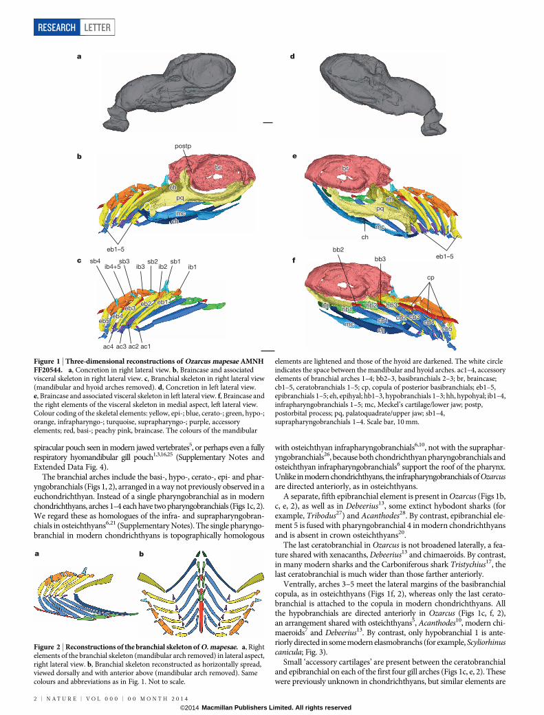
spiracular pouch seen in modern jawed vertebrates5, or perhaps even a fullyrespiratory hyomandibular gill pouch1,3,16,25 (Supplementary Notes andExtended Data Fig. 4).
The branchial arches include the basi-, hypo-, cerato-, epi- and phar-yngobranchials (Figs 1, 2), arranged in a way not previously observed in aeuchondrichthyan. Instead of a single pharyngobranchial as in modernchondrichthyans, arches 1–4 each have two pharyngobranchials (Figs 1c, 2).We regard these as homologues of the infra- and suprapharyngobran-chials in osteichthyans6,21 (Supplementary Notes). The single pharyngo-branchial in modern chondrichthyans is topographically homologous
with osteichthyan infrapharyngobranchials6,10, not with the supraphar-yngobranchials26, because both chondrichthyan pharyngobranchials andosteichthyan infrapharyngobranchials6 support the roof of the pharynx.Unlike in modern chondrichthyans, the infrapharyngobranchials of Ozarcusare directed anteriorly, as in osteichthyans.
A separate, fifth epibranchial element is present in Ozarcus (Figs 1b,c, e, 2), as well as in Debeerius13, some extinct hybodont sharks (forexample, Tribodus27) and Acanthodes28. By contrast, epibranchial ele-ment 5 is fused with pharyngobranchial 4 in modern chondrichthyansand is absent in crown osteichthyans20.
The last ceratobranchial in Ozarcus is not broadened laterally, a fea-ture shared with xenacanths, Debeerius13 and chimaeroids. By contrast,in many modern sharks and the Carboniferous shark Tristychius17, thelast ceratobranchial is much wider than those farther anteriorly.
Ventrally, arches 3–5 meet the lateral margins of the basibranchialcopula, as in osteichthyans (Figs 1f, 2), whereas only the last cerato-branchial is attached to the copula in modern chondrichthyans. Allthe hypobranchials are directed anteriorly in Ozarcus (Figs 1c, f, 2),an arrangement shared with osteichthyans5, Acanthodes10, modern chi-maeroids7 and Debeerius13. By contrast, only hypobranchial 1 is ante-riorly directed in some modern elasmobranchs (for example, Scyliorhinuscanicula; Fig. 3).
Small ‘accessory cartilages’ are present between the ceratobranchialand epibranchial on each of the first four gill arches (Figs 1c, e, 2). Thesewere previously unknown in chondrichthyans, but similar elements are
a
b
c bb3
bb2
cp
ib1ib2ib3sb2
ib4+5sb1sb3sb4
ac1ac2ac3ac4
eb1–5
postp
eb1–5
d
e
f
ch
pqpq
mcmc
eheh
chch
brbr
pq
mc
eh
ch
br brbr
eheh
pqpq
mcmc
br
eh
pq
mc
eb1eb1eb2eb2eb3eb3
eb4eb4eb5eb5
eb1eb2eb3
eb4eb5
hhhh
chchmcmc
hb1hb1hb2hb2 hb3hb3
cb1cb1 cb2cb2 cb3cb3cb4cb4
cb5cb5
hh
chmc
hb1hb2 hb3
cb1 cb2 cb3cb4
cb5
Figure 1 | Three-dimensional reconstructions of Ozarcus mapesae AMNHFF20544. a, Concretion in right lateral view. b, Braincase and associatedvisceral skeleton in right lateral view. c, Branchial skeleton in right lateral view(mandibular and hyoid arches removed). d, Concretion in left lateral view.e, Braincase and associated visceral skeleton in left lateral view. f, Braincase andthe right elements of the visceral skeleton in medial aspect, left lateral view.Colour coding of the skeletal elements: yellow, epi-; blue, cerato-; green, hypo-;orange, infrapharyngo-; turquoise, suprapharyngo-; purple, accessoryelements; red, basi-; peachy pink, braincase. The colours of the mandibular
elements are lightened and those of the hyoid are darkened. The white circleindicates the space between the mandibular and hyoid arches. ac1–4, accessoryelements of branchial arches 1–4; bb2–3, basibranchials 2–3; br, braincase;cb1–5, ceratobranchials 1–5; cp, copula of posterior basibranchials; eb1–5,epibranchials 1–5; eh, epihyal; hb1–3, hypobranchials 1–3; hh, hypohyal; ib1–4,infrapharyngobranchials 1–5; mc, Meckel’s cartilage/lower jaw; postp,postorbital process; pq, palatoquadrate/upper jaw; sb1–4,suprapharyngobranchials 1–4. Scale bar, 10 mm.
ba
Figure 2 | Reconstructions of the branchial skeleton of O. mapesae. a, Rightelements of the branchial skeleton (mandibular arch removed) in lateral aspect,right lateral view. b, Branchial skeleton reconstructed as horizontally spread,viewed dorsally and with anterior above (mandibular arch removed). Samecolours and abbreviations as in Fig. 1. Not to scale.
RESEARCH LETTER
2 | N A T U R E | V O L 0 0 0 | 0 0 M O N T H 2 0 1 4
Macmillan Publishers Limited. All rights reserved©2014
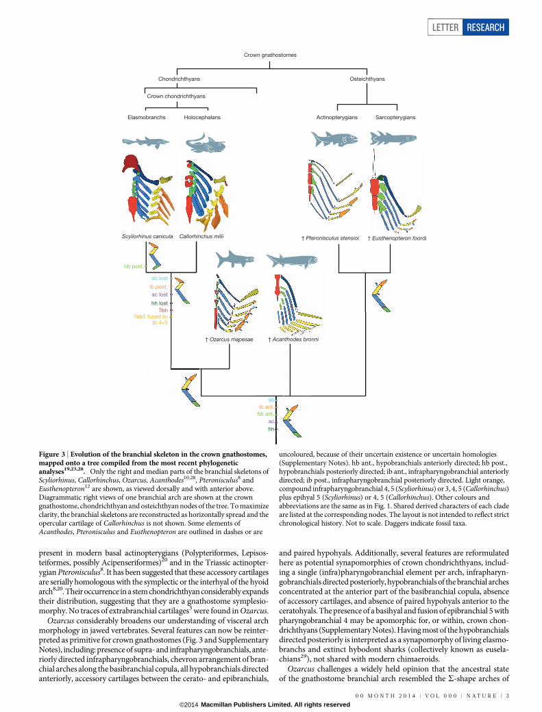
present in modern basal actinopterygians (Polypteriformes, Lepisos-teiformes, possibly Acipenseriformes)20 and in the Triassic actinopter-ygian Pteronisculus8. It has been suggested that these accessory cartilagesare serially homologous with the symplectic or the interhyal of the hyoidarch8,20. Their occurrence in a stem chondrichthyan considerably expandstheir distribution, suggesting that they are a gnathostome symplesio-morphy. No traces of extrabranchial cartilages3 were found in Ozarcus.
Ozarcus considerably broadens our understanding of visceral archmorphology in jawed vertebrates. Several features can now be reinter-preted as primitive for crown gnathostomes (Fig. 3 and SupplementaryNotes), including: presence of supra- and infrapharyngobranchials, ante-riorly directed infrapharyngobranchials, chevron arrangement of bran-chial arches along the basibranchial copula, all hypobranchials directedanteriorly, accessory cartilages between the cerato- and epibranchials,
and paired hypohyals. Additionally, several features are reformulatedhere as potential synapomorphies of crown chondrichthyans, includ-ing a single (infra)pharyngobranchial element per arch, infrapharyn-gobranchials directed posteriorly, hypobranchials of the branchial archesconcentrated at the anterior part of the basibranchial copula, absenceof accessory cartilages, and absence of paired hypohyals anterior to theceratohyals. The presence of a basihyal and fusion of epibranchial 5 withpharyngobranchial 4 may be apomorphic for, or within, crown chon-drichthyans (Supplementary Notes). Having most of the hypobranchialsdirected posteriorly is interpreted as a synapomorphy of living elasmo-branchs and extinct hybodont sharks (collectively known as eusela-chians29), not shared with modern chimaeroids.
Ozarcus challenges a widely held opinion that the ancestral stateof the gnathostome branchial arch resembled the S-shape arches of
Crown gnathostomes
Chondrichthyans
Callorhinchus milii † Pteronisculus stensioi
† Ozarcus mapesae † Acanthodes bronni
Crown chondrichthyans
Elasmobranchs Holocephalans Actinopterygians Sarcopterygians
† Eusthenopteron foordi
sb lost
sb
ib post.
ib ant.
ac lost
ac
hh lost
hh
Scyliorhinus canicula
?eb5 fused to ib 4+5
?bh
hb post.
hb ant.
Osteichthyans
Figure 3 | Evolution of the branchial skeleton in the crown gnathostomes,mapped onto a tree compiled from the most recent phylogeneticanalyses19,23,28. Only the right and median parts of the branchial skeletons ofScyliorhinus, Callorhinchus, Ozarcus, Acanthodes10,28, Pteronisculus8 andEusthenopteron12 are shown, as viewed dorsally and with anterior above.Diagrammatic right views of one branchial arch are shown at the crowngnathostome, chondrichthyan and osteichthyan nodes of the tree. To maximizeclarity, the branchial skeletons are reconstructed as horizontally spread and theopercular cartilage of Callorhinchus is not shown. Some elements ofAcanthodes, Pteronisculus and Eusthenopteron are outlined in dashes or are
uncoloured, because of their uncertain existence or uncertain homologies(Supplementary Notes). hb ant., hypobranchials anteriorly directed; hb post.,hypobranchials posteriorly directed; ib ant., infrapharyngobranchial anteriorlydirected; ib post., infrapharyngobranchial posteriorly directed. Light orange,compound infrapharyngobranchial 4, 5 (Scyliorhinus) or 3, 4, 5 (Callorhinchus)plus epihyal 5 (Scyliorhinus) or 4, 5 (Callorhinchus). Other colours andabbreviations are the same as in Fig. 1. Shared derived characters of each cladeare listed at the corresponding nodes. The layout is not intended to reflect strictchronological history. Not to scale. Daggers indicate fossil taxa.
LETTER RESEARCH
0 0 M O N T H 2 0 1 4 | V O L 0 0 0 | N A T U R E | 3
Macmillan Publishers Limited. All rights reserved©2014
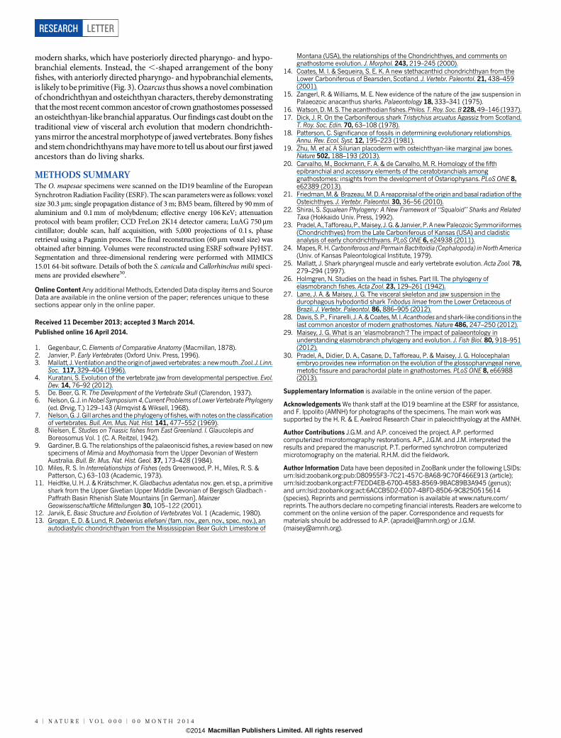
modern sharks, which have posteriorly directed pharyngo- and hypo-branchial elements. Instead, the ,-shaped arrangement of the bonyfishes, with anteriorly directed pharyngo- and hypobranchial elements,is likely to be primitive (Fig. 3). Ozarcus thus shows a novel combinationof chondrichthyan and osteichthyan characters, thereby demonstratingthat the most recent common ancestor of crown gnathostomes possessedan osteichthyan-like branchial apparatus. Our findings cast doubt on thetraditional view of visceral arch evolution that modern chondrichth-yans mirror the ancestral morphotype of jawed vertebrates. Bony fishesand stem chondrichthyans may have more to tell us about our first jawedancestors than do living sharks.
METHODS SUMMARYThe O. mapesae specimens were scanned on the ID19 beamline of the EuropeanSynchrotron Radiation Facility (ESRF). The scan parameters were as follows: voxelsize 30.3mm; single propagation distance of 3 m; BM5 beam, filtered by 90 mm ofaluminium and 0.1 mm of molybdenum; effective energy 106 KeV; attenuationprotocol with beam profiler; CCD FreLon 2K14 detector camera; LuAG 750mmcintillator; double scan, half acquisition, with 5,000 projections of 0.1 s, phaseretrieval using a Paganin process. The final reconstruction (60mm voxel size) wasobtained after binning. Volumes were reconstructed using ESRF software PyHST.Segmentation and three-dimensional rendering were performed with MIMICS15.01 64-bit software. Details of both the S. canicula and Callorhinchus milii speci-mens are provided elsewhere30.
Online Content Any additional Methods, Extended Data display items and SourceData are available in the online version of the paper; references unique to thesesections appear only in the online paper.
Received 11 December 2013; accepted 3 March 2014.
Published online 16 April 2014.
1. Gegenbaur, C. Elements of Comparative Anatomy (Macmillan, 1878).2. Janvier, P. Early Vertebrates (Oxford Univ. Press, 1996).3. Mallatt, J. Ventilationand theorigin of jawed vertebrates: anewmouth.Zool. J. Linn.
Soc. 117, 329–404 (1996).4. Kuratani, S. Evolution of the vertebrate jaw from developmental perspective. Evol.
Dev. 14, 76–92 (2012).5. De. Beer, G. R. The Development of the Vertebrate Skull (Clarendon, 1937).6. Nelson, G. J. inNobel Symposium4, Current Problems of Lower Vertebrate Phylogeny
(ed. Ørvig, T.) 129–143 (Almqvist & Wiksell, 1968).7. Nelson,G. J.Gill arches and the phylogenyof fishes,with notes on the classification
of vertebrates. Bull. Am. Mus. Nat. Hist. 141, 477–552 (1969).8. Nielsen, E. Studies on Triassic fishes from East Greenland. I. Glaucolepis and
Boreosomus Vol. 1 (C. A. Reitzel, 1942).9. Gardiner, B. G. The relationships of the palaeoniscid fishes, a review based on new
specimens of Mimia and Moythomasia from the Upper Devonian of WesternAustralia. Bull. Br. Mus. Nat. Hist. Geol. 37, 173–428 (1984).
10. Miles, R. S. In Interrelationships of Fishes (eds Greenwood, P. H., Miles, R. S. &Patterson, C.) 63–103 (Academic, 1973).
11. Heidtke, U. H. J. & Kratschmer, K. Gladbachus adentatus nov. gen. et sp., a primitiveshark from the Upper Givetian Upper Middle Devonian of Bergisch Gladbach -Paffrath Basin Rhenish Slate Mountains [in German]. MainzerGeowissenschaftliche Mitteilungen 30, 105–122 (2001).
12. Jarvik, E. Basic Structure and Evolution of Vertebrates Vol. 1 (Academic, 1980).13. Grogan, E. D. & Lund, R. Debeerius ellefseni (fam. nov., gen. nov., spec. nov.), an
autodiastylic chondrichthyan from the Mississippian Bear Gulch Limestone of
Montana (USA), the relationships of the Chondrichthyes, and comments ongnathostome evolution. J. Morphol. 243, 219–245 (2000).
14. Coates, M. I. & Sequeira, S. E. K. A new stethacanthid chondrichthyan from theLower Carboniferous of Bearsden, Scotland. J. Vertebr. Paleontol. 21, 438–459(2001).
15. Zangerl, R. & Williams, M. E. New evidence of the nature of the jaw suspension inPalaeozoic anacanthus sharks. Palaeontology 18, 333–341 (1975).
16. Watson, D. M. S. The acanthodian fishes. Philos. T. Roy. Soc. B 228, 49–146 (1937).17. Dick, J. R. On the Carboniferous shark Tristychius arcuatus Agassiz from Scotland.
T. Roy. Soc. Edin. 70, 63–108 (1978).18. Patterson, C. Significance of fossils in determining evolutionary relationships.
Annu. Rev. Ecol. Syst. 12, 195–223 (1981).19. Zhu, M. et al. A Silurian placoderm with osteichthyan-like marginal jaw bones.
Nature 502, 188–193 (2013).20. Carvalho, M., Bockmann, F. A. & de Carvalho, M. R. Homology of the fifth
epibranchial and accessory elements of the ceratobranchials amonggnathostomes: insights from the development of Ostariophysans. PLoS ONE 8,e62389 (2013).
21. Friedman,M.& Brazeau,M.D. A reappraisal of the origin and basal radiation of theOsteichthyes. J. Vertebr. Paleontol. 30, 36–56 (2010).
22. Shirai, S. Squalean Phylogeny: A New Framework of ‘‘Squaloid’’ Sharks and RelatedTaxa (Hokkaido Univ. Press, 1992).
23. Pradel, A., Tafforeau,P., Maisey, J.G.& Janvier, P. Anew PaleozoicSymmoriiformes(Chondrichthyes) from the Late Carboniferous of Kansas (USA) and cladisticanalysis of early chondrichthyans. PLoS ONE 6, e24938 (2011).
24. Mapes, R.H.Carboniferous and PermainBactritoidia (Cephalopoda) inNorthAmerica(Univ. of Kansas Paleontological Institute, 1979).
25. Mallatt, J. Shark pharyngeal muscle and early vertebrate evolution. Acta Zool. 78,279–294 (1997).
26. Holmgren, N. Studies on the head in fishes. Part III. The phylogeny ofelasmobranch fishes. Acta Zool. 23, 129–261 (1942).
27. Lane, J. A. & Maisey, J. G. The visceral skeleton and jaw suspension in thedurophagous hybodontid shark Tribodus limae from the Lower Cretaceous ofBrazil. J. Vertebr. Paleontol. 86, 886–905 (2012).
28. Davis, S. P., Finarelli, J. A.& Coates,M. I.Acanthodesandshark-likeconditions in thelast common ancestor of modern gnathostomes. Nature 486, 247–250 (2012).
29. Maisey, J. G. What is an ‘elasmobranch’? The impact of palaeontology inunderstanding elasmobranch phylogeny and evolution. J. Fish Biol. 80, 918–951(2012).
30. Pradel, A., Didier, D. A., Casane, D., Tafforeau, P. & Maisey, J. G. Holocephalanembryo provides new information on the evolution of the glossopharyngeal nerve,metotic fissure and parachordal plate in gnathostomes. PLoS ONE 8, e66988(2013).
Supplementary Information is available in the online version of the paper.
Acknowledgements We thank staff at the ID19 beamline at the ESRF for assistance,and F. Ippolito (AMNH) for photographs of the specimens. The main work wassupported by the H. R. & E. Axelrod Research Chair in paleoichthyology at the AMNH.
Author Contributions J.G.M. and A.P. conceived the project. A.P. performedcomputerized microtomography restorations. A.P., J.G.M. and J.M. interpreted theresults and prepared the manuscript. P.T. performed synchrotron computerizedmicrotomography on the material. R.H.M. did the fieldwork.
Author Information Data have been deposited in ZooBank under the following LSIDs:urn:lsid:zoobank.org:pub:D80955F3-7C21-457C-BA68-9C70F466E913 (article);urn:lsid:zoobank.org:act:F7EDD4EB-6700-4583-8569-9BAC89B3A945 (genus);and urn:lsid:zoobank.org:act:6ACCB5D2-E0D7-4BFD-85D6-9C8250515614(species). Reprints and permissions information is available at www.nature.com/reprints. The authors declare no competing financial interests. Readers are welcome tocomment on the online version of the paper. Correspondence and requests formaterials should be addressed to A.P. ([email protected]) or J.G.M.([email protected]).
RESEARCH LETTER
4 | N A T U R E | V O L 0 0 0 | 0 0 M O N T H 2 0 1 4
Macmillan Publishers Limited. All rights reserved©2014
METHODSLocality. The specimen was collected from the ARC-07 locality24 (section 22, T. 14N., R. 15 W.), from black dysoxic/anoxic shale of the Fayetteville Formation(Carboniferous, upper Mississippian, Chesterian, Pendleian stage, E1 zone 5 lowerNamurian A), near Leslie, Searcy County, Arkansas. Other fossils from the samelocality include invertebrates preserved by calcite, pyrite and phosphate, plants pre-served as permineralizations, coprolites, and vertebrates (mostly chondrichthyans).The chondrichthyan remains are mostly preserved in a hard phosphatic matrix thatis difficult to prepare by conventional means.Synchrotron. The Ozarcus specimens were scanned on the ID19 beamline of theEuropean Synchrotron Radiation Facility (ESRF), supported by glass balls contained
in a cylinder; voxel size 30.3mm, with a single propagation distance of 3 m; BM5 beam,filteredby90 mmofaluminiumand0.1 mmofmolybdenum;effectiveenergy106 KeV.We used an attenuation protocol with beam profiler. The detector camera was a CCDFreLon 2K14, and the scintillator a LuAG 750mm. We performed a double scan, halfacquisition, with 5,000 projections of 0.1 s, with phase retrieval using a Paganin process.The final reconstruction (60mm voxel size) was obtained after binning. Volumes werereconstructed using ESRF software PyHST. Subvolumes were corrected for ring arte-facts and concatenated to generate a single stack of 16-bit tif slices. Segmentation andthree-dimensional rendering were performed with MIMICS 15.01 64-bit software(Materialise). Details about the origin, preparation and scanning of both the S. caniculaand Callorhinchus milii specimens shown in Fig. 3 are provided elsewhere30.
LETTER RESEARCH
Macmillan Publishers Limited. All rights reserved©2014
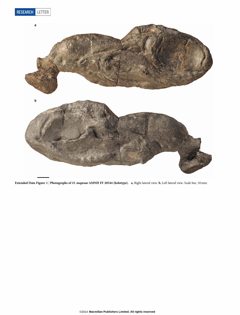
Extended Data Figure 1 | Photographs of O. mapesae AMNH FF 20544 (holotype). a, Right lateral view. b, Left lateral view. Scale bar, 10 mm.
RESEARCH LETTER
Macmillan Publishers Limited. All rights reserved©2014
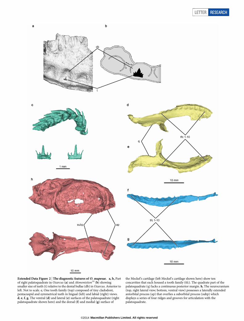
Extended Data Figure 2 | The diagnostic features of O. mapesae. a, b, Partof right palatoquadrate in Ozarcus (a) and Akmonistion14 (b) showingsmaller size of teeth (t) relative to the dental bullae (db) in Ozarcus. Anterior toleft. Not to scale. c, One tooth family (top) composed of tiny cladodont,pentacuspid and symmetrical teeth in lingual (left) and labial (right) views.d, e, f, g, The ventral (d) and lateral (e) surfaces of the palatoquadrate (rightpalatoquadrate shown here) and the dorsal (f) and medial (g) surface of
the Meckel’s cartilage (left Meckel’s cartilage shown here) show tenconcavities that each housed a tooth family (tfc). The quadrate part of thepalatoquadrate (q) lacks a continuous posterior margin. h, The neurocranium(top, right lateral view; bottom, ventral view) possesses a laterally extendedantorbital process (ap) that overlies a suborbital process (subp) whichdisplays a series of four ridges and grooves for articulation with thepalatoquadrate.
LETTER RESEARCH
Macmillan Publishers Limited. All rights reserved©2014
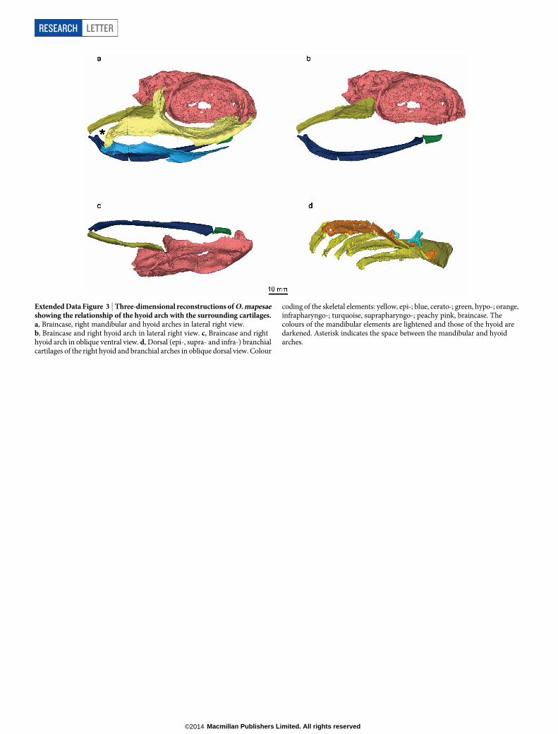
Extended Data Figure 3 | Three-dimensional reconstructions of O. mapesaeshowing the relationship of the hyoid arch with the surrounding cartilages.a, Braincase, right mandibular and hyoid arches in lateral right view.b, Braincase and right hyoid arch in lateral right view. c, Braincase and righthyoid arch in oblique ventral view. d, Dorsal (epi-, supra- and infra-) branchialcartilages of the right hyoid and branchial arches in oblique dorsal view. Colour
coding of the skeletal elements: yellow, epi-; blue, cerato-; green, hypo-; orange,infrapharyngo-; turquoise, suprapharyngo-; peachy pink, braincase. Thecolours of the mandibular elements are lightened and those of the hyoid aredarkened. Asterisk indicates the space between the mandibular and hyoidarches.
RESEARCH LETTER
Macmillan Publishers Limited. All rights reserved©2014
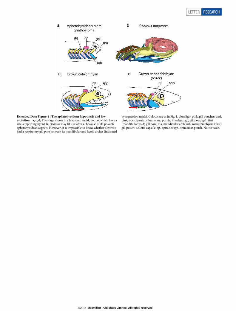
Extended Data Figure 4 | The aphetohyoidean hypothesis and jawevolution. a, c, d, The stage shown in a leads to c and d, both of which have ajaw-supporting hyoid. b, Ozarcus may fit just after a, because of its possibleaphetohyoidean aspects. However, it is impossible to know whether Ozarcushad a respiratory gill pore between its mandibular and hyoid arches (indicated
by a question mark). Colours are as in Fig. 1, plus: light pink, gill pouches; darkpink, otic capsule of braincase; purple, interhyal. gp, gill pore; gp1, first(mandibulohyoid) gill pore; ma, mandibular arch; mh, mandibulohyoid (first)gill pouch; oc, otic capsule; sp., spiracle; spp., spiracular pouch. Not to scale.
LETTER RESEARCH
Macmillan Publishers Limited. All rights reserved©2014
Related Documents