[CANCER RESEARCH 58. 3700-3705. August 15. 1998] A Novel Tumor Suppressor Locus on Chromosome 18q Involved in the Development of Human Lung Cancer1 Kimiko Takei, Takashi Kohno, Kunihiro llamada, Junko Takita, Masayuki Noguchi, Yoshihiro Matsuno, Setsuo Hirohashi, Hiroshi Uezato, and Jun Yokota2 Divisions of Biology ¡K.T., T. K., K. H., J. T.. J. Y.I and Pathology ¡M.N., Y. M., S. HJ. National Cancer Center Research Institute. Tokyo 104. and Department of Dermatology. School of Medicine. University of the Ryukyus. ¡K.T.. H. U.I. Okinawa 903-01 Japan ABSTRACT The high incidence of loss of heterozygosity (LOH) on chromosome 18q in advanced non-small cell lung carcinomas indicates the presence of tumor suppressor gene(s) on this chromosome arm, which plays an im portant role in the acquisition of malignant phenotypes in lung cancers. In the present study, we examined 62 lung cancer specimens and 54 lung cancer cell lines for alleile imbalance at 11 microsatellite loci to define common regions of 18q deletions. Allelic imbalance of 18q was detected in 24 (55.8%) non-small cell lung carcinoma specimens and in 6 (31.6%) small cell lung carcinoma specimens, whereas a similar frequency of LOH was statistically inferred to occur in cell lines by analyzing marker ho- mozygosity as an indirect measure of LOH. Five specimens and 11 cell lines showed partial or interstitial deletions of chromosome 18q, and 2 of them had homozygous deletions at the 18q21.1 region. A commonly deleted region was assigned between the DI8S46 and y953G12R loci. The size of this region is less than 1 Mb, and the coding exons of three candidate tumor suppressor genes, Smad2, Smad4, and DCC, were mapped outside the region. This result suggests that the common region harbors a novel tumor suppressor gene involved in the progression of lung INTRODUCTION Lung cancer is a major cause of cancer-related death in the world, and the overall survival rate has not improved significantly in the last 20 years. The understanding of the molecular pathogenesis of this disease should provide new and more sensitive means to diagnose and treat lung cancer patients. Genetic as well as biological studies of SCLC3 and NSCLC have indicated that multiple tumor suppressor genes, including p53, pió, RB. and PTEN, are involved in human lung carcinogenesis (1-3). We previously reported that the incidence of LOH on chromosome 18q in advanced-stage NSCLC was signifi cantly higher than that in early-stage NSCLC (4, 5). Frequent occur rence of 18q deletions in NSCLC was also shown by cytogenetic and comparative genomic hybridization analyses (6. 7). LOH on 18q was also observed in 30% of SCLCs (8). These results indicate that the tumor suppressor gene(s) on chromosome 18q plays an important role in the acquisition of malignant phenotypes in lung cancers. To date, three candidate tumor suppressor genes, Smad2, Smad4, and DCC, have been identified on chromosome 18q (9-12). The Smad2 and Smad4 genes share similarity to the Drosophila melano- gaster gene, MAD, which is known to reside in a pathway of trans forming growth factor ßsignaling. Smad4 alterations were observed Received 2/16/98: accepted 6/17/98. The costs of publication of this article were defrayed in part by the payment of page charges. This article must therefore be hereby marked aclverlisemenl in accordance with 18 U.S.C. Section 1734 solely to indicate this fact. ' Supported in part by a Grant-in-Aid from Ine Ministry of Health and Welfare for the 2nd-term Comprehensive 10-Year Strategy for Cancer Control and by Grants-in-Aid from the Ministry of Health and Welfare, the Ministry of Education. Science. Snorts and Culture of Japan, and the Naito Foundation. K. T. is a recipient of a Research Resident Fellowship from the Foundation for Promotion of Cancer Research. 2 To whom requests for reprints should be addressed, at Biology Division, National Cancer Center Research Institute. 1-1. Tsukiji 5-chome. Chuo-ku, Tokyo 104. Japan. Phone: 81 -3-3542-2511. extension 4650; Fax: 81 -3-3542-0807. ' The abbreviations used are: SCLC. small cell lung cancer; NSCLC. non-small cell lung cancer; LOH, loss of heterozygosity; Al, alleile imbalance. in a significant portion of pancreatic cancers and in a subset of other human cancers (11, 13), whereas Smad2 alterations were detected in some cases of colorectal cancers (12, 14). Genetic alterations of these genes were detected only in a limited fraction of lung cancers ( 14, 15). Thus, it is possible that genes other than the Smad2 and Smad4 genes function as tumor suppressors involved in lung cancer progression. The DCC gene was identified as a gene deleted in colorectal cancers. The gene encodes a transmembrane protein with four immunoglobulin and six fibronectin type III repeats in the extracellular domains that may function as a receptor for the axonal chemoattractant netrin-1 (16). However, to our knowledge, mutations of the DCC gene have not been reported in human lung cancer. Therefore, a target tumor suppressor gene on chromosome 18q, which is involved in human lung carcinogenesis, has not been defined. In the present study, we examined the region deleted hemizygously or homozygously in 62 lung cancer specimens and 54 lung cancer cell lines to define tumor suppressor loci on chromosome 18q. Sixteen cases showed partial or interstitial deletions on chromosome 18q; notably, two cases showed homozygous deletions at the 18q21.1 region. The size of a common region of 18q deletions was less than 1 Mb, and the region did not include the Smad2. Smad4, and DCC genes. This result suggested that a novel tumor suppressor gene is present in this region. MATERIALS AND METHODS Samples. Sixty-two pairs of tumors and adjacent noncancerous tissues were obtained from 43 patients with NSCLC and 19 patients with SCLC who were treated at the National Cancer Center Hospital (Tokyo, Japan). Forty- three NSCLCs. consisting of 29 primary tumors and 14 brain métastases,were histologically classified as 29 adenocarcinomas, 8 squamous cell carcinomas, 5 large cell carcinomas, and 1 adenosquamous carcinoma. Nineteen SCLCs contained 9 primary tumors and 10 métastases.The material to be analyzed was selected by a pathologist to ensure that the samples were macroscopically entirely tumorous and were chosen from an area devoid of necrotic tissue. Tumors and normal tissues were frozen and stored at -70°C until DNA extraction. Fifty-four lung cancer cell lines (34 NSCLCs and 20 SCLCs) were used in this study. NSCLC cell lines were A427, A549. PC-1, PC-3, PC-7, PC-9, PC-10. PC-13, PC-14, Lu65, Lu99, LCl-Sq, NCI-H23, NCI-H157, NCI-H322, NC1-H441, NCI-H520, NCI-H596, NCI-HI 155, Mai, Ma2, Ma3, MalO, Ma 12, Ma 17, Ma24, Ma25, Ma26, Ma29, VMRC-LCD, RERF-LCOK, ABC-1, EBC-1, and LCMS. The Ma29 cell line is derived from an adenocar- cinoma (stage Illb, T2N,M0) of the lung of a 55-year-old Japanese male who was admitted to the Osaka Prefectural Habikino Hospital (Osaka. Japan). SCLC cell lines were Lu24, Lul30, Lul34, Lul35, Lul38, Lul39, Lul40, Lul41, NCI-H69, NCI-H82, NCI-H209, NCI-H526, NCI-H774. NCI-H841, N230, N231. N417, LCMA, SBC-5. and MS18. Detailed information on these cell lines can be obtained upon request. High molecular weight DNA was prepared from the tumors and normal tissues by proteinase K digestion and phenol-chloroform extraction as described previously (4). Deletion Mapping of Chromosome 18q in Surgical Specimens. Analysis of AI was performed using a PCR-based approach using 11 microsatellite markers listed in Table 1. A microsatellite marker located 165 bp downstream from exon 7 of the DCC gene was used for the analysis of the DCC locus (17, 18). Detailed information on this marker can be obtained from the Genome Data Base (http://gdbwww.gdb.org/gdb/). Chromosomal locations and order- 3700 Research. on August 16, 2019. © 1998 American Association for Cancer cancerres.aacrjournals.org Downloaded from

Welcome message from author
This document is posted to help you gain knowledge. Please leave a comment to let me know what you think about it! Share it to your friends and learn new things together.
Transcript

[CANCER RESEARCH 58. 3700-3705. August 15. 1998]
A Novel Tumor Suppressor Locus on Chromosome 18q Involved in theDevelopment of Human Lung Cancer1
Kimiko Takei, Takashi Kohno, Kunihiro llamada, Junko Takita, Masayuki Noguchi, Yoshihiro Matsuno,Setsuo Hirohashi, Hiroshi Uezato, and Jun Yokota2
Divisions of Biology ¡K.T., T. K., K. H., J. T.. J. Y.I and Pathology ¡M.N., Y. M., S. HJ. National Cancer Center Research Institute. Tokyo 104. and Department of Dermatology.School of Medicine. University of the Ryukyus. ¡K.T.. H. U.I. Okinawa 903-01 Japan
ABSTRACT
The high incidence of loss of heterozygosity (LOH) on chromosome 18qin advanced non-small cell lung carcinomas indicates the presence of
tumor suppressor gene(s) on this chromosome arm, which plays an important role in the acquisition of malignant phenotypes in lung cancers. Inthe present study, we examined 62 lung cancer specimens and 54 lungcancer cell lines for alleile imbalance at 11 microsatellite loci to definecommon regions of 18q deletions. Allelic imbalance of 18q was detected in24 (55.8%) non-small cell lung carcinoma specimens and in 6 (31.6%)
small cell lung carcinoma specimens, whereas a similar frequency of LOHwas statistically inferred to occur in cell lines by analyzing marker ho-
mozygosity as an indirect measure of LOH. Five specimens and 11 celllines showed partial or interstitial deletions of chromosome 18q, and 2 ofthem had homozygous deletions at the 18q21.1 region. A commonlydeleted region was assigned between the DI8S46 and y953G12R loci. Thesize of this region is less than 1 Mb, and the coding exons of threecandidate tumor suppressor genes, Smad2, Smad4, and DCC, weremapped outside the region. This result suggests that the common regionharbors a novel tumor suppressor gene involved in the progression of lung
INTRODUCTION
Lung cancer is a major cause of cancer-related death in the world,
and the overall survival rate has not improved significantly in the last20 years. The understanding of the molecular pathogenesis of thisdisease should provide new and more sensitive means to diagnose andtreat lung cancer patients. Genetic as well as biological studies ofSCLC3 and NSCLC have indicated that multiple tumor suppressor
genes, including p53, pió, RB. and PTEN, are involved in human lungcarcinogenesis (1-3). We previously reported that the incidence ofLOH on chromosome 18q in advanced-stage NSCLC was significantly higher than that in early-stage NSCLC (4, 5). Frequent occur
rence of 18q deletions in NSCLC was also shown by cytogenetic andcomparative genomic hybridization analyses (6. 7). LOH on 18q wasalso observed in 30% of SCLCs (8). These results indicate that thetumor suppressor gene(s) on chromosome 18q plays an important rolein the acquisition of malignant phenotypes in lung cancers.
To date, three candidate tumor suppressor genes, Smad2, Smad4,and DCC, have been identified on chromosome 18q (9-12). TheSmad2 and Smad4 genes share similarity to the Drosophila melano-
gaster gene, MAD, which is known to reside in a pathway of transforming growth factor ßsignaling. Smad4 alterations were observed
Received 2/16/98: accepted 6/17/98.The costs of publication of this article were defrayed in part by the payment of page
charges. This article must therefore be hereby marked aclverlisemenl in accordance with18 U.S.C. Section 1734 solely to indicate this fact.
' Supported in part by a Grant-in-Aid from Ine Ministry of Health and Welfare for the
2nd-term Comprehensive 10-Year Strategy for Cancer Control and by Grants-in-Aid from
the Ministry of Health and Welfare, the Ministry of Education. Science. Snorts andCulture of Japan, and the Naito Foundation. K. T. is a recipient of a Research ResidentFellowship from the Foundation for Promotion of Cancer Research.
2 To whom requests for reprints should be addressed, at Biology Division, NationalCancer Center Research Institute. 1-1. Tsukiji 5-chome. Chuo-ku, Tokyo 104. Japan.Phone: 81-3-3542-2511. extension 4650; Fax: 81-3-3542-0807.
' The abbreviations used are: SCLC. small cell lung cancer; NSCLC. non-small cell
lung cancer; LOH, loss of heterozygosity; Al, alleile imbalance.
in a significant portion of pancreatic cancers and in a subset of otherhuman cancers (11, 13), whereas Smad2 alterations were detected insome cases of colorectal cancers (12, 14). Genetic alterations of thesegenes were detected only in a limited fraction of lung cancers ( 14, 15).Thus, it is possible that genes other than the Smad2 and Smad4 genesfunction as tumor suppressors involved in lung cancer progression.The DCC gene was identified as a gene deleted in colorectal cancers.The gene encodes a transmembrane protein with four immunoglobulinand six fibronectin type III repeats in the extracellular domains thatmay function as a receptor for the axonal chemoattractant netrin-1
(16). However, to our knowledge, mutations of the DCC gene havenot been reported in human lung cancer. Therefore, a target tumorsuppressor gene on chromosome 18q, which is involved in humanlung carcinogenesis, has not been defined.
In the present study, we examined the region deleted hemizygouslyor homozygously in 62 lung cancer specimens and 54 lung cancer celllines to define tumor suppressor loci on chromosome 18q. Sixteencases showed partial or interstitial deletions on chromosome 18q;notably, two cases showed homozygous deletions at the 18q21.1region. The size of a common region of 18q deletions was less than 1Mb, and the region did not include the Smad2. Smad4, and DCCgenes. This result suggested that a novel tumor suppressor gene ispresent in this region.
MATERIALS AND METHODS
Samples. Sixty-two pairs of tumors and adjacent noncancerous tissues
were obtained from 43 patients with NSCLC and 19 patients with SCLC whowere treated at the National Cancer Center Hospital (Tokyo, Japan). Forty-
three NSCLCs. consisting of 29 primary tumors and 14 brain métastases,werehistologically classified as 29 adenocarcinomas, 8 squamous cell carcinomas,5 large cell carcinomas, and 1 adenosquamous carcinoma. Nineteen SCLCscontained 9 primary tumors and 10 métastases.The material to be analyzedwas selected by a pathologist to ensure that the samples were macroscopicallyentirely tumorous and were chosen from an area devoid of necrotic tissue.Tumors and normal tissues were frozen and stored at -70°C until DNA
extraction. Fifty-four lung cancer cell lines (34 NSCLCs and 20 SCLCs) wereused in this study. NSCLC cell lines were A427, A549. PC-1, PC-3, PC-7,PC-9, PC-10. PC-13, PC-14, Lu65, Lu99, LCl-Sq, NCI-H23, NCI-H157,NCI-H322, NC1-H441, NCI-H520, NCI-H596, NCI-HI 155, Mai, Ma2, Ma3,
MalO, Ma 12, Ma 17, Ma24, Ma25, Ma26, Ma29, VMRC-LCD, RERF-LCOK,ABC-1, EBC-1, and LCMS. The Ma29 cell line is derived from an adenocar-cinoma (stage Illb, T2N,M0) of the lung of a 55-year-old Japanese male who
was admitted to the Osaka Prefectural Habikino Hospital (Osaka. Japan).SCLC cell lines were Lu24, Lul30, Lul34, Lul35, Lul38, Lul39, Lul40,Lul41, NCI-H69, NCI-H82, NCI-H209, NCI-H526, NCI-H774. NCI-H841,N230, N231. N417, LCMA, SBC-5. and MS18. Detailed information on these
cell lines can be obtained upon request. High molecular weight DNA wasprepared from the tumors and normal tissues by proteinase K digestion andphenol-chloroform extraction as described previously (4).
Deletion Mapping of Chromosome 18q in Surgical Specimens. Analysisof AI was performed using a PCR-based approach using 11 microsatellite
markers listed in Table 1. A microsatellite marker located 165 bp downstreamfrom exon 7 of the DCC gene was used for the analysis of the DCC locus (17,18). Detailed information on this marker can be obtained from the GenomeData Base (http://gdbwww.gdb.org/gdb/). Chromosomal locations and order-
3700
Research. on August 16, 2019. © 1998 American Association for Cancercancerres.aacrjournals.org Downloaded from
A TUMOR SUPPRESSOR LOCUS ON CHROMOSOME 18q
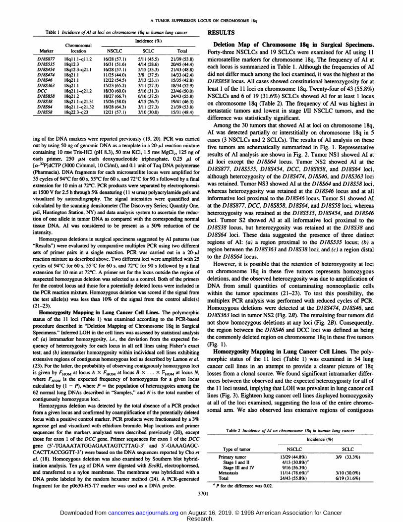
Table 1 Incidence of AI at loci on chromosome 18i/ in human lung cancer
MarkerDI8S877DI8S535D18S454ÜI8S474DI8S46DI8SJ63DCCDI8S858DI8S38D18S64DI8S58ChromosomallocationlSqll.l-qll.218ql2.3I8ql2.3-q21.1I8q21.118q21.1I8Ó21.II8q21.1-q2l.2I8q21.218q21.1-q21.3118q21.1-q21.3218q22.3-q23NSCLC16/28(57.1)16/31
(51.6)16/28(57.1)1
1/25(44.0)12/22(54.5)15/23(65.2)18/30(60.0)18/27(66.7)15/26(58.0)18/28(64.3)12/21
(57.1)Incidence
(%)SCLC5/1
1(45.5)4/14(28.6)5/15
(33.3)3/8(37.5)3/13(23.1)3/11(27.3)5/16(31.3)6/16(37.5)4/15(26.7)3/1
1(27.3)3/10(30.0)Total21/39(53.8)20/45
(44.4)21/43(48.8)14/33
(42.4)15/35(42.8)18/34(52.9)23/46
(50.0)24/43(55.8)19/41(46.3)21/39(53.8)15/31
(48.4)
ing of the DNA markers were reported previously (19, 20). PCR was carriedout by using 50 ng of genomic DNA as a template in a 2()-ju,lreaction mixturecontaining 10 mM Tris-HCl (pH 8.3), 50 mM KCI. 1.5 mM MgCl,, 125 ng of
each primer. 250 ¡IMeach deoxynucleotide triphosphate. 0.25 ¿il of[a-32P]dCTP (3000 Ci/mmol, 10 Ci/mi), and 0.1 unit of Taq DNA polymerase
(Pharmacia). DNA fragments for each microsatellite locus were amplified for35 cycles of 94°Cfor 60 s, 55°Cfor 60 s. and 72°Cfor 90 s followed by a finalextension for 10 min at 72°C.PCR products were separated by electrophoresis
at 15(X)V for 2.5 h through 5% denaturing ( 11 M urea) polyacrylamide gels andvisuali/.ed by autoradiography. The signal intensities were quantified andcalculated by the scanning densitometer (The Discovery Series; Quantity One.ptli. Huntington Station. NY) and data analysis system to ascertain the reduction of one alÃelein tumor DNA as compared with the corresponding normaltissue DNA. AI was considered to be present as a 50%- reduction of the
intensity.Homozygous deletions in surgical specimens suggested by AI patterns (see
"Results") were evaluated by comparative multiplex PCR using two different
sets of primer pairs in a single reaction. PCR was carried out in a 20-jul
reaction mixture as described above. Two different loci were amplified with 25cycles of 94°Cfor 60 s, 55°Cfor 60 s. and 72°Cfor 90 s followed by a finalextension for 10 min at 72°C.A primer set for the locus outside the region of
suspected homozygous deletion was selected as a control. Both of the primersfor the control locus and those for a potentially deleted locus were included inthe PCR reaction mixture. Homozygous deletion was scored if the signal fromthe test allele(s) was less than 10% of the signal from the control alÃele!s)(21-23).
Homozygosity Mapping in Lung Cancer Cell Lines. The polymorphicstatus of the 11 loci (Table 1) was examined according to the PCR-basedprocedure described in "Deletion Mapping of Chromosome 18q in SurgicalSpecimens." Inferred LOH in the cell lines was assessed by statistical analysis
of: (a) intramarker homozygosity, i.e., the deviation from the expected frequency of heterozygosity for each locus in all cell lines using Fisher's exact
test; and (h) intermarker homozygosity within individual cell lines exhibitingextensive regions of contiguous homozygous loci as described by Larson et al.(23). For the latter, the probability of observing contiguously homozygous lociis given by FHOM at locus A X FHOM at locus B X ... X FHOMat locus N,where FHOM is the expected frequency of homozygotes for a given locuscalculated by (1 —¿�P), where P = the population of hétérozygotesamong the62 normal lung DNAs described in "Samples." and N is the total number of
contiguously homozygous loci.Homozygous deletion was detected by the total absence of a PCR product
from a given locus and confirmed by coamplification of the potentially deletedlocus with a positive control marker. PCR products were fractionated by a 3%agarose gel and visualized with ethidium bromide. Map locations and primersequences for the markers analyzed were described previously (20), exceptthose for exon 1 of the DCC gene. Primer sequences for exon 1 of the DCCgene (5'-TGAAATATGGAGAATAGTCTTAG-3' and 5'-GAAAGAGC-CACTTACCGGTT-3') were based on the DNA sequences reported by Cho et
al. (18). Homozygous deletion was also examined by Southern blot hybridization analysis. Ten fj.g of DNA were digested with EcoRl. electrophoresed.and transferred to a nylon membrane. The membrane was hybridized with aDNA probe labeled by the random hexamer method (24). A PCR-generatedfragment for the p0630-H5-T7 marker was used as a DNA probe.
RESULTS
Deletion Map of Chromosome 18q in Surgical Specimens.Forty-three NSCLCs and 19 SCLCs were examined for AI using 11
microsatellite markers for chromosome 18q. The frequency of AI ateach locus is summarized in Table 1. Although the frequencies of AIdid not differ much among the loci examined, it was the highest at theD18S858 locus. All cases showed constitutional heterozygosity for atleast 1 of the 11 loci on chromosome 18q. Twenty-four of 43 (55.8%)
NSCLCs and 6 of 19 (31.6%) SCLCs showed AI for at least 1 locuson chromosome 18q (Table 2). The frequency of AI was highest inmetastatic tumors and lowest in stage I/II NSCLC tumors, and thedifference was statistically significant.
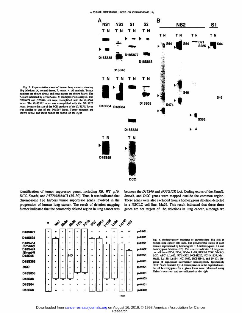
Among the 30 tumors that showed AI at loci on chromosome 18q,AI was detected partially or interstitially on chromosome 18q in 5cases (3 NSCLCs and 2 SCLCs). The results of AI analysis on thesefive tumors are schematically summarized in Fig. 1. Representativeresults of AI analysis are shown in Fig. 2. Tumor NS1 showed AI atall loci except the DI8S64 locus. Tumor NS2 showed AI at theD18S877, D18S535. DÌ8S454,DCC, DÌ8S858,and D18S64 loci,although heterozygosity of the D18S474, D18S46, and D18S363 lociwas retained. Tumor NS3 showed AI at the D18S64 and DI8S58 loci,whereas heterozygosity was retained at the D18S46 locus and at allinformative loci proximal to the DÌ8S46locus. Tumor SI showed AIat the D18S877, DCC, D18S858, D18S64, and D18S58 loci, whereasheterozygosity was retained at the D18S535. D18S454, and D18S46loci. Tumor S2 showed AI at all informative loci proximal to theD18S38 locus, but heterozygosity was retained at the DJ8S38 andD18S64 loci. These data suggested the presence of three distinctregions of AI: (a) a region proximal to the D18S535 locus; (b) aregion between the DI8S363 and DI8S38 loci: and (c) a region distalto the D18S64 locus.
However, it is possible that the retention of heterozygosity at locion chromosome 18q in these five tumors represents homozygousdeletions, and the observed heterozygosity was due to amplification ofDNA from small quantities of contaminating nonneoplastic cellswithin the tumor specimens (21-23). To test this possibility, the
multiplex PCR analysis was performed with reduced cycles of PCR.Homozygous deletions were detected at the D18S474. D18S46, andD18S363 loci in tumor NS2 (Fig. 2B). The remaining four tumors didnot show homozygous deletions at any loci (Fig. 2B). Consequently,the region between the D18S46 and DCC loci was defined as beingthe commonly deleted region on chromosome 18q in these five tumors(Fig. 1).
Homozygosity Mapping in Lung Cancer Cell Lines. The polymorphic status of the 11 loci (Table 1) was examined in 54 lungcancer cell lines in an attempt to provide a clearer picture of 18qlosses from a clonal source. We found significant intramarker differences between the observed and the expected heterozygosity for all ofthe 11 loci tested, implying that LOH was prevalent in lung cancer celllines (Fig. 3). Eighteen lung cancer cell lines displayed homozygosityat all of the loci examined, suggesting the loss of the entire chromosomal arm. We also observed less extensive regions of contiguous
Table 2 Incidence of AI on chromosome 18q in human lung cancer
Type oftumorPrimary
tumorStage 1 and 11Stage 111and IV
MetastasisTotalIncidence
(9NSCLC13/29(44.8%)
4/13(30.8%)"
9/16(56.3%)11/14(78.6%)"
24/43 (55.8%)Õ)SCLC3/9
(33.3%)3/10(30.0%)
6/19(31.6%)"P for the difference was 0.02.
3701
Research. on August 16, 2019. © 1998 American Association for Cancercancerres.aacrjournals.org Downloaded from

A TUMOR SUPPRESSOR LOCUS ON CHROMOSOME I8q
NSCLC SCLC
Fig. I. Deletion map of I8q in surgical specimens. Tumornumbers arc shown above, and markers are shown on the left.NS1, adenocarcinoma (stage III): NS2, adenocarcinoma (stageIV); NSJ. adenocarcinoma (brain metastasis); SI. SCLC (stageI); 52, SCLC (hilar lymph node metastasis). •¿�,AI; O. retentionof both alÃeles;—¿�,not informative. Regions of AI are indicated
by D. and a region of homo/ygous deletion is indicated by athick-bordered D. The commonly deleted region is shaded.
Asterisks indicate the loci showing homozygous deletions.
22
23
NS1 NS2 NS3 S1 S2
D18S877D18S535D18S454
(Smad2)D18S474(Smad4)D18S46D18S363DCCD18S858D18S38
D18S64
D18S58•••
•¿�•••—0•
o*o*o*
oooo
oo
homozygosity in 11 other cell lines, implying more restricted regionsof LOH. although the remaining 25 cell lines did not show significantcontiguous intermarker homozygosity. Statistical analyses of homozygosity patterns among the cell lines are presented in Table 3.Overall, the frequency of inferred LOH in the cell lines [21 of 34NSCLCs (61.8%) and 8 of 20 SCLCs (40.0%)] was similar to that ofAI in surgical specimens. A common region of inferred LOH wasassigned to the region between the D18S474 and DI8S858 loci (Fig.3), which overlapped with the common region of 18q deletions insurgical specimens.
Notably, a homozygous deletion was found at the DI8S46 locus inthe Ma29 cell line. The region of homozygous deletion in the Ma29cell line overlapped with that in the NS2 tumor; however, the size ofhomozygous deletion in Ma29 was much smaller than that in NS2,because the DI8S474 and DI8S363 loci deleted in NS2 were retainedin Ma29. To estimate the size of the homozygous deletion in the Ma29cell line in detail, we performed genomic PCR analysis using eightDNA markers that had been mapped in a YAC contig encompassingthe D18S46 locus (20). The region of the homozygous deletion wasmapped between the c9!7-46T3 and \953C12R markers, and the
homozygous deletion was confirmed by Southern blot hybridizationanalysis (Fig. 4). This region was mapped in the common region ofinferred LOH in lung cancer cell lines and overlapped with thecommonly deleted region in surgical specimens. Therefore, the regionbetween the D18S46 and \953G12R loci was defined as a commonregion of chromosome 18q deletions in human lung cancers.
Relative Location of the Smad4 and DCC Genes and Homozygous Deletions in Lung Tumors. The Smad4 gene was mappedbetween the c9J7-46T3 and DÌ8S474loci, and the orientation of thisgene is 18qter-5'-5m«i/4-3'-18cen (Ref. 20; see Fig. 4). Therefore,
coding exons of the Smad4 gene were mapped outside the deletion inthe Ma29 cell line. However, it is likely that the Smad4 gene wasincluded in the homozygous deletion in the NS2 tumor, because theproximal border of the deletion was mapped proximal to the D18S474locus (Fig. 1). The DCC gene was mapped distal to the DJ8S363locus, and the orientation of this gene is 18cen-5'-DCC-3'-18qter (20,
39). A microsatellite locus located in intron 7 of the DCC gene was
not homozygously deleted in the Ma29 cell line and the NS2 tumors(Figs. 1 and 3). However, it was still unclear whether the 5' end of the
DCC gene was homozygously deleted or retained in these tumors,because it had not yet been localized in the physical map of the18q21.1 region (20). Therefore, we also examined the status of exon1 of the DCC gene in these tumors to confirm that the 5' end of the
DCC gene was retained. DCC exon 1 was amplified from the Ma29cell line DNA by PCR (Fig. 4). The multiplex PCR analysis revealedthat DCC exon 1 was hemizygously but not homozygously deleted inthe NS2 tumor (data not shown). Therefore, it is most likely that all ofthe coding exons of the DCC gene were retained and located distal tothe region of the homozygous deletions in these tumors.
DISCUSSION
Tumor suppressor genes have been considered to be present in theregions of chromosomal deletions in human tumors. Because chromosome 18q was frequently deleted in advanced lung cancer in ourprevious studies (4, 5), it has been suggested that tumor suppressorgenes on chromosome 18q are involved in the genesis and progressionof lung cancer. However, we examined for LOH at only a single locus,DCC, in those studies. Therefore, in the present study, we examinedfor AI of 11 microsatellite loci on 18q in 62 lung cancer specimens.The result was consistent with our previous findings; AI was detectedin 48.4% (30 of 62) of lung cancers, and the frequency of AI inadvanced stages was significantly higher than that seen in early stagesin NSCLCs. Therefore, we further evaluated the status of LOH on 18qin 54 lung cancer cell lines statistically by analyzing marker homozygosity as an indirect measure of LOH. The frequency of inferred LOHin the cell lines was similar to that of AI in the surgical specimens,suggesting that deletions of the chromosome arm, rather than gains,are common in 18q alterations represented by AI in lung cancerspecimens.
During the analysis, we detected two overlapping homozygousdeletions at 18q21.1 in a specimen and a cell line. Homozygousdeletion is a genetic event for the inactivation of tumor suppressorgenes, and it has played a critical role as a molecular marker for the
3702
Research. on August 16, 2019. © 1998 American Association for Cancercancerres.aacrjournals.org Downloaded from
A TUMOR SUPPRESSOR LOCUS ON CHROMOSOME I8q
A BNS1 NS3 S1 S2
TN TN TN TN
D18S858D18S877
NS2 S1
TN
D18S858
TN TN
S64 "*"
TN
S64
Fig. 2. Representative cases of human lung cancers showingI8q deletions. N. normal tissue; T. tumor. A. Al analysis. Tumornumbers are shown above, and locus names are shown below. TheAls are indicated by arrowheads. B. multiplex PCR analysis. TheD18S474 and D18S46 loci were coamplified with the D18S64locus. The D18S363 locus was coamplified with the D2IS225locus, because the size of the PCR products of the DI8S363 locuswas similar to that of the DI8S64 locus. Tumor numbers areshown above, and locus names are shown on the right.
D18S46
TN TNTN TN
•¿� •¿�ft ^^^^H
¿ -
D18S64 D18S64D18S38
S46S46
S474
S363
T N
iD18S58
D18S535
T N
DCC
identification of tumor suppressor genes, including RB, WT, pió,DCC, Smad4, and PTEN/MMAC1 (25-30). Thus, it was indicated that
chromosome 18q harbors tumor suppressor genes involved in theprogression of human lung cancer. The result of deletion mappingfurther indicated that the commonly deleted region in lung cancer was
between the DJ8S46 and y953G12K loci. Coding exons of the Smadl,Smad4, and DCC genes were mapped outside the common region.These genes were also excluded from a homozygous deletion detectedin a NSCLC cell line, Ma29. This result indicated that these threegenes are not targets of 18q deletions in lung cancer, although we
D18S877
D18S535D18S454(Smad2)
D18S474(Smad4)
D18S46Ü1OOOO3nrr*D18S858D18S38
D18S64
D18S58---*--•-•f-HD---*-•-+-•--•--••f-*•--*•f--*-p<0.001
p=0.001p«0.001p-0
002p=0.001p<0
001p<0.001p<0.001p=0.002
Fig. 3. Homozygosity mapping of chromosome I8q loci inhuman lung cancer cell lines. The polymorphic status of eachlocus is represented by homozygote ( - ). hétérozygote( + ), and
homozygous deletion (HD). The asterisk indicates IS lung cancer cell lines (PC-1. PC-9. PC-14, Lu99, RERF-LCOK. VMRC-LCD. ABC-I. Lu65. NCI-H322. NC1-H520. NC1-H1155. Mai.Ma25. Lu 130. Lu 134. NC1-H69. NCI-H841, and N4I7). Re
gions of significant intermarker homozygosity (probability< I0~2) are bounded by D. Discrepancies in the expected num
ber of hétérozygotesfor a given locus were calculated usingFisher's exact test and are indicated on the right.
3703
Research. on August 16, 2019. © 1998 American Association for Cancercancerres.aacrjournals.org Downloaded from

A TUMOR SUPPRESSOR LOCUS ON CHROMOSOME 18q
Table 3 Regions of significata inienmtrker fwnwzygosity in lung cancer cell lines
Cellline18celllines'PC-3/PC-13PC-7/EBC-1NCI-H23Ma24/Ma29/Lu24Lu
140Ma2Lu
138Region
ofhomozygosity"DI8S877-D18S58DI8S877-DCCD/8S454-DI8S58DISS46-DI8S5fiD18S877-D18S858D18S474-DI8S58D1XS454-DI8S858D18S474-D18S858Probabilily'1.48
X10"7.70X10"1.40X10"9.76X10~2.35X10"4.57X10"2.20X10"7.26X 10"
" Marker loci that define the boundaries of the regions exhibiting continuous homozy-
gosity.'' Probability of contiguous loci exhibiting homozygosity by chance alone (see
"Materials and Methods").r Eighteen cell lines (PCI, PC9. PC14, Lu99, RERF-LCOK. VMRC-LCD. ABCI.
Lu65. NCI-H322, NCI-H520. NCI-HI 155. Mai. Ma25. Lul30, Lul34. NC1-H69. NCI-
H84I. and N417).
cannot completely exclude the possibility that the deletions inactivatethe Smad4 and/or DCC genes by disrupting upstream regulatorysequences of them. The size of this region was estimated as being lessthan 1 Mb. because the DÌ8S46and \953G12R loci were mappedwithin a YAC clone of 1150 kbp in size (20). An extensive search tornovel genes in the deleted region is now in progress in our laboratory.
Allelic losses on chromosome 18q occur frequently in various typesof human cancers, including pancreatic cancer, colorectal cancer,esophageal cancer, prostate cancer, gastric cancer, and neuroblastoma(31-33). The Smad4 gene has been defined as a target tumor suppres
sor gene of 18q deletions in pancreatic cancer, because homozygousdeletions as well as intragenic mutations of the gene have beenfrequently observed (11). However, Smad4 alterations are not frequent in other types of cancers (34-38). Alterations of the Smad2 and
DCC genes have only been detected in a subset of colorectal cancersto date (9, 10, 18). Thus, it is likely that the unknown tumor suppressor gene(s) is involved in the development and/or progression ofseveral human cancers. Deletion mapping of chromosome 18q incolorectal cancer and prostate cancer indicated that common regionsof 18q deletions were in the 18q21 region (38. 39). The region in
cen
•¿�Smad2
PJr*D18S474
Smad4 exon 11
Smad4 exon 1C917-46T3
p224-J22
p0630-H5-SP6
p313-N14
D18S46
p0630-H5-T7
y953G12R
D18S363f
•¿�DCC exon 1
•¿�DCC intron 7
tel
Fig. 4. Homozygous deletion map in the Ma29 cell line. The order of the markers wasreported previously (201. Homozygous deletion detected by genomic PCR of five DNAmarkers and by Southern blot hybridization analysis of the p0630-H5-T7 locus is shown.
—¿�23.1
—¿�9.4
—¿�6.6
—¿�4.4
—¿�2.3—¿�2.0
P0630-H5-T7
colorectal cancer included the Smad2, Smad4, and DCC genes,whereas that in prostate cancer included the Simid4 gene and not theSmutl2 and DCC genes. However, both of these regions also includedthe commonly deleted region in lung cancer defined in this study.Furthermore, chromosome 18q deletions occur preferentially in colorectal and prostate cancers of advanced stages, as is the case in lungcancer (38, 40). Therefore, it is highly possible that a novel tumorsuppressor gene inactivated in several types of human cancers ispresent in the common region defined in this study.
ACKNOWLEDGMENTS
We thank the following scientists for providing cell lines: Dr. Y. Hayata ofTokyo Medical College; Drs. T. Terasaki and S. Hirohashi of the NationalCancer Center Research Institute. Japan; Dr. M. Takada of I/umisano Municipal Hospital; and Drs. A. F. Gazdar and J. D. Minna of the University of TexasSouthwestern Medical Center. Cell lines were also obtained from the AmericanType Culture Collection.
REFERENCES
10,
Groeger. A. M., Esposito, V.. Mueller, M. R.. Caputi. M.. Kaiser, H. E„andGiordano. A. Advances in the understanding of lung cancer. Anticancer Res., 17:2519-2522, 1997.Yokota. J.. Adachi. J.. Sasaki. A., and Kohno, T. Genetic defects in cancer cells andcancer prone families. Eur. J. Cancer Prev.. 5<Suppl.2): 33-36. 1996.
Kohno. T.. Takahashi. M.. Manda. R., and Yokola. J. Inactivation of the PTEN/MMACl/TEPl gene in human lung cancers. Genes Chromosomes Cancer, 22:152-156. 1998.
Shiseki, M.. Kohno, T.. Nishikawa. R.. Sameshima. Y.. Mizoguchi. H.. and Yokota.J. Frequent allelic losses on chromosomes 2q. 18q, and 22q in advanced non-smallcell lung carcinoma. Cancer Res., 54: 5643-5648. 1994.
Shiseki. M.. Kohno. T.. Adachi. J.. Okazaki, T.. Olsuka, T.. Mizoguchi. H.. Noguchi.M.. Hirohashi. S.. and Yokota. J. Comparative allelotype of early stage non-small celllung carcinomas. Genes Chromosomes Cancer. 17: 71-77. 1996.Testa, J. R.. Siegfried. J. M.. Liu. Z.. Hunt. J. D.. Feder. M. M.. Litwin. S.. Zhou. J-y.,Taguchi. T.. and Keller. S. M. Cytogenetic analysis of 63 non-small cell lung
carcinomas: recurrent chromosome alterations aimed frequent and widespreadgenomic upheaval. Genes Chromosomes Cancer. //: 178-194. 1994.Petersen. I.. Bujard. M.. Petersen. S.. Wolf, G.. Goeze. A.. Schwendcl. A.. Langreek.H., Gellen. K.. Reichel. M.. Just. K., du Manoir. S.. Cremer, T.. Dietel, M., and Ried.T. Patterns of chromosomal imbalances in adenocarcinoma and squamous cell carcinoma of the lung. Cancer Res., 57: 2331-2335. 1997.Kawanishi. M.. Kohno. T.. Otsuka. T.. Adachi. J.. Soné.S.. Noguchi. M.. Hirohashi.S., and Yokota, J. Allelotype and replication error phenotype of small cell lungcarcinoma. Carcinogenesis (Lond.). 18: 2057-2062. 1997.Fearon. E. R.. Cho, K. R.. Nigro. J. M.. Kern. S. E.. Simons. J. W.. Ruppert, J. M.,Hamilton. S. R.. Preisinger. A. C.. Thomas, G.. Kinzler. K. W.. and Vogelstein. B.Identification of a chromosome 18q gene that is altered in colorectal cancers. Science(Washington DC). 247: 49-56. 1990.Eppert. K.. Scherer. S. W., Ozcelik. H.. Pirone. R.. Hoodless. P.. Kim, H.. Tsui, L-C,Bapat. B., Gallinger. S.. Andrulis. I. L.. Thomsen. G. H.. Wrana. J. L., and Attisano.L. MADR2 maps to 18q2l and encodes a TGF/3-regulated MAD-related protein thatis functionally mutated in colorectal carcinoma. Cell. X6: 543-552. 1996.
Hahn. S. A.. Schutte. M.. Hoque. A. T. M. S., Moskaluk. C. A., da Costa. L. T..Rozenbulum, E., Weinstein. C. L.. Fischer. A.. Yeo. C. J.. Hruban. R. H., and Kern.S. E. DPC4, a candidate tumor suppressor gene at human chromosome 18q21.1.Science (Washington DC). 271: 350-353. 1996.
Riggins. G. J.. Thiagalingam. S., Rozenblum. E.. Weinstein, C. L., Kern, S. E.,Hamilton. S. R.. Willson. J. K. V., Markowitz, S. D.. Kinzler. K. W., and Vogelstein.B. Mad-related genes in the human. Nat. Genet.. 13: 347-349, 1996.
Schutte. S. M.. Hruban. R. H.. Hedrick, L., Cho, K. R., Nadasdy, G. M., Weinstein.C. L., Bova, G. S., Issacs. W. B.. Cairns. P.. Nawroz. H.. Sidransky. D.. Casero. R. A.,Jr.. Meltzer. P. S., Hahn. S. A., and Kern, S. E. DPC4 gene in various tumor types.Cancer Res., 56: 2527-2530. 1996.
Nagatake. M., Takagi. Y.. Osada. H.. Uchida, K.. Milsudomi, T.. Saji. S., Shimokawa.K.. Takahashi. T.. and Takahashi. T. Somatic in riY«alterations of the DPC4 gene atI8q2l in human lung cancers. Cancer Res.. 56: 2718-2720. 1996.Uchida. K.. Nagatake, M.. Osada. H.. Yatabe, Y.. Kondo. M.. Mitsudomi, T.,Matsuda. A.. Takahashi, T.. and Takahashi, T. Somatic in vivo alterations of theJVI8-1 gene at 18q21 in human lung cancers. Cancer Res.. 56: 5583-5585. 1996.Masu. K-K.. Masu, M.. Hinck, L.. Leonardo, E. D.. Chan, S. S-Y.. Culotti. J. G.. andLavigne. M. T. Deleted in colorectal cancer (DCC) encodes a netrin receptor. Cell,87: 175-185, 1996.
Risinger. J. I., and Boyd. J. Dinucleotide repeat polymorphism in the human DCCgene at chromosome 18q21. Hum. Mol. Genet.. /: 657. 1992.Cho. K. R.. Oliner. J. D.. Simons. J. W.. Hedrick. L.. Fealon. E. R., Preisinger, A. C.,Hedge, P.. Silverman. G. A., and Vogelstein, B. The DCC gene: structural analysisand mutations in colorectal carcinomas. Genomics. 19: 525-531. 1994.
3704
Research. on August 16, 2019. © 1998 American Association for Cancercancerres.aacrjournals.org Downloaded from
A TUMOR SUPPRESSOR LOCUS ON CHROMOSOME I8q
19. Silverman, G. A.. Overhauser. J.. Gerken. S.. Aburomia. R.. O'Connell. P.. Krauter.
K. S.. Wadleigh. S. D. D.. Yoshikawa. T.. Collins. A. R., and van Kessel. A. G.Report of the fourth international workshop on human chromosome 18 mapping1996. Cytogenet. Cell Genet.. 75: 111-131. 1996.
20. Hahn. S. A.. Hoque. A. T. M. S.. Moskaluk. C. A., da Costa. L. T.. Shulle. M..Rozenblum. E.. Seymour. A. B.. Weinstein. C. L.. Yeo. C. J.. Hniban. R. H., andKern. S. E. Homozygous deletion map at 18q21.1 in pancreatic cancer. Cancer Res..56: 490-494. 1996.
21. Caims. P.. Tokino, K.. Eby. Y.. and Sidransky. D. Homo/ygous deletion of 9p2l inprimary human bladder tumors delected by comparative multiplex polymerase chainreaction. Cancer Res.. 54: 1422-1424. 1994.
22. Caims. P.. Polascik. T. J.. Eby. Y.. Tokino. K.. Califano. J.. Merlo. A.. Mao. L..Herath. J.. Jenkins. R.. Westra. W.. Rutter. J. L.. Buckler. A.. Gabrielson. E..Tockman. M.. Cho, K. R.. Hedrick. L.. Bova. G. S., Issacs. W.. Koch. W.. Schwab.D., and Sidransky. D. Frequency of homozygous deletion at />16/Cl)KN2 in primaryhuman tumors. Nat. Genet.. //: 210-212. 1995.
23. Larson. A. A.. Kem, S.. Curtiss. S.. Gordon. R.. Cavenee. W. K.. and Hampton, G. M.High-resolution analysis of chromosome 3p alterations in cervical carcinoma. CancerRes.. 57: 4082-4090. 1997.
24. Maniatis. T., Fritsch. F. F.. and Sambrock. J. Molecular Cloning: A Laboratory'
Manual. Cold Spring Harbor, NY: Cold Spring Harbor Laboratory Press, 1982.25. Friend. S. H.. Bernards. R.. Rogelj. S.. Weinberg. R. A.. Rapaport. J. M.. Albert.
D. M.. and Dryja. T. P. A human DNA segment with properties of the gene thatpredisposes to retinoblastoma and osteosarcoma. Nature (Lond.). 323: 643-646.
1986.26. Call. K. M.. Glaser. T.. Ito. C. Y.. Buckler. A. J.. Pelletier. J.. Haber. D. A.. Rose.
E. A.. Krai, A.. Yeger. H.. Lewis. W. H.. Jones. C.. and Housman. D. E. Isolation andcharacterization of a zinc finger polypeplide gene at the human chromosome 11Wilms' tumor locus. Cell. 60: 509-520. 1990.
27. Kamb. A., GruÃs,N. A.. Feldhaus. J. W.. Liu. Q.. Harshman. K.. Tavligan. S. V..Stocken. E.. Day, R. S.. 111.Johnson. B. E.. and Skolnick. M. H. A cell cycle regulatorpotentially involved in genesis of many tumor types. Science (Washington DC). 264:436-440, 1994.
28. Nobori. T.. Miura, K.. Wu. D. J.. Lois. A.. Takabayashi. K.. and Carson. D. A.Deletions of the cyclin-dependent kinase-4 inhibitor gene in multiple human cancers.Nature (Lond.). 368: 753-756, 1994.
29. Li. J.. Yen. C., Liaw. D.. Podsypanina. K.. Bose. S.. Wang. S. !.. Puc. J.. Miliaresis,C.. Rodgers. L.. McCombie. R.. Bigner. S. H.. Giovanella. B. C.. Ittmann. M.. Tycko.B.. Hibshoosh. H.. Wigler. M. H., and Parsons. R. PTEN. a putative protein tyrosinephosphatase gene mutated in human brain, breast, and prostate cancer. Science(Washington DC). 275: 1943-1947. 1997.
30. Steck. P. A.. Pershouse. M. A., Jasser. S. A.. Yung. W. K. A.. Lin. H.. Ligón,A. H.,Langford. L. A.. Baumgard. M. L.. Hattier. T.. Davis, T.. Frye. C.. Hu. R.. Swedlund.B.. Teng. D. H. F., and Tavtigam. S. V. Identification of a candidate tumoursuppressor gene, MMAC1. at chromosome I0q23.3 that is mutated in multipleadvanced cancers. Nat. Genet., 15: 356-362, 1997.
31. Rodriguez. E.. Sreekantaiah. C.. and Chaganti. R. S. K. Genetic changes in epithelialsolid neoplasia. Cancer Res.. 54: 3398-3406. 1994.
32. Hahn. S. A., Seymour. A. B.. Hoque, A. T. M. S., Shutte, M., da Costa. L. T.,Redston, M. S.. Caldas. C.. Weinslein. C. L.. Fisher, A., Yeo. C. J., Hniban. R. H.,and Kern. S. E. Allelotype of pancreatic adenocarcinoma using xenograt't enrichment.
Cancer Res.. 55: 4670-4675. 1995.
33. Takita. J.. Hayashi. Y.. Kohno. T.. Shiseki. M.. Yamaguchi. N.. Hanada. R..Yamamoto. K., and Yokota. J. Allelotype of neuroblastoma. Oncogene, //: 1829-
1834, 1995.34. Kong, X-T.. Choi. S. H., Inoue. A.. Xu. F.. Chen. T.. Takita. J., Yokota. J.. Bessho.
F., Yanagisawa, M.. Hanada. R., Yamamoto. K.. and Hayashi. Y. Expression andmutational analysis of the IK'C, DPC4. and MADR2/JVI8-1 genes in neuroblastoma.
Cancer Res.. 57: 3772-3778. 1997.
35. MacGrogan. D.. Pegram. M.. Slamon. D.. and Bookstein. R. Comparative mutationalanalysis of DPC4 (Smad4) in prostatic and coloréela!carcinomas. Oncogene. 15:1111-1114. 1997.
36. Maesawa. C.. Tamura. G.. Nishizuka. S., Iwaya. T.. Ogasawara. S.. Ishida. K.. Sakata.K.. Sato. N.. Ikeda. K.. Kimura. Y.. Saito. K.. and Satodate. R. MAD-related genes
on 18q21.1. Smtul2 and Snuitl4. are altered infrequently in esophageal squamous cellcarcinoma. Jpn. J. Cancer Res., 88: 340-343, 1997.
37. Powell, S. M.. Harper. J. C.. Hamilton. S. R.. Robinson. C. R.. and Cummings, O. W.Inactivation of SnuuH in gastric carcinomas. Cancer Res., 57: 4221-4224. 1997.
38. Ueda, T.. Komiya, A.. Emi. M.. Su/uki. H.. Shiraishi. T.. Yatani. R.. Masai. M.,Yasuda. K.. and Ito. H. Allelic losses on 18q2l are associated with progression andmetastasis in human prostate cancer. Genes Chromosomes Cancer. 20: 140-147.
1997.39. Thiagalingam. S.. Lengauer. C.. Leach. F. S., Shulte. M., Hahn, S. A.. Overhouser, J.,
Willson. J. K. V.. Markowits. S.. Hamilton. S. R.. Kern. S. E.. Kinzler. K. W., andVogelstein. B. Evaluation of candidate tumour suppressor genes on chromosome 18in coloreclal cancers. Nat. Genet.. 13: 343-346. 1996.
40. Ookawa. K.. Sakamoto. M.. Hirohashi. S.. Yoshida. Y.. Sugimura. T.. Terada. M.. andYokota. J. Concordant p53 and DCC alterations and alleile losses on chromosome 13qand I4q associated with liver métastasesof coloreclal carcinoma. Int. J. Cancer. 53:382-387. 1993.
3705
Research. on August 16, 2019. © 1998 American Association for Cancercancerres.aacrjournals.org Downloaded from
1998;58:3700-3705. Cancer Res Kimiko Takei, Takashi Kohno, Kunihiro Hamada, et al. Involved in the Development of Human Lung CancerA Novel Tumor Suppressor Locus on Chromosome 18q
Updated version
http://cancerres.aacrjournals.org/content/58/16/3700
Access the most recent version of this article at:
E-mail alerts related to this article or journal.Sign up to receive free email-alerts
Subscriptions
Reprints and
To order reprints of this article or to subscribe to the journal, contact the AACR Publications
Permissions
Rightslink site. Click on "Request Permissions" which will take you to the Copyright Clearance Center's (CCC)
.http://cancerres.aacrjournals.org/content/58/16/3700To request permission to re-use all or part of this article, use this link
Research. on August 16, 2019. © 1998 American Association for Cancercancerres.aacrjournals.org Downloaded from
Related Documents