A Novel Secretion Pathway of Salmonella enterica Acts as an Antivirulence Modulator during Salmonellosis Ohad Gal-Mor 1 , Deanna L. Gibson 2 , Dan Baluta 1 , Bruce A. Vallance 2 , B. Brett Finlay 1 * 1 Michael Smith Laboratories, University of British Columbia, Vancouver, British Columbia, Canada, 2 Division of Gastroenterology, University of British Columbia and BC Children’s Hospital, Vancouver, British Columbia, Canada Abstract Salmonella spp. are Gram-negative enteropathogenic bacteria that infect a variety of vertebrate hosts. Like any other living organism, protein secretion is a fundamental process essential for various aspects of Salmonella biology. Herein we report the identification and characterization of a horizontally acquired, autonomous and previously unreported secretion pathway. In Salmonella enterica serovar Typhimurium, this novel secretion pathway is encoded by STM1669 and STM1668, designated zirT and zirS, respectively. We show that ZirT is localized to the bacterial outer membrane, expected to adopt a compact b-barrel conformation, and functions as a translocator for ZirS. ZirS is an exoprotein, which is secreted into the extracellular environment in a ZirT-dependent manner. The ZirTS secretion pathway was found to share several important features with two-partner secretion (TPS) systems and members of the intimin/invasin family of adhesions. We show that zirTS expression is affected by zinc; and that in vivo, induction of zirT occurs distinctively in Salmonella colonizing the small intestine, but not in systemic sites. Additionally, strong expression of zirT takes place in Salmonella shed in fecal pellets during acute and persistent infections of mice. Inactivation of ZirTS results in a hypervirulence phenotype of Salmonella during oral infection of mice. Cumulatively, these results indicate that the ZirTS pathway plays a unique role as an antivirulence modulator during systemic disease and is involved in fine-tuning a host–pathogen balance during salmonellosis. Citation: Gal-Mor O, Gibson DL, Baluta D, Vallance BA, Finlay BB (2008) A Novel Secretion Pathway of Salmonella enterica Acts as an Antivirulence Modulator during Salmonellosis. PLoS Pathog 4(4): e1000036. doi:10.1371/journal.ppat.1000036 Editor: Guy Tran Van Nhieu, Pasteur Institute, France Received October 15, 2007; Accepted March 3, 2008; Published April 4, 2008 Copyright: ß 2008 Gal-Mor et al. This is an open-access article distributed under the terms of the Creative Commons Attribution License, which permits unrestricted use, distribution, and reproduction in any medium, provided the original author and source are credited. Funding: This work was supported by operating grants from the Canadian Institutes of Health Research (CIHR) to BBF and BAV, and the Howard Hughes Medical Institute (HHMI) (BBF). OG and DLG are recipients of postdoctoral fellowships from the Michael Smith Foundation for Health Research (MSFHR) and the Canadian Association of Gastroenterology partnered with CIHR and AstraZeneca (DLG). BBF is a CIHR Distinguished Investigator, an HHMI International Research Scholar, and the University of British Columbia Peter Wall Distinguished Professor. Competing Interests: The authors have declared that no competing interests exist. * E-mail: [email protected] Introduction Salmonella spp. are Gram-negative enteropathogenic bacteria that infect a variety of mammalian, avian and reptile hosts. Infection by this highly versatile pathogen can lead to different outcomes including asymptomatic carriage, gastroenteritis, or severe, life-threatening systemic disease, known as typhoid fever. The nature and the severity of the disease depend upon the serovar of the infecting Salmonella as well as the species and immunological status of the infected host [1]. The two hallmarks of Salmonella enterica serovar Typhimurium (S. Typhimurium) pathogenesis are the invasion of non-phagocytic cells such as epithelial cells of the intestinal mucosa, and the survival and replication inside infected phagocytic cells. Both mechanisms, as well as many of the virulence determinants used by S. Typhimurium, are directly linked to genes encoded within large horizontally acquired regions of the chromosome termed Salmonella pathogenicity islands. Protein secretion is a ubiquitous cellular function found in organisms of all kingdoms. Gram-negative bacteria secrete a wide range of proteins whose functions include biogenesis of organelles, nutrient acquisition, virulence, efflux of toxins, and injection of virulence factors (effectors) into host cells. Protein export from the bacterial cytoplasm to the surface or the extracellular milieu requires transport across the inner membrane (IM), periplasm, and outer membrane (OM) of the cell envelope. In Gram-negative bacteria, several secretion pathways have evolved to fulfill this task [2,3]. The auto-transporters (ATs) and the two-partner secretion (TPS) systems (often classified as the Type V Secretion System) have been the focus of much interest in recent years due to their prime role in virulence traits of Gram-negative pathogens [4,5]. ATs are single functional units consisting of modular domains including: an N-terminal signal sequence that targets the protein to the general secretion (Sec) machinery at the IM; the passenger domain, which harbors the specific effector function; and the C- terminal translocation unit that forms, once inserted into the OM, a b-barrel secondary structure that mediates the secretion of the passenger domain. ATs are synthesized as pre-pro-proteins, and after cleavage of the signal peptide, the pro-protein is released into the periplasm. The passenger domain is then exported through the OM via the translocation unit, often cleaved off and released into the extracellular milieu [6,7]. In contrast to the ATs, which are synthesized as a single polypeptide, in TPS systems the passenger domain and the transporter domain are translated as two separate proteins, referred to by the generic terms TpsA and TpsB, respectively [8]. TpsA proteins are synthesized with an N-terminal cleavable PLoS Pathogens | www.plospathogens.org 1 April 2008 | Volume 4 | Issue 4 | e1000036

Welcome message from author
This document is posted to help you gain knowledge. Please leave a comment to let me know what you think about it! Share it to your friends and learn new things together.
Transcript

A Novel Secretion Pathway of Salmonella enterica Actsas an Antivirulence Modulator during SalmonellosisOhad Gal-Mor1, Deanna L. Gibson2, Dan Baluta1, Bruce A. Vallance2, B. Brett Finlay1*
1 Michael Smith Laboratories, University of British Columbia, Vancouver, British Columbia, Canada, 2 Division of Gastroenterology, University of British Columbia and BC
Children’s Hospital, Vancouver, British Columbia, Canada
Abstract
Salmonella spp. are Gram-negative enteropathogenic bacteria that infect a variety of vertebrate hosts. Like any other livingorganism, protein secretion is a fundamental process essential for various aspects of Salmonella biology. Herein we reportthe identification and characterization of a horizontally acquired, autonomous and previously unreported secretionpathway. In Salmonella enterica serovar Typhimurium, this novel secretion pathway is encoded by STM1669 and STM1668,designated zirT and zirS, respectively. We show that ZirT is localized to the bacterial outer membrane, expected to adopt acompact b-barrel conformation, and functions as a translocator for ZirS. ZirS is an exoprotein, which is secreted into theextracellular environment in a ZirT-dependent manner. The ZirTS secretion pathway was found to share several importantfeatures with two-partner secretion (TPS) systems and members of the intimin/invasin family of adhesions. We show thatzirTS expression is affected by zinc; and that in vivo, induction of zirT occurs distinctively in Salmonella colonizing the smallintestine, but not in systemic sites. Additionally, strong expression of zirT takes place in Salmonella shed in fecal pelletsduring acute and persistent infections of mice. Inactivation of ZirTS results in a hypervirulence phenotype of Salmonelladuring oral infection of mice. Cumulatively, these results indicate that the ZirTS pathway plays a unique role as anantivirulence modulator during systemic disease and is involved in fine-tuning a host–pathogen balance duringsalmonellosis.
Citation: Gal-Mor O, Gibson DL, Baluta D, Vallance BA, Finlay BB (2008) A Novel Secretion Pathway of Salmonella enterica Acts as an Antivirulence Modulatorduring Salmonellosis. PLoS Pathog 4(4): e1000036. doi:10.1371/journal.ppat.1000036
Editor: Guy Tran Van Nhieu, Pasteur Institute, France
Received October 15, 2007; Accepted March 3, 2008; Published April 4, 2008
Copyright: � 2008 Gal-Mor et al. This is an open-access article distributed under the terms of the Creative Commons Attribution License, which permitsunrestricted use, distribution, and reproduction in any medium, provided the original author and source are credited.
Funding: This work was supported by operating grants from the Canadian Institutes of Health Research (CIHR) to BBF and BAV, and the Howard Hughes MedicalInstitute (HHMI) (BBF). OG and DLG are recipients of postdoctoral fellowships from the Michael Smith Foundation for Health Research (MSFHR) and the CanadianAssociation of Gastroenterology partnered with CIHR and AstraZeneca (DLG). BBF is a CIHR Distinguished Investigator, an HHMI International Research Scholar,and the University of British Columbia Peter Wall Distinguished Professor.
Competing Interests: The authors have declared that no competing interests exist.
* E-mail: [email protected]
Introduction
Salmonella spp. are Gram-negative enteropathogenic bacteria
that infect a variety of mammalian, avian and reptile hosts.
Infection by this highly versatile pathogen can lead to different
outcomes including asymptomatic carriage, gastroenteritis, or
severe, life-threatening systemic disease, known as typhoid fever.
The nature and the severity of the disease depend upon the
serovar of the infecting Salmonella as well as the species and
immunological status of the infected host [1].
The two hallmarks of Salmonella enterica serovar Typhimurium (S.
Typhimurium) pathogenesis are the invasion of non-phagocytic
cells such as epithelial cells of the intestinal mucosa, and the
survival and replication inside infected phagocytic cells. Both
mechanisms, as well as many of the virulence determinants used
by S. Typhimurium, are directly linked to genes encoded within
large horizontally acquired regions of the chromosome termed
Salmonella pathogenicity islands.
Protein secretion is a ubiquitous cellular function found in
organisms of all kingdoms. Gram-negative bacteria secrete a wide
range of proteins whose functions include biogenesis of organelles,
nutrient acquisition, virulence, efflux of toxins, and injection of
virulence factors (effectors) into host cells. Protein export from the
bacterial cytoplasm to the surface or the extracellular milieu
requires transport across the inner membrane (IM), periplasm, and
outer membrane (OM) of the cell envelope. In Gram-negative
bacteria, several secretion pathways have evolved to fulfill this
task [2,3]. The auto-transporters (ATs) and the two-partner
secretion (TPS) systems (often classified as the Type V Secretion
System) have been the focus of much interest in recent years due to
their prime role in virulence traits of Gram-negative pathogens
[4,5].
ATs are single functional units consisting of modular domains
including: an N-terminal signal sequence that targets the protein to
the general secretion (Sec) machinery at the IM; the passenger
domain, which harbors the specific effector function; and the C-
terminal translocation unit that forms, once inserted into the OM,
a b-barrel secondary structure that mediates the secretion of the
passenger domain. ATs are synthesized as pre-pro-proteins, and
after cleavage of the signal peptide, the pro-protein is released into
the periplasm. The passenger domain is then exported through the
OM via the translocation unit, often cleaved off and released into
the extracellular milieu [6,7].
In contrast to the ATs, which are synthesized as a single
polypeptide, in TPS systems the passenger domain and the
transporter domain are translated as two separate proteins,
referred to by the generic terms TpsA and TpsB, respectively
[8]. TpsA proteins are synthesized with an N-terminal cleavable
PLoS Pathogens | www.plospathogens.org 1 April 2008 | Volume 4 | Issue 4 | e1000036

signal peptide and transported across the IM by the Sec
machinery. Subsequently, TpsA substrates transit through the
periplasmic space to their cognate secretion partner (TpsB) which
then facilitates their secretion [9]. The TpsB cluster members
show characteristic features of integral OM proteins and like
TpsA, are thought to be exported across the IM by the Sec
apparatus [9]. Conserved amphipathic motifs throughout their
sequence indicate that TpsB proteins are likely to contain high
numbers of transmembrane b-strands [10]. This secondary
structure is believed to adopt a b-barrel conformation forming a
pore in the OM that enables the translocation of TpsA across the
OM into the extracellular environment.
Another group of proteins, which are conceptually analogous to
ATs is the intimin/invasin (Int/Inv) family of adhesins. These
family members are specialized OM proteins found in strains of
Yersinia spp. (Inv), pathogenic E. coli (Int), and Citrobacter spp. (Int)
that mediate adhesion of these pathogens to their hosts. Both
invasins and intimins are translocated from the cytoplasm across
the IM via the Sec-translocase and are related to each other both
in terms of sequence and structure. The structure of Int/Inv
includes a C-terminal C-type lectin receptor-binding domain,
which is separated from a membrane-embedded N-terminal
domain by several tandem Ig-like repeats, four in invasin and
three in intimin. The conserved N-terminus domain is believed to
form a b-barrel in the OM, which is used for the export of the C-
terminal region. The extracellular C-terminus of Int/Inv is
responsible for the receptor binding (Tir and b1 integrin,
respectively) [11,12]. Not much is known about the secretion
mechanisms of Int/Inv, but based on existing similarities with ATs
[13], it has been proposed that Int/Inv are secreted by an ATs-like
mechanism [14].
In this report we describe the identification and characterization
of a novel secretion pathway in Salmonella, named ZirTS. We
show that ZirTS share important characteristics with the TPS
systems and the Int/Inv family, and demonstrate that ZirTS play a
unique role as an antivirulence modulator during systemic disease
in mice.
Results
Identification of zirTS in a conserved Salmonella genomicisland
Many virulence factors are pathogen-specific, however, a
growing group of identified virulence determinants has been
shown to harbor homology to various eukaryotic proteins or
domains [15,16], presumably as a result of continuous co-
evolution with the eukaryotic host. Based on this idea, we
developed a bioinformatic screen aimed at identifying Salmonella
open reading frames (ORFs) that: (1) are absent from related non-
pathogenic bacterial genomes and; (2) possess homology to known
eukaryotic domains.
Screening the Salmonella Typhimurium LT2 genome while
applying these bioinformatic filters led to the identification of an
unknown ORF designated STM1668, located 26-bp downstream
to an ‘invasin-like’ annotated gene (STM1669). Herein we rename
STM1668 and STM1669 zirS and zirT, respectively, (see below).
No homologs of ZirS were found in the currently available
genome databases in any bacterial genome outside of the Salmonella
genus; however, weak homology was found to several eukaryotic
proteins including a human zinc finger protein (NP_065798, 24%
identity and 39% similarity over 199 amino acids). Additionally,
zirS was found to be A+T rich (59.7%) in comparison to the rest of
the S. Typhimurium genome (47%) and was located within a
previously identified genomic island, GEI 1664/1678 [17]. These
observations indicate that the zirS region was most likely acquired
by a lateral gene transfer event during the evolution of Salmonella.
Interestingly, a highly conserved organization of the zirS region
was found in all of the available Salmonella serovar genome
sequences including S. bongori (Figure 1), implying that the lateral
transfer event occurred before the divergence of S. enterica from the
species S. bongori (,35 to 40 million years ago), but after the split of
Salmonella from the genus Escherichia (,120 to 160 million years
ago) [18,19].
The neighboring ORFs adjacent to zirT and zirS include
STM1670 and STM1667. STM1670 is annotated as a putative
serine/threonine protein kinase and located 74-bp upstream to
zirT. STM1670 homologs are currently found only in Salmonella
databases, suggesting that it might be a unique Salmonella protein.
STM1667, which is encoded 98-bp downstream to zirS contains a
conserved peroxiredoxin domain and is annotated as a putative
thiol peroxidase.
ZirS is secreted into the extracellular milieu in a Sec-dependent manner
ZirS is predicted to be a 276 amino acid protein with an
estimated molecular mass of 30.8 kDa. Sequence analysis of ZirS
using the SignalP 3.0 program [20] (http://www.cbs.dtu.dk/
services/SignalP/) predicted a typical prokaryotic Sec-dependent
signal sequence at its N-proximal region, with a potential cleavage
site between amino acids 24 and 25 (VLA.DS). In Gram-negative
bacteria, the presence of a signal sequence suggests that the protein
is processed and exported across the IM in a Sec-dependent
fashion. This process involves the cleavage of the signal peptide by
a LepB leader peptidase (type I signal peptidase) and requires ATP
hydrolysis by a designated ATPase, which provides the driving
force for translocation (reviewed in [21]). To examine this
hypothesis experimentally, a tagged version of ZirS was construct-
ed using a C-terminal two-hemagglutinin (2HA) tag and cloned
into a low copy number vector in the presence of the upstream
gene, zirT (pOG-zirTS-HA). As illustrated in Figure 2A, expres-
sion of zirS-HA (in the presence of zirT) in S. Typhimurium led to
Author Summary
Bacteria of the Salmonella genus are important humanpathogens and a leading cause of food-borne illness. Likefor all other living organisms, protein secretion is afundamental process, which is required for many differentaspects of Salmonella biology including biogenesis oforganelles, nutrient acquisition, and virulence. In this workwe describe a new secretion pathway in Salmonellatermed ZirTS. This pathway consists of an exported protein(ZirS) and a designated membrane translocator (ZirT),which mediates the secretion of ZirS to the extracellularmilieu. Using a mouse model of Salmonella infection, wefound that the ZirTS system is induced in Salmonellacolonizing the small intestine and in Salmonella shed infecal pellets during acute and persistent infections.Interestingly, inactivation of ZirTS results in a hyperviru-lence phenotype of Salmonella during oral infection ofmice. These observations indicate that the ZirTS pathwayplays a unique role as an antivirulence modulator and isinvolved in fine-tuning host–pathogen interactions duringdisease. Our study elucidates an emerging theme inpathogenesis emphasizing the importance of pathogensto limit their effects upon the cells they infect in order toachieve a balance with their host.
A Novel Secretion Pathway of Salmonella
PLoS Pathogens | www.plospathogens.org 2 April 2008 | Volume 4 | Issue 4 | e1000036

Figure 1. zirS and zirT are organized in a conserved Salmonella genomic island. The zirTS corresponding region was compared betweendifferent Salmonella species and serovars, whose genome sequence is currently available, including: S. enterica serovar Typhimurium LT2; S. entericaserovar Choleraesuis str. SC-B67; S. enterica serovar Paratyphi A str. ATCC 9150; S. enterica serovar Typhi str. CT18; S. enterica serovar Enteritidis PT4NCTC 13349; S. enterica serovar Gallinarum 287/91 NCTC 13346; and S. bongori 12419 ATCC 43975. Spotted arrows indicate possible frameshiftmutations. ORF annotations (when available) and chromosomal position of the coresponding regions are indicated.doi:10.1371/journal.ppat.1000036.g001
Figure 2. ZirS is an exoprotein secreted in a Sec-dependent manner. A. ZirS is secreted into the extracellular environment. S. Typhimuriumstrains harboring ZirS-HA in the presence of zirT (pOG-zirTS-HA; ZirS) or the empty vector (pWSK29; vector) were grown in LB to late-logarithmicphase. Whole bacterial cell pellets (P), culture media supernatant (S), and the periplasmic fraction (PP) were analyzed by Western-blot using an anti-HA antibody (bottom panel). To show that the ZirS-HA detected in the supernatant is not a result of cells lysis, the same blot was reprobed for thecytoplasmic protein, DnaK (top panel). B. ZirS is secreted in a Sec translocase-dependent manner. S. Typhimurium harboring pOG-zirTS-HA (ZirS) orthe empty vector (vector) were grown in LB to late exponential phase, washed and incubated for additional 90 or 120 min in fresh LB supplementedwith (+) or lacking (2) 2 mM sodium azide. The cellular and the secreted levels of ZirS were evaluated in the whole bacterial cell pellets (P) and thesecreted fractions (S) by Western-blot using an anti-HA antibody (bottom panel). As a fractionation control, the blot was probed against the b-ATPasemembrane protein (top panel). Protein samples were normalized according to the optical density (O.D.600) of the cultures and separated on SDS-13.5% PAGE.doi:10.1371/journal.ppat.1000036.g002
A Novel Secretion Pathway of Salmonella
PLoS Pathogens | www.plospathogens.org 3 April 2008 | Volume 4 | Issue 4 | e1000036

prominent secretion of ZirS-HA into the medium as detected by
Western-blot analysis.
To assess the contribution of the Sec-translocon to the
extracellular export of ZirS, the secretion of ZirS-HA in the
absence and presence of azide was studied. Low concentrations of
azide (2 mM) specifically inhibit SecA, the ATPase component of
the Sec-complex, and therefore interfere with Sec-dependent
protein secretion, resulting in accumulation of pre-proteins in the
cytoplasm [22,23]. In performing this experiment, Salmonella strain
expressing ZirS-HA (pOG-zirTS-HA) that was grown in LB to late
logarithmic phase was washed, resuspended in fresh medium, and
incubated for 90 or 120 min in the presence or absence of 2 mM
sodium azide. Subsequently, the intracellular and the secreted
ZirS-HA were analyzed by Western-blot. As demonstrated in
Figure 2B, the presence of low concentrations of azide strongly
reduced the secretion of ZirS-HA into the medium and led to
accumulation of a higher molecular-weight (pre-ZirS-HA) isoform
in the cytoplasm. We concluded from these experiments that the
secretion of ZirS into the extracellular environment is dependent
on the function of the Sec-translocon and involved signal peptide
cleavage at the N-terminus of ZirS.
ZirT is homologous to members of the Int/Inv family ofadhesins
ZirT, encoded by the gene immediately upstream to zirS, is
predicted to be a 660 amino acid protein with an estimated
molecular mass of 72.7 kDa. In contrast to ZirS that showed no
prokaryotic homologs outside of the genus Salmonella, a bioinfor-
matic search against non-redundant protein databases revealed
several bacterial protein groups that share significant homology
with ZirT. All are known OM proteins including various invasins
and intimins from different Gram-negative pathogens (Figure 3).
More precise comparison of ZirT to these proteins revealed that
the sequence similarity is concentrated within the mid-N-terminal
region of ZirT, spanning from amino acid 88 to 368. Importantly,
Figure 3. ZirT is homologous to the Int/Inv family members. Amino acid sequence alignment of ZirT (NP_460627); Yersinia frederikseniipredicted invasin (ZP_00829972); Yersinia intermedia predicted invasins (ZP_00833278 and ZP_00835369); Yersinia pseudotuberculosis invasin(P11922); Escherichia coli O127:H6 intimin (P19809); Citrobacter rodentium intimin (Q07591); and Yersinia enterocolitica invasin (P19196) is presented.Sequence alignment was performed by ClustalW and the output was reformatted by BoxShade 3.21. Amino acid identity is shown in black and similaramino acids are shown in gray. The highly similar region spanning amino acids 88 to 368 of ZirT is highlighted.doi:10.1371/journal.ppat.1000036.g003
A Novel Secretion Pathway of Salmonella
PLoS Pathogens | www.plospathogens.org 4 April 2008 | Volume 4 | Issue 4 | e1000036

these homologous regions are thought to form porin-like b-barrels
in the bacterial OM [13].
ZirT is folded into a b-barrel protein in the outer-membrane
Further sequence analysis of ZirT also predicted a Sec-
dependent signal sequence in the N-terminus with a potential
cleavage site between amino acids 27 and 28 (VIA.DS), which
supported the possible export of ZirT from the cell cytoplasm. In
agreement with the sequence homology found, other localization
prediction tools (PSORTb v.2.0 http://www.psort.org/) [24]
suggested a subcellular localization of ZirT in the OM, with 31
predicted trans-membrane b-segments (TMBETA-NET, http://
psfs.cbrc.jp/tmbeta-net/) [25]. In order to investigate the
subcellular localization of ZirT, a C-terminus HA tagged version
was constructed and cellular fractionation analysis of Salmonella
cells expressing ZirT-HA (pOG-zirT-HA) was performed. This
experimental approach showed the localization of ZirT-HA in the
cellular membranes fraction (Figure 4A).
To further characterize the precise localization of ZirT in the cell
envelope, we utilized sucrose density gradient ultracentrifugation
fractionation. S. Typhimurium total membranes fraction was
isolated and subjected to ultracentrifugation through a sucrose
density gradient (30–60% sucrose, w/v). The specific localization
of ZirT was determined based on the presence of OmpA and b-
ATPase used as controls for the OM and the IM fractions,
respectively [26,27]. As shown in Figure 4B, ZirT-HA was found
to be distinctively localized into the OM fractions.
In general, many OM proteins with b-barrel structures exhibit
heat-modifiable electrophoretic mobility behavior, in which strong
resistance to denaturation in the presence of 1% SDS is observed,
unless heated to 100uC. Consequently, the folded and the compact
b-barrel conformations migrate more quickly in SDS-PAGE than
their denaturated forms [28]. As ZirT was found to have sequence
similarity to known b-barrel OM proteins and predicted to be
amphipathic b-strand rich, we investigated whether ZirT also
exhibited heat-modifiable electrophoretic mobility. When protein
extracts from Salmonella cells expressing ZirT-HA (pOG-zirT-HA)
were incubated in a sample buffer without boiling and analyzed by
SDS-PAGE, instead of running at its expected denatured position
of ,75 kDa, non-boiled ZirT-HA migrated mainly as a faster
protein band at ,62 kDa (and a secondary band at ,55 kDa).
Figure 4. ZirT is arranged into a b-barrel structure in the outer membrane. A. ZirT is localized into the cell envelope. S. Typhimurium SL1344expressing ZirT-HA was grown in LB to late-logarithmic phase followed by cellular fractionation as explained in the Materials and Methods section.Proteins from the whole bacterial cell pellets (P), cytoplasmic fraction (C), and membranes (M) were analyzed by SDS–10% PAGE and immunoblottedwith anti-HA. As a control for cytoplasmic proteins, the blot was probed with anti-DnaK. Antisera raised against the b-subunit of ATPase were used asa control for membrane proteins. B. ZirT is an outer membrane protein. Total membranes fraction (TM) was isolated from SL1344 expressing ZirT-HAby ultracentrifugation, applied to the top of a sucrose density gradient and subjected to ultracentrifugation at 100,000 g for 16 h. Fractions werecollected from the bottom of the gradient and aliquots were separated on SDS-10% PAGE followed by Western blotting. As controls for OM and IMproteins we used polyclonal antisera raised against OmpA (two OmpA isoforms are shown) and the b-subunit of ATPase, respectively. C. ZirT isexpected to adopt a compact b-barrel secondary structure. Total protein extract from whole cell lysate of S. Typhimurium expressing ZirT-HA wassubjected to heat-modifiable electrophoretic mobility analysis. Equal protein portions were heated for 10 min at 25, 37, 42, 65, and 100uC (left panel)or boiled for 0, 1, 2 or 4 min (right panel), immediately placed on ice and separated in SDS-8% PAGE followed by Western blotting using an anti-HAantibody. The ZirT-HA bands representing the folded and the denatured forms are indicated.doi:10.1371/journal.ppat.1000036.g004
A Novel Secretion Pathway of Salmonella
PLoS Pathogens | www.plospathogens.org 5 April 2008 | Volume 4 | Issue 4 | e1000036

Heat denaturation of the samples before analysis reproduced the
unfolded form in a time and temperature-dependent manner
(Figure 4C). These data suggest that the folded, mature ZirT is
arranged into b-barrel architecture in the OM.
It is noteworthy that Western-blot analyses against the
denatured tagged version of ZirT allowed the detection of 2–3
distinct molecular-weight bands (Figure 4). This observation might
indicate possible processing of ZirT, resulting in different protein
isoforms.
ZirS and ZirT compose a novel secretion system inSalmonella
We next focused our interest on examining possible interactions
between ZirT and ZirS. The secretion of ZirS-HA was, therefore,
analyzed while expressed from a low-copy number construct
harboring ZirS-HA alone (pOG-zirS-HA), or from a plasmid
containing both ZirT and ZirS-HA (pOG-zirTS-HA). This
assessment was done in three different S. Typhimurium genetic
backgrounds (wild-type, DzirS, and DzirT strains) using immuno-
blots against the cellular and the secreted protein fractions.
Interestingly, expression of ZirS-HA was observed in both the
presence and absence of ZirT, as evidenced in the cellular
fractions. However, in contrast to protein expression, secretion of
ZirS-HA was only detected when ZirT was co-expressed
(Figure 5A). Thus, secretion, but not expression, of ZirS was
found to be dependent on the presence of ZirT.
Unexpectedly, the same results were observed in all three
genetic backgrounds, including the wild-type and a DzirS mutant
strain, both carrying a chromosomal copy of zirT that did not seem
to support the secretion of ZirS-HA expressed from an episomal
construct. Two possible interpretations of this result were: (1) the
nature of the interaction between ZirS and ZirT requires specific
stoichiometry that was not achieved from chromosomal expression
of ZirT, or that (2) the secretion of ZirS demands the presence of
ZirT in cis. To examine these possibilities we analyzed the
expression and the secretion of ZirS-HA in the presence of ZirT
that was provided either in cis from the same episomal construct
(pOG-zirTS-HA) or in trans from a different vector with a similar
(low) copy-number (pOG-zirS-HA and pOG-zirT-4). As can be
seen in Figure 5B, providing ZirT either in cis or in trans from a
similar copy number vector was able to complement the secretion
of ZirS, implying that particular stoichiometry of ZirS and ZirT
might be required for efficient secretion of ZirS.
To gain further insight into the nature of ZirS secretion, we
expressed the HA-tagged version of ZirS alone (pOG-zirS-HA) or
together with ZirT (pOG-zirTS-HA), in a heterologous E. coli K-
12 host that does not possess any homologs of ZirTS (or any
neighboring genes). Introducing ZirS-HA alone into an E. coli host
resulted in detectable expression, but not secretion of ZirS-HA. In
contrast, introducing ZirS-HA together with ZirT led to
prominent secretion of ZirS-HA into the medium by E. coli
(Figure 5C). We concluded from this experiment that zirTS
encodes an autonomous and self-sufficient secretion system, in
which ZirS is secreted in a strict ZirT-dependent manner.
Cumulatively, the data presented describing the nature of ZirT
and ZirS are consistent with several key characteristics of TPS
systems and/or the Int/Inv family. These similarities includes: (1)
primary sequence homology to various intimins and invasins; (2)
ZirT being an outer membrane b-barrel protein, similar to TpsB
or the N-terminus module of the Int/Inv members; (3) like TpsA,
ZirS seems to be translocated from the cytoplasm across the IM
via the Sec translocase; (4) ZirT containing a prototypical N-
terminal signal sequence, as the TpsB and the Int/Inv members;
and (5) ZirS being secreted into the extracellular milieu in an
explicit ZirT-dependent manner, analogous to the relation
between TpsA and TpsB.
Nevertheless, despite the shared similarities, some fundamental
differences exist between the ZirTS and the compared systems (see
Discussion). Based on this, we suggest that the ZirTS secretion
system is functionally similar to the TPS pathway, but represents a
distinctive secretion pathway in Salmonella.
The transcription factor OxyR and zinc are involved inzirTS regulation
In order to understand better the regulation the ZirTS pathway,
we were interested in identifying different regulatory factors that
govern the expression of zirTS. Using the Virtual-Footprint
Figure 5. ZirS and ZirT compose an autonomous secretionsystem in Salmonella. A. The secretion of ZirS is ZirT-dependent. Wild-type S. Typhimurium (wild-type) or its isogenic strains harboring adeletion mutation in zirS (DzirS) or zirT (DzirT) expressing ZirS-HA only(pOG-zirS-HA; ZirS) or both ZirT and ZirS-HA (pOG-zirTS-HA; ZirS+ZirT)were grown in LB to late-logarithmic phase. To evaluate the expressionand the secretion of ZirS-HA, whole bacterial cell pellets (P) and culturemedia supernatant (S) were analyzed by immunoblot using antibodiesagainst hemagglutinin (bottom panel). To show that the ZirS-HAdetected in the supernatant is not a result of bacterial cell lysis and thatequal amounts of protein were loaded, the same blot was reprobed forthe bacterial cytoplasmic protein, DnaK (top panel). B. Complementa-tion of ZirS secretion in the presence of ZirT in cis and trans. Thesecretion of ZirS-HA was analyzed in wild-type S. Typhimurium (wild-type) or in a zirT mutant strain (DzirT) expressing ZirT and ZirS-HA eitherin cis from a single low copy number construct (pOG-zirTS-HA;ZirS+ZirT) or in trans, from two different, but similar-copy numberedvectors (pOG-zirS-HA and pOG-zirT-4; V1ZirS+V2ZirT). Whole bacterialcell pellets (P) and the secreted fractions (S) were analyzed by Western-blot using antibodies against HA (bottom panel) or DnaK (top panel). C.Expression of ZirS and ZirT in a heterologous host leads to the secretionof ZirS to the extracellular milieu. E. coli DH5a harboring an emptyvector (vector), ZirS-HA only (pOG-zirS-HA; ZirS), or ZirS-HA and ZirT(pOG-zirTS-HA; ZirS+ZirT) were grown in LB. Whole bacterial cell pellets(P) and the secreted fractions (S) were analyzed by Western-blot asoutlined above.doi:10.1371/journal.ppat.1000036.g005
A Novel Secretion Pathway of Salmonella
PLoS Pathogens | www.plospathogens.org 6 April 2008 | Volume 4 | Issue 4 | e1000036
program (http://www.prodoric.de/vfp/) [29] we were able to
identify a potential binding site for OxyR located 97-bp upstream
from the start codon of zirT. The OxyR transcription factor is a
LysR-type regulator that activates the expression of numerous
genes in response to oxidative stress [30]. To test whether OxyR
affects zirTS expression, S. Typhimurium SL1344 oxyR mutant
strain and a reporter-gene construct harboring a fusion between
zirTS and a promoterless b-galactosidase gene (zirTS::lacZ) were
constructed. Since zirTS::lacZ was found to be most strongly
induced in M9 minimal medium (pH 7.4) in comparison to LB
(742673 and 258621 M.U., respectively), the expression of this
reporter-gene fusion was compared in both strains under these
conditions. As demonstrated in Figure 6A, zirTS::lacZ expression
was found to be about 2.3 fold higher in the DoxyR background
Figure 6. The ZirTS pathway is negatively regulated by zinc and OxyR. A. OxyR negatively regulates the expression of zirTS. Wild-type S.Typhimurium SL1344 (wild-type) and an isogenic strain harboring a mutation in oxyR (oxyR) expressing zirTS::lacZ were grown in M9 minimal mediumto late stationary phase. b-galactosidase assay was performed to evaluate the expression of zirTS::lacZ in each strain. The presented values representthe average of at least 7 independent cultures with a standard deviation shown by the error bars. B. Expression of zirT and zirS in the oxyRbackground vs. the wild-type strain as determined by q-RT-PCR. RNA was harvested from SL1344 and the oxyR mutant strains grown in M9; reverse-transcribed and the expression of zirT and zirS was examined by quantitative real-time PCR. The fold change in the expression of zirT and zirS in theoxyR background, relative to their expression in the wild-type strain is presented. Expression was normalized using the housekeeping rpoD gene as acontrol. The results represent the average of 4 independent experiments (each included 3–5 replicates) with a standard deviation shown by the errorbars. C. Expression of zirTS in the presence of metals. S. Typhimurium SL1344 expressing zirTS::lacZ was grown in M9 medium (M9) or M9supplemented with the following metal salts: 0.1 mM MgSO4 (Mg), 0.1 mM FeCl3 (Fe3+), 0.1 mM FeSO4 (Fe2+), and ZnSO4 (Zn) in the indicatedconcentrations (0.01, 0.1, or 0.5 mM). Cultures were grown to a late stationary phase and b-galactosidase assay was performed to evaluate theexpression levels of zirTS::lacZ under each condition. The presented values represent the average of at least 8 independent cultures with a standarddeviation shown by the error bars. D. Expression in the presence of metal chelators. S. Typhimurium SL1344 expressing zirTS::lacZ was grown in LBbroth (LB) or LB supplemented with the following compounds: 1 mM ZnSO4 (LB+Zn), 0.1 mM metal chelators (DTPA, EDDA, or TPEN), metalschelators with 1 mM ZnSO4 (Zn), or metal chelators with 1 mM FeSO4 (Fe). Cultures were grown to a late stationary phase following by b-galactosidase assay. The presented values represent the average of at least 8 independent cultures with a standard deviation shown by the error bars.E. Expression of zirT and zirS in the presence and absence of zinc and metal chelators as determined by q-RT-PCR. RNA was harvested from wild-typeSalmonella cultures that were grown either in M9 medium in the presence or absence of 0.5 mM ZnSO4; or in LB supplemented with or lacking DTPA(0.1 mM) or TPEN (0.05 mM). RNA was reverse-transcribed and the expression of zirT and zirS was examined by real-time PCR. The fold change in theexpression of zirT and zirS in cultures that were grown in the presence of zinc (M9+Zn) or metal chelators (LB+DTPA/TPEN) relative to their expressionin the appropriate unsupplemented media is presented. Expression was normalized using the housekeeping rpoD gene as a control. The resultsrepresent the average of 3 independent experiments (each included 3–5 replicates) with a standard deviation shown by the error bars.doi:10.1371/journal.ppat.1000036.g006
A Novel Secretion Pathway of Salmonella
PLoS Pathogens | www.plospathogens.org 7 April 2008 | Volume 4 | Issue 4 | e1000036
than the wild-type strain (P,0.0001), suggesting that OxyR is
involved in the regulation of these genes. To further confirm these
results we applied a qualitative real-time PCR approach and
compared the abundance of zirT and zirS transcripts in the DoxyR
background vs. the wild-type strain. In agreement with the lacZ
reporter-gene results, the expression levels of zirT and zirS were
about 1.5 (P = 0.0033) and 2.5 (P,0.0001) fold, respectively,
higher in the DoxyR background, compared to the wild-type strain
(Figure 6B). Together, these results suggest that OxyR plays a role
as a negative regulator of the ZirTS pathway.
The induced expression of zirTS::lacZ in M9 minimal medium
in comparison to LB and the initial identification of ZirS as a
Salmonella protein, which presented some sequence homology to
eukaryotic zinc-binding proteins, prompted us to examine possible
effect of different metals ions on the regulation of zirTS. To
investigate this, we complemented defined M9 medium with
different metal salts and examined the expression levels of
zirTS::lacZ under these conditions. Addition of Mg2+, Fe2+ or
Fe3+ ions to the medium did not alter the expression of zirTS::lacZ;
however, addition of subinhibitory concentrations of Zn2+ ions
resulted in moderate but statistically significant (P,0.0001)
reduction of zirTS::lacZ expression, in a dose-dependent manner
(Figure 6C). These results implied that Zn2+ may repress the
expression of zirTS. If this assumption were true, we expected that
addition of metal chelators to LB broth would lead to an increased
expression of zirTS::lacZ. Indeed, addition of different divalent
metal chelators (DTPA, EDDA, and TPEN) resulted in a
significant (P,0.0001) increase of zirTS::lacZ expression. Further-
more, when the presence of these metal chelators was counter-
acted by the addition of excessive Zn2+, induction was prevented.
Addition of excessive Fe2+ did not prevent zirTS::lacZ induction,
indicating that the observed repression is zinc-specific (Figure 6D).
In order to further support these results, we implemented a
quantitative RT-PCR methodology and compared the abundance
of the zirT and zirS transcripts in minimal medium supplemented
with or lacking zinc, as well as in LB in the presence or absence of
metal chelators. As illustrated in Figure 6E, the addition of zinc
salt to an M9 defined medium, decreased the expression of zirT
and zirS by about 2 and 2.5 fold, respectively (P,0.0001). As
oppose to that, when the metal chelators DTPA was added to LB
broth, a moderate induction of zirS expression, by more than 2.6
fold (P,0.0001) was observed. Strikingly, when LB was supple-
mented with the intracellular zinc-specific chelator, TPEN [31],
stronger induction by more than 2 and 5 fold, was observed
(P,0.0001) in the expression of zirT and zirS, respectively.
Collectively, we concluded from these experiments that zinc
significantly contributes to negative regulation of the ZirTS
pathway and therefore we named STM1668 and STM1669, zinc
regulated secreted protein (zirS) and zinc regulated transporter
(zirT), respectively.
ZirTS modulates virulence during salmonellosis in themouse model
To investigate the role of ZirTS in vivo, we used the murine
model for systemic salmonellosis and evaluated the survival-time of
BALB/c mice infected orally with ,16106 cfu of wild-type, DzirS,
and DzirT S. Typhimurium strains. Surprisingly, the median
survival-times of DzirS and DzirT strains were 7.5 and 6 days,
respectively, while the median survival-time of the wild-type strain
was longer (8.5 days), implying the possibility that the DzirS and
DzirT strains might possess virulent capability higher than the
wild-type. However, although a trend was apparent, with a sample
size of 8 mice in each group, these differences were not statistically
significant (P.0.05).
In many cases, comparing survival time is not sensitive enough
to reveal virulence differences, especially when two virulent strains
are compared. In contrast, the competitive index (CI) approach
[32] is considered to be more sensitive to subtle differences. In
these experiments, mice were challenged orally with a mixed
inoculum containing equal numbers of wild-type bacteria and a
mutant strain carrying an in-frame deletion of zirS. Six days post
infection (p.i.) mice were sacrificed and the recovered cfu ratio
between the mutant and the wild-type strain (i.e. CI value) was
evaluated. As the main sites of Salmonella replication during
systemic infection are the spleen and the liver [33–35], the CI
geometrical mean was calculated for these sites. In 129X1/SvJ
mice, the mean CI was found to be 4.4 and 4.2 for the spleen and
liver, respectively (Figure 7A), indicating that the DzirS mutant
strain outcompeted the wild-type strain by more than 4 fold during
the infection. Comparable results were also obtained when a DzirT
mutant was competed against the wild-type strain (data not
shown). This CI analysis correlates with the single infection results,
which suggested a shorter survival-time of mice infected with DzirS
and DzirT mutant strains in comparison to the wild-type, and
together demonstrated a hypervirulent phenotype for DzirS and
DzirT mutant strains in mice.
Nramp1 (natural resistance-associated macrophage protein-1;
also known as Slc11a1) is a host resistance gene that provides
protection against several intracellular pathogens, including S.
Typhimurium [36]. In order to examine a possible effects of
Nramp1 on the observed hypervirulent phenotype, and to further
validate these results, we repeated the CI analysis in 129Sv/J
(Nramp1+/+) and isogenic Nramp1-deficient (Nramp12/2) mouse
genetic backgrounds. As can be seen in Figure 7B and C, a similar
trend was observed in these mouse strains, with an even more
pronounced difference in the Nramp12/2 background. In the
latter, the mean CI values were 10.3 and 12.7 in the spleen and the
liver respectively, demonstrating a significant overgrowth of the
DzirS mutant in comparison to the wild-type strain.
Next, we examined whether the apparent hypervirulence
behavior of the DzirS mutant is dependent on the route of
infection. To test this, 10 129X1/SvJ mice were infected
intraperitoneally (i.p.) with approximately 26104 cfu and sacri-
ficed 3 days p.i.. Intriguingly, as indicated in Figure 7D, when the
bacteria were administrated i.p., both strains reached equal
numbers and no growth advantage of the DzirS mutant was
observed, suggesting that following i.p. infection, wild-type and the
DzirS mutant are equally virulent. Similar results were obtained
during CI infection of a DzirT mutant versus a wild-type strain
(data not shown).
We infer from these experiments that the absence of the zirS (or
zirT) gene leads to hypervirulence of Salmonella in vivo, resulting in
a significant (3–12 fold, P,0.05) overgrowth of the DzirS strain in
systemic sites. Interestingly, the differential growth of the DzirS
mutant was evident only following oral infection but not when the
mice were infected i.p.. Since the lack of ZirTS leads to an
increased virulence of Salmonella, we propose that the ZirTS
secretion pathway functions as an antivirulence modulator during
systemic disease in mice.
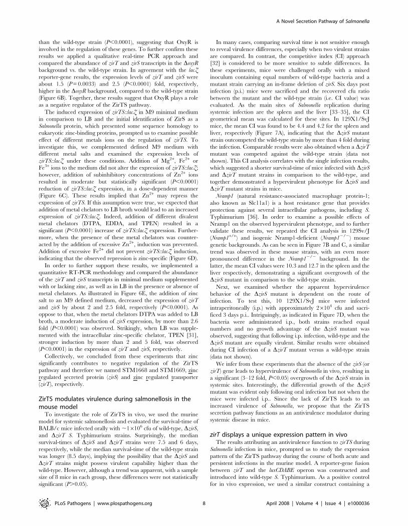
zirT displays a unique expression pattern in vivoThe results attributing an antivirulence function to zirTS during
Salmonella infection in mice, prompted us to study the expression
pattern of the ZirTS pathway during the course of both acute and
persistent infections in the murine model. A reporter-gene fusion
between zirT and the luxCDABE operon was constructed and
introduced into wild-type S. Typhimurium. As a positive control
for in vivo expression, we used a similar construct containing a
A Novel Secretion Pathway of Salmonella
PLoS Pathogens | www.plospathogens.org 8 April 2008 | Volume 4 | Issue 4 | e1000036

Salmonella sigma 70 (RpoD) dependent promoter [37]. Both
reporter strains, designated zirT::lux and rpoD::lux, were used to
infect C57BL/6 mice orally. Three days post-infection mice were
sacrificed and immediately examined for luciferase activity in the
gastrointestinal (GI) tract and systemic sites. Although zirT::lux had
about two logs lower expression levels in vitro in comparison to
rpoD::lux (data not shown), induced and distinct expression of
zirT::lux was observed in vivo. In contrast to rpoD::lux, which
showed strong expression in systemic sites (liver, spleen, and
mesenteric lymph nodes) and the small intestine, zirT::lux
expression was localized primarily within the small intestine,
while at systemic sites, zirT::lux expression was low, despite being
heavily colonized with Salmonella (Figure 8). These results suggest
that the in vivo expression of zirT is induced mainly throughout
the gastrointestinal tract of infected animals rather than at systemic
sites. This unique expression pattern is in agreement with the CI
experiments that showed a virulence difference between the wild-
type and a zirS mutant strain following oral, but not i.p., infection.
In light of these data, suggesting that zirT is not abundantly
induced at systemic sites; we propose that by administrating the
bacteria i.p., the induction of zirT in the GI tract was bypassed and
differences between the wild type and the zirTS mutants were not
noticeable.
Besides the induction in the small intestine, strong expression of
zirT::lux was apparent in the fecal pellets from both C57BL/6
(Nramp1 negative) mice that developed an acute systemic disease
and 129X1/SvJ (Nramp1 positive) mice carrying a persistent
Salmonella infection (Figure 9). Remarkably, the induced expression
of zirT::lux in Salmonella shed within fecal pellets of both mouse
strains was evident starting from day one p.i. and lasting at least 8
weeks p.i. in 129X1/SvJ mice during persistent Salmonella
infection. At 6 weeks p.i., due to low levels of Salmonella shedding
(as determined by cfu counts), expression of zirT::lux was not
detected in the feces, but reappeared at 8 weeks p.i., when high
numbers of Salmonella were shed. These results indicate strong and
continuous expression of zirT in the feces of shedding mice.
Figure 7. ZirS modulates Salmonella virulence during systemic disease in mice. The geometrical means of competitive index values areshown for the spleen and liver of mice that were infected orally (panels A–C) or i.p. (panel D). Six to seven weeks old 129X1/SvJ (panels A and D),Nramp1+/+ (panel B), or Nramp12/2 (panel C) female mice were infected with a 1:1 mixed inoculum of marked wild-type SL1344 strain and a DzirSmutant. For oral infection, mice were inoculated with 16106 cfu in 0.1 ml of infection buffer (0.1 M HEPES pH 8.0, 0.9% NaCl) and sacrificed after 6days. For i.p. infection, 26104 cfu were injected in 0.2 ml PBS and mice were sacrificed after 3 days. Panels A and D represent pooled results from 2independent experiments.doi:10.1371/journal.ppat.1000036.g007
A Novel Secretion Pathway of Salmonella
PLoS Pathogens | www.plospathogens.org 9 April 2008 | Volume 4 | Issue 4 | e1000036
Discussion
In this study we described the identification and characteriza-
tion of a Salmonella-conserved autonomous and previously
unidentified secretion pathway, termed ZirTS. We showed that
this novel secretion system consists of an OM protein, ZirT that is
expected to adopt a b-barrel structure and the exoprotein ZirS,
which is secreted into the extracellular environment in a strictly
ZirT-dependent manner.
The ZirTS secretion system was found to share several
important features with members of the TPS system and the
Int/Inv family. However, despite the mutual similarities, some
fundamental differences exist between the ZirTS and the
compared systems. Obviously, as opposed to Int/Inv members,
ZirTS function as a secretion system comprised of two separate
components. Likewise, comparison of ZirTS to TPS systems
reveals several hallmarks of TPS, which are absent from ZirTS.
The most characteristic feature of the TPS exoproteins is a
conserved N-terminal domain, known as the TPS domain. This
region has been identified in all of the TpsA proteins characterized
thus far and includes highly conserved NPNGI and NPNL motifs.
The TPS domain is necessary for secretion and has been proposed
to mediate recognition of the exoprotein by the transporter
[8,9,38–40]. An additional feature of TPS is the sequence
conservation of TpsB family members, particularly within the C-
terminus [41]. Amino acid sequence analysis and comparison of
ZirS and ZirT to other known TPS members indicated the lack of
these conserved modules in ZirS and ZirT and therefore
differentiate ZirTS from the TPS pathway. Based on this we
contend that zirTS encode a novel secretion pathway in Salmonella.
Adapting the currently accepted mechanism of TPS systems, we
propose the following model for the modus operandi of ZirTS: pre-
ZirS is translocated across the IM, via the Sec translocase. Upon
translocation, the signal peptide of pre-ZirS is cleaved off by a
specific periplasmic signal peptidase and the mature ZirS is
released in the periplasmic space. As was shown for other TPS
systems, there appears to be no accumulation of periplasmic
intermediates [8] and ZirS is expected to transit through the
periplasmic space, only briefly. Following Sec-dependent export
across the IM, ZirS interacts with ZirT and then is translocated
across the OM to the extracellular environment through a
hydrophilic b-barrel pore formed by ZirT.
One of many intriguing questions related to ZirTS is its
evolutionary origin. A conceivable scenario for the evolution of
such a system might be a molecular separation of an ancestral Int/
Inv related protein (or an AT) into two distinct functional
polypeptides, early in the evolution of Salmonella. Other circum-
stances that might explain the development of ZirTS include a
Figure 8. zirT is induced and displays a unique expression pattern in vivo. Two groups of female C57BL/6 mice were infected orally with,107 cfu of wild-type S. Typhimurium expressing rpoD::lux (A) or zirT::lux (B) reporter strains. At day 3 p.i. mice were sacrificed and their intact GI tractsas well as systemic organs were removed and imaged immediately. The detected bioluminescence signal is shown as pseudocolor images, withvariations in color representing light intensity at a given location. The color bar indicates relative signal intensity (as photons s21 cm22 sr21).Different organs are indicated as follow: liver (L); spleen (S); mesenteric lymph nodes (LN); colon (C); ileum (I); and jejunum (J). The experiment wasrepeated twice (with 9 mice in total for each reporter strain), and representative images are shown. Bacterial load is indicated by a cfu per organ in atable below each image.doi:10.1371/journal.ppat.1000036.g008
A Novel Secretion Pathway of Salmonella
PLoS Pathogens | www.plospathogens.org 10 April 2008 | Volume 4 | Issue 4 | e1000036

later acquisition of zirS into the genome and the ‘‘exploitation’’ of
an already existing OM b-barrel porin for its secretion.
Thus far, Int/Inv and TPS systems have been shown to be
primarily involved in different virulence traits of Gram-negative
pathogens. In contrast, the ZirTS secretion pathway demonstrated
an interesting and somewhat surprising antivirulence activity,
rather than a virulence determinant, in the murine salmonellosis
model. Presently, it is still unknown if the ZirTS pathway is unique
to Salmonella or related systems exist in other bacteria. A
bioinformatic search for non-invasin/intimin ZirT homologs has
led to the identification of a predicted 934 amino acid OM protein
(SG0602) in Sodalis glossinidius, which share 28% identity and 45%
similarity with ZirT (E-value 5e-47). The gene localized immedi-
ately downstream to SG0602 is expected to encode a 371 amino
acid protein (SG0603), with a predicted signal sequence.
Interestingly, S. glossinidius is an endosymbiont residing intracellu-
larly in tissues of the tsetse flies and utilize for cell invasion a
type III secretion system, which is phylogenetically related to
the SPI-1 type III secretion system of Salmonella [42]. Although
at this point, experimental evidence is still needed, de facto
secretion of SG0603 in a SG0602-dependant manner will indicate
a broader distribution of the ZirTS-like pathways among the
Enterobacteriaceae.
Despite the fact that ZirS is being secreted into the extracellular
environment, in mixed infection experiments, secretion of ZirS
from a wild-type strain did not seem to complement the phenotype
of a co-infecting zirS mutant. This observation may imply that
ZirS exerts its effect only topically or has a short in vivo half-life.
Another interesting feature of ZirTS is its location on a
horizontally acquired genomic island known as GEI 1664/1678
[17]. The acquisition of genomic islands by horizontal gene
transfer enables bacteria to rapidly gain complex functions from
other species and are crucial for the interaction of S. Typhimurium
with eukaryotic host cells. From an evolutionary and ecological
standpoint, infections caused by microbial pathogens that have
sustained a long-standing association with their hosts are most
often self-limiting or go unnoticed. Salmonella enterica, as an example
of such a pathogen, has maintained a coexistence with vertebrate
hosts for millions of years and evolved extremely sophisticated
mechanisms to engage vertebrate hosts. When examined at the
Figure 9. The expression of zirT is induced in shed Salmonella within fecal pellets during persistent and acute infection. S.Typhimurium expressing zirT::lux was used to infect 129X1/SvJ and C57BL/6 mice as explained above. (A) Fresh fecal pellets were collected from129X1/SvJ mice with persistent Salmonella infection and imaged immediately. Bioluminescence of fecal pellets is shown for 1, 28, 42, and 56 days p.i..An asterisk at day 42 p.i. indicates low levels of Salmonella shedding in the collected stool as was determined by cfu count. (B) In vivo imaging of129X1/SvJ mouse, 1 day p.i. The induced expression of zirT::lux in the stool is shown. (C) Bioluminescence of fecal pellets from C57BL/6 micedeveloping an acute Salmonella infection is shown for day 4 p.i. Fresh pellets were harvested directly from the animal prior to imaging and driedstools were collected from the cage.doi:10.1371/journal.ppat.1000036.g009
A Novel Secretion Pathway of Salmonella
PLoS Pathogens | www.plospathogens.org 11 April 2008 | Volume 4 | Issue 4 | e1000036

cellular and molecular levels, this functional interface reveals an
impressive array of bacterial determinants designed to manipulate
the host immune system, to sense the host environment or to
modulate a variety of cellular processes. The overall picture
emerging from close examination is perhaps one of balance, self
restraint and sophistication rather than one of uncontrolled
hostility towards its host [43]. Most studies in bacterial
pathogenesis are directed towards finding genes that promote
virulence in the host. Nonetheless, recent studies have demon-
strated that indeed, a fine balance during host infection is kept due
to the function of a subset of Salmonella genes known as
‘antivirulence genes’ [44]. Deletion mutations of these genes have
led to an overgrowth phenotype in the mouse model. Mouslim and
colleagues have shown that a PhoPQ regulated gene, pcgL, is
involved in stimulating the host immune system to prevent
bacterial overgrowth in mouse organs [45]. Another example of an
antivirulence gene is sciS. A sciS mutant strain has been shown to
display increased intracellular numbers in J774 macrophages and
hypervirulence in mice, when administered intragastrically [46]. A
null mutation in a Gifsy-2 phage harbored gene, grvA, increased
virulence as measured by competitive index experiments in mouse
spleens and small intestine [47]. Cumulatively, these studies
contribute to a recurring theme in pathogenesis emphasizing the
importance of pathogens to limit their effects upon the cells they
infect in order to achieve a balance with their host. These
examples prove that it is possible for an inactivated gene to lead to
an increase in bacterial numbers in host tissues. Increased bacterial
loads in the murine host would likely lead to more rapid sepsis and
toxic shock, thus increasing lethality and decreasing transmission
of the pathogen. The hypervirulence of DzirTS observed in the
mouse model during oral infection and its induction in the fecal
pellets at an early stage of the infection are consistent with this
concept. As with all Salmonella species, S. Typhimurium is primarily
transmitted through the fecal-oral route. Infected animals excrete
Salmonella in the feces, which will then gain access to an uninfected
host, starting a new infection cycle. Supported by induction of zirT
in the small intestine and its constant induction in feces, we
hypothesize that ZirTS play a role as a ‘virulence modulator’ in
the early stages of infection. We propose that ZirTS contribute to a
host-pathogen balance after the transmission from an infected to
naı̈ve host. The role ZirTS play during an early stage of the
infection may be in prevention of premature host death and,
perhaps, demonstrate at the molecular level, what was understood
by MacFarlane Burnet almost 70 years ago that ‘‘there is little
point in a microorganism destroying its host in a spectacular
fashion if this leaves it with no prospect of being ferried to other
vulnerable hosts’’ [48].
Materials and Methods
Bacterial strains and in vitro growth conditionsBacterial strains and plasmids used in this study are listed in
Table 1. S. Typhimurium SL1344 was used as the wild-type strain,
and all mutants used in this study were isogenic derivatives of
SL1344. Bacterial liquid cultures were maintained in Luria-
Bertani (LB) broth or M9-glucose minimal medium supplemented
with 0.0021% (w/v) histidine (since SL1344 is a histidine
auxotroph). The appropriate antibiotics were used at the following
concentrations: chloramphenicol, 25 mg ml21; kanamycin,
50 mg ml21; ampicillin, 100 mg ml21; and streptomycin,
100 mg ml21. The metal chelators diethylenetriaminepentaacetic
acid (DTPA), N,N9-ethylenediaminediacetic acid (EDDA), and
NN9N9-tetrakis(2-pyridylmethyl)ethylene diamine (TPEN) were
added to LB in the indicated concentrations.
Construction of S. Typhimurium SL1344 mutant strainsS. Typhimurium OG2006 carrying an in-frame deletion (amino
acid 9–270) of zirS, and OG2007 harboring a Kan cassette in zirT,
were generated by allelic exchange using the counter-selectable
suicide vector pRE112 [49] containing the levansucrase-encoding
sacB gene [50]. pRE112 based plasmids were transformed into E.
coli DH5alpir and then electroporated into E. coli SM10lpir [51]
that was used as the conjugative donor strain to S. Typhimurium
SL1344. Streptomycin/chloramphenicol-resistance merodiploid
colonies were grown for 4 h in LB broth without antibiotic
selection, diluted and then plated onto agar containing 1% (w/v)
tryptone, 0.5% (w/v) yeast extract, 5% (w/v) sucrose and
incubated at 30uC. Sucrose-resistant colonies were selected, and
the presence of the constructed mutation was confirmed by PCR.
An SL1344 oxyR mutant strain was constructed by P22
transduction from TA4101 (S. Typhimurium LT2). The growth
of these constructed strains was indistinguishable from the parental
strain while growing in liquid culture in vitro.
Generating hemagglutinin tagged proteins and reporter-gene constructs
Primers used in the study are listed in Table 2. Two-
hemagglutinin (2HA) tagged version of ZirS was constructed
using the primers OG-61 and OG-63. The resulting PCR product
containing the intact sequence of zirT following by zirS (without
the stop codon) was cloned into the vector pOG-WSHA after
digest with SacI and XbaI. The resulting plasmid harbors a C-
terminal fusion of the 2HA tag with ZirS (pOG-zirTS-HA). The
primers OG-62 and OG-63 were used to amplify a PCR fragment
(containing zirS and ,1-kb upstream to zirS) that was cloned using
SacI and XbaI into pOG-WSHA to generate pOG-zirS-HA. To
construct a 2HA tagged version of ZirT we used the primers OG-
61 and OG-158 to amplify a PCR fragment that was cloned using
SacI and XbaI into pOG-WSHA resulting in pOG-zirT-HA.
A reporter gene construct containing a translational fusion
between zirTS and a promoterless lacZ gene was generated using
the primers OG-89 and OG-92. The resulted PCR product was
cloned into pCR-Blunt (Invitrogen), digested with EcoRI and SmaI
and subcloned into pMC1403 to generate pOG-zirTS::lacZ.
Reporter-gene fusion of zirT with the luxCDABE operon from
Photorhabdus luminescens was generated by PCR amplification using
the primers OG-186 and OG-187. The resulted product was
cloned into pCR-Blunt (Invitrogen), digested with XhoI and BamHI
and cloned into pSC26 [37] to generate pOG-zirT::lux.
Secretion assay and immunoblottingTo examine the expression and secretion of ZirS and ZirT,
culture supernatant was filtered through a 0.2 mm pore-size filter
membrane, concentrated by precipitation with 10% (vol/vol)
trichloroacetic acid (TCA), and washed with acetone. The secreted
protein fraction and the corresponding bacterial cell pellets were
resuspended in 16 sodium dodecyl sulfate-polyacrylamide gel
electrophoresis (SDS-PAGE) sample buffer and subjected to
Western blot analysis using the appropriate primary antibodies:
rat monoclonal anti-HA (a-HA; 1:2,000; Roche Applied Science)
or mouse a-DnaK (1:2,000; Stressgen Biotechnologies). Rabbit
polyclonal antibodies against subunit beta of E. coli ATP synthase
and OmpA were generous gifts from Gabriele Deckers-Hebestreit
and Francisco Garcia-del Portillo, respectively, and were both
used in a 1:10,000 dilution. Goat a-rat, mouse, or rabbit
immunoglobulin G conjugated to horseradish peroxidase were
used as a secondary antibodies (1:7,500) followed by detection with
ECL reagents (Amersham Pharmacia).
A Novel Secretion Pathway of Salmonella
PLoS Pathogens | www.plospathogens.org 12 April 2008 | Volume 4 | Issue 4 | e1000036
b-galactosidase assaysb-galactosidase assays were performed according to [52]. The
assays were performed with 100 ml of culture, and the substrate for
b-galactosidase hydrolysis was o-nitrophenyl-b-D-galactopyranoside
(ONPG, Sigma). The background expression of the vector
(pMC1403) was 2.7660.5 and 1.2160.14 Miller Units (M.U.)
when cultures were grown in LB and M9 minimal medium,
respectively.
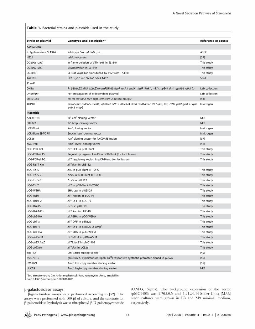
Table 1. Bacterial strains and plasmids used in the study.
Strain or plasmid Genotype and description* Reference or source
Salmonella
S. Typhimurium SL1344 wild-type Smr xyl hisG rpsL ATCC
NB24 ushA::res-cat-res [57]
OG2006 (zirS) In-frame deletion of STM1668 in SL1344 This study
OG2007 (zirT) STM1669::kan in SL1344 This study
OG2013 SL1344 oxyR::kan transduced by P22 from TA4101 This study
TA4101 LT2 oxyR1 zii-166::Tn5 SGSC1407 SGSC
E. coli
DH5a F- w80lacZDM15 D(lacZYA-argF)U169 deoR recA1 endA1 hsdR17(rk2, mk+) supE44 thi-1 gyrA96 relA1 l- Lab collection
DH5alpir For propagation of p-dependent plasmid Lab collection
SM10 lpir thi thr leu tonA lacY supE recA::RP4-2-Tc::Mu Kmlpir [51]
TOP10 mcrAD(mrr-hsdRMS-mcrBC) w80lacZ DM15 DlacX74 deoR recA1araD139 D(ara, leu) 7697 galU galK l- rpsLendA1 mupG
Invitrogen
Plasmids
pACYC184 Tcr Cmr cloning vector NEB
pBR322 Tcr Ampr cloning vector NEB
pCR-Blunt Kanr cloning vector Invitrogen
pCR-Blunt II-TOPO Zeocinr kanr cloning vector Invitrogen
pCS26 Kanr cloning vector for luxCDABE fusion [37]
pMC1403 Ampr lacZY cloning vector [58]
pOG-PCR-zirT zirT ORF in pCR-Blunt This study
pOG-PCR-zirTS Regulatory region of zirTS in pCR-Blunt (for lacZ fusion) This study
pOG-PCR-zirT-2 zirT regulatory region in pCR-Blunt (for lux fusion) This study
pOG-RzirT-Km zirT::kan in pRE112
pOG-TzirS zirS in pCR-Blunt II-TOPO This study
pOG-TzirS-2 DzirS in pCR-Blunt II-TOPO This study
pOG-TzirS-3 DzirS in pRE112 This study
pOG-TzirT zirT in pCR-Blunt II-TOPO This study
pOG-WSHA 2HA tag in pWSK29 This study
pOG-UzirT zirT region in pUC-19 This study
pOG-UzirT-2 zirT ORF in pUC-19 This study
pOG-UzirTS zirTS in pUC-19 This study
pOG-UzirT-Km zirT::kan in pUC-19 This study
pOG-zirS-HA zirS-2HA in pOG-WSHA This study
pOG-zirT-3 zirT ORF in pBR322 This study
pOG-zirT-4 zirT ORF in pBR322 D Ampr This study
pOG-zirT-HA zirT-2HA in pOG-WSHA This study
pOG-zirTS-HA zirTS-2HA in pOG-WSHA This study
pOG-zirTS::lacZ zirTS::lacZ in pMC1403 This study
pOG-zirT::lux zirT::lux in pCS26 This study
pRE112 Cmr sacB1 suicide vector [49]
pSIG70-16 rpoD::lux S. Typhimurium RpoD (s70) responsive synthetic promoter cloned in pCS26 [56]
pWSK29 Ampr low copy number cloning vector [59]
pUC19 Ampr high-copy number cloning vector NEB
*Sm, streptomycin; Cm, chloramphenicol; Kan, kanamycin; Amp, ampicillin.doi:10.1371/journal.ppat.1000036.t001
A Novel Secretion Pathway of Salmonella
PLoS Pathogens | www.plospathogens.org 13 April 2008 | Volume 4 | Issue 4 | e1000036

Quantitative real-time PCR analysisSalmonella RNA was extracted from mid-exponential phase
cultures using the Qiagen RNAprotect Bacteria Reagent and the
RNeasy mini kit according to the manufacture instructions,
including an on-column DNase digest using the RNase-free
DNase set (Qiagen). The quantity and quality of the extracted
RNA were determined by a ND-1000 spectrophotometer
(NanoDrop Technologies). To diminish any genomic DNA
contamination, RNA was secondly treated with an RNase-free
DNase I (Invitrogen). 0.5 mg of DNase I-treated RNA was
subjected to a first strand cDNA synthesis using the QuantiTect
Reverse Transcription Kit (Qiagen). Real-time PCR reactions
were performed in an Applied Biosystems 7500 Fast Real-time
PCR System. Each reaction was carried out in a total volume of
10 ml on a 96-well optical reaction plate (Applied Biosystems)
containing 5 ml FastStart Universal SYBR Green Master (ROX)
mix (Roche Applied Science); 1 ml cDNA; and two gene-specific
primers in a final concentration of 0.3 mM each. Real-time cycling
conditions were as follows: 50uC for 2 min; 95uC for 10 min; and
40 cycles of 95uC for 15 s, 60uC for 1 min. No-template and no
reverse-transcriptase controls were included for each primers set
and template. Melting curve analysis verified that each reaction
contained a single PCR product. Reported gene expression levels
were normalized to transcripts of rpoD, a housekeeping gene that
serves as an internal control. Gene-specific primers were designed
using PRIMER3 software (http://primer3.sourceforge.net/), are
listed in Table 2, and correspond to the following genes: rpoD, OG-
220 and OG-221; zirT, OG-216 and OG-217, OG-216 and OG-
229; zirS, OG-212 and OG-215, OG-228 and OG230.
Subcellular fractionationS. Typhimurium SL1344 strains expressing ZirT-HA or ZirTS-
HA were grown for 3 h in LB to late log phase (O.D.600,1.0).
Cells were harvested at 5,000 g for 10 minutes at 4uC, and washed
with ice-cold phosphate buffered saline (PBS). All the following
steps were performed at 4uC. Cells were resuspended in 1 ml of
cold lysis buffer [50mM Tris (pH 8.0), 20% (w/v) sucrose,
protease inhibitor cocktail (Roche Applied Science), and lysozyme
(100 mg/ml)] and incubated on ice for 1 h to generate sphero-
plasts. MgSO4 was added to final a concentration of 20 mM and
spheroplasts were collected by centrifugation for 10 min at
5,000 g. The supernatant containing the periplasmic fraction was
recovered and the pellet containing the cytoplasm and the
membranes fractions was resuspended in 1 ml cold sonication
buffer [50 mM Tris (pH 8.0), 20 mM MgSO4, RNase A (10 mg/
ml), DNase I (5 mg/ml), and protease inhibitor cocktail] and lysed
by sonication. Unlysed bacteria were removed by low-speed
centrifugation at 5,000 g for 10 min. The supernatant was
recovered and subjected to ultracentrifugation for 1 h at
100,000 g (in a TLA 100.3 fixed angle rotor in Beckman TL100
ultracentrifuge) to pellet the membrane fractions. The supernatant
represented the cytoplasmic fraction was recovered and the
membrane pellet was washed in cold sonication buffer, repelleted
for 30 min at 100,000 g and resuspended. This fraction repre-
sented the total membranes fraction.
Sucrose density gradientS. Typhimurium membranes were prepared from 100 ml
cultures expressing ZirT-HA that were grown to late log phase
(O.D.600,1.0) in LB. Cells were harvested at 5,000 g for
10 minutes at 4uC, and washed with 20 ml of ice-cold PBS. All
steps were performed at 4uC afterwards. Cells were resuspended in
5 ml of cold PBS containing protease inhibitor cocktail and passed
twice through a French Pressure cell at 10000 psi. Debris was
removed by centrifugation at 5,000 g for 10 min and the clarified
cell extract was centrifuged for 1 h at 100,000 g (30,000 rpm in a
SW41Ti rotor; Beckman). Membrane pellets were resuspended in
250 ml of PBS by repeated passage through a syringe equipped
with a 25 gauge needle. To separate inner and outer membranes,
200 ml of membranes were layered on top of a discontinuous
sucrose gradient composed of 0.5 ml of 60%, 1 ml of 55%, 2.4 ml
of 50%, 2.5 ml of 45%, 2.4 ml of 40%, 1.4 ml of 35%, and 0.8 ml
of 30% sucrose in PBS (w/v) and centrifuged for 16 hours at
100,000 g in a Beckman SW41Ti rotor. Membrane fractions
(800 ml) were recovered from the gradient by using a 20 gauge
needle from the bottom of the gradient. Fractions aliquots were
separated on a SDS-10% Polyacrylamide gel. The presence of
OmpA and the b-subunit of ATPase in the different fractions were
used as markers for the analysis of the inner and outer membranes
using Western-blot with the respective antibodies.
Mice and S. Typhimurium infection129X1/SvJ female mice were purchased from the Jackson
Laboratories. A breeding colony of inbred strain of 129Sv/J
(Nramp1+/+) mice and isogenic Nramp1-deficient (Nramp12/2)
strain have been previously described [53] and are maintained at
the University of British Columbia Animal Facility. All the mice
were kept in sterilized filter-topped cages and given food and water
ad libitum. Experiments were carried out under specific-pathogen-
free conditions according to the standard animal care guidelines
and protocols of the UBC Animal Care Committee and the
Canadian Council on Use of Laboratory Animals. For competitive
index (CI) infection experiments, 6–7 weeks old female mice were
infected orally or i.p. with mixed inoculum containing a marked
wild-type strain resistant to chloramphenicol (ushA::cat, NB24) and
a DzirS or DzirT mutant strain (OG2006 and OG2007,
respectively). For oral infection, mice were administered with
16106 cfu in 0.1 ml of infection buffer (0.1 M HEPES pH 8.0,
0.9% NaCl) and sacrificed after 6 days. For i.p. infection, 26104
cfu were injected in 0.2 ml PBS and mice were sacrificed after 3
Table 2. List of primers used in the study.
Primer Sequence (59-39)
OG-61 GAGCTCGGGCATAATTTCACAGGCGG
OG-62 GAGCTCCAGCAAAATCCGCATTACGG
OG-63 TCTAGATAGTCTTCCTCTGATGGAAATTTC
OG-89 GAATTCGGGCATAATTTCACAGGCGG
OG-92 CCCGGGGATAATAAGATTATGTTTCGAC
OG-158 TCTAGAATTCGCATCAGAGGTTGCGG
OG-186 GTCGAGATAGTGCATTCTTGCCTGCC
OG-187 GGATCCTGTATTGTCTCCTGCTATATCC
OG-212 TTTCCGGGAATGACTATTGC
OG-215 ATCTGGGCTTGGCGTATTAG
OG-216 GATGCCGACATCCGTTATTC
OG-217 TCCCGATATCCTGCTCTGAC
OG-220 GTGAAATGGGCACTGTTGAACTG
OG-221 TTCCAGCAGATAGGTAATGGCTTC
OG-228 GCCAAGCCCAGATTTAACAG
OG-229 AAAGCCATATGCCGGACGC
OG-230 ACGCGCTACAGTCTTATTAATG
doi:10.1371/journal.ppat.1000036.t002
A Novel Secretion Pathway of Salmonella
PLoS Pathogens | www.plospathogens.org 14 April 2008 | Volume 4 | Issue 4 | e1000036

days. Spleen and liver were homogenized in cold PBS, diluted and
plated on LB plates containing streptomycin for determination of
total Salmonella cfu. Colonies were replica-plated under chloram-
phenicol selection for enumeration of SL1344 ushA::cat. The
competitive index was calculated as [mutant/wild-type]output/
[mutant/wild-type]input. CI experiments, in which mice were co-
infected with the chromosomally marked strain (SL1344 ushA::cat)
and an unmarked SL1344 strain, or strains harboring mutation in
non-virulent genes, demonstrated a CI value of 1, indicating equal
virulence capability in mice [54,55].
Bioluminescence imaging of Salmonella during murineinfection
Wild-type Salmonella Typhimurium harboring pOG-zirT::lux or
pSIG70-16, carrying the lux operon under an RpoD dependent
promoter [56] as an expression control were grown for 16 h in
LB+Kan at 37 uC. Female C57BL/6 mice were orally infected
with 26107 cfu of the reporter strains in 0.1 ml of PBS. At 3 days
p.i. mice were anaesthetized with 2% isofluorane carried in 2% O2
and imaged using IVIS 100 (Xenogen Imaging Technologies).
Greyscale reference images taken under low illumination were
collected and overlaid with images capturing the emission of
photons from the lux-expressing bioluminescent S. Typhimurium
using Living Image Software version 2.50 (Xenogen). To
determine the total numbers of colonizing Salmonella (cfu), the
spleen, liver, MLN, ileum, caecum and colon were homogenized
in PBS using a high-speed mixer mill (MM301; Retsch), diluted
and spread plated on LB agar supplemented with streptomycin.
Statistical analysisData of the b-galactosidase assays are expressed as mean6-
standard deviation. The statistical significance between two mean
values was calculated by the unpaired t-test with two-tailed P
value. The statistical significance of the measured mean fold-
change by the qRT-PCR was evaluated by t-test against
hypothetical value of 1 with two-tailed P value. Data of the CI
experiments in mice are expressed as geometrical mean. The
statistical significance was calculated by the Wilcoxon Signed
Rank Test, against theoretical median of 1 with two-tailed P value.
P,0.05 was considered to be statistically significant.
Acknowledgments
We would like to thank Dr. Wanyin Deng for helpful discussions and
insightful comments and the members of the Finlay lab for critical reading
of the manuscript. We are grateful to Dr. Guntram Grassl and Yuling Li
for excellent assistance with the animal studies, to Aaron White and
Michael Surette for the pSIG70-16 plasmid and to Francisco Garcia-del
Portillo and Gabriele Deckers-Hebestreit for the antibodies against OmpA
and the beta subunit of E. coli ATP synthase, respectively.
Author Contributions
Conceived and designed the experiments: OG DG BV BF. Performed the
experiments: OG DG DB. Analyzed the data: OG DG. Wrote the paper:
OG DG BF.
References
1. Kingsley RA, Baumler AJ (2002) Pathogenicity islands and host adaptation of
Salmonella serovars. Curr Top Microbiol Immunol 264: 67–87.
2. Economou A, Christie PJ, Fernandez RC, Palmer T, Plano GV, et al. (2006)
Secretion by numbers: Protein traffic in prokaryotes. Mol Microbiol 62:
308–319.
3. Kostakioti M, Newman CL, Thanassi DG, Stathopoulos C (2005) Mechanisms
of protein export across the bacterial outer membrane. J Bacteriol 187:
4306–4314.
4. Henderson IR, Nataro JP, Kaper JB, Meyer TF, Farrand SK, et al. (2000)
Renaming protein secretion in the gram-negative bacteria. Trends Microbiol 8:
352.
5. Henderson IR, Cappello R, Nataro JP (2000) Autotransporter proteins,
evolution and redefining protein secretion. Trends Microbiol 8: 529–532.
6. Desvaux M, Parham NJ, Henderson IR (2004) The autotransporter secretion
system. Res Microbiol 155: 53–60.
7. Rutherford N, Mourez M (2006) Surface display of proteins by gram-negative
bacterial autotransporters. Microb Cell Fact 5: 22.
8. Jacob-Dubuisson F, Fernandez R, Coutte L (2004) Protein secretion through
autotransporter and two-partner pathways. Biochim Biophys Acta 1694:
235–257.
9. Jacob-Dubuisson F, Locht C, Antoine R (2001) Two-partner secretion in Gram-
negative bacteria: a thrifty, specific pathway for large virulence proteins. Mol
Microbiol 40: 306–313.
10. Clantin B, Delattre AS, Rucktooa P, Saint N, Meli AC, et al. (2007) Structure of
the membrane protein FhaC: a member of the Omp85-TpsB transporter
superfamily. Science 317: 957–961.
11. Niemann HH, Schubert WD, Heinz DW (2004) Adhesins and invasins of
pathogenic bacteria: a structural view. Microbes Infect 6: 101–112.
12. Remaut H, Waksman G (2004) Structural biology of bacterial pathogenesis.
Curr Opin Struct Biol 14: 161–170.
13. Newman CL, Stathopoulos C (2004) Autotransporter and two-partner secretion:
delivery of large-size virulence factors by gram-negative bacterial pathogens. Crit
Rev Microbiol 30: 275–286.
14. Touze T, Hayward RD, Eswaran J, Leong JM, Koronakis V (2004)
Self-association of EPEC intimin mediated by the beta-barrel-containing
anchor domain: a role in clustering of the Tir receptor. Mol Microbiol 51:
73–87.
15. Sikora S, Strongin A, Godzik A (2005) Convergent evolution as a mechanism for
pathogenic adaptation. Trends Microbiol 13: 522–527.
16. Bruggemann H, Cazalet C, Buchrieser C (2006) Adaptation of Legionella
pneumophila to the host environment: role of protein secretion, effectors and
eukaryotic-like proteins. Curr Opin Microbiol 9: 86–94.
17. McClelland M, Sanderson KE, Spieth J, Clifton SW, Latreille P, et al. (2001)
Complete genome sequence of Salmonella enterica serovar Typhimurium LT2.
Nature 413: 852–856.
18. Baumler AJ (1997) The record of horizontal gene transfer in Salmonella. Trends
Microbiol 5: 318–322.
19. Cotter PA, DiRita VJ (2000) Bacterial virulence gene regulation: an evolutionary
perspective. Annu Rev Microbiol 54: 519–565.
20. Bendtsen JD, Nielsen H, von Heijne G, Brunak S (2004) Improved prediction of
signal peptides: SignalP 3.0. J Mol Biol 340: 783–795.
21. Manting EH, Driessen AJ (2000) Escherichia coli translocase: the unravelling of a
molecular machine. Mol Microbiol 37: 226–238.
22. Huie JL, Silhavy TJ (1995) Suppression of signal sequence defects and azide
resistance in Escherichia coli commonly result from the same mutations in secA.
J Bacteriol 177: 3518–3526.
23. Senior AE (1990) The proton-translocating ATPase of Escherichia coli. Annu Rev
Biophys Biophys Chem 19: 7–41.
24. Gardy JL, Laird MR, Chen F, Rey S, Walsh CJ, et al. (2005) PSORTb v.2.0:
expanded prediction of bacterial protein subcellular localization and insights
gained from comparative proteome analysis. Bioinformatics 21: 617–623.
25. Gromiha MM, Ahmad S, Suwa M (2004) Neural network-based prediction of
transmembrane beta-strand segments in outer membrane proteins. J Comput
Chem 25: 762–767.
26. Freudl R, Cole ST (1983) Cloning and molecular characterization of the ompA
gene from Salmonella typhimurium. Eur J Biochem 134: 497–502.
27. Altendorf K, Stalz W, Greie J, Deckers-Hebestreit G (2000) Structure and
function of the F(o) complex of the ATP synthase from Escherichia coli. J Exp Biol
203: 19–28.
28. Schnaitman CA (1973) Outer membrane proteins of Escherichia coli. I. Effect of
preparative conditions on the migration of protein in polyacrylamide gels. Arch
Biochem Biophys 157: 541–552.
29. Alkema WB, Lenhard B, Wasserman WW (2004) Regulog analysis: detection of
conserved regulatory networks across bacteria: application to Staphylococcus aureus.
Genome Res 14: 1362–1373.
30. Storz G, Imlay JA (1999) Oxidative stress. Curr Opin Microbiol 2: 188–194.
31. Outten CE, O’Halloran TV (2001) Femtomolar sensitivity of metalloregulatory
proteins controlling zinc homeostasis. Science 292: 2488–2492.
32. Beuzon CR, Holden DW (2001) Use of mixed infections with Salmonella strains to
study virulence genes and their interactions in vivo. Microbes Infect 3:
1345–1352.
33. Salcedo SP, Noursadeghi M, Cohen J, Holden DW (2001) Intracellular
replication of Salmonella typhimurium strains in specific subsets of splenic
macrophages in vivo. Cell Microbiol 3: 587–597.
A Novel Secretion Pathway of Salmonella
PLoS Pathogens | www.plospathogens.org 15 April 2008 | Volume 4 | Issue 4 | e1000036

34. Matsui H, Eguchi M, Kikuchi Y (2000) Use of confocal microscopy to detect
Salmonella typhimurium within host cells associated with Spv-mediated intracellularproliferation. Microb Pathog 29: 53–59.
35. Richter-Dahlfors A, Buchan AM, Finlay BB (1997) Murine salmonellosis studied
by confocal microscopy: Salmonella typhimurium resides intracellularly insidemacrophages and exerts a cytotoxic effect on phagocytes in vivo. J Exp Med 186:
569–580.36. Gruenheid S, Gros P (2000) Genetic susceptibility to intracellular infections:
Nramp1, macrophage function and divalent cations transport. Curr Opin
Microbiol 3: 43–48.37. Bjarnason J, Southward CM, Surette MG (2003) Genomic profiling of iron-
responsive genes in Salmonella enterica serovar typhimurium by high-throughputscreening of a random promoter library. J Bacteriol 185: 4973–4982.
38. Kajava AV, Cheng N, Cleaver R, Kessel M, Simon MN, et al. (2001) Beta-helixmodel for the filamentous haemagglutinin adhesin of Bordetella pertussis and
related bacterial secretory proteins. Mol Microbiol 42: 279–292.
39. Hodak H, Clantin B, Willery E, Villeret V, Locht C, et al. (2006) Secretionsignal of the filamentous haemagglutinin, a model two-partner secretion
substrate. Mol Microbiol 61: 368–382.40. Clantin B, Hodak H, Willery E, Locht C, Jacob-Dubuisson F, et al. (2004) The
crystal structure of filamentous hemagglutinin secretion domain and its
implications for the two-partner secretion pathway. Proc Natl Acad Sci U S A101: 6194–6199.
41. Yen MR, Peabody CR, Partovi SM, Zhai Y, Tseng YH, et al. (2002) Protein-translocating outer membrane porins of Gram-negative bacteria. Biochim
Biophys Acta 1562: 6–31.42. Dale C, Young SA, Haydon DT, Welburn SC (2001) The insect endosymbiont
Sodalis glossinidius utilizes a type III secretion system for cell invasion. Proc Natl
Acad Sci U S A 98: 1883–1888.43. Galan JE (2001) Salmonella interactions with host cells: type III secretion at work.
Annu Rev Cell Dev Biol 17: 53–86.44. Foreman-Wykert AK, Miller JF (2003) Hypervirulence and pathogen fitness.
Trends Microbiol 11: 105–108.
45. Mouslim C, Hilbert F, Huang H, Groisman EA (2002) Conflicting needs for aSalmonella hypervirulence gene in host and non-host environments. Mol
Microbiol 45: 1019–1027.46. Parsons DA, Heffron F (2005) sciS, an icmF homolog in Salmonella enterica serovar
Typhimurium, limits intracellular replication and decreases virulence. InfectImmun 73: 4338–4345.
47. Ho TD, Slauch JM (2001) Characterization of grvA, an antivirulence gene on the
gifsy-2 phage in Salmonella enterica serovar typhimurium. J Bacteriol 183:611–620.
48. Burnet M (1940) Biological Aspects of Infectious Disease. Cambridge:
Cambridge University press.49. Edwards RA, Keller LH, Schifferli DM (1998) Improved allelic exchange vectors
and their use to analyze 987P fimbria gene expression. Gene 207: 149–157.50. Pelicic V, Reyrat JM, Gicquel B (1996) Generation of unmarked directed
mutations in mycobacteria, using sucrose counter-selectable suicide vectors. Mol
Microbiol 20: 919–925.51. Miller VL, Mekalanos JJ (1988) A novel suicide vector and its use in construction
of insertion mutations: osmoregulation of outer membrane proteins andvirulence determinants in Vibrio cholerae requires toxR. J Bacteriol 170:
2575–2583.52. Miller JH (1972) Experiments in molecular biology. Cold Spring Harbor, N.Y:
Cold Spring Harbor Laboratory.
53. Vidal S, Tremblay ML, Govoni G, Gauthier S, Sebastiani G, et al. (1995) TheIty/Lsh/Bcg locus: natural resistance to infection with intracellular parasites is
abrogated by disruption of the Nramp1 gene. J Exp Med 182: 655–666.54. Gal-Mor O, Valdez Y, Finlay BB (2006) The temperature-sensing protein TlpA
is repressed by PhoP and dispensable for virulence of Salmonella enterica serovar
Typhimurium in mice. Microbes Infect 8: 2154–2162.55. Coombes BK, Wickham ME, Lowden MJ, Brown NF, Finlay BB (2005)
Negative regulation of Salmonella pathogenicity island 2 is required for contextualcontrol of virulence during typhoid. Proc Natl Acad Sci U S A 102:
17460–17465.56. Kim W, Surette MG (2006) Coordinated regulation of two independent cell-cell
signaling systems and swarmer differentiation in Salmonella enterica serovar
Typhimurium. J Bacteriol 188: 431–440.57. Brown NF, Vallance BA, Coombes BK, Valdez Y, Coburn BA, et al. (2005)
Salmonella Pathogenicity Island 2 Is Expressed Prior to Penetrating the Intestine.PLoS Pathog 1: e32.
58. Casadaban MJ, Chou J, Cohen SN (1980) In vitro gene fusions that join an
enzymatically active beta-galactosidase segment to amino-terminal fragments ofexogenous proteins: Escherichia coli plasmid vectors for the detection and cloning
of translational initiation signals. J Bacteriol 143: 971–980.59. Wang RF, Kushner SR (1991) Construction of versatile low-copy-number
vectors for cloning, sequencing and gene expression in Escherichia coli. Gene 100:195–199.
A Novel Secretion Pathway of Salmonella
PLoS Pathogens | www.plospathogens.org 16 April 2008 | Volume 4 | Issue 4 | e1000036
Related Documents