ORIGINAL ARTICLE A novel mouse model of hepatocarcinogenesis triggered by AID causing deleterious p53 mutations A Takai 1 , T Toyoshima 3 , M Uemura 3 , Y Kitawaki 2 , H Marusawa 1 , H Hiai 3 , S Yamada 4 , IM Okazaki 2 , T Honjo 2 , T Chiba 1 and K Kinoshita 3 1 Department of Gastroenterology and Hepatology, Graduate School of Medicine, Kyoto University, Kyoto, Japan; 2 Department of Immunology and Genomic Medicine, Graduate School of Medicine, Kyoto University, Kyoto, Japan; 3 Shiga Medical Center Research Institute, Moriyama, Japan and 4 Mikasa Laboratory, Immuno-Biological Laboratories Co., Ltd, Mikasa, Japan Activation-induced cytidine deaminase (AID), the only enzyme that is known to be able to induce mutations in the human genome, is required for somatic hypermutation and class-switch recombination in B lymphocytes. Recently, we showed that AID is implicated in the pathogenesis of human cancers including hepatitis C virus (HCV)-induced human hepatocellular carcinoma (HCC). In this study, we established a new AID transgenic mouse model (TNAP-AID) in which AID is expressed in cells producing tissue-nonspecific alkaline phosphatase (TNAP), which is a marker of primordial germ cells and immature stem cells, including ES cells. High expression of TNAP was found in the liver of the embryos and adults of TNAP-AID mice. HCC developed in 27% of these mice at the age of approximately 90 weeks. The HCC that developed in TNAP-AID mice expressed a-fetoprotein and had deleterious mutations in the tumour suppressor gene Trp53, some of which corresponded to those found in human cancer. In conclusion, TNAP-AID is a mouse model that spontaneously develops HCC, sharing genetic and phenotypic features with human HCC, which develops in the inflamed liver as a result of the accumulation of genetic changes. Oncogene advance online publication, 10 November 2008; doi:10.1038/onc.2008.415 Keywords: hepatic cancer; stem cell marker; activation- induced cytidine deaminase; Cre recombinase; somatic mutation; animal model Introduction It is widely recognized that mutations of oncogenes, tumour suppressor genes and genomic stability genes play pivotal roles in cancer development (Vogelstein and Kinzler, 2004). Despite remarkable progress in our knowledge of the molecular mechanisms of individual cancer-related genes, surprisingly little is known about the fundamental aspects of when and how mutations are introduced into what kind of cell populations (for example, differentiated cells versus tissue stem cells). To address this problem, a new mechanism of mutagenesis for cancer development has recently been proposed (Kinoshita and Nonaka, 2006; Marusawa, 2008). It hypothesizes that at least some cancer-related mutations are introduced by activation-induced cytidine deaminase (AID), an enzyme that is expressed in activated B lymphocytes, and is required for somatic hypermutation (SHM) and class-switch recombination of antibody genes (Honjo et al., 2002). The hypothesis is based on the following observations: (1) AID can induce mutations in non-B cells (Yoshikawa et al., 2002); (2) AID transgenic mice develop various tumours, including T-cell lymphoma and lung microadenoma (Okazaki et al., 2003) and (3) AID can be induced in human hepatic, gastric and biliary epithelial cells when stimulated with pro-inflammatory cytokines, such as transforming growth factor-b and tumor necrosis factor-a, and when challenged with pathogens, such as hepatitis C virus (HCV) and Helicobacter pylori. AID is detected in the liver, stomach and bile duct in humans (Kou et al., 2006; Endo et al., 2007; Matsumoto et al., 2007; Komori et al., 2008). On the basis of this evidence, AID is a potential candidate for a mutagen in human cancers. To substantiate this hypothesis, there is an urgent need for the establishment of an AID transgenic mouse model that recapitulates the development of human cancer. However, we have found earlier that it is difficult to analyse epithelial tumours of tissues other than lymphoid malignancies in a transgenic mouse model with constitutive and ubiquitous AID expression because of early death of the mice from lethal T-cell lymphoma (Okazaki et al., 2003). We generated earlier AID transgenic mice that can express AID conditionally in a Cre-recombianase- dependent manner (AID conditional transgenic, AID cTg) and reported B-cell-specific AID transgenic mice Received 30 June 2008; revised 30 September 2008; accepted 14 October 2008 Correspondence: Dr K Kinoshita, Evolutionary Medicine, Shiga Medical Center Research Institute, 5/4/1930, Moriyama, Shiga 524-8524, Japan. E-mail: [email protected] Oncogene (2008), 1–10 & 2008 Macmillan Publishers Limited All rights reserved 0950-9232/08 $32.00 www.nature.com/onc

Welcome message from author
This document is posted to help you gain knowledge. Please leave a comment to let me know what you think about it! Share it to your friends and learn new things together.
Transcript
ORIGINAL ARTICLE
A novel mouse model of hepatocarcinogenesis triggered by AID causing
deleterious p53 mutations
A Takai1, T Toyoshima3, M Uemura3, Y Kitawaki2, H Marusawa1, H Hiai3, S Yamada4,IM Okazaki2, T Honjo2, T Chiba1 and K Kinoshita3
1Department of Gastroenterology and Hepatology, Graduate School of Medicine, Kyoto University, Kyoto, Japan; 2Departmentof Immunology and Genomic Medicine, Graduate School of Medicine, Kyoto University, Kyoto, Japan; 3Shiga Medical CenterResearch Institute, Moriyama, Japan and 4Mikasa Laboratory, Immuno-Biological Laboratories Co., Ltd, Mikasa, Japan
Activation-induced cytidine deaminase (AID), the onlyenzyme that is known to be able to induce mutations in thehuman genome, is required for somatic hypermutation andclass-switch recombination in B lymphocytes. Recently,we showed that AID is implicated in the pathogenesis ofhuman cancers including hepatitis C virus (HCV)-inducedhuman hepatocellular carcinoma (HCC). In this study,we established a new AID transgenic mouse model(TNAP-AID) in which AID is expressed in cells producingtissue-nonspecific alkaline phosphatase (TNAP), which isa marker of primordial germ cells and immature stemcells, including ES cells. High expression of TNAP wasfound in the liver of the embryos and adults of TNAP-AIDmice. HCC developed in 27% of these mice at the age ofapproximately 90 weeks. The HCC that developedin TNAP-AID mice expressed a-fetoprotein and haddeleterious mutations in the tumour suppressor geneTrp53, some of which corresponded to those found inhuman cancer. In conclusion, TNAP-AID is a mousemodel that spontaneously develops HCC, sharing geneticand phenotypic features with human HCC, which developsin the inflamed liver as a result of the accumulation ofgenetic changes.Oncogene advance online publication, 10 November 2008;doi:10.1038/onc.2008.415
Keywords: hepatic cancer; stem cell marker; activation-induced cytidine deaminase; Cre recombinase; somaticmutation; animal model
Introduction
It is widely recognized that mutations of oncogenes,tumour suppressor genes and genomic stability genes
play pivotal roles in cancer development (Vogelstein andKinzler, 2004). Despite remarkable progress in ourknowledge of the molecular mechanisms of individualcancer-related genes, surprisingly little is known aboutthe fundamental aspects of when and how mutationsare introduced into what kind of cell populations(for example, differentiated cells versus tissue stem cells).
To address this problem, a new mechanism ofmutagenesis for cancer development has recently beenproposed (Kinoshita and Nonaka, 2006; Marusawa,2008). It hypothesizes that at least some cancer-relatedmutations are introduced by activation-induced cytidinedeaminase (AID), an enzyme that is expressed inactivated B lymphocytes, and is required for somatichypermutation (SHM) and class-switch recombinationof antibody genes (Honjo et al., 2002). The hypothesis isbased on the following observations: (1) AID can inducemutations in non-B cells (Yoshikawa et al., 2002);(2) AID transgenic mice develop various tumours,including T-cell lymphoma and lung microadenoma(Okazaki et al., 2003) and (3) AID can be induced inhuman hepatic, gastric and biliary epithelial cells whenstimulated with pro-inflammatory cytokines, such astransforming growth factor-b and tumor necrosisfactor-a, and when challenged with pathogens, such ashepatitis C virus (HCV) and Helicobacter pylori. AID isdetected in the liver, stomach and bile duct in humans(Kou et al., 2006; Endo et al., 2007; Matsumoto et al.,2007; Komori et al., 2008). On the basis of this evidence,AID is a potential candidate for a mutagen in humancancers.
To substantiate this hypothesis, there is an urgentneed for the establishment of an AID transgenic mousemodel that recapitulates the development of humancancer. However, we have found earlier that it is difficultto analyse epithelial tumours of tissues other thanlymphoid malignancies in a transgenic mouse modelwith constitutive and ubiquitous AID expressionbecause of early death of the mice from lethal T-celllymphoma (Okazaki et al., 2003).
We generated earlier AID transgenic mice that canexpress AID conditionally in a Cre-recombianase-dependent manner (AID conditional transgenic, AIDcTg) and reported B-cell-specific AID transgenic mice
Received 30 June 2008; revised 30 September 2008; accepted 14 October2008
Correspondence: Dr K Kinoshita, Evolutionary Medicine, ShigaMedical Center Research Institute, 5/4/1930, Moriyama,Shiga 524-8524, Japan.E-mail: [email protected]
Oncogene (2008), 1–10& 2008 Macmillan Publishers Limited All rights reserved 0950-9232/08 $32.00
www.nature.com/onc
obtained by crossing AID cTg mice with B-cell-specificCre transgenic mice (Muto et al., 2006). In this study, weused a different Cre transgenic mouse line that expressesCre in cells producing tissue nonspecific alkalinephosphatase (TNAP), as the mating partner of AIDcTg mice.
TNAP is a member of the alkaline phosphatase familyencoded by the Akp2 gene and alternatively designatedalkaline phosphatase 2, liver. As TNAP is also known asa marker of primordial germ cells and immature stemcells, including ES cells (Urven et al., 1993; Kues et al.,2005; Wang et al., 2005), we initially speculated thatAID expression in TNAP-positive cells might contributeto the development of tumours of germ cell origin.However, the resulting mice frequently developedhepatocellular carcinoma (HCC) but not germ celltumours or lethal lymphomas. Histological and mole-cular analyses of HCC revealed genetic and phenotypicsimilarities to human tumours, including deleteriousmutations in the p53 gene and the expression ofa-fetoprotein, a well-known marker of human HCC.This mouse model should be useful for studies on theprevention and treatment of hepatic cancer, which is amajor health concern worldwide.
Results
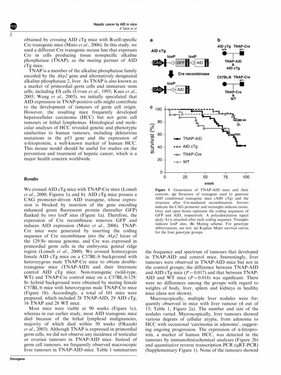
We crossed AID cTg mice with TNAP-Cre mice (Lomeliet al., 2000; Figures 1a and b). AID cTg mice possess aCAG promoter-driven AID transgene, whose expres-sion is blocked by insertion of the gene encodingenhanced green fluorescent protein (hereafter GFP)flanked by two loxP sites (Figure 1a). Therefore, theexpression of Cre recombinase removes GFP andinduces AID expression (Muto et al., 2006). TNAP-Cre mice were generated by inserting the codingsequence of Cre recombinase into the Akp2 locus ofthe 129/Sv mouse genome, and Cre was expressed inprimordial germ cells in the embryonic genital ridgeregion (Lomeli et al., 2000). We crossed homozygousfemale AID cTg mice on a C57BL/6 background withheterozygous male TNAP-Cre mice to obtain double-transgenic mice (TNAP-AID) and their littermatecontrol AID cTg mice. Non-transgenic (wild-type,WT) and TNAP-Cre control mice on a C57BL/6:129/Sv hybrid background were obtained by mating femaleC57BL/6 mice with heterozygous male TNAP-Cre mice(Figure 1b). Accordingly, a total of 101 mice wereprepared, which included 28 TNAP-AID, 29 AID cTg,16 TNAP and 28 WT mice.
Most mice were viable at 90 weeks (Figure 1c),whereas in our earlier study, most AID transgenic micedied because of the lethal lymphoid malignancies,majority of which died within 50 weeks (Okazakiet al., 2003). Although TNAP is expressed in primordialgerm cells, we did not observe any incidence of testicularor ovarian tumours in TNAP-AID mice. Instead ofgerm cell tumours, we frequently observed macroscopicliver tumours in TNAP-AID mice. Table 1 summarizes
the frequency and spectrum of tumours that developedin TNAP-AID and control mice. Interestingly, livertumours were observed in TNAP-AID mice but not inthe control groups; the difference between TNAP-AIDand AID cTg mice (P¼ 0.017) and that between TNAP-AID and WT mice (P¼ 0.018) was significant. Therewere no differences among the groups with regard toweights of body, liver, spleen and kidneys in healthymice (data not shown).
Macroscopically, multiple liver nodules were fre-quently observed in mice with liver tumour (4 out of15; Table 1, Figure 2a). The number and size of thenodules varied. Microscopically, liver tumours showedvarious degrees of cellular atypia, from adenoma toHCC with occasional ‘carcinoma in adenoma’, suggest-ing ongoing progression. The expression of a-fetopro-tein, a marker of human HCC, was detected in thetumours by immunohistochemical analyses (Figure 2b)and quantitative reverse transcription PCR (qRT-PCR)(Supplementary Figure 1). None of the tumours showed
Figure 1 Generation of TNAP-AID mice and theircontrols. (a) Structure of transgene used to generateAID conditional transgenic mice (AID cTg) and thestructure after Cre-mediated recombination. Arrowsindicate the CAG promoter and rectangles indicate exons.Grey and open boxes represent the coding sequences ofGFP and AID, respectively. A polyadenylation signal(poly A) is attached after each coding sequence. Trianglesindicate loxP sites. (b) Mating scheme. For genotypeabbreviations, see text. (c) Kaplan–Meier survival curvesfor the four genotype groups.
Hepatic cancer by AID in miceA Takai et al
2
Oncogene

nuclear localization of b-catenin protein (data notshown).
Sequences of some tumour suppressor genes andoncogenes in the tumours were determined. Significantnumbers of mutations were observed in the Trp53 geneencoding the p53 protein (Table 2). The non-transcribedregion upstream of the promoter did not containmutations, an observation that is consistent with thetranscription dependence of mutagenesis by AID(Yoshikawa et al., 2002). Majority of mutations weresingle-base substitutions with significant GC bias,another footprint of AID (Figure 3a) (Yoshikawaet al., 2002). There was no significant correlationbetween the distribution of mutations and those ofSHM hot spot motifs (RGYW/WRCY) and DNAsecondary structures (Supplementary Figure 2). Inter-estingly, the mutation frequency was the highest incancer tissues followed by non-cancerous TNAP-AIDliver and lowest in normal liver of WT mice, whichcorresponds to the PCR error rate or the backgroundrate in this assay. The increase in mutation frequency inHCC compared with non-cancerous liver was because ofthe increase in single-base insertion events mostly at thehomonucleotide tracts, leading to frameshift mutationsin the 50-half of the coding region (Table 2 andFigure 3b). Most of TNAP-AID liver mutationsoccurred in the DNA-binding domain of p53, which iscritical for its function (Table 3).
Although small volumes (approximately 50 mm3) ofnormal and cancerous liver tissues were dissected formutation analysis, the identified mutations were quitediverse, suggesting that the sampled tissue blockcontained multiple HCC nodules, each representingmonoclonal expansion. Such heterogeneity was verifiedby two independent series of PCR, plasmid cloning andsequencing for the same sample: there were few overlapsof mutations. For example, 8 and 7 clones derived fromHCC of mouse TA113 contained nonsynonymous pointmutations out of sequenced 27 and 40 clones in the firstand second experiment, respectively. The mutationpatterns were completely different except C808G(R270G), which appeared thrice and once in the firstand the second trial, respectively. Table 3 lists thecombined results of the two experiments. The mostfrequent mutations were C790T(R264W) andC808G(R270G), the latter corresponding to one of thesecond most important mutational targets (R273) inhuman cancers (Figure 3b, top). Despite extensivesequencing, no mutations were found in Myc, Pten
and Cdkn2a (p16 and p19Arf) genes (SupplementaryFigure 3). The Trp53 mutation frequency in the thymuswas significantly lower than in the liver of TNAP-AIDmice, which is consistent with the lack of lethal T-celllymphomagenesis (Table 2).
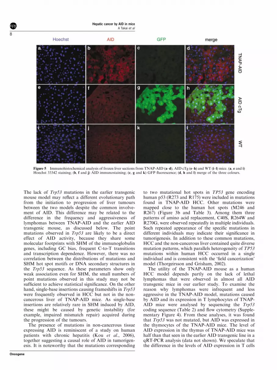
To determine why liver tumours developed, TNAPexpression in various organs was examined by qRT-PCR in E14.5 embryos and 53- to 74-week-old adultmice of strain C57BL/6. TNAP was ubiquitouslyexpressed at a relatively high level in the liver andintestine of the embryos and in the liver and testis of theadults (Figure 4a). In agreement with this widedistribution of TNAP expression, GFP expression inTNAP-AID mice was absent in most organs in adults(Figure 4c). The substantial levels of AID expressionwere confirmed by qRT-PCR (Figure 4b) and westernblotting (Figure 4d) in various organs of the adultTNAP-AID mice but not of AID cTg mice. Thepresence of AID protein and loss of GFP were detectedin the non-cancerous liver sections of TNAP-AID mice(Figure 5). Taken together, these findings suggest thatconstitutive AID expression in hepatic lineage, includingfetal and adult liver, contributes to the high incidence ofliver tumours.
As reported for AID transgenic mice with theubiquitous promoter, lymphomas were observed inTNAP-AID mice (Table 1). Although the frequency oflymphomas in the TNAP-AID group was higher thanthose in the other groups, the differences betweenTNAP-AID and AID cTg mice (P¼ 0.508) and thosebetween TNAP-AID and WT mice (P¼ 0.150) were notstatistically significant. The survival curve analysis(Figure 1c) suggested that there was no significantdifference between the TNAP-AID and TNAP-Cregroups (P¼ 0.515), indicating that the lymphomas inTNAP-AID mice were less aggressive than those inthe transgenic mice with ubiquitous AID expression.Histologically, the lymphomas in TNAP-AID miceexhibited nodular appearance, which was differentfrom that of the diffusely infiltrating lymphoma thatdevelops in the ubiquitous promoter-driven AID trans-genic mice (data not shown). This difference suggests thatdifferent mechanisms underlie tumorigenesis of lymphoidcells between these mice. We confirmed that GFPexpression and Cre-mediated excision of GFP occurredefficiently in CD3-positive T cells of AID cTg andTNAP-AID mice, respectively (Supplementary Figure 4).This suggests that AID is expressed in T cells of TNAP-AID mice.
Table 1 Frequencies of tumours observed in TNAP-AID and control mice
Genotype Mean age at killing (weeks) HCC Lymphoma Lung cancer Stomach cancer
TNAP-AID (15) 88.5 26.7% (4) 40.0% (6) 6.7% (1) 6.7% (1)AID-cTg (24) 89.9 0.0% (0) 29.2% (7) 0.0% (0) 0.0% (0)TNAP-Cre (14) 88.4 0.0% (0) 0.0% (0) 0.0% (0) 0.0% (0)WT (23) 89.1 0.0% (0) 17.4% (4) 0.0% (0) 0.0% (0)
Abbreviations: HCC, hepatocellular carcinoma; WT, wild type.Frequencies were calculated from the numbers of mice with macroscopic tumours of the indicated organs at approximately 90 weeks of age.Numbers in parentheses are the number of individuals killed for tumour inspection (genotype column) and those with macroscopic tumours (rightfour columns).
Hepatic cancer by AID in miceA Takai et al
3
Oncogene
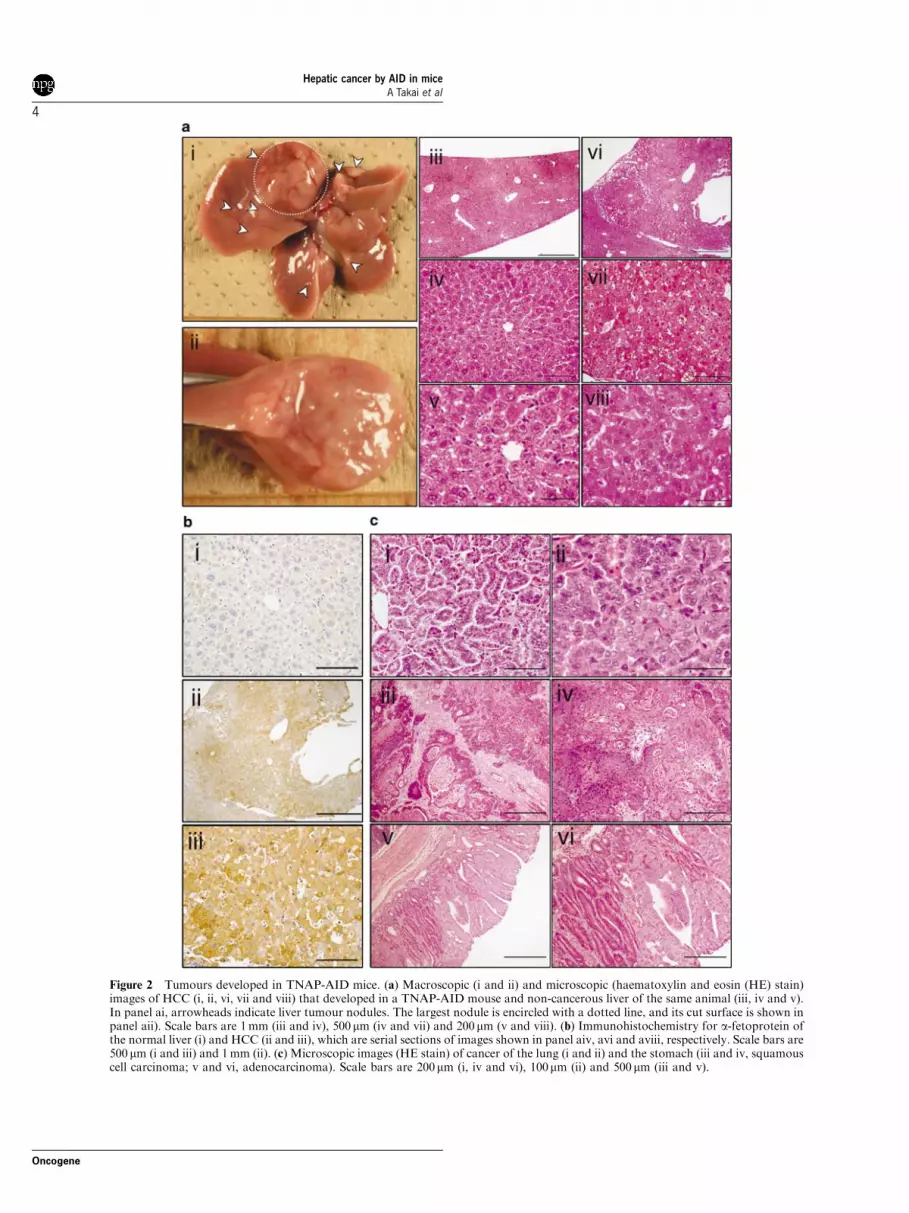
Figure 2 Tumours developed in TNAP-AID mice. (a) Macroscopic (i and ii) and microscopic (haematoxylin and eosin (HE) stain)images of HCC (i, ii, vi, vii and viii) that developed in a TNAP-AID mouse and non-cancerous liver of the same animal (iii, iv and v).In panel ai, arrowheads indicate liver tumour nodules. The largest nodule is encircled with a dotted line, and its cut surface is shown inpanel aii). Scale bars are 1 mm (iii and iv), 500mm (iv and vii) and 200mm (v and viii). (b) Immunohistochemistry for a-fetoprotein ofthe normal liver (i) and HCC (ii and iii), which are serial sections of images shown in panel aiv, avi and aviii, respectively. Scale bars are500mm (i and iii) and 1 mm (ii). (c) Microscopic images (HE stain) of cancer of the lung (i and ii) and the stomach (iii and iv, squamouscell carcinoma; v and vi, adenocarcinoma). Scale bars are 200mm (i, iv and vi), 100mm (ii) and 500mm (iii and v).
Hepatic cancer by AID in miceA Takai et al
4
Oncogene
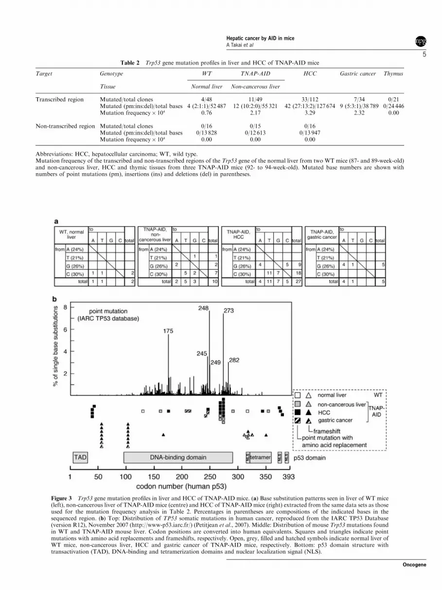
Table 2 Trp53 gene mutation profiles in liver and HCC of TNAP-AID mice
Target Genotype WT TNAP-AID HCC Gastric cancer Thymus
Tissue Normal liver Non-cancerous liver
Transcribed region Mutated/total clones 4/48 11/49 33/112 7/34 0/21Mutated (pm:ins:del)/total bases 4 (2:1:1)/52 487 12 (10:2:0)/55 321 42 (27:13:2)/127 674 9 (5:3:1)/38 789 0/24 446Mutation frequency� 104 0.76 2.17 3.29 2.32 0.00
Non-transcribed region Mutated/total clones 0/16 0/15 0/16Mutated (pm:ins:del)/total bases 0/13 828 0/12 613 0/13 947Mutation frequency� 104 0.00 0.00 0.00
Abbreviations: HCC, hepatocellular carcinoma; WT, wild type.Mutation frequency of the transcribed and non-transcribed regions of the Trp53 gene of the normal liver from two WT mice (87- and 89-week-old)and non-cancerous liver, HCC and thymic tissues from three TNAP-AID mice (92- to 94-week-old). Mutated base numbers are shown withnumbers of point mutations (pm), insertions (ins) and deletions (del) in parentheses.
Figure 3 Trp53 gene mutation profiles in liver and HCC of TNAP-AID mice. (a) Base substitution patterns seen in liver of WT mice(left), non-cancerous liver of TNAP-AID mice (centre) and HCC of TNAP-AID mice (right) extracted from the same data sets as thoseused for the mutation frequency analysis in Table 2. Percentages in parentheses are compositions of the indicated bases in thesequenced region. (b) Top: Distribution of TP53 somatic mutations in human cancer, reproduced from the IARC TP53 Database(version R12), November 2007 (http://www-p53.iarc.fr/) (Petitjean et al., 2007). Middle: Distribution of mouse Trp53 mutations foundin WT and TNAP-AID mouse liver. Codon positions are converted into human equivalents. Squares and triangles indicate pointmutations with amino acid replacements and frameshifts, respectively. Open, grey, filled and hatched symbols indicate normal liver ofWT mice, non-cancerous liver, HCC and gastric cancer of TNAP-AID mice, respectively. Bottom: p53 domain structure withtransactivation (TAD), DNA-binding and tetramerization domains and nuclear localization signal (NLS).
Hepatic cancer by AID in miceA Takai et al
5
Oncogene

Macroscopic tumours of other organs includedone case of lung adenocarcinoma and one case ofgastric cancer (squamous cell carcinoma) (Table 1 andFigure 2c). These rare tumours were restricted to theTNAP-AID group and likely to be caused by AIDexpression. However, the small number of tumoursprecludes a quantitative discussion on causal relation-ships, except for liver tumours. Sequencing analysisrevealed that Trp53mutation frequency in gastric cancertissue was 2.32/104, a comparable level to that observedin HCC tissues (Tables 2 and 3 as well as Figure 3).Unfortunately, we could not carry out mutationanalyses on the lung cancer tissue because of its smallsize. We did not examine microscopic tumours exceptfor subpleural lung microadenomas, which wereobserved in the TNAP-AID mice, a finding similar tothat in the AID transgenic mice in our earlier study(Okazaki et al., 2003). In addition, two cases of gastricadenocarcinoma were found incidentally by microscopicexamination (Figure 2c), suggesting that many micro-scopic tumours may have been overlooked.
Discussion
Here we describe TNAP-AID mice as a novel model ofhepatocarcinogenesis, which occurs as a result of theaccumulation of mutations in chronically inflamed liverin humans, often caused by infection with hepatitis
viruses. The TNAP-AID mouse model exhibits severalcharacteristics of human HCC, in that the mice developHCC spontaneously and the HCC tissue expressesa-fetoprotein and that it has the p53 gene mutations,some of which cause the same amino acid replacementsas those seen in human tumours. Earlier HCC mousemodels include mice with genetic modifications of Lkb1,Mdr2, Aox and Pten genes (Fan et al., 1998; Nakauet al., 2002; Horie et al., 2004; Katzenellenbogen et al.,2006), transgenic mice overexpressing c-myc, transform-ing growth factor-a, transforming growth factor-b1,HBx of hepatitis B virus and HCV core (Sandgren et al.,1989; Kim et al., 1991; Murakami et al., 1993; Koikeet al., 1994, 2002; Factor et al., 1997; Riehle et al., 2008)and chemical- or diet-induced HCC (Sell, 2001; Maedaet al., 2005; Ma et al., 2006; Sakurai et al., 2006). Incontrast to these models, our TNAP-AID model isunique because it does not arbitrarily target specificoncogenes, tumour suppressors or stability genes.In addition, it does not use chemical mutagens orspecific dietary conditions that are not associated withhuman HCC. The development of Trp53 mutations isanother feature of this model because it is unprecedentedin any earlier mouse HCC models as mentioned above.
Liver tumours were occasionally observed in ourearlier AID transgenic mice (Endo et al., 2007).However, it was difficult to use these mice as a livertumour model because of early death from lethallymphomas. Trials to detect Trp53 mutations in theliver tumour failed (data not shown). Even the T-cell
Table 3 Predicted amino acid replacements observed in three TNAP-AID mice (TA113, TA114 and TA128), corresponding human mutations andaffected domains of p53
Tissue Mutation Mouse ID Mutated/totalclone
Human-equivalentcodon
Domain Functional assaysa Counta
Nucleotide Codon Transactivationclass
Structure-basedprediction
TNAP-AID,non-cancerous liver
G472A A158T TA113 1/26 A161T DNA binding Partially functional Non-functional 73
C790T R264W TA128 2/23 R267W DNA binding Non-functional Non-functional 25C808G R270G TA128 2/23 R273G DNA binding Non-functional Non-functional 14G860A R287H TA113 1/26 R290H DNA binding Supertrans NA 29C1049T A350V TA113 1/26 A353V Tetramerization Supertrans NA 0
TNAP-AID, HCC G119C C40S TA113 2/67 None Transactivation NA NA NATA114 1/33
G127A D43N TA113 1/67 D42N Transactivation Functional NA 0C532T R178C TA114 1/33 R175C DNA binding Partially functional Non-functional 24G729A M243I TA113 1/67 M246I DNA binding Non-functional Functional 32C790T R264W TA113 3/67 R267W DNA binding Non-functional Non-functional 25
TA114 1/33
C808G R270G TA113 4/67 R273G DNA binding Non-functional Non-functional 14TA114 2/33
G1020C E340D TA113 1/67 E343D Tetramerization Functional NA 0G1112A G371D TA113 1/67 G374D Regulation/NLS Functional NA 0C1118G S373C TA113 1/67 S376C Regulation/NLS Functional NA 0G1127C R376P TA113 1/67 R379P Regulation Partially functional NA 0
TNAP-AID, gastric cancer G613T D205Y TA114 1/34 D208Y DNA binding Non-functional Functional 1G629A R210H TA114 1/34 R213Q DNA binding Non-functional Non-functional 34G716A C239Y TA114 1/34 C242Y DNA binding Non-functional Non-functional 51G729A M243I TA114 1/34 M246I DNA binding Non-functional Functional 32
Abbreviation: NA, data not available.For details, visit the IARC website.aBased on the data presented in the IARC website and described in the legend of Figure 3. Functional assays are predicted as impacts of mutationsin yeast assays and structural computation. Count is the number of the entries in the database.
Hepatic cancer by AID in miceA Takai et al
6
Oncogene

lymphomas that developed in the earlier AID transgenicmice did not harbour Trp53 mutations (Kotani et al.,2005). Therefore, the TNAP-AID mouse model is thefirst model in which Trp53 mutations can be detectedunequivocally in non-cancerous tissues as well as incancerous tissues that express AID. In spite of thesedifferences, the two independent AID transgenic micelines commonly developed liver tumours, making itunlikely that liver tumours in TNAP-AID mice are
ascribed to unexpected variables such as the transgeneintegration sites and the segregation of relevant lociupon crossing of mice with different genetic back-grounds. However, the possibility that these variablessomewhat modified the tumour frequency cannot beexcluded.
The overlap of mutational hot spots between TNAP-AID and human HCC (Table 3 and Figure 3b) suggeststhat these mutations induced by AID have critical roles.
Figure 4 Expression analysis of TNAP-AID mice. (a) Results of qRT-PCR for TNAP gene (Akp2) calibrated by the amount ofribosomal 18S RNA for indicated organs of adult mice (53- and 74-week-old) and four E14.5 embryos. The graph shows averageresults for the indicated groups. (b) Results of qRT-PCR for transgene-derived AID expression. (c) GFP fluorescence in frozen sectionsof GFP from the indicated organs and animals. A mosaic pattern of GFP expression in the liver of AID cTg mice was always observed,which may be attributed to the integration site of the transgene. (d) Western blot analysis using anti-AID monoclonal antibody MAID-1 for the lysates of indicated organs of 42-week-old TNAP-AID and AID cTg mice. Arrowhead indicates the position of the AID band.Comparable intensities of nonspecific bands indicate equal loading of extracts between TNAP-AID and AID cTg lanes.
Hepatic cancer by AID in miceA Takai et al
7
Oncogene
The lack of Trp53 mutations in the earlier transgenicmouse model may reflect a different evolutionary pathfrom the initiation to progression of liver tumoursbetween the two models despite the common involve-ment of AID. This difference may be related to thedifference in the frequency and aggressiveness oflymphomas between TNAP-AID and the earlier AIDtransgenic mouse, as discussed below. The pointmutations observed in Trp53 are likely to be a directeffect of AID activity, because they share somemolecular footprints with SHM of the immunoglobulingenes, including GC bias, frequent C-to-T transitionsand transcription dependence. However, there was nocorrelation between the distributions of mutations andSHM hot spot motifs or DNA secondary structures inthe Trp53 sequence. As these parameters show onlyweak association even for SHM, the small numbers ofpoint mutations observed in this study may not besufficient to achieve statistical significance. On the otherhand, single-base insertions causing frameshifts in Trp53were frequently observed in HCC but not in the non-cancerous liver of TNAP-AID mice. As single-baseinsertions are relatively rare in SHM induced by AID,these might be caused by genetic instability (forexample, impaired mismatch repair) acquired duringthe progression of the tumours.
The presence of mutations in non-cancerous tissueexpressing AID is reminiscent of a study on humanpatients with chronic hepatitis (Kou et al., 2006),together suggesting a causal role of AID in tumorigen-esis. It is noteworthy that the mutations corresponding
to two mutational hot spots in TP53 gene encodinghuman p53 (R273 and R175) were included in mutationsfound in TNAP-AID HCC. Other mutations weremapped close to the human hot spots (M246 andR267) (Figure 3b and Table 3). Among them threepatterns of amino acid replacement, C40S, R264W andR270G, were observed repeatedly in multiple individuals.Such repeated appearance of the specific mutations indifferent individuals may indicate their significance intumorigenesis. In addition to these common mutations,HCC and the non-cancerous liver contained quite diversemutation patterns, which parallels heterogeneity of TP53mutations within human HCC occurred in a singleindividual and is consistent with the ‘field cancerization’model (Thorgeirsson and Grisham, 2002).
The utility of the TNAP-AID mouse as a humanHCC model depends partly on the lack of lethallymphomas that were observed in almost all AIDtransgenic mice in our earlier study. To examine thereason why lymphomas were infrequent and lessaggressive in the TNAP-AID model, mutations causedby AID and its expression in T lymphocytes of TNAP-AID mice were analysed by sequencing the Trp53coding sequence (Table 2) and flow cytometry (Supple-mentary Figure 4). From these analyses, it was foundthat Trp53 was not mutated, but AID was expressed inthe thymocytes of the TNAP-AID mice. The level ofAID expression in the thymus of TNAP-AID mice washalf than that seen in the earlier AID transgenic line in aqRT-PCR analysis (data not shown). We speculate thatthe difference in the levels of AID expression in T cells
Figure 5 Immunohistochemical analysis of frozen liver sections from TNAP-AID (a–d), AID cTg (e–h) and WT (i–l) mice. (a, e and i)Hoechst 33342 staining; (b, f and j) AID immunostaining; (c, g and k) GFP fluorescence; (d, h and l) merge of the three colours.
Hepatic cancer by AID in miceA Takai et al
8
Oncogene

may be one of the reasons for this, although thedifference in the genetic background (C57BL/6:129/Svhybrid versus pure C57BL/6) cannot be excluded.
The TNAP-AID mice did not develop germ celltumours despite the expression of AID in the testes(Figure 4d) and the absence of GFP in the seminiferoustubules and oocytes of the ovaries (data not shown). Wereported earlier the lack of lymphomagenesis in B-cell-specific AID transgenic mice obtained by crossing thesame AID cTg line with CD19-Cre mice (Muto et al.,2006), suggesting that some protective mechanism mightexist in B cells, which have physiological expression ofAID. A similar reason might apply to germ cells becauseAID has been reported to be expressed physiologicallyin the human testis (Schreck et al., 2006) and mouseovary (Morgan et al., 2004).
To conclude, we generated a new line of AIDtransgenic mice that develop HCC but not lethal T-celllymphoma. We consider that the TNAP-AID mousemodel is useful for human HCC studies because it hasbeen shown that AID is induced in the humanpre-cancerous conditions of chronic hepatitis and cirrho-sis (Kou et al., 2006). The relatively long latency beforeHCC development in this model is reasonable, consider-ing that it is a physiological recapitulation of a humantumour phenotype that takes decades to develop.
Materials and methods
MiceThe AID cTg mice (line 20) on a C57BL/6 background (Mutoet al., 2006) were self-crossed to obtain homozygous transgenicmice, which were maintained in a specific pathogen-freefacility at the Kyoto University Faculty of Medicine andShiga Medical Center. This mouse line has been depositedat the Riken Bioresource Center (Tsukuba, Japan; No.RBRC00892). The TNAP-Cre mice (Lomeli et al., 2000) weregifted by Dr Andras Nagy (Mount Sinai Hospital, Toronto,Canada) and maintained by self-crossing between the hetero-zygous mice. C57BL/6 mice were purchased from ShimizuLaboratory Supplies Co., Ltd (Kyoto, Japan). All mice werefed ad libitum and were killed by cervical dislocation forcensoring, or observed until spontaneous death. Uponcensoring and spontaneous death, the numbers of macroscopictumours were counted after laparotomy and thoracotomy.Kaplan–Meier survival curves were analysed using Prism 4.0software (Graphpad, San Diego, CA, USA). All animalexperiments were approved by the Ethical Committee forAnimal Experiments and performed under the Guidelines forAnimal Experiments of Kyoto University and Shiga MedicalCenter.
Histology and immunohistochemistryParaffin-embedded mouse organs were sectioned and stainedwith haematoxylin and eosin. a-Fetoprotein was detectedusing anti-a-fetoprotein antibody (Dako, Glostrup, Denmark)and an ABC kit (Vector, Burlingame, CA, USA) for paraffin-embedded sections of formalin-fixed liver. For AID immuno-staining, freshly isolated liver was fixed in 4% paraformalde-hyde in phosphate-buffered saline on ice for 2 h and immersedin 30% sucrose for 18 h. After embedding in OCT compound(Sakura Fineteck Japan, Tokyo, Japan) in liquid nitrogen,
8-mm sections were sliced and mounted on glass slides. Hoechst33342 dye was used to visualize nuclei. AID protein wasdetected using the monoclonal anti-AID antibody, MAID-2(Tsuji et al., 2008), with peroxidase-labelled donkey F(ab0)2anti-rat IgG (Jackson ImmunoResearch, West Grove, PA,USA) and signal amplification using TSA Plus DNP and TSAPlus TMR kits (Perkin Elmer, Waltham, MA, USA). Imagestaken with a fluorescence microscope (DM5000B; Leica,Wetzlar, Germany) were merged using Photoshop (Adobe,San Jose, CA, USA).
Mutation analysisCancerous and non-cancerous liver tissues of approximatevolume of 50 mm3 were macroscopically dissected, frozen inliquid nitrogen and powdered with a frozen-cell crusher (Cryo-press; Microtec, Funabashi, Japan). Genomic DNA and totalRNA were extracted using the QIAamp DNA Mini kit(Qiagen, Duesseldorf, Germany) and the QuickGene kit (Fuji,Tokyo, Japan) from liquid nitrogen-frozen tissues that hadbeen dissected macroscopically from non-cancerous livers andcancerous nodules. Sequencing of Trp53 was performed asdescribed earlier (Matsumoto et al., 2007) except for primersfor the non-transcribed regions, sequences of which are shownin Supplementary Figure 5. Mutations of the Myc, Pten andCdkn2a genes were analysed as described (Matsumoto et al.,2007) except for primers, sequences of which are shown inSupplementary Figure 5. Statistical significance was assessedby Fisher’s exact test using Stata 8.2 software (Stata Corp,College Station, TX, USA). Searching for SHM hot spotmotifs and DNA secondary structure analyses were performedusing GENETYX-MAC 14 (Genetyx Corp., Tokyo, Japan)and mfold software (Zuker, 2003), respectively.
qRT-PCRcDNA was synthesized using the iScript kit (Bio-Rad, Hercules,CA, USA). Quantitative PCR (qPCR) was performed usingQuantiTect reagent (Qiagen) and a real-time thermal cycler(Mx3000P; Stratagene, La Jolla, CA, USA). Oligonucleotidesequences are shown in Supplementary Figure 5.
Western blottingMouse organs were dissected, frozen in liquid nitrogen andpowdered with a frozen-cell crusher (Cryo-press). Proteinswere extracted from the tissue powder using 10 mM sodiumphosphate (pH 6.8), 200 mM NaCl, 1.5 mM MgCl2 and 0.2 mM
EDTA with a protease inhibitor cocktail (Complete; RocheDiagnostics, Mannheim, Germany). Protein of 100 mg per lanewas applied to a 5–20% polyacrylamide gel (Bio-Rad),electrophoresed and blotted on to Hybond P membrane(GE Healthcare, Buckinghamshire, UK). Signals weregenerated using peroxidase-labelled anti-rat IgG (JacksonImmunoResearch) and the ECL Plus system (GE Healthcare)and detected with a LAS-3000 mini image analysis system(Fuji).
Acknowledgements
We thank Dr Andras Nagy for his generous gift of TNAP-Cremice, Dr Takashi Shinohara for suggesting the choice of theCre mouse strain, Dr Yoshinobu Toda for the preparation oftissue sections and Dr Masayuki Tsuji for technical help withthe immunohistochemical analyses. We also thank Dr SidoniaFagarasan for critical reading of the manuscript and discus-sions. This study was supported by Grant-in-Aid for ScientificResearch (17013042 and 18390122) and the Takeda ScienceFoundation.
Hepatic cancer by AID in miceA Takai et al
9
Oncogene

References
Endo Y, Marusawa H, Kinoshita K, Morisawa T, Sakurai T, OkazakiIM et al. (2007). Expression of activation-induced cytidinedeaminase in human hepatocytes via NF-kappaB signaling.Oncogene 26: 5587–5595.
Factor VM, Kao CY, Santoni-Rugiu E, Woitach JT, Jensen MR,Thorgeirsson SS. (1997). Constitutive expression of mature trans-forming growth factor beta1 in the liver accelerates hepatocarcino-genesis in transgenic mice. Cancer Res 57: 2089–2095.
Fan CY, Pan J, Usuda N, Yeldandi AV, Rao MS, Reddy JK. (1998).Steatohepatitis, spontaneous peroxisome proliferation and livertumors in mice lacking peroxisomal fatty acyl-CoA oxidase.Implications for peroxisome proliferator-activated receptor alphanatural ligand metabolism. J Biol Chem 273: 15639–15645.
Honjo T, Kinoshita K, Muramatsu M. (2002). Molecular mechanismof class switch recombination: linkage with somatic hypermutation.Annu Rev Immunol 20: 165–196.
Horie Y, Suzuki A, Kataoka E, Sasaki T, Hamada K, Sasaki J et al.(2004). Hepatocyte-specific Pten deficiency results in steatohepatitisand hepatocellular carcinomas. J Clin Invest 113: 1774–1783.
Katzenellenbogen M, Pappo O, Barash H, Klopstock N, Mizrahi L,Olam D et al. (2006). Multiple adaptive mechanisms to chronic liverdisease revealed at early stages of liver carcinogenesis in the Mdr2-knockout mice. Cancer Res 66: 4001–4010.
Kim CM, Koike K, Saito I, Miyamura T, Jay G. (1991). HBx gene ofhepatitis B virus induces liver cancer in transgenic mice. Nature 351:317–320.
Kinoshita K, Nonaka T. (2006). The dark side of activation-inducedcytidine deaminase: relationship with leukemia and beyond. Int J
Hematol 83: 201–207.Koike K, Moriya K, Iino S, Yotsuyanagi H, Endo Y, Miyamura Tet al. (1994). High-level expression of hepatitis B virus HBx gene andhepatocarcinogenesis in transgenic mice. Hepatology 19: 810–819.
Koike K, Moriya K, Kimura S. (2002). Role of hepatitis C virus in thedevelopment of hepatocellular carcinoma: transgenic approach toviral hepatocarcinogenesis. J Gastroenterol Hepatol 17: 394–400.
Komori J, Marusawa H, Machimoto T, Endo Y, Kinoshita K, Kou Tet al. (2008). Activation-induced cytidine deaminase links bileduct inflammation to human cholangiocarcinoma. Hepatology 47:888–896.
Kotani A, Okazaki IM, Muramatsu M, Kinoshita K, Begum NA,Nakajima T et al. (2005). A target selection of somatic hypermuta-tions is regulated similarly between T and B cells upon activation-induced cytidine deaminase expression. Proc Natl Acad Sci USA
102: 4506–4511.Kou T, Marusawa H, Kinoshita K, Endo Y, Okazaki IM, Ueda Yet al. (2006). Expression of activation-induced cytidine deaminase inhuman hepatocytes during hepatocarcinogenesis. Int J Cancer 120:469–476.
Kues WA, Petersen B, Mysegades W, Carnwath JW, Niemann H.(2005). Isolation of murine and porcine fetal stem cells from somatictissue. Biol Reprod 72: 1020–1028.
Lomeli H, Ramos-Mejia V, Gertsenstein M, Lobe CG, Nagy A.(2000). Targeted insertion of Cre recombinase into the TNAP gene:excision in primordial germ cells. Genesis 26: 116–117.
Ma W, Xia X, Stafford LJ, Yu C, Wang F, LeSage G et al. (2006).Expression of GCIP in transgenic mice decreases susceptibility tochemical hepatocarcinogenesis. Oncogene 25: 4207–4216.
Maeda S, Kamata H, Luo JL, Leffert H, Karin M. (2005). IKKbetacouples hepatocyte death to cytokine-driven compensatory prolifera-tion that promotes chemical hepatocarcinogenesis. Cell 121: 977–990.
Marusawa H. (2008). Aberrant AID expression and human cancerdevelopment. Int J Biochem Cell Biol 40: 1399–1402.
Matsumoto Y, Marusawa H, Kinoshita K, Endo Y, Kou T, MorisawaT et al. (2007). Helicobacter pylori infection triggers aberrantexpression of activation-induced cytidine deaminase in gastricepithelium. Nat Med 13: 470–476.
Morgan HD, Dean W, Coker HA, Reik W, Petersen-Mahrt SK.(2004). Activation-induced cytidine deaminase deaminates 5-methyl-cytosine in DNA and is expressed in pluripotent tissues: implicationsfor epigenetic reprogramming. J Biol Chem 279: 52353–52360.
Murakami H, Sanderson ND, Nagy P, Marino PA, Merlino G,Thorgeirsson SS. (1993). Transgenic mouse model for synergisticeffects of nuclear oncogenes and growth factors in tumorigenesis:interaction of c-myc and transforming growth factor alpha inhepatic oncogenesis. Cancer Res 53: 1719–1723.
Muto T, Okazaki IM, Yamada S, Tanaka Y, Kinoshita K, MuramatsuM et al. (2006). Negative regulation of activation-induced cytidinedeaminase in B cells. Proc Natl Acad Sci USA 103: 2752–2757.
Nakau M, Miyoshi H, Seldin MF, Imamura M, Oshima M, TaketoMM. (2002). Hepatocellular carcinoma caused by loss of hetero-zygosity in Lkb1 gene knockout mice. Cancer Res 62: 4549–4553.
Okazaki IM, Hiai H, Kakazu N, Yamada S, Muramatsu M, KinoshitaK et al. (2003). Constitutive expression of AID leads to tumorigen-esis. J Exp Med 197: 1173–1181.
Petitjean A, Mathe E, Kato S, Ishioka C, Tavtigian SV, Hainaut Pet al. (2007). Impact of mutant p53 functional properties on TP53mutation patterns and tumor phenotype: lessons from recentdevelopments in the IARC TP53 database. Hum Mutat 28: 622–629.
Riehle KJ, Campbell JS, McMahan RS, Johnson MM, Beyer RP,Bammler TK et al. (2008). Regulation of liver regeneration andhepatocarcinogenesis by suppressor of cytokine signaling 3. J Exp
Med 205: 91–103.Sakurai T, Maeda S, Chang L, Karin M. (2006). Loss of hepatic NF-
kappa B activity enhances chemical hepatocarcinogenesis throughsustained c-Jun N-terminal kinase 1 activation. Proc Natl Acad Sci
USA 103: 10544–10551.Sandgren EP, Quaife CJ, Pinkert CA, Palmiter RD, Brinster RL.
(1989). Oncogene-induced liver neoplasia in transgenic mice.Oncogene 4: 715–724.
Schreck S, Buettner M, Kremmer E, Bogdan M, Herbst H, NiedobitekG. (2006). Activation-induced cytidine deaminase (AID) is expressedin normal spermatogenesis but only infrequently in testicular germcell tumours. J Pathol 210: 26–31.
Sell S. (2001). Heterogeneity and plasticity of hepatocyte lineage cells.Hepatology 33: 738–750.
Thorgeirsson SS, Grisham JW. (2002). Molecular pathogenesis ofhuman hepatocellular carcinoma. Nat Genet 31: 339–346.
Tsuji M, Suzuki K, Kitamura H, Maruya M, Kinoshita K, Ivanov IIet al. (2008). Requirement for lymphoid tissue-inducer cells inisolated follicle formation and T cell-independent immunoglobulin ageneration in the gut. Immunity 29: 261–271.
Urven LE, Weng DE, Schumaker AL, Gearhart JD, McCarrey JR.(1993). Differential gene expression in fetal mouse germ cells. Biol
Reprod 48: 564–574.Vogelstein B, Kinzler KW. (2004). Cancer genes and the pathways they
control. Nat Med 10: 789–799.Wang L, Duan E, Sung LY, Jeong BS, Yang X, Tian XC. (2005).
Generation and characterization of pluripotent stem cells fromcloned bovine embryos. Biol Reprod 73: 149–155.
Yoshikawa K, Okazaki IM, Eto T, Kinoshita K, Muramatsu M,Nagaoka H et al. (2002). AID enzyme-induced hypermutation in anactively transcribed gene in fibroblasts. Science 296: 2033–2036.
Zuker M. (2003). Mfold web server for nucleic acid folding andhybridization prediction. Nucleic Acids Res 31: 3406–3415.
Supplementary Information accompanies the paper on the Oncogene website (http://www.nature.com/onc)
Hepatic cancer by AID in miceA Takai et al
10
Oncogene
Related Documents


