Research Article A Novel Homozygous Mutation in the EC1/EC2 Interaction Domain of the Gap Junction Complex Connexon 26 Leads to Profound Hearing Impairment Ralf Birkenhäger, Nicola Prera, Antje Aschendorff, Roland Laszig, and Susan Arndt Department of Otorhinolaryngology, Head and Neck Surgery, University Medical Center Freiburg, Killianstraße 5, 79106 Freiburg, Germany Correspondence should be addressed to Ralf Birkenh¨ ager; [email protected] Received 29 April 2013; Revised 27 September 2013; Accepted 14 October 2013; Published 16 January 2014 Academic Editor: Brad Upham Copyright © 2014 Ralf Birkenh¨ ager et al. is is an open access article distributed under the Creative Commons Attribution License, which permits unrestricted use, distribution, and reproduction in any medium, provided the original work is properly cited. To date, about 165 genetic loci or genes have been identified which are associated with nonsyndromal hearing impairment. In about half the cases, genetic defects in the GJB2 gene (connexin 26) are the most common cause of inner-ear deafness. e genes GJB2 and GJB6 are localized on chromosome 13q11-12 in tandem orientation. Connexins belong to the group of “gap junction” proteins, which form connexons, each consisting of six connexin molecules. ese are responsible for the exchange of ions and smaller molecules between neighboring cells. Mutational analysis in genes GJB2 and GJB6 was brought by direct sequencing of the coding exons including the intron transitions. Here we show in the participating extended family a homozygous mutation c.506G>A, (TGC>TAC) p.Cys169Tyr, in the GJB2 gene, which could be proven for the first time and led to nonsyndromal severe hearing impairment in the afflicted patients. e mutation is located in the EC1/EC2 interaction complex of the gap junction connexon 26 complex and interrupts the K + circulation and therefore the ion homeostasis in the inner ear. e homozygous mutation p.Cys169Tyr identified here provides a novel insight into the structure-function relationship of the gap junction complex connexin/connexon 26. 1. Introduction Severe prelingual sensorineural hearing impairment is diag- nosed in about 2 of 1000 neonates. Nearly 50% of these cases are genetic in origin. Two-thirds of the cases are non- syndromal. 80% of the genetically caused hearing losses follow autosomal recessive transmission [1]. To date, about 140 genetic loci have been identified in connection with hearing loss, of which 49 genes have been identified and characterized (connexin-deafness homepage). 50% of all autosomal recessive inherited hearing losses show mutations in the GJB2 gene (MIM 121011) [2]. is gene consists of two exons and is localized in tandem orientation with the GJB6 gene (MIM 604418) at genetic locus DFNB1 A/B (MIM 220290, MIM 612645) on chromosome 13q12. Six connexins form a molecular complex, a so-called connexon, which is localized in the cell membrane and, as a “gap junction” with the corresponding connexon of neighboring cells, enables the exchange of metabolites and potassium. is electrolyte exchange is of decisive importance for the electrical potential in the cochlea [3]. To date, more than 91 different mutations in the GJB2 gene have been proven in connection with hearing loss (connexin-deafness homepage, which lists, however, only mutations characterized until 2003). A recently published article by Hilgert et al. [4] reports that about 220 mutations in the GJB2 gene have been described worldwide. Among the most frequently occurring mutations in the GJB2 gene are 30/35delG, 167delT, 235delC, L90P, E47X, and M34delT [5–7]. ree large deletions have also been characterized thus far, involving part of the GJB6 gene and the associated chromosomal downstream region, which also lead to serious non-syndromal hearing impairment. Here we describe a novel homozygotic missense mutation in the GJB2 gene and characterize its influence on the tertiary structure of the connexon-connexon interaction domain of the connexin 26 protein, which led to non-syndromal Hindawi Publishing Corporation BioMed Research International Volume 2014, Article ID 307976, 7 pages http://dx.doi.org/10.1155/2014/307976

Welcome message from author
This document is posted to help you gain knowledge. Please leave a comment to let me know what you think about it! Share it to your friends and learn new things together.
Transcript
Research ArticleA Novel Homozygous Mutation in the EC1/EC2 InteractionDomain of the Gap Junction Complex Connexon 26 Leads toProfound Hearing Impairment
Ralf Birkenhäger, Nicola Prera, Antje Aschendorff, Roland Laszig, and Susan Arndt
Department of Otorhinolaryngology, Head and Neck Surgery, University Medical Center Freiburg, Killianstraße 5,79106 Freiburg, Germany
Correspondence should be addressed to Ralf Birkenhager; [email protected]
Received 29 April 2013; Revised 27 September 2013; Accepted 14 October 2013; Published 16 January 2014
Academic Editor: Brad Upham
Copyright © 2014 Ralf Birkenhager et al. This is an open access article distributed under the Creative Commons AttributionLicense, which permits unrestricted use, distribution, and reproduction in any medium, provided the original work is properlycited.
To date, about 165 genetic loci or genes have been identified which are associated with nonsyndromal hearing impairment. Inabout half the cases, genetic defects in the GJB2 gene (connexin 26) are the most common cause of inner-ear deafness. The genesGJB2 and GJB6 are localized on chromosome 13q11-12 in tandem orientation. Connexins belong to the group of “gap junction”proteins, which form connexons, each consisting of six connexin molecules. These are responsible for the exchange of ions andsmaller molecules between neighboring cells. Mutational analysis in genes GJB2 and GJB6 was brought by direct sequencing ofthe coding exons including the intron transitions. Here we show in the participating extended family a homozygous mutationc.506G>A, (TGC>TAC) p.Cys169Tyr, in the GJB2 gene, which could be proven for the first time and led to nonsyndromal severehearing impairment in the afflicted patients. The mutation is located in the EC1/EC2 interaction complex of the gap junctionconnexon 26 complex and interrupts the K+ circulation and therefore the ion homeostasis in the inner ear. The homozygousmutation p.Cys169Tyr identified here provides a novel insight into the structure-function relationship of the gap junction complexconnexin/connexon 26.
1. Introduction
Severe prelingual sensorineural hearing impairment is diag-nosed in about 2 of 1000 neonates. Nearly 50% of thesecases are genetic in origin. Two-thirds of the cases are non-syndromal. 80% of the genetically caused hearing lossesfollow autosomal recessive transmission [1]. To date, about140 genetic loci have been identified in connection withhearing loss, of which 49 genes have been identified andcharacterized (connexin-deafness homepage). 50% of allautosomal recessive inherited hearing losses show mutationsin the GJB2 gene (MIM 121011) [2]. This gene consists oftwo exons and is localized in tandem orientation with theGJB6 gene (MIM 604418) at genetic locus DFNB1 A/B (MIM220290, MIM 612645) on chromosome 13q12. Six connexinsform a molecular complex, a so-called connexon, which islocalized in the cell membrane and, as a “gap junction” withthe corresponding connexon of neighboring cells, enables
the exchange of metabolites and potassium. This electrolyteexchange is of decisive importance for the electrical potentialin the cochlea [3]. To date,more than 91 differentmutations inthe GJB2 gene have been proven in connection with hearingloss (connexin-deafness homepage,which lists, however, onlymutations characterized until 2003). A recently publishedarticle by Hilgert et al. [4] reports that about 220 mutationsin the GJB2 gene have been described worldwide. Amongthe most frequently occurring mutations in the GJB2 geneare 30/35delG, 167delT, 235delC, L90P, E47X, and M34delT[5–7]. Three large deletions have also been characterizedthus far, involving part of the GJB6 gene and the associatedchromosomal downstream region, which also lead to seriousnon-syndromal hearing impairment.
Herewe describe a novel homozygoticmissensemutationin theGJB2 gene and characterize its influence on the tertiarystructure of the connexon-connexon interaction domainof the connexin 26 protein, which led to non-syndromal
Hindawi Publishing CorporationBioMed Research InternationalVolume 2014, Article ID 307976, 7 pageshttp://dx.doi.org/10.1155/2014/307976

2 BioMed Research International
prelingual deafness in an extended consanguineous Arabianfamily from the Middle East.
2. Methods
2.1. Patient 1. The patient, a boy, was brought to our depart-ment at the age of 14 months with suspected congenital high-grade deafness on both sides. At the age of 6 months, hehad been fitted with hearing aids on both sides, which hedid not tolerate. The child occasionally reacted to very close,loud noises; he was clearly face oriented in communication.There was no clinical evidence of a syndromal disease, andthe pregnancy was normal. As part of preop evaluationfor cochlear implantation, electrocochleography was per-formed on both sides after adenotomy and paracentesis.The compound action potentials were negative, and cochlearmicrophonics could be recorded on both sides starting at110 dBHL. No potentials could be recorded in brainstemelectric response audiometry (BERA). Hearing loss has alsobeen found in the sister and paternal grandfather.There is noclinical evidence of a syndromal disease (Figure 1).
2.2. Patient 2. The patient was brought to our departmentat the age of 13 months with suspected congenital deafness.The patient did not react to sounds; he vocalized andfollowed attentively with his eyes. There were no previousdiseases such as meningitis or recurrent otitides. The ENTexamination revealed a nonirritative tympanic membrane onboth sides, with no evidence of middle ear effusion. In thepedaudiological examination with hearing aids, he perceivedthe offered tones only in the deep-tone frequency rangebetween 75 and 100 dB; no reactions could be recorded onthe left side. Preevaluation for cochlear implantation wasthen performed. The electrocochleography revealed negativecompound action potentials on both sides; cochlear micro-phonics could be recorded on both sides starting at 110 dBHL.Brainstem electric response audiometry (BERA) found nopotentials on either side.The father andmother are both deaf.There is also familiar deafness in an uncle, a great-uncle, and2 cousins in the father’s family (Figure 1).
To rule out malformations, radiological examination ofthe skull was performed on both patients with CT andMRT scans. Examination showed nothing conspicuous; boththe bony structures of the petrous bone and the internalauditory canal were without pathological findings (Figure 1).Subsequently, serious prelingual, bilateral impairment ofsound perception (non-syndromal deafness) was diagnosedin both cases. Both patients underwent cochlear implantation(Nucleus Contour advanced electrode Cochlear Ltd., Sydney,Australia) at the age of 14 (patient II-3) and 13 (patient II-6)months, respectively.
2.3. Gene/Mutation Analysis. The Ethics Committee of theUniversity of Freiburg approved this project (no. 161/02-07/2003/Birkenhager). Genomic DNA was extracted fromperipheral blood leukocytes of the patients (II-3, II-6), themother (I-2) of patient II-3, and the parents (I-4, I-5) ofpatient II-6 (Figure 1), using standard methods (Qiagen).
Primer and PCR conditions were selected according toprocedures optimized previously for sequence analysis of thecoding exon of theGJB2 gene, including the intron transitionsand deletion analysis of the GJB6 gene [8]. Sequencingof the PCR products was done with standard proceduresand analyzed in an automated DNA sequencer AmershamMegaBACE 500 (Amersham Biosciences).
3. Results
High-resolution computed tomography of the petrous bonerevealed no morphological anomalies in the sense of malfor-mation of the cochlea or the vestibular system in either case asthe cause of prelingual non-syndromal hearing impairment(Figure 2).
Sequence analyses of the coding exons and the introntransitions of genes GJB2 and GJB6 and deletion analysesshowed the same homozygotic mutation in both patients(Figure 3(a)). The mutations c.506G>A, (TGC>TAC),Cys169Tyr in the GJB2 gene were identified in both patients.This mutation was proven heterozygous in the parents (I-2,I-4, and I-5) of the patients (Figure 3(b)). No mutation wasidentified in the GJB6 gene; the known deletions were alsonot detected in the GJB6 gene.
Novel missense mutations can be evaluated for possiblepathogenic protein effects by prediction tools such SIFTand PolyPhen [9]. The PolyPhen [http://genetics.bwh.har-vard.edu/pph2/] and SIFT [http://sift-bioinformatics.soft112.com/] prediction tools offer an in silico mechanism to inves-tigate the potential pathogenicity of novel missense varia-tions. When analyzing the mutation using the PolyPhen andSIFT tools, it is predicted that the new mutation p.Cys169Tyrprobably changed the conformation of the connexin-26protein. Both tools describe the fact that a change in theamino acid leads to a loss of function of the connexin 26protein. A substitution of a tyrosine for the highly conservedcysteine changes the three-dimensional arrangement of theEC1 and EC2 subdomain, in the intracytoplasm domain ofthe protein complex of connexin 26, leading to a defectiveprotein and associated deafness. This leads to a change inthe extracellular domain, so that the interaction betweenconnexon complexes is no longer possible, thus interruptingthe potassium cycle in the inner ear, and ultimately the ionhomeostasis can no longer be maintained.
4. Discussion
The causes of a non-syndromal prelingual inner-ear hearingimpairment or deafness cannot always be unequivocallydiagnosed at the molecular level, since more than 140 dif-ferent genes and genetic loci involved in the developmentand function of hearing are currently under discussion(Hereditary Hearing Loss Homepage). Since gene GJB2 wasidentified and characterized, it could be shown that defects,that is, mutations, are present in this gene in more thanhalf of all cases. To date, about 220 recessive mutationshave been described [4]. Nine dominant mutations havealso been identified, which are usually associated with skin

BioMed Research International 3
∗+ +
∗+ ∗
+++
∗+
??
II-3 II-4 II-5 II-6
I-1 I-2 I-4 I-5
Figure 1: Pedigree of the family: patients are carriers of the homozygotic mutation (II-3 and II-6) (black); parents of the patients are eachcarrier of the heterozygotic mutation (I-2, I-4, and I-5) (half black). (∗) Members of the family are also hearing impaired, but the clinicalcourse indicates other causes (grey and half grey); (+) no genetic material was available; (?) no information available.
(a) (b)
(c)
Figure 2: High-resolution computed tomography of the inner-ear structures of the patients: (a) II-3 and (b) II-5. There is no evidence ofmalformations of the cochlea or the petrous bone; (c) control image.
diseases, such as Keratitis-Ichthyosis-Deafness Syndrome(MIM 148210) [10], Vohwinkel’s Syndrome (MIN124500)[11], and palmoplantar hyperkeratosis (MIN148350) [12].Interestingly, these are arranged exclusively in the highly-preserved CNX domain of the connexin 26 gene betweenamino acids 42 and 75 in the protein. By contrast, onlydeletions have been found in the GJB6 gene to date [13].Connexins belong to a group of integral membrane proteins,of which six oligomerize and form intercellular canals, thatis, “gap junction” complexes.These canals enable an exchangeof low-molecular metabolites <1-2 kD, signal molecules, and
ions between neighboring cells. These “gap junction” proteincomplexes have thus far been identified in various vertebratecell types.
Gap junctions play important roles in different biolog-ical processes. Gap junction channels are formed by theinteraction of two hemichannels, connexons, each of whichis composed of six connexin molecules surrounding thecentral pore. In the human system, 21 different connexinshave been characterized, each has distinct physiologicalactivities. Electrophysiological studies have demonstratedthat gap junctions have multiple gating mechanisms. Gap

4 BioMed Research International
G T G A A G T A C A A C G C C
167 168 169 170 171
AlaAsnTyrLysVal
(a)
167 168 169 170 171
AlaAsnLysVal
GAG T G A A G T C A A C G C C
Cys/Tyra
(b)
G T G A A G T C A A C G C C
167 168 169 170 171
AlaAsnLysVal CysG
(c)
Figure 3: Electrogram of the mutation; (a) homozygote (c.506G>A, (TGC>TAC), Cys169Tyr) in patients (II-3, II-6); (b) heterozygote in theparents (I-2, I-4, and I-5); (c) control.
junctions can be gated by membrane voltage and by chemicalfactors [14]. A variety of mutations of connexin genes havebeen shown to be associated with a wide range of inheriteddiseases, for example, deafness, skin diseases, developmentalabnormalities, and so forth [15, 16]. X-ray crystallography ofthe connexin 26 has provided structural details. Connexin26 contains four transmembrane helices (TM1–TM4), twoextracellular loops (E1, E2), and an N-terminal helix [15].The E1 and E2 domains compose together the extracellularcomplex of the connexon hemichannel; this complex con-tributes to interhemichannel interactions of the connexons.The E1 and E2 complex of one hemichannel interacts with thecorresponding E1 and E2 complex of the other hemichannel.Six conserved cysteine residues form three intramoleculardisulphide bonds between E1 and E2; these bonds stabilizethe structure of the extracellular region. It is conspicuousthat the motifs of extracellular cysteine are conserved in allhuman connexins. Amino acid substitutions in any of themlead to a loss of functional gap junction channels in Xenopusoocyte experiments.This is probably because of the structuraldisorder of the extracellular region [17]. Amino acid residues
that contribute to inter-hemichannel interactions are highlyconserved in all connexins. Mutations of the residues thatcontribute to inter-hemichannel interactions are associatedwith human diseases, as in the case for mutations of aminoacid residues involved in interactions that stabilize connexinand connexon structures [18, 19]. Oshima et al. [18] analyzedthe roles of cysteine 64 residue by expressing mutant connex-ins in insect Sf9 and HeLa cells. Residue cysteine 64 is thethird cysteine in the E1 domain, and the mutation cysteine 64results in a loss of the electric coupling activity and causedthe most profound defects among all mutations examined.They suggested that the mutated cysteine 64 has a decisiveinfluence onoligomerization and/or protein folding andplaysan important role in connexon assembly [17].The importanceof disulfide bonds in the extracellular domains has also beenstudied in other proteins, and the substitutions of cysteineresulted in a reduced stability [18]. All these experimentssuggest that cysteine forms extracellular disulfide bridges inconnexin, which is important to maintain a stable connexinstructure that is suitable for connexon hemichannels forma-tion.
BioMed Research International 5
In the absence of functional studies, a common approachto investigate missense mutations is by multiple comple-mentary means: a database and the literature search, anevaluation of conservations across species, family studies, andresearch tools such as SIFT and PolyPhen [9]. Invertebrateshave similar protein complexes with the same secondarystructure, but with lower sequence identity [20]. So far,13 different connexins have been characterized. Hydropa-thy analyses show that all have the same transmembranaltopology, four transmembranal helices, an intracellular andtwo extracellular loops, N- and C-termini of the protein arelocated in the cytoplasm. The sequence similarity amongthe various isoforms is about 50–80%; the transmembranalhelices are particularly well preserved [21]. By contrast,the intracellular domains, as well as the N- and C-termini,are highly variable. Both extracellular domains contain threespecific cysteine residuals which are highly preserved in allconnexins, forming intramolecular disulfide bridges betweenthe cysteine residuals 53 and 180, 60 and 174, and also 64and 169 [22].These three disulfide bridges probably stabilize aspecific three-dimensional structure of the two extracellulardomains in connexin 26 molecules of a connexon complex,which is prerequisite to the interaction with a connexonof a neighboring cell. The novel homozygotic mutationc.506G>A, (TGC>TAC), Cys169Tyr, described here for thefirst time, localized in the second extracellular domain ofthe connexin 26 protein, leads to an amino acid exchange atposition 169 of the connexin 26 protein of cysteine to tyrosine.In addition, as derived from the structure of the connexin26 channel at 3.5 A resolution, recently published by Maedaet al. 2009 [23], it is clearly demonstrated that the aminoacid is important for the three-dimensional structure of theextracellular domain and thus for the interaction betweentwo connexons. The C169Y mutation occurs in a highlyconserved region of the second extracellular domain andaffects one of three cysteine residuals involved in the subunitdisulfide bonds that are crucial for the connexon-connexoninteraction [24]. Moreover, extracellular cysteines are criticalfor the correct folding of the protein and essential for propercorrect channel function [25]. The amino acid Cys169 isinvolved in one of three disulfide bridges which stabilizethe three-dimensional structure between the extracellulardomains E1 and E2 of the protein connexin 26. Cys169Tyris fundamental for the interaction between two connexincomplexes of adjacent cells. All evidences point to the fact thatthe characterized amino acid exchange Cys 169Tyr is a realmutation and is not a polymorphism as described by Murgiaet al. (connexin-deafness homepage).
Several multiple sequence alignment tools are availablefor evaluating the evolutionary conservation of amino acidsacross gene families and species. Multiple sequence align-ment analyses are based largely on the assumptions thatevolutionarily-conserved amino acids are more likely tobe functionally important than nonconserved amino acidsand substitutions involving chemically similar amino acidsare less likely to be damaging than substitutions involv-ing chemically different amino acids. However, multiplesequence alignment analyses can only provide an illustrationof the degree of evolutionary conservation of an amino
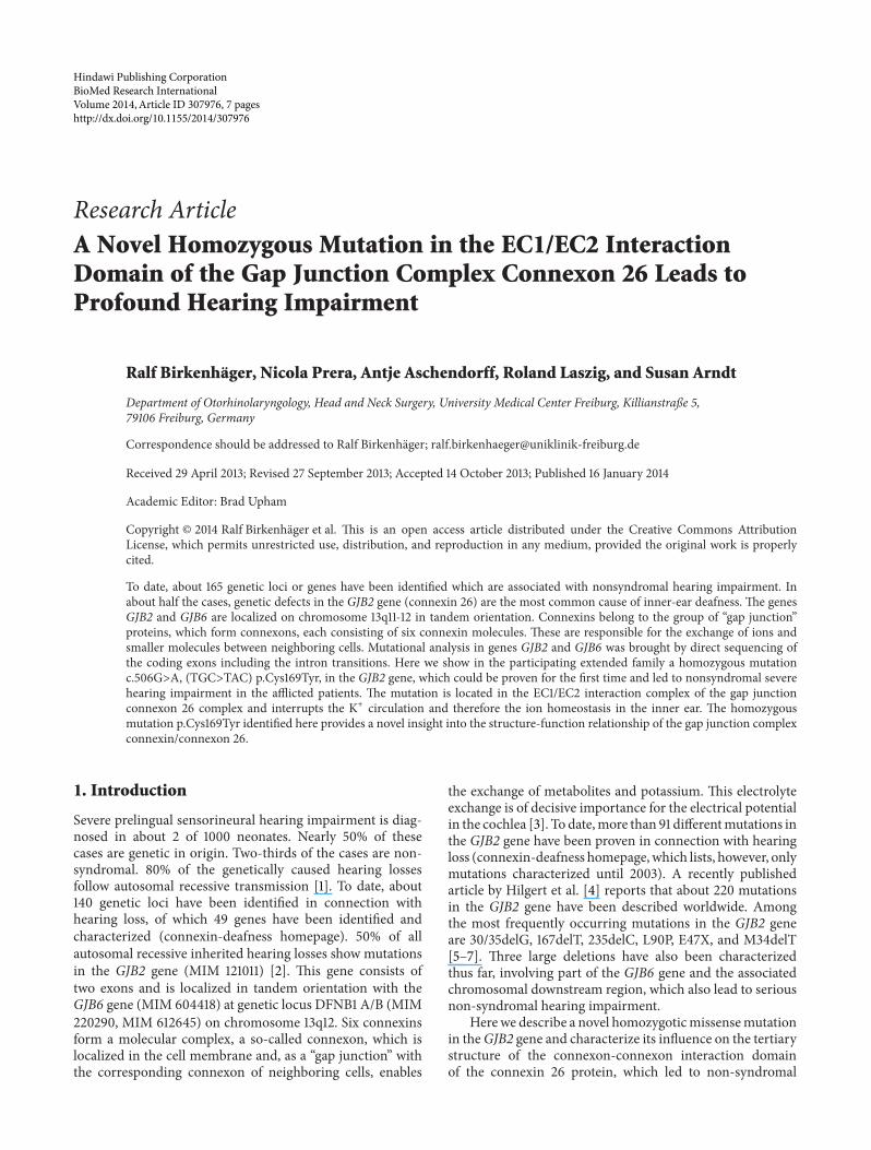
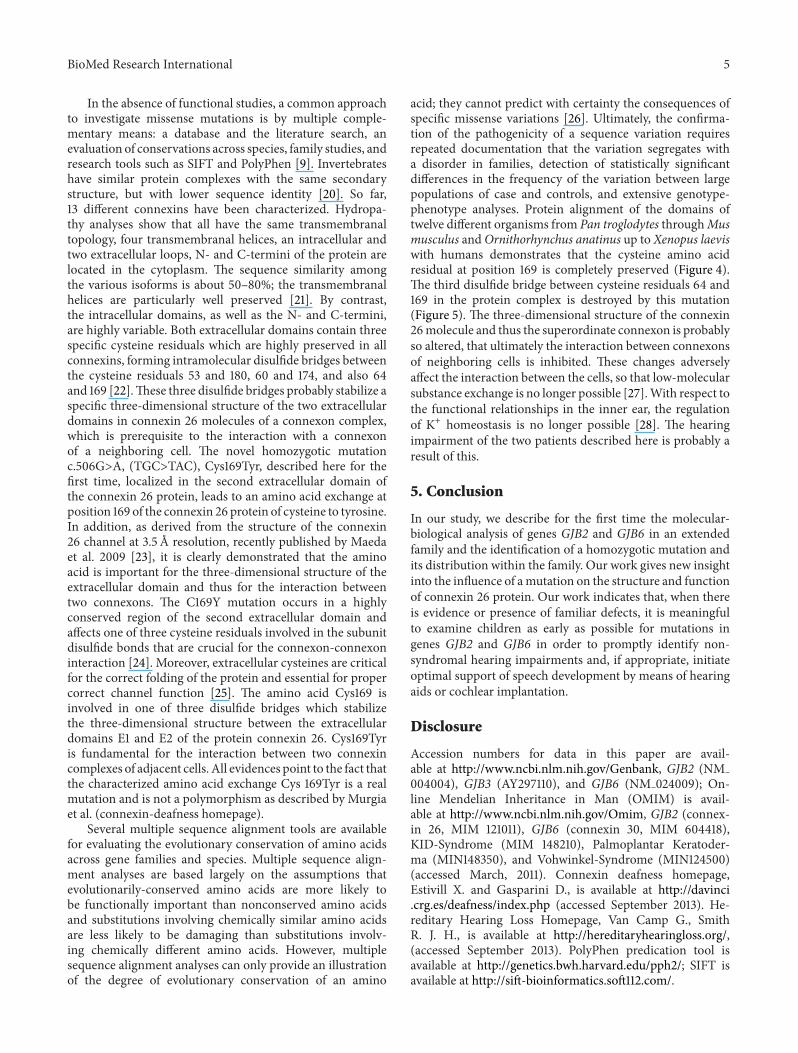
acid; they cannot predict with certainty the consequences ofspecific missense variations [26]. Ultimately, the confirma-tion of the pathogenicity of a sequence variation requiresrepeated documentation that the variation segregates witha disorder in families, detection of statistically significantdifferences in the frequency of the variation between largepopulations of case and controls, and extensive genotype-phenotype analyses. Protein alignment of the domains oftwelve different organisms from Pan troglodytes throughMusmusculus andOrnithorhynchus anatinus up to Xenopus laeviswith humans demonstrates that the cysteine amino acidresidual at position 169 is completely preserved (Figure 4).The third disulfide bridge between cysteine residuals 64 and169 in the protein complex is destroyed by this mutation(Figure 5). The three-dimensional structure of the connexin26molecule and thus the superordinate connexon is probablyso altered, that ultimately the interaction between connexonsof neighboring cells is inhibited. These changes adverselyaffect the interaction between the cells, so that low-molecularsubstance exchange is no longer possible [27].With respect tothe functional relationships in the inner ear, the regulationof K+ homeostasis is no longer possible [28]. The hearingimpairment of the two patients described here is probably aresult of this.
5. Conclusion
In our study, we describe for the first time the molecular-biological analysis of genes GJB2 and GJB6 in an extendedfamily and the identification of a homozygotic mutation andits distribution within the family. Our work gives new insightinto the influence of amutation on the structure and functionof connexin 26 protein. Our work indicates that, when thereis evidence or presence of familiar defects, it is meaningfulto examine children as early as possible for mutations ingenes GJB2 and GJB6 in order to promptly identify non-syndromal hearing impairments and, if appropriate, initiateoptimal support of speech development by means of hearingaids or cochlear implantation.
Disclosure
Accession numbers for data in this paper are avail-able at http://www.ncbi.nlm.nih.gov/Genbank, GJB2 (NM004004), GJB3 (AY297110), and GJB6 (NM 024009); On-line Mendelian Inheritance in Man (OMIM) is avail-able at http://www.ncbi.nlm.nih.gov/Omim, GJB2 (connex-in 26, MIM 121011), GJB6 (connexin 30, MIM 604418),KID-Syndrome (MIM 148210), Palmoplantar Keratoder-ma (MIN148350), and Vohwinkel-Syndrome (MIN124500)(accessed March, 2011). Connexin deafness homepage,Estivill X. and Gasparini D., is available at http://davinci.crg.es/deafness/index.php (accessed September 2013). He-reditary Hearing Loss Homepage, Van Camp G., SmithR. J. H., is available at http://hereditaryhearingloss.org/,(accessed September 2013). PolyPhen predication tool isavailable at http://genetics.bwh.harvard.edu/pph2/; SIFT isavailable at http://sift-bioinformatics.soft112.com/.

6 BioMed Research International
TM1 TM2 TM3 TM4
EVWGDEQADFVCNTLQPGCKNVCYDHYFPISHIR........YVMYDGFSMQRLVKCNAWPCPNTVDCFVSRPTEKTVFT
CNX
226 AA
Homo sapiensPongo pygmaeus Macaca mulattaHylobates larBos taurusEquus caballusCanis familiarisOvis ariesMus musculusCricetulus griseusOrnithorhynchus anatinusXenopus laevis
Consensus sequence
YVMYDGFSMQRLVKCNAWPCPNTVDCFVSRPTEKTVFT
YVMYDGFSMQRLVKCNAWPCPNTVDCFVSRPTEKTVFT
YVMYDGFSMQRLVKCNAWPCPNTVDCFVSRPTEKTVFT
YVMYDGFSMQRLVKCNAWPCPNTVDCFVSRPTEKTVFT
YVMYDGFAMQRLVKCNAWPCPNTVDCFVSRPTEKTVFT
YVMYDGFAMQRLVKCNAWPCPNTVDCFVSRPTEKTVFT
YVMYDGFSMQRLVKCNAWPCPNTVDCFVSRPTEKTVFT
YVMYDGFAMQRLVKCNAWPCPNTVDCFVSRPTEKTVFT
YIMYNGFFMQRLVKCNAWPCPNTVDCFISRPTEKTVFT
YIMYNGFFMQRLVKCNAWPCPNTVDCFISRPTEKTVFT
YFMYNGFSMTRVVKCNAWPCPNTVDCFVSRPTEKTVFT
YYLYSGFHMPRLVQCNNWPCPNVVDCFISRPTEKTVFT
∗ ∗ :∗:∗∗ ∗∗ ∗∗∗.∗∗∗∗:∗∗∗∗∗∗∗∗∗∗∗∗
(a)
(b)
(c)
(d)c.506G>A, (TGC>TAC) p.Cys169Tyr
∗ :∗.
–COOHNH2–
Figure 4: Alignment of the second extracelluar domain of the connexin 26 protein of various organisms; the identified mutation (c.506G>A,(TGC>TAC), Cys169Tyr) is strongly preserved.
Cys169
Cys64
EC1 EC2
TM4TM1 TM 2TM3
Figure 5: Structure analysis of the extracelluar interaction domain of a single connexin molecule; the three disulfide bridges are shown[23, 29].
Conflict of Interests
The authors do not have any financial interest in relationto the work. No financial support has been provided bycompanies toward the completion of the work. The authorsstate that they have no conflict of interests regarding thecontent of the paper and disclose that there is no affiliation
with any organization that has a direct interest, particularly afinancial interest, in the subjectmatter ormaterials discussed.
Acknowledgments
The authors wish to extend special thanks to the membersof the affected family, who assisted the study with blood

BioMed Research International 7
samples.Their thanks also go toMrs. S.Weis for her excellenttechnical work.
References
[1] R. L. Snoeckx, P. L. M. Huygen, D. Feldmann et al., “GJB2mutations and degree of hearing loss: a multicenter study,”American Journal of Human Genetics, vol. 77, no. 6, pp. 945–957,2005.
[2] H.-Y. Tang, P. Fang, P. A. Ward et al., “DNA sequence analysisof GJB2, encoding connexin 26: observations from a populationof hearing impaired cases and variable carrier rates, complexgenotypes, and ethnic stratification of alleles among controls,”American Journal of Medical Genetics A, vol. 140, no. 22, pp.2401–2415, 2006.
[3] Q. Wei and H. Huang, “Insights into the role of cell-celljunctions in physiology and disease,” International Review ofCell and Molecular Biology, vol. 306, pp. 187–221, 2013.
[4] N. Hilgert, R. J. H. Smith, and G. van Camp, “Forty-six genescausing nonsyndromic hearing impairment: which ones shouldbe analyzed in DNA diagnostics?”Mutation Research, vol. 681,no. 2-3, pp. 189–196, 2009.
[5] F. J. del Castillo and I. del Castillo, “The DFNB1 subtypeof autosomal recessive non-syndromic hearing impairment,”Frontiers in Bioscience, vol. 16, no. 8, pp. 3252–3274, 2011.
[6] S. Battelino, B. Repic Lampret, M. Zargi, and K. T. Podkrajsek,“Novel connexin 30 and connexin 26 mutational spectrumin patients with progressive sensorineural hearing loss,” TheJournal of Laryngology & Otology, vol. 126, no. 8, pp. 763–769,2012.
[7] V. Dalamon, V. Lotersztein, A. Beheran et al., “GJB2 andGJB6 genes: molecular study and identification of novel GJB2mutations in the hearing-impaired argentinean population,”Audiology and Neurotology, vol. 15, no. 3, pp. 194–202, 2010.
[8] R. Birkenhager, A. J. Zimmer, W. Maier, and J. Schipper,“Pseudodominants of two recessive connexin mutations innon-syndromic sensorineural hearing loss?” Laryngo- Rhino-Otologie, vol. 85, no. 3, pp. 191–196, 2006.
[9] G. V. Putcha, B. A. Bejjani, S. Bleoo et al., “A multicenter studyof the frequency and distribution of GJB2 and GJB6 mutationsin a large North American cohort,” Genetics in Medicine, vol. 9,no. 7, pp. 413–426, 2007.
[10] P. V. Mhaske, N. A. Levit, L. Li et al., “The human Cx26-D50A and Cx26-A88V mutations causing keratitis-ichthyosis-deafness syndrome display increased hemichannel activity,”American Journal of Physiology: Cell Physiology, vol. 304, no. 12,pp. 1150–1158, 2013.
[11] E. A. de Zwart-Storm, M. van Geel, E. Veysey et al., “Anovel missense mutation in GJB2, p.Tyr65His, causes severeVohwinkel syndrome,” British Journal of Dermatology, vol. 164,no. 1, pp. 197–199, 2011.
[12] R. Birkenhager, N. Lublinghoff, E. Prera, C. Schild, A. Aschen-dorff, and S. Arndt, “Autosomal dominant prelingual hearingloss with palmoplantar keratoderma syndrome: Variability inclinical expression from mutations of R75W and R75Q in theGJB2 gene,” American Journal of Medical Genetics A, vol. 152,no. 7, pp. 1798–1802, 2010.
[13] I. del Castillo, M. Villamar, M. A. Moreno-Pelayo et al., “Adeletion involving the connexin 30 gene in nonsyndromichearing impairment,”TheNew England Journal ofMedicine, vol.346, no. 4, pp. 243–249, 2002.
[14] J. C. Saez, K. A. Schalper, M. A. Retamal, J. A. Orellana, K. F.Shoji, and M. V. L. Bennett, “Cell membrane permeabilizationvia connexin hemichannels in living and dying cells,” Experi-mental Cell Research, vol. 316, no. 15, pp. 2377–2389, 2010.
[15] S. Nakagawa, S. Maeda, and T. Tsukihara, “Structural andfunctional studies of gap junction channels,” Current Opinionin Structural Biology, vol. 20, no. 4, pp. 423–430, 2010.
[16] D. W. Laird, “The gap junction proteome and its relationship todisease,” Trends in Cell Biology, vol. 20, no. 2, pp. 92–101, 2010.
[17] G. Dahl, E. Levine, C. Rabadan-Diehl, and R.Werner, “Cell/cellchannel formation involves disulfide exchange,” European Jour-nal of Biochemistry, vol. 197, no. 1, pp. 141–144, 1991.
[18] A. Oshima, T. Doi, K. Mitsuoka, S. Maeda, and Y. Fujiyoshi,“Roles of Met-34, Cys-64, and Arg-75 in the assembly ofhuman connexin 26: implication for key amino acid residuesfor channel formation and function,” The Journal of BiologicalChemistry, vol. 278, no. 3, pp. 1807–1816, 2003.
[19] C. Ambrosi, A. E. Walker, A. D. Depriest et al., “Analysisof trafficking, stability and function of human connexin 26gap junction channels with deafness-causing mutations in thefourth transmembrane helix,” PLoS ONE, vol. 8, no. 8, ArticleID e70916, 2013.
[20] P. Phelan, J. P. Bacon, J. A. Davies, L. A. Stebbings, and M.G. Todman, “Innexins: a family of invertebrate gap-junctionproteins,” Trends in Genetics, vol. 14, no. 9, pp. 348–349, 1998.
[21] R. Bruzzone, T. W. White, and D. L. Paul, “Connections withconnexins: the molecular basis of direct intercellular signaling,”European Journal of Biochemistry, vol. 238, no. 1, pp. 1–27, 1996.
[22] D. A. Goodenough, J. A. Goliger, and D. L. Paul, “Connexins,connexons, and intercellular communication,” Annual Reviewof Biochemistry, vol. 65, pp. 475–502, 1996.
[23] S. Maeda, S. Nakagawa, M. Suga et al., “Structure of theconnexin 26 gap junction channel at 3.5 A resolution,” Nature,vol. 458, no. 7238, pp. 597–602, 2009.
[24] P. Primignani, L. Trotta, P. Castorina et al., “Analysis of the GJB2and GJB6 genes in Italian patients with nonsyndromic hearingloss: frequencies, novel mutations, genotypes, and degree ofhearing loss,” Genetic Testing and Molecular Biomarkers, vol. 13,no. 2, pp. 209–217, 2009.
[25] C. I. Foote, L. Zhou, X. Zhu, and B. J. Nicholson, “The pattern ofdisulfide linkages in the extracellular loop regions of connexin32 suggests a model for the docking interface of gap junctions,”Journal of Cell Biology, vol. 140, no. 5, pp. 1187–1197, 1998.
[26] M. P. Miller and S. Kumar, “Understanding human diseasemutations through the use of interspecific genetic variation,”Human Molecular Genetics, vol. 10, no. 21, pp. 2319–2328, 2001.
[27] R. S. Mani, A. Ganapathy, R. Jalvi et al., “Functional con-sequences of novel connexin 26 mutations associated withhereditary hearing loss,” European Journal of Human Genetics,vol. 17, no. 4, pp. 502–509, 2009.
[28] Q. Wei and H. Huang, “Insights into the role of cell-celljunctions in physiology and disease,” International Review ofCell and Molecular Biology, vol. 306, pp. 187–221, 2013.
[29] A.Marchler-Bauer, C. Zheng, F. Chitsaz et al., “CDD: conserveddomains and protein three-dimensional structure,” NucleicAcids Research, vol. 41, no. 1, pp. D384–D352, 2013.
Related Documents


