A Novel Defensive Mechanism against Acetaminophen Toxicity in the Mouse Lateral Nasal Gland: Role of CYP2A5-Mediated Regulation of Testosterone Homeostasis and Salivary Androgen-Binding Protein Expression Xin Zhou, Yuan Wei, Fang Xie, Christina M. Laukaitis, Robert C. Karn, Kerri Kluetzman, Jun Gu, Qing-Yu Zhang, Dean W. Roberts, and Xinxin Ding Wadsworth Center, New York State Department of Health, and School of Public Health, State University of New York at Albany, Albany, New York (X.Z, Y.W., F.X., K.K., J.G., Q.-Y.Z., X.D.); College of Medicine, University of Arizona, Tucson, Arizona (C.M.L., R.C.K.); and Department of Pediatrics, University of Arkansas for Medical Sciences, Little Rock, Arkansas (D.W.R.) Received November 16, 2010; accepted January 19, 2011 ABSTRACT To identify novel factors or mechanisms that are important for the resistance of tissues to chemical toxicity, we have determined the mechanisms underlying the previously observed increases in re- sistance to acetaminophen (APAP) toxicity in the lateral nasal gland (LNG) of the male Cyp2g1-null/Cyp2a5-low mouse. Initial studies established that Cyp2a5-null mice, but not a newly gen- erated strain of Cyp2g1-null mice, were resistant to APAP toxicity in the LNG; therefore, subsequent studies were focused on the Cyp2a5-null mice. Compared with the wild-type (WT) male mouse, the Cyp2a5-null male mouse had intact capability to me- tabolize APAP to reactive intermediates in the LNG, as well as unaltered circulating levels of APAP, APAP-GSH, APAP-glucuro- nide, and APAP-sulfate. However, it displayed reduced tissue levels of APAP and APAP-GSH and increased tissue levels of testosterone and salivary androgen-binding protein (ABP) in the LNG. Furthermore, we found that ABP was able to compete with GSH and cellular proteins for adduction with reactive metabolites of APAP in vitro. The amounts of APAP-ABP adducts formed in vivo were greater, whereas the amounts of APAP adducts formed with other cellular proteins were substantially lower, in the LNG of APAP-treated male Cyp2a5-null mice compared with the LNG of APAP-treated male WT mice. We propose that through its critical role in testosterone metabolism, CYP2A5 regulates 1) the bioavail- ability of APAP and APAP-GSH (presumably through modulation of the rates of xenobiotic excretion from the LNG) and 2) the expression of ABP, which can quench reactive APAP metabolites and thereby spare critical cellular proteins from inactivation. Introduction Acetaminophen (APAP) is widely used as a model com- pound for studying the mechanisms of chemical toxicity. In mice, APAP causes toxicity in several organs, including the liver, kidney, lung, the nasal olfactory mucosa and respira- tory mucosa, and the lateral nasal gland (LNG) (Zhuo et al., 2004; Gu et al., 2005). The LNG, found in most mammalian species, including humans, is one of the largest anterior nasal glands (Moe and Bojsen-Moller, 1971). The gland is believed to play important roles in the sense of smell through secretion of odorant binding proteins (Pes et al., 1998) and in the immune barrier function of the olfactory mucosa through secretion of immunoglobulin A (Getchell and Mellert, 1991). The metabolic basis of APAP toxicity in the liver is reason- ably well understood (for review, see Hinson et al., 2004). P450-mediated metabolic activation of APAP generates a toxic intermediate, N-acetyl-p-benzoquinoneimine, that can be detoxified via conjugation with cellular GSH. Under over- dose conditions, there is greater formation of N-acetyl-p- This work was supported in part by the National Institutes of Health National Institute of Environmental Health Sciences [Grant ES007462] (to X.D.); the National Institutes of Health National Institute of Diabetes and Digestive and Kidney Diseases [Grant DK081406] (to D.W.R.); the Arkansas Children’s Hospital Research Institute and the Arkansas Biosciences Insti- tute, the major research component of the Tobacco Settlement Proceeds Act of 2000 (to D.W.R.); the National Institutes of Health National Cancer Institute/ University of Arizona SPORE in GI Cancer [Grants CA95060, CA023074] (to C.M.L.); and the National Institutes of Health National Institute of Child Health and Human Development [Grant 5F33-HD055016-02] (to R.C.K.). Article, publication date, and citation information can be found at http://molpharm.aspetjournals.org. doi:10.1124/mol.110.070045. ABBREVIATIONS: APAP, acetaminophen; P450, cytochrome P450; PCR, polymerase chain reaction; HPLC, high-performance liquid chroma- tography; LC-MS, liquid chromatography-mass spectrometry; LNG, lateral nasal gland; APAP-G, acetaminophen-glucuronide; APAP-S, acet- aminophen-sulfate; ABP, salivary androgen-binding protein; WT, wild type; B6, C57BL/6; kbp, kilobase pair(s); ES, embryonic stem; E2-Glu, 17-estradiol-3-(-D-glucuronide); GAPDH, glyceraldehyde 3-phosphate dehydrogenase. 0026-895X/11/7904-710–723$20.00 MOLECULAR PHARMACOLOGY Vol. 79, No. 4 Copyright © 2011 The American Society for Pharmacology and Experimental Therapeutics 70045/3677947 Mol Pharmacol 79:710–723, 2011 Printed in U.S.A. 710

Welcome message from author
This document is posted to help you gain knowledge. Please leave a comment to let me know what you think about it! Share it to your friends and learn new things together.
Transcript

A Novel Defensive Mechanism against Acetaminophen Toxicityin the Mouse Lateral Nasal Gland: Role of CYP2A5-MediatedRegulation of Testosterone Homeostasis and SalivaryAndrogen-Binding Protein Expression
Xin Zhou, Yuan Wei, Fang Xie, Christina M. Laukaitis, Robert C. Karn, Kerri Kluetzman,Jun Gu, Qing-Yu Zhang, Dean W. Roberts, and Xinxin DingWadsworth Center, New York State Department of Health, and School of Public Health, State University of New York at Albany,Albany, New York (X.Z, Y.W., F.X., K.K., J.G., Q.-Y.Z., X.D.); College of Medicine, University of Arizona, Tucson, Arizona (C.M.L.,R.C.K.); and Department of Pediatrics, University of Arkansas for Medical Sciences, Little Rock, Arkansas (D.W.R.)
Received November 16, 2010; accepted January 19, 2011
ABSTRACTTo identify novel factors or mechanisms that are important for theresistance of tissues to chemical toxicity, we have determined themechanisms underlying the previously observed increases in re-sistance to acetaminophen (APAP) toxicity in the lateral nasalgland (LNG) of the male Cyp2g1-null/Cyp2a5-low mouse. Initialstudies established that Cyp2a5-null mice, but not a newly gen-erated strain of Cyp2g1-null mice, were resistant to APAP toxicityin the LNG; therefore, subsequent studies were focused on theCyp2a5-null mice. Compared with the wild-type (WT) malemouse, the Cyp2a5-null male mouse had intact capability to me-tabolize APAP to reactive intermediates in the LNG, as well asunaltered circulating levels of APAP, APAP-GSH, APAP-glucuro-nide, and APAP-sulfate. However, it displayed reduced tissuelevels of APAP and APAP-GSH and increased tissue levels of
testosterone and salivary androgen-binding protein (ABP) in theLNG. Furthermore, we found that ABP was able to compete withGSH and cellular proteins for adduction with reactive metabolitesof APAP in vitro. The amounts of APAP-ABP adducts formed invivo were greater, whereas the amounts of APAP adducts formedwith other cellular proteins were substantially lower, in the LNG ofAPAP-treated male Cyp2a5-null mice compared with the LNG ofAPAP-treated male WT mice. We propose that through its criticalrole in testosterone metabolism, CYP2A5 regulates 1) the bioavail-ability of APAP and APAP-GSH (presumably through modulationof the rates of xenobiotic excretion from the LNG) and 2) theexpression of ABP, which can quench reactive APAP metabolitesand thereby spare critical cellular proteins from inactivation.
IntroductionAcetaminophen (APAP) is widely used as a model com-
pound for studying the mechanisms of chemical toxicity. Inmice, APAP causes toxicity in several organs, including the
liver, kidney, lung, the nasal olfactory mucosa and respira-tory mucosa, and the lateral nasal gland (LNG) (Zhuo et al.,2004; Gu et al., 2005). The LNG, found in most mammalianspecies, including humans, is one of the largest anteriornasal glands (Moe and Bojsen-Moller, 1971). The gland isbelieved to play important roles in the sense of smell throughsecretion of odorant binding proteins (Pes et al., 1998) and inthe immune barrier function of the olfactory mucosa throughsecretion of immunoglobulin A (Getchell and Mellert, 1991).
The metabolic basis of APAP toxicity in the liver is reason-ably well understood (for review, see Hinson et al., 2004).P450-mediated metabolic activation of APAP generates atoxic intermediate, N-acetyl-p-benzoquinoneimine, that canbe detoxified via conjugation with cellular GSH. Under over-dose conditions, there is greater formation of N-acetyl-p-
This work was supported in part by the National Institutes of HealthNational Institute of Environmental Health Sciences [Grant ES007462] (toX.D.); the National Institutes of Health National Institute of Diabetes andDigestive and Kidney Diseases [Grant DK081406] (to D.W.R.); the ArkansasChildren’s Hospital Research Institute and the Arkansas Biosciences Insti-tute, the major research component of the Tobacco Settlement Proceeds Act of2000 (to D.W.R.); the National Institutes of Health National Cancer Institute/University of Arizona SPORE in GI Cancer [Grants CA95060, CA023074] (toC.M.L.); and the National Institutes of Health National Institute of ChildHealth and Human Development [Grant 5F33-HD055016-02] (to R.C.K.).
Article, publication date, and citation information can be found athttp://molpharm.aspetjournals.org.
doi:10.1124/mol.110.070045.
ABBREVIATIONS: APAP, acetaminophen; P450, cytochrome P450; PCR, polymerase chain reaction; HPLC, high-performance liquid chroma-tography; LC-MS, liquid chromatography-mass spectrometry; LNG, lateral nasal gland; APAP-G, acetaminophen-glucuronide; APAP-S, acet-aminophen-sulfate; ABP, salivary androgen-binding protein; WT, wild type; B6, C57BL/6; kbp, kilobase pair(s); ES, embryonic stem; E2-Glu,17�-estradiol-3-(�-D-glucuronide); GAPDH, glyceraldehyde 3-phosphate dehydrogenase.
0026-895X/11/7904-710–723$20.00MOLECULAR PHARMACOLOGY Vol. 79, No. 4Copyright © 2011 The American Society for Pharmacology and Experimental Therapeutics 70045/3677947Mol Pharmacol 79:710–723, 2011 Printed in U.S.A.
710
benzoquinoneimine, which leads to GSH depletion, adductionand inactivation of cellular macromolecules, increased cellu-lar oxidative stress, and eventually cell death.
The mechanisms of APAP toxicity in extrahepatic tissuesare complicated by possible contributions from toxic APAPmetabolites generated in multiple tissue sources, includingthe liver and the extrahepatic target organ, such as the lungand the olfactory mucosa, where APAP-metabolizing en-zymes are also expressed. The contributions of liver P450enzymes to APAP toxicity in the kidney, lung, nasal mucosa,and LNG were determined previously through the use of theliver-specific P450 reductase knockout mouse model (Gu etal., 2005). The results of that study indicated that, althoughAPAP toxicity in the olfactory mucosa was independent ofAPAP metabolic activation in the liver, APAP toxicity in thelung, kidney, and LNG was at least partly dependent onmetabolism of APAP by hepatic P450 enzymes.
In mice, CYP2G1 and CYP2A5 are two of the most abun-dant P450s expressed in the olfactory mucosa (Gu et al.,1998). CYP2A5, but not CYP2G1, is also expressed in theLNG (Zhuo et al., 2004). Heterologously expressed mouseCYP2G1 and CYP2A5 exhibited high activity toward endog-enous sex steroid hormones, such as testosterone and proges-terone (Gu et al., 1999), and several xenobiotic compounds,including APAP (Gu et al., 1998). In a previously generatedCyp2g1-null mouse model (Zhuo et al., 2004), the expressionof Cyp2a5 was suppressed in several tissues, including theliver, kidney, and the LNG, although not in the olfactorymucosa, as a result of the insertion of a neomycin resistancegene in the Cyp2g1 locus (Zhou et al., 2010); hereafter, wewill refer to that mouse strain as Cyp2g1-null/Cyp2a5-low toavoid confusion with the newly generated Cyp2g1-null mouse(in this study) and the Cyp2a5-null mouse (Zhou et al., 2010).Unexpectedly, in the male Cyp2g1-null/Cyp2a5-low mouse,the LNG, but not the olfactory mucosa, was resistant toAPAP toxicity; furthermore, the increases in resistance toAPAP toxicity did not arise from reduced metabolic activa-tion of APAP in the target tissue (Zhuo et al., 2004). Thesefindings led us to propose that olfactory mucosa CYP2G1(through a paracrine pathway) or LNG CYP2A5 may indi-rectly influence resistance of the LNG to chemical toxicity,possibly by regulating gene expression in the LNG throughsteroid hormones or other endogenous P450 substrates andtheir metabolites.
The aim of the present study was to test the precedinghypothesis, with a broader goal of identifying novel factorsand mechanisms that are important for the resistance oftissues to chemical toxicity. Our initial studies establishedthat male Cyp2a5-null mice, but not male Cyp2g1-null mice,were resistant to APAP toxicity in the LNG. Meanwhile, agenomic analysis of gene expression changes in the LNG ofthe male Cyp2g1-null/Cyp2a5-low mice identified salivaryandrogen binding protein (ABP) as one of the most highlyup-regulated genes. Mouse ABP, a member of the secretoglo-bin family, is capable of binding sex steroids having thesaturated A ring of testosterone with high affinity (Karn1998). Therefore, subsequent studies were focused on assess-ing the impact of Cyp2a5 knockout on APAP metabolism andtestosterone homeostasis, and on establishing mechanisticlinks among CYP2A5, testosterone, ABP, and resistance ofLNG to APAP toxicity in the male Cyp2a5-null mouse. Thesestudies led to several novel findings related to tissue-specific
mechanisms against xenobiotic toxicity in the LNG and, pos-sibly, other secretory glands. The implications of these find-ings are discussed, followed by the proposal of a novel defen-sive mechanism against xenobiotic toxicity.
Materials and MethodsReagents and Animals. Acetaminophen, p-acetamidophenyl
�-D-glucuronide sodium salt, acetaminophen sulfate potassium salt,�-estradiol 3-(�-D-glucuronide) sodium salt, and NADPH were pur-chased from Sigma-Aldrich (St. Louis, MO). The sources of testos-terone, 16�-hydroxyprogesterone, and all testosterone metabolitestandards were the same as described previously (Ding and Coon,1994; Zhou et al., 2009). 1,2-D2-testosterone was obtained from Cam-bridge Isotope Laboratories (Andover, MA). All solvents (acetonitrile,methanol, and water) were of high-performance liquid chromatogra-phy (HPLC) grade (Thermo Fisher Scientific, Waltham, MA). Allprocedures involving animals were approved by the InstitutionalAnimal Care and Use Committee of the Wadsworth Center. Wild-type (WT) C57BL/6J (B6), WT 129/Sv, transgenic Cyp2g1-null/Cyp2a5-low (on mixed B6 and 129/Sv genetic background (B6N2);Zhuo et al., 2004), Cyp2g1-null (on B6 genetic background; seeGeneration of the Cyp2g1-Null Mouse), and Cyp2a5-null (on B6genetic background; Zhou et al., 2010) mice were obtained frombreeding stocks maintained at the Wadsworth Center.
Generation of the Cyp2g1-Null Mouse. The targeting vector(Fig. 1) was prepared in the pMC-lox-neo-lox vector (Millipore, Bil-lerica, MA), in which the neo was flanked by two loxP sites in thesame orientation (i.e., floxed); the same vector was used for thegeneration of the Cyp2a5-null mice (Zhou et al., 2010). A 6.4-kilobasepair (kbp) PstI fragment, encompassing regions upstream of exon 3,and a 8.4-kbp BamHI fragment, consisting of exons 4 to 8 of theCyp2g1 gene, were obtained from the mouse bacterial artificial chro-mosome clone RP24-238K2 (from the B6 strain; BACPAC Resources,Oakland, CA). The two Cyp2g1 fragments were cloned into the pMC-lox-neo-lox vector, at the ApaI-EcoRV (for the PstI fragment) andNotI-PmeI (for the BamHI fragment) sites after subcloning into apCR-Script Amp SK(�) vector (Stratagene, La Jolla, CA); the finaltargeting construct was linearized with ApaI before electroporationinto embryonic stem (ES) cells. The Bruce4 (B6-derived) ES cells(Kontgen et al., 1993), kindly provided by Dr. Colin Stewart (Na-tional Cancer Institute, Frederick, MD), were used for electropora-tion at the Transgenic and Knockout Core Facility of the WadsworthCenter. Procedures for ES cell selection and blastocyst injection wereessentially the same as described for the generation of the Cyp2a5-null mouse (Zhou et al., 2010). Positive ES cell clones were identifiedusing PCR [with primers 5�-actaacgaaagggcaagtttgg-3� (upstream ofthe 6.7-kbp PstI fragment) and 5�-cgatctagaggtaccataacttcgt-3�(within the vector region) and an annealing temperature of 62°C]and confirmed by Southern blot analysis (data not shown) with bothinternal (a 350-base-pair ApaI-EcoRV fragment within the vectorregion) and external probes (a 1.2-kbp XbaI fragment upstream ofexon 1 of the Cyp2g1 gene).
ES cells from a homologous recombinant clone (no. 72) were usedfor subsequent injection into the blastocyst cavity of albino B6(Cg)-Tyrc-2J/J embryos, from which a chimeric male was generated.Adult Cyp2g1-null-neo male chimeras were bred with EIIa-Cre(Lakso et al., 1996) female mice (The Jackson Laboratory, Bar Har-bor, ME) for Cre recombinase-mediated deletion of the floxed neogene in zygote. F1 pups that were heterozygous for the Cyp2g1-nullallele (without neo), which also contained the EIIa-Cre transgene[Cyp2g1(�/�)/EIIa-Cre(�/�)], were then bred with B6 female miceto generate Cyp2g1(�/�)/EIIa-Cre(�/�) mice. Homozygous Cyp2g1-null mice were produced by cross-breeding between heterozygouslittermates. CYP2G1 expression was determined by RNA-PCR, withgene-specific PCR primers (5�-ctgaccgtccttcgaaactttggg-3� and 5�-gggcttgctcatctccacaaagc-3�; annealing temperature of 65°C), which
A Novel Defensive Mechanism against Acetaminophen Toxicity 711

amplify sequences (�250 base pairs) corresponding to Cyp2g1 exons3 and 4; PCR products were validated by sequence analysis.
Microarray Hybridization and Data Analysis. Microarrayanalysis was performed with use of the Mouse Expression Set 430AGeneChip (Affymetrix, Santa Clara, CA) arrays. Procedures for RNApreparation, array hybridization, and data analysis were essentiallyidentical to those described previously for hepatic gene expression(Weng et al., 2005). Data sets were normalized using GeneChip-robust multichip analysis, and analysis for statistical significancewas performed using the unpaired t test in the Genetraffic UNO 3.2software (Iobion Informatics; La Jolla, CA). The averaged changevalues from multiple chips, for annotated genes with significantlychanged expression (p � 0.05; change �1.5-fold; compared with bothB6 WT and 129/Sv WT strains) were tabulated together with genesymbol and gene name. The array data are accessible through NCBI’sGEO Series accession number GSE26056 (http://www.ncbi.nlm.nih.gov/geo/query/acc.cgi?acc�GSE26056).
RNA-PCR Analysis. Tissues were collected between 9:00 and10:00 AM local time. Total RNA was isolated with the use of theRNeasy Mini kit (QIAGEN, Valencia, CA). All RNA samples weretreated with DNase I (Invitrogen, Carlsbad, CA) before reverse tran-scription. Real-time RNA-PCR was performed according to the gen-eral protocol described elsewhere for analysis of P450 gene expres-sion (Zhang et al., 2005), with use of an ABI 7500 Fast Real-TimePCR System and SYBR Green core reagents (Applied Biosystems,Foster City, CA). The PCR primers used were described previouslyby Wada et al. (2000) for lipocalin-type prostaglandin D2 synthase orby Zhou et al. (2009) for Abpa27 and glyceraldehyde 3-phosphatedehydrogenase (GAPDH). PCR products were validated by sequenceanalysis, and PCR specificity was confirmed by analysis of reactionproducts on agarose gels. One of the samples was serially diluted forconstruction of a standard curve. Experiments were performed induplicate, and the results were corrected on the basis of the levels ofGAPDH mRNA present in the same RNA preparation.
In Vivo Studies in Mice. Two- to 3-month-old male mice weregiven a single intraperitoneal injection of APAP in warm saline (Gu etal., 2005) at �10:00 AM, after overnight fasting, at a dose of 400 mg/kg.For determination of the levels of APAP, APAP-GSH, acetaminophen-glucuronide (APAP-G), and acetaminophen-sulfate (APAP-S), bloodsamples were collected by cardiac puncture, and the LNG was dissectedas described previously (Zhuo et al., 2004), at 15 min, 1 h, or 2 h afterAPAP injection. For determination of nonprotein thiols and APAP-protein adducts, mice were killed at 2 h after APAP injection. Forhistopathological study of APAP toxicity, mice were killed at 24 h afterAPAP administration, and the nasal tissues were dissected, fixed inBouin’s fixative, and sectioned, essentially as described previously(Zhuo et al., 2004).
Plasma (�10 �l) and LNG from individual mice were used fordetermination of APAP level according to the method describedpreviously (Gu et al., 2005). Liver from individual mice and pooledLNG from two mice were used for determination of nonprotein thiols,according to the method of Tonge et al. (1998); GSH was used as astandard.
Determination of Plasma and Tissue Levels of APAP-GSH,APAP-G, and APAP-S. LNG from individual mice was homoge-nized in 1 ml of water. The internal standard, 17�-estradiol-3-(�-D-glucuronide) (E2-Glu), was added to tissue homogenate or dilutedplasma samples at 1 ng/ml. The resultant mixture was extractedwith an Isolute Extraction Cartridge (C18, 1 ml/100 mg; Biotage,Charlottesville, VA). Samples were eluted from the cartridge with0.5 ml of methanol, dried with nitrogen, and reconstituted in 20%(v/v) acetonitrile in water for liquid chromatography-mass spectrom-etry (LC-MS) analysis. Quantification was carried out with use ofcalibration curves, constructed by adding APAP-GSH, APAP-G,APAP-S, and E2-Glu to untreated mouse plasma or LNG homoge-nate. E2-Glu was not detected in nonspiked plasma or tissue sam-ples, and recoveries for all four analytes were �70% in the concen-tration ranges found in the plasma and tissue samples.
An LC-MS system consisting of an Agilent 1200 Series HPLC andan ABI 4000 Q-Trap mass spectrometer (Applied Biosystems) with a3.5-�m Symmetry C18 column (2.1 � 150 mm; Waters, Milford, MA)was used. The detection method for APAP-G and APAP-S was mod-ified from that described by Lee et al. (2009). The mobile phaseconsisted of solvent A (0.05% acetic acid in water) and solvent B(acetonitrile). The column was equilibrated with 70% A/30% B; thesamples were eluted at a flow rate of 0.25 ml/min with 30% B for 1min, followed by linear increases from 30% B to 100% B between 2and 8 min and then by 100% B for 4 min. The retention time was 4.2min for APAP-G, 12.1 min for APAP-S, and 8.8 min for the internalstandard, E2-Glu. The mass spectrometer was operated in the neg-ative ion mode, using electrospray ionization. The parent/product ionpairs of m/z 326/150 (for APAP-G), 230/150 (for APAP-S), and 417/271 (for E2-Glu) were measured in the multiple reaction monitoringscan mode. The parameters for the chamber were as follows: curtaingas, Pg � 30 psi; heated nebulizer temperature, 350°C; ion sprayvoltage, �4000 V; nebulizer gas, Pg � 50 psi; turbo gas, Pg � 50 psi;declustering potential, �50 V; and entrance potential, �5 V.
APAP-GSH was determined using the same mobile phase, column,and solvent gradient as for the determination of APAP-G andAPAP-S. The retention time of APAP-GSH was 9.8 min. The massspectrometer was operated in the positive mode, using electrospayionization. The parent/product ion of m/z 457/328 was measured inthe multiple reaction monitoring scan mode. The parameters for thechamber were as follows: curtain gas, Pg � 25 psi; heated nebulizertemperature, 400°C; ion spray voltage, 5000 V; nebulizer gas andturbo gas, Pg � 50 psi; declustering potential, 50 V; and entrancepotential, 10 V.
Immunoblot Analysis. LNGs from 10 2- to 3-month-old malemice were pooled for preparation of subcellular fractionations. Tis-sue homogenate, postmitochondrial S9 fraction, cytosol (supernatantfraction after 100,000g centrifugation, possibly also containing se-cretory vesicles and secreted mucus), and microsomal fractions wereobtained, essentially as described previously (Ding and Coon, 1990).Immunoblot analysis was performed with the following antibodies:goat anti-rat CYP3A2, rabbit anti-rat P450-reductase, and goat anti-rat 1A1/2 (BD Gentest, Woburn, MA), rabbit anti-human CYP2E1(Oxford Biomedical Research, Oxford, MI), rabbit anti-mouseCYP2A5 (Gu et al.,1998), rabbit anti-mouse ABP (Dlouhy et al.,1986), and rabbit anti-human lipocalin-type prostaglandin D2 syn-thase (Santa Cruz Biotechnology, Santa Cruz, CA). Immunoblotanalysis was carried out under reducing conditions (Laemmli 1970)essentially as described previously (Ding and Coon, 1990). The in-tensity of the detected bands was quantified with the use of a den-sitometer (Zhuo et al., 2004).
The levels of APAP-protein adducts were determined with a rabbitanti-APAP antibody (Roberts et al., 1987; Potter et al., 1989); theantiserum (diluted 1/1000 with phosphate-buffered saline plus 0.5%Tween 20) was preincubated with 20 �g of S9 protein from saline-treated B6 WT mice for 1 h at 4°C to reduce nonspecific binding insamples from APAP-treated mice. The levels of calnexin and GAPDHwere also determined as loading controls for microsomes and cytosol,respectively, using rabbit anti-human calnexin and goat anti-rabbitGAPDH (GenScript, Piscataway, NJ). Incubation with these latterantibodies was performed after stripping of the anti-APAP antibodyfrom the blots, through incubation of the blots with a stripping buffer(62.5 mM Tris-HCl, pH 6.8, 2% SDS, and 100 mM �-mercaptoetha-nol) at 50°C for 30 min.
Determination of Microsomal APAP Metabolism. Metabolicactivation of APAP was assayed by a determination of the rates offormation of APAP-GSH adduct; APAP-GSH was measured by use ofHPLC with UV detection, as described previously (Gu et al., 1998).Reaction mixtures contained 100 mM potassium phosphate buffer,pH 7.6, 0.5 mM APAP, 0.5 mg/ml microsomal protein, 10 mM GSH,and 1 mM NADPH in a final volume of 0.2 ml. Reactions were carriedout at 37°C for 10 min for liver and olfactory mucosa or for 30 min forLNG.
712 Zhou et al.

For determinations of the ability of LNG cytosol preparation orABP to inhibit APAP-GSH formation, increasing amounts of LNGcytosolic protein (0–3 mg/ml) or partially purified mouse ABP (0–32�g/ml) were added to reaction mixtures containing olfactory mucosamicrosomes. Boiled LNG cytosol preparation or boiled ABP wasincluded as negative controls. Mouse ABP was purified from thepooled saliva of �70-day-old WT B6 male mice, according to a pro-tocol described previously (Karn and Russell, 1993), with a yieldestimated to be at least 90%. Aliquots of the various reaction mix-tures were also analyzed on immunoblots for detection of APAP-protein adducts, as described above; the reactions were stopped bythe addition of �2 volumes of the sample loading buffer for SDS-polyacrylamide gel electrophoresis, followed by immediate heat de-naturation of the proteins.
Other Methods. Serum and LNG testosterone levels were deter-mined as described previously (Zhou et al., 2009). Serum (obtainedthrough cardiac puncture) and tissues from individual mice were usedfor testosterone determination. Metabolism of testosterone was assayedessentially as described previously (Zhou et al., 2009). Quantitativeanalysis of testosterone metabolites was carried out using LC-MS; theretention times for 15�-hydroxytestosterone, 15�-hydroxytestosterone,2�-hydroxytestosterone, and 16�-hydroxytestosterone were 10.0, 11.7,15.1, and 21.5 min, respectively. For determination of the rates oftestosterone disappearance, 10 nM testosterone was used, and thereactions were carried out for up to 15 min; the rates were linear withtime under this condition. As internal standard, 1 ng of 1,2-D2-testosterone was added to each sample after termination of the enzymereaction. For electrophoretic analysis of the purified ABP protein, sam-ples were resolved on a SDS-polyacrylamide gel under reducing condi-tions (Laemmli, 1970), and the gel was stained with silver by using thePlusOne Silver Staining Kit (GE Healthcare, Chalfont St. Giles, Buck-inghamshire, UK). A prestained protein marker (Precision Plus ProteinStandards; Bio-Rad Laboratories, Hercules, CA) was used for size de-termination for silver-stained gels and for immunoblots.
ResultsCyp2a5-Null, but not Cyp2g1-Null, Mice Are Resis-
tant to APAP Toxicity in the LNG. To determine theability of CYP2G1 and CYP2A5 to influence APAP toxicity inthe LNG, we compared APAP toxicity in WT B6, Cyp2a5-null, and Cyp2g1-null male mice. Male mice were studiedbecause the original finding of an increased LNG resistanceto APAP toxicity was made with male Cyp2g1-null/Cyp2a5-low mice (Zhuo et al., 2004). The production and initial char-acterization of the Cyp2a5-null mouse have been reportedelsewhere (Zhou et al., 2010), whereas the Cyp2g1-nullmouse has not been described previously. We used the samegene targeting strategy for the production of the Cyp2g1-nullmouse as was used previously for the production of theCyp2g1-null/Cyp2a5-low mouse (Zhuo et al., 2004), exceptthat the neomycin-resistance (neo) gene, which was used toreplace the Cyp2g1 exon 3 and was presumably responsiblefor the suppression of the neighboring Cyp2a5 gene in theCyp2g1-null/Cyp2a5-low mouse, was removed in the Cyp2g1-null mouse through Cre-mediated recombination in zygotes.
The structures of the WT Cyp2g1 allele, the targetingconstruct, the Cyp2g1-null allele with a floxed neo inserted,and the Cyp2g1-null allele without neo are shown in Fig. 1A.The structural integrity of the targeted Cyp2g1 allele and theabsence of random integration of the targeting constructwere confirmed by Southern blot analysis using both internaland external probes (data not shown). Absence of Cyp2g1expression in the Cyp2g1-null mice was confirmed by PCRanalysis of CYP2G1 mRNA in the olfactory mucosa of both
male and female mice (Fig. 1B). The normal CYP2A5 expres-sion in the Cyp2g1-null mice confirms our previous hypoth-esis that the lowered CYP2A5 expression seen in the originalCyp2g1-null/Cyp2a5-low mouse was due to the presence ofneo at the Cyp2g1 exon 3 (Zhuo et al., 2004) and was vali-dated by immunoblot analysis of CYP2A5 protein in theolfactory mucosa, LNG, and liver (Fig. 1C).
The Cyp2a5-null mice and the Cyp2g1-null mice, whichwere both produced by using ES cells derived from the B6strain, are maintained on a B6 genetic background. Bothmodels exhibited normal growth rates and reproductive abil-ity and no structural abnormality was found in their olfac-tory mucosa and LNG upon histological analysis (data notshown). The Cyp2g1-null, Cyp2a5-null, and WT B6 malemice were then used for APAP toxicity studies. As shown inFig. 2, the LNG of both WT (A and D) and Cyp2g1-null mice
A
Targeting construct
5' 3'neo
PP Vector
Wild-type Cyp2g1 allele
'3'53 6 9
1 kbp
PP
1 2
A
5 31 2 6
neo
Cyp2g1-null allele with floxed neo
'3'51 2 6 9
neo
P P P
Probe INeoR
Cyp2g1-null allele without neoPP
BWT Cyp2g1-null
'3'56 91 2
Probe E2g1F 2g1R
OM
LNG
WT Cyp2g1-null
C Anti-CYP2A5
M F M F LNG
Liver
Fig. 1. Targeted disruption of the mouse Cyp2g1 gene. A, structures ofthe WT Cyp2g1 allele, the targeting vector, the Cyp2g1 allele with afloxed neo insertion, and the Cyp2g1 allele without neo. Positions of thePCR primers (2g1F, 2g1R, and NeoR) used for genotyping, as well as thePst I restriction sites (P) and the external (Probe E) and internal (ProbeI) probes used for Southern blot analysis, are indicated. Triangles repre-sent loxP sites; selected exons are numbered below. B, absence ofCYP2G1 expression in the Cyp2g1-null mice. RNA-PCR was performedusing total RNA prepared from the olfactory mucosa of adult male (M) orfemale (F) WT B6 or Cyp2g1-null mice. PCR products were analyzedon an agarose gel and visualized by staining with ethidium bromide.C, normal CYP2A5 expression in the Cyp2g1-null mice. Immunoblotanalysis was performed for microsomal proteins (5 �g per lane for LNGand 1 �g per lane for olfactory mucosa and liver) of the WT B6 andCyp2g1-null mice, with use of an anti-CYP2A5 antibody. Microsomeswere prepared from pooled olfactory mucosa, LNG, or liver from five malemice (2 months old). Densitometric analysis (not shown) indicated thatthe maximal difference in band intensity between samples from WT andthe Cyp2g1-null groups was less than 10%. Typical results are shown.
A Novel Defensive Mechanism against Acetaminophen Toxicity 713

(B and E) was severely damaged at 24 h after a singleinjection of APAP (400 mg/kg i.p.), resulting in extensivetissue necrosis, as described previously for APAP-treated WTB6 or 129/Sv mice (Zhuo et al., 2004). In contrast, the LNG ofAPAP-treated Cyp2a5-null mice was intact (C and F), whichindicated that CYP2A5, but not CYP2G1, influences APAPtoxicity in the LNG. This finding ruled out the previouslyproposed possibility that CYP2G1, which is located only inthe olfactory mucosa, can act through a paracrine pathway toindirectly influence resistance of the LNG to chemical toxic-ity (Zhuo et al., 2004).
The resistance of the Cyp2a5-null mice to APAP-inducedLNG toxicity was also evident in experiments that measuredtissue levels of total nonprotein thiols in the LNG. As shownin Table 1, at 2 h after APAP treatment, LNG total nonpro-tein thiol levels were decreased by 69% in WT B6 mice butwere unchanged in the Cyp2a5-null mice compared withsaline-treated mice. For a comparison, substantial (86–87%)decreases in total nonprotein thiol levels were observed inthe livers of both WT and Cyp2a5-null mice, thus document-ing tissue-specific protection of the LNG against APAP tox-icity in the Cyp2a5-null mice.
The Cyp2a5-Null Mouse Does Not Show Alterationsin Rates of APAP Metabolic Activation in the LNG orin Systemic APAP Clearance, but It Does Show De-
creased APAP and APAP-GSH Levels in the LNG. Inefforts to identify the mechanisms underlying the observedresistance of the LNG against APAP toxicity in the Cyp2a5-null mice, we sought to determine whether the Cyp2a5-nullmice had decreased rates of APAP metabolic activation in thetarget tissue or in the liver or else somehow had decreasedcirculating levels of APAP. In vitro studies using microsomesprepared from the liver and LNG of WT and Cyp2a5-nullmale mice indicated that the rates of APAP metabolic acti-vation, measured by the rates of formation of APAP-GSH,were not different in the two mouse strain, for either liver orLNG (Table 2). This result, which was consistent with pre-vious findings of an unaltered metabolic activation of APAPin the LNG of Cyp2g1-null/Cyp2a5-low mice (Zhuo et al.,2004), confirmed that the resistance seen in the Cyp2a5-nullmice was not due to a decrease in rates of target tissuemetabolic activation in the LNG or to a decrease in APAPmetabolic activation in the liver. In this connection, immu-noblot analysis revealed that, in addition to CYP2A5, theenzymes CYP2E1 and CYP3A, which are also capable ofactivating APAP (Patten et al., 1993), are expressed in theLNG; the LNG expression levels of these latter enzymes, aswell as that of the P450 reductase, were not different be-tween the WT and Cyp2a5-null mice (data not shown).
The levels of APAP and APAP-GSH in the plasma and
CBA
SC
PSC PSC
MS SMSM
*FED
**
Fig. 2. Histological analysis of APAP tox-icity in the LNG. Male, 2-month-old B6WT, Cyp2g1-null, and Cyp2a5-null mice(eight in each group) were treated, at 9:00to 10:00 AM after overnight fasting, witha single injection of APAP (400 mg/kg i.p.)in warm saline. Control groups (notshown) received the vehicle only. Micewere killed at 24 h after the APAP injec-tion. All sections were at the level of thesecond palatal ridge (level 5 in Young,1981). Typical images are shown, at ei-ther 100� (A–C) or 400� (D–F, for boxedarea only) magnification. LNG in APAP-treated WT (A) and Cyp2g1-null (B) miceexhibited extensive cell necrosis, as indi-cated by the widespread appearance ofpale, ragged secretory cells (PSC) anddark, shrunken duct cells (*), and apopto-tic glandular epithelial cells (arrows);LNG in APAP-treated Cyp2a5-nullmouse (C) had normal, palely stainingduct cells with open face nuclei andplump secretary cells (SC) with bright eo-sinophilic cytoplasmic granules. MS,maxillary sinus.
TABLE 1Effects of APAP treatment on the levels of total nonprotein thiols in the liver and the LNGTwo- to 3-month-old male WT B6 and Cyp2a5-null mice were fasted overnight before a single injection of APAP (400 mg/kg i.p.) in saline or of saline alone. Tissues weredissected from individual mice at 2 h after APAP injection. The values shown represent means S.D.
Tissue & TreatmentWT Cyp2a5-Null
Total Nonprotein Thiols APAP/Saline Total Nonprotein Thiols APAP/Saline
�mol/g tissue % �mol/g tissue %
LNG (n � 4)Saline 1.02 0.06 0.97 0.12APAP 0.32 0.03a 31.3 0.90 0.02 92.7
Liver (n � 8)Saline 6.35 0.85 6.20 0.45APAP 0.91 0.08a 14.3 0.82 0.07a 13.2
a P � 0.01, APAP versus saline for a given tissue of the same mouse strain.
714 Zhou et al.

LNG were examined at various times after dosing, to deter-mine whether circulating or target-tissue bioavailability ofAPAP was by some means decreased in the Cyp2a5-nullmouse, leading to decreased LNG toxicity. Plasma APAP(Fig. 3A) or APAP-GSH (Fig. 3C) levels were not different inWT and Cyp2a5-null mice at any of the three time pointsexamined; plasma levels of major phase II metabolites ofAPAP (APAP-S, formed by sulfotransferase, and APAP-G,formed by UDP-glucuronosyltransferase) were also not dif-ferent in the two mouse strains (Fig. 3E, 3G). However, LNGAPAP (Fig. 3B) and APAP-GSH (Fig. 3D) levels were reducedat all three time points, by �40 and 50%, respectively, at 2 hafter dosing in the Cyp2a5-null mice compared with the WTmice, whereas APAP-S and APAP-G levels in the LNG werenot different in WT and Cyp2a5-null mice at any of the timepoints examined (Fig. 3, F and H). Taken together, theseresults indicate that, although the disruption to Cyp2a5caused no changes in rates of APAP metabolic activation inthe LNG or changes in the rates of systemic APAP clearance,it did lead to a sizable decrease in the tissue burdens of APAPand APAP-GSH in the LNG.
It is noteworthy that APAP-G was the predominant me-tabolite in the circulation, whereas APAP-S and APAP-GSHwere detected at much lower abundance (Fig. 3). The levels ofAPAP-G and APAP-S in the LNG were �1/10 of that in theplasma, whereas the levels of APAP were approximately thesame as those in the plasma; this discrepancy may reflectdifferential abilities of APAP and its conjugated metabolitesto distribute from the blood to tissues, as well as lowerextents of conjugation of APAP via the -GSH, -S, and -Gpathways in the LNG compared with the extents in othertissues that contribute to plasma levels of these AP metabolites.
The levels of APAP-GSH in the LNG were also lower thanthose in the plasma. However, the time course for thechanges in plasma levels differed in APAP and APAP-GSH;whereas APAP levels were maximal at 15 min after injection(the first time point monitored) and decreased thereafter, theAPAP-GSH levels peaked at 60 min. Intriguingly, a similardivergence in the apparent Tmax values was not observed forAPAP and APAP-GSH levels in the LNG, where both APAPand APAP-GSH levels were highest at 15 min after dosing.Possible explanations for the difference in the apparent Tmax
of APAP-GSH in plasma and in LNG may include an in-creased mucus secretion from the LNG upon exposure toAPAP and a lag in the further degradation of the APAP-GSHby peptidases in the LNG in both WT and Cyp2a5-null mice.
Up-Regulation of ABP Expression in the LNG of theCyp2g1-Null/Cyp2a5-Low and Cyp2a5-Null Mice. De-creases in the levels of LNG APAP (a substrate for local
TABLE 2In vitro metabolism of APAP by liver and LNG microsomes from WTand Cyp2a5-null miceRates of formation of the major in vitro APAP metabolite, APAP-GSH, were deter-mined. Reaction mixtures contained 100 mM potassium phosphate buffer, pH 7.6,0.5 mM APAP, 10 mM GSH, 0.5 mg/ml liver or LNG microsomal protein from2-month-old male mice, and 1.0 mM NADPH. The values presented are means S.D. (n � 3).
StrainRates of APAP-GSH Formation
Liver LNG
nmol � min�1 � mg protein�1
WT 0.46 0.04 0.08 0.01Cyp2a5-null 0.43 0.02 0.07 0.01
)
400 WTCyp2a5-null
A
)
500B
Plas
ma
APA
P le
vel (
µg/m
l )
100
200
300
NG
APA
P le
vel (
µg/g
tiss
ue
100
200
300
40075%
62%*
**
**
63%
evel
(µg/
ml)
40
50
(µg/
g tis
sue)
4
5C D
56%
15 60 120
P
015 60 120
LN
0
15 60 120
Plas
ma
APA
P-G
SH l e
0
10
20
30
15 60 120
LNG
APA
P-G
SH le
vel
0
1
2
3 **
**
*
56%
62%
47%
AP-
S le
vel (
µg/m
l)
20
30
40
S le
vel (
µg/g
tiss
ue)
2
3
4FE15 60 120 15 60 120
600
)
60G H
15 60 120
Plas
ma
APA
0
10
15 60 120
LNG
APA
P-S
0
1
asm
a A
PAP-
G le
vel (
µg/m
l)
200
300
400
500
G A
PAP-
G le
vel (
µg/g
tiss
ue)
20
30
40
50
15 60 120
Pla
0
100
15 60 120
LNG
0
10
Time after APAP administration (min)Fig. 3. Levels of APAP, APAP-GSH, APAP-S, and APAP-G in theplasma and LNG of WT and Cyp2a5-null mice at various times afterAPAP treatment. Two- to 3-month-old male mice, either B6 WT orCyp2a5-null, were fasted overnight before a single injection of APAP(400 mg/kg i.p.). LNG and plasma were obtained from treated mice at15 min, 1 h, or 2 h after APAP injection for determination of APAP (Aand B; n � 6), APAP-GSH (C and D; n � 4), APAP-S (E and F; n � 4),and APAP-G (G and H; n � 4) levels. Values represent means S.D.��, P � 0.01; �, P � 0.05, Student’s t test, for comparisons between WTand Cyp2a5-null mice.
A Novel Defensive Mechanism against Acetaminophen Toxicity 715

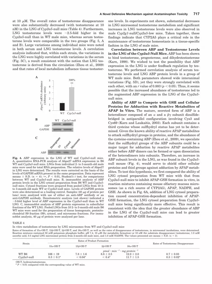
metabolic activation) and APAP-GSH [potentially involved inthe GSH depletion through the �-glutamyl cycle, as sug-gested by our previous finding that APAP toxicity in the LNGis significantly influenced by APAP metabolic activation inthe liver (Gu et al., 2005)] could both contribute to decreasedAPAP toxicity in that tissue. However, the relatively smallextents of the observed decreases suggested that the mecha-nisms underlying the resistance of Cyp2a5-null mouse toAPAP toxicity involve additional molecular events beyondthe metabolic activation step. In that connection, we hadperformed a microarray analysis of gene expression differ-ences between the LNG of the Cyp2g1-null/Cyp2a5-low malemice (on mixed B6 and 129/Sv genetic background) and theLNG of the WT B6 or 129/Sv male mice. This identified twogenes encoding subunits of the complex androgen-bindingprotein system. These are Abpa27 and Abpbg27, the productsof which form a disulfide-bridged dimeric protein describedpreviously in mouse salivary glands and saliva (Dlouhy et al.,1987; Laukaitis et al., 2008 and references therein). Thesetwo genes are among those that had the greatest up-regula-tion in the Cyp2g1-null/Cyp2a5-low mice compared withmice of either WT strain (Table 3). The up-regulation of bothABP mRNA and protein expression was confirmed by quan-titative RNA-PCR and by immunoblot analysis, respectively,in the Cyp2g1-null/Cyp2a5-low mice (not shown) and in theCyp2a5-null mice (Fig. 4). In the Cyp2a5-null mouse, mRNAlevels for Abpa27, encoding a major ABP � subunit detectedin the LNG (Zhou et al., 2009), were �3-fold higher than inWT B6 mice (Fig. 4A), and the levels of ABP protein in thecytosol preparation were also �3-fold higher (Fig. 4B). Thecytosolic localization of ABP protein was confirmed by immu-noblot analysis of various subcellular fractions (Fig. 4C).
As shown in Table 3, the expression of lipocalin-type pros-taglandin D2 synthase was also highly up-regulated in theCyp2g1-null/Cyp2a5-low mice; this up-regulation was subse-
quently confirmed at both mRNA and protein levels for theCyp2g1-null/Cyp2a5-low mice, but not for the Cyp2a5-nullmice (data not shown). The disparate results from the twomouse strains indicate that lipocalin-type prostaglandin D2
synthase is not relevant to the resistance to APAP toxicity inthe Cyp2g1-null/Cyp2a5-low or the Cyp2a5-null mice. Thepotential roles in the altered resistance to APAP toxicity ofthe other genes that were found to be up- or down-regulatedin the LNG of the Cyp2g1-null/Cyp2a5-low mice (Table 3)have not been examined. It is noteworthy that althoughABCC5 (MRP5), an organic anion transporter capable oftransporting GSH conjugates such as S-(2, 4-dinitrophenyl)-GSH (Wijnholds et al., 2000), seemed to be up-regulated(Table 3), its signal intensity on the gene array was quite low,and the corresponding protein was not detected in the LNGby immunoblot analysis (data not shown).
Impact of Cyp2a5 Deletion on Testosterone Metabo-lism and Homeostasis in the LNG. Our recent findingthat the LNG of male mice contains unusually high levels oftestosterone together with the known ability of CYP2A5 tometabolize testosterone prompted us to examine testosteronelevels in the LNG of the Cyp2a5-null mice. It is noteworthythat although recombinant CYP2A5 can metabolize testos-terone (Gu et al., 1999), the loss of CYP2A5 expression didnot lead to a large decrease in the overall rates of testoster-one metabolism by hepatic microsomes in vitro or a signifi-cant change in circulating levels of testosterone in vivo (Zhouet al., 2010). As shown in Table 4, in the LNG of the Cyp2a5-null mice, in vitro microsomal testosterone hydroxylase ac-tivities in the formation of the most abundant metabolite,15�-hydroxytestosterone, and two minor products, 15�-hy-droxytestosterone and 2�-hydroxytestosterone (all knownmetabolites by CYP2A5), were essentially abolished,whereas the rates of formation of another minor product,16�-hydroxytestosterone, were unchanged, with testosterone
TABLE 3Genes that were differentially expressed in the LNG of Cyp2g1-null/Cyp2a5-low and WT miceRNA from 2-month-old male mice was used for microarray analysis, with use of the Affymetrix Mouse Expression Set 430A GeneChip arrays. LNGs from 10 mice were pooledfor the preparation of each RNA sample, and a total of nine LNG RNA samples were analyzed (four from B6 WT, two from WT 129/Sv, and three from Cyp2g1-null/Cyp2a5-low). Genes with significantly changed expression (P � 0.05), and with change values either greater than 1.5-fold or smaller than 0.67-fold in comparisons of bothCyp2g1-null/Cyp2a5-low with B6 and Cyp2g1-null/Cyp2a5-low with 129/Sv in at least one probe set, are shown. Probe sets for which none of the three groups had averagedexpression values greater than 60 (after GC-RMA normalization) were excluded.
Gene SymbolChange
Gene NameCyp2g1-Null/Cyp2a5-Low:129/Sv Cyp2g1-Null/Cyp2a5-low:B6
fold
Ptgds 7.67/12.39 8.72/12.37 L-type prostaglandin D2 synthaseAbpa 7.45 1.75 Androgen binding protein, �a
Abpb 6.18 2.05 Androgen binding protein, �b
S100a10 2.70 1.68 S100 calcium binding protein A10Sdc4 2.25 4.78 Syndecan 4Cebpd 1.90 1.62 CCAAT/enhancer binding protein, deltaAbp1 1.71 2.01 Amiloride binding protein 1Calml4 1.70 1.91 Calmodulin-like 4Ramp1 1.66 1.56 Receptor activity modifying proteinFoxa1 1.62 1.69 Forkhead box A1Thbs1 1.57 1.66 Thrombospondin 1Abcc5 1.57 1.59 ATP binding cassette, subfamily C, member 5Ccl28 1.54/1.69 1.68/1.52 C-C motif ligand 28Igfbp5 1.53 1.69 Insulin-like growth factor binding protein 1D6WSU176e 0.63 0.65 DNA segment Chr6, Wayne State Univ. 176, expressedPnliprp1 0.62 0.51 Pancreatic lipase-related protein 1Dhrs 0.56 0.58 Dehydrogenase/reductase, member 7Adh1 0.53 0.33 Alcohol dehydrogenase 1Cyp2a5 0.02 0.03 Cytochrome P450, 2A5a The probes used are common for a number of Abpa genes, including Abpa27.b The probes used are common for a number of Abpb genes, including Abpbg27.
716 Zhou et al.
at 10 �M. The overall rates of testosterone disappearancewere also substantially decreased (with testosterone at 10nM) in the LNG of Cyp2a5-null mice (Table 4). Furthermore,LNG testosterone levels were �2.5-fold higher in theCyp2a5-null than in WT male mice, whereas serum testos-terone levels were comparable in the two groups (Fig. 5, Aand B). Large variations among individual mice were notedin both serum and LNG testosterone levels. A correlationanalysis indicated that, within each strain, the variations inthe LNG were highly correlated with variations in the serum(Fig. 5C), a result consistent with the notion that LNG tes-tosterone is derived from the circulation (Zhou et al., 2009)and that rates of local metabolism influence tissue testoster-
one levels. In experiments not shown, substantial decreasesin LNG microsomal testosterone metabolism and significantincreases in LNG testosterone levels were also observed inmale Cyp2g1-null/Cyp2a5-low mice. Taken together, thesefindings indicate that CYP2A5 plays a critical role in themaintenance of testosterone homeostasis in a tissue-specificfashion in the LNG of male mice.
Correlation between ABP and Testosterone Levelsin the LNG of the Cyp2a5-Null Mice. ABP has been shownto bind testosterone, progesterone, and dihydrotestosterone(Karn, 1998). We wished to test the possibility that ABPexpression in the LNG is under feedback regulation by tes-tosterone. We performed correlation analysis of serum tes-tosterone levels and LNG ABP protein levels in a group ofWT male mice. Both parameters showed wide interanimalvariations (Fig. 5D), yet they were strongly correlated witheach other, with an r value of 0.883 (p � 0.05). Thus, it seemspossible that the increased abundance of testosterone led tothe augmented ABP expression in the LNG of the Cyp2a5-null mice.
Ability of ABP to Compete with GSH and CellularProteins for Adduction with Reactive Metabolites ofAPAP In Vitro. The mature, secreted form of ABP is aheterodimer composed of an � and a �� subunit disulfide-bridged in antiparallel configuration involving Cys3 andCys69 (Karn and Laukaitis, 2003). Each subunit contains athird cysteine whose sulfhydryl status has yet to be deter-mined. Given the known ability of reactive APAP metabolitesto attack sulfhydryl groups in proteins, and the abundance ofthe cysteine-containing ABP (Zhou et al., 2009), we speculatethat the sulfhydryl groups of the ABP subunits could be amajor target for adduction by reactive APAP metaboliteseither before ABP dimers can be formed or upon dissociationof the heterodimers into subunits. Therefore, an increase inABP subunit levels in the LNG, as was found in the Cyp2a5-null mouse (Fig. 4), would serve to shield other cellularproteins and thiol groups against adduction by APAP metab-olites. To test this hypothesis, we first compared the ability ofLNG cytosol preparation from WT mice with that fromCyp2a5-null mice to inhibit APAP-GSH formation in vitro, inreaction mixtures containing mouse olfactory mucosa micro-somes (as a rich source of CYP2A5), APAP, NADPH, andGSH. As shown in Fig. 6A, addition of LNG cytosol prepara-tion caused concentration-dependent inhibition of APAP-GSH formation, the LNG cytosol preparation from Cyp2a5-null mice being significantly more effective. This result isconsistent with the idea that the greater abundance of ABPin the LNG of the Cyp2a5-null mice can lead to greaterinhibition of APAP-GSH formation.
A
4
5**
300%
unda
nce
ts)
0
1
2
3
Abpa
27m
RN
A a
b u(A
rbitr
ary
unit
WT Cyp2a5-null
WT Cyp2a5-null
ABPGAPDH
B
C
ABP
Fig. 4. ABP expression in the LNG of WT and Cyp2a5-null mice.A, quantitative RNA-PCR analysis of Abpa27 mRNA expression in B6WT and Cyp2a5-null mice. LNGs from individual 2- to 3-month-old malemice were used for total RNA preparation. The relative levels of Abpa27in LNG were determined. The results were corrected on the basis of thelevels of GAPDH mRNA present in the same preparation. Data representmeans S.D. (n � 4); ��, P � 0.01, Student’s t test, for comparisonsbetween WT and Cyp2a5-null mice. B, immunoblot analysis of ABPprotein levels in the LNG cytosol preparation from B6 WT and Cyp2a5-null mice. Cytosol fractions were prepared from pooled LNGs from 10 2-to 3-month-old male WT or Cyp2a5-null mice. Levels of GAPDH proteinwere also determined as a loading control. Samples (40 �g of protein perlane) were analyzed with use of either an anti-ABP antibody or ananti-GAPDH antibody. Densitometric analysis (not shown) indicated a�3-fold higher level of ABP expression in the Cyp2a5-null than in WTLNG. C, immunoblot analysis of ABP protein expression in subcellularfractions of the WT LNG. Pooled LNGs from 10 2- to 3-month-old male B6WT mice were used for the preparation of tissue homogenate, postmito-chondrial S9 fraction (S9), cytosol, and microsome fractions. For immu-noblot analysis, 40 �g of protein were analyzed per lane.
TABLE 4In vitro metabolism of testosterone by LNG microsomes from WT and Cyp2a5-null miceRates of formation of 15�-OH-T, 15�-OH-T, 2�-OH-T, and 16�-OH-T, as well as the rates of disappearance of testosterone, in microsomal incubations, were determined.Reaction mixtures contained 50 mM potassium phosphate buffer, pH 7.4, 10 �M (for metabolite formation) or 10 nM (for substrate disappearance) testosterone, 1.0 mMascorbic acid, 0.5 mg/ml LNG microsomal protein from 2-month-old male mice, and 1.0 mM NADPH. The values presented are means S.D. (n � 3).
StrainRates of Product Formation
Rates of Testosterone Disappearance15�-OH-T 15�-OH-T 2�-OH-T 16�-OH-T
pmol � min�1 � mg protein�1
WT B6 106 19 7.8 1.6 8.8 2.5 12.0 2.0 0.7 0.03Cyp2a5-null 0.3 0.1a � 0.04a � 0.4a 11.7 1.5 0.3 0.02a
OH-T, hydroxytestosterone.a P � 0.01 compared with the corresponding value of WT mice.
A Novel Defensive Mechanism against Acetaminophen Toxicity 717

However, more direct evidence was needed to demonstratethe ability of ABP to form APAP adducts. We next added ABPprepared from mouse saliva to reaction mixtures containingolfactory mucosa microsomes, APAP, NADPH, and GSH. Thepreparation of partially purified ABP exhibited a cluster ofpartially resolved, prominent bands in the 10-kDa region(which corresponded well with the position of ABP subunitsdetected by immunoblot analysis, and presumably repre-sents a mix of the � and � subunits of various ABP isoforms),as well as a few minor bands (not recognized by the anti-ABPantibody) in higher molecular weight regions upon electro-phoretic analysis (under reducing conditions) and silverstaining (Fig. 6B). Addition of the ABP preparation causedconcentration-dependent inhibition of APAP-GSH formation(Fig. 6C). As was found for boiled LNG cytosolic proteins,boiled ABP was incapable of inhibiting APAP-GSH forma-tion. This finding suggests that heat denaturation will makethe sulfhydryl groups in ABP and other cytosolic proteinsinaccessible to the reactive APAP metabolites.
In Fig. 6C, the highest concentration of salivary ABP usedwas �33 �g/ml (�1.65 �M), which was much lower than thelevels of APAP (0.5 mM) added to the assays but not too farfrom the amounts of APAP-GSH that were blocked fromformation (�10 �M), if we consider the possible involvementof multiple Cys residues of each ABP subunit as targets forAPAP adduction. Thus, the inhibitory effect is more than canbe explained by noncovalent binding of APAP to the ABPheterodimer. The inhibitory effect was also not due to a direct
inhibition of the formation of the reactive intermediate, be-cause the inhibitory effects can be overcome by addition ofgreater amounts of GSH (data not shown).
Addition of salivary ABP to reactions without GSH led totime-dependent formation of a unique APAP adduct, detectedby an antibody to APAP (Fig. 6D). We believe that thisrepresents an adduct of APAP with an ABP subunit (APAP-ABP) on the basis of its migration rate, identical to that ofABP subunits on immunoblot, as well as its detection by bothanti-APAP and anti-ABP and its increases in abundancewith the increases in the amounts of ABP added (Fig. 6E). Asfurther controls, formation of the APAP-ABP adduct wasnearly abolished by the addition of an excess amount (10mM) of GSH (Fig. 6D) or when either APAP or NADPH wasomitted (data not shown). In addition, APAP-ABP and otherAPAP-protein adducts were detected when LNG cytosolpreparation was added to olfactory mucosa microsomal reac-tions with APAP (Fig. 6E); notably, few APAP adducts withmicrosomal proteins were detected, most likely because ofthe relatively low amounts of total microsomal protein usedin the assay. It is noteworthy that addition of salivary ABP toreactions containing LNG cytosol preparation led to in-creases in the abundance of APAP-ABP, and decreases in theabundance of the other APAP-protein adducts. This result isconsistent with the ability of ABP subunits to compete withcellular proteins for binding to reactive APAP metabolites.
The inhibitory effects of ABP addition on APAP-GSH for-mation (Fig. 6C) and on APAP-protein adduct formation (Fig.
A
tero
ne le
vels
(ng/
ml )
0.5
1.0 B
n e le
vels
(ng/
g pr
otei
n )
30
40
50
60**
WT Cyp2a5-null
Seru
m te
stos
t
0.0WT Cyp2a5-null
LNG
test
oste
ron
0
10
20
CC
one
leve
l (ng
/g p
rote
in)
80100120140160180 WT
Cyp2a5-null
danc
e (a
rbitr
ary
unit)
2
3
4
5D
Serum testosterone level (ng/ml)0 1 8 9 10
LNG
test
oste
r o
0204060
Serum testosterone level (ng/ml)0 1 2 3 4
AB
P ab
und
0
1
Fig. 5. Testosterone levels in the serumand LNG of WT and Cyp2a5-null malemice. Serum (A) and LNG (B) testoster-one levels were determined for 2- to3-month-old male B6 WT and Cyp2a5-null mouse. The values shown are themedians, together with the 25% (lowerbar) and 75% (upper bar) percentilemarks. Testosterone levels in the LNG(but not the levels in the serum) weresignificantly higher in the Cyp2a5-nullthan in the WT mice (n � 8); ��, P �0.01, Mann-Whitney rank sum test.C, correlation of LNG testosterone lev-els with serum testosterone levels.LNG and serum testosterone levels foreach mouse were plotted. The correla-tion coefficient (r) for LNG versus se-rum is 1.000 (P � 0.01) in WT, and0.976 in Cyp2a5-null (P � 0.01) mice;Spearman rank order correlation test.D, correlation of LNG ABP protein lev-els with serum testosterone levels.LNG cytosol ABP levels determined byimmunoblot analysis and serum testos-terone levels were determined for ninemale B6 mouse (2- to 3-month old). Thecorrelation coefficient (r) for LNG ABPversus serum is 0.883 (P � 0.05); Spear-man rank-order correlation test.
718 Zhou et al.

6E) were consistent, except that the blockage of cytosolicprotein adduct formation was more complete because of theabsence of added GSH. It is noteworthy that because thesmaller proteins typically have poorer retention on the nitro-cellulose membrane than the larger proteins do during elec-
trophoretic transfer, the intensity of the APAP-ABP bandwas relatively low (e.g., Fig. 6E, last lane), and it did notmatch the intensity of the cytosolic protein adducts detectedin the higher molecular weight region in the absence of addedABP (Fig. 6E, lane 4).
AP-
GSH
form
atio
n m
in/m
g pr
otei
n)
2.5
3.0
3.5
4.0WTCyp2a5-null
**
BA
3750
250 kDABP Marker
100
D
Amount of LNG cytosol protein added (mg/ml)0.0 0.5 1.0 1.5 2.0 2.5 3.0
Rat
e of
AP A
(nm
ol/m
0.0
1.5
2.0 ****
101525
37
Anti-APAPAnti-ABP
10 kD
10 kD
440 44 44 44 ABP (µg/ml)100 0 0 0GSH (mM)3030 0 10 30Time (min)
D
AP-
GSH
form
atio
n /m
in/m
g pr
otei
n)
2.5
3.0
3.5
4.0 Boiled ABPNative ABP
C
Amount of ABP protein added (µg/ml)0 5 10 15 20 25 30 35
Rat
e of
AP
( nm
ol/m
0.0
1.5
2.0
EE
__ _ __ +LNG Cytosol + +
100
Anti-APAP 50
250 kD
+ _ + + + + APAP + ++_ + + + + NADPH + +
00 0 0 15 44 4451)lm/gµ( PBA
Anti-ABP
Anti-APAP
10 kD
10
25
37
APAP-ABP15
Fig. 6. Ability of ABP to compete with GSH and cellular proteins for adduction with APAP in vitro. Reaction mixtures contained 100 mM potassiumphosphate buffer, pH 7.6, 0.5 mM APAP, 0.5 mg/ml olfactory mucosa microsomal protein of 2- to 3-month-old male B6 WT mice, and other componentsas indicated. Unless otherwise indicated, reactions were carried out for 10 min at 37°C. A, differential inhibition of APAP-GSH formation by LNGcytosol preparation from WT and Cyp2a5-null mice. Rates of in vitro formation of APAP-GSH were determined in the presence of 1.0 mM NADPH,0.2 mM GSH, and increasing amounts of LNG cytosolic protein (0–3 mg/ml), prepared from 2-month-old male WT or Cyp2a5-null mice. The valuespresented are means S.D. (n � 3); �� P � 0.01, Student’s t test. In negative control reactions, the addition of boiled LNG cytosol preparation from2-month-old male WT mice (at 3 mg/ml) did not affect rates of APAP-GSH formation (data not shown). B, gel-electrophoretic analysis and silverstaining of partially purified ABP protein. A preparation of ABP (�2 �g of protein) purified from mouse saliva was analyzed under reducing conditionon a 10% polyacrylamide gel. Selected fragments of a prestained protein size marker are labeled. C, inhibition of APAP-GSH formation by purifiedABP. Rates of formation of APAP-GSH were determined in the presence of 1.0 mM NADPH, 0.2 mM GSH, and increasing amounts of either boiledor native ABP (at 0–33 �g/ml; amounts equivalent to those of ABP present in 0 to 3 mg/ml LNG cytosol protein from Cyp2a5-null mice). The valuespresented are averages for duplicated samples at each ABP concentration. D, time-dependent formation of APAP-ABP protein adduct in vitro.Reactions were carried out for 0 to 30 min in the presence of 1.0 mM NADPH, 0 or 44 �g/ml ABP, and 0 or 10 mM GSH. Aliquots of the reactionmixtures, equivalent to 5 �g of the olfactory mucosa microsomal protein per lane, were analyzed on immunoblots, with use of either an anti-APAP oran anti-ABP. E, Competition between ABP and other LNG cytosolic proteins for adduction with APAP. Reactions were performed as in C, except thatLNG cytosol preparation from Cyp2a5-null mice was added at 1.5 mg/ml, whereas the concentration of ABP varied between 0 and 44 �g/ml. Additionof ABP to reactions containing LNG cytosol led to increases in the abundance of APAP-ABP, and decreases in the abundance of the other APAP-proteinadducts.
A Novel Defensive Mechanism against Acetaminophen Toxicity 719

Increased Formation of APAP-ABP Protein Adduct,Accompanied by Decreased Formation of APAP Ad-ducts with Other Cellular Proteins, in the LNG ofAPAP-Treated Cyp2a5-Null Mice, Compared with theLNG of APAP-Treated WT Mice. The ability of ABP toform APAP adducts in the LNG in vivo in APAP-treated micewas then examined. As shown in Fig. 7A, after a treatment ofthe animals with a toxic dose of APAP (400 mg/kg), manybands representing APAP-protein adducts were detectedwith the anti-APAP antibody in the microsomal or cytosolicfractions of the LNG from WT mice, including a band at �10kDa, which was detected in the cytosol preparation both byanti-APAP (Fig. 7A) and by anti-ABP (Fig. 7B). The intensityof the APAP-ABP band was greater (�2-fold; Fig. 7A),whereas the intensities of the other adduct bands in eithermicrosome or cytosol fractions were much reduced, in theLNG of APAP-treated Cyp2a5-null mice. These resultsstrongly support the notion that the increased ABP subunitabundance in the Cyp2a5-null LNG serves to quench greateramounts of reactive APAP metabolites, thus sparing the
other perhaps more critical cellular proteins from adduction byAPAP metabolites and protecting the cells against necrosis.
DiscussionThe discovery that Cyp2a5 deletion leads to significant
increases in testosterone levels in the LNG indicates thatCYP2A5 is the major enzyme for testosterone metabolism inthe LNG. The effects of Cyp2a5 knockout on testosteronehomeostasis in the LNG is tissue-specific, given the lack of asignificant change in overall rates of hepatic testosteronemetabolism and in circulating testosterone levels (Zhou etal., 2010). Viewed from a mechanistic standpoint, the tissue-specific impact of Cyp2a5 knockout on testosterone homeo-stasis in the LNG is clearly related to the dependence of theLNG, but not the liver, on CYP2A5 for testosterone degrada-tion. Considering the fact that CYP2A5 is expressed in nu-merous other tissues, including kidney, brain, small intes-tine, olfactory mucosa, lung, other parts of the respiratorytract, and the salivary and lacrimal glands (Piras et al., 2003;Su and Ding, 2004), it will be interesting to determinewhether androgen metabolism and homeostasis is altered inother organs of the Cyp2a5-null mice. It is noteworthy that ofthe two human orthologs of mouse CYP2A5, CYP2A13 is alsoefficient in catalyzing testosterone hydroxylation (von Wey-marn et al., 2005), whereas CYP2A6 is not active in thisreaction (Liu et al., 1996); the role of human CYP2A13 inandrogen metabolism and homeostasis remains to be explored.
The molecular mechanism responsible for the up-regula-tion of ABP expression in the LNG of male Cyp2a5-null miceis not clear at this point; however, the up-regulation seems tobe pretranslational, given the increased ABP mRNA levels inthe null mice. Testosterone-stimulated synthesis and secre-tion of steroid hormone (androgen) binding protein (a proteinunrelated to ABP) has been reported in Sertoli cells of testes(Louis and Fritz, 1977). Currently, we do not know whetherABP expression in the LNG can be activated by testosterone,despite the observed positive correlation between serum tes-tosterone levels and LNG ABP abundance among adult malemice. Furthermore, it remains to be determined whether theabundance of ABP is also a critical factor for the elevatedtestosterone level in the LNG of the Cyp2a5-null mice. As wehad suggested previously, a decrease in testosterone degra-dation may be necessary, but not sufficient, for an increasedaccumulation of testosterone in an organ (Zhou et al., 2009).Therefore, additional studies are warranted to further dem-onstrate activation of ABP expression by testosterone, and toclarify whether the high abundance of testosterone in theLNG of WT mice and the further elevation of testosteronelevel in the LNG of the Cyp2a5-null mice are dependent onABP.
The up-regulation of ABP subunits provides one direct linkbetween Cyp2a5 deletion and increased resistance to APAPtoxicity in the LNG, given our demonstration that ABP sub-units can form conjugates with APAP and thereby protectother cellular proteins and thiol groups against the attack byAPAP reactive intermediates. Several salient features ofABP make it an ideal protector against thiol-reactive chem-icals: 1) it is abundant in the LNG (�1% total cytosolicprotein); 2) it is a secretory protein, thus capable of carryingthe conjugated chemical intermediates out of the gland to thenasal mucus; and 3) it contains three cysteine residues per
A
WT li
WT APAP
Null APAP kD
WT li
WT APAP
Null APAP
LNG microsome LNG cytosol
saline APAP APAP250150
50
75
100
kDsaline APAP APAP
Anti-APAP
10
25
37
B
Anti-ABP
WT saline
WT APAP
Null APAP
LNG cytosol
10 kD
Anti-Calnexin Anti-GAPDH
Anti-GAPDH
Fig. 7. Immunoblot detection of APAP-protein adducts in the LNG ofAPAP-treated mice. Two-month-old male mice were fasted overnightbefore an injection of APAP (at 400 mg/kg i.p.); mice in the control groupwere treated with saline. Microsomal and cytosol preparations wereobtained from the pooled LNGs of four mice; tissues were obtained at 2 hafter the APAP treatment. Microsomal and cytosol (A) samples (20 and 30�g of protein, respectively, in each lane), from either B6 WT or Cyp2a5-null (Null) mice, were analyzed on 10% SDS-polyacrylamide gels, andAPAP adducts were detected on immunoblots by using a polyclonal anti-APAP antiserum. B, the levels of ABP protein were determined on aseparate blot for the same cytosol samples that were used in A to dem-onstrate induction of the ABP protein in the Cyp2a5-null mice. The levelsof calnexin (for microsomes, A) and GAPDH (for cytosol, A and B) werealso determined, after stripping of the blot to remove the anti-APAP oranti-ABP antibodies, for demonstration of equal protein loading amongthe three samples. Densitometric analysis (not shown) indicated a �2.5-fold higher level of APAP-ABP (A) and a 2.8-fold higher level of ABP (B)in the LNG cytosol from APAP-treated Cyp2a5-null mice, compared withAPAP-treated WT mice.
720 Zhou et al.
subunit, so it can quench several reactive molecules at thesame time. It is noteworthy that other cysteine-containingsecretory proteins may also be able to serve as a protectoragainst thiol-reactive intermediates and, in fact, we observedother proteins that reacted with APAP antibody in micro-somes and cytosol, as expected based on previous findings inother tissues (e.g., Gu et al., 2005). Nonetheless, other thanAPAP-ABP, few other APAP-protein adducts were found tobe increased in the LNG of APAP-treated Cyp2a5-null micecompared with the LNG of APAP-treated WT mice (Fig. 7).This suggests that other secretory proteins do not make asubstantial contribution to the increased protection againstAPAP toxicity in the LNG of Cyp2a5-null mice.
ABP up-regulation alone may not fully explain the nearlytotal protection of the LNG against APAP-induced tissuedamage and nonprotein thiol depletion in the Cyp2a5-nullmice. Two other factors might also have been important.First, APAP levels in the LNG were significantly decreasedin the Cyp2a5-null LNG compared with the WT LNG,whereas circulating APAP levels were not different in thetwo mouse strains; this decrease in LNG APAP bioavailabil-ity would reduce the amounts of N-acetyl-p-benzoquinonei-mine produced in situ and thus contribute to a reduction inadduct formation and in toxicity. Second, APAP-GSH levelsin the LNG were substantially lower in the Cyp2a5-null LNGthan in the WT LNG, whereas circulating APAP-GSH levelswere not different in the two mouse strains. It is known thatAPAP-GSH, through its degradation product APAP-Cys, canenter the �-glutamyl cycle and cause depletion of GSH (e.g.,Stern et al., 2005a). Thus, a decrease in APAP-GSH levelwould reduce the extent of GSH depletion and increase re-sistance of the tissue to APAP toxicity. In this connection, theability of liver-generated APAP-GSH and APAP-Cys to in-duce toxicity in the kidney is well documented (Stern et al.,2005a, 2005b).
The mechanistic link between Cyp2a5 deletion and thereduction in levels of APAP and APAP-GSH in the LNG
remains to be identified. We speculate that the up-regulationof testosterone levels led to an increased rate of excretion ofcytosolic content (including APAP and APAP-GSH) from theCyp2a5-null LNG compared with the WT LNG. In this con-nection, it has been reported by others that testosterone canstimulate secretion from rat lacrimal glands (Sullivan et al.,1990). For APAP, which is lipid-soluble and thus readilyabsorbed, neither circulating levels nor rates of local metab-olism were altered; therefore, the reduced LNG APAP level ismost likely due to increased rate of excretion. The decrease inLNG APAP level would contribute to a decrease in LNGAPAP-GSH level. However, given the low rates of in situAPAP metabolism, and the likelihood of a transfer of circu-lating APAP-GSH to the LNG, as implied by our earlierfinding that APAP toxicity in the LNG is partly dependent onAPAP metabolism in the liver (Gu et al., 2005), it is conceiv-able that an increase in APAP-GSH excretion from the LNGmight have been the more dominant mechanism. On theother hand, the levels of APAP-G and APAP-S were notdifferent in Cyp2a5-null and WT mice, a result that arguesagainst an increase in excretion. However, differential trans-port of the various APAP conjugates has been reported inother organ systems (e.g., Chen et al., 2003). It is possiblethat, in the LNG, APAP-G and APAP-S are transported moreinto the blood, whereas APAP and APAP-GSH are more af-fected by excretion to the nasal mucus. The excretion may havebeen more affected by CYP2A5-mediated changes in testoster-one homeostasis. In any case, further studies on the ability oftestosterone to stimulate LNG excretion and to influence tissueburden of APAP and various APAP metabolites are warranted.
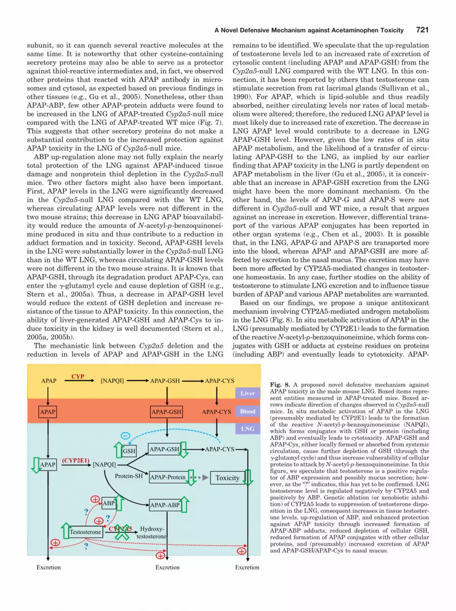
Based on our findings, we propose a unique antitoxicantmechanism involving CYP2A5-mediated androgen metabolismin the LNG (Fig. 8). In situ metabolic activation of APAP in theLNG (presumably mediated by CYP2E1) leads to the formationof the reactive N-acetyl-p-benzoquinoneimine, which forms con-jugates with GSH or adducts at cysteine residues on proteins(including ABP) and eventually leads to cytotoxicity. APAP-
APAP-GSHCYP
APAP-CYSAPAP [NAPQI]
APAP
Liver
BloodAPAP-GSH APAP-CYS
[ Q ]
LNG
APAP [NAPQI]
GSH
Protein-SH APAP-Protein
APAP-GSH
(CYP2E1)
Toxicity
APAP-CYS
APAP-ABP
Testosterone
ABP
Hydroxy-testosterone
CYP2A5+
+?
?
Excretion
testosterone
Excretion Excretion
++ +?
Fig. 8. A proposed novel defensive mechanism againstAPAP toxicity in the male mouse LNG. Boxed items repre-sent entities measured in APAP-treated mice. Boxed ar-rows indicate direction of changes observed in Cyp2a5-nullmice. In situ metabolic activation of APAP in the LNG(presumably mediated by CYP2E1) leads to the formationof the reactive N-acetyl-p-benzoquinoneimine (NAPQI),which forms conjugates with GSH or protein (includingABP) and eventually leads to cytotoxicity. APAP-GSH andAPAP-Cys, either locally formed or absorbed from systemiccirculation, cause further depletion of GSH (through the�-glutamyl cycle) and thus increase vulnerability of cellularproteins to attack by N-acetyl-p-benzoquinoneimine. In thisfigure, we speculate that testosterone is a positive regula-tor of ABP expression and possibly mucus secretion; how-ever, as the “?” indicates, this has yet to be confirmed. LNGtestosterone level is regulated negatively by CYP2A5 andpositively by ABP. Genetic ablation (or xenobiotic inhibi-tion) of CYP2A5 leads to suppression of testosterone dispo-sition in the LNG, consequent increases in tissue testoster-one levels, up-regulation of ABP, and enhanced protectionagainst APAP toxicity through increased formation ofAPAP-ABP adducts, reduced depletion of cellular GSH,reduced formation of APAP conjugates with other cellularproteins, and (presumably) increased excretion of APAPand APAP-GSH/APAP-Cys to nasal mucus.
A Novel Defensive Mechanism against Acetaminophen Toxicity 721
GSH and APAP-Cys, either locally formed or absorbed fromsystemic circulation, cause further depletion of GSH (throughthe �-glutamyl cycle) and thus increase vulnerability of cellularproteins to attacks by N-acetyl-p-benzoquinoneimine. Testos-terone, which is abundant in the LNG, may be a positive regu-lator of ABP expression and possibly mucus secretion; however,LNG testosterone level is regulated negatively by CYP2A5(through metabolism) and positively by ABP (through directbinding and sequestration). Genetic ablation of Cyp2a5 leads tosuppression of testosterone disposition in the LNG, thus caus-ing increases in tissue testosterone levels, up-regulation ofABP, and enhanced protection against APAP toxicity. Protec-tion occurs through increased formation of APAP-ABP adducts,reduced depletion of cellular GSH, reduced formation of APAPconjugates with other cellular proteins, and reduced tissue bur-den of APAP and APAP-GSH; the decrease in APAP and APAP-GSH is achieved at least partly through increased excretion ofAPAP and APAP-GSH/APAP-Cys to nasal mucus. As a furtherextension of this mechanism, xenobiotic inhibition of CYP2A5(or other androgen-metabolizing enzymes) may also lead tosuppression of testosterone disposition, which may initiate anadaptive response, as seen in the Cyp2a5-null mice, for en-hanced protection against potential xenobiotic toxicity in theLNG (and possibly other secretory glands or organs). We haveno direct evidence for aspects of this model; however, furtherconfirmation and characterization of this putative, novel defen-sive mechanism, which may be exploited for prevention of xe-nobiotic toxicity, are under way.
It should be noted that the proposed antitoxicant mecha-nism is based on studies with male mice. It remains to bedetermined whether the exact same mechanism also appliesto female mice. Female Cyp2a5-null mice also showed up-regulation of ABP expression and increased resistance toAPAP toxicity in the LNG, compared with WT female mice(X. Zhou and X. Ding, unpublished results). However, it is notclear whether testosterone, which is at much lower abun-dance in female mice than in males, or progesterone, anotherABP ligand, which is abundant in female mice, is involved.
Acknowledgments
We thank Drs. Xiuling Zhang, Cheng Fang, and Jaime D’Agostinoof the Wadsworth Center and Dr. Lindsay Hough of the AlbanyMedical College for helpful discussions, and Weizhu Yang for tech-nical assistance. We gratefully acknowledge the use of the Trans-genic and Knockout Mouse Core, the Microarray Core, the Biochem-istry Core, and the Histopathology Core of the Wadsworth Center.
Authorship Contributions
Participated in research design: Zhou and Ding.Conducted experiments: Zhou, Wei, Xie, Kluetzman, Gu, and
Zhang.Contributed new reagents or analytic tools: Laukaitis, Karn, and
Roberts.Performed data analysis: Zhou and Ding.Wrote or contributed to the writing of the manuscript: Zhou, Lau-
kaitis, Karn, Roberts, and Ding.
ReferencesChen C, Hennig GE, and Manautou JE (2003) Hepatobiliary excretion of acetamin-
ophen glutathione conjugate and its derivatives in transport-deficient (TR�) hy-perbilirubinemic rats. Drug Metab Dispos 31:798–804.
Ding X and Coon MJ (1990) Immunochemical characterization of multiple forms ofcytochrome P-450 in rabbit nasal microsomes and evidence for tissue-specificexpression of P-450s NMa and NMb. Mol Pharmacol 37:489–496.
Ding X and Coon MJ (1994) Steroid metabolism by rabbit olfactory-specific P4502G1. Arch Biochem Biophys 315:454–459.
Dlouhy SR, Nichols WC, and Karn RC (1986) Production of an antibody to mousesalivary androgen binding protein (ABP) and its use in identifying a prostateprotein produced by a gene distinct from Abp. Biochem Genet 24:743–763.
Dlouhy SR, Taylor BA, and Karn RC (1987) The genes for mouse salivary androgen-binding protein (ABP) subunits alpha and gamma are located on chromosome 7.Genetics 115:535–543.
Getchell ML and Mellert TK (1991) Olfactory mucus secretion, in Smell and Taste inHealth and Disease (Getchell TV, Doty RL, Bartoshuk LM, and Snow JB Jr eds),pp 83–95, Raven Press, New York.
Gu J, Zhang QY, Genter MB, Lipinskas TW, Negishi M, Nebert DW, and Ding X(1998) Purification and characterization of heterologously expressed mouseCYP2A5 and CYP2G1: role in metabolic activation of acetaminophen and 2,6-dichlorobenzonitrile in mouse olfactory mucosal microsomes. J Pharmacol ExpTher 285:1287–1295.
Gu J, Dudley C, Su T, Spink DC, Zhang QY, Moss RL, and Ding X (1999) CytochromeP450 and steroid hydroxylase activity in mouse olfactory and vomeronasal mucosa.Biochem Biophys Res Commun 266:262–267.
Gu J, Cui H, Behr M, Zhang L, Zhang QY, Yang W, Hinson JA, and Ding X (2005)In vivo mechanisms of tissue-selective drug toxicity: effects of liver-specific knock-out of the NADPH-cytochrome P450 reductase gene on acetaminophen toxicity inkidney, lung, and nasal mucosa. Mol Pharmacol 67:623–630.
Hinson JA, Reid AB, McCullough SS, and James LP (2004) Acetaminophen-inducedhepatotoxicity: role of metabolic activation, reactive oxygen/nitrogen species, andmitochondrial permeability transition. Drug Metab Rev 36:805–822.
Karn RC (1998) Steroid binding by mouse salivary proteins. Biochem Genet 36:105–117.
Karn RC and Laukaitis CM (2003) Characterization of two forms of mouse salivaryandrogen-binding protein (ABP): implications for evolutionary relationships andligand-binding function. Biochemistry 42:7162–7170.
Karn RC and Russell R (1993) The amino acid sequence of the alpha subunit ofmouse salivary androgen-binding protein (ABP), with a comparison to the partialsequence of the beta subunit and to other ligand-binding proteins. Biochem Genet31:307–319.
Kontgen F, Suss G, Stewart C, Steinmetz M, and Bluethmann H (1993) Targeteddisruption of the MHC class II Aa gene in C57BL/6 mice. Int Immunol 5:957–964.
Lakso M, Pichel JG, Gorman JR, Sauer B, Okamoto Y, Lee E, Alt FW, and WestphalH (1996) Efficient in vivo manipulation of mouse genomic sequences at the zygotestage. Proc Natl Acad Sci USA 93:5860–5865.
Laemmli UK (1970) Cleavage of structural proteins during the assembly of the headof bacteriophage T4. Nature 227:680–685.
Laukaitis CM, Heger A, Blakley TD, Munclinger P, Ponting CP, and Karn RC (2008)Rapid bursts of androgen-binding protein (Abp) gene duplication occurred inde-pendently in diverse mammals. BMC Evol Biol 8:46–62.
Lee JK, Abe K, Bridges AS, Patel NJ, Raub TJ, Pollack GM, and Brouwer KL (2009)Sex-dependent disposition of acetaminophen sulfate and glucuronide in the in situperfused mouse liver. Drug Metab Dispos 37:1916–1921.
Liu C, Zhuo X, Gonzalez FJ, and Ding X (1996) Baculovirus-mediated expression andcharacterization of rat CYP2A3 and human CYP2a6: role in metabolic activationof nasal toxicants. Mol Pharmacol 50:781–788.
Louis BG and Fritz IB (1977) Stimulation by androgens of the production of andro-gen binding protein by cultured sertoli cells. Mol Cell Endocrinol 7:9–16.
Moe H and Bojsen-Moller F (1971) The fine structure of the lateral nasal gland(Steno’s gland) of the Rat. J Ultrastruct Res 36:127–148.
Patten CJ, Thomas PE, Guy RL, Lee M, Gonzalez FJ, Guengerich FP, and Yang CS(1993) Cytochrome P450 enzymes involved in acetaminophen activation by rat andhuman liver microsomes and their kinetics. Chem Res Toxicol 6:511–518.
Pes D, Mameli M, Andreini I, Krieger J, Weber M, Breer H, and Pelosi P (1998)Cloning and expression of odorant-binding proteins Ia and Ib from mouse nasaltissue. Gene 212:49–55.
Piras E, Franzen A, Fernandez EL, Bergstrom U, Raffalli-Mathieu F, Lang M, andBrittebo EB (2003) Cell-specific expression of CYP2A5 in the mouse respiratorytract: effects of olfactory toxicants. J Histochem Cytochem 51:1545–1555.
Potter DW, Pumford NR, Hinson JA, Benson RW, and Roberts DW (1989) Epitopecharacterization of acetaminophen bound to protein and nonprotein sulfhydrylgroups by an enzyme-linked immunosorbent assay. J Pharmacol Exp Ther 248:182–189.
Roberts DW, Pumford NR, Potter DW, Benson RW, and Hinson JA (1987) A sensitiveimmunochemical assay for acetaminophen-protein adducts. J Pharmacol Exp Ther241:527–533.
Stern ST, Bruno MK, Horton RA, Hill DW, Roberts JC, and Cohen SD (2005a)Contribution of acetaminophen-cysteine to acetaminophen nephrotoxicity II. Pos-sible involvement of the gamma-glutamyl cycle. Toxicol Appl Pharmacol 202:160–171.
Stern ST, Bruno MK, Hennig GE, Horton RA, Roberts JC, and Cohen SD (2005b)Contribution of acetaminophen-cysteine to acetaminophen nephrotoxicity in CD-1mice: I. Enhancement of acetaminophen nephrotoxicity by acetaminophen-cysteine. Toxicol Appl Pharmacol 202:151–159.
Su T and Ding X (2004) Regulation of the cytochrome P450 2A genes. Toxicol ApplPharmacol 199:285–294.
Sullivan DA, Kelleher RS, Vaerman JP, and Hann LE (1990) Androgen regulation ofsecretory component synthesis by lacrimal gland acinar cells in vitro. J Immunol145:4238–4244.
Tonge RP, Kelly EJ, Bruschi SA, Kalhorn T, Eaton DL, Nebert DW, and Nelson SD(1998) Role of CYP1A2 in the hepatotoxicity of acetaminophen: investigationsusing Cyp1a2 null mice. Toxicol Appl Pharmacol 153:102–108.
von Weymarn LB, Zhang QY, Ding X, and Hollenberg PF (2005) Effects of 8-me-thoxypsoralen on cytochrome P450 2A13. Carcinogenesis 26:621–629.
Wada R, Tifft CJ, and Proia RL (2000) Microglial activation precedes acute neuro-
722 Zhou et al.

degeneration in Sandhoff disease and is suppressed by bone marrow transplanta-tion. Proc Natl Acad Sci USA 97:10954–10959.
Weng Y, DiRusso CC, Reilly AA, Black PN, and Ding X (2005) Hepatic gene expres-sion changes in mouse models with liver-specific deletion or global suppression ofthe NADPH-cytochrome P450 reductase gene mechanistic implications for theregulation of microsomal cytochrome P450 and the fatty liver phenotype. J BiolChem 280:31686–31698.
Wijnholds J, Mol CA, van Deemter L, de Haas M, Scheffer GL, Baas F, Beijnen JH,Scheper RJ, Hatse S, De Clercq E, et al. (2000) Multidrug-resistance protein 5 isa multispecific organic anion transporter able to transport nucleotide analogs.Proc Natl Acad Sci USA 97:7476–7481.
Young JT (1981) Histopathologic examination of the rat nasal cavity. Fundam ApplToxicol 1:309–312.
Zhang X, Zhang QY, Liu D, Su T, Weng Y, Ling G, Chen Y, Gu J, Schilling B, andDing X (2005) Expression of cytochrome P450 and other biotransformation genesin fetal and adult human nasal mucosa. Drug Metab Dispos 33:1423–1428.
Zhou X, Zhang X, Weng Y, Fang C, Kaminsky L, and Ding X (2009) High abundanceof testosterone and salivary androgen-binding protein in the lateral nasal gland ofmale mice. J Steroid Biochem Mol Biol 117:81–86.
Zhou X, Zhuo X, Xie F, Kluetzman K, Shu YZ, Humphreys WG, and Ding X (2010)Role of CYP2A5 in the clearance of nicotine and cotinine: insights from studies ona Cyp2a5-null mouse model. J Pharmacol Exp Ther 332:578–587.
Zhuo X, Gu J, Behr MJ, Swiatek PJ, Cui H, Zhang QY, Xie Y, Collins DN, and DingX (2004) Targeted disruption of the olfactory mucosa-specific Cyp2g1 gene: impacton acetaminophen toxicity in the lateral nasal gland, and tissue-selective effects onCyp2a5 expression. J Pharmacol Exp Ther 308:719–728.
Address correspondence to: Dr. Xinxin Ding, Wadsworth Center, New YorkState Department of Health, Empire State Plaza, Box 509, Albany, NY 12201-0509. E-mail: [email protected]
A Novel Defensive Mechanism against Acetaminophen Toxicity 723
Related Documents
![r a p hy &S Chromatography · receptor than does testosterone [2-4]. 5α-Dihydrotestosterone is the most potent endogenous androgen. Androstanalone is white color powder, molecular](https://static.cupdf.com/doc/110x72/5e7ea66a3c3ca174262d7b94/r-a-p-hy-s-chromatography-receptor-than-does-testosterone-2-4-5-dihydrotestosterone.jpg)