Hirabayashi and Hirokazu Kawagishi Dohra, Kenta Moriwaki, Eiji Miyoshi, Jun Yuka Kobayashi, Hiroaki Tateno, Hideo Pholiota squarrosa the Mushroom A Novel Core Fucose-specific Lectin from Glycobiology and Extracellular Matrices: doi: 10.1074/jbc.M111.327692 originally published online August 7, 2012 2012, 287:33973-33982. J. Biol. Chem. 10.1074/jbc.M111.327692 Access the most updated version of this article at doi: . JBC Affinity Sites Find articles, minireviews, Reflections and Classics on similar topics on the Alerts: When a correction for this article is posted • When this article is cited • to choose from all of JBC's e-mail alerts Click here Supplemental material: http://www.jbc.org/content/suppl/2012/08/23/M111.327692.DC1.html http://www.jbc.org/content/287/41/33973.full.html#ref-list-1 This article cites 51 references, 19 of which can be accessed free at at OSAKA UNIVERSITY on December 23, 2013 http://www.jbc.org/ Downloaded from at OSAKA UNIVERSITY on December 23, 2013 http://www.jbc.org/ Downloaded from

Welcome message from author
This document is posted to help you gain knowledge. Please leave a comment to let me know what you think about it! Share it to your friends and learn new things together.
Transcript

Hirabayashi and Hirokazu KawagishiDohra, Kenta Moriwaki, Eiji Miyoshi, Jun Yuka Kobayashi, Hiroaki Tateno, Hideo
Pholiota squarrosathe Mushroom A Novel Core Fucose-specific Lectin fromGlycobiology and Extracellular Matrices:
doi: 10.1074/jbc.M111.327692 originally published online August 7, 20122012, 287:33973-33982.J. Biol. Chem.
10.1074/jbc.M111.327692Access the most updated version of this article at doi:
.JBC Affinity SitesFind articles, minireviews, Reflections and Classics on similar topics on the
Alerts:
When a correction for this article is posted•
When this article is cited•
to choose from all of JBC's e-mail alertsClick here
Supplemental material:
http://www.jbc.org/content/suppl/2012/08/23/M111.327692.DC1.html
http://www.jbc.org/content/287/41/33973.full.html#ref-list-1
This article cites 51 references, 19 of which can be accessed free at
at OSA
KA
UN
IVE
RSIT
Y on D
ecember 23, 2013
http://ww
w.jbc.org/
Dow
nloaded from
at OSA
KA
UN
IVE
RSIT
Y on D
ecember 23, 2013
http://ww
w.jbc.org/
Dow
nloaded from
A Novel Core Fucose-specific Lectin from the MushroomPholiota squarrosa□S
Received for publication, April 5, 2012, and in revised form, August 4, 2012 Published, JBC Papers in Press, August 7, 2012, DOI 10.1074/jbc.M111.327692
Yuka Kobayashi‡1, Hiroaki Tateno§, Hideo Dohra¶, Kenta Moriwaki�, Eiji Miyoshi�, Jun Hirabayashi§,and Hirokazu Kawagishi**‡‡2
From the ‡J-Oil Mills, Inc., 11, Kagetoricho, Totsuka-ku, Yokohama, Kanagawa 245-0064, Japan, §Research Center for MedicalGlycoscience, National Institute of Advanced Industrial Science and Technology, Central 2, 1-1-1 Umezono, Ibaraki 305-8568,Japan, ¶Institute for Genetic Research and Biotechnology, **Graduate School of Science and Technology, and ‡‡Department ofApplied Biological Chemistry, Faculty of Agriculture, Shizuoka University, 836 Ohya, Suruga-ku, Shizuoka 422-8529, Japan,and the �Department of Molecular Biochemistry and Clinical Investigation, Osaka University Graduate School of Medicine, 1-7,Yamada-oka, Suita, Osaka 565-0871, Japan
Background: Fuc-�1–6 oligosaccharide has a variety of biological functions.Results: Purification of a novel Fuc� 1–6-specific lectin from the mushroom Pholiota squarrosa.Conclusion: The lectin binds only to core �1–6-fucosylated N-glycans and not to the other types of fucosylatedoligosaccharides.Significance: The lectin will be a promising tool for analyzing the biological functions of �1–6 fucosylation.
Fuc�1–6 oligosaccharide has a variety of biological functionsand serves as a biomarker for hepatocellular carcinoma becauseof the elevated presence of fucosylated �-fetoprotein (AFP) inthis type of cancer. In this study we purified a novel Fuc�1–6-specific lectin from the mushroom Pholiota squarrosa by ion-exchange chromatography and affinity chromatography on thy-roglobulin-agarose. Thepurified lectinwas designated asPhoSL(P. squarrosa lectin). SDS-PAGE,MALDI-TOFmass spectrom-etry, andN-terminal amino acid sequencing indicate thatPhoSLhas a molecular mass of 4.5 kDa and consists of 40 amino acids(NH2-APVPVTKLVCDGDTYKCTAYLDFGDGRWVAQWDT-NVFHTG-OH). Isoelectric focusing of the lectin showed bandsnear pI 4.0. The lectin activity was stable between pH 2.0 and11.0 and at temperatures ranging from 0 to 100 °C for incuba-tion times of 30min.When PhoSLwas investigated with frontalaffinity chromatography using 132 pyridylaminated oligosac-charides, it was found that the lectin binds only to core �1–6-fucosylated N-glycans and not to other types of fucosylated oli-gosaccharides, such as �1–2-, �1–3-, and �1–4-fucosylatedglycans. Furthermore, PhoSL bound to �1–6-fucosylated AFPbut not to non-fucosylated AFP. In addition, PhoSL was able todemonstrate the differential expression of �1–6 fucosylationbetween primary and metastatic colon cancer tissues. Thus,PhoSLwill be a promising tool for analyzing the biological func-tions of �1–6 fucosylation and evaluating Fuc�1–6 oligosac-charides as cancer biomarkers.
Fucose is a monosaccharide that is found on glycoproteinsand glycolipids in vertebrates, invertebrates, plants, andmicro-organisms. Fucosylation comprises the transfer of a fucose res-
idue to oligosaccharides and glycoproteins and is one of themost important oligosaccharide modifications involved in can-cer and inflammation (1). Fucosylation is divided into severaltypes, including �1–2, �1–3, �1–4, and �1–6 fucosylation.Among them, �1–6 fucosylation, which is referred to as corefucosylation, is a cancer biomarker for hepatocellular carci-noma (HCC)3 because of the elevated presence of �1–6-fuco-sylated AFP (AFP-L3) in this type of cancer. The �1–6 fucosy-lation of glycoproteins is catalyzed by �1–6 fucosyltransferase(FucT8), which transfers an L-fucose residue to the reducingterminal N-acetylglucosamine on N-glycans via an �1–6-link-age (2). This oligosaccharide structure can be detected by lectinaffinity electrophoresis using Lens culinaris agglutinin (LCA),which has an affinity to core-fucosylated mono- and bi-anten-nary N-glycans (3–5). Therefore, the detection of AFP-L3 bythismethod has been clinically used tomake a differential diag-nosis of HCC from liver cirrhosis (6–8). LCA can be used foraffinity chromatography, but using it for lectin blot analysis toevaluate cellular fucosylation can be difficult because of its lowsugar binding specificity. Conventionally, in addition to LCA,other commercially available core fucose-binding lectins, suchas Pisum sativum agglutinin (9), Aleuria aurantia lectin (AAL)(10–13), Narcissus pseudonarcissus agglutinin, Vicia fabaagglutinin (14–16), and Aspergillus oryzae lectin (12, 17, 18)have been used in studies on glycobiology. However, mostfucose-binding lectins recognize any type of fucosylation, andLCA binds not only to fucose but also to mannose residues inN-glycans (3).A. oryzae lectin has been reported to be�1–6 fucose-specific
but in fact also binds�1–2 fucose residues in lectinmicroarraysand in lectin frontal chromatography (12). Therefore, there is a
□S This article contains supplemental Fig. S1–S5.1 To whom correspondence may be addressed. Tel.: 81-45-852-4001; Fax:
81-45-852-6357; E-mail: [email protected] To whom correspondence may be addressed. Tel.: and Fax: 81-54-238-4885;
E-mail: [email protected].
3 The abbreviations used are: HCC, hepatocellular carcinoma; PhoSL, P. squar-rosa lectin; LCA, Lens culinaris agglutinin; AAL, A. aurantia lectin; RSL,R. stolonifer lectin; PA, pyridylaminated; FAC, frontal affinity chromatogra-phy; AFP, �-fetoprotein; AFP-L3, �1– 6-fucosylated fetoprotein; Bt, effec-tive ligand constant based on concentration dependence analysis.
THE JOURNAL OF BIOLOGICAL CHEMISTRY VOL. 287, NO. 41, pp. 33973–33982, October 5, 2012© 2012 by The American Society for Biochemistry and Molecular Biology, Inc. Published in the U.S.A.
OCTOBER 5, 2012 • VOLUME 287 • NUMBER 41 JOURNAL OF BIOLOGICAL CHEMISTRY 33973
at OSA
KA
UN
IVE
RSIT
Y on D
ecember 23, 2013
http://ww
w.jbc.org/
Dow
nloaded from

real need for novel �1–6 fucose-binding lectins with a strictbinding specificity.In the course of our continued screening for newmushroom
lectins (20, 21),4 we found lectin activity for core fucose in theextracts of the mushroom Pholiota squarrosa and succeeded inthe purification of a core fucose-binding lectin from the mush-room. Here, we describe the isolation, characterization, andbiological activity of this core fucose-binding lectin.
EXPERIMENTAL PROCEDURES
Materials—Fruiting bodies of P. squarrosa were collectedfrom Tochigi, Fukushima, and Miyagi prefectures, Japan, andidentified by HyphaGenesis Inc. (MEX-1083). The fruitingbodies were frozen upon collection and stored at �20 °C.DEAE-Sepharose Fast Flow was purchased from GE Health-care. Butyl-Toyopearl and TSK-GEL G3000SWXL were pur-chased from Tosoh (Tokyo, Japan). MALDI-TOFmass spectrawere acquired on an AutoFlex mass spectrometer (Bruker Dal-tonics Inc., Billerica, MA). Erythrocytes were products of Nip-pon Biotest Laboratories Inc. (Tokyo, Japan) and Biotest AG(Dreieich, Germany). All of the sugars and glycoproteins usedfor the hemagglutinating inhibition tests and ELISA were pur-chased fromNacalai Tesque (Kyoto, Japan),Wako Pure Chem-ical Industries, Ltd. (Osaka, Japan), Calbiochem, and Sigma.Pyridylaminated (PA) oligosaccharides for frontal affinity chro-matography (FAC) analysis were purchased from Takara BioInc. (Shiga, Japan) and Masuda Chemical Industries Co., Ltd.(Kagawa, Japan). HiTrap NHS-activated Sepharose was pur-chased from GE Healthcare. Stainless steel empty miniaturecolumns (inner diameter, 2 mm; length, 10 mm; bed volume,31.4 �l) were obtained from Shimadzu Co. (Kyoto, Japan). TheHuh-7D12 cell linewas purchased fromDSPharmaBiomedicalCo., Ltd. (Osaka, Japan). AFP from human cord serum was aproduct of SCIPAC Ltd. (Sittingbourne, UK). N-Glycosidase Fwas purchased from Roche Applied Science. Human serumsamples for the study were prepared by KAC Co., Ltd. (Kyoto,Japan) with informed consent from the patients. The L. culi-naris agglutinin lectin affinity HPLC column (LA-LCA, 0.46 �15 cm), biotinylated LCA, and biotinylated AAL were productsof J-Oil mills, Inc. (Tokyo, Japan).Preparation of Affinity Adsorbent—Thyroglobulin and anti-
human AFP antibody NB-011 (Nippon Biotest Laboratory,Inc.) were conjugated to HiTrap NHS-activated Sepharoseaccording to the manufacturer’s protocols.Purification of PhoSL—All of the procedureswere carried out
at 4 °C.After defrosting, the fruiting bodies ofP. squarrosawerehomogenized and then extracted overnight with 10 mM Tris-HCl buffer (pH 7.2) containing 0.1% (v/v) sodium sulfite. Thehomogenate was centrifuged at 8500 � g for 20 min, and theresultant supernatant was applied to a DEAE-Sepharose col-umn (2.5 � 5 cm) equilibrated with the same buffer. Afterunbound materials were washed with the buffer, the boundfraction was desorbed with a linear gradient elution of NaCl (0,0.05, 0.1, 0.2, 0.5, and 1 M) in the same buffer. The lectin-con-taining fraction eluted with 0.2 M NaCl was concentrated byultrafiltration and lyophilized. The lyophilized fraction was
redissolved in PBS and applied to a column of thyroglobulin-agarose (2.5 � 15 cm) equilibrated with PBS. The column wasexhaustively washed with the same buffer, and the adsorbedlectin was desorbedwith 0.2 M ammonia. The eluate was imme-diately neutralized with 1 M HCl, dialyzed extensively againstdistilledwater, and lyophilized. Approximately 2.7mgof PhoSLwas obtained from 100 g of the fresh fruiting bodies.SDS-PAGE—SDS-PAGE (PhastGel Gradient 10–15 and
Highdensity) was performed according to themethod of Laem-mli (22). Samples were heated in the presence or absence of2-mercaptoethanol for 5 min at 100 °C. Gels were stained withCoomassie Brilliant Blue. The molecular mass standards, XL-Ladder Broad (APRO life Science Institute, Inc., Tokushima,Japan) and smart peptide protein standard (GenScriptUSA Inc.Piscataway, NJ), were used. Gels were also stained with Glyco-protein Staining kit and GelCode Phosphoprotein Staining kit(Thermo Fisher Scientific Inc., Waltham, MA) for glycosylatedand phosphorylated proteins, respectively.Gel Filtration for Estimation of Molecular Mass—Gel filtra-
tion by HPLC was carried out on a TSK-gel G3000SWXL col-umn (7.8 � 300 mm) at 25 °C in 20 mM phosphate buffer (pH7.4) containing 20% acetonitrile at a flow rate of 0.5 ml/min.Fractions were collected by monitoring the absorbance at 280nm. The molecular mass was calibrated with standard proteins(Sigma).Isoelectric Focusing—Isoelectric focusingwas performed on a
PhastGel IEF gel (pH 3–9) using Phastsystem (GE Healthcare).The pI standards were purchased from GE Healthcare.N-terminal Sequence Analysis—The N-terminal amino acid
of the intact protein was analyzed on a PPSQ-21A protein pep-tide sequencer (Shimadzu).Bioinformatics Analysis—A sequence homology search was
performed using the BLAST program (www.ncbi.nlm.nih.gov).Peptide Synthesis—A peptide possessing the sequence deter-
mined by N-terminal sequence analysis (NH2-APVPVTKLVCD-GDTYKCTAYLDFGDGRWVAQWDTNVFHTG-OH) was syn-thesizedchemicallybyTorayResearchCenter (Tokyo, Japan).Thecrude synthesized peptide was purified by reverse-phase HPLCusing an ODS column (Wakosil-II 5C18HG, 2 � 25 cm) (WakoPure Chemical Industries) with a linear gradient of 10–90%CH3CN, 0.05% trifluoroacetic acid in H2O at a flow rate of 1ml/min. The effluent was monitored at 215 nm. The solvent wasremoved by evaporation at room temperature, leaving behind thepeptide as a residual powder.Thermostability, pH Stability, and Metal Cation Require-
ments—The thermostability and pH stability of the lectin wereexamined as described previously (23–26). Briefly, samples inPBS (0.1 mg/ml) were heated for 30 min at 4, 30, 40, 50, 60, 70,80, or 100 °C, cooled on ice, and titrated. In addition, the sam-ples in PBS were heated for 0.5, 1, 2, 3, 6, or 12 h at 4, 60, 80, or100 °C, cooled on ice, and titrated. The pH stability of the lectinwas measured by incubating the samples in different buffers(0.1mg/ml) for 12 h at 4 °C, dialyzed against PBS, and titrated inPBS. The following buffers were used: 50 mM glycine-HClbuffer (pH 2.0, 3.0), 50 mM sodium acetate buffer (pH 4.0, 5.0),50 mM sodium phosphate buffer (pH 6.0, 7.0), 50 mM Tris-HClbuffer (pH8.0), and 50mMglycine-NaOHbuffer (pH9.0–11.0).To examine the metal cation requirements for hemagglutina-4 Y. Kobayashi and H. Kawagishi, unpublished data.
Core Fucose-specific Lectin from P. squarrosa
33974 JOURNAL OF BIOLOGICAL CHEMISTRY VOLUME 287 • NUMBER 41 • OCTOBER 5, 2012
at OSA
KA
UN
IVE
RSIT
Y on D
ecember 23, 2013
http://ww
w.jbc.org/
Dow
nloaded from
tion by the lectin, the sample (0.1 mg/ml) was incubated in 10mM EDTA for 1 h at room temperature, dialyzed against PBS,and titrated. Afterward, 0.1 M metal cation (CaCl2, FeCl2,MgCl2, MnCl2, or ZnCl2) was added to the demetalized lectin,and the solutionwas incubated for 1 h at room temperature andtitrated.Solubility—The solubility of the lectins, PhoSL and LCA, was
measured by incubating each sample in various buffers (1mg/ml) for 12 h at 4 °C, monitoring absorbance at 280 nm, andtitrating in PBS. The following buffers were used: PBS, 10 mM
Tris-HCl buffer (pH 7.4), 10 mM sodium phosphate buffer (pH7.4), 10 mM potassium phosphate buffer (pH 7.4), 10 mM
sodium citrate buffer (pH 7.4), and 50 mM Veronal buffer (pH8.6).Hemagglutination and Inhibition Assay—The hemaggluti-
nating activity of lectin was determined using a 2-fold serialdilution procedure using intact erythrocytes. Briefly, 20 �l of alectin-containing solution was added to the far left wells of a96-well microtiter plate with U-shaped wells (Greiner), and a2-fold serial dilution in PBS was made down the plate. There-after, 20 �l of a 3% solution of erythrocytes was added to eachwell, and hemagglutination was allowed to proceed for 1 h atroom temperature. The hemagglutination titer was determinedas the reciprocal of the highest dilution in which hemaggluti-nation was observed. Results were interpreted as follows: tightbutton � negative; spread red blood cells � positive.
Inhibition was expressed as the minimum concentration ofeach sugar or glycoprotein required to inhibit hemagglutina-tion of titer 4 of the lectin using rabbit erythrocytes. Briefly, 10�l concentrations of each sugar- or glycoprotein-containingsolution was added to the wells of a 96-well microtiter platewith U-shaped wells (Greiner), and a 2-fold serial dilution inPBSwasmade down the plate. Next, 10�l of titer 4 of the lectindissolved in PBS was added to each well, and the reaction wasallowed to proceed for 1 h at room temperature. 20 �l of a 3%solution of erythrocytes was added to each well, and hemagglu-tination was allowed to proceed for 1 h at room temperature.The hemagglutination inhibitory concentration of each sugarwas expressed as the reciprocal of the highest dilution in whichinhibition of hemagglutination was observed.FAC Analysis—The principle and protocol of FAC analysis
have been described previously (27–29). Briefly, the lectin andthe peptide were each dissolved in 0.2 M NaHCO3 containing0.5 MNaCl (pH 8.3) and coupled to aHiTrapN-hydroxysuccin-imide-activated Sepharose. After washing and deactivation ofexcess active groups by 0.5 M Tris containing 0.5 M NaCl (pH8.3), the lectin- or peptide-immobilized Sepharose beads weresuspended in 10 mM Tris-HCl buffer (pH 7.4) containing 0.8%NaCl (TBS); the slurry was packed into a stainless steel column(2.0 � 10 mm) and connected to the FAC-1 machine that hadbeen specially designed and manufactured by Shimadzu. Theamount of immobilized protein was determined by measuringthe amount of uncoupled protein in the washing solutions bythe method of Bradford (30). The flow rate and the columntemperature were kept at 125 �l/min and 25 °C, respectively.After equilibration with TBS, an excess volume (0.3 ml) of PA-glycans (3.8 or 4.5 nM) was successively injected into the col-umns by an autosampling system. Elution of PA-glycans was
monitored bymeasuring fluorescence (excitation and emissionwavelengths, 310 and 380 nm, respectively). The elution frontrelative to that of a standard oligosaccharide (PA-05, Man�1-3(Man�1-6)Man�1-4GlcNAc�1-4GlcNAc-PA), i.e. V � V0,was then determined.V is the elution volume of each PA sugar.PA-05, which has no affinity to the lectin, was used for thedetermination of V0. For obtaining the effective ligand content(Bt) of each lectin column, concentration dependence analy-sis was performed using varying concentrations (0.5–10�M) of PA402 (Masuda Chemicals, Takamatsu, Japan). Woolf-Hofstee plots were made using the V � V0 values, and the Btvalues were thus obtained. Next, using the Bt values for eachlectin column, Kd values for a series of glycans were calculatedfrom the equationKd � Bt/(V�V0), ifKd �� [A]0. In this studybinding affinity is discussed using an association constant (Ka),where Ka is the inverse of Kd (Ka � 1/Kd).Cell Culture and Purification of AFP-L3—Huh7 cells were
cultured in Dulbecco’s modified Eagle’s medium (Sigma) con-taining 10% fetal bovine serum (Invitrogen) and 1/100 penicil-lin-streptomycin solution (�100) (Wako PureChemical Indus-tries) at 37 °C and 5% CO2. The cultured supernatant wascentrifuged at 8500 � g for 20 min and filtered. The resultantsupernatant was applied to a column of anti-human AFP anti-body NB-011 (Nippon Biotest Laboratories)-immobilized Sep-harose (5 ml) equilibrated with PBS. After unbound materialswere washed with the buffer, the bound fraction was desorbedwith 0.1 M glycine-HCl buffer (pH 3.0). The eluant was imme-diately neutralized with 1 M Tris-HCl (pH 9.0). The fractionswere concentrated and applied to an LA-LCA column (0.46 �15 cm) equilibrated with 50 mM Tris-H2SO4 (pH 7.2). The col-umn was exhaustively washed with the same buffer, and theadsorbed AFPwas eluted by a linear gradient of methyl �-man-noside (0 to 0.2 M) in the buffer. The eluant was dialyzed exten-sively against distilled water, ultrafiltered, and concentrated.Deglycosylation of the Anti-human AFP Antibody—Anti-hu-
man AFP antibody NB-011 (150 �g) was incubated with 25units ofN-glycosidase F (Roche Applied Science) in a total vol-ume of 1ml of glycosidase reaction buffer (20mM sodiumphos-phate buffer (pH 7.4)) containing 0.5% n-octyl glucoside(Dojindo Laboratories, Kumamoto, Japan) for 24 h at 37 °C.The deglycosylated antibody was stored in aliquots at 4 °C with0.1% sodium azide (31).Biotinylation of Lectin—PhoSL was incubated with biotin
amidocaproate N-hydroxysuccinimide ester (Sigma) in 0.1 M
NaHCO3 with haptenic sugar for 12 h at 4 °C and then desaltedwith Sephadex G-25 columns (GE Healthcare).ELISA—Interaction between glycosylated proteins and
L-Fuc-specific lectins was detected by ELISA. Ninety-six-wellELISA plates (Greiner Bio-One, Frickenhausen, Germany)were coated by adding 25 �l of each diluted protein or glyco-protein (100 ng/ml) in 0.1 M carbonate buffer (pH 9.5) per well,and the plates were then incubated overnight at 4 °C. Subse-quently, the plateswere blockedwith PBS containing 1%bovineserum albumin (BSA) for 1.5 h at room temperature and thenrinsed with wash buffer (PBS containing 0.05% Tween 20, pH7.4) 3 times before the addition of each biotinylated lectin inblocking buffer (1 �g/ml). After incubation for 1 h at roomtemperature, the plates were washed 3 times before the addi-
Core Fucose-specific Lectin from P. squarrosa
OCTOBER 5, 2012 • VOLUME 287 • NUMBER 41 JOURNAL OF BIOLOGICAL CHEMISTRY 33975
at OSA
KA
UN
IVE
RSIT
Y on D
ecember 23, 2013
http://ww
w.jbc.org/
Dow
nloaded from

tion of horseradish peroxidase-streptavidin (Vector Laborato-ries Inc., Burlingame, CA). After the plates were washed,3,3�,5,5�-tetramethylbenzine microwell peroxidase substratesystem (KPL) was used for colorimetric analysis, and theabsorbance was measured at 450 nm.Human Serum Samples Information—Human serum sam-
ples for the study were prepared by KAC Co., Ltd., withinformed consent from the patients. The normal volunteerswere NV-1 (sample ID S018282, female, age 41), NV-2 (sampleID S01828, male, age 46), and NV-3 (sample ID S018290, male,age 29). The HCC patients were HCC-1 (sample ID S09119,male, age 71, Grade 00, TNMT3NxM0, Stage III, CEA 2.3, AFP6.9), HCC-2 (sample ID S09069, male, age 44, Grade G3, TNMT1N0M0, Stage N/A, CEA N/A, AFP 956.31), and HCC-3(sample ID S09227, male, age 50, Grade G2, TNM T3N0M0,Stage III, CEA 0.606, AFP 10.66). Human serum samples werepretreated with Proteome Purify™ 12 (R&D Systems, Inc. Min-neapolis, MN).Antibody-Lectin Sandwich Assay—Ninety-six-well ELISA
plates were coated by adding 25 �l of diluted deglycosylatedantibody in 0.1 M carbonate buffer (pH 9.5) per well, and theplates were then incubated overnight at 4 °C. The next day theplates were blocked for 1.5 h at room temperature with 150 �lof blocking buffer (PBS containing 1% BSA) and then rinsedwith wash buffer (PBS containing 0.05% Tween 20, pH 7.4) 3times. Each sample in the blocking buffer or the human serumsamples was allowed to react for 1.5 h at 37 °C, and the plateswere washed 3 times before the addition of each lectin in theblocking buffer (1 �g/ml). After incubation for 1 h at roomtemperature, the plates were washed 3 times before the addi-tion of horseradish peroxidase-streptavidin. After the plateswere washed, 3,3�,5,5�-tetramethylbenzine microwell peroxi-dase substrate system was used for colorimetric analysis, andthe absorbance was measured at 450 nm.Human Colon Cancer Array Analysis—Human colon cancer
array slides carrying primary or metastatic colon cancers wereobtained from KURABO Industries Ltd. (Osaka, Japan). Intotal, 124 colon cancer tissues, including 79 primary and 45metastatic cancer tissues and 11 normal colon tissues, weresubjected to immunohistochemical analysis. Tissuemicroarrayslides were deparaffinized with xylene and ethanol. The slideswere pretreated with avidin/biotin solution and then with per-oxidase blocking reagent (DAKO, Carpinteria, CA). Afterwashing twice with PBS, the slides were incubated in TBST (10mM Tris-HCl buffer (pH 7.4) containing 0.8% NaCl and 0.05%Tween 20) containing 5%BSAat 4 °Covernight.Next, the slideswere incubatedwith biotinylatedAAL (5.0�g/ml) or PhoSL (50�g/ml) for 1 h at room temperature. The slides were thenwashed 3 times with PBS and incubated with the ABC kit (Vec-tor Laboratories) for 30 min at room temperature. After wash-ing 3 times with PBS, positive staining was visualized usingdiaminobenzidine (DAKO). Statistical analysis was performedusing the �2 test.
RESULTS
Isolation and Molecular Properties of PhoSL—A lectin waspurified from extracts of fruiting bodies of P. squarrosa usingDEAE-Sepharose and affinity chromatography on thyroglobu-
lin-agarose. Because very little of the lectin activity was recov-ered from the affinity support (the thyroglobulin-agarose) byelution with the haptenic sugar, L-fucose, even at a concentra-tion of 0.2 M, the lectin was eluted with 0.2 M ammonia. Thepurified lectin, designated as PhoSL, gave a band with a mass of4.5 kDa on SDS-PAGE in the presence (Fig. 1A, lane 1, and B,lane 1) and two bands with masses of 4.5 and 14 kDa in theabsence of 2-mercaptoethanol (Fig. 1B, lane 2). Isoelectricfocusing gave a band near pI 4.0 (Fig. 1C). The use of assay kitsfor the detection of glycoproteins and phosphoproteins showedno significant bands on themembrane, suggesting that very lowor undetectable levels of glycosylation and phosphorylationwere present in the protein (data not shown). MALDI-TOFmass analysis of PhoSL yielded molecular ions from m/z 4,229to 4,455 and small peaks at m/z 8,932 and 13,373 (data notshown). HPLC gel filtration of the intact lectin gave a peak at anelution volume corresponding to a molecular mass of 14 kDa(supplemental Fig. S2).N-terminal Amino Acid Sequence Analysis—N-terminal
amino acid sequence analysis of PhoSL gave the 40-amino acidsequence NH2-APVPVTKLVCDGDTYKCTAYLDFGDGRW-VAQWDTNVFHTG-OH (Fig. 2). The amino acid sequence ofPhoSL was analyzed by the BLAST program, and the sequenceshowed homology to a lectin from Rhizopus stolonifer (RSL)(85%) (Fig. 2).Stability of PhoSL—The lectin activity of PhoSL was extraor-
dinarily stable over a wide range of temperatures between 4 and100 °C at an incubation time of 30 min (Fig. 3A). The activitywas also retained at 60 °C for 12 h and at 80 °C for 6 h (Fig. 3B).Half of the lectin activity was maintained even at 100 °C for 3 h(Fig. 3B). Similarly, the lectin activity was very stable over thewide range of tested pH values (pH 2.0–11.0) (Fig. 3C). Treat-ment with EDTA or the addition of the metal cations CaCl2,FeCl2, MgCl2, MnCl2, or ZnCl2 did not produce any changes inthe lectin activity. This result indicates that the lectin does notrequire metal ions for binding (date not shown). PhoSL wassoluble in all the buffers used; however, LCA was soluble in 10mM Tris-HCl buffer (pH 7.4), 10 mM sodium phosphate buffer(pH 7.4), and 50 mM Veronal buffer (pH 8.6) but not soluble in
FIGURE 1. Characterization of PhoSL. A, shown is an SDS-PAGE linear gradi-ent gel (10 –15%). Lane M indicates marker proteins; lane 1, purified PhoSLunder reducing conditions with 2-mercaptoethanol. B, shown is a high den-sity SDS-PAGE gel. Lane pM indicates marker peptides; lane 1, purified PhoSLunder reducing conditions with 2-mercaptoethanol; lane 2, purified PhoSLnon-reduced; lane M, marker proteins. C, shown is isoelectric focusing ofPhoSL. Lane M indicates marker proteins; lane 1, PhoSL.
Core Fucose-specific Lectin from P. squarrosa
33976 JOURNAL OF BIOLOGICAL CHEMISTRY VOLUME 287 • NUMBER 41 • OCTOBER 5, 2012
at OSA
KA
UN
IVE
RSIT
Y on D
ecember 23, 2013
http://ww
w.jbc.org/
Dow
nloaded from

PBS, 10 mM potassium phosphate buffer (pH 7.4), or 10 mM
sodium citrate buffer (pH 7.4) (data not shown).Hemagglutination and InhibitionAssays—As shown inTable
1, PhoSL agglutinated intact erythrocytes from rabbit, horse,pig, goose, and guinea pig. As shown in Table 2, various mono-saccharides, oligosaccharides, and glycopeptides were able toinhibit the hemagglutination activity of PhoSL. None of themono- and oligosaccharides used bound to PhoSL. Among thetested glycoproteins, only IgG and thyroglobulin inhibitedthe hemagglutination activity of PhoSL. A peptide possessingthe determined amino acid sequence was synthesized chemi-cally. The synthetic peptide did not agglutinate intact rabbiterythrocytes (data not shown).FACAnalysis of PhoSL—The detailed sugar binding specific-
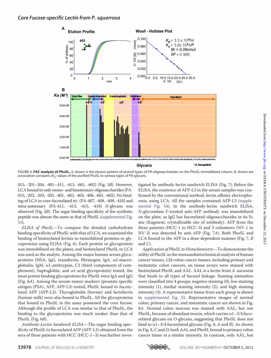
ity of PhoSL was also elucidated by FAC analysis. Among 132kinds of PA-glycans used (supplemental Fig. S1), only the 21glycans possessing the core �1–6 fucose bound to the lectin(Fig. 4B). The Bt andKd valueswere determined to be 0.09 nmol
and 3.3 � 10�6 M, respectively, for the immobilized PhoSL (1mg/ml) using PA-402 (Fig. 4A). The strength of affinity of eachPA-glycan for the immobilized lectin is shown as a Ka value(M�1) in Figs. 4B and 5B. Man�1-3(Man�1-6 Man�1-4GlcNAc�(Fuc�1-6)1-4GlcNAc-PA (PA-15, Ka � 5.0 � 10�5
M�1) showed the strongest affinity to the immobilized lectin (Fig.4B). The sialylatedN-glycans�Neu5Ac�2-3Gal�1-4GlcNAc�1-2Man�1-3(Neu5Ac�2-3Gal�1-4GlcNAc�1-2Man�1-6)Man�1-4GlcNAc�1-4 (Fuc�1-6) GlcNAc-PA (PA-601, Ka � 2.4 � 10�5
M�1 and 602, Ka � 1.2 � 10�5 M�1) also bound to the lectin. Incontrast, O-glycans having L-Fuc (PA-718 to PA-723, PA-726 toPA-731, PA-739, PA-909) or Fuc�1–3 linkages (PA-419 and -420)did not show any significant affinity to the immobilized PhoSL.The detailed oligosaccharide binding specificity of PhoSL
was compared with that of LCA, which has been reported pre-viously (Fig. 5) (3). For an easier understanding of the structuralelements required for the recognition of PhoSL and LCA, theKa values for a series of core-fucosylated glycans have beenarranged in the order of affinity strength, and the core-fucosy-lated glycans have been represented using the “GRYP” code pro-posed in a previous report (3, 32). In this system the branch posi-tions ofN-glycans (GlcNAc) are numbered from I toVI accordingto the corresponding mammalian GlcNAc-transferases, and thenonreducing end sugars are shown in different colors: GlcNAc(blue), Gal (yellow), andNeuAc (purple). The presence of the corefucose (�1–6Fuc) is emphasized with another box colored in red.Both PhoSL and LCA showed high specificity for mannose-type(PA-015),mono- (PA-201, -401, -402), and bi-antennary (PA-202,-203, -403, -404, -405, -406, -601, -602)N-glycans containing corefucose. PhoSL recognized not only mono- or biantennary oligo-saccharides but also tri- or tetra-antennary oligosaccharides (PA-
FIGURE 2. Multiple alignment of PhoSL and RSL. The residues in the first rowdescribe the amino acid sequence of PhoSL, and those in the second rowdescribe the amino acid sequence of RSL. Shown in gray shading are theamino acid residues that are identical between PhoSL and RSL.
FIGURE 3. The stability of PhoSL over a range of temperature and pHs. Thestability of PhoSL was investigated over a broad range of temperatures for 30min (A), long incubation times for four different temperatures (B), and incu-bation in different pH buffers (C).
TABLE 1Agglutination profiles of PhoSL(0.5 mg/ml)
Group oferythrocytes Titera
Rabbit 29Sheep NAb
Bovine NAHorse 28Pig 28Chicken NAGoose 210Guinea pig 28Human A NAHuman B NAHuman O NA
a The hemagglutination titer was defined as the reciprocal of the highest dilutionexhibiting hemagglutination.
b NA, not agglutinated.
TABLE 2Inhibition of PhoSL-mediated hemagglutination by glycoproteins
Inhibitora MICb
�g/mlThyroglobulin 125IgG 250
a Glucose, galactose, mannose, fucose, L-fucose, xylose, L-rhamnose, GlcNAc, Gal-NAc, ManNAc, LacNAc, lactose, maltose, fructose, saccharose, melibiose, andraffinose did not inhibit at concentrations up to 0.2 M. N-Acetylneuraminic acidand N-glycolylneuramic acid did not inhibit at concentrations up to 20 mM. Fe-tuin, asialo-fetuin, �1-acid glycoprotein, transferrin, BSM, asialo-BSM, PSM,asialo-PSM, and albumin did not inhibit at concentrations up to 500 �g/ml.
b Minimum inhibitor concentration required for inhibition of four hemagglutina-tion dose of the lectin.
Core Fucose-specific Lectin from P. squarrosa
OCTOBER 5, 2012 • VOLUME 287 • NUMBER 41 JOURNAL OF BIOLOGICAL CHEMISTRY 33977
at OSA
KA
UN
IVE
RSIT
Y on D
ecember 23, 2013
http://ww
w.jbc.org/
Dow
nloaded from
015, -201–204, -401–411, -413, -601, -602) (Fig. 5B). However,LCAbound to onlymono- andbiantennary oligosaccharides (PA-015, -202, -203, -205, -401, -402, -403, -406, -601, -602). No bind-ing of LCA to core-fucosylated tri- (PA-407, -408, -409, -410) andtetra-antennary (PA-411, -413, -415, -418) N-glycans wasobserved (Fig. 5B). The sugar binding specificity of the syntheticpeptide was almost the same as that of PhoSL (supplemental Fig.S3).ELISA of PhoSL—To compare the detailed carbohydrate
binding specificity of PhoSLwith that of LCA, we examined thebinding of biotinylated lectins to immobilized proteins or gly-coproteins using ELISA (Fig. 6). Each protein or glycoproteinwas immobilized on the plates, and biotinylated PhoSL or LCAwas used as the analyte. Among the major human serum glyco-proteins (HSA, IgG, transferrin, fibrinogen, IgA, �2-macro-globulin, IgM, �1-antitrypsin, C3 (third components of com-plement), haptoglobin, and �1-acid glycoprotein) tested, themost potent binding glycoproteins for PhoSLwere IgA and IgG(Fig. 6A). Among the serum tumor markers (prostate specificantigen (PSA), AFP, AFP-L3) tested, PhoSL bound to fucosy-lated AFP (AFP-L3). Thyroglobulin (bovine) and lactoferrin(human milk) were also bound to PhoSL. All the glycoproteinsthat bound to PhoSL in the assay possessed the core fucose.Although the profile of LCA was similar to that of PhoSL, thebinding to the glycoproteins was much weaker than that ofPhoSL (Fig. 6B).Antibody-Lectin Sandwich ELISA—The sugar binding spec-
ificity of PhoSL to fucosylated AFP (AFP-L3) obtained from thesera of three patients with HCC (HCC-1–3) was further inves-
tigated by antibody-lectin sandwich ELISA (Fig. 7). Before theELISA, the existence of AFP-L3 in the serum samples was con-firmed by the conventional method, lectin affinity electropho-resis, using LCA. All the samples contained AFP-L3 (supple-mental Fig. S4). In the antibody-lectin sandwich ELISA,N-glycosidase F-treated anti-AFP antibody was immobilizedon the plate, as IgG has fucosylated oligosaccharides in its Fcsite (fragment, crystallizable site of antibody). AFP from thethree patients (HCC-1 to HCC-3) and 3 volunteers (NV-1 toNV-3) was detected by anti-AFP (Fig. 7A). Both PhoSL andLCA bound to the AFP in a dose-dependent manner (Fig. 7, Band C).Application of PhoSL toHistochemistry—Todemonstrate the
utility of PhoSL in the immunohistochemical analysis of humancancer tissues, 124 colon cancer tissues, including primary andmetastatic colon cancers, on tissue arrays were stained withbiotinylated PhoSL and AAL. AAL is a lectin from A. aurantiathat binds to all types of fucosyl linkage. Staining intensitieswere classified into 4 groups: negative staining (0), low stainingintensity (1), medial staining intensity (2), and high stainingintensity (3). A representative tissue from each group is shownin supplemental Fig. S5. Representative images of normalcolon, primary cancer, and metastatic cancer are shown in Fig.8A. Normal colon mucosa was stained with AAL, but notPhoSL, because of abundantmucin, which carries�1–3/4 fuco-sylated glycans on O-glycans, suggesting that PhoSL does notbind to �1–3/4 fucosylated glycans (Fig. 8, A and B). As shownin Fig. 8,C andD, bothAAL and PhoSL bound to primary coloncancer tissue at a similar intensity. In contrast, only AAL, but
FIGURE 4. FAC analysis of PhoSL. A, shown is the elusion pattern of several types of PA-oligosaccharides on the PhoSL-immobilized column. B, shown areassociation constants (Ka) values of the purified PhoSL to various types of PA-glycans.
Core Fucose-specific Lectin from P. squarrosa
33978 JOURNAL OF BIOLOGICAL CHEMISTRY VOLUME 287 • NUMBER 41 • OCTOBER 5, 2012
at OSA
KA
UN
IVE
RSIT
Y on D
ecember 23, 2013
http://ww
w.jbc.org/
Dow
nloaded from

not PhoSL, bound to metastatic colon cancer tissue (Fig. 8, Eand F). The staining intensities of all tissues examined in thisstudy are summarized in Table 3. Approximately 70% of theprimary and metastatic cancer tissues were classified into ahigh intensity group after AAL staining (Fig. 8, G and H, andTable 3). No difference in AAL staining intensity was observedbetween primary and metastatic cancer tissues. In contrast,PhoSL exhibited a significantly lower binding capacity to themetastatic cancer tissues than the primary tissues (Fig. 8,G andH, and Table 3). Only 25% of the metastatic tissues showedmedial and strong intensities (group 2 and 3) after PhoSL stain-ing despite the fact that 84% of these tissues were stained withAAL.
DISCUSSION
PhoSL was purified from the edible mushroom P. squarrosa.The lectin gave a band with a mass of 4.5 kDa on SDS-PAGE in
the presence (Fig. 1,A, lane 1, andB, lane 1) and two bandswithmasses of 4.5 and 14 kDa in the absence of 2-mercaptoethanol(Fig. 1B, lane 2). Its primary structure consisted of 40 aminoacids, as determined by N-terminal amino acid sequence anal-
FIGURE 5. Comparative analysis of glycan binding specificity of PhoSLand LCA by the GRYP code. A, shown are definitions of the GRYP code forrepresenting the branch positions and non-reducing end residues. The non-reducing end sugars and the core fucose are shown in different colors in theleft panel. Each branch is numbered from I to VI corresponding to GlcNActransferases, as shown in the middle panel. B, shown are bar graph represen-tations of the association constants (Ka) of PhoSL toward core-fucosylatedN-glycans. Numbers at the bottom of the bar graphs correspond to the sugarnumbers indicated in supplemental Fig. S2. C, shown are bar graph represen-tations of the association constants (Ka) of LCA.
FIGURE 6. Binding of PhoSL and LCA to immobilized glycoproteins byELISA. A, shown is binding activity of biotin-labeled PhoSL and various immo-bilized glycoproteins. B, shown is binding activity of biotin-labeled LCA andvarious immobilized glycoproteins. The immobilized glycoproteins are: HAS,human serum albumin; Fib, fibrinogen IgA Immunoglobulin A; �2M, �2-mac-roglobulin; A1AT, �1-antitrypsin; C3, third components of complement; HP,haptoglobin; AGP, �1-acid glycoprotein; PSA, prostate specific antigen; AFP-L3, 1– 6-fucosylated fetoprotein; LF, lactoferrin; FT, fetuin; BSM, bovine sub-maxillary gland mucin; PSM, porcine stomach mucin; Inv, invertase; OVA,ovalbumin.
FIGURE 7. Antibody-lectin sandwich ELISA using purified AFP-L3 and thesera of HCC patients and normal volunteers. ELISA with Anti-AFP (A),PhoSL (B), and LCA (C).
Core Fucose-specific Lectin from P. squarrosa
OCTOBER 5, 2012 • VOLUME 287 • NUMBER 41 JOURNAL OF BIOLOGICAL CHEMISTRY 33979
at OSA
KA
UN
IVE
RSIT
Y on D
ecember 23, 2013
http://ww
w.jbc.org/
Dow
nloaded from

ysis. The synthetic PhoSL peptide corresponding to the deter-mined sequence exhibited identical binding specificity to nativePhoSL in FAC analysis (supplemental Fig. S2) but did notexhibit hemagglutination activity. HPLC gel filtration of theintact lectin gave a peak at an elution volume corresponding toamolecularmass of 14 kDa. PhoSLpossessedno sugar chains orphosphate groups. All the results mentioned above indicatedthat PhoSL is composed of three or four 4.5-kDa subunits withS-S linkage and exhibits true polyvalent binding during agglu-
tination of erythrocytes and/or precipitation of appropriate cellsurface polysaccharides, but the oligomeric form is not neces-sarily in direct binding assays (FAC analysis, ELISA, etc.).The BLAST search revealed that PhoSL has 85% sequence
homology (22/26 amino acids) with the �1–6-linked fucose-specific lectin from R. stolonifer. RSL has also been isolated as acore fucose-specific lectin. RSL has high affinity toward saccha-rides with �1–6Fuc and weak affinity toward saccharides with�1–2Fuc, �1–3Fuc, and �1–4Fuc (33).
The unique property of PhoSL is its strict sugar binding spec-ificity to �1–6Fuc (Fig. 4B). �1–6 fucosylation is one of themost important oligosaccharide modifications in carcinogene-sis; however, although many studies related to fucosylationhave been conducted, they have not completely clarified thedifference between �1–2, �1–3, or �1–4 fucosylation and�1–6 fucosylation. A hindrance to this clarification has beenthe lack of a tool for the specific detection of �1–6 fucosyllinkage; AAL, which is used in many studies, recognizes alltypes of fucosyl linkages (13, 17).The FAC results indicate that PhoSL recognizes �1–6 fuco-
syl linkages exclusively, that all the �1–6 oligosaccharides werebound to the lectin, and that LCA could not bind to some �1–6oligosaccharides (Figs. 4 and 5). In addition, the Ka value ofPhoSL was 3.2 � 105 M�1 for the fully galactosylated, bianten-nary N-glycan with a core fucose (PA-405), which is the majorN-glycan in AFP-L3 from Huh7 cells and HCC patients (Fig.4B) (34). On the other hand, the Ka value of the bindingbetween LCA and PA-405 was 4.7� 104 M�1 (3). The affinity ofPhoSL toward the oligosaccharide was higher than that of LCA.Furthermore, LCA also bound to non-fucosylated, high man-nose-type N-glycans. For example, LCA showed affinity for alarger high mannose-type N-glycan, Man8 (Man�1-2Man�1-2Man�1-3 (Man�1-2Man�1-6 (Man�1-3) Man�1-6)Man�1–4GlcNAc�1–4 (Fuc�1–6) GlcNAc-PA, PA-012),with a Ka of 2.5 � 104 M�1 (3). LCA is now the only commer-cially available diagnostic agent that can detect �1–6 fucosyl-linked sugar chains specifically.The superiority of PhoSL over LCAwas confirmed by ELISA
using biotin-labeled PhoSL and LCA and immobilized glyco-proteins. Although the specificity of both lectins showed simi-lar tendencies, the binding strength of PhoSL to the �1–6-fu-cosylated glycoproteins was greater than that of LCA (Fig. 6).The promising potential of PhoSL as a diagnostic agentwas alsoshown by antibody-lectin sandwich ELISA using AFP-L3 andthe partially purified AFP from the sera of HCC patients andnormal volunteers (NV) (Fig. 7). The sensitivity and selectivityof PhoSL to AFP-L3 in the sera of HCCs and normal volunteerswere much greater than those of LCA and even anti-AFP (Fig.
FIGURE 8. Immunohistochemical analysis of human colon cancer tissueswith PhoSL and AAL. Human colon cancer tissue arrays comprising normalcolon tissues (A and B), primary colon cancers (C and D), and metastatic coloncancers (E and F) were subjected to immunohistochemical analysis withPhoSL and AAL. G and H show the ratio of the numbers in each staining-intensity group to the total number is shown.
TABLE 3Staining intensities of AAL and PhoSL in human colon cancer tissue analyses
AALa PhoSLb
Staining intensity Normal Primary Metastatic Normal Primary Metastatic
0 0 1 (1.3%) 3 (6.7%) 7 1 (1.3%) 12 (26.7%)1 0 13 (16.5%) 7 (15.6%) 4 11 (13.9%) 22 (48.9%)2 5 12 (15.2%) 2 (4.4%) 0 41 (51.9%) 7 (15.6%)3 6 53 (67.1%) 33 (73.3%) 0 26 (32.9%) 4 (8.9%)Total 11 79 45 11 79 45
a not significant.b p � 0.01, compared between primary and metastatic cancers (�2 test).
Core Fucose-specific Lectin from P. squarrosa
33980 JOURNAL OF BIOLOGICAL CHEMISTRY VOLUME 287 • NUMBER 41 • OCTOBER 5, 2012
at OSA
KA
UN
IVE
RSIT
Y on D
ecember 23, 2013
http://ww
w.jbc.org/
Dow
nloaded from
7). AFP is a biomarker that was discovered in 1963 by Abelev(35) and belongs to the albumin-like superfamily. This protein,whose molecular mass is 65 kDa, consists of 590 amino acidsand has a biantennary sugar chain at Asn232 (36). A variety ofsugar chains are on the protein, andAFP-L3 is one of them. Thestructure of AFP-L3 has been determined to be GlcNAc�1-2Man�1-3 (Gal�1-4GlcNAc�1-2Man�1-6)Man�1-4GlcNAc�1-4(Fuc�1-6) GlcNAc-AFP by lentil lectin affinity electrophoresis.Because slightly increased serum concentrations of total AFPhave been observed in patients with chronic hepatitis and livercirrhosis, conditions that are known to be associated with pre-malignant HCC lesions, a wide overlap in total AFP has beenobserved betweenHCCand such benign liver diseases. The seraof patients with HCC are known to contain relatively largeamounts of AFP-L3. Therefore, AFP-L3 has been recognized asa specific marker for HCC. Furthermore, analysis using thismarker could be useful formonitoring treatment responses anddisease recurrence and could also be a tool for recognition ofHCC earlier than that possible by using imagingmodalities (34,37–42). In recent years, in addition to AFP, new biomarkerspossessing fucosides have been discovered. For example, Golgiprotein 73 (GP73) content in the blood of patients with HCCwas found to be elevated, and the protein was �1–6-hyperfu-cosylated (43–46). In addition, patients with liver cirrhosis andliver cancer had increased levels of triantennary glycan contain-ing outer arm (�1–3)-fucosylation in �-antitrypsin in theblood, but increases in core (�1–6)-fucosylationwere observedonly on �1-antitrypsin from patients with liver cancer (47).Physiological functions of the core fucose has been investigatedrecently. The lack of core fucosylation of transforming growthfactor-�1 receptors induces severe growth retardation anddeath during postnatal development (48, 49). Mutations of theGDP-mannose-4,6-dehydratase gene that plays a pivotal role infucosylation in human colon cancer resulted in resistance toTRAIL-induced apoptosis followed by escape from immunesurveillance. This pathway by GDP-mannose-4,6-dehydratasemutation could be a novel type of cancer progression throughcellular fucosylation and natural killer cell-mediated tumorsurveillance. However the cellular fucosylation type has notbeen determined yet (50, 51).Fig. 8 shows that AAL bound to both primary andmetastatic
colon cancer tissues with a similar intensity. However, PhoSLbound to the primary colon cancer tissuesmore strongly than itdid to the metastatic tissues. These results suggest that in somecases �1–6 fucosylation is increased in the early phase of coloncancer development and subsequently decreased in the meta-static phase. The decreased expression of �1–6 fucosylation inmetastatic cancer tissues may be responsible for the escape ofcancer cells from natural killer cell-mediated tumor surveil-lance. The mechanism and meaning underlying the decreasedexpression of �1–6 fucosylation in metastatic cancer tissuesshould be revealed in a future study. PhoSL, the lectin charac-terized in this study, may be useful for the detection of AFP-L3and other new biomarkers and for determining the physiolog-ical functions of oligosaccharides (19).In summary, PhoSL very strongly and specifically binds to
Fuc�-oligosaccharides.Moreover, it is highly stable over a widerange of pHs and temperatures and is highly soluble in various
buffers. These advantages indicate that PhoSL can become apowerful tool to analyze biological functions of core fucosesand serve as a diagnostic agent in the near future.
REFERENCES1. Miyoshi, E., Moriwaki, K., and Nakagawa, T. (2008) Biological function of
fucosylation in cancer biology. J. Biochem. 143, 725–7292. UozumiN,Yanagidani S,Miyoshi E, IharaY, SakumaT,GaoCX,TeshimaT,
Fujii S, Shiba T, and Taniguchi, N. (1996) Purification and cDNA cloning ofporcine brain GDP-L-Fuc:N-acetyl-�-D-glucosaminide �136fucosyl-transferase. J. Biol. Chem. 271, 27810–27817
3. Tateno, H., Nakamura-Tsuruta, S., and Hirabayashi, J. (2009) Compara-tive analysis of core-fucose-binding lectins from Lens culinaris and Pisumsativum using frontal affinity chromatography. Glycobiology 19, 527–536
4. Howard, I. K. (1971) Studies on a phytohemagglutinin from the lentil. II.Multiple forms of Lens culinaris hemagglutinin. J. Biol. Chem. 246,1590–1595
5. Foriers, A., Lebrun, E., Van Rapenbusch, R., de Neve, R., and Strosberg,A. D. (1981) The structure of the lentil (Lens culinaris) lectin. Amino acidsequence determination and prediction of the secondary structure. J. Biol.Chem. 256, 5550–5560
6. Aoyagi, Y. (1995) Carbohydrate-based measurements on �-fetoprotein inthe early diagnosis of hepatocellular carcinoma.Glycoconj. J. 12, 194–199
7. Ichikawa, E., Kuriyama, S., Yuji, J., Masaki, T., Uchida, N., Nishioka, M.,and Taketa, K. (2006) Further resolution of �-fetoprotein glycoforms bytwo-dimensional isoelectric focusing and lectin affinity electrophoresis.Electrophoresis 27, 3480–3487
8. Taketa, K. (1990) �-Fetoprotein. Reevaluation in hepatology. Hepatology12, 1420–1432
9. Debray, H., Decout, D., Strecker, G., Spik, G., and Montreuil, J. (1981)Specificity of twelve lectins toward oligosaccharides and glycopeptidesrelated to N-glycosylproteins. Eur. J. Biochem. 117, 41–55
10. Amano, J., Osanai,M.,Orita, T., Sugahara, D., andOsumi, K. (2009) Struc-tural determination by negative-ionMALDI-QIT-TOFMSn after pyrenederivatization of variously fucosylated oligosaccharides with branched de-caose cores from human milk. Glycobiology 19, 601–614
11. Harada, H., Kamei, M., Tokumoto, Y., Yui, S., Koyama, F., Kochibe, N.,Endo, T., and Kobata, A. (1987) Systematic fractionation of oligosaccha-rides of human immunoglobulin G by serial affinity chromatography onimmobilized lectin columns. Anal. Biochem. 164, 374–381
12. Matsumura, K., Higashida, K., Hata, Y., Kominami, J., Nakamura-Tsuruta,S., and Hirabayashi, J. (2009) Comparative analysis of oligosaccharidespecificities of fucose-specific lectins from Aspergillus oryzae and Aleuriaaurantia using frontal affinity chromatography. Anal. Biochem. 386,217–221
13. Yamashita, K., Kochibe, N., Ohkura, T., Ueda, I., and Kobata, A. (1985)Fractionation of L-fucose-containing oligosaccharides on immobilizedAleuria aurantia lectin. J. Biol. Chem. 260, 4688–4693
14. Matsumoto, I., Uehara, Y., Jimbo, A., and Seno, N. (1983) Immunochemi-cal and spectral studies on Vicia faba agglutinin. J. Biochem. 93, 763–769
15. Jordinson,M., El-Hariry, I., Calnan, D., Calam, J., and Pignatelli, M. (1999)Vicia faba agglutinin, the lectin present in broad beans, stimulates differ-entiation of undifferentiated colon cancer cells. Gut 44, 709–714
16. Allen, A. K., Desai, N. N., and Neuberger, A. (1976) Purification of theglycoprotein lectin from the broad bean (Vicia faba) and a comparison ofits properties with lectins of similar specificity. Biochem. J. 155, 127–135
17. Matsumura, K., Higashida, K., Ishida, H., Hata, Y., Yamamoto, K., Shigeta,M.,Mizuno-Horikawa, Y.,Wang, X.,Miyoshi, E., Gu, J., andTaniguchi, N.(2007) Carbohydrate binding specificity of a fucose-specific lectin fromAspergillus oryzae. A novel probe for core fucose. J. Biol. Chem. 282,15700–15708
18. Mun, J. Y., Lee, K. J., Kim, Y. J., Kwon, O., Kim, S. J., Lee, S. G., Park, W. S.,Heo, W. D., and Oh, D. B. (2012) Development of fluorescent probes forthe detection of fucosylated N-glycans using an Aspergillus oryzae lectin.Appl. Microbiol. Biotechnol. 93, 251–260
19. Matsumoto, H., Shinzaki, S., Narisada, M., Kawamoto, S., Kuwamoto, K.,Moriwaki, K., Kanke, F., Satomura, S., Kumada, T., andMiyoshi, E. (2010)
Core Fucose-specific Lectin from P. squarrosa
OCTOBER 5, 2012 • VOLUME 287 • NUMBER 41 JOURNAL OF BIOLOGICAL CHEMISTRY 33981
at OSA
KA
UN
IVE
RSIT
Y on D
ecember 23, 2013
http://ww
w.jbc.org/
Dow
nloaded from

Clinical application of a lectin antibody ELISA to measure fucosylatedhaptoglobin in sera of patients with pancreatic cancer. Clin. Chem. Lab.Med. 48, 505–512
20. Kawagishi, H. (1995) Mushroom lectins. Food Rev. Int. 11, 63–6821. Kobayashi, Y., Ishizaki, T., and Kawagishi, H. (2004) Screening for lectins
in wild and cultivated mushrooms from Japan and their sugar bindingspecificities. Int. J. Med. Mushr. 6, 113–125
22. Laemmli, U. K. (1970) Cleavage of structural proteins during the assemblyof the head of bacteriophage T4. Nature 227, 680–685
23. Kawagishi, H., Yamawaki, M., Isobe, S., Usui, T., Kimura, A., and Chiba, S.(1994) Two lectins from the marine sponge Halichondria okadai. An N-acetyl-sugar-specific lectin (HOL-I) and an N-acetyllactosamine-specificlectin (HOL-II). J. Biol. Chem. 269, 1375–1379
24. Kawagishi, H., and Mizuno, T. (1988) Purification and properties of a�-galactosyl-specific lectin from the fruiting bodies of ischnoderma res-inosus. FEBS Lett. 227, 99–102
25. Kobayashi, Y., Kobayashi, K., Umehara, K., Dohra, H.,Murata, T., Usui, T.,and Kawagishi, H. (2004) Purification, characterization, and sugar bindingspecificity of anN-glycolylneuraminic acid-specific lectin from themush-room Chlorophyllum molybdites. J. Biol. Chem. 279, 53048–53055
26. Horibe M, Kobayashi Y, Dohra H, Morita T, Murata T, Usui T, Naka-mura-Tsuruta S, Kamei M, Hirabayashi J, Matsuura M, Yamada M, Sai-kawa Y, Hashimoto K, Nakata M, and Kawagishi, H. (2010) Toxic isolec-tins from the mushroom Boletus venenatus. Phytochemistry 71, 648–657
27. Hirabayashi, J., Arata, Y., and Kasai, K. (2000) Reinforcement of frontalaffinity chromatography for effective analysis of lectin-oligosaccharide in-teractions. J. Chromatogr. A. 890, 261–271
28. Tateno, H., Nakamura-Tsuruta, S., and Hirabayashi, J. (2007) Frontal affinitychromatography. Sugar-protein interactions.Nat. Protoc. 2, 2529–2537
29. Nakamura-Tsuruta, S., Uchiyama, N., and Hirabayashi, J. (2006) High-throughput analysis of lectin-oligosaccharide interactions by automatedfrontal affinity chromatography.Methods Enzymol. 415, 311–325
30. Bradford, M. M. (1976) A rapid and sensitive method for the quantitationof microgram quantities of protein utilizing the principle of protein-dyebinding. Anal. Biochem. 72, 248–254
31. Lundy, F. T., andWisdom,G. B. (1999) An antibody-lectin sandwich assayfor quantifying protein glycoforms.Mol. Biotechnol. 12, 203–206
32. Nakamura-Tsuruta, S., Kominami, J., Kamei, M., Koyama, Y., Suzuki, T.,Isemura, M., and Hirabayashi, J. (2006) Comparative analysis by frontalaffinity chromatography of oligosaccharide specificity of GlcNAc-bindinglectins, Griffonia simplicifolia lectin-II (GSL-II) and Boletopsis leucomelaslectin (BLL) J. Biochem. 140, 285–291
33. Oda, Y., Senaha, T., Matsuno, Y., Nakajima, K., Naka, R., Kinoshita, M.,Honda, E., Furuta, I., and Kakehi, K. (2003) A new fungal lectin recognizing�(1–6)-linked fucose in theN-glycan. J. Biol. Chem. 278, 32439–32447
34. Nakagawa, T., Miyoshi, E., Yakushijin, T., Hiramatsu, N., Igura, T.,Hayashi, N., Taniguchi, N., and Kondo, A. (2008) Glycomic analysis of�-fetoprotein L3 in hepatoma cell lines and hepatocellular carcinoma pa-tients. J. Proteome Res. 7, 2222–2233
35. Abelev, G. I., Perova, S. D., Khramkova, N. I., Postnikova, Z. A., and Irlin,I. S. (1963) Production of embryonal �-globulin by transplantable mousehepatomas. Transplantation 1, 174–180
36. Morinaga, T., Sakai,M.,Wegmann, T. G., andTamaoki, T. (1983) Primarystructures of human �-fetoprotein and its mRNA. Proc. Natl. Acad. Sci.U.S.A. 80, 4604–4608
37. Taketa, K., and Hirai, H. (1989) Lectin affinity electrophoresis of �-feto-protein in cancer diagnosis. Electrophoresis 10, 562–567
38. Li, D., Mallory, T., and Satomura, S. (2001) AFP-L3. A new generation oftumor marker for hepatocellular carcinoma. Clin. Chim. Acta 313, 15–19
39. Yamagata, Y., Shimizu, K., Nakamura, K., Henmi, F., Satomura, S., Mat-suura, S., and Tanaka, M. (2003) Simultaneous determination of percent-age of Lens culinaris agglutinin-reactive �-fetoprotein and �-fetoprotein
concentration using the LiBASys clinical auto-analyzer. Clin. Chim. Acta327, 59–67
40. Sterling, R. K., Jeffers, L., Gordon, F., Sherman, M., Venook, A. P., Reddy,K. R., Satomura, S., and Schwartz, M. E. (2007) Clinical utility of AFP-L3%measurement in North American patients with HCV-related cirrhosis.Am. J. Gastroenterol. 102, 2196–2205
41. Kagebayashi, C., Yamaguchi, I., Akinaga, A., Kitano, H., Yokoyama, K.,Satomura, M., Kurosawa, T., Watanabe, M., Kawabata, T., Chang, W., Li,C., Bousse, L., Wada, H. G., and Satomura, S. (2009) Automated immu-noassay system for AFP-L3% using on-chip electrokinetic reaction andseparation by affinity electrophoresis. Anal. Biochem. 388, 306–311
42. Tamura, Y., Igarashi, M., Kawai, H., Suda, T., Satomura, S., and Aoyagi, Y.(2010) Clinical advantage of highly sensitive on-chip immunoassay forfucosylated fraction of �-fetoprotein in patients with hepatocellular car-cinoma. Dig. Dis. Sci. 55, 3576–3583
43. Morota, K., Nakagawa, M., Sekiya, R., Hemken, P. M., Sokoll, L. J., Elliott,D., Chan, D. W., and Dowell, B. L. (2011) A comparative evaluation ofGolgi protein-73, fucosylated hemopexin, �-fetoprotein, and PIVKA-II inthe serum of patients with chronic hepatitis, cirrhosis, and hepatocellularcarcinoma. Clin. Chem. Lab. Med. 49, 711–718
44. Norton, P. A., Comunale, M. A., Krakover, J., Rodemich, L., Pirog, N.,D’Amelio, A., Philip, R., Mehta, A. S., and Block, T. M. (2008) N-Linkedglycosylation of the liver cancer biomarker GP73. J. Cell. Biochem. 104,136–149
45. Wang,M., Long, R. E., Comunale, M. A., Junaidi, O., Marrero, J., Di Bisce-glie, A. M., Block, T. M., and Mehta, A. S. (2009) Novel fucosylated bio-markers for the early detection of hepatocellular carcinoma. Cancer Epi-demiol. Biomarkers Prev. 18, 1914–1921
46. Yamamoto, K., Imamura, H., Matsuyama, Y., Kume, Y., Ikeda, H., Nor-man, G. L., Shums, Z., Aoki, T., Hasegawa, K., Beck, Y., Sugawara, Y., andKokudo, N. (2010) AFP, AFP-L3, DCP, and GP73 as markers for monitor-ing treatment response and recurrence and as surrogate markers of clin-icopathological variables of HCC. J. Gastroenterol. 45, 1272–1282
47. Comunale, M. A., Rodemich-Betesh, L., Hafner, J., Wang, M., Norton, P.,Di Bisceglie, A. M., Block, T., and Mehta, A. (2010) Linkage specific fuco-sylation of alpha-1-antitrypsin in liver cirrhosis and cancer patients. Im-plications for a biomarker of hepatocellular carcinoma. PLoS One 5,12419–12422
48. Fukuda, T., Hashimoto, H., Okayasu, N., Kameyama, A., Onogi, H., Naka-gawasai, O., Nakazawa, T., Kurosawa, T., Hao, Y., Isaji, T., Tadano, T.,Narimatsu, H., Taniguchi, N., and Gu, J. (2011) �1,6-Fucosyltransferase-deficient mice exhibit multiple behavioral abnormalities associated with aschizophrenia-like phenotype. Importance of the balance between the do-pamine and serotonin systems. J. Biol. Chem. 286, 18434–18443
49. Wang, X., Inoue, S., Gu, J., Miyoshi, E., Noda, K., Li, W., Mizuno-Horikawa, Y., Nakano,M., Asahi,M., Takahashi,M., Uozumi, N., Ihara, S.,Lee, S. H., Ikeda, Y., Yamaguchi, Y., Aze, Y., Tomiyama, Y., Fujii, J., Suzuki,K., Kondo, A., Shapiro, S. D., Lopez-Otin, C., Kuwaki, T., Okabe, M.,Honke, K., and Taniguchi, N. (2005) Dysregulation of TGF-�1 receptoractivation leads to abnormal lung development and emphysema-like phe-notype in core fucose-deficient mice. Proc. Natl. Acad. Sci. U.S.A. 102,15791–15796
50. Moriwaki, K., Noda, K., Furukawa, Y., Ohshima, K., Uchiyama, A., Naka-gawa, T., Taniguchi, N., Daigo, Y., Nakamura, Y., Hayashi, N., and Miyo-shi, E. (2009) Deficiency of GMDS leads to escape fromNK cell-mediatedtumor surveillance through modulation of TRAIL signaling.Gastroenter-ology 137, 188–198
51. Moriwaki, K., Shinzaki, S., and Miyoshi, E. (2011) GDP-mannose-4,6-de-hydratase (GMDS) deficiency renders colon cancer cells resistant to tu-mor necrosis factor-related apoptosis-inducing ligand (TRAIL) receptor-and CD95-mediated apoptosis by inhibiting complex II formation. J. Biol.Chem. 286, 43123–43133
Core Fucose-specific Lectin from P. squarrosa
33982 JOURNAL OF BIOLOGICAL CHEMISTRY VOLUME 287 • NUMBER 41 • OCTOBER 5, 2012
at OSA
KA
UN
IVE
RSIT
Y on D
ecember 23, 2013
http://ww
w.jbc.org/
Dow
nloaded from
Related Documents


![Best Management Practices for Minimizing Nitrate Leaching ...downloads.hindawi.com/journals/tswj/2001/102320.pdf · Zebra Plant Aphelandra squarrosa 1,998[11] Begonia Begonia x hiemlis](https://static.cupdf.com/doc/110x72/5f1e50b079782533df2cdb91/best-management-practices-for-minimizing-nitrate-leaching-zebra-plant-aphelandra.jpg)