nanomaterials Article A Novel Coloration of Polyester Fabric through Green Silver Nanoparticles (G-AgNPs@PET) K. M. Faridul Hasan 1, *, Md. Nahid Pervez 2 , Md. Eman Talukder 3 , Mst. Zakia Sultana 2 , Sakil Mahmud 4 , Md. Mostakim Meraz 5 , Vipul Bansal 6 and Cao Genyang 1, * 1 State Key Laboratory of New Textile Materials and Advanced Processing Technologies, Wuhan Textile University, Wuhan 430200, China 2 School of Chemistry and Chemical Engineering, Wuhan Textile University, Wuhan 430200, China; [email protected] (M.N.P.); [email protected] (M.Z.S.) 3 Guangzhou Institute of Advanced Technology, Chinese Academy of Sciences, Nansha, Guangzhou 511458, China; [email protected] 4 Ningbo Institute of Material Technology and Engineering, Chinese Academy of Sciences, Ningbo 315201, China; [email protected] 5 College of Chemical and Chemistry Engineering, Xiamen University, Xiamen 361005, China; [email protected] 6 Sir Ian Potter NanoBioSensing Facility, NanoBiotechnology Research Laboratory, School of Science, RMIT University, Melbourne, VIC 3000, Australia; [email protected] * Correspondence: [email protected] (K.M.F.H.); [email protected] (C.G.); Tel.: +86-132-07102972 (K.M.F.H.); +86-181-64066991 (C.G.) Received: 15 February 2019; Accepted: 20 March 2019; Published: 8 April 2019 Abstract: This paper reports a novel route for the coloration of polyester fabric with green synthesized silver nanoparticles (G-AgNPs@PET) using chitosan as a natural eco-friendly reductant. The formation of AgNPs was confirmed by UV-visible spectroscopy. The morphologies and average particles size of G-AgNPs was investigated by transmission electron microscope (TEM) analysis. The uniform deposition of G-AgNPs on the PET fabric surface was confirmed with scanning electron microscopy (SEM) and Fourier transform infrared (FT-IR) spectroscopy. The thermal properties were investigated using a thermogravimetric analyzer (TGA). The coloration and fastness properties of fabric were found to be significantly improved, a result related to the surface plasmon resonance of G-AgNPs. The antibacterial properties of fabric were also found to be excellent as more than 80% bacterial reduction was noticed even after 10 washing cycles. Overall, the proposed coating process using green nanoparticles can contribute to low-cost production of sustainable textiles. Keywords: green silver nanoparticles; polyester; chitosan; coloration; sustainable textile 1. Introduction Textile coloration can be summarized as the relatively simple application of colorant(s) to a substrate in a solution medium. The coloring process typically involves a number of operations including dyeing, pigmenting and printing [1] and as a result releases large amounts of contaminated colored wastewater, which is considered to be a significant environmental pollutant due to its recalcitrant chemical nature (e.g., dyes/pigments) [2,3]. Many efforts are ongoing regarding the establishment of green textile coloration in terms of cost effectiveness and producing harmless non-contaminant materials associated with the subsequent process [4–6]. In recent years, the introduction of nanotechnology in the textile coloration process has provided a viable option to meet the current scenario [7–9]. Nanoparticles have received significant attention as an emerging class of colorant for textiles. Compared to conventional synthetic or natural dyes, nanoparticles have no chromophore (responsible Nanomaterials 2019, 9, 569; doi:10.3390/nano9040569 www.mdpi.com/journal/nanomaterials

Welcome message from author
This document is posted to help you gain knowledge. Please leave a comment to let me know what you think about it! Share it to your friends and learn new things together.
Transcript

nanomaterials
Article
A Novel Coloration of Polyester Fabric through GreenSilver Nanoparticles (G-AgNPs@PET)
K. M. Faridul Hasan 1,*, Md. Nahid Pervez 2 , Md. Eman Talukder 3, Mst. Zakia Sultana 2,Sakil Mahmud 4, Md. Mostakim Meraz 5 , Vipul Bansal 6 and Cao Genyang 1,*
1 State Key Laboratory of New Textile Materials and Advanced Processing Technologies, Wuhan TextileUniversity, Wuhan 430200, China
2 School of Chemistry and Chemical Engineering, Wuhan Textile University, Wuhan 430200, China;[email protected] (M.N.P.); [email protected] (M.Z.S.)
3 Guangzhou Institute of Advanced Technology, Chinese Academy of Sciences, Nansha,Guangzhou 511458, China; [email protected]
4 Ningbo Institute of Material Technology and Engineering, Chinese Academy of Sciences,Ningbo 315201, China; [email protected]
5 College of Chemical and Chemistry Engineering, Xiamen University, Xiamen 361005, China;[email protected]
6 Sir Ian Potter NanoBioSensing Facility, NanoBiotechnology Research Laboratory, School of Science, RMITUniversity, Melbourne, VIC 3000, Australia; [email protected]
* Correspondence: [email protected] (K.M.F.H.); [email protected] (C.G.);Tel.: +86-132-07102972 (K.M.F.H.); +86-181-64066991 (C.G.)
Received: 15 February 2019; Accepted: 20 March 2019; Published: 8 April 2019�����������������
Abstract: This paper reports a novel route for the coloration of polyester fabric with greensynthesized silver nanoparticles (G-AgNPs@PET) using chitosan as a natural eco-friendly reductant.The formation of AgNPs was confirmed by UV-visible spectroscopy. The morphologies and averageparticles size of G-AgNPs was investigated by transmission electron microscope (TEM) analysis.The uniform deposition of G-AgNPs on the PET fabric surface was confirmed with scanning electronmicroscopy (SEM) and Fourier transform infrared (FT-IR) spectroscopy. The thermal properties wereinvestigated using a thermogravimetric analyzer (TGA). The coloration and fastness properties offabric were found to be significantly improved, a result related to the surface plasmon resonance ofG-AgNPs. The antibacterial properties of fabric were also found to be excellent as more than 80%bacterial reduction was noticed even after 10 washing cycles. Overall, the proposed coating processusing green nanoparticles can contribute to low-cost production of sustainable textiles.
Keywords: green silver nanoparticles; polyester; chitosan; coloration; sustainable textile
1. Introduction
Textile coloration can be summarized as the relatively simple application of colorant(s) to asubstrate in a solution medium. The coloring process typically involves a number of operationsincluding dyeing, pigmenting and printing [1] and as a result releases large amounts of contaminatedcolored wastewater, which is considered to be a significant environmental pollutant due to its recalcitrantchemical nature (e.g., dyes/pigments) [2,3]. Many efforts are ongoing regarding the establishment ofgreen textile coloration in terms of cost effectiveness and producing harmless non-contaminant materialsassociated with the subsequent process [4–6]. In recent years, the introduction of nanotechnology in thetextile coloration process has provided a viable option to meet the current scenario [7–9].
Nanoparticles have received significant attention as an emerging class of colorant for textiles.Compared to conventional synthetic or natural dyes, nanoparticles have no chromophore (responsible
Nanomaterials 2019, 9, 569; doi:10.3390/nano9040569 www.mdpi.com/journal/nanomaterials
Nanomaterials 2019, 9, 569 2 of 13
for color) but rather their color properties are meditated by shape and size [10]. Among thesenanoparticles, the majority of this research has focused on silver nanoparticles (AgNPs) as a sustainabletextile coloring agent [11–13] as well as determining their ultimate fate after discharge into theenvironment [14]. AgNPs show excellent color stability due to their distinctive localized surfaceplasmon resonance (LSPR) properties [15]. The significant connection between the morphology ofsilver nanoparticles and their color is exceptionally useful for their utilization in various textile finishingapplications [16]. Control of the morphology of AgNPs is a promising method to tailor the LSPR bandand successfully tune the color of silver nanoparticles. As indicated by the literature, there are threemethodologies available for fabric coloration using nanoparticles: fabrics impregnated with a colloidalnanoparticles solution, formation of fabric/nanoparticle nanocomposites during the spinning process,and in situ synthesis of nanoparticles on fabric [17]. The functionalization of synthetic fabrics throughAgNPs has received significant attention, particularly with respect to polyester-based fabrics [18,19].Some research of note in this regard focuses on AgNP-treated polyester fabrics for improvingtheir regular performance. For example, Radetic et al. [20] employed corona treatment (electricaldischarge at atmospheric pressure) on polyester and polyamide textile materials to improve their bindinteraction prior to application of silver nanoparticles. They found that both corona-pretreated fabricshad high laundering durability and significant antibacterial activity after modification of AgNPs.Gorensek et al. [21] further reported that AgNPs on Ar/N2 (50%:50%) plasma-pretreated polyesterfabric demonstrated high dyeability and acceptable antibacterial properties against P. aeruginosa, E. coliand S. faecalis.
Up to now, numerous synthesis routes have been established for the preparation of AgNPs fortextile coloration [22–24]. The majority of AgNPs synthesized have been found to be associated withhazardous chemicals and yield toxic organic byproducts. Many recent studies focus on the greensynthesis of AgNPs for coloration of textiles with advanced applications for coloration [25,26]. In thiscontext, chitosan may be a viable green stabilizing agent for AgNPs with remarkable antimicrobialproperties against vegetative bacteria [27–30]. It is well known that chitosan is a polymeric compoundthat enables surface binding to nanoparticles with high antibacterial properties due to its functionalgroups [31]. Therefore, chitosan-mediated AgNPs could be a potential green coloration source for thetextile industry.
The present study focused on developing chitosan-mediated green silver nanoparticles forpolyester fabric coloration (G-AgNPs@PET) and, to the best of authors’ knowledge, presents sucha focus for the first time in the literature. The characteristics of G-AgNPs in solution phase werestudied by UV-visible spectrophotometer and Fourier transfer infrared spectroscopy (FTIR). Moreover,color characteristics and antibacterial properties of the treated fabrics were also evaluated. In sum,we developed a simple, green and feasible route to produce green coloration of polyester-based fabricfor multifunctional applications.
2. Materials and Methods
2.1. Materials
The 100% gray plain weave polyester (PET) fabric with 25 warps and 20 wefts in cm was suppliedby Esquel Group (Gaoming, China). Silver nitrate (AgNO3) (99.98%), chitosan and ascorbic acid(99%) were purchased from the Sinopharm Chemical Reagent company limited (Shanghai, China).All chemicals were used without further purification.
2.2. Preparation and Coloration of G-AgNPs@PET
Initially, chitosan-mediated green silver nanoparticles (G-AgNPs) were synthesized by adding1.5 g AgNO3 to 0.65 g chitosan in 50 mL solution, with the final solution being magnetically stirred at70 ◦C for 50 min. The formation of G-AgNPs was confirmed by observing the change of color solutionfrom colorless to yellow and the sample was termed as yellow colored G-AgNPs solution. Further,
Nanomaterials 2019, 9, 569 3 of 13
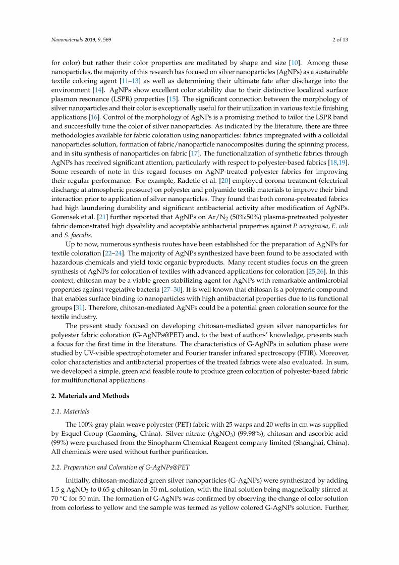
by adding 3.5 g AgNO3 and 3.3 g AgNO3 with 0.5 g ascorbic acid to the same solution, two types ofcolored AgNPs solution were observed, red and blue, respectively, which were termed as red coloredG-AgNPs and blue colored G-AgNPs solutions. For a better synthesis protocol, the solution wasfurther stirred for 90 min at 70 ◦C. The green coloration of PET fabric through as-prepared G-AgNPswas initiated by immersing the PET (5 g) into the prepared AgNPs solution under ambient conditions.Then, the solution was shaken for 45 min. The liquor ratio of AgNPs solution to fabric was 1:20.The coloration was performed in a laboratory dyeing machine bath at 135 ◦C temperature for 45 min.After coloration, the fabric was rinsed again and dried at ambient temperature in an oven dryer.Scheme 1 represents the synthesis and coloration procedure.
Nanomaterials 2019, 9, x FOR PEER REVIEW 3 of 12
70 °C for 50 min. The formation of G-AgNPs was confirmed by observing the change of color solution from colorless to yellow and the sample was termed as yellow colored G-AgNPs solution. Further, by adding 3.5 g AgNO3 and 3.3 g AgNO3 with 0.5 g ascorbic acid to the same solution, two types of colored AgNPs solution were observed, red and blue, respectively, which were termed as red colored G-AgNPs and blue colored G-AgNPs solutions. For a better synthesis protocol, the solution was further stirred for 90 min at 70 °C. The green coloration of PET fabric through as-prepared G-AgNPs was initiated by immersing the PET (5 g) into the prepared AgNPs solution under ambient conditions. Then, the solution was shaken for 45 min. The liquor ratio of AgNPs solution to fabric was 1:20. The coloration was performed in a laboratory dyeing machine bath at 135 °C temperature for 45 min. After coloration, the fabric was rinsed again and dried at ambient temperature in an oven dryer. Scheme 1 represents the synthesis and coloration procedure.
Scheme 1. G-AgNPs synthesis and coloration on PET fabric.
2.3. Characterization
The UV-Vis extinction spectra were measured using a UV–vis spectrophotometer (Shimadzu, Japan) at 300–600 nm. The morphologies of G-AgNPs were determined by transmission electron microscope (TEM, HT7700, Hitachi High Technologies America, Inc., Schaumburg, IL, USA) operating at 100 kV. The diameter and size distribution of G-AgNPs were calculated by Image J software (National Institutes of Health (NIH), Bethesda, MD, USA) using TEM images. Fourier transform infrared (FT-IR) spectra of G-AgNPs@PET were recorded in KBr pellets using an FT-IR spectrophotometer (Bruker Optics, Ettlingen, Germany) in the wavelength range of 4000–500 cm−1. The morphologies of samples were investigated using a scanning electron microscope (SEM, S-4800, Hitachi Ltd., Tokyo, Japan) at 5 kV. Thermal analysis was measured using a thermogravimetric analyzer (TGA V5.1A DUPONT2000). The samples were heated from 50 °C to 600 °C at the rate of 10 °C min−1 under the nitrogen (N2) atmosphere.
2.4. Color Measurement and Color Fastness
The color properties (reflectance curve, K/S and CIE L*a*b* values) of G-AgNPs@PET fabrics were evaluated using a Macbeth Color-Eye7000A spectrophotometer (Gretag Macbeth, Singapore) within the visible spectrum (λ = 400–700 nm) at D65 daylight and 10° standard observer. Wash fastness of the prepared samples was tested according to ISO 105-CO3 (1989) method [32]. Light fastness was tested according to ISO 105-BO2 (1989) method [33]. Dry and wet rubbing fastness of the samples were tested according to ISO 105-X12 (1987) method [34].
2.5. Antibacterial Properties
Scheme 1. G-AgNPs synthesis and coloration on PET fabric.
2.3. Characterization
The UV-Vis extinction spectra were measured using a UV–vis spectrophotometer (Shimadzu,Japan) at 300–600 nm. The morphologies of G-AgNPs were determined by transmission electronmicroscope (TEM, HT7700, Hitachi High Technologies America, Inc., Schaumburg, IL, USA) operatingat 100 kV. The diameter and size distribution of G-AgNPs were calculated by Image J software (NationalInstitutes of Health (NIH), Bethesda, MD, USA) using TEM images. Fourier transform infrared(FT-IR) spectra of G-AgNPs@PET were recorded in KBr pellets using an FT-IR spectrophotometer(Bruker Optics, Ettlingen, Germany) in the wavelength range of 4000–500 cm−1. The morphologies ofsamples were investigated using a scanning electron microscope (SEM, S-4800, Hitachi Ltd., Tokyo,Japan) at 5 kV. Thermal analysis was measured using a thermogravimetric analyzer (TGA V5.1ADUPONT2000). The samples were heated from 50 ◦C to 600 ◦C at the rate of 10 ◦C min−1 under thenitrogen (N2) atmosphere.
2.4. Color Measurement and Color Fastness
The color properties (reflectance curve, K/S and CIE L*a*b* values) of G-AgNPs@PET fabricswere evaluated using a Macbeth Color-Eye7000A spectrophotometer (Gretag Macbeth, Singapore)within the visible spectrum (λ = 400–700 nm) at D65 daylight and 10◦ standard observer. Wash fastnessof the prepared samples was tested according to ISO 105-CO3 (1989) method [32]. Light fastness wastested according to ISO 105-BO2 (1989) method [33]. Dry and wet rubbing fastness of the sampleswere tested according to ISO 105-X12 (1987) method [34].
Nanomaterials 2019, 9, 569 4 of 13
2.5. Antibacterial Properties
The antibacterial efficiency of G-AgNPs-treated fabrics was initially measured by a disc diffusionassay method (zone of inhibition assay) against Escherichia coli (Gram-negative) and Staphylococcus aureus(Gram-positive). The quantitative analysis of the antibacterial activity in terms of bacterial reductionpercentage was done according to test method GB/T 20944.3-2008 [35]. The laundering durability(repeated washing cycles) of the antibacterial activity of G-AgNPs treated fabrics was also investigatedfor up to 5 and 10 washing cycles.
3. Results and Discussion
3.1. UV-Vis Extinction Spectra of G-AgNPs
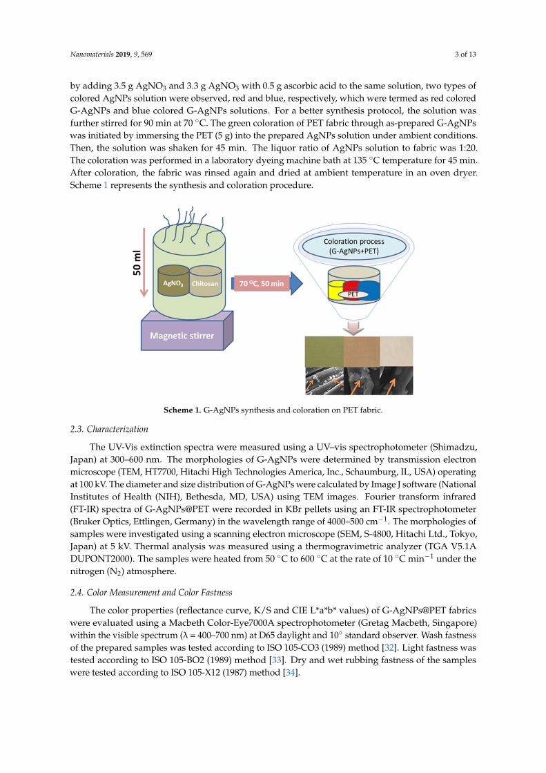
In general, a solution color change is considered to be a common indicator for the confirmation ofmetal nanoparticles. Figure 1 presents the UV-Vis extinction spectra of G-AgNPs with three differentcolors (yellow, red and blue); successful formation of G-AgNPs via Ag+ ion reduction was confirmed.The presented three different color extinction spectra were related to yellow, red and blue (Figure 1a–c)and their main extinction bands corresponded to 432, 547 and 597 nm, respectively. This phenomenonmay be ascribed to dipole plasmon plane bands of the G-AgNPs.
Nanomaterials 2019, 9, x FOR PEER REVIEW 4 of 12
The antibacterial efficiency of G-AgNPs-treated fabrics was initially measured by a disc
diffusion assay method (zone of inhibition assay) against Escherichia coli (Gram-negative) and
Staphylococcus aureus (Gram-positive). The quantitative analysis of the antibacterial activity in terms
of bacterial reduction percentage was done according to test method GB/T 20944.3-2008 [35]. The
laundering durability (repeated washing cycles) of the antibacterial activity of G-AgNPs treated
fabrics was also investigated for up to 5 and 10 washing cycles.
3. Results and Discussion
3.1. UV-Vis Extinction Spectra of G-AgNPs
In general, a solution color change is considered to be a common indicator for the confirmation
of metal nanoparticles. Figure 1 presents the UV-Vis extinction spectra of G-AgNPs with three
different colors (yellow, red and blue); successful formation of G-AgNPs via Ag+ ion reduction was
confirmed. The presented three different color extinction spectra were related to yellow, red and
blue (Figure 1a–c) and their main extinction bands corresponded to 432, 547 and 597 nm,
respectively. This phenomenon may be ascribed to dipole plasmon plane bands of the G-AgNPs.
Figure 1. UV-Vis extinction spectra of: (a) yellow G-AgNPs; (b) red G-AgNPs; and (c) blue
G-AgNPs.
3.2. TEM Images of G-AgNPs
The morphology and size distribution of the G-AgNPs was investigated by TEM image, as
shown in Figure 2a–c. In Figure 2a, the TEM image suggests that the morphologies of yellow
colored G-AgNPs showed spherical shape with well dispersed. In Figure 2b, in the case of red
colored G-AgNPs, the morphology was also spherical but dispersed in sponge form. In Figure 2c,
the TEM image of blue colored G-AgNPs reveals that also light spherical shape with reflection
dispersed. The average particle size of G-AgNPs observed size distribution in a narrow size. In
comparison with the results of three colored G-AgNPs particle size, yellow colored G-AgNPs
(Figure 2d) exhibited smaller average particle size (14.51 nm), red colored G-AGNPs (Figure 2e)
showed a bit higher average particle size (15.60 nm) and blue colored G-AgNPs demonstrated the
biggest average particle size (15.86 nm). It was noticed that the majority of average particles (90%)
were positioned between 10 and 20 nm, which is a sign of sufficient homogeneity and good
dispersion of G-AgNPs. As a result, it was expected that G-AgNPs treated fabrics would also have
well dispersed nanoparticles with high homogeneity, which could be further confirmed by SEM
analysis.
Figure 1. UV-Vis extinction spectra of: (a) yellow G-AgNPs; (b) red G-AgNPs; and (c) blue G-AgNPs.
3.2. TEM Images of G-AgNPs
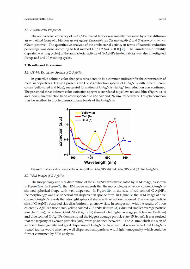
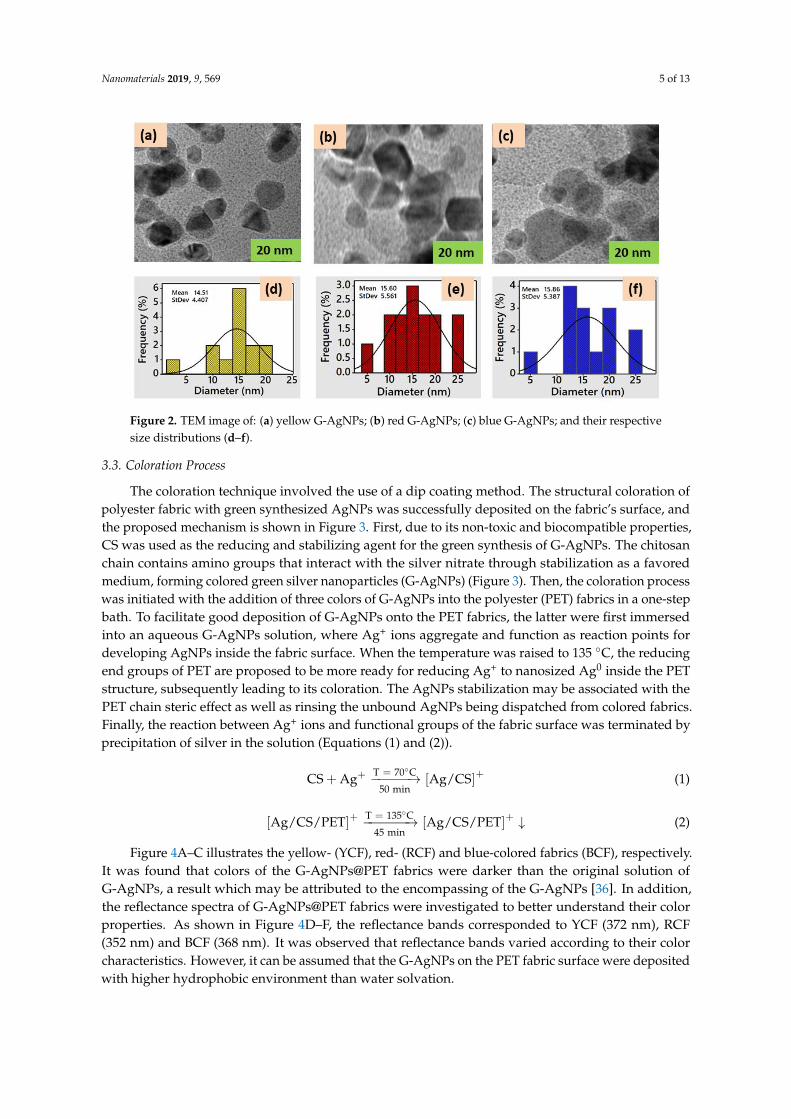
The morphology and size distribution of the G-AgNPs was investigated by TEM image, as shownin Figure 2a–c. In Figure 2a, the TEM image suggests that the morphologies of yellow colored G-AgNPsshowed spherical shape with well dispersed. In Figure 2b, in the case of red colored G-AgNPs,the morphology was also spherical but dispersed in sponge form. In Figure 2c, the TEM image of bluecolored G-AgNPs reveals that also light spherical shape with reflection dispersed. The average particlesize of G-AgNPs observed size distribution in a narrow size. In comparison with the results of threecolored G-AgNPs particle size, yellow colored G-AgNPs (Figure 2d) exhibited smaller average particlesize (14.51 nm), red colored G-AGNPs (Figure 2e) showed a bit higher average particle size (15.60 nm)and blue colored G-AgNPs demonstrated the biggest average particle size (15.86 nm). It was noticedthat the majority of average particles (90%) were positioned between 10 and 20 nm, which is a sign ofsufficient homogeneity and good dispersion of G-AgNPs. As a result, it was expected that G-AgNPstreated fabrics would also have well dispersed nanoparticles with high homogeneity, which could befurther confirmed by SEM analysis.
Nanomaterials 2019, 9, 569 5 of 13Nanomaterials 2019, 9, x FOR PEER REVIEW 5 of 12
Figure 2. TEM image of: (a) yellow G-AgNPs; (b) red G-AgNPs; (c) blue G-AgNPs; and their
respective size distributions (d–f).
3.3. Coloration Process
The coloration technique involved the use of a dip coating method. The structural coloration of
polyester fabric with green synthesized AgNPs was successfully deposited on the fabric’s surface,
and the proposed mechanism is shown in Figure 3. First, due to its non-toxic and biocompatible
properties, CS was used as the reducing and stabilizing agent for the green synthesis of G-AgNPs.
The chitosan chain contains amino groups that interact with the silver nitrate through stabilization
as a favored medium, forming colored green silver nanoparticles (G-AgNPs) (Figure 3). Then, the
coloration process was initiated with the addition of three colors of G-AgNPs into the polyester
(PET) fabrics in a one-step bath. To facilitate good deposition of G-AgNPs onto the PET fabrics, the
latter were first immersed into an aqueous G-AgNPs solution, where Ag+ ions aggregate and
function as reaction points for developing AgNPs inside the fabric surface. When the temperature
was raised to 135 °C, the reducing end groups of PET are proposed to be more ready for reducing
Ag+ to nanosized Ag0 inside the PET structure, subsequently leading to its coloration. The AgNPs
stabilization may be associated with the PET chain steric effect as well as rinsing the unbound
AgNPs being dispatched from colored fabrics. Finally, the reaction between Ag+ ions and functional
groups of the fabric surface was terminated by precipitation of silver in the solution (Equations (1)
and (2)).
oT 70 C
50 minC AS Ag g / CS
+=++ ⎯⎯⎯⎯→ (1)
oT 135 C
45 minAg / CS / PET Ag / CS / PET
+ +=⎯⎯⎯⎯→ (2)
Figure 4A–C illustrates the yellow- (YCF), red- (RCF) and blue-colored fabrics (BCF),
respectively. It was found that colors of the G-AgNPs@PET fabrics were darker than the original
solution of G-AgNPs, a result which may be attributed to the encompassing of the G-AgNPs [36]. In
addition, the reflectance spectra of G-AgNPs@PET fabrics were investigated to better understand
their color properties. As shown in Figure 4D–F, the reflectance bands corresponded to YCF (372
nm), RCF (352 nm) and BCF (368 nm). It was observed that reflectance bands varied according to
their color characteristics. However, it can be assumed that the G-AgNPs on the PET fabric surface
were deposited with higher hydrophobic environment than water solvation.
Figure 2. TEM image of: (a) yellow G-AgNPs; (b) red G-AgNPs; (c) blue G-AgNPs; and their respectivesize distributions (d–f).
3.3. Coloration Process
The coloration technique involved the use of a dip coating method. The structural coloration ofpolyester fabric with green synthesized AgNPs was successfully deposited on the fabric’s surface, andthe proposed mechanism is shown in Figure 3. First, due to its non-toxic and biocompatible properties,CS was used as the reducing and stabilizing agent for the green synthesis of G-AgNPs. The chitosanchain contains amino groups that interact with the silver nitrate through stabilization as a favoredmedium, forming colored green silver nanoparticles (G-AgNPs) (Figure 3). Then, the coloration processwas initiated with the addition of three colors of G-AgNPs into the polyester (PET) fabrics in a one-stepbath. To facilitate good deposition of G-AgNPs onto the PET fabrics, the latter were first immersedinto an aqueous G-AgNPs solution, where Ag+ ions aggregate and function as reaction points fordeveloping AgNPs inside the fabric surface. When the temperature was raised to 135 ◦C, the reducingend groups of PET are proposed to be more ready for reducing Ag+ to nanosized Ag0 inside the PETstructure, subsequently leading to its coloration. The AgNPs stabilization may be associated with thePET chain steric effect as well as rinsing the unbound AgNPs being dispatched from colored fabrics.Finally, the reaction between Ag+ ions and functional groups of the fabric surface was terminated byprecipitation of silver in the solution (Equations (1) and (2)).
CS + Ag+ T = 70◦C−−−−−→50 min
[Ag/CS]+ (1)
[Ag/CS/PET]+ T = 135◦C−−−−−−→45 min
[Ag/CS/PET]+ ↓ (2)
Figure 4A–C illustrates the yellow- (YCF), red- (RCF) and blue-colored fabrics (BCF), respectively.It was found that colors of the G-AgNPs@PET fabrics were darker than the original solution ofG-AgNPs, a result which may be attributed to the encompassing of the G-AgNPs [36]. In addition,the reflectance spectra of G-AgNPs@PET fabrics were investigated to better understand their colorproperties. As shown in Figure 4D–F, the reflectance bands corresponded to YCF (372 nm), RCF(352 nm) and BCF (368 nm). It was observed that reflectance bands varied according to their colorcharacteristics. However, it can be assumed that the G-AgNPs on the PET fabric surface were depositedwith higher hydrophobic environment than water solvation.

Nanomaterials 2019, 9, 569 6 of 13Nanomaterials 2019, 9, x FOR PEER REVIEW 6 of 12
Figure 3. Proposed mechanism of G-AgNPs@PET.
Figure 4. (A–C) Photographs; and (D–F) reflectance curves of the colored fabric.
3.4. Color Measurement and Fastness Properties
Colorimetric data were measured for G-AgNPs@PET to study the effect of G-AgNPs on the color properties (Table 1). YCF demonstrated decreased L* values with higher b* than RCF and BCF, indicating that the color became darker and more saturated in the case of YCF. On the other hand, redness (a* values) was comparatively higher for RCF and BCF compared to YCF, most likely due to the presence of a reddish tone on the fabric surface. As expected, the color strength (K/S) values were higher for YCF than RCF and BCF, a result support by the decreased L* values.
Figure 3. Proposed mechanism of G-AgNPs@PET.
Nanomaterials 2019, 9, x FOR PEER REVIEW 6 of 12
Figure 3. Proposed mechanism of G-AgNPs@PET.
Figure 4. (A–C) Photographs; and (D–F) reflectance curves of the colored fabric.
3.4. Color Measurement and Fastness Properties
Colorimetric data were measured for G-AgNPs@PET to study the effect of G-AgNPs on the
color properties (Table 1). YCF demonstrated decreased L* values with higher b* than RCF and
BCF, indicating that the color became darker and more saturated in the case of YCF. On the other
hand, redness (a* values) was comparatively higher for RCF and BCF compared to YCF, most likely
due to the presence of a reddish tone on the fabric surface. As expected, the color strength (K/S)
values were higher for YCF than RCF and BCF, a result support by the decreased L* values.
Figure 4. (A–C) Photographs; and (D–F) reflectance curves of the colored fabric.
3.4. Color Measurement and Fastness Properties
Colorimetric data were measured for G-AgNPs@PET to study the effect of G-AgNPs on thecolor properties (Table 1). YCF demonstrated decreased L* values with higher b* than RCF and BCF,indicating that the color became darker and more saturated in the case of YCF. On the other hand,redness (a* values) was comparatively higher for RCF and BCF compared to YCF, most likely due tothe presence of a reddish tone on the fabric surface. As expected, the color strength (K/S) values werehigher for YCF than RCF and BCF, a result support by the decreased L* values.

Nanomaterials 2019, 9, 569 7 of 13
Table 1. Color measurements and K/S values.
Samples L* a* b* K/S
YCF 59.57 9.71 27.34 3.84RCF 59.71 10.39 20.72 2.33BCF 64.39 12.31 17.21 1.22
The color fastness (washing, rubbing and light fastness) is related to the color-fading nature offabric, a property which is a crucial factor for commercial end uses. The color-fastness propertiesof G-AgNPs@PET fabrics were determined (Table 2). With respect to washing fastness, all coloredfabrics showed good fastness ratings in terms of demonstrating little color change (rating above 4).The light fastness of fabrics was acquired with a rating above 5, which is also satisfactory. The rubbingfastness (wet and dry) of colored fabrics was achieved in the acceptable range. Overall, all of the aboveresults were determined to be acceptable according to the color-fastness rating of the Chinese NationalStandards for Textiles [25].
Table 2. Fastness properties of the colored fabrics.
Samples WF LF RF
wet dry
YCF 4–5 5–6 4 4–5RCF 4 5 3–4 4BCF 4 5 4 4
WF, Wash Fastness; LF, Light Fastness; RF, Rubbing Fastness.
3.5. FT-IR Analysis
Fourier transform infrared spectroscopy (FTIR) was used to investigate the presence of functionalgroups of raw and G-AgNPs-treated PET fabric (Figure 5a–d). For the spectrum of RF (Figure 5a),the peaks at 3402 and 2970 cm−1 were assigned to intermolecular O–H bonded to C=O groups andC–H stretching in the polyester chain [37]. The peak at 1719 cm−1 was ascribed to the C=O groupof aromatic ester linkage, while the peaks at 1244 and 1094 cm−1 can be assigned to C=O ester bondstretching vibration [38]. The small peak at 1488 cm−1 was associated with C=C stretching vibrationand the end characteristic band at 771 cm−1 corresponded to in-plane C–O stretching vibration.As shown in Figure 5b–d, after G-AgNPs treatment, the intensity of the bands at 3000–3500 cm−1
region increased and widened compared to RF. The band at 1719 cm−1 was shifted to 1717, 1713and 1711 cm−1 (Figure 5b–d, respectively) for polyester and another band at 771 cm−1 also shifted to785, 788 and 790 cm−1 (Figure 5b–d, respectively), results associated with C–H bending vibrations ofbenzene rings in the polyester due to incorporation of the G-AgNPs on the PET surface. These resultsindicated that there were some interactions between the G-AgNPs and PET.
3.6. TGA Analysis
Thermal measurements of raw and G-AgNPs-treated PET fabrics were determined usingthermogravimetric analysis (TGA) (Figure 6a–d). Figure 6a shows the thermogram of RF. It wasobserved that decomposition of RF was in two stages; the initial stage involved 3% weight loss within10–250 ◦C due to moisture absorption of the fabric. The second stage was ascribed to the most weightloss that was observed within 278–350 ◦C due to depolymerization of the polyester. Figure 6b shows aTGA curve for the YCF sample; the two stages of degradation behavior also occurred but shifted to ahigher temperature range compared to RF. Maximum weight loss was observed in the second stagewithin 288–382 ◦C. Figure 6c illustrates the TGA curve for the RCF sample; although a similar initial3% weight loss stage to the RF was observed, in the second stage, a higher degradation temperatureof 268–358 ◦C was acquired compared to RF. Figure 6d shows the TGA curve for BCF, with the same

Nanomaterials 2019, 9, 569 8 of 13
thermogram but a higher degradation at the initial temperature (4%) and second stage at 266–356 ◦C.From the curve, it can be seen that incorporation of G-AgNPs on the PET fabric surface increasedits thermal stability, with YCF demonstrating the highest thermal stability. The TGA curve providesconfirmation that our experimental samples have good flame-retardant protection properties.
Nanomaterials 2019, 9, x FOR PEER REVIEW 8 of 12
on the PET fabric surface increased its thermal stability, with YCF demonstrating the highest
thermal stability. The TGA curve provides confirmation that our experimental samples have good
flame-retardant protection properties.
Figure 5. FT-IR spectra of: (a) RF; (b) YCF; (c) RCF; and (d) BCF.
Figure 6. TGA curves of: (a) RF; (b) YCF; (c) RCF; and (d) BCF.
Figure 5. FT-IR spectra of: (a) RF; (b) YCF; (c) RCF; and (d) BCF.
Nanomaterials 2019, 9, x FOR PEER REVIEW 8 of 12
on the PET fabric surface increased its thermal stability, with YCF demonstrating the highest
thermal stability. The TGA curve provides confirmation that our experimental samples have good
flame-retardant protection properties.
Figure 5. FT-IR spectra of: (a) RF; (b) YCF; (c) RCF; and (d) BCF.
Figure 6. TGA curves of: (a) RF; (b) YCF; (c) RCF; and (d) BCF.
Figure 6. TGA curves of: (a) RF; (b) YCF; (c) RCF; and (d) BCF.
Nanomaterials 2019, 9, 569 9 of 13
3.7. SEM Analysis
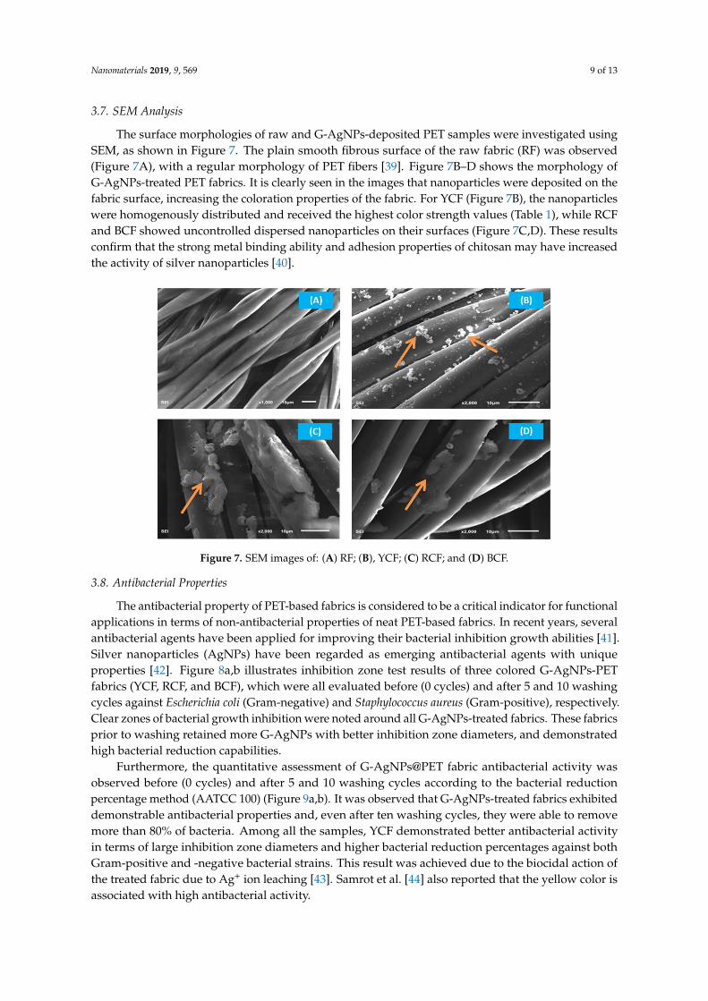
The surface morphologies of raw and G-AgNPs-deposited PET samples were investigated usingSEM, as shown in Figure 7. The plain smooth fibrous surface of the raw fabric (RF) was observed(Figure 7A), with a regular morphology of PET fibers [39]. Figure 7B–D shows the morphology ofG-AgNPs-treated PET fabrics. It is clearly seen in the images that nanoparticles were deposited on thefabric surface, increasing the coloration properties of the fabric. For YCF (Figure 7B), the nanoparticleswere homogenously distributed and received the highest color strength values (Table 1), while RCFand BCF showed uncontrolled dispersed nanoparticles on their surfaces (Figure 7C,D). These resultsconfirm that the strong metal binding ability and adhesion properties of chitosan may have increasedthe activity of silver nanoparticles [40].
Nanomaterials 2019, 9, x FOR PEER REVIEW 9 of 12
3.7. SEM Analysis
The surface morphologies of raw and G-AgNPs-deposited PET samples were investigated
using SEM, as shown in Figure 7. The plain smooth fibrous surface of the raw fabric (RF) was
observed (Figure 7A), with a regular morphology of PET fibers [39]. Figure 7B–D shows the
morphology of G-AgNPs-treated PET fabrics. It is clearly seen in the images that nanoparticles were
deposited on the fabric surface, increasing the coloration properties of the fabric. For YCF (Figure
7B), the nanoparticles were homogenously distributed and received the highest color strength
values (Table 1), while RCF and BCF showed uncontrolled dispersed nanoparticles on their surfaces
(Figure 7C,D). These results confirm that the strong metal binding ability and adhesion properties
of chitosan may have increased the activity of silver nanoparticles [40].
Figure 7. SEM images of: (A) RF; (B), YCF; (C) RCF; and (D) BCF.
3.8. Antibacterial Properties
The antibacterial property of PET-based fabrics is considered to be a critical indicator for
functional applications in terms of non-antibacterial properties of neat PET-based fabrics. In recent
years, several antibacterial agents have been applied for improving their bacterial inhibition growth
abilities [41]. Silver nanoparticles (AgNPs) have been regarded as emerging antibacterial agents
with unique properties [42]. Figure 8a,b illustrates inhibition zone test results of three colored
G-AgNPs-PET fabrics (YCF, RCF, and BCF), which were all evaluated before (0 cycles) and after 5
and 10 washing cycles against Escherichia coli (Gram-negative) and Staphylococcus aureus
(Gram-positive), respectively. Clear zones of bacterial growth inhibition were noted around all
G-AgNPs-treated fabrics. These fabrics prior to washing retained more G-AgNPs with better
inhibition zone diameters, and demonstrated high bacterial reduction capabilities.
Furthermore, the quantitative assessment of G-AgNPs@PET fabric antibacterial activity was
observed before (0 cycles) and after 5 and 10 washing cycles according to the bacterial reduction
percentage method (AATCC 100) (Figure 9a,b). It was observed that G-AgNPs-treated fabrics
exhibited demonstrable antibacterial properties and, even after ten washing cycles, they were able
to remove more than 80% of bacteria. Among all the samples, YCF demonstrated better
antibacterial activity in terms of large inhibition zone diameters and higher bacterial reduction
percentages against both Gram-positive and -negative bacterial strains. This result was achieved
due to the biocidal action of the treated fabric due to Ag+ ion leaching [43]. Samrot et al. [44] also
reported that the yellow color is associated with high antibacterial activity.
Figure 7. SEM images of: (A) RF; (B), YCF; (C) RCF; and (D) BCF.
3.8. Antibacterial Properties
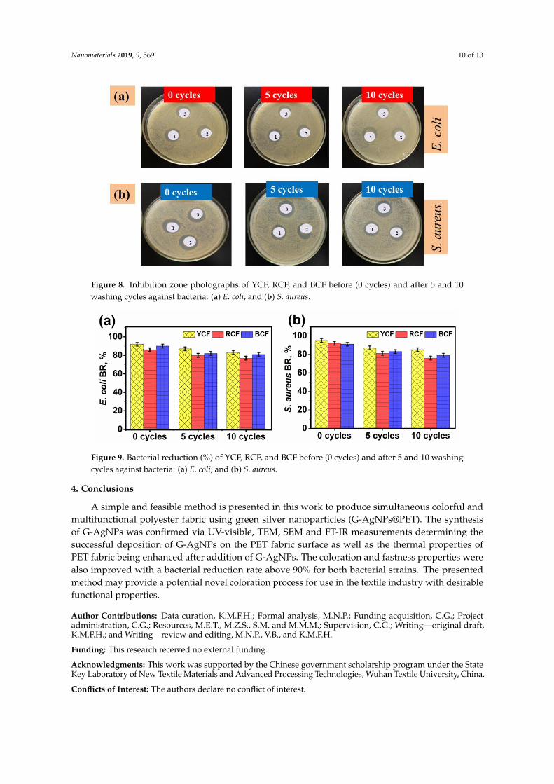
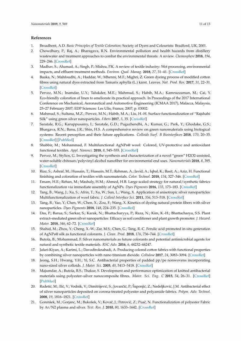
The antibacterial property of PET-based fabrics is considered to be a critical indicator for functionalapplications in terms of non-antibacterial properties of neat PET-based fabrics. In recent years, severalantibacterial agents have been applied for improving their bacterial inhibition growth abilities [41].Silver nanoparticles (AgNPs) have been regarded as emerging antibacterial agents with uniqueproperties [42]. Figure 8a,b illustrates inhibition zone test results of three colored G-AgNPs-PETfabrics (YCF, RCF, and BCF), which were all evaluated before (0 cycles) and after 5 and 10 washingcycles against Escherichia coli (Gram-negative) and Staphylococcus aureus (Gram-positive), respectively.Clear zones of bacterial growth inhibition were noted around all G-AgNPs-treated fabrics. These fabricsprior to washing retained more G-AgNPs with better inhibition zone diameters, and demonstratedhigh bacterial reduction capabilities.
Furthermore, the quantitative assessment of G-AgNPs@PET fabric antibacterial activity wasobserved before (0 cycles) and after 5 and 10 washing cycles according to the bacterial reductionpercentage method (AATCC 100) (Figure 9a,b). It was observed that G-AgNPs-treated fabrics exhibiteddemonstrable antibacterial properties and, even after ten washing cycles, they were able to removemore than 80% of bacteria. Among all the samples, YCF demonstrated better antibacterial activityin terms of large inhibition zone diameters and higher bacterial reduction percentages against bothGram-positive and -negative bacterial strains. This result was achieved due to the biocidal action ofthe treated fabric due to Ag+ ion leaching [43]. Samrot et al. [44] also reported that the yellow color isassociated with high antibacterial activity.
Nanomaterials 2019, 9, 569 10 of 13Nanomaterials 2019, 9, x FOR PEER REVIEW 10 of 12
Figure 8. Inhibition zone photographs of YCF, RCF, and BCF before (0 cycles) and after 5 and 10
washing cycles against bacteria : (a) E. coli; and (b) S. aureus.
Figure 9. Bacterial reduction (%) of YCF, RCF, and BCF before (0 cycles) and after 5 and 10 washing
cycles against bacteria: (a) E. coli; and (b) S. aureus.
4. Conclusions
A simple and feasible method is presented in this work to produce simultaneous colorful and
multifunctional polyester fabric using green silver nanoparticles (G-AgNPs@PET). The synthesis of
G-AgNPs was confirmed via UV-visible, TEM, SEM and FT-IR measurements determining the
successful deposition of G-AgNPs on the PET fabric surface as well as the thermal properties of PET
fabric being enhanced after addition of G-AgNPs. The coloration and fastness properties were also
improved with a bacterial reduction rate above 90% for both bacterial strains. The presented
method may provide a potential novel coloration process for use in the textile industry with
desirable functional properties.
Author Contributions: Data curation, K.M.F.H.; Formal analysis, M.N.P.; Funding acquisition, C.G.; Project
administration, C.G.; Resources, M.E.T., M.Z.S., S.M. and M.M.M.; Supervision, C.G.; Writing—original draft,
K.M.F.H.; and Writing—review and editing, M.N.P., V.B., and K.M.F.H.
Funding: This research received no external funding.
Acknowledgments: This work was supported by the Chinese government scholarship program under the State
Key Laboratory of New Textile Materials and Advanced Processing Technologies, Wuhan Textile University,
China.
Conflicts of Interest: The authors declare no conflict of interest.
Figure 8. Inhibition zone photographs of YCF, RCF, and BCF before (0 cycles) and after 5 and 10washing cycles against bacteria: (a) E. coli; and (b) S. aureus.
Nanomaterials 2019, 9, x FOR PEER REVIEW 10 of 12
Figure 8. Inhibition zone photographs of YCF, RCF, and BCF before (0 cycles) and after 5 and 10
washing cycles against bacteria : (a) E. coli; and (b) S. aureus.
Figure 9. Bacterial reduction (%) of YCF, RCF, and BCF before (0 cycles) and after 5 and 10 washing
cycles against bacteria: (a) E. coli; and (b) S. aureus.
4. Conclusions
A simple and feasible method is presented in this work to produce simultaneous colorful and
multifunctional polyester fabric using green silver nanoparticles (G-AgNPs@PET). The synthesis of
G-AgNPs was confirmed via UV-visible, TEM, SEM and FT-IR measurements determining the
successful deposition of G-AgNPs on the PET fabric surface as well as the thermal properties of PET
fabric being enhanced after addition of G-AgNPs. The coloration and fastness properties were also
improved with a bacterial reduction rate above 90% for both bacterial strains. The presented
method may provide a potential novel coloration process for use in the textile industry with
desirable functional properties.
Author Contributions: Data curation, K.M.F.H.; Formal analysis, M.N.P.; Funding acquisition, C.G.; Project
administration, C.G.; Resources, M.E.T., M.Z.S., S.M. and M.M.M.; Supervision, C.G.; Writing—original draft,
K.M.F.H.; and Writing—review and editing, M.N.P., V.B., and K.M.F.H.
Funding: This research received no external funding.
Acknowledgments: This work was supported by the Chinese government scholarship program under the State
Key Laboratory of New Textile Materials and Advanced Processing Technologies, Wuhan Textile University,
China.
Conflicts of Interest: The authors declare no conflict of interest.
Figure 9. Bacterial reduction (%) of YCF, RCF, and BCF before (0 cycles) and after 5 and 10 washingcycles against bacteria: (a) E. coli; and (b) S. aureus.
4. Conclusions
A simple and feasible method is presented in this work to produce simultaneous colorful andmultifunctional polyester fabric using green silver nanoparticles (G-AgNPs@PET). The synthesisof G-AgNPs was confirmed via UV-visible, TEM, SEM and FT-IR measurements determining thesuccessful deposition of G-AgNPs on the PET fabric surface as well as the thermal properties ofPET fabric being enhanced after addition of G-AgNPs. The coloration and fastness properties werealso improved with a bacterial reduction rate above 90% for both bacterial strains. The presentedmethod may provide a potential novel coloration process for use in the textile industry with desirablefunctional properties.
Author Contributions: Data curation, K.M.F.H.; Formal analysis, M.N.P.; Funding acquisition, C.G.; Projectadministration, C.G.; Resources, M.E.T., M.Z.S., S.M. and M.M.M.; Supervision, C.G.; Writing—original draft,K.M.F.H.; and Writing—review and editing, M.N.P., V.B., and K.M.F.H.
Funding: This research received no external funding.
Acknowledgments: This work was supported by the Chinese government scholarship program under the StateKey Laboratory of New Textile Materials and Advanced Processing Technologies, Wuhan Textile University, China.
Conflicts of Interest: The authors declare no conflict of interest.
Nanomaterials 2019, 9, 569 11 of 13
References
1. Broadbent, A.D. Basic Principles of Textile Coloration; Society of Dyers and Colourists: Bradford, UK, 2001.2. Chowdhary, P.; Raj, A.; Bharagava, R.N. Environmental pollution and health hazards from distillery
wastewater and treatment approaches to combat the environmental threats: A review. Chemosphere 2018, 194,229–246. [CrossRef]
3. Madhav, S.; Ahamad, A.; Singh, P.; Mishra, P.K. A review of textile industry: Wet processing, environmentalimpacts, and effluent treatment methods. Environ. Qual. Manag. 2018, 27, 31–41. [CrossRef]
4. Baaka, N.; Mahfoudhi, A.; Haddar, W.; Mhenni, M.F.; Mighri, Z. Green dyeing process of modified cottonfibres using natural dyes extracted from Tamarix aphylla (L.) karst. Leaves. Nat. Prod. Res. 2017, 31, 22–31.[CrossRef]
5. Pervez, M.N.; Inamdar, U.Y.; Talukder, M.E.; Mahmud, S.; Habib, M.A.; Kamruzzaman, M.; Cai, Y.Eco-friendly coloration of linen to ameliorate its practical approach. In Proceedings of the 2017 InternationalConference on Mechanical, Aeronautical and Automotive Engineering (ICMAA 2017), Malacca, Malaysia,25–27 February 2007; EDP Sciences: Les Ulis, France, 2007; p. 03002.
6. Mahmud, S.; Sultana, M.Z.; Pervez, M.N.; Habib, M.A.; Liu, H.-H. Surface functionalization of “RajshahiSilk” using green silver nanoparticles. Fibers 2017, 5, 35. [CrossRef]
7. Saratale, R.G.; Karuppusamy, I.; Saratale, G.D.; Pugazhendhi, A.; Kumar, G.; Park, Y.; Ghodake, G.S.;Bhargava, R.N.; Banu, J.R.; Shin, H.S. A comprehensive review on green nanomaterials using biologicalsystems: Recent perception and their future applications. Colloids Surf. B Biointerfaces 2018, 170, 20–35.[CrossRef] [PubMed]
8. Shabbir, M.; Mohammad, F. Multifunctional AgNPs@ wool: Colored, UV-protective and antioxidantfunctional textiles. Appl. Nanosci. 2018, 8, 545–555. [CrossRef]
9. Pervez, M.; Stylios, G. Investigating the synthesis and characterization of a novel “green” H2O2-assisted,water-soluble chitosan/polyvinyl alcohol nanofiber for environmental end uses. Nanomaterials 2018, 8, 395.[CrossRef]
10. Riaz, S.; Ashraf, M.; Hussain, T.; Hussain, M.T.; Rehman, A.; Javid, A.; Iqbal, K.; Basit, A.; Aziz, H. Functionalfinishing and coloration of textiles with nanomaterials. Color. Technol. 2018, 134, 327–346. [CrossRef]
11. Emam, H.E.; Rehan, M.; Mashaly, H.M.; Ahmed, H.B. Large scaled strategy for natural/synthetic fabricsfunctionalization via immediate assembly of AgNPs. Dyes Pigments 2016, 133, 173–183. [CrossRef]
12. Tang, B.; Wang, J.; Xu, S.; Afrin, T.; Xu, W.; Sun, L.; Wang, X. Application of anisotropic silver nanoparticles:Multifunctionalization of wool fabric. J. Colloid Interface Sci. 2011, 356, 513–518. [CrossRef]
13. Tang, B.; Yao, Y.; Chen, W.; Chen, X.; Zou, F.; Wang, X. Kinetics of dyeing natural protein fibers with silvernanoparticles. Dyes Pigments 2018, 148, 224–235. [CrossRef]
14. Das, P.; Barua, S.; Sarkar, S.; Karak, N.; Bhattacharyya, P.; Raza, N.; Kim, K.-H.; Bhattacharya, S.S. Plantextract–mediated green silver nanoparticles: Efficacy as soil conditioner and plant growth promoter. J. Hazard.Mater. 2018, 346, 62–72. [CrossRef]
15. Shahid, M.; Zhou, Y.; Cheng, X.-W.; Zar, M.S.; Chen, G.; Tang, R.-C. Ferulic acid promoted in-situ generationof AgNPs@ silk as functional colorants. J. Clean. Prod. 2018, 176, 736–744. [CrossRef]
16. Butola, B.; Mohammad, F. Silver nanomaterials as future colorants and potential antimicrobial agents fornatural and synthetic textile materials. RSC Adv. 2016, 6, 44232–44247.
17. Jafari-Kiyan, A.; Karimi, L.; Davodiroknabadi, A. Producing colored cotton fabrics with functional propertiesby combining silver nanoparticles with nano titanium dioxide. Cellulose 2017, 24, 3083–3094. [CrossRef]
18. Jeong, S.H.; Hwang, Y.H.; Yi, S.C. Antibacterial properties of padded pp/pe nonwovens incorporatingnano-sized silver colloids. J. Mater. Sci. 2005, 40, 5413–5418. [CrossRef]
19. Majumdar, A.; Butola, B.S.; Thakur, S. Development and performance optimization of knitted antibacterialmaterials using polyester–silver nanocomposite fibres. Mater. Sci. Eng. C 2015, 54, 26–31. [CrossRef][PubMed]
20. Radetic, M.; Ilic, V.; Vodnik, V.; Dimitrijevic, S.; Jovancic, P.; Šaponjic, Z.; Nedeljkovic, J.M. Antibacterial effectof silver nanoparticles deposited on corona-treated polyester and polyamide fabrics. Polym. Adv. Technol.2008, 19, 1816–1821. [CrossRef]
21. Gorenšek, M.; Gorjanc, M.; Bukošek, V.; Kovac, J.; Petrovic, Z.; Puac, N. Functionalization of polyester Fabricby Ar/N2 plasma and silver. Text. Res. J. 2010, 80, 1633–1642. [CrossRef]
Nanomaterials 2019, 9, 569 12 of 13
22. Perelshtein, I.; Applerot, G.; Perkas, N.; Guibert, G.; Mikhailov, S.; Gedanken, A. Sonochemical coatingof silver nanoparticles on textile fabrics (nylon, polyester and cotton) and their antibacterial activity.Nanotechnology 2008, 19, 245705. [CrossRef]
23. Tang, B.; Zhang, M.; Hou, X.; Li, J.; Sun, L.; Wang, X. Coloration of cotton fibers with anisotropic silvernanoparticles. Ind. Eng. Chem. Res. 2012, 51, 12807–12813. [CrossRef]
24. Hassan, M.M.; Koyama, K. Multifunctional acrylic fibers prepared via in-situ formed silver nanoparticles:Physicochemical, UV radiation protection, and antistatic properties. Dyes Pigments 2018, 159, 517–526.[CrossRef]
25. Shahid, M.; Cheng, X.-W.; Tang, R.-C.; Chen, G. Silk functionalization by caffeic acid assisted in-situgeneration of silver nanoparticles. Dyes Pigments 2017, 137, 277–283. [CrossRef]
26. Mahmud, S.; Pervez, M.N.; Sultana, M.Z.; Habib, M.A.; Liu, H.-H. Wool functionalization by using greensynthesized silver nanoparticles. Orient. J. Chem. 2017, 33, 2198–2208. [CrossRef]
27. Alfaro-González, B.; Ulate, D.; Alvarado, R.; Argüello-Miranda, O. Chitosan-silver nanoparticles as anapproach to control bacterial proliferation, spores and antibiotic-resistant bacteria. Biomed. Phys. Eng. Express2018, 4, 035011. [CrossRef]
28. Nithya, A.; JeevaKumari, H.L.; Rokesh, K.; Ruckmani, K.; Jeganathan, K.; Jothivenkatachalam, K. A versatileeffect of chitosan-silver nanocomposite for surface plasmonic photocatalytic and antibacterial activity.J. Photochem. Photobiol. B Biol. 2015, 153, 412–422. [CrossRef]
29. Xu, Q.; Zheng, W.; Duan, P.; Chen, J.; Zhang, Y.; Fu, F.; Diao, H.; Liu, X. One-pot fabrication of durableantibacterial cotton fabric coated with silver nanoparticles via carboxymethyl chitosan as a binder andstabilizer. Carbohydr. Polym. 2019, 204, 42–49. [CrossRef]
30. Kumar-Krishnan, S.; Prokhorov, E.; Hernández-Iturriaga, M.; Mota-Morales, J.D.; Vázquez-Lepe, M.;Kovalenko, Y.; Sanchez, I.C.; Luna-Bárcenas, G. Chitosan/silver nanocomposites: Synergistic antibacterialaction of silver nanoparticles and silver ions. Eur. Polym. J. 2015, 67, 242–251. [CrossRef]
31. Huang, X.; Bao, X.; Liu, Y.; Wang, Z.; Hu, Q. Catechol-functional chitosan/silver nanoparticle composite as ahighly effective antibacterial agent with species-specific mechanisms. Sci. Rep. 2017, 7, 1860. [CrossRef]
32. ISO 105-c03: Textiles–Tests for Colour Fastness–Partc03: Colour Fastness to Washing: Test 3; ISO: Geneva,Switzerland, 1989.
33. ISO 105-b02: Textiles–Tests for Colour Fastness–Partb02: Colour Fastness to Artificial Light: Xenon Arc FadingLamp Test; ISO: Geneva, Switzerland, 1988.
34. ISO 105-x12: Textiles–Tests for Colour Fastness–Partx12: Colour Fastness to Rubbing; ISO: Geneva, Switzerland,1987.
35. GB/T 20944.3. Textiles—Evaluation for Antibacterial Activity—Part 3: Shake Flask Method; North Yuehai WeiChemical Co., Ltd.: Shenzhen, China, 2008.
36. Perelshtein, I.; Applerot, G.; Perkas, N.; Wehrschetz-Sigl, E.; Hasmann, A.; Guebitz, G.; Gedanken, A.Antibacterial properties of an in situ generated and simultaneously deposited nanocrystalline ZnO on fabrics.ACS Appl. Mater. Interfaces 2008, 1, 361–366. [CrossRef]
37. Mock, J.J.; Smith, D.R.; Schultz, S. Local refractive index dependence of plasmon resonance spectra fromindividual nanoparticles. Nano Lett. 2003, 3, 485–491. [CrossRef]
38. Liu, Z.; Li, J.; Zhao, X.; Li, Z.; Li, Q. Surface coating for flame retardancy and pyrolysis behavior of polyesterfabric based on calcium alginate nanocomposites. Nanomaterials 2018, 8, 875. [CrossRef]
39. Liu, C.; Li, X.; Li, X.; Xu, T.; Song, C.; Ogino, K.; Gu, Z. Preparation of conductive polyester fibers usingcontinuous two-step plating silver. Materials 2018, 11, 2033. [CrossRef]
40. Montaser, A.S.; Mahmoud, F.A. Preparation of chitosan-grafted-polyvinyl acetate metal nanocomposite forproducing multifunctional textile cotton fabrics. Int. J. Biol. Macromol. 2019, 124, 659–666. [CrossRef]
41. Zhou, J.; Fei, X.; Li, C.; Yu, S.; Hu, Z.; Xiang, H.; Sun, B.; Zhu, M. Integrating Nano-Cu2O@ ZrP into in situpolymerized polyethylene terephthalate (PET) fibers with enhanced mechanical properties and antibacterialactivities. Polymers 2019, 11, 113. [CrossRef]
42. Jaworski, S.; Wierzbicki, M.; Sawosz, E.; Jung, A.; Gielerak, G.; Biernat, J.; Jaremek, H.; ojkowski, W.;Wozniak, B.; Wojnarowicz, J. Graphene oxide-based nanocomposites decorated with silver nanoparticles asan antibacterial agent. Nanoscale Res. Lett. 2018, 13, 1–17. [CrossRef]

Nanomaterials 2019, 9, 569 13 of 13
43. Gedik, G.; Aksit, A.; Engin, B.; Paksu, U. Production of metal oxide containing antibacterial coated textilematerial and investigation of the mechanism of action. Fibers Polym. 2018, 19, 2548–2563. [CrossRef]
44. Samrot, A.V.; Shobana, N.; Jenna, R. Antibacterial and antioxidant activity of different staged ripenedfruit of Capsicum annuum and its green synthesized silver nanoparticles. BioNanoScience 2018, 8, 632–646.[CrossRef]
© 2019 by the authors. Licensee MDPI, Basel, Switzerland. This article is an open accessarticle distributed under the terms and conditions of the Creative Commons Attribution(CC BY) license (http://creativecommons.org/licenses/by/4.0/).
Related Documents