Plant Physiol. (1994) 104: 109-118 A Nove1 Pathogen- and Wound-lnducible Tobacco (Nicotiana tabacum) Protein with Antifungal Activity Anne S. Ponstein*, Sandra A. Bres-Vloemans, Marianne B. Sela-Buurlage, Peter J.M. van den Elzen, Leo S. Melchers, and Ben J.C. Cornelissen' MOGEN lnternational NV, 2333 CB Leiden, The Netherlands A nove1 pathogen- and wound-inducible antifungal protein of 20 kD was purified from tobacco (Nicotiana tabacum) Samsun NN leaves inoculated with tobacco mosaic virus (TMV). The protein, designated CBPZO, was purified by chitin-affinity chromatography and gel filtration. In vitro assays demonstrated that CBPZO exhibits antifungal activity toward Trichoderma viride and Fusarium solani by causing lysis of the germ tubes and/or growth inhibition. In addition it was shown that CBPZO acts synergistically with a tobacco class I chitinase against F. solani and with a tobacco class I &1,3- glucanase against F. solani and Alternaria radicina. Analysis of the protein and corresponding cDNAs revealed that CBPZO contains an N-terminal chitin-binding domain that is present also in the class I chitinases of tobacco, the putative wound-induced (WIN) proteins of potato, W l N l and WINZ, and severa1 plant lectins. The C-terminal domain of CBPZO showed high identity with tobacco pathogenesis-related(PR) proteins, PR-4a and PR-4b, tomato PR- P2, and potato W l N l and WIN2. CBPZO is synthesized as a prepro- protein, which is processed into the mature protein by the removal of an N-terminal signal peptide and a C-terminal propeptide, most likely involved in the vacuolar targeting of the protein. The intra- cellular localization of CBPZO and its induction upon TMV infection and wounding indicate that CBPZO is the first class I PR-4 type protein purified. In plants resistance against pathogens can be induced by a variety of biotic and abiotic elicitors (Sequeira, 1983). For example, infection of tobacco with a necrotizing strain of TMV leads to the induction of resistance against a broad range of pathogens including fungi, bacteria, and viruses. Resistance is induced both locally around the site of infection and systemically in noninfected parts of the plant. Concom- itant with resistance, the synthesis of a large number of proteins including the so-called PR proteins is induced. The PR proteins form a group of proteins that are pathogen induced in the infected parts of the plant, although they may be synthesized in other parts of the plant constitutively or during specific developmental stages (Van Loon, 1990). PR proteins have been classified into five different groups (re- viewed by Bol et al., 1990; Linthorst, 1991). Most groups can be subdivided into two or more classes. Generally, class I proteins are localized in the vacuole of the plant cell, whereas class I1 proteins are present extracellularly. The class I and Present address: Institute for Molecular Cell Biology, BioCentrum Amsterdam, Kruislaan 318, 1098 SM Amsterdam, The Netherlands. * Corresponding author; fax 31-71-221471. 1 o9 class I1 proteins are related both structurally and immunolog- ically but differ in their induction pattems (Brederode et al., 1991; Ward et al., 1991). Two of the five groups of PR proteins have known enzymic functions: the PR-2 group consists of /3-1,3-glucanases (Kauffmann et al., 1987) and the PR-3 proteins exhibit chiti- nase activity (Legrand et al., 1987). A third class of PR-2 proteins (Payne et al., 1990b) and two new classes of PR-3 proteins (Lawton et al., 1992; Collinge et al., 1993) have been described recently. It has been shown that chitinases and p- 1,3-glucanases from various plant sources are able to inhibit funga1 growth in vitro (Mauch et al., 1988). This is also true for the class I chitinases and ,f3-1,3-glucanases from tobacco. However, both class I1 hydrolases seem to lack detectable amounts of antifungal activity in in vitro assays (Sela-Buur- lage et al., 1993). As yet the biochemical basis for the differ- ence in the antifungal effect of class I and class I1 proteins is not known. In the case of the tobacco PR-3 polypeptides the main structural difference is the presence in the class I proteins of a chitin-binding domain and a hinge region that are both absent in the mature class I1 proteins (Linthorst et al., 1990b; Shinshi et al., 1990; Linthorst, 1991; Collinge et al., 1993). Class 111 chitinases seem to lack antifungal activity as well (Vogelsang and Barz, 1993). Chitin-binding proteins, such as hevein (Van Parijs et al., 1991), stinging nettle lectin (Broekaert et al., 1989), and some antimicrobial peptides purified from Amaranthus caudatus seed (Broekqert et al., 1992), have been shown to exhibit antifungal activity. These chitin-binding proteins a11 lack detectable levels of chitinase activity. In contrast to the lectins mentioned above, wheat germ agglutinin, consisting of four hevein domains in tandem (Raikhel and Wilkins, 1987), was reported to lack antifungal activity (Schlumbaum et al., 1986; Chrispeels and Raikhel, 1991). In this paper we report the purification of a chitin-binding protein of 20 kD from tobacco (Nicotiana tabacum) Samsun NN. Structural and immunological data indicate that this protein belongs to the PR-4 group. The polypeptide is located intracellularly like a11 tobacco class I proteins. The pathogen- and stress-induction pattem of CBP2O mRNA resembles the induction pattem of class I PR proteins rather than the pattem Abbreviations: CBP20, 20-kD chitin-binding protein; CTPP, C- terminal propeptide; EF, extracellular fluid; ODU, optical density units; PR, pathogenesis related; PVDF, polyvinylidene difluoride; TMV, tobacco mosaic virus; WIN, wound induced. www.plant.org on October 6, 2015 - Published by www.plantphysiol.org Downloaded from Copyright © 1994 American Society of Plant Biologists. All rights reserved.

Welcome message from author
This document is posted to help you gain knowledge. Please leave a comment to let me know what you think about it! Share it to your friends and learn new things together.
Transcript
Plant Physiol. (1994) 104: 109-118
A Nove1 Pathogen- and Wound-lnducible Tobacco (Nicotiana tabacum) Protein with Antifungal Activity
Anne S. Ponstein*, Sandra A. Bres-Vloemans, Marianne B. Sela-Buurlage, Peter J.M. van den Elzen, Leo S. Melchers, and Ben J.C. Cornelissen'
MOGEN lnternational NV, 2333 CB Leiden, The Netherlands
A nove1 pathogen- and wound-inducible antifungal protein of 20 kD was purified from tobacco (Nicotiana tabacum) Samsun NN leaves inoculated with tobacco mosaic virus (TMV). The protein, designated CBPZO, was purified by chitin-affinity chromatography and gel filtration. In vitro assays demonstrated that CBPZO exhibits antifungal activity toward Trichoderma viride and Fusarium solani by causing lysis of the germ tubes and/or growth inhibition. In addition it was shown that CBPZO acts synergistically with a tobacco class I chitinase against F. solani and with a tobacco class I &1,3- glucanase against F. solani and Alternaria radicina. Analysis of the protein and corresponding cDNAs revealed that CBPZO contains an N-terminal chitin-binding domain that i s present also in the class I chitinases of tobacco, the putative wound-induced (WIN) proteins of potato, WlN l and WINZ, and severa1 plant lectins. The C-terminal domain of CBPZO showed high identity with tobacco pathogenesis-related (PR) proteins, PR-4a and PR-4b, tomato PR- P2, and potato WlNl and WIN2. CBPZO is synthesized as a prepro- protein, which i s processed into the mature protein by the removal of an N-terminal signal peptide and a C-terminal propeptide, most likely involved in the vacuolar targeting of the protein. The intra- cellular localization of CBPZO and its induction upon TMV infection and wounding indicate that CBPZO is the first class I PR-4 type protein purified.
In plants resistance against pathogens can be induced by a variety of biotic and abiotic elicitors (Sequeira, 1983). For example, infection of tobacco with a necrotizing strain of TMV leads to the induction of resistance against a broad range of pathogens including fungi, bacteria, and viruses. Resistance is induced both locally around the site of infection and systemically in noninfected parts of the plant. Concom- itant with resistance, the synthesis of a large number of proteins including the so-called PR proteins is induced. The PR proteins form a group of proteins that are pathogen induced in the infected parts of the plant, although they may be synthesized in other parts of the plant constitutively or during specific developmental stages (Van Loon, 1990). PR proteins have been classified into five different groups (re- viewed by Bol et al., 1990; Linthorst, 1991). Most groups can be subdivided into two or more classes. Generally, class I proteins are localized in the vacuole of the plant cell, whereas class I1 proteins are present extracellularly. The class I and
Present address: Institute for Molecular Cell Biology, BioCentrum Amsterdam, Kruislaan 318, 1098 SM Amsterdam, The Netherlands.
* Corresponding author; fax 31-71-221471. 1 o9
class I1 proteins are related both structurally and immunolog- ically but differ in their induction pattems (Brederode et al., 1991; Ward et al., 1991).
Two of the five groups of PR proteins have known enzymic functions: the PR-2 group consists of /3-1,3-glucanases (Kauffmann et al., 1987) and the PR-3 proteins exhibit chiti- nase activity (Legrand et al., 1987). A third class of PR-2 proteins (Payne et al., 1990b) and two new classes of PR-3 proteins (Lawton et al., 1992; Collinge et al., 1993) have been described recently. It has been shown that chitinases and p- 1,3-glucanases from various plant sources are able to inhibit funga1 growth in vitro (Mauch et al., 1988). This is also true for the class I chitinases and ,f3-1,3-glucanases from tobacco. However, both class I1 hydrolases seem to lack detectable amounts of antifungal activity in in vitro assays (Sela-Buur- lage et al., 1993). As yet the biochemical basis for the differ- ence in the antifungal effect of class I and class I1 proteins is not known. In the case of the tobacco PR-3 polypeptides the main structural difference is the presence in the class I proteins of a chitin-binding domain and a hinge region that are both absent in the mature class I1 proteins (Linthorst et al., 1990b; Shinshi et al., 1990; Linthorst, 1991; Collinge et al., 1993). Class 111 chitinases seem to lack antifungal activity as well (Vogelsang and Barz, 1993). Chitin-binding proteins, such as hevein (Van Parijs et al., 1991), stinging nettle lectin (Broekaert et al., 1989), and some antimicrobial peptides purified from Amaranthus caudatus seed (Broekqert et al., 1992), have been shown to exhibit antifungal activity. These chitin-binding proteins a11 lack detectable levels of chitinase activity. In contrast to the lectins mentioned above, wheat germ agglutinin, consisting of four hevein domains in tandem (Raikhel and Wilkins, 1987), was reported to lack antifungal activity (Schlumbaum et al., 1986; Chrispeels and Raikhel, 1991).
In this paper we report the purification of a chitin-binding protein of 20 kD from tobacco (Nicotiana tabacum) Samsun NN. Structural and immunological data indicate that this protein belongs to the PR-4 group. The polypeptide is located intracellularly like a11 tobacco class I proteins. The pathogen- and stress-induction pattem of CBP2O mRNA resembles the induction pattem of class I PR proteins rather than the pattem
Abbreviations: CBP20, 20-kD chitin-binding protein; CTPP, C- terminal propeptide; EF, extracellular fluid; ODU, optical density units; PR, pathogenesis related; PVDF, polyvinylidene difluoride; TMV, tobacco mosaic virus; WIN, wound induced.
www.plant.org on October 6, 2015 - Published by www.plantphysiol.orgDownloaded from Copyright © 1994 American Society of Plant Biologists. All rights reserved.
110 Ponstein et al. Plant Physiol. Vol. 104, 1994
of the class I1 PR proteins. Consequently, CBP2O was classi- fied as a class I PR-4 protein. The purified protein was shown to inhibit growth of several fungi and to act synergistic- ally with both a tobacco class I chitinase and a class I 8- 1,3-glucanase.
MATERIALS AND METHODS
Biological Materiais
Tobacco (Nicotiana tabacum cv Samsun NN) was grown at 24OC in an artificially illuminated room (12,000 luxht plant height) with a 16-h photoperiod.
Purification of the Class I Chitinases and CBPZO
Proteins were extracted from tobacco leaves 7 d after infection with TMV (Woloshuk et al., 1991). The protein extract was desalted by passage through a G-25 column and partly separated by cation-exchange chromatography as de- scribed by Woloshuk et al. (1991). Fractions containing chi- tinase activity were pooled and concentrated by ultrafiltration through a YMlO (Amicon) membrane (molecular mass cutoff 10 kD). The concentrated solution was brought to 20 m~ NaHC03. The pH of the protein solution was adjusted to 8.3 by the addition of 1 M NaOH, and the proteins were further dialyzed to 20 m1.4 NaHCO, (pH 8.3). Chitin-affinity purifi- cation was performed essentially as described by Broekaert et al. (1988). Proteins were allowed to adsorb to a matrix (50 mL) of regenerated chitin (Molano et al., 1977) equilibrated in 20 m~ NaHC03 for 1 h at 4OC under continuous stirring. A column was poured, and unbound proteins were washed off by passage of 100 mL of 20 mM NaHC03 (pH 8.3). The column was further washed with 100 mL of 20 m~ Na acetate (pH 5.2), and bound proteins were eluted by the passage of 20 m~ acetic acid (pH 3.5). Fractions of about 4 mL each were collected at a flow rate of 1 mL min-’.
Fractions containing chitin-binding proteins were further purified by gel filtration chromatography. The chitin eluate was concentrated (to about 1 mL) and loaded in several runs onto a Superdex-75 column (HR 10/30; Pharmacia) equili- brated in 50 m~ KHPOr buffer (pH 7.0), containing 0.2 M
NaCl. Gel filtration was camed out at 0.5 mL min-’ in the same buffer, and fractions (0.5 mL) were collected. The gel filtration column was calibrated with BSA (68 kD), carbonic anhydrase (29 kD), and Cyt c reductase (12.5 kD). Two protein peaks at apparent molecular masses of 15 to 15.5 and 8 kD, containing the class I chitinases and CBP20, respec- tively, were obtained. The two peaks were pooled separately and rechromatographed (usually twice). The basic chitinases were purified as previously described (Sela-Buurlage et al., 1993).
Purification of PR-4a,b
The proteins that did not bind to the S-Sepharose column (see above) were dialyzed to 20 m~ Tris-HC1 (pH 8.0) and allowed to flow through a Q-Sepharose column equilibrated in the same buffer (Woloshuk et al., 1991). The flow through was collected, concentrated, and dialyzed to 25 m~ di- ethanolamine-HCl (pH 9.7). The resulting protein solution
was loaded onto a Mono P column (Pharmacia) equilibrated in the same buffer. Bound proteins were eluted with a de- creasing pH gradient produced by a 10% solution of Poly- buffer 96 (pH 6.0). PR-4a,b (formerly called PR-rl,r2) was readily eluted as deduced from the electrophoretic pattem of the protein fractions on 10% native gels. The PR-4a,b- con- taining fractions were concentrated and passed through the Superdex-75 gel filtration column as described above. PR- 4a,b-containing fractions were pooled and used for antifungal assays.
Protein Analysis
EFs were isolated as described by De Wit and Spikman (1982). ‘-EF” and “total” fractions were prepared as described by Melchers et al. (1993). Protein concentrations were deter- mined according to the method of Bradford (1976) with BSA as the standard.
Chitinase activity measurements were canied out with dye- labeled CM-chitin (Wirth and Wolf, 1990). Mixtures of sub- strate (100 pL of a 2 mg mL-’ solution per assay) in 50 m~ KHP04 buffer (pH 6.4) and appropriate amounts of en- zyme were incubated at 37OC for 30 min in a final volume of 200 pL. The reaction was tenninated by the addition of 100 pL of 1 M HCl, causing precipitation of the nonde- graded substrate. The reaction vials were cooled on ice for 10 min and centrifuged (5 min in a Eppendorf centrifuge). The resulting supematant (200 pL) was pipetted into a microtiter dish (96 wells), and A540 was read. The A (ex- pressed in ODU) was taken as a measure for enzyme activity. Lysozyme activity measurements were camed out in 50 m~ KHPOl buffer (pH 6.0) as described by Selsted and Martinez (1980).
Electrophoretic analysis was performed by the use of 10% native or 12.5% SDS-polyacrylamide gels (Laemmli, 1970). Gels were either stained with Coomassie brilliant blue G-250 (Neuhoff et al., 1988) or transferred to nitrocellulose filters to allow for immunological detection. The transfer buffer consisted of 48 m~ Tris, 39 m~ Gly, 20% (v/v) methanol, and 0.0375% (w/v) SDS. Immunodetection was performed according to the enhanced chemiluminescence westem blotting protocol provided by Amersham. The antiserum to the PR-4a,b analog from tomato (PR-P2) was kindly pro- vided by Matthieu Joosten (Wageningen, The Netherlands; Joosten et al., 1990). The antisera against PR-3a from tobacco (Linthorst et al., 1990b) and PR-P2 from tomato were produced in rabbits. The antisera were dduted 1:5000 and 1 : 1000, respectively.
Protein Sequence Determinations
Purified CBP2O was used to determine the amino acid sequence of the mature protein. Intemal sequences were obtained after digestion of 10 Kg with N-chlorosuccinimide/ urea (Lischwe and Ochs, 1982) or V8 protease (Boehringer Mannheim; Cleveland et al., 1977). Trypsin digestion was camed out on 40 pg of purified CBP20 as described earlier (Yokosawa and Ishii, 1979; Ishii et al., 1983; Kumazaki et al., 1986).
To obtain the amino acid sequences, digested protein sam- www.plant.org on October 6, 2015 - Published by www.plantphysiol.orgDownloaded from
Copyright © 1994 American Society of Plant Biologists. All rights reserved.
Antifungal Activity of 20-kD Chitin-Binding Protein 111
pies were separated on 17.5% SDS-polyacrylamide gels asdescribed by Moos et al. (1988) and electroblotted to PVDFmembranes according to the method of Matsudaira (1987).Proteins were visualized by Coomassie brilliant blue R-250staining (Matsudaira, 1987). Protein bands of interest werecut out and sequenced by Eurosequence (Groningen, TheNetherlands), using Edman degradation on an Applied Bio-systems 4 77A protein sequencer.
Antifungal Assays
Fusarium solani, Trichoderma viride, and Alternaria radicinawere maintained and in vitro assays performed as describedby Sela-Buurlage et al. (1993).
Screening of a cDNA Library and DNA Sequence AnalysisA tobacco cDNA library, prepared from polyadenylated
RNA isolated from TMV-infected Samsun NN tobaccoleaves, was made using a ZAP-cDNA synthesis kit (Strata-gene catalog Nos. 200400 and 200401) and was kindly pro-vided by Dr. Huub J,M. Linthorst (Linthorst et al., 1991).DNA (1 Mg) isolated from the tobacco XZAP library was usedin a PCR with oligonucleotides LS20 (5'-CAGCTATGAC-CATGATTACG-3') and LS46 [5'-CTCGAATTCGG(A/T)-CCIACIGG(A/T)CC(G/A)TA(G/A)AAAGCIGTCCA-3']to amplify a partial CBP20 cDNA fragment. The PCR productwas cloned after digestion with EcoRI into a pBS vector toyield clone pMOG684. From this clone a specific CBP20probe (187 bp) was amplified in a PCR using the oligonucle-otides LS48 (5'-CTCGAATTCGGCACGAGGATCCTCT-ATTTC-3') and LS49 (5'-CTCGAATTCCACTGCACTGGCTTTGGCAGC-3 ' ) . Recombinant DNA procedures wereperformed as described by Maniatis et al. (1982). The nucleo-tide sequence of the different cDNA clones was determinedusing the double-stranded DNA sequencing method (Chenand Seeburg, 1985).
Northern and Southern Blot AnalysisTobacco genomic DNA (10 /ng) was digested with either
Sstl, Pstl, Htndlll, or EcoRI restriction endonucleases, andfragments were separated on a 1% agarose gel and trans-ferred onto Hybond N+ membranes. Hybridization was per-formed with either the 32P-labeled cDNA of clone cfcp20-44or PR-4a, and the membranes were washed in 0.5X SSCcontaining 0.1% SDS at 55°C. Total RNA was isolated fromthe leaves of healthy Samsun NN tobacco plants and fromstressed plants using phenol extraction and LiCl precipitation(Verwoerd et al., 1989). RNA (15 jug), denatured by glyoxaltreatment, was separated on a 1.5% agarose gel, blotted, andcross-linked onto a GeneScreen membrane, and hybridizedto a 32P-labeled insert of clone cbp20-44. The northern blotwas washed in O.lx SSC at 65°C.
RESULTS
Identification and Characterization of a NovelChitin-Binding Protein
Seven days after inoculation of Samsun NN tobacco plantswith TMV, proteins were extracted from the infected leaves.
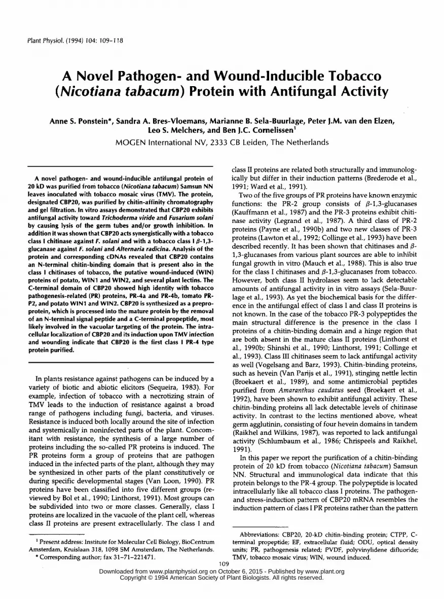
The leaf extract was passed through a Sephadex G-25 column(Fig. 1, lane A) and a cation-exchange column (Fig. 1, laneB). Chitin-binding proteins were isolated by the subsequentuse of affinity chromatography. Proteins were allowed tobind to insoluble chitin at pH 8.3 at 4°C. Bound proteinswere eluted by lowering the pH of the mobile phase to 3.5.Three proteins of 20, 32, and 34 kD were present in theeluate (Fig. 1, lane C). The 32- and 34-kD proteins wereidentified as the two class I chitinases that are known to beinduced in tobacco upon infection with TMV (Legrand et al.,1987) and known to reversibly bind to chitin (Broekaert etal., 1988; Sela-Buurlage et al., 1993). The third protein ofabout 20 kD appeared to be a novel chitin-binding protein.This protein (referred to as CBP20) was purified to homoge-neity by three rounds of gel-filtration chromatography. Theclass I chitinases eluted at an apparent molecular mass of 15to 15.5 kD, and CBP20 eluted at an apparent molecular massof 8 kD. Usually, baseline separation occurred after thesecond passage. The CBP20-containing fractions resultingafter the third passage were pooled and appeared to beelectrophoretically pure (Fig. 1, lane E). Usually 100 /*g ofpure CBP20 was obtained from about 400 g of TMV-infectedtobacco leaves (containing about 100 mg of protein). Inextracts of healthy tobacco leaves no chitin-binding proteinswere detected (data not shown).
Because CBP20 bound to chitin, it seemed obvious to assaythis protein for chitinase activity. To this end we used thechitinase assay described by Wirth and Wolf (1990). Someactivity was found to be associated with CBP20: 0.03 to 0.05ODU Mg"1 of protein (data not shown). However, this activityis extremely low compared to the activity measured for theclass I chitinases (approximately 3 ODU Mg"1)- Because somechitinases exert lysozyme activity, we tested CBP20 for suchan activity as well. Activity measurements indicated that
Mr E Mr
101 —71 —
44—
29 —
18 —
15 —
*
fFigure 1. Purification of CBP20. Protein samples were taken atdifferent stages in the purification procedure of CBP20 and ana-lyzed on 12.5% SDS-polyacrylamide gels. A sample of the crudedesalted leaf extract is shown in lane A. Protein pools obtainedafter S-Sepharose cation-exchange chromatography, chitin-affinitychromatography, and gel-filtration chromatography are shown inlanes B, C, and D, respectively. In lane E, 3 ^g of purified proteinwas loaded. The lanes indicated by Mr show prestained markers.The corresponding molecular masses in kD are as indicated. www.plant.org on October 6, 2015 - Published by www.plantphysiol.orgDownloaded from
Copyright © 1994 American Society of Plant Biologists. All rights reserved.
112 Ponstein et al. Plant Physiol. Vol. 104, 1994
CBP20 exhibited some lysozyme activity: 0.01 ODU Mg"1 ofprotein. This specific activity is in the same range as thelysozyme activity associated with the class I chitinases butabout 1000-fold lower than the specific activity for hen egglysozyme measured under the same reaction conditions.
CBP20 Inhibits Fungal Growth in Vitro
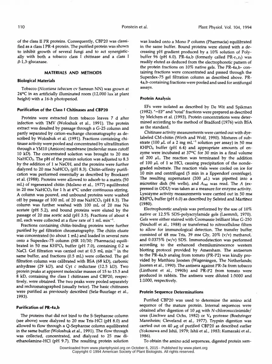
The antifungal activity associated with several chitin-bind-ing proteins prompted us to test the effect of CBP20 onfungal growth. To this end 24-well microtiter plates wereused (Sela-Buurlage et al., 1993). Spores of either T. viride, F.solani, or A. radicina were pipetted onto potato dextrose agarand allowed to germinate for 6 to 16 h. Purified and filter-sterilized protein solutions were added to the pregerminatedspores. The ability to lyse the fungus was studied 1 h afterthe addition of protein. Growth inhibition was scored 2 to 3d later. T. viride appeared to be the most sensitive fungus.Almost complete lysis of the germ tubes was observed im-mediately after the addition of small amounts (1 Mg/well =6.7 Mg mL~l) of CBP20 (data not shown). Consequently,growth was severely inhibited (data not shown). Hyphal tipsof F. solani showed no lysis in the presence of up to 10 Mg ofCBP20 per well, although swelling of the hyphal tips wasobserved microscopically (data not shown). However, someeffect of purified CBP20 (1 Mg/well) was observed on thegrowth of F. solani (Fig. 2). The addition of CBP20 (up to 5jig/well) to spores of A. radicina did not result in lysis either.Unlike F. solani, growth of A. radicina was not affected in thepresence of CBP20 (data not shown).
CBP20
(ug)Class I Glu (fjg) Class I Chi (jyg)
0.5 0.5
1
(den)
Figure 2. Antifungal activity of CBP20. The effect of purified CBP20and combinations of CBP20 with a class /3-1,3-glucanase (Clu) or aclass I chitinase (Chi) on the in vitro growth of F. solani. Amountsof protein (in Mg) per well are indicated. The effect of the proteincombinations should be compared to the effect of the individualproteins (left column and top row) and heat-denatured (den) controlsamples (bottom row).
Because chitin-binding proteins are known to act synergis-tically with chitinases (Broekaert et al., 1989; Hejgaard et al.,1992) and chitinases and (3-1,3-glucanases are known to actsynergistically as well (Mauch et al., 1988; Sela-Buurlage etal., 1993), we studied the effect of CBP20 in combinationwith these two enzymes. The addition of 0.5 Mg of class I /3-1,3-glucanase alone resulted in a small amount of lysis (<5%)and some growth inhibition in the case of F. solani (Fig. 2).The combination of 1 Mg of CBP20 and 0.5 Mg of 0-1,3-glucanase showed a strong lysing activity (about 70%). Also,a severe effect on growth was visible (Fig. 2). From thesedata we conclude that CBP20 and the class I 0-1,3-glucanaseact synergistically. Addition of 0.5 Mg of class I chitinase alonedid not cause lysis of the germ tubes or interfere with thegrowth of F. solani (Fig. 2). The combination of 1 Mg of CBP20and 0.5 Mg of class I chitinase did not cause lysis either butinhibited the growth of F. solani in a synergistic manner (Fig.2). The effect of the latter combination was even more potentthan the combination of CBP20 and the /3-1,3-glucanase.
The addition of 0.5 Mg/well of class 10-1,3-glucanase aloneinhibited growth of A. radicina substantially, whereas 0.1 Mgof class 10-1,3-glucanase was ineffective in inhibiting fungalgrowth. Combinations of CBP20 (5 Mg/well) and /3-1,3-glu-canase (0.1 Mg/well) inhibited the growth of A. radicina (about35%), resulting in more condensed growing mycelia (data notshown). Apparently, both proteins acted synergistically.Combining 5 Mg of CBP20 with a class I chitinase (0.5 Mg/well) did not result in enhanced growth inhibition comparedto the effect of the class I chitinase alone. Thus, in the caseof A. radicina no synergism was observed between CBP20and the class I chitinase. In all cases heat inactivation of theprotein mixtures eliminated their antifungal effect.
CBP20 Is C-Terminally Processed
To further characterize CBP20, amino acid sequences weredetermined.The protein was separated on a 12.5% SDS-polyacrylamide gel and electroblotted to a PVDF membraneto allow for N-terminal sequencing. However, no sequencedata became available, probably due to the presence of amodified Gin residue interfering with Edman degradation(see below). An internal sequence was obtained after digest-ing CBP20 with the Glu-C-specific endoproteinase (V8 pro-tease) from Staphylococcus aureus (Cleveland et al., 1977).The major reaction product was a 17-kD peptide that gavethe following amino acid sequence (CBP-PEP1): Tyr-(Ala/Gly)-Ser-Pro-Ser-Gln-Gly-X-Gln-Ser-Gln-(Arg)-Ser-Gly-Gly-Gly-Gly-Gly-Gly-Gly-Gly-Gly-Gly-Gly-Gly-Gly-Ala-Gln-Asn. From the specificity of the protease it can be pre-dicted that a Glu residue precedes the first given amino acidresidue. The amino acid indicated as X is most likely a Cysresidue, because no special care was taken to alkylate the Cysresidues prior to sequence analysis. The amino acid residuesgiven in parentheses were not characterized unequivocally.
A second internal amino acid sequence was obtainedafter chemical digestion of CBP20 with N-chlorosuccinimide(Lischwe and Ochs, 1982). A band of 9 to 11 kD was usedfor the sequence determination (CBP-PEP2): Thr-Ala-Phe-Tyr-Gly-Pro-Val-Gly-Pro-(Pro/Arg)-Gly-Arg-Asp-Ser-X-Gly-Lys-(Gly)-Leu-?-Val-Thr-Asn. Because N-chlorosucci- www.plant.org on October 6, 2015 - Published by www.plantphysiol.orgDownloaded from
Copyright © 1994 American Society of Plant Biologists. All rights reserved.

Antifungal Activity of 20-kD Chitin-Binding Protein 113
nimide cleaves at the C terminus of Trp residues, it is assumed that this amino acid precedes the given sequence. The amino acid indicated as X is again most likely a Cys residue as argued above. At the position of the question mark no amino acid identification could be made. A third amino acid se- quence representing the C tenninus of CBP20 was obtained as follows. CBP20 (4 nmol) was digested with trypsin, and the C-terminal peptide was purified from the digest via affinity chromatography on an immobilized anhydrotrypsin column (Yokosawa and Ishii, 1979; Ishii et al., 1983; Kuma- zaki et al., 1986). Sequencing of this peptide revealed the following amino acid sequence (CBP-PEP3): (G1y)-(His)-Leu- Ile-Val-Asn-Tyr-Glu-Phe-Val-Asn-Cys-Gly- Asp- Asn, which is presumably preceded by a Lys or an Arg residue.
To isolate clones corresponding to CBP20 a XZAP cDNA library of tobacco leaves infected with TMV was screened. A partial cDNA fragment was amplified from a total XZAP- cDNA library by using the oligonucleotides LS20 and LS46 in a PCR. The oligonucleotide LS20 was complementary to the pSK vector sequence of the XZAP arm at the 5’ end of the cDNA insert, and oligonucleotide LS46 was based on the amino acid sequence of fragment CBP-PEP2. The amplified DNA fragment (407 bp) was cloned as an EcoRI fragment into the EcoRI-linearized vector pBlueScript to yield clone pMOG684. The nucleotide sequence of the cloned EcoRI fragment confirmed that a partial CBP20 cDNA clone was isolated. To ensure the use of a CBP2O-specific DNA probe, a 187-bp PCR fragment was amplified from plasmid pMOG684 using the oligonucleotides LS48 and LS49. This PCR fragment, encoding the N-terminal part of CBP20 up to the Gly-rich region present in CBP-PEPI, was subsequently used to screen a tobacco cDNA library.
The screening for CBP20-like cDNAs resulted in the iso- lation of severa1 positive clones. Five clones containing 800 bp of cDNA inserts were sequenced. The derived sequences indicated that two types of cDNA clones had been isolated. The nucleotide sequence and the deduced amino acid se- quence of the two different types of cDNA clones are shown in Figure 3. Clone cbp20-44 represents a nearly full-length cDNA. Because the deduced amino acid sequence exactly matched the detennined sequences CBP-PEP1, CBP-PEP2, and CBP-PEP3 (Fig. 3), we concluded that cbp20-44 is a true CBP20 cDNA clone. Clone cbp20-52 encodes a protein con- taining two additional amino acid residues (Gly, Ala) in the Gly-rich region compared to CBP-PEP2 (Fig. 3).
Both cDNAs code at their 5’ end for a putative signal peptide that may be involved in transport of the protein across the membrane of the ER. The cleavage site would be between Alaz2 and Gln23, analogous to the cleavage sites of other PR proteins, including /3-1,3-glucanases (Shinshi et al., 1988; Linthorst et al., 1990a), PR-1 proteins (Cornelissen et al., 1987), chitinases (Linthorst et al., 1990b; Payne et al., 1990a), and class I1 PR-4 proteins (Linthorst et al., 1991). The resulting N-terminal Gln residue is readily modified, prob- ably resulting in ineffective Edman degradation of the mature protein (see above). As mentioned above, CBP-PEP3 repre- sents the C terminus of the mature CBP20 protein. However, as compared to this peptide the deduced amino acid sequence extends for another 11 residues. Apparently mature CBP20 is synthesized as a preproprotein from which the N-terminal
52. seq 44 .seq 44 . pep 52. pep
52. seq 44 .seq 44 .pep 52.pep
5Z .seq 44.seq 44 .pep
52. seq 44 .seq 44.pep
52. seq 44 .seq 44.pep 52.pep
44 .seq 44 . pep
52 .seq 44 . seq 44 . pep
44 .seq 44.pep
52. seq 44 .seq 44.pep
52. seq 44 .seq 4 4 . pep
52. seq 44 . seq 44. pep 52. pep
5 2 . seq 4 4 . seq
52. seq 44 . seq
44. seq
44 .seq
A AT C ACAACACCAGTTCAAACACTTTGAAAAAT _._.______________.__________ GGGAAAGCTAAGTACTCTTTTGCTTGTTCTfi
G K L S T L L L V L M F A
G G A T T C ATCCTCTATTTCATAGCCGCAGGTGCCAACGCACAGCAGTGCGGAAGGCAAAGGGGAGGA I L Y F ’ I A A G A N A Q Q C G R Q R G G V V A
T A GCCTTATGCAGTGGAAACTTGTGCTGCAGCCAATTTGGGTGGTGTGGGTCTACACCGGAA A L C S G N L C C S Q F G W C G S T P E
1 TACTGTTCTCCTAGCCAAGGCTGCCAAAG~CAGTGCAGTGGCGG~GGAGGCGGCGGTGGA Y C S P S Q G’C Q S Q C S G G G G G G G
GGCGGA C G C GGTGGC------GGTGGTGGTGGTGCGCAAAACGTTAGGGC~CATATCATATATATAAC u- - G G G G A O N ’ V R A T Y H I Y N
CCGCAGAATGTTGGGTGGGATTTGTATGCAGTTAGTGCGTACTGCTCAACTTGGGATGGT P Q N V G W O L Y A V S A Y C S T W D G
G A
A T AACAAGCCTTTGGCATGGCGGAGGAAGTATGGTTGGACTGCATTCTGTGGCCCTGTTGGA N K P L A W R R K Y G D T A F C G P V G
- CCTCGTGGCCGAGACTCTTGTGGCAAATGCTTAAGGGTGACAAATACAGGCACAGGAGCT P R G R D S C G K C L R V T N T G T G A
L CAGACCACAGTGAGAATCGTGGATCAATGCAGCAATGGCGGACTAGACTTGGACGTTAAT Q T T V R I V O Q C S N G G L D L O V N
T C GTTTTCCGGCAGCTCGACACAGACGGAAGAGGGAATCAACGCGGCCATCTTATTGTGAAC V F R Q L D T D G R G N Q R G H L I V N
G G TACGAGTTTGTTAATTGTGGTGACAATATGAATGTTCTGCTATCCCCAGTTGACAAAGAA Y E F V N C G O N M N V L L S P V O K E
A V
T AT G AGTCT GACG C A TA GTA TAAGAAGCCATCGATGCCCATGTTTTAGTCTTTGACGGCCCAAATAAAAGTAAAAGAACG
____.._..___________~~~~... . . .~~~~~~~~~~~~...--------
ATATGTAAAAGGAAAAAGAAAATAAAGTTGCTTTG~GGGTTAGGCAATTCCAATTTCTA
TATAAGAATGTCTTTCGTTTGGGAATAATGAGGTGACGTGTGTATGCGAATATTGTGATT
TTAAATAAAGAATCGCAGTGGGACAGTATTTGTTGGTCTCATTCCGAAAAAAAAAAAAAA
60
120
180
240
300
360
420
480
540
600
660
720
780
8 4 0
900
Figure 3. Sequence of cbp20 clones. Two cbp20 clones (designated cbp20-44 and cbp20-52) were isolated and sequenced (44.seq and 52.seq, respectively). T h e deduced amino acid is also given (44.pep and 52.pep, respectively). Nucleotides and deduced amino acid residues of cbp20-52 are given only when they differed from the cbp20-44 sequence. T h e peptide sequences obtained after Ve protease treatment (CBP-PEPl), N-chlorosuccinimidelurea diges- tion (CBP-PEPZ), and trypsin digestion (CBP-PEP3) are underlined. The putative N-terminal signal peptide cleavage site and the C- terminal cleavage site of the propeptide are indicated by arrow- heads. The primers used in the PCR reactions are underlined.
signal peptide and the CTPP are cleaved off. The CTPP most likely functions as a vacuolar targeting signal, because the CTPPs of class I chitinases, class I @-1,3-glucanase, and AP24 were shown to be involved in vacuolar targeting (Neuhaus et al., 1991; Melchers et al., 1993).
CBPZO 1s a Class I PR-4 Protein
Comparison of the deduced amino acid sequence of CBP20 with sequences stored in the SwissProt protein sequence data base revealed striking similarity with the primary structures of the putative wound-inducible proteins encoded by the winl and win2 genes from potato (Stanford et al., 1989) and of prohevein from the rubber tree (Broekaert et al., 1990). Prohevein is the precursor of hevein, a small lectin found in
www.plant.org on October 6, 2015 - Published by www.plantphysiol.orgDownloaded from Copyright © 1994 American Society of Plant Biologists. All rights reserved.
114 Ponstein et al. Plant Physiol. Vol. 104, 1994
WINl K R T. T STPTPS SHIH2 GN T. S .POP Q S
Hevein E A K PN W D DHN N . KDS . -E V SCBP20-44 QQCGRQRGGALCSGNLCCSQFGWCGSTPEYCSPSQGCQSQCSGGGGGGGGGGGGG
PR4a —————————————————————————————————————————————————————— QSPR-P2 —————————————————————————————————————————————————————— QS
^ ————————————————— -hevein doMin— ————————— - —————— X — -hing* region —— >
WINlWIN2
HeveinCBP20-44
PR4aPR-P2
WINlWIN2
HeveinCBP20-44
PR4aPR-P2
WIH1WIH2
HeveinCBP20-44
PR4aPR-P2
N A S KN A Y S
S L L S D H N A A Y S S A HAQNVRATYHIYNPQNVGWDLYAVSAYCSTWDGNKPLAWRRKYGWTAFCGPVGPRG
T S L I N R A F A A D Q AT L IN RTA V A AD E R AT
8 I N HQR I Q I V Q
QS S K K YE ITRDSCGKCLRVTNTGTGAQTTVRIVDQCSHGGLDLDVNVFRQLDTDGRGNQRGHLIQ V R T N N V Y Q TQ A R T E R N R N L Y N N
Q D ————————————Q V P VQ D SF PLFSVMKSSVIN
VNYEFVNCGDNMNV.LLSPVDKE
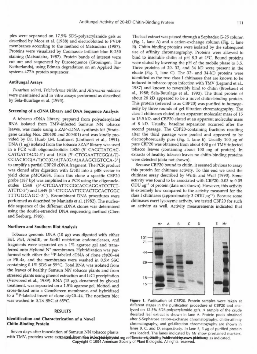
Figure 4. Sequence comparison of several PR-4 proteins. The se-quence of proCBP20 is given and compared to several PR-4 pro-teins. Amino acid residues of tobacco PR-4a and tomato PR-P2(Linthorst et al., 1991), prohevein (Broekaert et al., 1990), and theputative proWIN! and proWIN2 proteins (Stanford et al., 1989)that are different from those in proCBP20 are indicated. Gapsintroduced to optimize the alignment of the proteins are indicatedas dots, whereas amino acids missing from the sequences areindicated by hyphens.
the latex of the rubber tree. A comparison of these fourproteins is shown in Figure 4. In all cases the N-terminalsignal peptide (not included in Fig. 4) is followed by a chitin-binding domain that is connected to a C-terminal domain bya hinge region. High identity of the CBP20 proprotein se-quence was found with the putative WINl and WIN2 pro-proteins and prohevein (86.9, 87.5, and 70.5%, respectively).In contrast to the hinge regions of prohevein and the putativeWIN proteins, the hinge region of CBP20 is extremely Glyrich. In clone cbp20-52,14 of 15 amino acid residues are Gly,and in clone cbp20-44 all 13 residues are Gly (Fig. 4). Inaddition to the above similarities, the N-terminal sequenceof CBP20 was found to be very similar to the chitin-bindingdomain of tobacco class I chitinases (Linthorst et al., 1990b;Shinshi et al., 1990) and stinging nettle lectin (Broekaert etal., 1989) (data not shown). In view of the chitin-bindingcapacity of CBP20, this similarity is not unexpected.
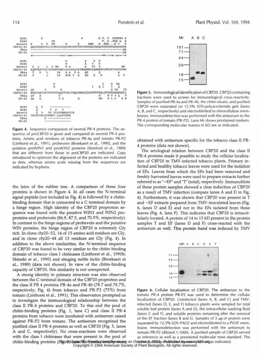
A strong identity in primary structure was also observedbetween the C-terminal domain of the CBP20 proprotein andthe class II PR 4 proteins PR-4a and PR-4b (78.7 and 78.7%,respectively; Fig. 4) from tobacco and PR-P2 (75%) fromtomato (Linthorst et al., 1991). This observation prompted usto investigate the immunological relationship between theclass II PR-4 proteins and CBP20. To this end the pool ofchitin-binding proteins (Fig. 1, lane C) and class II PR-4proteins from tobacco were incubated with antiserum raisedagainst PR-P2 from tomato. The antiserum recognized thepurified class II PR-4 proteins as well as CBP20 (Fig. 5, lanesA and C, respectively). No cross-reactions were observedwith the class I chitinases that were present in the pool ofchitin-binding proteins (Fig. 5, lane B). Similar results were
Mr
101 —71 —44—
29—
18 —15 —
A B C
Figure 5. Immunological identification of CBP20. CBP20-containingfractions were used to screen for immunological cross-reactivity.Samples of purified PR-4a and PR-4b, the chitin eluate, and purifiedCBP20 were separated on 12.5% SDS-polyacrylamide gels (lanesA, B, and C, respectively) and electroblotted to nitrocellulose mem-branes. Immunodetection was performed with the antiserum to thePR-4 protein of tomato (PR-P2). Lane Mr shows prestained markers.The corresponding molecular masses in kD are as indicated.
obtained with antiserum specific for the tobacco class II PR-4 proteins (data not shown).
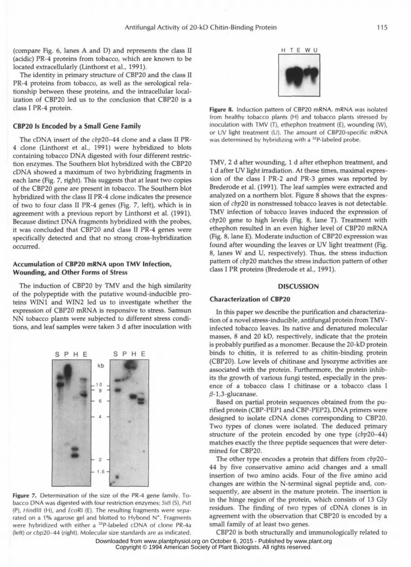
The serological relation between CBP20 and the class IIPR-4 proteins made it possible to study the cellular localiza-tion of CBP20 in TMV-infected tobacco plants. Primary in-fected and healthy tobacco leaves were used for the isolationof EFs. Leaves from which the EFs had been removed andfreshly harvested leaves were used to prepare extracts furtherreferred to as *—EF" and *T" (total), respectively. Immunoblotsof these protein samples showed a clear induction of CBP20as a result of TMV infection (compare lanes A and D in Fig.6). Furthermore, it was shown that CBP20 was present in Tand —EF extracts prepared from TMV-inoculated leaves (Fig.6, lanes D and E) and not in the EFs isolated from theseleaves (Fig. 6, lane F). This indicates that CBP20 is intracel-lularly located. A protein of 14 to 15 kD present in the proteinsamples T and EF (lanes D and F) cross-reacted with theantiserum as well. This protein band was induced by TMV
C B P A B C
71-
44-
18-15-
Figure 6. Cellular localization of CBP20. The antiserum to thetomato PR-4 protein PR-P2 was used to determine the cellularlocalization of CBP20. Uninfected (lanes A, B, and C) and TMV-infected (lanes D, E, and F) tobacco plants were sampled for totalsoluble leaf protein (lanes A and D), the extracellular washing fluid(lanes C and F), and soluble proteins remaining after the removalof the EF fraction (lanes B and E). Samples of 5 Mg of protein wereseparated by 12.5% SDS-PACE and electroblotted to a PVDF mem-brane. Immunodetection was performed with the antiserum totomato PR-P2 (diluted 1:1000). A purified sample of CBP20 servedas reference as well as a prestained molecular mass standard. Thecorresponding molecular masses in kD are as indicated. www.plant.org on October 6, 2015 - Published by www.plantphysiol.orgDownloaded from
Copyright © 1994 American Society of Plant Biologists. All rights reserved.
Antifungal Activity of 20-kD Chitin-Binding Protein 115
(compare Fig. 6, lanes A and D) and represents the class II(acidic) PR-4 proteins from tobacco, which are known to belocated extracellularly (Linthorst et al., 1991).
The identity in primary structure of CBP20 and the class IIPR-4 proteins from tobacco, as well as the serological rela-tionship between these proteins, and the intracellular local-ization of CBP20 led us to the conclusion that CBP20 is aclass I PR-4 protein.
CBP20 Is Encoded by a Small Gene Family
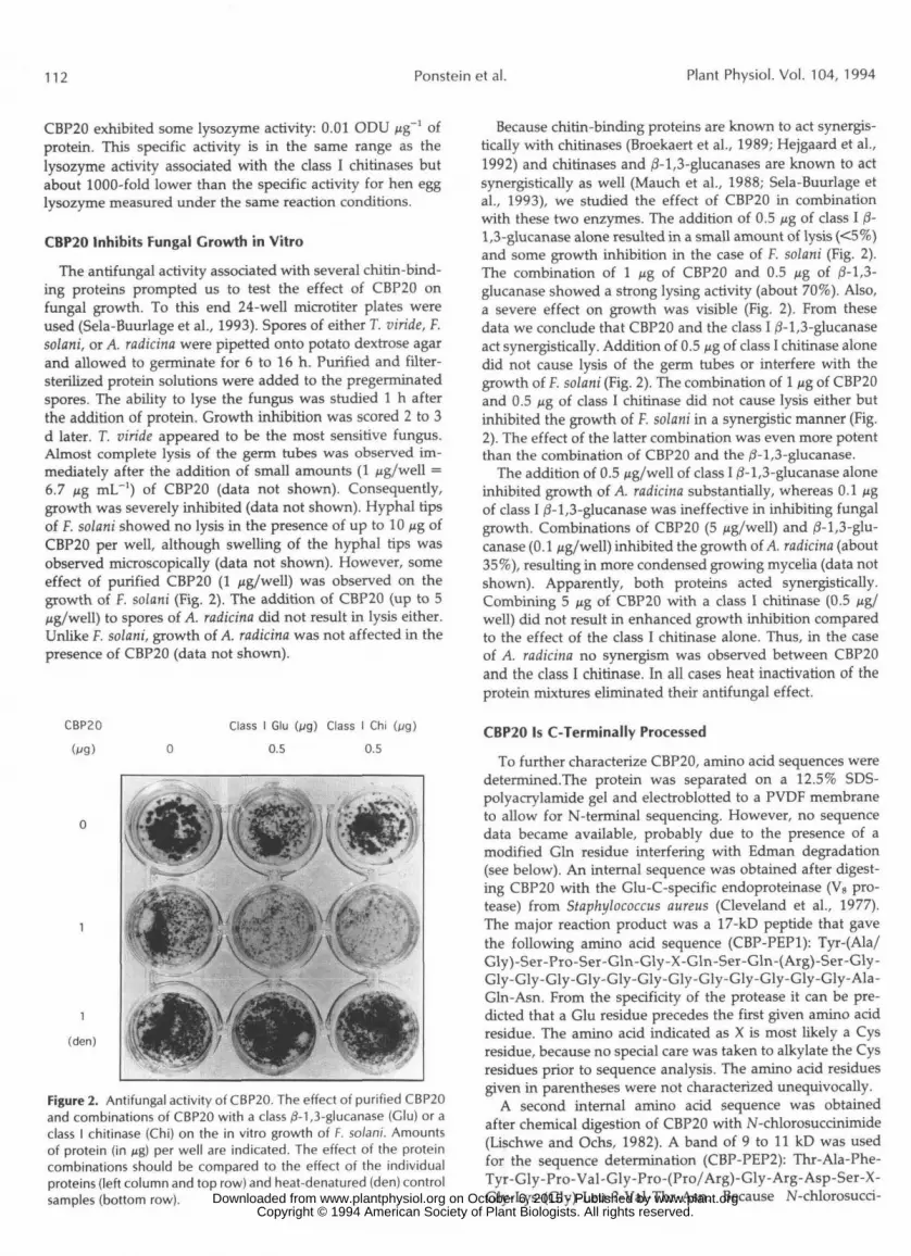
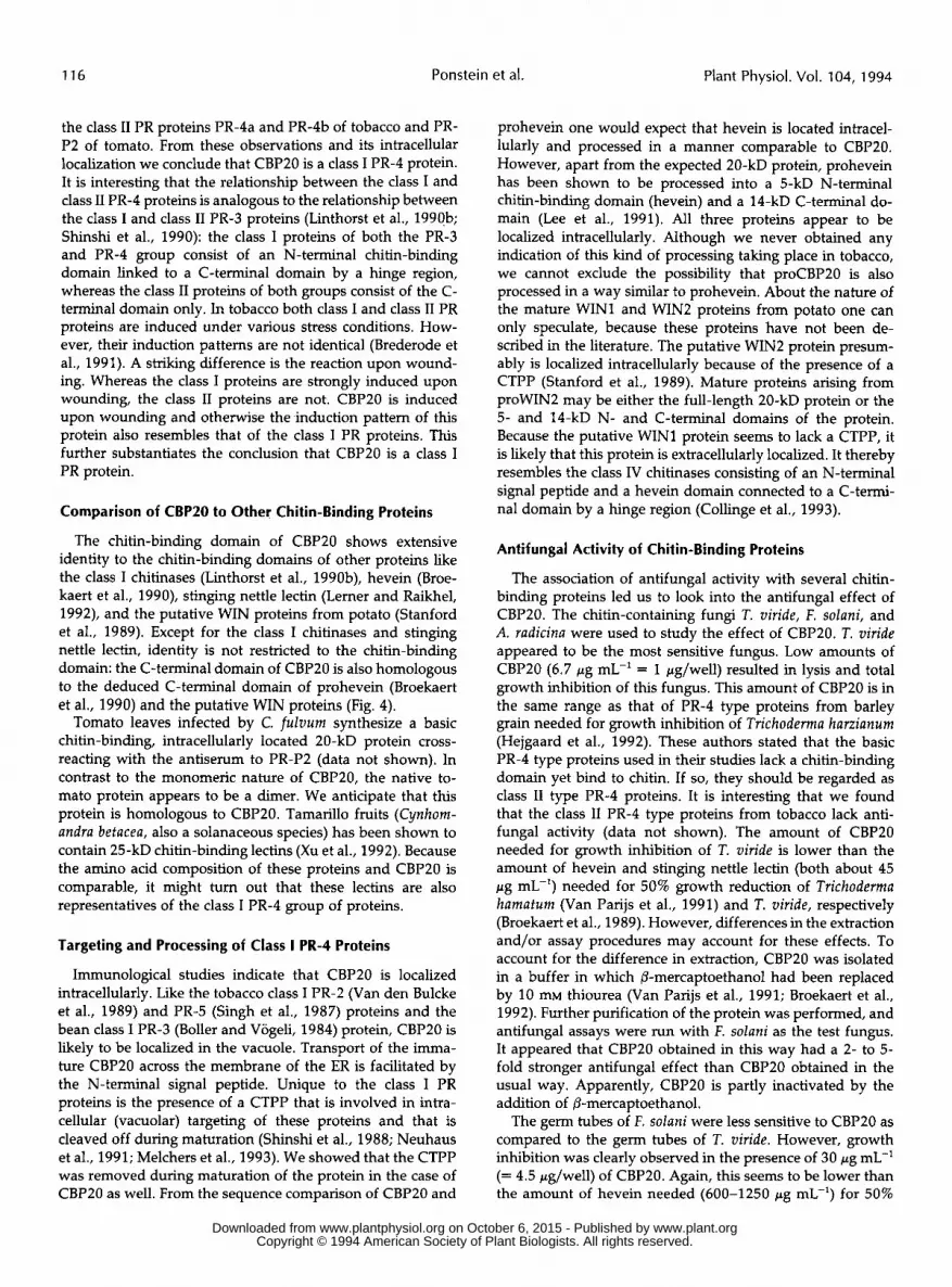
The cDNA insert of the cbp20-44 clone and a class II PR-4 clone (Linthorst et al., 1991) were hybridized to blotscontaining tobacco DNA digested with four different restric-tion enzymes. The Southern blot hybridized with the CBP20cDNA showed a maximum of two hybridizing fragments ineach lane (Fig. 7, right). This suggests that at least two copiesof the CBP20 gene are present in tobacco. The Southern blothybridized with the class II PR-4 clone indicates the presenceof two to four class II PR-4 genes (Fig. 7, left), which is inagreement with a previous report by Linthorst et al. (1991).Because distinct DNA fragments hybridized with the probes,it was concluded that CBP20 and class II PR-4 genes werespecifically detected and that no strong cross-hybridizationoccurred.
Accumulation of CBP20 mRNA upon TMV Infection,Wounding, and Other Forms of Stress
The induction of CBP20 by TMV and the high similarityof the polypeptide with the putative wound-inducible pro-teins WIN1 and WIN2 led us to investigate whether theexpression of CBP20 mRNA is responsive to stress. SamsunNN tobacco plants were subjected to different stress condi-tions, and leaf samples were taken 3 d after inoculation with
H T E W U
S P H E S P H E
kb
- 6 -
- 1 .6 -
Figure 7. Determination of the size of the PR-4 gene family. To-bacco DNA was digested with four restriction enzymes: Sstl (S), Pstl(P), H/ndlll (H), and EcoRI (E). The resulting fragments were sepa-rated on a 1% agarose gel and blotted to Hybond N+. Fragmentswere hybridized with either a 32P-labeled cDNA of clone PR-4a(left) or cbp20-44 (right). Molecular size standards are as indicated.
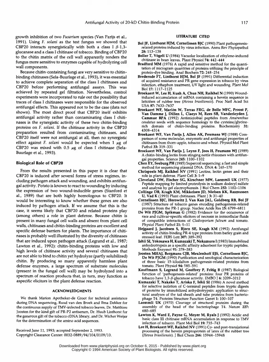
Figure 8. Induction pattern of CBP20 mRNA. mRNA was isolatedfrom healthy tobacco plants (H) and tobacco plants stressed byinoculation with TMV (T), ethephon treatment (E), wounding (W),or UV light treatment (U). The amount of CBP20-specific mRNAwas determined by hybridizing with a 32P-labeled probe.
TMV, 2 d after wounding, 1 d after ethephon treatment, and1 d after UV light irradiation. At these times, maximal expres-sion of the class I PR-2 and PR-3 genes was reported byBrederode et al. (1991). The leaf samples were extracted andanalyzed on a northern blot. Figure 8 shows that the expres-sion of cbp20 in nonsrressed tobacco leaves is not detectable.TMV infection of tobacco leaves induced the expression ofcbp20 gene to high levels (Fig. 8, lane T). Treatment withethephon resulted in an even higher level of CBP20 mRNA(Fig. 8, lane E). Moderate induction of CBP20 expression wasfound after wounding the leaves or UV light treatment (Fig.8, lanes W and U, respectively). Thus, the stress inductionpattern of cbp20 matches the stress induction pattern of otherclass I PR proteins (Brederode et al., 1991).
DISCUSSION
Characterization of CBP20
In this paper we describe the purification and characteriza-tion of a novel stress-inducible, antifungal protein from TMV-infected tobacco leaves. Its native and denatured molecularmasses, 8 and 20 kD, respectively, indicate that the proteinis probably purified as a monomer. Because the 20-kD proteinbinds to chitin, it is referred to as chitin-binding protein(CBP20). Low levels of chitinase and lysozyme activities areassociated with the protein. Furthermore, the protein inhib-its the growth of various fungi tested, especially in the pres-ence of a tobacco class I chitinase or a tobacco class I(8-1,3-glucanase.
Based on partial protein sequences obtained from the pu-rified protein (CBP-PEP1 and CBP-PEP2), DNA primers weredesigned to isolate cDNA clones corresponding to CBP20.Two types of clones were isolated. The deduced primarystructure of the protein encoded by one type (cbp20-44)matches exactly the three peptide sequences that were deter-mined for CBP20.
The other type encodes a protein that differs from cbp20-44 by five conservative amino acid changes and a smallinsertion of two amino acids. Four of the five amino acidchanges are within the N-terminal signal peptide and, con-sequently, are absent in the mature protein. The insertion isin the hinge region of the protein, which consists of 13 Glyresidues. The finding of two types of cDNA clones is inagreement with the observation that CBP20 is encoded by asmall family of at least two genes.
CBP20 is both structurally and immunologically related to www.plant.org on October 6, 2015 - Published by www.plantphysiol.orgDownloaded from
Copyright © 1994 American Society of Plant Biologists. All rights reserved.
116 Ponstein et al. Plant Physiol. Vol. 104, 1994
the class I1 PR proteins PR-4a and PR-4b of tobacco and PR- P2 of tomato. From these observations and its intracellular localization we conclude that CBP20 is a class I PR-4 protein. It is interesting that the relationship between the class I and class I1 PR-4 proteins is analogous to the relationship between the class I and class I1 PR-3 proteins (Linthorst et al., 1990b; Shinshi et al., 1990): the class I proteins of both the PR-3 and PR-4 group consist of an N-terminal chitin-binding domain linked to a C-terminal domain by a hinge region, whereas the class I1 proteins of both groups consist of the C- terminal domain only. In tobacco both class I and class I1 PR proteins are induced under various stress conditions. How- ever, their induction pattems are not identical (Brederode et al., 1991). A striking difference is the reaction upon wound- ing. Whereas the class I proteins are strongly induced upon wounding, the class I1 proteins are not. CBP20 is induced upon wounding and otherwise the induction pattem of this protein also resembles that of the class I PR proteins. This further substantiates the conclusion that CBP20 is a class I PR protein.
Comparison of CBPZO to Other Chitin-Binding Proteins
The chitin-binding domain of CBP20 shows extensive identity to the chitin-binding domains of other proteins like the class I chitinases (Linthorst et al., 1990b), hevein (Broe- kaert et al., 1990), stinging nettle lectin (Lemer and Raikhel, 1992), and the putative WIN proteins from potato (Stanford et al., 1989). Except for the class I chitinases and stinging nettle lectin, identity is not restricted to the chitin-binding domain: the C-terminal domain of CBP20 is also homologous to the deduced C-terminal domain of prohevein (Broekaert et al., 1990) and the putative WIN proteins (Fig. 4).
Tomato leaves infected by C. fuluum synthesize a basic chitin-binding, intracellularly located 20-kD protein cross- reacting with the antiserum to PR-P2 (data not shown). In contrast to the monomeric nature of CBP20, the native to- mato protein appears to be a dimer. We anticipate that this protein is homologous to CBP20. Tamarillo fruits (Cynhom- andra betacea, also a solanaceous species) has been shown to contain 25-kD chitin-binding lectins (Xu et al., 1992). Because the amino acid composition of these proteins and CBP20 is comparable, it might tum out that these lectins are also representatives of the class I PR-4 group of proteins.
Targeting and Processing of Class I PR-4 Proteins
Immunological studies indicate that CBP20 is localized intracellularly. Like the tobacco class I PR-2 (Van den Bulcke et al., 1989) and PR-5 (Singh et al., 1987) proteins and the bean class I PR-3 (Boller and Vogeli, 1984) protein, CBP20 is likely to be localized in the vacuole. Transport of the imma- ture CBP20 across the membrane of the ER is facilitated by the N-terminal signal peptide. Unique to the class I PR proteins is the presence of a CTPP that is involved in intra- cellular (vacuolar) targeting of these proteins and that is cleaved off during maturation (Shinshi et al., 1988; Neuhaus et al., 1991; Melchers et al., 1993). We showed that the CTPP was removed during maturation of the protein in the case of CBPZO as well. From the sequence comparison of CBP20 and
prohevein one would expect that hevein is located intracel- lularly and processed in a manner comparable to CBP20. However, apart from the expected 20-kD protein, prohevein has been shown to be processed into a 5-kD N-terminal chitin-binding domain (hevein) and a 14-kD C-terminal do- main (Lee et al., 1991). A11 three proteins appear to be localized intracellularly. Although we never obtained any indication of this kind of processing taking place in tobacco, we cannot exclude the possibility that proCBP20 is also processed in a way similar to prohevein. About the nature of the mature WINl and WIN2 proteins from potato one can only speculate, because these proteins have not been de- scribed in the literature. The putative WIN2 protein presum- ably is localized intracellularly because of the presence of a CTPP (Stanford et al., 1989). Mature proteins arising from proWIN2 may be either the full-length 20-kD protein or the 5- and 14-kD N- and C-terminal domains of the protein. Because the putative WINl protein seems to lack a CTPP, it is likely that this protein is extracellularly localized. It thereby resembles the class IV chitinases consisting of an N-terminal signal peptide and a hevein domain connected to a C-termi- na1 domain by a hinge region (Collinge et al., 1993).
Antifungal Activity of Chitin-Binding Proteins
The association of antifungal activity with severa1 chitin- binding proteins led us to look into the antifungal effect of CBP20. The chitin-containing fungi T. viride, F. solani, and A. radicina were used to study the effect of CBP20. T. viride appeared to be the most sensitive fungus. Low amounts of CBP20 (6.7 pg mL-' = 1 pg/well) resulted in lysis and total growth inhibition of this fungus. This amount of CBP2O is in the same range as that of PR-4 type proteins from barley grain needed for growth inhibition of Trichoderma harzianum (Hejgaard et al., 1992). These authors stated that the basic PR-4 type proteins used in their studies lack a chitin-binding domain yet bind to chitin. If so, they should be regarded as class I1 type PR-4 proteins. It is interesting that we found that the class I1 PR-4 type proteins from tobacco lack anti- funga1 activity (data not shown). The amount of CBP2O needed for growth inhibition of T. viride is lower than the amount of hevein and stinging nettle lectin (both about 45 pg mL-') needed for 50% growth reduction of Trichoderma hamatum (Van Parijs et al., 1991) and T. viride, respectively (Broekaert et al., 1989). However, differences in the extraction and/or assay procedures may account for these effects. To account for the difference in extraction, CBP20 was isolated in a buffer in which (3-mercaptoethanol had been replaced by 10 m thiourea (Van Parijs et al., 1991; Broekaert et al., 1992). Further purification of the protein was performed, and antifungal assays were run with F. solani as the test fungus. It appeared that CBP20 obtained in this way had a 2- to 5- fold stronger antifungal effect than CBP2O obtained in the usual way. Apparently, CBP20 is partly inactivated by the addition of (3-mercaptoethanol.
The germ tubes of F. solani were less sensitive to CBP20 as compared to the germ tubes of T. viride. However, growth inhibition was clearly observed in the presence of 30 pg mL-' (= 4.5 Kg/well) of CBP20. Again, this seems to be lower than the amount of hevein needed (600-1250 pg mL-') for 50%
www.plant.org on October 6, 2015 - Published by www.plantphysiol.orgDownloaded from Copyright © 1994 American Society of Plant Biologists. All rights reserved.
Antifungal Activity of 20-kD Chitin-Binding Protein 117
growth inhibition of two Fusarium species (Van Parijs et al., 1991). Using F. solani as the test fungus we showed that CBPZO interacts synergistically with both a class I p-1,3- glucanase and a class I chitinase of tobacco. Binding of CBP20 to the chitin matrix of the cell wall apparently renders the fungus more sensitive to enzymes capable of hydrolyzing cell wall components.
Because chitin-containing fungi are very sensitive to chitin- binding chitinases (Sela-Buurlage et al., 1993), it was essential to achieve complete separation of the class I chitinases and CBP20 before performing antifungal assays. This was achieved by repeated gel filtration. Nevertheless, control experiments were incorporated to rule out the possibility that traces of class I chitinases were responsible for the observed antifungal effects. This appeared not to be the case (data not shown). The most direct proof that CBP20 itself exhibits antifungal activity rather than contaminating class I chiti- nases is the synergistic activity of these two chitin-binding proteins on F. solani. If the chitinase activity in the CBP20 preparation resulted from contaminating chitinases, and CBPZO itself were not antifungal at all, then no synergistic effect against F. solani would be expected when 1 pg of CBP20 was mixed with 0.5 r g of class I chitinase (Sela- Buurlage et al., 1993).
Biological Role of CBPZO
From the results presented in this paper it is clear that CBP20 is induced after several forms of stress regimes, in- cluding pathogen attack and wounding, and exhibits antifun- gal activity. Potato is known to react to wounding by inducing the expression of two wound-inducible genes (Stanford et al., 1989) that are both highly homologous to CBP20. It would be interesting to know whether these genes are also induced by pathogen attack. If we assume that this is the case, it seems likely that CBP20 and related proteins play (among others) a role in plant defense. Because chitin is present in many fungal cell walls and absent from plant cell walls, chitinases and chitin-binding proteins are excellent and specific defense bamers for plants. The importance of chiti- nases is probably well illustrated by the diversity of chitinases that are induced upon pathogen attack (Legrand et al., 1987; Lawton et al., 1992): chitin-binding proteins with low and high levels of chitinase activity and several chitinases that are not able to bind to chitin yet hydrolyze (partly solubilized) chitin. By producing so many apparently harmless plant defense enzymes, a large spectrum of substrate molecules (present in the fungal cell wall) may be hydrolyzed into a spectrum of reaction products that, in tum, may function as aspecific elicitors in the plant defense reaction.
ACKNOWLEDCMENTS
We thank Marion Apotheker-de Groot for technical assistance during DNA sequencing, Ruud van den Broek and Ilma Dekker for the continuous supply of TMV-infected tobacco leaves, Dr. Matthieu Joosten for the kind gift of PR-P2 antiserum, Dr. Huub Linthorst for the generous gift of the tobacco cDNA library, and Dr. Wicher Weijer for the determination of the amino acid sequences.
Received June 11, 1993; accepted September 2, 1993. Copyright Clearance Center: 0032-0889/94/104/0109/1 O
LITERATURE ClTED
Bol JF, Linthorst HJM, Cornelissen BJC (1990) Plant pathogenesis- related proteins induced by virus infection. Annu Rev Phytopathol
Boller T, Vogeli U (1984) Vacuolar localization of ethylene-induced chitinase in bean leaves. Plant Physiol74 442-444
Bradford MM (1976) A rapid and sensitive method for the quanti- tation of microgram quantities of proteins utilizing the principle of protein-dye binding. Ana1 Biochem 7 2 248-254
Brederode FT, Linthorst HJM, Bol JF (1991) Differential induction of acquired resistance and PR gene expression in tobacco by virus infection, ethephon treatment, UV light and wounding. Plant Mo1 Biol 17: 1117-1125
Broekaert W, Lee H, Kush A, Chua NH, Raikhel N (1990) Wound- induced accumulation of mRNA containing a hevein sequence in laticifers of rubber tree (Hevea brasiliensis). Proc Natl Acad Sci USA 87: 7633-7637
Broekaert WF, Marien W, Terras FRG, de Bolle MFC, Proost P, Van Damme J, Dillen L, Claeys M, Rees SB, Vanderleyden J, Cammue BPA (1992) Antimicrobial peptides from Amaranthus caudafus seeds with sequence homology to the cysteine/glycine- rich domain of chitin-binding proteins. Biochemistry 31:
Broekaert WF, Van Parijs J, Allen AK, Peumans WJ (1988) Com- parison of some molecular, enzymatic and antifungal properties of chitinases from thom-auple, tobacco and wheat. Phvsiol Mo1 Plant
2 8 113-138
4308-4314
1.
Pathol33 319-331 Broekaert WF. Van Pariis I. Levns F. 100s H. Peumans WI(1989)
A chitin-binhing lectin ;rim singing nettle rhizomes with antifun: gal properties. Science 245 1100-1102
Chen EY, Seeburg PH (1985) Supercoil sequencing: a fast and simple method for sequencing plasmid DNA. DNA 4 165-170
Chrispeels MJ, Raikhel NV (1991) Lectins, lectin genes and their role in plant defense. Plant Cel l3 1-9
Cleveland DW, Fischer SG, Kirschner MW, Laemmli UK (1977) Peptide mapping by limited proteolysis in sodium dodecyl sulfate and analysis by gel electrophoresis. J Biol Chem 252 1102-1106
Collinge DB, Kragh KM, Mikkelsen JD, Nielsen KK, Rasmussen U, Vad K (1993) Plant chitinases. Plant J 3 31-40
Cornelissen BJC, Horowitz J, Van Kan JAL, Goldberg RB, Bol JF (1987) Structure of tobacco genes encoding pathogenesis-related proteins from the PR-1 group. Nucleic Acids Res 1 5 6799-6811
De Wit PJGM, Spikman G (1982) Evidence for the occurrence of race and cultivar-specific elicitors of necrosis in intracellular fluids of compatible interactions of Cladosporium fulvum and tomato. Physiol Plant Pathol21: -1-11
Hejgaard J, Jacobsen S, Bjorn SE, Kragh KM (1992) Antifungal activity of chitin-binding PR-4 type proteins from barley grain and stressed leaf. FEBS Lett 307: 389-392
Ishii SI, Yokosawa H, Kumazaki T, Nakamura I(1983) Immobilized anhydrotrypsin as a specific affinity adsorbent for tryptic peptides. Methods Enzymol91: 378-383
Joosten MHAJ, Bergmans CJB, Meulenhoff EJS, Cornelissen BJC, De Wit PJGM (1990) Purification and serological characterization of three basic 15-kilodalton pathogenesis-related proteins from tomato. Plant Physiol94 585-591
Kauffmann S, Legrand M, Geoffrey P, Fritig B (1987) Biological function of 'pathogenesis-related proteins: four PR proteins of tobacco have 1,3-P-glucanase activity. EMBO J 6 3209-3212
Kumazaki T, Nakako T, Ariska F, Ishii SI (1986) A nove1 method for selective isolation of C-terminal peptides from tryptic digests of proteins by immobilized anhydrotrypsin: application to struc- tural analyses of the tail sheath and tube proteins from bacterio- phage T4. Proteins Structure Function Genet 1: 100-107
Laemmli UK (1970) Cleavage of structural proteins during the assembly of the head of the bacteriophage T4. Nature 227:
Lawton K, Ward E, Payne G, Moyer M, Ryals J (1992) Acidic and basic class 111 chitinase mRNA accumulation in response to TMV infection of tobacco. Plant Mo1 Bioll9 735-743
Lee H, Broekaert WF, Raikhel NV (1991) Co- and post-translational processing of the hevein preproprotein of latex of the rubber tree (Hevea brasiliensis). J Biol Chem 266: 15944-15948
6 8 O - 6 8 5
www.plant.org on October 6, 2015 - Published by www.plantphysiol.orgDownloaded from Copyright © 1994 American Society of Plant Biologists. All rights reserved.
118 Ponstein et al. Plant Physiol. Vol. 104, 1994
Legrand M, Kauffmann S, Geoffroy P, Fritig B (1987) Biological function of pathogenesis-related proteins: four tobacco pathogen- esis-related proteins are chitinases. Proc Natl Acad Sci USA 8 4
Lerner DR, Raikhel NV (1992) The gene for stinging nettle lectin (Urfica dioica agglutinin) encodes both a lectin and a chitinase. J Biol Chem 267: 11085-11091
Linthorst HJM (1991) Pathogenesis-related proteins of plants. Crit Rev Plant Sci 1 0 123-150
Linthorst HJM, Danhash N, Brederode FT, Van Kan JAL, De Wit PJGM, Bol JF (1991) Tobacco and tomato PR proteins homologous to win and pro-hevein lack the 'hevein" domain. Mo1 Plant- Microbe Interact 4 586-592
Linthorst HJM, Melchers LS, Mayer A, Van Roekel JSC, Cornelis- sen BJC, Bol JF (1990a) Analysis of gene families encoding acidic and basic /3-1,3-glucanases of tobacco. Proc Natl Acad Sci USA 87:
Linthorst HJM, Van Loon LC, Van Rossum CMA, Mayer A, Bol JF, Van Roekel JSC, Meulenhoff EJS, Cornelissen BJC (1990b) Analysis of acidic and basic chitinases from tobacco and petunia and their constitutive expression in transgenic tobacco. Mo1 Plant- Microbe Interact 3 252-258
Lischwe MA, Ochs D (1982) A new method for partia1 peptide mapping using N-chlorosuccinimide/urea and peptide silver stain- ing in sodium dodecyl sulfate-polyacrylamide gels. Anal Biochem 127: 453-457
Maniatis T, Fritsch EF, Sambrook J (1982) A Laboratory Manual. Cold Spring Harbor Laboratory, Cold Spring Harbor, NY
Matsudaira P (1987) Sequence from picomole quantities of proteins electroblotted onto polyvinylidene difluoride membranes. J Biol Chem 262: 10035-10038
Mauch F, Mauch-Mani B, Boller T (1988) Antifungal hydrolases in pea tissue 11. Inhibition of fungal growth by combinations of chitinase and 8-1,3-glucanase in bean leaves. Plant Physiol 88
Melchers LS, Sela-Buurlage MB, Vloemans SA, Woloshuk CP, Van Roekel JSC, Pen J, Van den Elzen PJM, Cornelissen BJC (1993) Extracellular targeting of the vacuolar tobacco proteins AP24, chitinase and (3-1,3-glucanase in transgenic plants. Plant Mo1 Biol 21: 583-593
Molano J, Duran A, Cabib E (1977) A rapid and sensitive assay for chitinase using tritiated chitin. Anal Biochem 8 3 648-656
Moos M, Nguyen NY, Liu TY (1988) Reproducible high yield sequencing of proteins electrophoretically separated and trans- ferred to an inert support. J Biol Chem 263 6005-6008
Neuhaus J-M, Sticher L, Meins F, Boller T (1991) A short C- terminal sequence is necessary and sufficient for the targeting of chitinases to the plant vacuole. Proc Natl Acad Sci USA 88:
Neuhoff V, Arold N, Taube D, Ehrhardt W (1988) Improved staining of proteins in polyacrylamide gels including isoelectric focusing gels with clear background at nanogram sensitivity using Coomassie brilliant blue G-250 and R-250. Electrophoresis 9
Payne G, Ahl P, Moyer M, Harper A, Bech J, Meins F, Ryals J (1990a) Isolation of complementary DNA clones encoding patho- genesis-related proteins P and Q, two acidic chitinases from to- bacco. Proc Natl Acad Sci USA 87: 98-102
Payne G, Ward E, Gaffney T, Ahl Goy P, Moyer M, Harper A, Meins F, Ryals J (1990b) Evidence for a third structural class of P-1,3-glucanase in tobacco. Plant Mo1 Biol15 797-808
6750-6754
8756-8760
936-942
10362-10366
255-262
Raikhel NV, Wilkins TA (1987) Isolation and characterization of a cDNA clone encoding wheat germ agglutinin. Proc Natl Acad Sci
Schlumbaum A, Mauch F, Vogeli U, Boller T (1986) Plant chiti- nases are potent inhibitors of fungal growth. Nature 324 365-367
Sela-Buurlage MB, Ponstein AS, Vloemans SA, Melchers LS, Van den Elzen PJM, Cornelissen BJC (1993) Only specific tobacco chitinases and P-1,3-glucanases exhibit antifungal activity. Plant Physiol101: 857-863
Selsted ME, Martinez RJ (1980) A simple and ultrasensitive assay for the quantitative determination of lysozyme in the picogram range. Anal Biochem 109 67-70
Sequeira L (1983) Mechanisms of induced resistance in plants. Annu Rev Microbiol37: 51-97
Shinshi H, Neuhaus J-M, Ryals J, Meins F (1990) Structure of a tobacco endochitinase gene: evidence that different chitinase genes can arise by transposition of sequences encoding a cystein-rich domain. Plant Mo1 Bioll4 357-368
Shinshi H, Wenzler H, Neuhaus J-M, Felix G, Hofsteenge J, Meins F (1988) Evidence for N- and C-terminal processing of a plant defense-related enzyme: primary structure of tobacco prepro-8- 1,3-glucanase. Proc Natl Acad Sci USA 8 5 5541-5545
Singh NK, Bracker CA, Hasegawa PM, Handa AK, Buckel S, Hermodson MA, Pfankoch E, Regnier FE, Bressan RA (1987) Characterization of osmotin: a thaumatin-like protein associated with osmotic adaptation in plant cells. Plant Physiol85: 529-536
Stanford A, Bevan M, Northcote D (1989) Differential expression within a family of nove1 wound-induced genes in potato. Mo1 Gen Genet 215: 200-208
Van den Bulcke M, Bauw G, Castresana C, Van Montagu M, Vandekerckhove J (1989) Characterization of vacuolar and extra- cellular P(1,3)-glucanases of tobacco: evidence for a strictly com- partmentalized plant defense system. Proc Natl Acad Sci USA 86
Van Loon LC (1990) The nomenclature of pathogenesis-related proteins. Physiol Mo1 Plant Pathol 37: 229-230
Van Parijs J, Broekaert WF, Goldstein IJ, Peumans WJ (1991) Hevein: an antifungal protein from rubber-tree (Hevea brasiliensis) latex. Planta 183: 258-264
Verwoerd TC, Dekker BMM, Hoekema A (1989) A small-scale procedure for the rapid isolation of plant RNAs. Nucleic Acids Res 17: 2362
Vogelsang R, Barz W (1993) Purification, characterization and dif- ferential hormonal regulation of a P-1,3-glucanase and two chiti- nases from chickpea (Cicer arietinum L.). Planta 189 60-69
Ward ER, Uknes SC, Williams SC, Dincher SS, Wiederhold DL, Alexander DC, Ahl Goy P, Metraux J, Ryals JA (1991) Coordi- nate gene activity in response to agents that induce systemic acquired resistance. Plant Cel l3 1085-1094
Wirth SJ, Wolf GA (1990) Dye-labelled substrates for the assay and detection of chitinase and lysozyme activity. J Microbiol Methods
Woloshuk CP, Meulenhoff JS, Sela-Buurlage M, Van den Elzen PJM, Cornelissen BJC (1991) Pathogen-induced proteins with inhibitory activity toward Phytophthora infestam. Plant Cell 3:
Yokosawa H, Ishii SI (1979) Immobilized anhydrotrypsin as a biospecific adsorbent for the peptides produced by trypsin-like proteases. Anal Biochem 98: 198-203
Xu C, Moore CH, Fountain DW, Yu PL (1992) Purification and characterization of a new lectin from tamarillo fruit (Cynhomandra betacea). Plant Sci 81: 183-189
USA 8 4 6745-6749
2673-2677
12: 197-205
619-628
www.plant.org on October 6, 2015 - Published by www.plantphysiol.orgDownloaded from Copyright © 1994 American Society of Plant Biologists. All rights reserved.
Related Documents


