Sensors 2008, 8, 8423-8452; DOI: 10.3390/s8128423 sensors ISSN 1424-8220 www.mdpi.com/journal/sensors Review A Nonoxidative Electrochemical Sensor Based on a Self-Doped Polyaniline/Carbon Nanotube Composite for Sensitive and Selective Detection of the Neurotransmitter Dopamine: A Review Shah R. Ali, Rishi R. Parajuli, Yetunde Balogun, Yufeng Ma, and Huixin He * Chemistry Department, Rutgers University. 73 Warren St. Newark, NJ 07102, USA; E-mails: [email protected]; [email protected]; [email protected]; * Author to whom correspondence should be addressed; E-Mail: [email protected] Received: 29 July 2008; in revised form: 12 December 2008 / Accepted: 16 December 2008 / Published: 18 December 2008 Abstract: Most of the current techniques for in vivo detection of dopamine exploit the ease of oxidation of this compound. The major problem during the detection is the presence of a high concentration of ascorbic acid that is oxidized at nearly the same potential as dopamine on bare electrodes. Furthermore, the oxidation product of dopamine reacts with ascorbic acid present in samples and regenerates dopamine again, which severely limits the accuracy of the detection. Meanwhile, the product could also form a melanin-like insulating film on the electrode surface, which decreases the sensitivity of the electrode. Various surface modifications on the electrode, new materials for making the electrodes, and new electrochemical techniques have been exploited to solve these problems. Recently we developed a new electrochemical detection method that did not rely on direct oxidation of dopamine on electrodes, which may naturally solve these problems. This approach takes advantage of the high performance of our newly developed poly(anilineboronic acid)/carbon nanotube composite and the excellent permselectivity of the ion-exchange polymer Nafion. The high affinity binding of dopamine to the boronic acid groups of the polymer affects the electrochemical properties of the polyaniline backbone, which act as the basis for the transduction mechanism of this non-oxidative dopamine sensor. The unique reduction capability and high conductivity of single-stranded DNA functionalized single-walled carbon nanotubes greatly improved the electrochemical activity of the polymer in a physiologically-relevant buffer, and the large surface area of the carbon nanotubes increased OPEN ACCESS

Welcome message from author
This document is posted to help you gain knowledge. Please leave a comment to let me know what you think about it! Share it to your friends and learn new things together.
Transcript
Sensors 2008, 8, 8423-8452; DOI: 10.3390/s8128423
sensors ISSN 1424-8220
www.mdpi.com/journal/sensors
Review
A Nonoxidative Electrochemical Sensor Based on a Self-Doped Polyaniline/Carbon Nanotube Composite for Sensitive and Selective Detection of the Neurotransmitter Dopamine: A Review
Shah R. Ali, Rishi R. Parajuli, Yetunde Balogun, Yufeng Ma, and Huixin He *
Chemistry Department, Rutgers University. 73 Warren St. Newark, NJ 07102, USA;
E-mails: [email protected]; [email protected];
* Author to whom correspondence should be addressed; E-Mail: [email protected]
Received: 29 July 2008; in revised form: 12 December 2008 / Accepted: 16 December 2008 /
Published: 18 December 2008
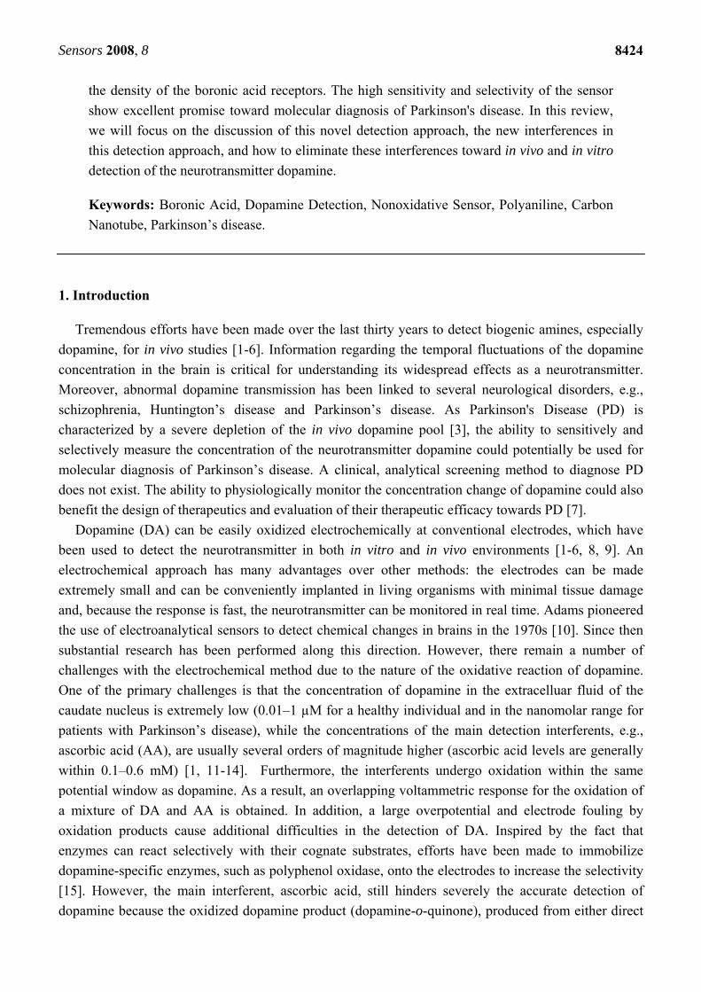
Abstract: Most of the current techniques for in vivo detection of dopamine exploit the ease
of oxidation of this compound. The major problem during the detection is the presence of a
high concentration of ascorbic acid that is oxidized at nearly the same potential as dopamine
on bare electrodes. Furthermore, the oxidation product of dopamine reacts with ascorbic
acid present in samples and regenerates dopamine again, which severely limits the accuracy
of the detection. Meanwhile, the product could also form a melanin-like insulating film on
the electrode surface, which decreases the sensitivity of the electrode. Various surface
modifications on the electrode, new materials for making the electrodes, and new
electrochemical techniques have been exploited to solve these problems. Recently we
developed a new electrochemical detection method that did not rely on direct oxidation of
dopamine on electrodes, which may naturally solve these problems. This approach takes
advantage of the high performance of our newly developed poly(anilineboronic acid)/carbon
nanotube composite and the excellent permselectivity of the ion-exchange polymer Nafion.
The high affinity binding of dopamine to the boronic acid groups of the polymer affects the
electrochemical properties of the polyaniline backbone, which act as the basis for the
transduction mechanism of this non-oxidative dopamine sensor. The unique reduction
capability and high conductivity of single-stranded DNA functionalized single-walled
carbon nanotubes greatly improved the electrochemical activity of the polymer in a
physiologically-relevant buffer, and the large surface area of the carbon nanotubes increased
OPEN ACCESS
Sensors 2008, 8
8424
the density of the boronic acid receptors. The high sensitivity and selectivity of the sensor
show excellent promise toward molecular diagnosis of Parkinson's disease. In this review,
we will focus on the discussion of this novel detection approach, the new interferences in
this detection approach, and how to eliminate these interferences toward in vivo and in vitro
detection of the neurotransmitter dopamine.
Keywords: Boronic Acid, Dopamine Detection, Nonoxidative Sensor, Polyaniline, Carbon
Nanotube, Parkinson’s disease.
1. Introduction
Tremendous efforts have been made over the last thirty years to detect biogenic amines, especially
dopamine, for in vivo studies [1-6]. Information regarding the temporal fluctuations of the dopamine
concentration in the brain is critical for understanding its widespread effects as a neurotransmitter.
Moreover, abnormal dopamine transmission has been linked to several neurological disorders, e.g.,
schizophrenia, Huntington’s disease and Parkinson’s disease. As Parkinson's Disease (PD) is
characterized by a severe depletion of the in vivo dopamine pool [3], the ability to sensitively and
selectively measure the concentration of the neurotransmitter dopamine could potentially be used for
molecular diagnosis of Parkinson’s disease. A clinical, analytical screening method to diagnose PD
does not exist. The ability to physiologically monitor the concentration change of dopamine could also
benefit the design of therapeutics and evaluation of their therapeutic efficacy towards PD [7].
Dopamine (DA) can be easily oxidized electrochemically at conventional electrodes, which have
been used to detect the neurotransmitter in both in vitro and in vivo environments [1-6, 8, 9]. An
electrochemical approach has many advantages over other methods: the electrodes can be made
extremely small and can be conveniently implanted in living organisms with minimal tissue damage
and, because the response is fast, the neurotransmitter can be monitored in real time. Adams pioneered
the use of electroanalytical sensors to detect chemical changes in brains in the 1970s [10]. Since then
substantial research has been performed along this direction. However, there remain a number of
challenges with the electrochemical method due to the nature of the oxidative reaction of dopamine.
One of the primary challenges is that the concentration of dopamine in the extracelluar fluid of the
caudate nucleus is extremely low (0.01–1 µM for a healthy individual and in the nanomolar range for
patients with Parkinson’s disease), while the concentrations of the main detection interferents, e.g.,
ascorbic acid (AA), are usually several orders of magnitude higher (ascorbic acid levels are generally
within 0.1–0.6 mM) [1, 11-14]. Furthermore, the interferents undergo oxidation within the same
potential window as dopamine. As a result, an overlapping voltammetric response for the oxidation of
a mixture of DA and AA is obtained. In addition, a large overpotential and electrode fouling by
oxidation products cause additional difficulties in the detection of DA. Inspired by the fact that
enzymes can react selectively with their cognate substrates, efforts have been made to immobilize
dopamine-specific enzymes, such as polyphenol oxidase, onto the electrodes to increase the selectivity
[15]. However, the main interferent, ascorbic acid, still hinders severely the accurate detection of
dopamine because the oxidized dopamine product (dopamine-o-quinone), produced from either direct
Sensors 2008, 8
8425
oxidation at the electrode or by the enzymes immobilized on the electrode, can catalytically oxidize
ascorbic acid nearby to regenerate dopamine that becomes available again for oxidation [16].
A variety of surface modification approaches has been exploited to resolve these problems. One
type of surface modification exploited the opposite polarities of DA and AA [17-24]. The modified
film on the electrode can attract one and repel the other. Another set of surface modification is to
incorporate electrochemical mediators, including some novel nanomaterials, such as Au nanoparticles
and carbon nanotubes, in the modified layer [25-28]. These approaches have shown great success in
selectivity by inhibiting the interference reactions or promoting dopamine oxidation at different
potentials. Special efforts have been paid to promote AA oxidation ahead of DA, which can effectively
avoid the dopamine regeneration problem. Simultaneous detection of AA and DA, and even uric acid,
were demonstrated [19-24]. Furthermore, due to the largely reduced overpotential and the existence of
the surface modification layer, the electrodes show excellent stability against electrode fouling [20,
27]. However, the detection limits in most of the reports are larger than 10 nM, which is not sensitive
enough for diagnosis of Parkinson’s disease.
New materials, such as carbon nanotubes [29] and boron-doped diamond [30] have been used to
fabricate novel electrodes for DA detection. New techniques have also been developed in an attempt to
solve the aforementioned problems, such as hybrid amperometric and conductometric measurements
based on conducting polymer nanojunctions [31], fast scan cyclic voltammetry (FSCV) and the
relevant data treatments [1].
FSCV has been the most successful approach for in vivo detection of catecholamine fluctuations
because of its high sensitivity and selectivity [1, 11, 12, 32, 33]. In this technique, the potential applied
to a carbon-fiber microelectrode is cycled at scan rates faster than 100 V/s. Compounds with slower
electron transfer rates, like ascorbic acid, can be distinguished very easily from biogenic amines at
high scan rates because the oxidation of ascorbate is drawn out to high potentials. To a certain extent,
this technique also avoids the problems of dopamine regeneration and electrode fouling due to the fast
turn-around potential applied to the electrodes [11, 34]. More recently, the carbon-fiber
microelectrodes have been modified by covalent attachment of molecules via diazonium salt reduction.
For example, the electrodes were modified with anionic functional groups such as carboxyphenyl [35],
phenylacetate [36], or sulfobenzene [37]. Electrodes modified in this way show further improved
sensitivity and selectivity to catecholamines over ascorbic acid without slowing down the response
time of the detection.
However, at high scan rates the majority of the current detected at the carbon-fiber working
electrode is a nonfaradic current that arises from charging of the double layer. Even though techniques
to subtract this large background current have been developed [38, 39], FSCV is typically only used to
observe concentration changes over the time course of a minute. This is because the background
current is only stable for a brief time. Therefore, it is not possible to measure basal concentrations of
dopamine due to the differential nature of the technique. Readers interested in this area are referred to
an extensive review about current development in detection of neurotransmitters and monitoring their
rapid concentration dynamics within neural tissue [1].
While oxidative dopamine sensors are the most widely used platforms for the electrochemical
detection of dopamine and are among the most successful in elucidating fundamental information
concerning physiological and neurological transmission of dopamine, approaches that could alleviate
Sensors 2008, 8
8426
all the afore-mentioned problems are attractive. One of the approaches is an electrochemical detection
method that does not rely on the direct oxidation or reduction of dopamine itself (refers to
nonoxidative dopamine sensors).
The first non-oxidative dopamine sensor was reported by Beni et al. [40]. They selectively detected
dopamine electrochemically at the interface between two immiscible electrolyte solutions. The two
liquid phases consisted of an aqueous layer in which both ascorbate and dopamine were dissolved, and
an organic layer formed by 1,2-dichloroethane. The crown ether dibenzo-18-crown-6 was used as an
ionophore that could transport the positively charged neurotransmitter dopamine (the amine group
from dopamine binds the crown ether) from the aqueous layer to the organic layer. The transfer of
ionized dopamine across the interface is accompanied with a potential difference, which produced a
current that can be measured in a voltammogram. As ascorbate cannot be transported to the organic
layer since it lacks affinity to the ionophore, it could not contribute to the measured amperometric
signal and therefore the interference from ascorbate was eliminated. Strawbridge et al. detected the
oxidation of phenylboronic acid, a diol-binding molecule, whose oxidation potential shifted upon
binding with dopamine. AA interference was eliminated because AA did not interact with
phenylboronic acid [41]. Recently this idea was applied by Wu et al. to increase the detection
sensitivity by modification of the electrodes with a layer of multiwalled carbon nanotubes that were
covalently linked to a phenylboronic acid derivative through carbodiimide coupling [42]. Fabre et al.
extended this principle by using a polyaniline derivative, poly(anilineboronic acid), which
preferentially binds dopamine over ascorbic acid by exploiting boronic acid-diol chemistry [43].
Formation of the resulting boronate ester changed the electrochemical behavior of the polyaniline
backbone, which was used to transduce the binding event. However, the detection limits of these
techniques were in the micromolar range, and therefore these detection methods are not sensitive
enough for molecular diagnosis of Parkinson’s disease.
Recently, we draw on the findings that single-stranded DNA (ss-DNA) can disperse bundled carbon
nanotubes in aqueous solution, resulting in helical wrapping of single-walled carbon nanotubes by ss-
DNA (ss-DNA/SWNTs) [44, 45]. The solubility and the unique surface properties of the ss-
DNA/SWNTs permit “in-situ” electrochemical polymerization of 3-aminophenylboronic acid
monomers to produce poly(anilineboronic acid)/ss-DNA/SWNT nanocomposites [46]. The
nanocomposite shows excellent properties through synergistic effects of the component materials. The
electrocatalytic reductive ability of the ss-DNA/SWNTs and their strong interaction with the
polyaniline backbone significantly improved the stability of poly(anilineboronic acid) (PABA) [47].
The conductivity and electrochemical activity in neutral solution were greatly enhanced. The large
surface area of the carbon nanotubes greatly increased the density of the boronic acid functional groups
available for sensitive detection of the target analyte. Taking advantage of these remarkable properties
of the nanocomposite, we developed a non-oxidative dopamine sensing approach, with which
dopamine concentrations as low as 1 nM were detected with cyclic voltammetry and 40 pM with
differential pulse voltammetry [48]. In this review, we will summarize our recent works along this line.
First we will discuss the unique roles of carbon nanotubes in enhancing the performance of the
conducting polymer, poly(anilineboronic acid), followed by a discussion of the dopamine transduction
mechanism and ascorbic acid interference mechanism in this non-oxidative approach. Finally, we will
Sensors 2008, 8
8427
discuss how to eliminate the interference of ascorbic acid toward the goal of molecular diagnosis of
Parkinson disease.
2. Development of a nonoxidative dopamine sensor based on a nanocomposite of carbon
nanotube/poly(anilineboronic acid).
2.1. Carbon nanotube/poly(anilineboronic acid) composites, multiple roles of single walled carbon
nanotubes dispersed and functionalized by single stranded DNA.
Conducting polymers are attractive for sensor applications because their electronic and
electrochemical properties are highly sensitive to molecular interactions, which provide excellent
signal transduction for molecular detection [49]. Among conducting polymers, polyaniline is unique
since it is environmentally stable and easy to fabricate. It has been applied widely in chemical sensors
[50-52] but not as much in biosensors [53-55]. The reason is that native polyaniline is neither
electrochemically active nor conductive in neutral solutions, which is a standard prerequisite for
biosensor applications. It is also limited both in the variety of molecules that can be detected and in the
selectivity of the detection. Major breakthroughs in this field were the discoveries of self-doped
polyaniline [56-59] and polyelectrolyte-anion-doped polyaniline [54, 55, 60], which brought
polyaniline into the biosensor field due to the improved redox activity and conductivity in neutral pH
solutions. However, compared to the parent polyaniline, the electrochemical activity, conductivity, and
the chemical and mechanical stabilities of both self-doped polyaniline and bulky polyelectrolyte-doped
polyaniline are greatly reduced, due to steric effects.
We found in this work that the stability of a self-doped polyaniline, poly(anilineboronic acid), is
greatly improved when it is polymerized in-situ with ss-DNA-wrapped single walled carbon nanotubes
(ss-DNA/SWNTs) (see Scheme 1). The redox properties of the polyaniline backbone are conserved in
neutral solutions (pH 7.4), and the sensitivity for biomolecular detection is significantly enhanced. We
found that the ss-DNA/SWNTs performed multiple roles in the greatly improved properties of the self-
doped polyaniline both during and after the polymerization, which makes this work unique compared
to previously reported conducting polymer/carbon nanotube composites [61-67]. First, they acted as
effective catalytic molecular templates during polymerization of the self-doped polyaniline to increase
the polymerization speed and improve the quality of the polymer. Second, they functioned as novel
active stabilizers after the polymerization. One difficulty in working with self-doped polyaniline is that
the fully oxidized pernigraniline form is not stable in aqueous solution and is difficult to completely
reduce to the partially oxidized emeraldine and fully reduced leucoemeraldine states [55, 68-71]. In
our case, pernigraniline was readily reduced in the electrochemical experiments due to the
electrocatalytic reductive ability of ss-DNA/SWNTs [72, 73]. This ease in reduction significantly
enhanced the stability of the film. Furthermore, the ss-DNA/SWNTs also acted as conductive
polyanionic doping agents in the resulting polyaniline film that showed enhanced conductivity and
redox activity. Finally, the large surface area of the carbon nanotubes greatly increased the density of
the functional groups available for sensitive detection of the target analytes.
Sensors 2008, 8
8428
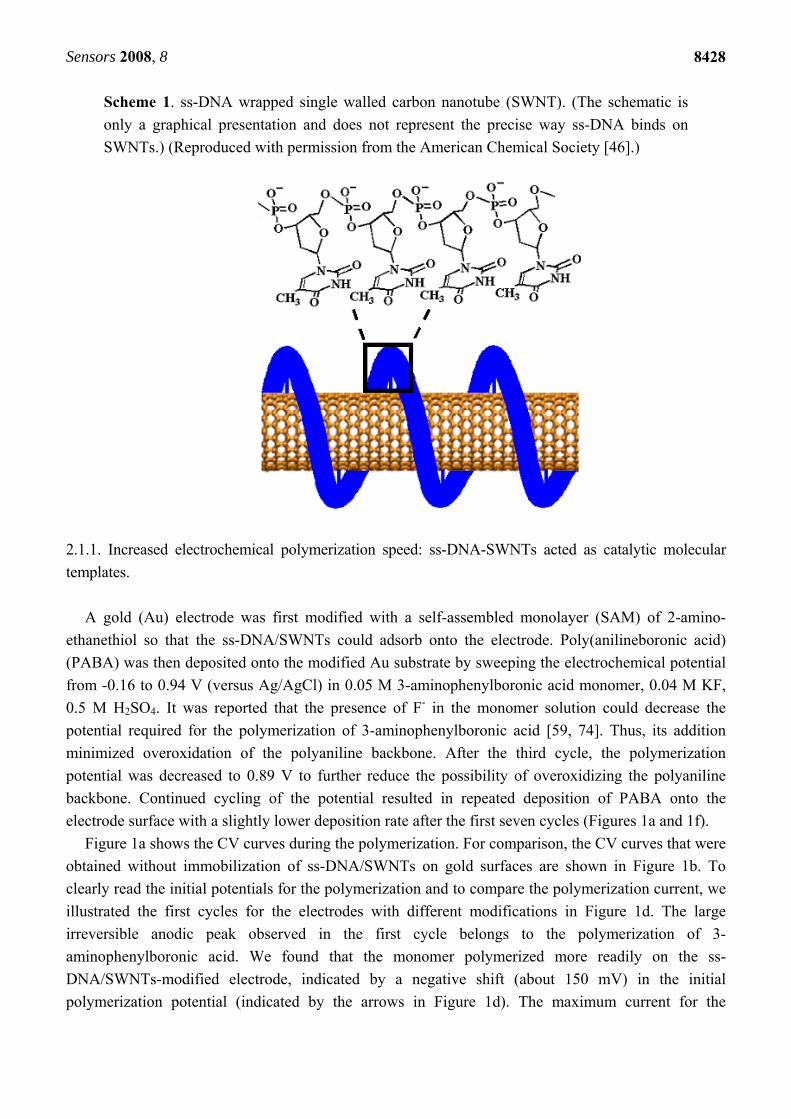
Scheme 1. ss-DNA wrapped single walled carbon nanotube (SWNT). (The schematic is
only a graphical presentation and does not represent the precise way ss-DNA binds on
SWNTs.) (Reproduced with permission from the American Chemical Society [46].)
2.1.1. Increased electrochemical polymerization speed: ss-DNA-SWNTs acted as catalytic molecular
templates.
A gold (Au) electrode was first modified with a self-assembled monolayer (SAM) of 2-amino-
ethanethiol so that the ss-DNA/SWNTs could adsorb onto the electrode. Poly(anilineboronic acid)
(PABA) was then deposited onto the modified Au substrate by sweeping the electrochemical potential
from -0.16 to 0.94 V (versus Ag/AgCl) in 0.05 M 3-aminophenylboronic acid monomer, 0.04 M KF,
0.5 M H2SO4. It was reported that the presence of F- in the monomer solution could decrease the
potential required for the polymerization of 3-aminophenylboronic acid [59, 74]. Thus, its addition
minimized overoxidation of the polyaniline backbone. After the third cycle, the polymerization
potential was decreased to 0.89 V to further reduce the possibility of overoxidizing the polyaniline
backbone. Continued cycling of the potential resulted in repeated deposition of PABA onto the
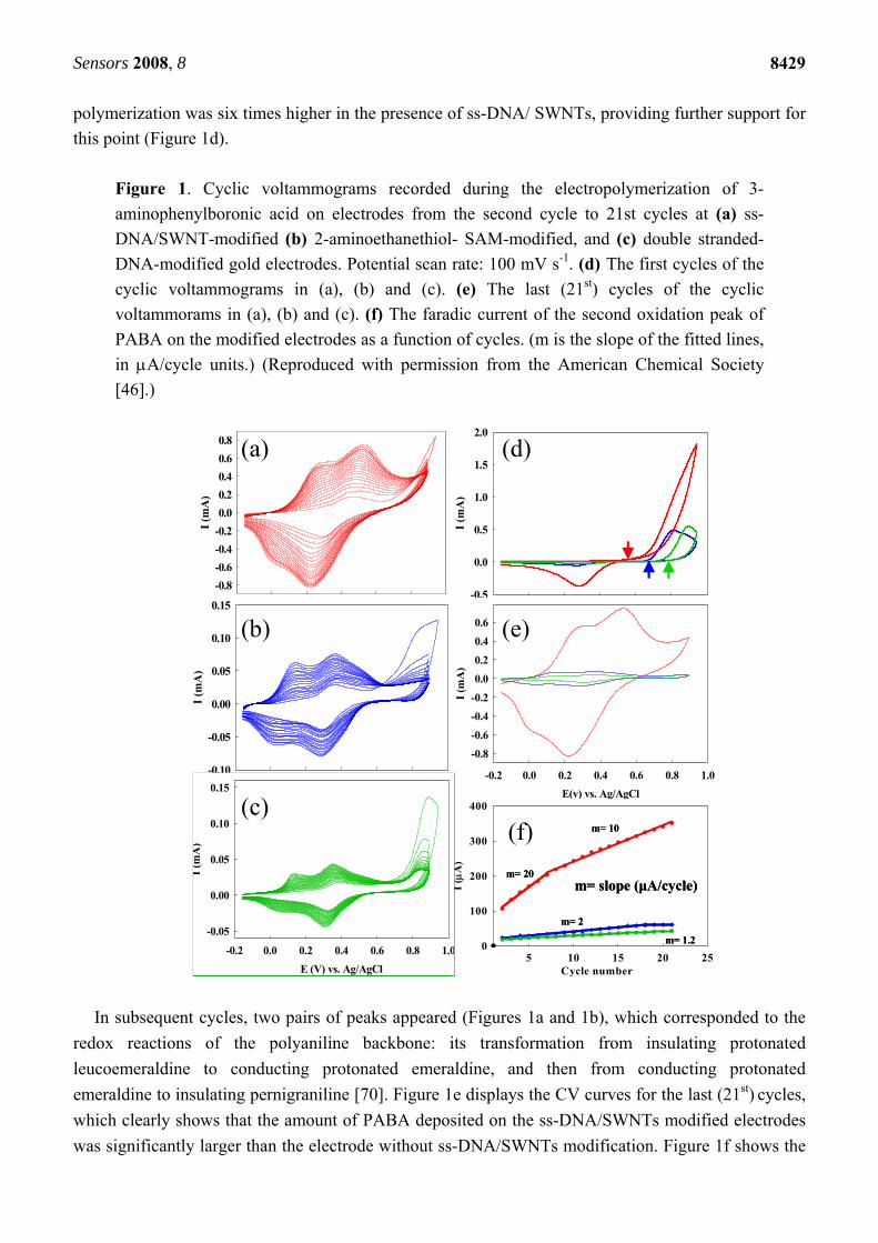
electrode surface with a slightly lower deposition rate after the first seven cycles (Figures 1a and 1f).
Figure 1a shows the CV curves during the polymerization. For comparison, the CV curves that were
obtained without immobilization of ss-DNA/SWNTs on gold surfaces are shown in Figure 1b. To
clearly read the initial potentials for the polymerization and to compare the polymerization current, we
illustrated the first cycles for the electrodes with different modifications in Figure 1d. The large
irreversible anodic peak observed in the first cycle belongs to the polymerization of 3-
aminophenylboronic acid. We found that the monomer polymerized more readily on the ss-
DNA/SWNTs-modified electrode, indicated by a negative shift (about 150 mV) in the initial
polymerization potential (indicated by the arrows in Figure 1d). The maximum current for the
Sensors 2008, 8
8429
polymerization was six times higher in the presence of ss-DNA/ SWNTs, providing further support for
this point (Figure 1d).
Figure 1. Cyclic voltammograms recorded during the electropolymerization of 3-
aminophenylboronic acid on electrodes from the second cycle to 21st cycles at (a) ss-
DNA/SWNT-modified (b) 2-aminoethanethiol- SAM-modified, and (c) double stranded-
DNA-modified gold electrodes. Potential scan rate: 100 mV s-1. (d) The first cycles of the
cyclic voltammograms in (a), (b) and (c). (e) The last (21st) cycles of the cyclic
voltammorams in (a), (b) and (c). (f) The faradic current of the second oxidation peak of
PABA on the modified electrodes as a function of cycles. (m is the slope of the fitted lines,
in A/cycle units.) (Reproduced with permission from the American Chemical Society
[46].)
I (m
A)
-0.8
-0.6
-0.4
-0.2
0.0
0.2
0.4
0.6
0.8
I (m
A)
-0.10
-0.05
0.00
0.05
0.10
0.15
I (m
A)
-0.5
0.0
0.5
1.0
1.5
2.0
I (m
A)
-0.5
0.0
0.5
1.0
1.5
2.0
(a) (d)
(b)
E(v) vs. Ag/AgCl
-0.2 0.0 0.2 0.4 0.6 0.8 1.0
I (m
A)
-0.8
-0.6
-0.4
-0.2
0.0
0.2
0.4
0.6 (e)
Cycle number5 10 15 20 25
I (
A)
0
100
200
300
400
m= 10
m= 2
m= 1.2
m= slope (μA/cycle)m= 20
Cycle number5 10 15 20 25
I (
A)
0
100
200
300
400
m= 10
m= 2
m= 1.2
m= slope (μA/cycle)m= 20
m= 10
m= 2
m= 1.2
m= slope (μA/cycle)m= 20
(f)
E (V) vs. Ag/AgCl
-0.2 0.0 0.2 0.4 0.6 0.8 1.0
I (m
A)
-0.05
0.00
0.05
0.10
0.15
(c)
In subsequent cycles, two pairs of peaks appeared (Figures 1a and 1b), which corresponded to the
redox reactions of the polyaniline backbone: its transformation from insulating protonated
leucoemeraldine to conducting protonated emeraldine, and then from conducting protonated
emeraldine to insulating pernigraniline [70]. Figure 1e displays the CV curves for the last (21st) cycles,
which clearly shows that the amount of PABA deposited on the ss-DNA/SWNTs modified electrodes
was significantly larger than the electrode without ss-DNA/SWNTs modification. Figure 1f shows the
Sensors 2008, 8
8430
current (the faradic current for the transformation from emeraldine to pernigraniline) as a function of
the CV cycle numbers for each of the electrodes with different modifications. Without ss-
DNA/SWNTs, the faradic current grew in intensity during the first 17 cycles and leveled off
afterwards (Figure 1f), indicating that PABA was deposited onto the electrodes only in the initial
stages of the experiment. At the ss-DNA/ SWNT modified electrodes, the PABA current increased
during all cycles, with a slower increase rate after the 7th cycle. From the slopes of the current vs. cycle
number in Figure 1f, we can estimate the current increase speed, which is ten times faster than that at
the electrode without ss-DNA/SWNTs. After seven cycles, the current increase rate lowered to 10 A
per cycle, which is still five times higher than that at the electrode without ss-DNA/SWNTs, where the
rate remained 2 A per cycle throughout. We attribute the faster deposition in the first seven cycles,
especially in the first cycle, to the pre-concentration of the monomers by the carbon nanotubes and to
the larger surface area conferred to the electrode by the ss-DNA/SWNTs.
We also used UV-Vis-Near IR spectroscopy to monitor in-situ the chemical polymerization process
of 3-aminophenylboronic acid monomers (ABA) in the presence and absence of ss-DNA/SWNTs. We
found that the polymerization process can be up to 4,500 times faster when ABA was polymerized in
the presence of ss-DNA/SWNTs [75]. We attributed this remarkable catalytic behavior of the ss-
DNA/SWNTs to the electronic interaction between the monomers and ss-DNA/SWNTs. ABA
monomers pre-concentrated along the carbon nanotubes and formed SWNTABA complexes, which
act as the polymerization precursors. Due to the electron richness of the ss-DNA/SWNTs, the ABA
monomers had higher electron density, which greatly facilitated the polymerization. During the
electrochemical polymerization, we found a negative shift (about 150 mV) in the initial polymerization
potential (indicated by the arrows in Figure 1d), suggesting that the catalytic effect of ss-DNA/SWNTs
also occurs during the electrochemical polymerization process [46].
2.1.2. Improved conductivity: ss-DNA-SWNTs acted as a unique conductive doping agent.
A prerequisite for continuous electrochemical deposition of polymer film is that the produced film
must be conductive in each cycle of deposition; otherwise, the deposition would self-terminate. The
redox current of the polyaniline backbone increased much faster on the electrode with ss-
DNA/SWNTs than at the electrodes with ds-DNA and with the 2-aminoethanethiol monolayer only,
respectively (Figures 1a, 1b, 1c and 1f). Figure 1e clearly shows that more PABA was deposited on the
electrode with ss-DNA/ SWNTs. We attribute the increase in deposition in the subsequent cycles to the
increased surface area of the electrodes imparted by the carbon nanotubes and to the better
conductivity of the resulting nanocomposite film. Although we could not characterize the conductivity
and the molecular structure of PABA in the composite on the gold electrode formed by
electrochemical polymerization, we have characterized the composites fabricated by in-situ chemical
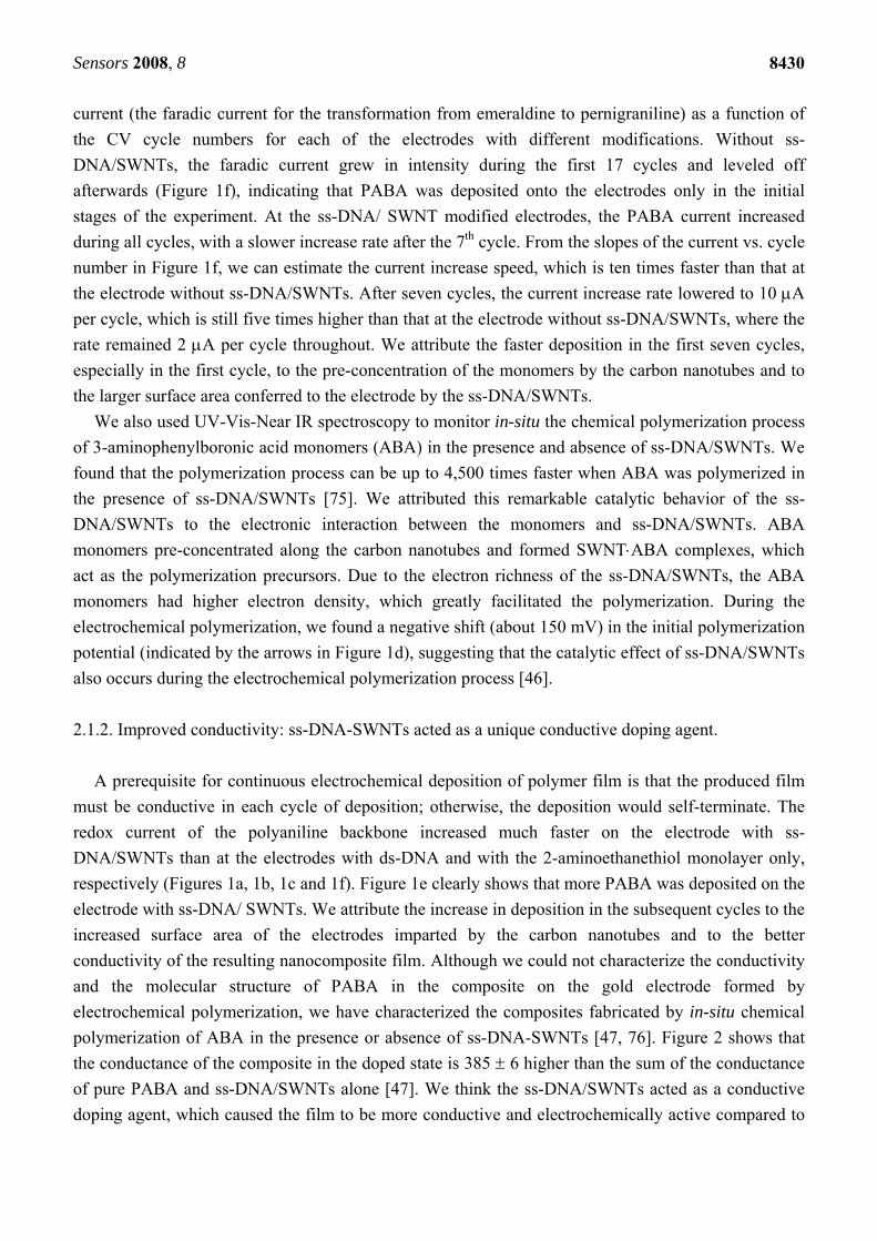
polymerization of ABA in the presence or absence of ss-DNA-SWNTs [47, 76]. Figure 2 shows that
the conductance of the composite in the doped state is 385 6 higher than the sum of the conductance
of pure PABA and ss-DNA/SWNTs alone [47]. We think the ss-DNA/SWNTs acted as a conductive
doping agent, which caused the film to be more conductive and electrochemically active compared to
Sensors 2008, 8
8431
the PABA film doped with a nonconductive polyanionic doping agent (i.e. ds-DNA) and small doping
agents [77].
Figure 2. Typical I-V characteristic curves of the (a) PABA-ss-DNA/SWNTs composite
film, (b) PABA alone, and ss-DNA/SWNT alone before and after treatment with NH3.
(Reproduced with permission from the American Chemical Society [47].)
ss-DNA/SWNTs before NH3
treatment ( )
ss-DNA/SWNTs after NH3
treatment ( )
ss-DNA/SWNTs before NH3
treatment ( )
ss-DNA/SWNTs after NH3
treatment ( )
The better conductivity of the nanocomposite film is also due to the improved conductivity of the
PABA component [47, 76]. Recently, we demonstrated that the contact resistance between carbon
nanotubes can be largely decreased by in situ polymerization of a thin skin of PABA around and along
the ss-DNA/SWNTs. The conductivity of the composite film after percolation can be two magnitudes
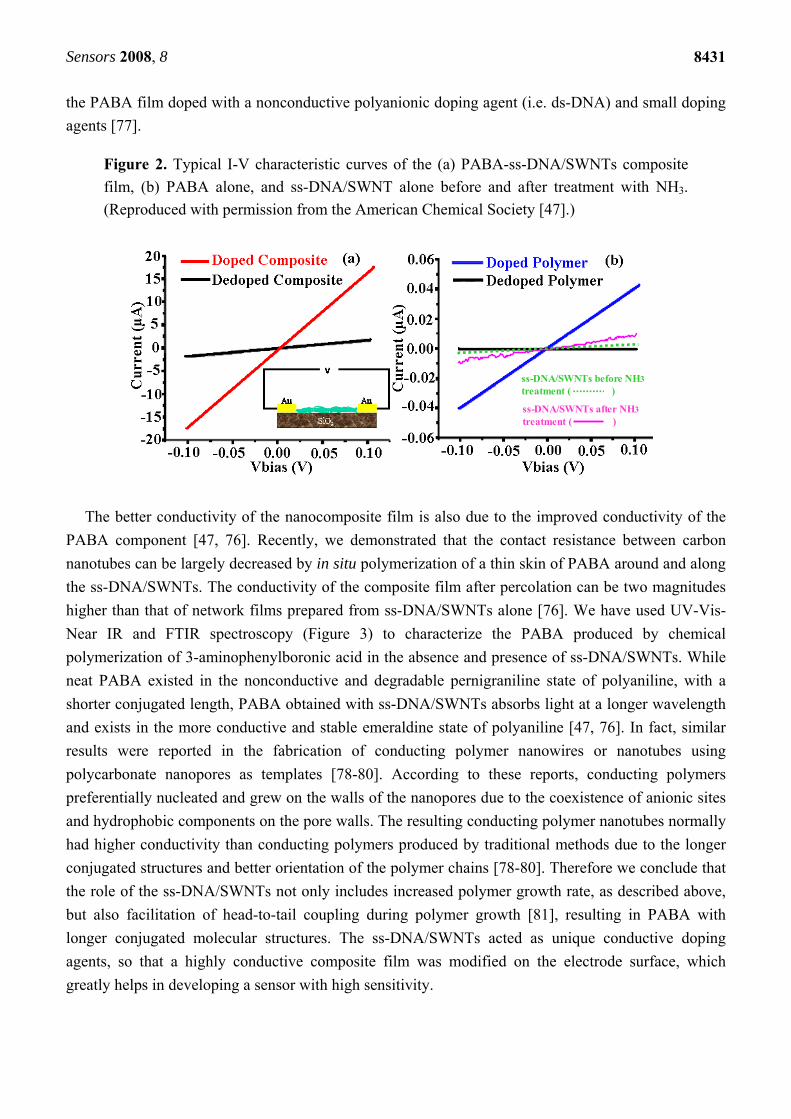
higher than that of network films prepared from ss-DNA/SWNTs alone [76]. We have used UV-Vis-
Near IR and FTIR spectroscopy (Figure 3) to characterize the PABA produced by chemical
polymerization of 3-aminophenylboronic acid in the absence and presence of ss-DNA/SWNTs. While
neat PABA existed in the nonconductive and degradable pernigraniline state of polyaniline, with a
shorter conjugated length, PABA obtained with ss-DNA/SWNTs absorbs light at a longer wavelength
and exists in the more conductive and stable emeraldine state of polyaniline [47, 76]. In fact, similar
results were reported in the fabrication of conducting polymer nanowires or nanotubes using
polycarbonate nanopores as templates [78-80]. According to these reports, conducting polymers
preferentially nucleated and grew on the walls of the nanopores due to the coexistence of anionic sites
and hydrophobic components on the pore walls. The resulting conducting polymer nanotubes normally
had higher conductivity than conducting polymers produced by traditional methods due to the longer
conjugated structures and better orientation of the polymer chains [78-80]. Therefore we conclude that
the role of the ss-DNA/SWNTs not only includes increased polymer growth rate, as described above,
but also facilitation of head-to-tail coupling during polymer growth [81], resulting in PABA with
longer conjugated molecular structures. The ss-DNA/SWNTs acted as unique conductive doping
agents, so that a highly conductive composite film was modified on the electrode surface, which
greatly helps in developing a sensor with high sensitivity.
Sensors 2008, 8
8432
Figure 3. (a) UV-vis spectra of the PABA/ss-DNA/SWNT composite (red) and the pure
PABA (inset, blue) before (-) and after (···) treatment with NaBH4; (b) FTIR spectra of the
composite (red) and the pure PABA (blue) and the pure PABA after reduction with NaBH4
(green). The spectra were normalized with the 1570 cm-1 peak. Baselines for the absorption
height ratio measurements are shown. (Reproduced with permission from the American
Chemical Society [47].)
(a)(a) (b)(b)
2.1.3. Enhanced Stability: ss-DNA/SWNTs acted as active stabilizer.
Toward the goal of biosensor applications, which require operation under physiological conditions,
the deep green PABA film obtained at the ss-DNA/SWNTs electrode was first stabilized in 0.5 M
H2SO4 and then in 0.01 M phosphate buffered saline (PBS, pH 7.4) by sweeping the potential between
0.04 and 0.79 V until the cyclic voltammetry (CV) curves were stabilized. Interestingly we found that
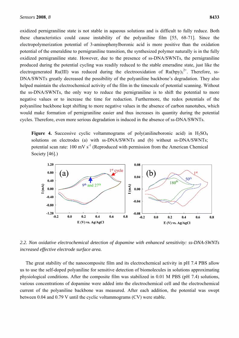
the stability of the resulting films was also increased by the presence of ss-DNA/SWNTs, as is
indicated by the fact that the CV curves were easily stabilized in both H2SO4 (Figure 4a) and PBS for
the PABA on the ss-DNA/SWNTs modified electrodes. In the absence of the ss-DNA/SWNTs, the
current decreased continuously upon cycling, and the oxidation and reduction peaks of the polyaniline
backbone also shifted significantly to negative potentials (Figure 4b). Figure 4b shows that after 180
cycles, the redox peaks of polyaniline are still decreasing and the peak positions are still shifting.
Holding the film at -0.3 V for 5 minutes prior to the scan caused the CV response to be recovered to a
certain extent. This behavior was often observed when the parent polyaniline film was cycled in higher
pH solutions [55, 68], and it was attributed to the inability to reduce the fully oxidized pernigraniline
completely over the timescale of the cyclic voltammetry experiment. On the contrary, with the ss-
DNA/SWNTs on the electrode, the CV curve of the 9th cycle is almost identical to the curves obtained
from previous cycles, indicating that the film was stabilized by the 9th cycle. We ascribe this enhanced
stability to the synergistic interaction of ss-DNA and SWNTs, which results in the electrocatalytic
reductive ability of the ss-DNA/SWNTs [72, 73]. Different from carbon nanotubes alone, ss-DNA-
wrapped carbon nanotubes are surprisingly effective electron donors, instead of electron acceceptors
[61, 82], and can be readily oxidized by strong oxidants such as KMnO4 and K2IrCl6 [72]. Recently the
electrocatalytic reduction ability of the ss-DNA/SWNTs was demonstrated in the study of
electrochemical oxidation of Ru(bpy)32+. The ss-DNA/SWNTs reduced electrogenerated Ru(III)
during electrooxidation of chemical Ru(bpy)32+ [73]. Polyaniline has three states, and the fully
Sensors 2008, 8
8433
oxidized pernigraniline state is not stable in aqueous solutions and is difficult to fully reduce. Both
these characteristics could cause instability of the polyaniline film [55, 68-71]. Since the
electropolymerization potential of 3-aminophenylboronic acid is more positive than the oxidation
potential of the emeraldine to pernigraniline transition, the synthesized polymer naturally is in the fully
oxidized pernigraniline state. However, due to the presence of ss-DNA/SWNTs, the pernigraniline
produced during the potential cycling was readily reduced to the stable emeradine state, just like the
electrogenerated Ru(III) was reduced during the electrooxidation of Ru(bpy)32+. Therefore, ss-
DNA/SWNTs greatly decreased the possibility of the polyaniline backbone’s degradation. They also
helped maintain the electrochemical activity of the film in the timescale of potential scanning. Without
the ss-DNA/SWNTs, the only way to reduce the pernigraniline is to shift the potential to more
negative values or to increase the time for reduction. Furthermore, the redox potentials of the
polyaniline backbone kept shifting to more negative values in the absence of carbon nanotubes, which
would make formation of pernigraniline easier and thus increases its quantity during the potential
cycles. Therefore, even more serious degradation is induced in the absence of ss-DNA/SWNTs.
Figure 4. Successive cyclic voltammograms of poly(anilineboronic acid) in H2SO4
solutions on electrodes (a) with ss-DNA/SWNTs and (b) without ss-DNA/SWNTs;
potential scan rate: 100 mV s-1 (Reproduced with permission from the American Chemical
Society [46].)
2.2. Non oxidative electrochemical detection of dopamine with enhanced sensitivity: ss-DNA-SWNTs
increased effective electrode surface area.
The great stability of the nanocomposite film and its electrochemical activity in pH 7.4 PBS allow
us to use the self-doped polyaniline for sensitive detection of biomolecules in solutions approximating
physiological conditions. After the composite film was stabilized in 0.01 M PBS (pH 7.4) solutions,
various concentrations of dopamine were added into the electrochemical cell and the electrochemical
current of the polyaniline backbone was measured. After each addition, the potential was swept
between 0.04 and 0.79 V until the cyclic voltammograms (CV) were stable.
Sensors 2008, 8
8434
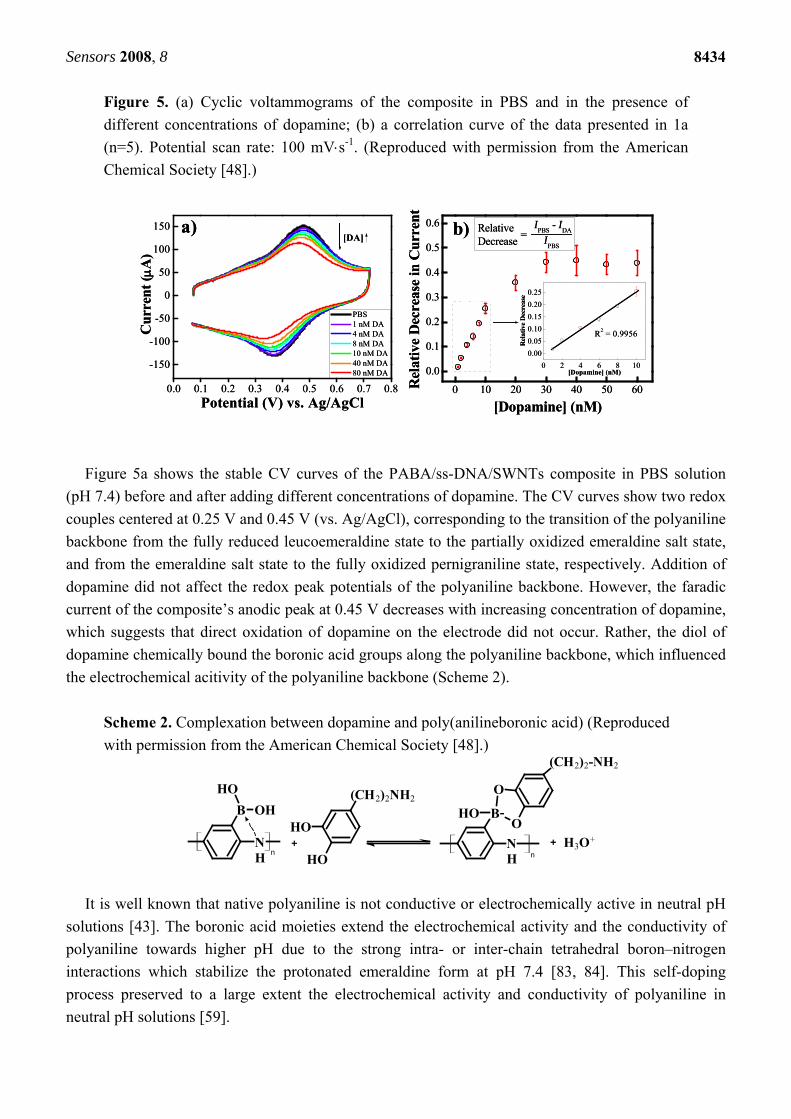
Figure 5. (a) Cyclic voltammograms of the composite in PBS and in the presence of
different concentrations of dopamine; (b) a correlation curve of the data presented in 1a
(n=5). Potential scan rate: 100 mVs-1. (Reproduced with permission from the American
Chemical Society [48].)
0.0 0.1 0.2 0.3 0.4 0.5 0.6 0.7 0.8
-150
-100
-50
0
50
100
150
PBS 1 nM DA 4 nM DA 8 nM DA 10 nM DA 40 nM DA 80 nM DA
[DA]
Cur
rent
(A
)
Potential (V) vs. Ag/AgCl
a)
0 10 20 30 40 50 60
0.0
0.1
0.2
0.3
0.4
0.5
0.6
IPBS
IPBS
- IDA=
RelativeDecrease
Rel
ativ
e D
ecre
ase
in C
urre
nt[Dopamine] (nM)
0 2 4 6 8 10
0.00
0.05
0.10
0.15
0.20
0.25
R2 = 0.9956
Rel
ativ
e D
ecre
ase
[Dopamine] (nM)
b)
Figure 5a shows the stable CV curves of the PABA/ss-DNA/SWNTs composite in PBS solution
(pH 7.4) before and after adding different concentrations of dopamine. The CV curves show two redox
couples centered at 0.25 V and 0.45 V (vs. Ag/AgCl), corresponding to the transition of the polyaniline
backbone from the fully reduced leucoemeraldine state to the partially oxidized emeraldine salt state,
and from the emeraldine salt state to the fully oxidized pernigraniline state, respectively. Addition of
dopamine did not affect the redox peak potentials of the polyaniline backbone. However, the faradic
current of the composite’s anodic peak at 0.45 V decreases with increasing concentration of dopamine,
which suggests that direct oxidation of dopamine on the electrode did not occur. Rather, the diol of
dopamine chemically bound the boronic acid groups along the polyaniline backbone, which influenced
the electrochemical acitivity of the polyaniline backbone (Scheme 2).
Scheme 2. Complexation between dopamine and poly(anilineboronic acid) (Reproduced
with permission from the American Chemical Society [48].)
OH
OH
(CH2)2NH2
OH
NH
B-O
O
(CH2)2-NH2
OH
NH
B
OH
H3O+
n+ +
n
It is well known that native polyaniline is not conductive or electrochemically active in neutral pH
solutions [43]. The boronic acid moieties extend the electrochemical activity and the conductivity of
polyaniline towards higher pH due to the strong intra- or inter-chain tetrahedral boron–nitrogen
interactions which stabilize the protonated emeraldine form at pH 7.4 [83, 84]. This self-doping
process preserved to a large extent the electrochemical activity and conductivity of polyaniline in
neutral pH solutions [59].
Sensors 2008, 8
8435
The high binding affinity between dopamine and boronic acid can affect the electrochemistry of the
polyaniline backbone in different and seemingly divergent modes, and thus require clarification. On
one hand, the conversion of the boronic acid to the boronate ester complex along the polyaniline
backbone interrupts the intra- or inter-chain tetrahedral boron–nitrogen interactions, which decreases
the self-doping and therefore the electrochemical activity of the polymer in neutral solutions.
Furthermore, the steric effect of the formed anionic ester also hinders the electrochemical activity of
the polyaniline backbone [85]. This is because oxidation and reduction of polyaniline during cyclic
voltammetry are accompanied by conformational changes of the polymer backbone, which become
less energetically favorable as large molecules are introduced along the backbone.
On the other hand, formation of the boronate complex eliminates the electron withdrawing nature of
the boron’s vacant p-orbital and instead, leads to an increase in the electron donating ability of the
boron in the boronate substitutent groups. Increasing the electron donating ability of the substitutent is
expected to stabilize the acid form of the quinone diimine group along the polymer backbone, a
trademark of the conductive form of polyaniline (emeraldine salt), which means an enhanced self-
doping ability. Therefore, the electrochemical activity of the polyaniline backbone should be enhanced
upon binding. It becomes apparent that these two effects on the electrochemical activity of the
polyaniline backbone offset one another. The net effect depends on the relative magnitude of the
influences.
These multifaceted effects on electrochemical activity were observed recently in a boronic acid-
substituted bipyridine Fe(II) complex [85]. Fabre et al. found that the apparent formal potential of the
ferrocene/ferrocenium redox couple decreased and the redox current increased upon formation of the
electron-donating boronate anion structure with F-, while with fructose an increase of the formal
potential and decrease of redox current were observed. The authors ascribed these contrary changes to
the different steric effects imparted by the F- and fructose to the pyridine backbone in spite of their
similar abilities to convert the boronic acid groups from electron withdrawing to electron donating
substituents. Upon binding of dopamine, we found that the redox current decreased, suggesting that the
steric effect of the formed anionic ester played the more important role. The resulting ester hindered
the electrochemical activity of the polyaniline backbone, which is in agreement with the report by
Fabre et al. [85].
It was reported that formation of the anionic ester could reduce the Ka of the protonated quinone
diimine groups in the polyaniline backbone, and reduction of the Ka caused a positive shift in the
potential of the electrochemical conversion of emeraldine to penigraniline. Shoji et al. developed a
PABA-based potentiometric sensor for detection of saccharides using the potential shift of PABA as
the transduction principle [86, 87]. Therefore, we expected a decrease of the oxidation current and a
concomitant positive shift in the peak potential upon dopamine binding. However, our data does not
show appreciable shifts in Epa upon dopamine binding. This is likely due to the low concentration of
dopamine (nanomolar range) used in this study: the formation of a small amount of anionic ester could
not induce an observable potential shift. In fact, both a decrease in the faradic current of the anodic
peak as well as an increase in the potential of the anodic peak was observed when 1 M dopamine was
added to the electrochemical cell.
The faradic current of the anodic peak (IPBS) at E=0.45 V was recorded from the last cycle of CV
curves at which the composite is stabilized in PBS solution, and the faradic current in the last CV
Sensors 2008, 8
8436
cycles of each dopamine addition (IDA). Figure 5b shows the relative decrease of the faradic current of
the composite in PBS as a function of the concentration of dopamine. The equation that was used to
calculate each of the data points is shown as an inset in this figure. The correlation curve demonstrates
an initial increasing linear region followed by a relatively horizontal regime. The linear portion, which
extends from 1 nM to 10 nM, is very reproducible and has a very small standard deviation (standard
error bars displayed on the curve; n=5). The theoretical detection limit (defined as the concentration
that generates a signal three times larger than the noise) of this composite towards dopamine using
cyclic voltammetry is 0.6 nM. Above the concentration of 30 nM the horizontal regime predominates:
error bars for data points in this region are large because the saturation limits were quite different for
different films. We do not know the exact reason yet at present, but we think that the saturation limits
depend on the degree of aggregation of the ss-DNA-wrapped carbon nanotubes during deposition and
drying on the electrode surfaces, which consequently influenced the amount and the quality of the
PABA deposited on the electrode. Since the linear sensing range is in the appropriate regime for
studying in vivo dopamine levels, our method holds great potential for molecular diagnosis of
Parkinson’s disease.
The sensitivity towards dopamine increased by 104 compared to the previous report in which neat
PABA was used to modify the electrodes in a detection platform of micro-electrochemical transistors
[43]. We believe that the ss-DNA/SWNTs in the composite increased the effective electrode surface
area, causing a higher density of boronic acid groups available for dopamine binding and thus
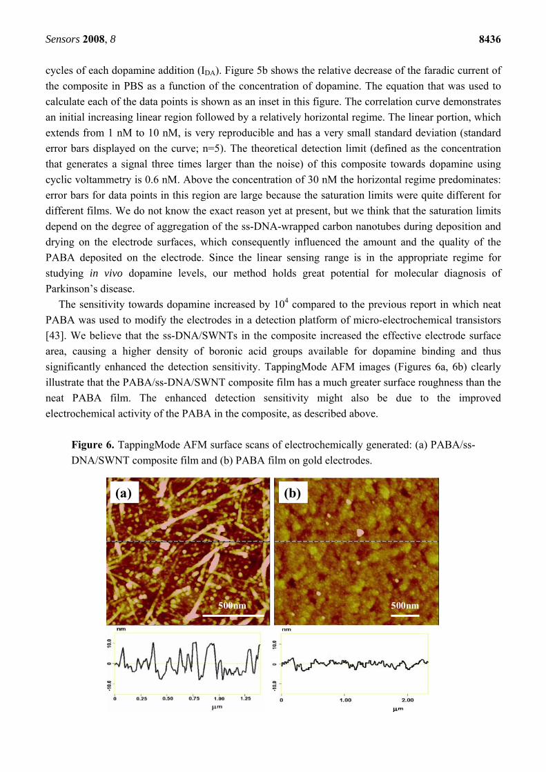
significantly enhanced the detection sensitivity. TappingMode AFM images (Figures 6a, 6b) clearly
illustrate that the PABA/ss-DNA/SWNT composite film has a much greater surface roughness than the
neat PABA film. The enhanced detection sensitivity might also be due to the improved
electrochemical activity of the PABA in the composite, as described above.
Figure 6. TappingMode AFM surface scans of electrochemically generated: (a) PABA/ss-
DNA/SWNT composite film and (b) PABA film on gold electrodes.
(a) (b)
500nm 500nm
(a) (b)
500nm 500nm
Sensors 2008, 8
8437
We should mention that even higher detection sensitivity can be reached by optimizing the
detection techniques. For example, a micro-electrochemical transistor as a detection platform normally
has higher sensitivity because the conductivity of polyaniline can be changed by many orders of
magnitude when its redox states are switched [51, 60, 88-90]. This large change in conductance of the
polymer leads to amplification of the detection signal. Another technique, differential pulse
voltammetry (DPV) [91], can greatly decrease the background charging currents, and in turn also
increase the detection sensitivity. Our preliminary study demonstrates that the detection limit can be
improved by two orders of magnitude when DPV instead of CV was applied to detect dopamine.
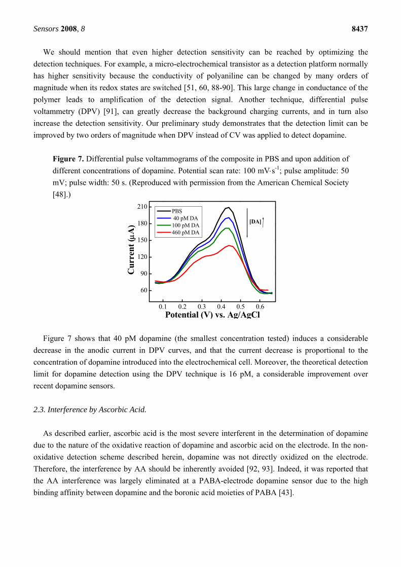
Figure 7. Differential pulse voltammograms of the composite in PBS and upon addition of
different concentrations of dopamine. Potential scan rate: 100 mVs-1; pulse amplitude: 50
mV; pulse width: 50 s. (Reproduced with permission from the American Chemical Society
[48].)
0.1 0.2 0.3 0.4 0.5 0.6
60
90
120
150
180
210
[DA]
Cur
rent
(A
)
Potential (V) vs. Ag/AgCl
PBS 40 pM DA 100 pM DA 460 pM DA
Figure 7 shows that 40 pM dopamine (the smallest concentration tested) induces a considerable
decrease in the anodic current in DPV curves, and that the current decrease is proportional to the
concentration of dopamine introduced into the electrochemical cell. Moreover, the theoretical detection
limit for dopamine detection using the DPV technique is 16 pM, a considerable improvement over
recent dopamine sensors.
2.3. Interference by Ascorbic Acid.
As described earlier, ascorbic acid is the most severe interferent in the determination of dopamine
due to the nature of the oxidative reaction of dopamine and ascorbic acid on the electrode. In the non-
oxidative detection scheme described herein, dopamine was not directly oxidized on the electrode.
Therefore, the interference by AA should be inherently avoided [92, 93]. Indeed, it was reported that
the AA interference was largely eliminated at a PABA-electrode dopamine sensor due to the high
binding affinity between dopamine and the boronic acid moieties of PABA [43].
Sensors 2008, 8
8438
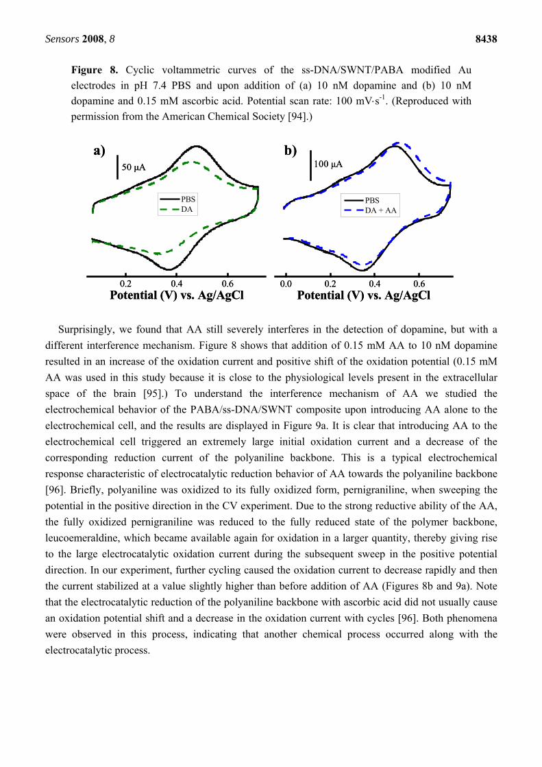
Figure 8. Cyclic voltammetric curves of the ss-DNA/SWNT/PABA modified Au
electrodes in pH 7.4 PBS and upon addition of (a) 10 nM dopamine and (b) 10 nM
dopamine and 0.15 mM ascorbic acid. Potential scan rate: 100 mVs-1. (Reproduced with
permission from the American Chemical Society [94].)
0.2 0.4 0.6Potential (V) vs. Ag/AgCl
PBS DA
0.0 0.2 0.4 0.6Potential (V) vs. Ag/AgCl
PBS DA + AA
a) b)50 μA 100 μA
0.2 0.4 0.6Potential (V) vs. Ag/AgCl
PBS DA
0.0 0.2 0.4 0.6Potential (V) vs. Ag/AgCl
PBS DA + AA
a) b)50 μA 100 μA
Surprisingly, we found that AA still severely interferes in the detection of dopamine, but with a
different interference mechanism. Figure 8 shows that addition of 0.15 mM AA to 10 nM dopamine
resulted in an increase of the oxidation current and positive shift of the oxidation potential (0.15 mM
AA was used in this study because it is close to the physiological levels present in the extracellular
space of the brain [95].) To understand the interference mechanism of AA we studied the
electrochemical behavior of the PABA/ss-DNA/SWNT composite upon introducing AA alone to the
electrochemical cell, and the results are displayed in Figure 9a. It is clear that introducing AA to the
electrochemical cell triggered an extremely large initial oxidation current and a decrease of the
corresponding reduction current of the polyaniline backbone. This is a typical electrochemical
response characteristic of electrocatalytic reduction behavior of AA towards the polyaniline backbone
[96]. Briefly, polyaniline was oxidized to its fully oxidized form, pernigraniline, when sweeping the
potential in the positive direction in the CV experiment. Due to the strong reductive ability of the AA,
the fully oxidized pernigraniline was reduced to the fully reduced state of the polymer backbone,
leucoemeraldine, which became available again for oxidation in a larger quantity, thereby giving rise
to the large electrocatalytic oxidation current during the subsequent sweep in the positive potential
direction. In our experiment, further cycling caused the oxidation current to decrease rapidly and then
the current stabilized at a value slightly higher than before addition of AA (Figures 8b and 9a). Note
that the electrocatalytic reduction of the polyaniline backbone with ascorbic acid did not usually cause
an oxidation potential shift and a decrease in the oxidation current with cycles [96]. Both phenomena
were observed in this process, indicating that another chemical process occurred along with the
electrocatalytic process.
Sensors 2008, 8
8439
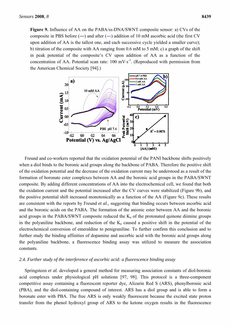
Figure 9. Influence of AA on the PABA/ss-DNA/SWNT composite sensor: a) CVs of the
composite in PBS before (―) and after (―) addition of 10 mM ascorbic acid (the first CV
upon addition of AA is the tallest one, and each successive cycle yielded a smaller curve); b) titration of the composite with AA ranging from 0.6 mM to 5 mM; c) a graph of the shift
in peak potential of the composite’s CV upon addition of AA as a function of the
concentration of AA. Potential scan rate: 100 mVs-1. (Reproduced with permission from the American Chemical Society [94].)
-0.2 0.0 0.2 0.4 0.6
-50
0
50
100
pH 7.4 PBS 0.6 mM AA 2 mM AA 5 mM AA
Cur
rent
(A
)
Potential (mV)
b)
0 1 2 3 4 50
10
20
30
40
R2 = 0.9878
[AA] (mM)
Pea
k P
oten
tial
Sh
ift
(mV
) c)
-0.2 0.0 0.2 0.4 0.6-1.0
-0.5
0.0
0.5
1.0
1.5
2.0
a)
Potential (V) vs. Ag/AgCl
Cur
rent
(m
A) 10 mM AA
PBS pH 7.4
-0.2 0.0 0.2 0.4 0.6
-50
0
50
100
pH 7.4 PBS 0.6 mM AA 2 mM AA 5 mM AA
Cur
rent
(A
)
Potential (mV)
b)
-0.2 0.0 0.2 0.4 0.6
-50
0
50
100
pH 7.4 PBS 0.6 mM AA 2 mM AA 5 mM AA
Cur
rent
(A
)
Potential (mV)
b)
0 1 2 3 4 50
10
20
30
40
R2 = 0.9878
[AA] (mM)
Pea
k P
oten
tial
Sh
ift
(mV
) c)
0 1 2 3 4 50
10
20
30
40
R2 = 0.9878
[AA] (mM)
Pea
k P
oten
tial
Sh
ift
(mV
)
0 1 2 3 4 50
10
20
30
40
R2 = 0.9878
[AA] (mM)
Pea
k P
oten
tial
Sh
ift
(mV
) c)
-0.2 0.0 0.2 0.4 0.6-1.0
-0.5
0.0
0.5
1.0
1.5
2.0
a)
Potential (V) vs. Ag/AgCl
Cur
rent
(m
A) 10 mM AA
PBS pH 7.4
-0.2 0.0 0.2 0.4 0.6-1.0
-0.5
0.0
0.5
1.0
1.5
2.0
a)
Potential (V) vs. Ag/AgCl
Cur
rent
(m
A) 10 mM AA
PBS pH 7.4
Freund and co-workers reported that the oxidation potential of the PANI backbone shifts positively
when a diol binds to the boronic acid groups along the backbone of PABA. Therefore the positive shift
of the oxidation potential and the decrease of the oxidation current may be understood as a result of the formation of boronate ester complexes between AA and the boronic acid groups in the PABA/SWNT
composite. By adding different concentrations of AA into the electrochemical cell, we found that both
the oxidation current and the potential increased after the CV curves were stabilized (Figure 9b), and the positive potential shift increased monotonically as a function of the AA (Figure 9c). These results
are consistent with the reports by Freund et al., suggesting that binding occurs between ascorbic acid
and the boronic acids on the PABA. The formation of the anionic ester between AA and the boronic acid groups in the PABA/SWNT composite reduced the Ka of the protonated quinone diimine groups
in the polyaniline backbone, and reduction of the Ka caused a positive shift in the potential of the
electrochemical conversion of emeraldine to penigraniline. To further confirm this conclusion and to further study the binding affinities of dopamine and ascorbic acid with the boronic acid groups along
the polyaniline backbone, a fluorescence binding assay was utilized to measure the association
constants.
2.4. Further study of the interference of ascorbic acid: a fluorescence binding assay
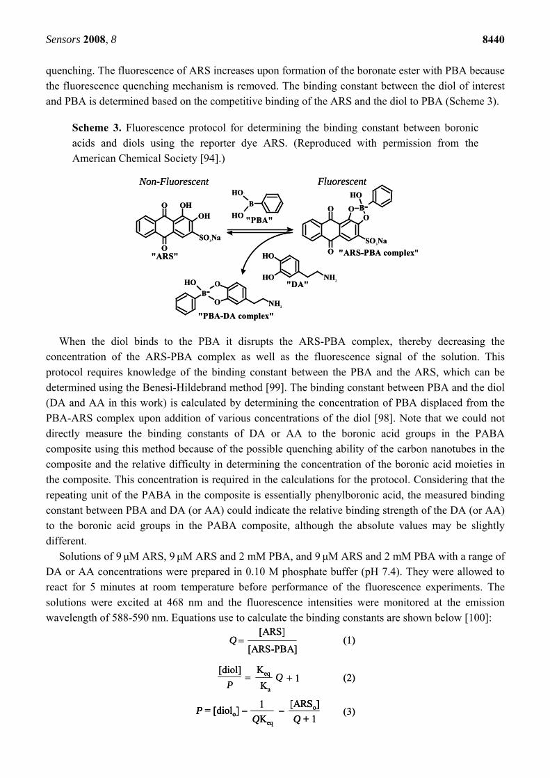
Springsteen et al. developed a general method for measuring association constants of diol-boronic
acid complexes under physiological pH solutions [97, 98]. This protocol is a three-component competitive assay containing a fluorescent reporter dye, Alizarin Red S (ARS), phenylboronic acid
(PBA), and the diol-containing compound of interest. ARS has a diol group and is able to form a
boronate ester with PBA. The free ARS is only weakly fluorescent because the excited state proton transfer from the phenol hydroxyl group of ARS to the ketone oxygen results in the fluorescence
Sensors 2008, 8
8440
quenching. The fluorescence of ARS increases upon formation of the boronate ester with PBA because
the fluorescence quenching mechanism is removed. The binding constant between the diol of interest
and PBA is determined based on the competitive binding of the ARS and the diol to PBA (Scheme 3).
Scheme 3. Fluorescence protocol for determining the binding constant between boronic
acids and diols using the reporter dye ARS. (Reproduced with permission from the
American Chemical Society [94].)
O
O OHOH
SO3Na
OH
OH NH2
OH
OH
BOH
O
O OO
SO3Na
B-
Non-Fluorescent Fluorescent
"ARS" "ARS-PBA complex"
"PBA"
"DA"OH
O
OB-
NH2
"PBA-DA complex"
O
O OHOH
SO3Na
OH
OH NH2
OH
OH
BOH
O
O OO
SO3Na
B-
Non-Fluorescent Fluorescent
"ARS" "ARS-PBA complex"
"PBA"
"DA"OH
O
OB-
NH2
"PBA-DA complex"
When the diol binds to the PBA it disrupts the ARS-PBA complex, thereby decreasing the
concentration of the ARS-PBA complex as well as the fluorescence signal of the solution. This
protocol requires knowledge of the binding constant between the PBA and the ARS, which can be determined using the Benesi-Hildebrand method [99]. The binding constant between PBA and the diol
(DA and AA in this work) is calculated by determining the concentration of PBA displaced from the
PBA-ARS complex upon addition of various concentrations of the diol [98]. Note that we could not directly measure the binding constants of DA or AA to the boronic acid groups in the PABA
composite using this method because of the possible quenching ability of the carbon nanotubes in the
composite and the relative difficulty in determining the concentration of the boronic acid moieties in the composite. This concentration is required in the calculations for the protocol. Considering that the
repeating unit of the PABA in the composite is essentially phenylboronic acid, the measured binding
constant between PBA and DA (or AA) could indicate the relative binding strength of the DA (or AA) to the boronic acid groups in the PABA composite, although the absolute values may be slightly
different.
Solutions of 9 μM ARS, 9 μM ARS and 2 mM PBA, and 9 μM ARS and 2 mM PBA with a range of DA or AA concentrations were prepared in 0.10 M phosphate buffer (pH 7.4). They were allowed to
react for 5 minutes at room temperature before performance of the fluorescence experiments. The
solutions were excited at 468 nm and the fluorescence intensities were monitored at the emission wavelength of 588-590 nm. Equations use to calculate the binding constants are shown below [100]:
Q =[ARS]
[ARS-PBA]
[diol]
P=
Keq
Ka
Q + 1
(1)
(2)
P = [diolo] –1
QKeq
–[ARSo]
Q + 1(3)
Q =[ARS]
[ARS-PBA]
[diol]
P=
Keq
Ka
Q + 1
(1)
(2)
P = [diolo] –1
QKeq
–[ARSo]
Q + 1P = [diolo] –
1
QKeq
–[ARSo]
Q + 1(3)
Sensors 2008, 8
8441
where Keq is the association constant of the ARS-PBA complex (determined by the Benesi-Hildebrand
method), Ka is the association constant of the boronic acid–diol complex, [diolo] is the total diol
concentration, [ARSo] is the total ARS concentration, Q is the ratio of the concentration of
uncomplexed ARS to complexed ARS (Equation 1), and P is defined by Equation 3. The Ka of the
boronic acid–diol complex was determined by plotting [diol]/P vs. Q, and dividing Ka by the slope of
the plot, as per Equation 2. The fluorescence emission spectra were obtained at a Cary Eclipse
fluorescence spectrophotometer (Varian).
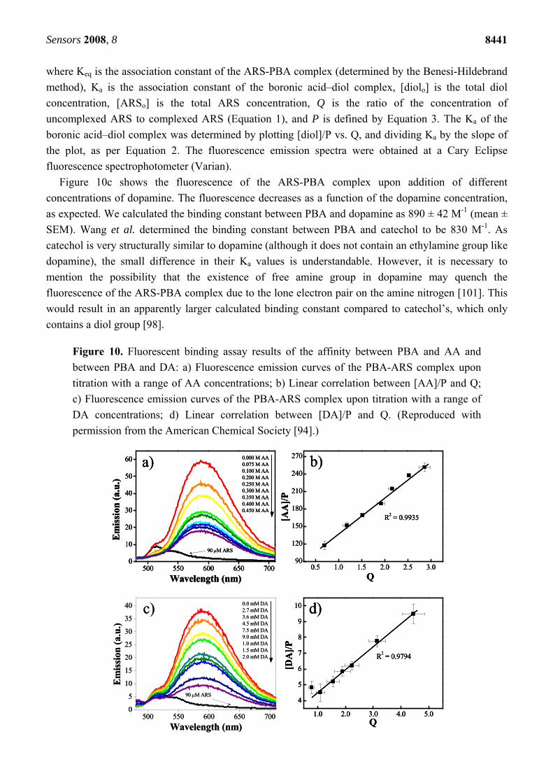
Figure 10c shows the fluorescence of the ARS-PBA complex upon addition of different
concentrations of dopamine. The fluorescence decreases as a function of the dopamine concentration,
as expected. We calculated the binding constant between PBA and dopamine as 890 ± 42 M-1 (mean ±
SEM). Wang et al. determined the binding constant between PBA and catechol to be 830 M-1. As
catechol is very structurally similar to dopamine (although it does not contain an ethylamine group like
dopamine), the small difference in their Ka values is understandable. However, it is necessary to
mention the possibility that the existence of free amine group in dopamine may quench the
fluorescence of the ARS-PBA complex due to the lone electron pair on the amine nitrogen [101]. This
would result in an apparently larger calculated binding constant compared to catechol’s, which only
contains a diol group [98].
Figure 10. Fluorescent binding assay results of the affinity between PBA and AA and
between PBA and DA: a) Fluorescence emission curves of the PBA-ARS complex upon
titration with a range of AA concentrations; b) Linear correlation between [AA]/P and Q;
c) Fluorescence emission curves of the PBA-ARS complex upon titration with a range of
DA concentrations; d) Linear correlation between [DA]/P and Q. (Reproduced with
permission from the American Chemical Society [94].)
1.0 2.0 3.0 4.0 5.0
4
5
6
7
8
9
10
R2 = 0.9794
[DA
]/P
Q
d)
0.5 1.0 1.5 2.0 2.5 3.090
120
150
180
210
240
270
R2 = 0.9935
[AA
]/P
Q
b)
500 550 600 650 7000
5
10
15
20
25
30
35
40 0.0 mM DA2.7 mM DA3.6 mM DA4.5 mM DA7.5 mM DA9.0 mM DA1.0 mM DA1.5 mM DA2.0 mM DA
90 M ARS
Em
issi
on (
a.u.
)
Wavelength (nm)
c)
500 550 600 650 7000
10
20
30
40
50
60
90 M ARS
0.000 M AA0.075 M AA0.100 M AA0.200 M AA0.250 M AA0.300 M AA0.350 M AA0.400 M AA0.450 M AA
Em
issi
on (
a.u.
)
Wavelength (nm)
a)
1.0 2.0 3.0 4.0 5.0
4
5
6
7
8
9
10
R2 = 0.9794
[DA
]/P
Q
d)
1.0 2.0 3.0 4.0 5.0
4
5
6
7
8
9
10
R2 = 0.9794
[DA
]/P
Q
d)
0.5 1.0 1.5 2.0 2.5 3.090
120
150
180
210
240
270
R2 = 0.9935
[AA
]/P
Q
b)
0.5 1.0 1.5 2.0 2.5 3.090
120
150
180
210
240
270
R2 = 0.9935
[AA
]/P
Q
b)
500 550 600 650 7000
5
10
15
20
25
30
35
40 0.0 mM DA2.7 mM DA3.6 mM DA4.5 mM DA7.5 mM DA9.0 mM DA1.0 mM DA1.5 mM DA2.0 mM DA
90 M ARS
Em
issi
on (
a.u.
)
Wavelength (nm)
c)
500 550 600 650 7000
10
20
30
40
50
60
90 M ARS
0.000 M AA0.075 M AA0.100 M AA0.200 M AA0.250 M AA0.300 M AA0.350 M AA0.400 M AA0.450 M AA
Em
issi
on (
a.u.
)
Wavelength (nm)
a)
500 550 600 650 7000
10
20
30
40
50
60
90 M ARS
0.000 M AA0.075 M AA0.100 M AA0.200 M AA0.250 M AA0.300 M AA0.350 M AA0.400 M AA0.450 M AA
Em
issi
on (
a.u.
)
Wavelength (nm)
a)
Sensors 2008, 8
8442
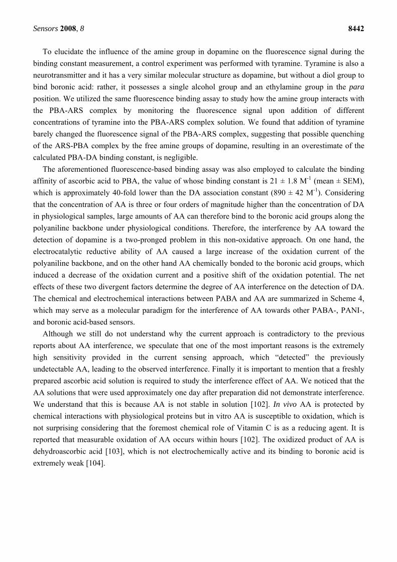
To elucidate the influence of the amine group in dopamine on the fluorescence signal during the
binding constant measurement, a control experiment was performed with tyramine. Tyramine is also a
neurotransmitter and it has a very similar molecular structure as dopamine, but without a diol group to
bind boronic acid: rather, it possesses a single alcohol group and an ethylamine group in the para
position. We utilized the same fluorescence binding assay to study how the amine group interacts with
the PBA-ARS complex by monitoring the fluorescence signal upon addition of different
concentrations of tyramine into the PBA-ARS complex solution. We found that addition of tyramine
barely changed the fluorescence signal of the PBA-ARS complex, suggesting that possible quenching
of the ARS-PBA complex by the free amine groups of dopamine, resulting in an overestimate of the
calculated PBA-DA binding constant, is negligible.
The aforementioned fluorescence-based binding assay was also employed to calculate the binding
affinity of ascorbic acid to PBA, the value of whose binding constant is 21 ± 1.8 M-1 (mean ± SEM),
which is approximately 40-fold lower than the DA association constant (890 ± 42 M-1). Considering
that the concentration of AA is three or four orders of magnitude higher than the concentration of DA
in physiological samples, large amounts of AA can therefore bind to the boronic acid groups along the
polyaniline backbone under physiological conditions. Therefore, the interference by AA toward the
detection of dopamine is a two-pronged problem in this non-oxidative approach. On one hand, the
electrocatalytic reductive ability of AA caused a large increase of the oxidation current of the
polyaniline backbone, and on the other hand AA chemically bonded to the boronic acid groups, which
induced a decrease of the oxidation current and a positive shift of the oxidation potential. The net
effects of these two divergent factors determine the degree of AA interference on the detection of DA.
The chemical and electrochemical interactions between PABA and AA are summarized in Scheme 4,
which may serve as a molecular paradigm for the interference of AA towards other PABA-, PANI-,
and boronic acid-based sensors.
Although we still do not understand why the current approach is contradictory to the previous
reports about AA interference, we speculate that one of the most important reasons is the extremely
high sensitivity provided in the current sensing approach, which “detected” the previously
undetectable AA, leading to the observed interference. Finally it is important to mention that a freshly
prepared ascorbic acid solution is required to study the interference effect of AA. We noticed that the
AA solutions that were used approximately one day after preparation did not demonstrate interference.
We understand that this is because AA is not stable in solution [102]. In vivo AA is protected by
chemical interactions with physiological proteins but in vitro AA is susceptible to oxidation, which is
not surprising considering that the foremost chemical role of Vitamin C is as a reducing agent. It is
reported that measurable oxidation of AA occurs within hours [102]. The oxidized product of AA is
dehydroascorbic acid [103], which is not electrochemically active and its binding to boronic acid is
extremely weak [104].
Sensors 2008, 8
8443
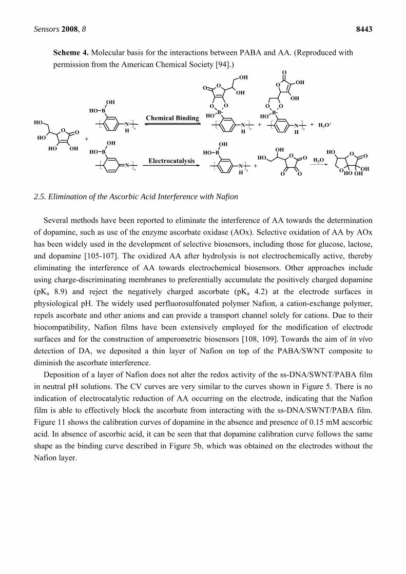
Scheme 4. Molecular basis for the interactions between PABA and AA. (Reproduced with
permission from the American Chemical Society [94].)
O
OH OH
OOH
OH NH
B
OH
OHOH
NH
B-OO
OOOH
OH
OH
NH
B-OO
O
O
OH
OH
H3O+
O
OO
O
OH
OH H2OO O
O OHOHOH
OH
N
B
OH
OH
NH
B
OH
OH
n
n nn
n
+
+
+ +
Electrocatalysis
Chemical Binding
2.5. Elimination of the Ascorbic Acid Interference with Nafion
Several methods have been reported to eliminate the interference of AA towards the determination
of dopamine, such as use of the enzyme ascorbate oxidase (AOx). Selective oxidation of AA by AOx
has been widely used in the development of selective biosensors, including those for glucose, lactose,
and dopamine [105-107]. The oxidized AA after hydrolysis is not electrochemically active, thereby
eliminating the interference of AA towards electrochemical biosensors. Other approaches include
using charge-discriminating membranes to preferentially accumulate the positively charged dopamine
(pKa 8.9) and reject the negatively charged ascorbate (pKa 4.2) at the electrode surfaces in
physiological pH. The widely used perfluorosulfonated polymer Nafion, a cation-exchange polymer,
repels ascorbate and other anions and can provide a transport channel solely for cations. Due to their
biocompatibility, Nafion films have been extensively employed for the modification of electrode
surfaces and for the construction of amperometric biosensors [108, 109]. Towards the aim of in vivo
detection of DA, we deposited a thin layer of Nafion on top of the PABA/SWNT composite to
diminish the ascorbate interference.
Deposition of a layer of Nafion does not alter the redox activity of the ss-DNA/SWNT/PABA film
in neutral pH solutions. The CV curves are very similar to the curves shown in Figure 5. There is no
indication of electrocatalytic reduction of AA occurring on the electrode, indicating that the Nafion
film is able to effectively block the ascorbate from interacting with the ss-DNA/SWNT/PABA film.
Figure 11 shows the calibration curves of dopamine in the absence and presence of 0.15 mM acscorbic
acid. In absence of ascorbic acid, it can be seen that that dopamine calibration curve follows the same
shape as the binding curve described in Figure 5b, which was obtained on the electrodes without the
Nafion layer.
Sensors 2008, 8
8444
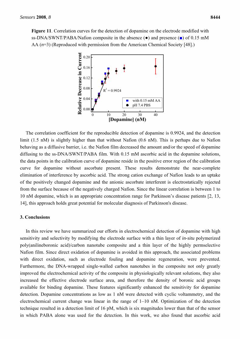
Figure 11. Correlation curves for the detection of dopamine on the electrode modified with
ss-DNA/SWNT/PABA/Nafion composite in the absence (●) and presence (■) of 0.15 mM
AA (n=3) (Reproduced with permission from the American Chemical Society [48].)
0 10 20 30 40
0.00
0.04
0.08
0.12
0.16
0.20
R2 = 0.9924
Rel
ativ
e D
ecre
ase
in C
urre
nt
[Dopamine] (nM)
with 0.15 mM AA pH 7.4 PBS
The correlation coefficient for the reproducible detection of dopamine is 0.9924, and the detection
limit (1.5 nM) is slightly higher than that without Nafion (0.6 nM). This is perhaps due to Nafion
behaving as a diffusive barrier, i.e. the Nafion film decreased the amount and/or the speed of dopamine
diffusing to the ss-DNA/SWNT/PABA film. With 0.15 mM ascorbic acid in the dopamine solutions,
the data points in the calibration curve of dopamine reside in the positive error region of the calibration
curve for dopamine without ascorbate present. These results demonstrate the near-complete
elimination of interference by ascorbic acid. The strong cation exchange of Nafion leads to an uptake
of the positively changed dopamine and the anionic ascorbate interferent is electrostatically rejected
from the surface because of the negatively charged Nafion. Since the linear correlation is between 1 to
10 nM dopamine, which is an appropriate concentration range for Parkinson’s disease patients [2, 13,
14], this approach holds great potential for molecular diagnosis of Parkinson's disease.
3. Conclusions
In this review we have summarized our efforts in electrochemical detection of dopamine with high
sensitivity and selectivity by modifying the electrode surface with a thin layer of in-situ polymerized
poly(anilineboronic acid)/carbon nanotube composite and a thin layer of the highly permselective
Nafion film. Since direct oxidation of dopamine is avoided in this approach, the associated problems
with direct oxidation, such as electrode fouling and dopamine regeneration, were prevented.
Furthermore, the DNA-wrapped single-walled carbon nanotubes in the composite not only greatly
improved the electrochemical activity of the composite in physiologically relevant solutions, they also
increased the effective electrode surface area, and therefore the density of boronic acid groups
available for binding dopamine. These features significantly enhanced the sensitivity for dopamine
detection. Dopamine concentrations as low as 1 nM were detected with cyclic voltammetry, and the
electrochemical current change was linear in the range of 1–10 nM. Optimization of the detection
technique resulted in a detection limit of 16 pM, which is six magnitudes lower than that of the sensor
in which PABA alone was used for the detection. In this work, we also found that ascorbic acid
Sensors 2008, 8
8445
interfered with the detection if Nafion was not deposited on the electrode, which is contrary to
previous reports of nonoxidative PABA-based sensors. We studied the mechanism of interference by
ascorbic acid and the results show that the interference mechanism is very different from the
approaches relying on direct oxidation of dopamine at the electrode. The ascorbic acid was able to
electrocatalytically reduce the fully oxidized polyaniline backbone during the electrochemical
oxidation process. The ascorbic acid was also able to bind to the boronic acid groups through its planar
diol as dopamine does, although its binding affinity is lower. Coating a thin layer of Nafion on top of
the composite eliminated the interference from ascorbic acid. The strong cation exchange of Nafion
leads to an uptake of positively charged DA and the negative charges on the Nafion film
electrostatically rejected the ingress of the negatively charged ascorbate to the PABA/carbon nanotube
composite. The sensitivity of the sensor along with its improved selectivity might allow for its
potential use in the diagnosis of Parkinson's disease. A clear understanding of the dopamine
transduction mechanism and AA interference mechanism in this non-oxidative approach is essential to
eliminate other interferences toward in vivo and in vitro detection of dopamine, which is the long term
goal of our continuing efforts.
Acknowledgements
We acknowledge the donors of the American Chemical Society Petroleum Research Fund and
National Science Foundation under CHE-0750201 for partial support of this research. S.R.A.
acknowledges an Undergraduate Research Fellowship by Rutgers University (2004-2005). Y. B.
acknowledges the American Chemical Society Petroleum Research Fund for the Supplement for
Underrepresented Minority Research (SUMR).
References and Notes
1. Robinson, D.L.; Hermans, A.; Seipel, A.T.; Wightman, R.M. Monitoring Rapid Chemical
Communication in the Brain. Chem. Rev. 2008, 108, 2554-2584.
2. Venton, B.J.; Wightman, R.M. Psychoanalytical Electrochemistry: Dopamine and Behavior.
Anal. Chem. 2003, 75, 414A-421A.
3. Wightman, R.M.; May, L.J.; Michael, A.C. Detection of Dopamine Dynamics in the Brain. Anal.
Chem. 1988, 60, 769A-779A.
4. Troyer, K.P.; Heien, M.L.A.V.; Venton, B.J.; Wightman, R.M. Neurochemistry and
Electroanalytical Probes. Curr. Opin. Chem. Biol. 2002, 6, 696-703.
5. Heien, M.L.A.V.; Khan, A.S.; Ariansen, J.L.; Cheer, J.F.; Phillips, P.E.M.; Wassum, K.M.;
Wightman, R.M. Real-time Measurement of Dopamine Fluctuations After Cocaine in the Brain
of Behaving Rats. Proc. Natl Acad. Sci. USA 2005, 102, 10023-10028.
6. Phillips, P.E.M.; Stuber, G.D.; Heien, M.L.A.V.; Wightman, R.M.; Carelli, R.M. Subsecond
Dopamine Release Promotes Cocaine Seeking. Nature 2003, 422, 614-617.
Sensors 2008, 8
8446
7. Cui, H.F.; Ye, J.S.; Chen, Y.; Chong, S.C.; Sheu, F.S. Microelectrode Array Biochip: Tool for in
vitro Drug Screening Based on the Detection of a Drug Effect on Dopamine Release from PC12
Cells. Anal. Chem. 2006, 78, 6347-6355.
8. Heien, M.L.A.V.; Phillips, P.E.M.; Stuber, G.D.; Seipel, A.T.; Wightman, R.M. Overoxidation of
Carbon-fiber Microelectrodes Enhances Dopamine Adsorption and Increases Sensitivity. Analyst
2003, 128, 1413 - 1419.
9. Yoo, J.-S.; Park, S.-M. Programmed Potential Sweep Voltammetry for Lower Detection Limits.
Anal. Chem. 2005, 77, 3694-3699.
10. Adams, R.N. Probing Brain Chemistry with Electroanalytical Techniques. Anal. Chem. 1976, 48,
1126A-1138A.
11. Venton, B.J.; Troyer, K.P.; Wightman, R.M. Response Times of Carbon Fibre Microelectrodes to
Dynamic Changes in Catecholamine Concentration. Anal. Chem. 2002, 74, 539-546.
12. Troyer, K.P.; Heien, M.L.A.V.; Venton, B.J.; Wightman, R.M. Neurochemistry and
Electroanalytical Probes. Curr. Opin. Chem. Biol. 2002, 6, 696-703.
13. Justice, J.B. Quantitative Microdialysis of Neurotransmitters. J. Neurosci. Meth. 1993, 48, 263-
276.
14. O'Neill, R.D. Microvoltammetric Techniques and Sensors for Monitoring Neurochemical
Dynamics in vivo: A Review. Analyst 1994, 119, 767-779.
15. Cooper, J.M.; Foreman, P.L.; Glidle, A.; Ling, T.W.; Pritchard, D.J. Glutamate Oxidase Enzyme
Electrodes: Microsensors for Neurotransmitter Determination Using Electrochemically
Polymerized Permselective Films. J. Electroanal. Chem. 1995, 388, 143-149.
16. Tse, D.C.S.; McCreery, R.L.; Adams, R.N. Potential Oxidative Pathways of Brain
Catecholaminest. J. Med. Chem. 1976, 19, 37-40.
17. Zhang, Y.; Cai, Y.; Su, S. Determination of Dopamine in the Presence of Ascorbic Acid by
Poly(styrene sulfonic acid) Sodium Salt/Single Walled Carbon Nanotube Film Modified Glassy
Carbon Electrode. Anal. BioChem. 2006, 350, 285-291.
18. Chen, S.M.; Chzo, W.Y. Simultaneous Voltammetric Detection of Dopamine and Ascorbic Acid
Using Didodecyldimethylammonium Bromide (DDAB) Film-Modified Electrodes. J.
Electroanal. Chem. 2006, 587, 226-234.
19. Kang, G.; Lin, X. RNA Modified Electrodes for Simultaneous Determination of Dopamine and
Uric Acid in the Presence of High Amounts of Ascorbic Acid. Electroanal. 2006, 18, 2458-2466.
20. Lin, X.; Li, Y. Simultaneous Electroanalysis of Dopamine, Ascorbic Acid and Uric Acid by Poly
(vinyl alcohol) Covalently Modified Glassy Carbon Electrode. Sens. Actuat. B 2006, 115, 134-
139.
21. Balamurugan, A.; Chen, S.M. Poly(3, 4-ethylenedioxythiophene-co-(5-amino-2-
naphthalenesulfonic acid)) ( PEDOT-PANS) Film Modified Glassy Carbon Electrode for
Selective Detection of Dopamine in the Presence of Ascorbic Acid and Uric Acid. Anal. Chim.
Acta 2007, 596, 92-98.
22. Xiao, Y.; Guo, C.; Li, C.M.; Li, Y.; Zhang, J.; Xue, R.; Zhang, S. Highly Sensitive and Selective
Method to Detect Dopamine in the Presence of Ascorbic Acid by a New Polymeric Composite
Film. Anal. Biochem. 2007, 371, 229-237.
Sensors 2008, 8
8447
23. Yin, T.; Wei, W.; Zeng, J. Selective Detection of Dopamine in the Presence of Ascorbic Acid by
Use of Glassy-Carbon Electrode Modified with both Polyaniline Film and Multi-walled Carbon
Nanotubes with Incorporated cyclodextrin. Anal. Bioanal. Chem. 2006, 386, 2087-2094.
24. Bustos, E.B.; Jimenez, M.G.G.; Diaz-Sanchez, B.R.; Juaristi, E.; Chapman, T.W.; Godinez, L.A.
Glassy Carbon Electrodes Modified with Composites of Starburst-PAMAM Dendrimers
Containing Metal Nanoparticles for Amperometric Detection of Dopamine in Urine. Talanta
2007, 72, 1586-1592.
25. Raoof, J.B.; Ojani, R.; Rashid-Nadimi, S. Voltammetric Determination of Ascorbic Acid and
Dopamine in the Same Sample at the Surface of a Carbon Paste Electrode Modified with
Polypyrrole/Ferrocyanide Films. Electrochim. Acta 2005, 50, 4694-4698.
26. Shahrokhian, S.; Zare-Mehrjardi, H.R. Application of Thionine-Nafion Supported on Multi-
Walled Carbon Nanotube for Preparation of a Modified Electrode in Simultaneous Voltammetric
Detection of Dopamine and Ascorbic Acid. Electrochim. Acta 2007, 52, 6310-6317.
27. Gopalan, A.I.; Lee, K.P.; Manesh, K.M.; Santhosh, P.; Kim, J.H.; Kang, J.S. Electrochemical
Determination of Dopamine and Ascorbic Acid at a Novel Gold Nanoparticles Distributed
Poly(4-aminothiophenol) Modified Electrode. Talanta 2007, 71, 1774-1781.
28. Liu, A.; Honma, I.; Zhou, H. Simultaneous Voltammetric Detection of Dopamine and Uric Acid
at Their Physiological Level in the Presence of Ascorbic Acid Using Poly(acrylic acid)-
Multiwalled Carbon Nanotube Composite-Covered Glassy-Carbon Electrode. Biosens.
Bioelectron. 2007, 23, 74-80.
29. Boo, H.; Jeong, R.A.; Park, S.; Kim, K.S.; An, K.H.; Lee, Y.H.; Han, J.H.; Kim, H.C.; Chung,
T.D. Electrochemical Nanoneedle Biosensor Based on Multiwall Carbon Nanotube. Anal. Chem.
2006, 78, 617-620.
30. Suzuki, A.; Ivandini, T.A.; Yoshimi, K.; Fujishima, A.; Oyama, G.; Nakazato, T.; Hattori, N.;
Kitazawa, S.; Einaga, Y. Fabrication, Characterization, and Application of Boron-Doped
Diamond Microelectrodes for in vivo Dopamine Detection. Anal. Chem. 2007, 79, 8608-8615.
31. Forzani, E.S.; Li, X.L.; Tao, N.J. Hybrid Amperometric and Conductometric Chemical Sensor
Based on Conducting Polymer Nanojunctions. Anal. Chem. 2007, 79, 5217-5224.
32. Heien, M.L.A.V.; Khan, A.S.; Ariansen, J.L.C.; J. F.; Phillips, P.E.M.; Wassum, K.M.;
Wightman, R.M. Real-Time Measurement of Dopamine Fluctuations After Cocaine in the Brain
of Behaving Rats. Proc. Natl. Acad. Sci. 2005, 102, 10023-10028.
33. Heien, M.L.A.V.; Johnson, M.A.; Wightman, R.M. Resolving Neurotransmitter Detected by
Fast-Scan Cyclic Voltammetry. Anal. Chem. 2004, 76, 5697-5704.
34. Lane, R.F.; Hubbard, A.T. Differential Double Pulse Voltammetry at Chemically Modified
Platium Electrodes for in vivo Determination of Catecholamines. Anal. Chem. 1976, 48, 1287-
1293.
35. Bath, B.D.; Martin, H.B.; Wightman, R.M.; Anderson, M.R. Dopamine Adsorption at Surface
Modified Carbon-Fiber Electrodes. Langmuir 2001, 17, 7032-7039.
36. Downard, A.J.; Roddick, A.D.; Bond, A.M. Covalent Modification of Carbon Electrodes for
Voltammetric Differentiation of Dopamine and Ascorbic Acid. Analy. Chim. Acta 1995, 317,
303-310.
Sensors 2008, 8
8448
37. Hermans, A.; Seipel, A.T.; Miller, C.E.; Wightman, R.M. Carbon-Fiber Microelectrodes
Modified iwth 4-Sulfobenzene Have Increased Sensitivity and Selectivity for Catechlamines.
Langmuir 2006, 22, 1964-1969.
38. Howell, J.O.; Kuhr, W.G.; Ensman, R.E.; Wightman, R.M. Background Substraction for Rapid
Scan Voltammetry. J. Electroanal. Chem. 1986, 209, 77-90.
39. Hermans, A.; Keithley, R.B.; Kita, J.M.; Sombers, L.A.; Wightman, R.M. Dopamine Detection
with Fast-Scan Cyclic Voltammetry Used with Analog Background Subtraction. Anal. Chem.
2008, 80, 4040-4048.
40. Beni, V.; Ghita, M.; Arrigan, D.W.M. Cyclic and Pulse Voltammetric Study of Dopamine at the
Interface between Two Immiscible Electrolyte Solutions. Biosens. Bioelectron. 2005, 20, 2097-
2103.
41. Strawbridge, S.M.; Green, S.J.; Tucker, J.H.R. Electrochemical Detection of Catechol and
Dopamine as Their Phenylboronate Ester Derivatives. Chem. Commun. 2000, 2393-2394.
42. Wu, W.; Zhu, H.; Fan, L.; Liu, D.; Renneberga, R.; Yang, S. Sensitive Dopamine Recognition by
Boronic Acid Functionalized Multiwalled Carbon Nanotubes. Chem. Comm. 2007, 2345–2347.
43. Fabre, B.; Taillebois, L. Poly(aniline boronic acid)-based conductimetric sensor of dopamine.
Chem. Commun. 2003, 2982-2983.
44. Zheng, M.; Jagota, A.; Strano, M.S.; Santos, A.P.; Barone, P.; Chou, S.G.; Diner, B.A.;
Dresselhaus, M.S.; Mclean, R.S.; Onoa, G.B.; Samsonidze, G.G.; Semke, E.D.; Usrey, M.;
Walls, D.J. Structure-Based Carbon Nanotube Sorting by Sequence-Dependent DNA Assembly.
Science 2003, 302, 1545-1548.
45. Zheng, M.; Jagota, A.; Semke, E.D.; Diner, B.A.; Mclean, R.S.; Lustig, S.R.; Richardson, R.E.;
Tassi, N.G. DNA-assisted Dispersion and Separation of Carbon Nanotubes. Nat. Mater. 2003, 2,
338-342.
46. Ma, Y.F.; Ali, S.R.; Dodoo, A.S.; He, H.X. Enhanced Sensitivity for Biosensors: Multiple
Functions of DNA Wrapped Single Walled Carbon Nanotubes in Self-Doped Polyaniline
Nanocomposites. J. Phys. Chem. B 2006, 110, 16359-16365.
47. Ma, Y.F.; Ali, S.R.; Wang, L.; Chiu, P.L.; Mendelsohn, R.; He, H.X. In-situ Fabrication of A
Water-Soluble, Self-Doped Polyaniline Nanocomposite: the Unique Role of DNA Functionalized
Single-Walled Carbon Nanotubes. J. Am. Chem. Soc. 2006, 128, 12064-12065.
48. Ali, S.R.; Ma, Y.F.; Parajuli, R.R.; Balogan, Y.; Lai, W.Y.-C.; He, H.X. A Non-Oxidative Sensor
Based on a Self-Doped Polyaniline/Carbon Nanotube Composite for Highly Sensitive and
Selective Detection of the Neurotransmitter Dopamine. Anal. Chem. 2007, 79, 2583-2587.
49. Persaud, K.C.; Pelosi, P. Sensor Arrays Using Conducting Polymers for an Artificial Nose.
Kluwer Academic Publishers: Boston, 1992.
50. Ma, Y.F.; Zhang, J.M.; Zhang, G.J.; He, H.X. Polyaniline Nanowires on Si surfaces Fabricated
with DNA Templates. J. Am. Chem. Soc. 2004, 126, 7097-7101.
51. Paul, E.W.; Ricco, A.J.; Wrighton, M.S. Resistance of Polyaniline Films as A Function of
Electrochemical Potential and theFabrication of Polyaniline-Based Microelectronic Devices. J.
Phys. Chem. 1985, 89, 1441-1447.
Sensors 2008, 8
8449
52. Virji, S.; Huang, J.X.; Kaner, R.B.; Weiller, B.H. Polyaniline Nanofiber Gas Sensors:
Examination of Responce Mechanisms. Nano Lett. 2004, 4, 491-496.
53. Sukeerthi, S.; Contractor, A.Q. Molecular Sensors and Sensor Arrays Based on Polyaniline
Microtubules. Anal. Chem. 1999, 71, 2231-2236.
54. Bartlett, P.N.; Simon, E. Poly(aniline)-Poly(acrylate) Composite Films as Modified Electrodes
for the Oxidation of NADH. Phys. Chem. Chem. Phys. 2000, 2, 2599-2606.
55. Bartlett, P.N.; Wang, J.H. Electroactivity, Stability and Application in an Enzyme Switch at pH 7
of Poly(aniline)-Poly(styrenesulfonate) Composite Films. J. Chem. Soc. Faraday Trans. 1996,
92, 4137-4143.
56. Yue, J.; Wang, Z.H.; Cromack, K.R.; Epstein, A.J.; MacDiarmid, A.G. Effect of Sulfonic Acid
Group on Polyaniline Backbone. J. Am. Chem. Soc. 1991, 113, 2665-2671.
57. Chan, H.S.O.; Ho, P.K.H.; Ng, S.C.; Tan, B.T.G.; Tan, K.L. A New Water-Solube, Self-Doping
Conducting Polyaniline from Poly(o-aminobenzylphosphonic acid) and Its Sodium Salts:
Synthesis and Characterization. J. Am. Chem. Soc. 1995, 117, 8517-8523.
58. Wei, X.-L.; Wang, Y.Z.; Long, S.M.; Bobeczko, C.; Epstein, A.J. Synthesis and Physical
Properties of Highly Sulfonated Polyaniline. J. Am. Chem. Soc. 1996, 118, 2545-2555.
59. Deore, B.A.; Hachey, S.; Freund, M.S. Electroactivity of Electrochemically Synthesized
Poly(Aniline Boronic Acid) as a Function of pH: Role of Self-Doping. Chem. Mater. 2004, 16,
1427-1432.
60. Bartlett, P.N.; Astier, Y. Microelectrochemical Enzyme Transistors. Chem. Commun. 2000, 105-
112.
61. Zengin, H.; Zhou, W.; Jin, J.; Czerw, R.; Smith, J.D.W.; Echegoyen, L.; Carroll, D.L.; Foulger,
S.H.; Ballato, J. Carbon Nanotube Doped Polyaniline. Adv. Mater. 2002, 14, 1480-1483.
62. Cochet, M.; Maser, W.K.; Benito, A.M.; Callejas, M.A.; Martínez, M.T.; Benoit, J.-M.;
Schreiber, J.; Chauvet, O. Synthesis of a New Polyaniline/Nanotube Composite: “in-situ"
Polymerisation and Charge Transfer through Site-Selective Interaction. Chem. Comm. 2001,
1450-1451.
63. Chen, G.Z.; Shaffer, M.S.P.; Coleby, D.; Dixon, G.; Zhou, W.; Fray, D.J.; Windle, A.H. Carbon
Nanotube and Polypyrrole Composites: Coating and Doping. Adv. Mater. 2000, 12, 522-526.
64. Li, X.H.; Wu, B.; Huang, J.-E.; Zhang, J.; Liu, Z.F.; Li, H.L. Fabrication and Characterization of
Well-Dispersed Single-Walled Carbon Nanotube/Polyaniline Composites. Carbon 2002, 411,
1670-1673.
65. Lou, X.; Detrembleur, C.; Pagnoulle, C.; Jérôme, R.; Bocharova, V.; Kiriy, A.; Stamm, M.
Surface Modification of Multiwalled Carbon Nanotubes by Poly(2-vinylpyridine): Dispersion,
Selective Deposition, and Decoration of the Nanotubes. Adv. Mater. 2004, 16, 2123-2127.
66. An, K.H.; Jeong, S.Y.; Hwang, H.R.; Lee, Y.H. Enhanced Sensitive of a Gas Sensor
Incorporating Single-Walled Carbon Nanotube-Polypyrrole Nanocomposites. Adv. Mater. 2004,
16, 1005-1009.
67. Zhao, B.; Hu, H.; Haddon, R.C. Synthesis and Properties of a Water-Soluble Single-Wall Carbon
Nanotube-Poly(m-aminobenzene sulfonic acid) Graft Copolymer. Adv. Funct. Mater. 2004, 14,
71-76.
Sensors 2008, 8
8450
68. Nyholm, L.; Peter, L.M. Wide pH Range Microelectrode Study of the Electrochemical Behaviour
of Polyaniline Films in Buffered Solutions. J. Chem. Soc. Faraday Trans. 1994, 90, 149-154.
69. Dinh, H.N.; Ding, J.; Xia, S.J.; Birss, V.I. Multi-technique Study of the Anodic Degradation of
Polyaniline Films. J. Electroanal. Chem. 1998, 459, 45-56.
70. Kobayashi, E.; Yoneyama, H.; Tamura, H. Polyaniline Film-Coated Electrodes as
Electrochromic Display Devices. J. Electroanal. Chem. 1984, 161, 419-423.
71. Orata, D.; Buttry, D.A. Determination of Ion Populations and Solvent Content as Functions of
Redox State and pH in Polyaniline. J. Am. Chem. Soc. 1987, 109, 3574-3581.
72. Zheng, M.; Diner, B.A. Solution Redox Chemistry of Carbon Nanotubes. J. Am. Chem. Soc.
2004, 126, 15490-15494.
73. Napier, M.E.; Hull, D.O.; Thorp, H.H. Electrocatalytic Oxidation of DNA-Wrapped Carbon
Nanotubes. J. Am. Chem. Soc. 2005, 127, 11952-11953.
74. Shoji, E.; Freund, M.S. Potentiometric Saccharide Detection Based on the pKa Change of
Poly(aniline boronic acid). J. Am. Chem. Soc. 2002, 124, 12486-12493.
75. Ma, Y.F.; Chiu, P.L.; Chen, A.M.; Serrano, A.; Ali, S.R.; He, H.X. The Electronic Role of DNA
Functionalized Carbon Nanotubes: Efficacy for in-situ Polymerization of Conducting Polymer
Nanocomposite. J. Am. Chem. Soc. 2008, 130, 7921-7928.
76. Ma, Y.F.; Cheung, W.; Wei, D.; Bogozi, A.; Chiu, P.L.; Wang, L.; Pontoriero, F.; Mendelsohn,
R.; He, H.X. Improved Conductivity of Carbon Nanotube Networks by In Situ Polymerization of
a Thin Skin of Conducting Polymer. ACS Nano 2008, 2, 1197-1204.
77. Wang, J.; Dai, J.H.; Yarlagadda, T. Carbon Nanotube-Conducting Polymer Composite
Nanowires. Langmuir 2005, 21, 9-12.
78. Martin, C.R. Nanomaterials: A Membrane-Based Synthetic Approach. Science 1994, 266, 1961-
1966.
79. Van Dyke, L.S.; Martin, C.R. Electrochemical Investigation of Electronically Conductive
Polymers: Controlling the Supermolecular Structure Allows Charge Transport Rates to Be
Enhanced. Langmuir 1990, 6, 1118-1123.
80. Parthasarathy, R.; Martin, C.R. Template-Synthesized Polyaniline Microtubules. Chem. Mater.
1994, 6, 1627-1632.
81. Liu, W.; Kumar, J.; Tripathy, S.; Senecal, K.J.; Samuelson, L. Enzymatically Synthesized
Conducting Polyaniline. J. Am. Chem. Soc. 1999, 121, 71-78.
82. Sun, Y.; Wilson, S.R.; Schuster, D.I. High Dissolution and Strong Light Emission of Carbon
Nanotubes in Aromatic Amine Solvents. J. Am. Chem. Soc. 2001, 123, 5348-5349.
83. Nicolas, M.; Fabre, B.; Marchand, G.; Simonet, J. New Boronic-Acid- and Boronate-Substituted
Aromatic Compounds as Precursors of Fluoride-Responsive Conjugated Polymer Films. Eur. J.
Org. Chem. 2000, 1703-1710.
84. Pringsheim, E.; Terpetschnig, E.; Piletsky, S.A.; Wolfbeis, O.S. A PANI with Near-Infrared
Optical Response to Saccharides. Adv. Mater. 1999, 11, 865-868.
85. Nicolas, M.; Fabre, B.; Simonet, J. Electrochemical sensing of fluoride and sugars with a boronic
acid-substituted bipyridine Fe(II) complex in solution and attached onto an electrode surface.
Electrochim. Acta 2001, 46, 1179-1190.
Sensors 2008, 8
8451
86. Shoji, E.; Freund, M.S. Potentiometric Saccharide Detection Based on the pKa Changes of
Poly(aniline boronic acid). J. Am. Chem. Soc. 2002, 124, 12486-12493.
87. Shoji, E.; Freund, M.S. Potentiometric Sensors Based on the Inductive Effect on the pKa of
Poly(aniline): A Nonenzymatic Glucose Sensor. J. Am. Chem. Soc. 2001, 123, 3383-3384.
88. He, H.X.; Zhu, J.S.; Tao, N.J.; Amlani, I.; Nagahara, L.A.; Tsui, R. A Conducting Polymer
Nanojunction Switch. J. Am. Chem. Soc. 2001, 123, 7730-7731.
89. Ofer, D.; Crooks, R.M.; Wrighton, M.S. Potential Dependence of the Conductivity of Highly
Oxidized Polythiophenes, Polypyrroles, and Polyanilines: Finite Windows of High Conductivity.
J. Am. Chem. Soc. 1990, 112, 7869.
90. Huang, J.; Wrighton, M.S. Microelectrochemical Multitransistor Devices Based on Electrostatic
Binding of Electroactive Anionic Metal Complexes in Protonated Poly(4-vinylpyridine): Devices
That Can Detect and Distinguish up to Three Species Simultaneously. Anal. Chem. 1993, 65,
2740.
91. Allen, J.B.; Larry, R.F. Electrochemical Methods: Fundamentals and Applications. 2nd Ed.
Wiley, New York, 2001.
92. Arrigan, D.W.M.; Ghita, M.; Beni, V. Selective voltammetric detection of dopamine in the
presence of ascorbate. Chem. Commun. 2004, 732-733.
93. Beni, V.; Ghita, M.; Arrigan, D.W.M. Cyclic and pulse voltammetry study of dopamine at the
interface between two immiscible electrolyte solutions. Biosens. Bioelectron. 2005, 20, 2097-
2103.
94. Ali, S.K.; Parajuli, R.R.; Ma, Y.F.; Balogan, Y.; Lai, W.Y.-C.; He, H.X. The Interference of
Ascorbic Acid in Dopamine Detection by a Non-oxidative Approach. J. Phy. Chem. B, 2007,
111, 12275-12281.
95. Rahman, M.A.; Kwon, N.H.; Won, M.S.; Choe, E.S.; Shim, Y.B. Functionalized Conducting
Polymer as an Enzyme-Immobilizing Substrate: An Amperometric Glutamate Microbiosensor
for in Vivo Measurements. Anal. Chem. 2005, 77, 4854-4860.
96. Granot, E.; Katz, E.; Basnar, B.; Willner, I. Enhanced Bioelectrocatalysis Using Au-
Nanoparticle/Polyaniline Hybrid Systems in Thin Films and Microstructured Rods Assembled on
Electrodes. Chem. Mater. 2005, 17, 4600-4609.
97. Springsteen, G.; Wang, B. Alizarin Red S. as a general reporter for studying the binding of
boronic acids with carbohydrates. Chem. Commun. 2001, 1608-1609.
98. Springsteen, G.; Wang, B. A detailed examination of boronic acid-diol complexation.
Tetrahedron, 2002, 58, 5291-5300.
99. Benesi, H.A.; Hildebrand, J.H. A Spectrophotometric Investigation of the Interaction of Iodine
with Aromatic Hydrocarbons. J. Am. Chem. Soc. 1949, 71, 2703.
100. Connors, K. Binding Constants. Wiley: New York, 1987.
101. James, T.D.; Samankumara Sandanayake, K.R.A.; Iguchi, R.; Shinkai, S. Novel Saccharide-
Photoinduced Electron Transfer Sensors Based on the Interaction of Boronic Acid and Amine. J.
Am. Chem. Soc. 1995, 117, 8982-8987.
102. Dhariwal, K.R.; Hartzell, W.O.; Levine, M. Ascorbic Acid and dehydroascorbic acid
measurements in human plasma and serum. Am. J. Clin. Nutr. 1991, 54, 712-716.
Sensors 2008, 8
8452
103. Huang, J.; Agus, D.B.; Winfree, C.J.; Kiss, S.; Mack, W.J.; McTaggart, R.A.; Choudhri, T.F.;
Kim, L.J.; Mocco, J.; Pinsky, D.J. Fox, W.D.; Israe, R.J.; Boyd, T.A.; Golde, D.W.; Jr., Connolly,
E.S. Dehydroascorbic acid, a blood–brain barrier transportable form of vitamin C, mediates
potent cerebroprotection in experimental stroke. Proc. Natl. Acad. Sci. 2001, 98, 11720–11724.
104. Palmisano, F.; Zambonin, P.G. Ascorbic Acid Interferences in Hydrogen Peroxide Detecting
Biosensors Based on Electrochemically Immobilized Enzymes. Anal. Chem. 1993, 65, 2690-
2692.
105. Anzai, J.-I.; Takeshita, H.; Kobayashi, Y.; Osa, T.; Hoshi, T. Layer-by-layer construction of
Enzyme Multilayers on an Electrode for the Preparation of Glucose and Lactose Sensors:
Elimination of Ascorbate Interference by Means of an Ascorbate Oxidase Monolayer. Anal.
Chem. 1998, 70, 811-817.
106. Lisdat, F.; Wollenberger, U.; Makower, A.; Hortnagl, H.; Pfeiffer, D.; Scheller, F.W.
Catecholamine detection using enzymatic amplification. Biosens. Bioelectron. 1997, 12, 1199-
1211.
107. Rahman, M.A.; Kwon, N.-H.; Won, M.-S.; Choe, E.S.; Shim, Y.-B. Functionalized Conducting
Polymer as an Enzyme-Immobilizing Substrate: An Amperometric Glutamate Microbiosensor
for in Vivo Measurements. Anal. Chem. 2005, 77, 4854-4860.
108. Harrison, J.; Turner, R.F.B.; Baltes, H.P. Characterization of Perfluorosulfonic Acid Polymer
Coated Enzyme Electrodes and a Miniaturized Integrated Potentiostat for Glucose Analysis in
While Blood. Anal. Chem. 1988, 60, 2002-2006.
109. Wang, J.; Tuzhi, P. Selectivity and Sensitivity Improvements at Perfluorinated
Ionomer/Cellulose Acetate Bilayer Electrodes. Anal. Chem. 1986, 58, 3257-3261.
© 2008 by the authors; licensee Molecular Diversity Preservation International, Basel, Switzerland.
This article is an open-access article distributed under the terms and conditions of the Creative
Commons Attribution license (http://creativecommons.org/licenses/by/3.0/).
Related Documents