Laser-activated perfluorocarbon nanodroplets: A new tool for blood brain barrier opening Kristina A. Hallam, Emory University Eleanor M. Donnelly, Georgia Institute of Technology Andrei B. Karpiouk, Georgia Institute of Technology Robin K. Hartman, Georgia Institute of Technology Stanislav Emelianov, Emory University Journal Title: Biomedical Optics Express Volume: Volume 9, Number 9 Publisher: Optical Society of America: Open Access Journals | 2018-09-01, Pages 4527-4538 Type of Work: Article | Final Publisher PDF Publisher DOI: 10.1364/BOE.9.004527 Permanent URL: https://pid.emory.edu/ark:/25593/tdmn4 Final published version: http://dx.doi.org/10.1364/BOE.9.004527 Copyright information: © 2018 Optical Society of America. This is an Open Access work distributed under the terms of the Creative Commons Attribution 4.0 International License (https://creativecommons.org/licenses/by/4.0/). Accessed September 16, 2022 11:21 AM EDT

Welcome message from author
This document is posted to help you gain knowledge. Please leave a comment to let me know what you think about it! Share it to your friends and learn new things together.
Transcript

Laser-activated perfluorocarbon nanodroplets: Anew tool for blood brain barrier openingKristina A. Hallam, Emory UniversityEleanor M. Donnelly, Georgia Institute of TechnologyAndrei B. Karpiouk, Georgia Institute of TechnologyRobin K. Hartman, Georgia Institute of TechnologyStanislav Emelianov, Emory University
Journal Title: Biomedical Optics Express
Volume: Volume 9, Number 9
Publisher: Optical Society of America: Open Access Journals | 2018-09-01,Pages 4527-4538
Type of Work: Article | Final Publisher PDF
Publisher DOI: 10.1364/BOE.9.004527
Permanent URL: https://pid.emory.edu/ark:/25593/tdmn4
Final published version: http://dx.doi.org/10.1364/BOE.9.004527
Copyright information:
© 2018 Optical Society of America.
This is an Open Access work distributed under the terms of the CreativeCommons Attribution 4.0 International License(https://creativecommons.org/licenses/by/4.0/).
Accessed September 16, 2022 11:21 AM EDT
Laser-activated perfluorocarbon nanodroplets: a new tool for blood brain barrier opening
KRISTINA A. HALLAM,1,2 ELEANOR M. DONNELLY,2 ANDREI B. KARPIOUK,2 ROBIN K. HARTMAN,2 AND STANISLAV Y. EMELIANOV
1,2,* 1Wallace H. Coulter Department of Biomedical Engineering, Georgia Institute of Technology and Emory University School of Medicine, Atlanta, GA, USA 2School of Electrical and Computer Engineering, Georgia Institute of Technology, Atlanta, GA, USA *[email protected]
Abstract: A major obstacle in the monitoring and treatment of neurological diseases is the blood brain barrier (BBB), a semipermeable barrier that prevents the delivery of many therapeutics and imaging contrast agents to the brain. In this work, we explored the possibility of laser-activated perfluorocarbon nanodroplets (PFCnDs) to open the BBB and deliver agents to the brain tissue. Specifically, near infrared (NIR) dye-loaded PFCnDs comprised of a perfluorocarbon (PFC) core with a boiling point above physiological temperature were repeatedly vaporized and recondensed from liquid droplet to gas bubble under pulsed laser excitation. As a result, this pulse-to-pulse repeated behavior enabled the recurring interaction of PFCnDs with the endothelial lining of the BBB, allowing for a BBB opening and extravasation of dye into the brain tissue. The blood brain barrier opening and delivery of agents to tissue was confirmed on the macro and the molecular level by evaluating Evans Blue staining, ultrasound-guided photoacoustic (USPA) imaging, and histological tissue analysis. The demonstrated PFCnD-assisted pulsed laser method for BBB opening, therefore, represents a tool that has the potential to enable non-invasive, cost-effective, and efficient image-guided delivery of contrast and therapeutic agents to the brain. © 2018 Optical Society of America under the terms of the OSA Open Access Publishing Agreement
OCIS codes: (170.0170) Medical optics and biotechnology; (170.5120) Photoacoustic imaging; (170.7170) Ultrasound.
References and links
1. C. Poon, D. McMahon, and K. Hynynen, “Noninvasive and targeted delivery of therapeutics to the brain using focused ultrasound,” Neuropharmacology 120, 20–37 (2017).
2. M. Aryal, C. D. Arvanitis, P. M. Alexander, and N. McDannold, “Ultrasound-mediated blood-brain barrier disruption for targeted drug delivery in the central nervous system,” Adv. Drug Deliv. Rev. 72, 94–109 (2014).
3. N. Vykhodtseva, N. McDannold, and K. Hynynen, “Progress and problems in the application of focused ultrasound for blood-brain barrier disruption,” Ultrasonics 48(4), 279–296 (2008).
4. T. D. Azad, J. Pan, I. D. Connolly, A. Remington, C. M. Wilson, and G. A. Grant, “Therapeutic strategies toimprove drug delivery across the blood-brain barrier,” Neurosurg. Focus 38(3), E9 (2015).
5. K. Hynynen, N. McDannold, N. Vykhodtseva, and F. A. Jolesz, “Non-invasive opening of BBB by focusedultrasound,” Acta Neurochir. Suppl. (Wien) 86, 555–558 (2003).
6. N. Sheikov, N. McDannold, N. Vykhodtseva, F. Jolesz, and K. Hynynen, “Cellular mechanisms of the blood-brain barrier opening induced by ultrasound in presence of microbubbles,” Ultrasound Med. Biol. 30(7), 979–989 (2004).
7. J. H. Hwang, J. Tu, A. A. Brayman, T. J. Matula, and L. A. Crum, “Correlation between inertial cavitation dose and endothelial cell damage in vivo,” Ultrasound Med. Biol. 32(10), 1611–1619 (2006).
8. Y. S. Tung, F. Vlachos, J. A. Feshitan, M. A. Borden, and E. E. Konofagou, “The mechanism of interactionbetween focused ultrasound and microbubbles in blood-brain barrier opening in mice,” J. Acoust. Soc. Am. 130(5), 3059–3067 (2011).
9. A. Carpentier, M. Canney, A. Vignot, V. Reina, K. Beccaria, C. Horodyckid, C. Karachi, D. Leclercq, C. Lafon, J. Y. Chapelon, L. Capelle, P. Cornu, M. Sanson, K. Hoang-Xuan, J. Y. Delattre, and A. Idbaih, “Clinical trial of blood-brain barrier disruption by pulsed ultrasound,” Sci. Transl. Med. 8(343), 343re2 (2016).
Vol. 9, No. 9 | 1 Sep 2018 | BIOMEDICAL OPTICS EXPRESS 4527
#334739 https://doi.org/10.1364/BOE.9.004527 Journal © 2018 Received 11 Jun 2018; revised 8 Aug 2018; accepted 20 Aug 2018; published 29 Aug 2018
10. N. Lipsman, S. Ironside, R. Alkins, A. Bethune, Y. Huang, J. Perry, A. Sahgal, M. Trudeau, K. Hynynen, and T. Mainprize, “SCDT-51. initial experience of blood-brain barrier opening for chemotherapeutic-drug delivery to brain tumors by MR-guided focused ultrasound,” Neuro-Oncol. 19(6), vi275 (2017).
11. E. Strohm, M. Rui, I. Gorelikov, N. Matsuura, and M. Kolios, “Vaporization of perfluorocarbon droplets using optical irradiation,” Biomed. Opt. Express 2(6), 1432–1442 (2011).
12. P. S. Sheeran and P. A. Dayton, “Phase-change contrast agents for imaging and therapy,” Curr. Pharm. Des. 18(15), 2152–2165 (2012).
13. K. Wilson, K. Homan, and S. Emelianov, “Biomedical photoacoustics beyond thermal expansion using triggered nanodroplet vaporization for contrast-enhanced imaging,” Nat. Commun. 3(1), 618 (2012).
14. C. C. Chen, P. S. Sheeran, S. Y. Wu, O. O. Olumolade, P. A. Dayton, and E. E. Konofagou, “Targeted drug delivery with focused ultrasound-induced blood-brain barrier opening using acoustically-activated nanodroplets,” J. Control. Release 172(3), 795–804 (2013).
15. S. Y. Wu, S. M. Fix, C. B. Arena, C. C. Chen, W. Zheng, O. O. Olumolade, V. Papadopoulou, A. Novell, P. A. Dayton, and E. E. Konofagou, “Focused ultrasound-facilitated brain drug delivery using optimized nanodroplets: vaporization efficiency dictates large molecular delivery,” Phys. Med. Biol. 63(3), 035002 (2018).
16. N. Rapoport, “Drug-loaded perfluorocarbon nanodroplets for ultrasound-mediated drug delivery,” Adv. Exp. Med. Biol. 880, 221–241 (2016).
17. A. Hannah, G. Luke, K. Wilson, K. Homan, and S. Emelianov, “Indocyanine green-loaded photoacoustic nanodroplets: dual contrast nanoconstructs for enhanced photoacoustic and ultrasound imaging,” ACS Nano 8(1), 250–259 (2014).
18. D. Y. Santiesteban, D. S. Dumani, D. Profili, and S. Y. Emelianov, “Copper sulfide perfluorocarbon nanodroplets as clinically relevant photoacoustic/ultrasound imaging agents,” Nano Lett. 17(10), 5984–5989 (2017).
19. G. P. Luke, A. S. Hannah, and S. Y. Emelianov, “Super-resolution ultrasound imaging in vivo with transient laser-activated nanodroplets,” Nano Lett. 16(4), 2556–2559 (2016).
20. C. Y. Lin and W. G. Pitt, “Acoustic droplet vaporization in biology and medicine,” BioMed Res. Int. 2013, 404361 (2013).
21. A. S. Hannah, D. VanderLaan, Y. S. Chen, and S. Y. Emelianov, “Photoacoustic and ultrasound imaging using dual contrast perfluorocarbon nanodroplets triggered by laser pulses at 1064 nm,” Biomed. Opt. Express 5(9), 3042–3052 (2014).
22. H. Yoon, K. A. Hallam, C. Yoon, and S. Y. Emelianov, “Super-resolution imaging with ultrafast ultrasound imaging of optically triggered perfluorohexane nanodroplets,” IEEE Trans. Ultrason. Ferroelectr. Freq. Control 1, 1 (2018).
23. T. Kobus, N. Vykhodtseva, M. Pilatou, Y. Zhang, and N. McDannold, “Safety Validation of Repeated Blood-Brain Barrier Disruption Using Focused Ultrasound,” Ultrasound Med. Biol. 42(2), 481–492 (2016).
24. F.-Y. Yang, W.-M. Fu, R.-S. Yang, H.-C. Liou, K.-H. Kang, and W.-L. Lin, “Quantitative evaluation of focused ultrasound with a contrast agent on blood-brain barrier disruption,” Ultrasound Med. Biol. 33(9), 1421–1427 (2007).
25. N. McDannold, N. Vykhodtseva, and K. Hynynen, “Use of ultrasound pulses combined with Definity for targeted blood-brain barrier disruption: a feasibility study,” Ultrasound Med. Biol. 33(4), 584–590 (2007).
26. J. J. Choi, J. A. Feshitan, B. Baseri, S. Wang, Y. S. Tung, M. A. Borden, and E. E. Konofagou, “Microbubble-size dependence of focused ultrasound-induced blood-brain barrier opening in mice in vivo,” IEEE Trans. Biomed. Eng. 57(1), 145–154 (2010).
27. Y. H. Tan, M. Liu, B. Nolting, J. G. Go, J. Gervay-Hague, and G. Y. Liu, “A nanoengineering approach for investigation and regulation of protein immobilization,” ACS Nano 2(11), 2374–2384 (2008).
28. K. Namdee, M. Carrasco-Teja, M. B. Fish, P. Charoenphol, and O. Eniola-Adefeso, “Effect of variation in hemorheology between human and animal blood on the binding efficacy of vascular-targeted carriers,” Sci. Rep. 5(1), 11631 (2015).
29. A. Roggan, M. Friebel, K. Do Rschel, A. Hahn, and G. Mu Ller, “Optical properties of circulating human blood in the wavelength range 400-2500 nm,” J. Biomed. Opt. 4(1), 36–46 (1999).
30. D. Weber-Adrian, E. Thévenot, M. A. O’Reilly, W. Oakden, M. K. Akens, N. Ellens, K. Markham-Coultes, A. Burgess, J. Finkelstein, A. J. Yee, C. M. Whyne, K. D. Foust, B. K. Kaspar, G. J. Stanisz, R. Chopra, K. Hynynen, and I. Aubert, “Gene delivery to the spinal cord using MRI-guided focused ultrasound,” Gene Ther. 22(7), 568–577 (2015).
31. A. H. Payne, G. W. Hawryluk, Y. Anzai, H. Odéen, M. A. Ostlie, E. C. Reichert, A. J. Stump, S. Minoshima, and D. J. Cross, “Magnetic resonance imaging-guided focused ultrasound to increase localized blood-spinal cord barrier permeability,” Neural Regen. Res. 12(12), 2045–2049 (2017).
32. P. S. Sheeran, N. Matsuura, M. A. Borden, R. Williams, T. O. Matsunaga, P. N. Burns, and P. A. Dayton, “Methods of generating submicrometer phase-shift perfluorocarbon droplets for applications in medical ultrasonography,” IEEE Trans. Ultrason. Ferroelectr. Freq. Control 64(1), 252–263 (2017).
33. M. Seo, R. Williams, and N. Matsuura, “Size reduction of cosolvent-infused microbubbles to form acoustically responsive monodisperse perfluorocarbon nanodroplets,” Lab Chip 15(17), 3581–3590 (2015).
34. A. S. Hannah, G. P. Luke, and S. Y. Emelianov, “Blinking phase-change nanocapsules enable background-free ultrasound imaging,” Theranostics 6(11), 1866–1876 (2016).
Vol. 9, No. 9 | 1 Sep 2018 | BIOMEDICAL OPTICS EXPRESS 4528
35. H. Yoon, S. K. Yarmoska, A. S. Hannah, C. Yoon, K. A. Hallam, and S. Y. Emelianov, “Contrast-enhanced ultrasound imaging in vivo with laser-activated nanodroplets,” Med. Phys. 44(7), 3444–3449 (2017).
1. Introduction
Noninvasive treatment and monitoring of neurological disease is greatly hindered by the blood brain barrier (BBB), which often blocks delivery of therapeutics and contrast agents to the brain tissue [1,2]. To overcome this barrier, many methods of delivering contrast and therapeutics to the brain for disease diagnosis and treatment have been developed and are currently used in the clinic [2–4]. However, the methods developed, such as surgical resection, device implantation, or chemical manipulation of tissue via pharmacological substances, have challenges and pitfalls. These methods can be invasive, and their effects can result in widespread opening of the BBB, which is not always desirable, as this greatly increases the risk of complications such as infection [2,3]. Furthermore, these interventions often do little to prevent the recurrence rate of high mortality diseases, like glioblastoma [4]. This main challenge of overcoming the BBB is difficult, as it prevents the passage of large molecules (>500 Da) from entering into the brain tissue [1,2]. In recent years, focused ultrasound (FUS) has been developed as an alternative method to transiently open BBB by using the interaction between the FUS field and microbubbles [1,3,5–7]. Although the biological mechanisms of this method are not fully understood, it is widely believed that the oscillation or cavitation of microbubbles, which interact with the endothelial lining of the brain vasculature, stretch tight junctions and increase the uptake through mechanotransduction pathways, allowing for larger molecules to pass through the BBB [1,6,8]. Because of its localized effect, BBB opening occurs only where the FUS field and microbubbles are present, and delivery of contrast and therapeutic agents can be targeted to precise locations. FUS mediated delivery of therapeutics to the brain has shown promise, resulting in initial clinical trials [9,10].
Another possible, noninvasive means of opening the BBB is through the use of perfluorocarbon nanodroplets (PFCnDs). PFCnDs are unique particles, that when activated by electromagnetic or acoustic energy, can phase change form liquid to gas [11–13]. They are composed of a liquid core, a shell, and when activated via laser light, an optical trigger [11,13]. PFCnDs, activated by an FUS field and converted to gas microbubble in situ have been investigated to examine their ability to open the blood brain barrier [14,15]. Acoustically activated PFCnDs phase change to microbubbles when exposed to sufficient rarefractional pressure provided by FUS and are capable of providing BBB opening through the phase change and stable cavitation of the produced microbubbles [14]. Because of their similarity to microbubbles in their gaseous state, it was demonstrated that PFCnDs are capable of producing similar BBB opening effects [14,15]. Although acoustic droplet vaporization (ADV) enables BBB opening, it involves the use of below body temperature perfluorocarbon such as perfluorobutane (boiling point −1.7°C) or octafluoropropane (boiling point −36.7°C) which can result in premature, spontaneous droplet vaporization in vivo [15]. These lower boiling point PFCs are used for acoustic droplet vaporization because PFCs with boiling points above body temperature would require potentially unsafe, higher pressure acoustic waves to cause phase change [12]. Thus, there are limitations to the use of ADV as a means to open the BBB.
In general, PFCnDs are multifaceted as they are small in size (i.e. hundreds of nanometers) in their liquid state and are capable of carrying therapeutics or other particles, which are either encapsulated or attached to the droplet shell [16]. More importantly, PFCnDs act as a versatile agent when activated to their gaseous phase via multiple forms of energy including electromagnetic waves such as pulsed laser light [12,13,17]. Specifically, the absorption of laser energy by the droplet’s optical trigger causes localized heating and expansion and produces a photoacoustic pressure wave, with these phenomena together phase changing a PFCnD from liquid to gas [13]. Vaporization under a laser pulse of low and,
Vol. 9, No. 9 | 1 Sep 2018 | BIOMEDICAL OPTICS EXPRESS 4529
therefore, safe fluence produces ultrasound (US) and photoacoustic (PA) contrast, resulting in the development of laser-activated PFCnDs as dynamic US and PA imaging contrast agents [12,13,17–19]. Laser activation of PFCnDs due to the optical trigger located within the droplet allows for implementation of higher boiling point PFCnDs, such as perfluoropentane (boiling point 29°C) or perfluorohexane (boiling point 57°C) [20]. Thus, laser-activated PFCnDs can be synthesized with many different components, using varying combinations of photoabsorbers, perfluorocarbon (PFC) species, as well as PFCnD sizes, creating application-specific PFCnDs [13,17,18,21].
Laser-activated PFCnDs have the potential to aid in the diagnosis and treatment of neurological diseases. Indeed, optically activated PFCnDs are an attractive tool for BBB opening, as they can be selectively and repeatedly vaporized at the particular wavelength at which their optical trigger absorbs the most laser light energy [13,17,18]. To enable successful BBB opening, PFCnD parameters (size, PFC species, and concentration) and laser parameters (fluence and lasing duration) must be selected appropriately in order to achieve BBB opening. In this work, we harness the characteristics of NIR dye-loaded perfluorohexane nanodroplets (PFHnDs) to open the BBB and deliver substances to the tissue.
2. Materials and methods
2.1 Synthesis of laser-activated perfluorohexane nanodroplets (PFHnDs)
Due to its boiling point being above body temperature (37°C), perfluorohexane (FluoroMed, L.P.) was used as the core of the synthesized PFCnDs [20]. In addition to 0.3 mL of perfluorohexane (PFH), 3 mL 1X PBS, 1 mL Zonyl FSO fluorosurfactant (1% v/v, Sigma Aldrich), and 2 mg Epolight 3072 dye (Epolin, Inc.) – a near infrared (NIR) dye with a peak absorption around 1064 nm – were used. All materials were added to a 7 mL scintillation vial and vortexed for 10 seconds (Vortex Mixer, Fisher Scientific). The vial was placed in an ice bath and sonicated using a probe sonicator (Q500, QSonica LLC) for a total of 60 seconds at the lowest sonicator amplitude of 1. The emulsion was then transferred to two, 2 mL tubes and spun in a mini centrifuge (Mini-Spin, Eppendorf) at 400 rcf for 2 min to remove excess dye and size separate PFHnDs. The supernatant containing smaller sized PFHnDs was transferred to another 2 mL tube and spun again at 400 rcf for 4 min. To concentrate the droplets, the supernatant resulting from this second centrifugation step was removed and the pellet was resuspended in 0.5 mL 1X PBS using a water bath sonicator (VWR, 180 W). When preparing larger sized PFHnDs for BBB opening that would enable larger (i.e. micrometer) sized constructs to extravasate, droplets were sonicated and size separated as described above, but the pellet of the first centrifugation step was kept, resuspended in 1 mL 1X PBS, and the supernatant discarded. For in vivo experiments, the droplets were left under UV light for 30 minutes for sterilization. A spectrophotometer (Evolution 220, Thermo Scientific) was used to measure the absorbance of PFHnDs both with and without NIR dye, and the blank droplet spectra was subtracted from the spectra of the PFHnDs containing NIR dye. Droplet zeta potential and size were measured in PBS using a dynamic light scattering (DLS) instrument (Zetasizer Nano ZS, Malvern Instruments Ltd.).
2.2 Preparation of the imaging phantom containing laser-activated PFHnDs
To evaluate droplet vaporization and recondenstation dynamics, a polyacrylamide phantom containing solely PFHnDs was fabricated. The polyacrylamide phantom was prepared by first adding 127 mL nanopure water (Thermo Scientific), 42.5 mL 40% polyacrylamide solution (VWR), and 1.7 mL 10% aqueous ammonium persulfate (Sigma Aldrich) to a Büchner flask and mixing with a magnetic stir bar and stir plate. The solution was degassed by sealing the top of the flask with a rubber stopper, attaching the flask to a vacuum, and sonicating using the water bath sonicator for 10 minutes. The solution was returned to the stir plate and 170 µL PFHnDs were added to the solution followed by 212.5 µL of tetramethylethylenediamine
Vol. 9, No. 9 | 1 Sep 2018 | BIOMEDICAL OPTICS EXPRESS 4530
(Sigma Aldrich). The phantom was poured into a plastic container and allowed to solidify before removal from the mold.
2.3 Imaging and processing of laser-induced PFHnD vaporization and recondensation dynamics
To visualize the phase change dynamics of PFHnDs, the prepared polyacrylamide phantom embedded with PFHnDs was imaged using a Verasonics Vantage 256 ultrasound system (Verasonics Inc.) and an Nd:YAG Phocus laser (10 Hz, 5-7 ns pulse length, Opotek Inc.) operating at 1064 nm and a fluence of 46 mJ/cm2. An array transducer operating at 5 MHz (L11-4v, Verasonics Inc.) was used to capture ultrasound at a frame rate of 500 frames per second. Photoacoustic signal was captured at the rate of 10 Hz (laser PRF). Ultrasound data was collected for six frames after each laser pulse (12 milliseconds total), and pixels containing droplets were identified using compounded harmonic ultrasound difference images, similar to a previously described method [22]. To visualize PFHnD recondensation dynamics over time, linear ultrasound intensity of the identified droplet pixels was plotted over multiple laser pulses.
2.4 In vivo blood brain barrier opening using laser-activated PFHnDs
All animal studies were conducted under the protocol approved by the Institutional Animal Care and Use Committee at Georgia Institute of Technology. Prior to anesthesia, an injection of sustained released buprenorphine (IP, 0.8 mg/kg) was administered to each animal (C57BL/6 mouse). Mice were anesthetized using a combination of isoflurane (2%) and oxygen (0.6 L/min). Mice were positioned in a stereotax in the prone position, on a heating pad (Stoelting Co). Hair from the scalp was removed through shaving and depilatory cream. Proparacaine (0.5%, Henry Schein) was applied to the eyes and a retro-orbital injection of 50 µL 3% w/v of sterile filtered Evans Blue (EB) (Sigma Aldrich) was administered with or without a co-injection of 50 µL PFHnDs (~108 droplets, measured using a Malvern NanoSight NS300), depending on the animal group. The animal groups included mice irradiated with 1064 nm light and co-injected with EB dye and PFHnDs (experimental group, n = 10), mice irradiated with 1064 nm light and injected with EB dye only (i.e., no PFHnDs, control group, n = 3), and mice co-injected with EB dye and PFHnDs but not irradiated (another control group, n = 3). Animals were positioned beneath an unfocused 1.5 mm core diameter optical fiber (0.39 NA, Thorlabs, Inc.) to enable irradiation of the right side of the brain. Irradiation was performed using a Vibrant laser (10 Hz, 5-7 ns pulse length, Opotek Inc.) with a wavelength of 1064 nm and a fluence of 70 mJ/cm2. Irradiation was performed for 60 seconds, falling within a similar time range to microbubble induced FUS BBB opening sonication [3,23]. After laser irradiation, mice were observed for 4 hours, with no gross behavioral damage observed. Animals were then sacrificed and perfused with 1X PBS (pH 6.8) followed by 4% paraformaldehyde (PFA). Heads were removed and post-fixed overnight in 4% PFA solution. After 24 hours, brains were excised and whole brain photographs were taken.
2.5 Ex vivo ultrasound and photoacoustic imaging
Fixed, excised mouse brains were imaged using a photoacoustic and ultrasound imaging system (Vevo LAZR, FUJIFILM VisualSonics Inc.) with a 40 MHz ultrasound and photoacoustic imaging probe (LZ-550, FUJIFILM VisualSonics Inc.). The system’s tunable ND:YAG laser (20 Hz, 5-7 ns pulse length) was operated at a wavelength of 1064 nm and at a fluence of 10-12 mJ/cm2, and specimens were imaged at a USPA frame rate of 5 frames per second. Tissue samples were placed on top of an 8% gelatin base in a container filled with degassed water. Images were acquired by performing coronal and sagittal 3D USPA imaging with a distance step size of 0.1 mm. 3D USPA imaging at 1064 nm was performed both for the experimental and two control groups.
Vol. 9, No. 9 | 1 Sep 2018 | BIOMEDICAL OPTICS EXPRESS 4531
2.6 Histology and immunohistochemistry
After remaining in 4% PFA solution for 24 hours at 4°C, brains were transferred to a solution of 30% sucrose and stored at 4°C for 5 days. Brains were snap frozen, and 20 µm coronal sections were cut using a cryostat (Leica CM 1860, Leica Biosystems). For each brain, 72 sections were cut and analyzed, spanning a total volume of 1.44 mm. Standard hematoxylin (Sigma Aldrich, Gill No.2) and eosin (VWR) (H&E) staining was performed. H&E photomicrographs were captured using the bright field mode of a Zeiss AxioObserver Z1 Microscope. IHC was also performed using DAPI (4',6-diamidino-2-phenylindole, Invitrogen) and a secondary antibody of goat anti-mouse IgG (H + L) tagged with Alexa Fluor 488 (Invitrogen). IHC photomicrographs were captured using a Zeiss Laser Scanning Confocal Microscope 700.
3. Results
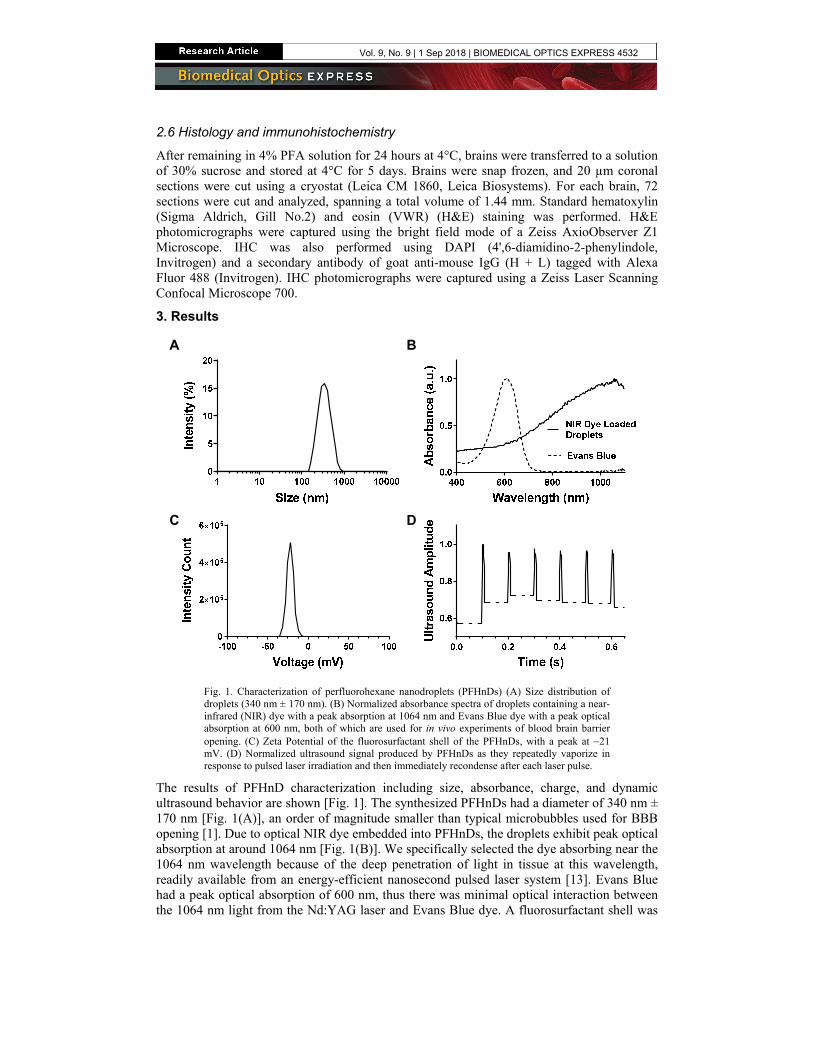
Fig. 1. Characterization of perfluorohexane nanodroplets (PFHnDs) (A) Size distribution of droplets (340 nm ± 170 nm). (B) Normalized absorbance spectra of droplets containing a near-infrared (NIR) dye with a peak absorption at 1064 nm and Evans Blue dye with a peak optical absorption at 600 nm, both of which are used for in vivo experiments of blood brain barrier opening. (C) Zeta Potential of the fluorosurfactant shell of the PFHnDs, with a peak at −21 mV. (D) Normalized ultrasound signal produced by PFHnDs as they repeatedly vaporize in response to pulsed laser irradiation and then immediately recondense after each laser pulse.
The results of PFHnD characterization including size, absorbance, charge, and dynamic ultrasound behavior are shown [Fig. 1]. The synthesized PFHnDs had a diameter of 340 nm ± 170 nm [Fig. 1(A)], an order of magnitude smaller than typical microbubbles used for BBB opening [1]. Due to optical NIR dye embedded into PFHnDs, the droplets exhibit peak optical absorption at around 1064 nm [Fig. 1(B)]. We specifically selected the dye absorbing near the 1064 nm wavelength because of the deep penetration of light in tissue at this wavelength, readily available from an energy-efficient nanosecond pulsed laser system [13]. Evans Blue had a peak optical absorption of 600 nm, thus there was minimal optical interaction between the 1064 nm light from the Nd:YAG laser and Evans Blue dye. A fluorosurfactant shell was
A B
C D
Vol. 9, No. 9 | 1 Sep 2018 | BIOMEDICAL OPTICS EXPRESS 4532
used to encappotential whe
The synthto pulsed lasevia the linear dashed lines PFHnDs quicDuring this pthese particulPFHnD reconrecondensatioHz. As a resmicrobubbles
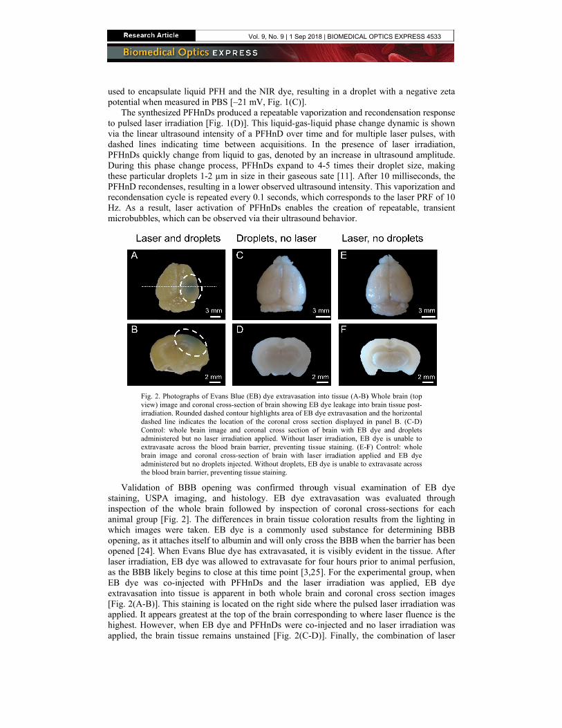
Fig. 2view) irradiadasheContradminextravbrain adminthe bl
Validationstaining, USPinspection ofanimal group which imageopening, as it opened [24]. laser irradiatioas the BBB liEB dye wasextravasation [Fig. 2(A-B)]applied. It apphighest. Howapplied, the b
psulate liquid Pn measured in esized PFHnD
er irradiation [Fultrasound intindicating tim
ckly change frophase change par droplets 1-2
ndenses, resultion cycle is repesult, laser activ, which can be
2. Photographs of image and corona
ation. Rounded dad line indicates th
rol: whole brain inistered but no lasvasate across the
image and coronnistered but no droood brain barrier,
n of BBB opPA imaging, f the whole br
[Fig. 2]. The s were taken. attaches itself When Evans Bon, EB dye waikely begins to co-injected winto tissue is
. This stainingpears greatest ever, when EB
brain tissue rem
PFH and the NPBS [–21 mV
Ds produced a rFig. 1(D)]. Thitensity of a PFme between aom liquid to gaprocess, PFHn2 µm in size ining in a lower oeated every 0.1vation of PFH
e observed via t
Evans Blue (EB) al cross-section ofashed contour highhe location of theimage and coronaser irradiation appblood brain barri
nal cross-section ooplets injected. Wipreventing tissue s
pening was coand histology
rain followed differences inEB dye is a
f to albumin andBlue dye has eas allowed to e
o close at this twith PFHnDs apparent in b
g is located on at the top of th
B dye and PFHmains unstaine
NIR dye, result, Fig. 1(C)].
repeatable vapois liquid-gas-liFHnD over timacquisitions. Inas, denoted by
nDs expand to n their gaseousobserved ultras1 seconds, whicHnDs enables their ultrasoun
dye extravasation f brain showing EBhlights area of EB de coronal cross seal cross section oplied. Without laser, preventing tisof brain with lasithout droplets, EBstaining.
onfirmed throuy. EB dye ex
by inspectionn brain tissue c
commonly ud will only cro
extravasated, it extravasate for ime point [3,2
and the laseboth whole brathe right side whe brain corresHnDs were co-ed [Fig. 2(C-D
ting in a dropl
orization and riquid phase chame and for muln the presenc
y an increase in4-5 times the
s sate [11]. Aftsound intensitych correspondsthe creation o
nd behavior.
n into tissue (A-B)B dye leakage intodye extravasation ection displayed iof brain with EB ser irradiation, EBsue staining. (E-Fer irradiation app
B dye is unable to
ugh visual exxtravasation wn of coronal ccoloration resuused substanceoss the BBB wht is visibly evidr four hours pri5]. For the exper irradiation ain and coronawhere the pulssponding to wh-injected and n
D)]. Finally, th
let with a nega
recondensationange dynamic ltiple laser pulce of laser irrn ultrasound ameir droplet sizefter 10 millisecy. This vaporizs to the laser Pof repeatable,
) Whole brain (topo brain tissue post-and the horizontal
in panel B. (C-D)dye and droplets
B dye is unable toF) Control: wholeplied and EB dyeextravasate across
xamination of was evaluatedcross-sections
ults from the lie for determinhen the barrierdent in the tissior to animal pperimental growas applied,
al cross sectiosed laser irradiahere laser fluenno laser irradiahe combination
ative zeta
response is shown
lses, with radiation, mplitude.
e, making conds, the zation and PRF of 10
transient
p -l ) s o e e s
EB dye d through
for each ighting in ing BBB
r has been sue. After perfusion, oup, when
EB dye n images ation was nce is the ation was n of laser
Vol. 9, No. 9 | 1 Sep 2018 | BIOMEDICAL OPTICS EXPRESS 4533
irradiation anno EB dye exwith laser irra
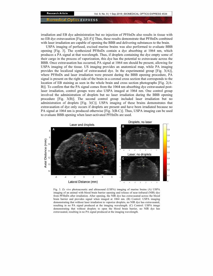
USPA imopening [Figproduces a PAtheir cargo inBBB. Once exUSPA imaginprovides the where PFHnDsignal is preselocation of EBB)]. To confirlaser irradiatiinvolved the procedure [Fadministrationextravasation PA signal at 1to evaluate BB
Fig. 3imagifrom brain demonresultidemonextrav
d EB dye admtravasation [Fi
adiation are capaging of perfu. 3]. The syntA signal at tha
n the process ofxtravasation hang of the tissulocalized sign
Ds and laser ient on the rightB staining as srm that the PAion, control gradministration
Fig. 3(B)]. Tn of droplets of dye only o
1064 nm is proBB opening wh
3. Ex vivo photoang of an animal wPFHnDs after irrabarrier and prov
nstrating that withing in no PA signstrating that wivasated, resulting i
ministration butig. 2(E-F)] Thupable of openinused, excised mthesized PFHn
at wavelength. f vaporization,as occurred, PAue. US imaginnal of extravasrradiation wert side of the braseen in the wh
A signal comes roups were alsn of droplets he second co[Fig. 3(C)]. U
occurs if dropleoduced otherwihen laser-activ
coustic and ultraswith blood brain baadiation. After opevides signal whenhout laser irradiatiognal produced at tthout droplets toin no PA signal pro
t no injection ous, these resultsng the BBB anmurine brains wnDs contain aThus, if drople, this dye has tA signal at 106
ng provides an sated dye. In re present duriain in a corona
hole brain and from the 1064
so USPA imagbut no laser ontrol group USPA imagingets are present ise [Fig. 3(B-C
vated PFHnDs a
sound (USPA) imarrier opening andening, the NIR dy
imaged at 1064 on to vaporize drothe imaging wave
o open the bloododuced at the imag
of PFHnDs alss demonstrate
nd delivering suwas also perfo
a dye absorbinets containing the potential to64 nm should b
n anatomical mthe experimen
ing the BBB oal cross sectioncross section
4 nm absorbingged at 1064 nirradiation durincluded las
g of these brt and have beenC)]. Thus, USPare used.
maging of murine d release of near-inye has extravasated nm. (B) Control
oplets, no NIR dyeelength. (C) Contd brain barrier, nging wavelength.
so results in tisthat PFHnDs c
ubstances to thormed to evalung at 1064 nmthe dye empty
o extravasate abe present, allo
map, while PAntal group [Fiopening proce
n that corresponphotographs [F
g dye extravasanm. One contrring the BBB
ser irradiationrains demonstrn irradiated be
PA imaging can
brains (A) USPAnfrared (NIR) dyed across the bloodl: USPA imaginge has extravasated,trol: USPA imageno NIR dye has
ssue with combined e brain.
uate BBB m, which y some of across the owing for
A imaging ig. 3(A)], edure, PA nds to the Fig. 2(A-ated post-rol group opening but no rates that ecause no n be used
A e d g , e s
Vol. 9, No. 9 | 1 Sep 2018 | BIOMEDICAL OPTICS EXPRESS 4534
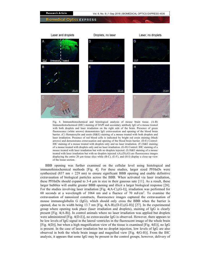
Fig. Immuwith bfluorebarrielaser arrowIHC sof a mmousetreateddisplaof the
BBB opeimmunohistocsynthesized (extravasation these PFHnDlarger bubbleFor the studie60 seconds aextravasation mouse immuopened, due tgroup where present [Fig. were administbe low levels [Fig. 4(D)], bis present. In observed in banalysis, it ap
4. Immunohistocunohistochemical (both droplets and
escence (white arrer. (C) Hematoxyliirradiation. Presen
ws) and demonstratstaining of a mousmouse treated with e treated with lased with laser irradia
aying the entire 20 tissue section.
ening was furchemical meth837 nm ± 229of biological
s should expans will enable ges involving laat a waveleng
of nanosizednoglobulin G to its width beopening took 4(A-B)]. In cotered [Fig. 4(Dof IgG signal iut when a highthe case of las
both the wholeppears that som
chemical and his(IHC) staining of Dd laser irradiationrows) demonstratein and eosin (H&Ence of red blood ctes extravasation ase treated with drodroplets only and
er irradiation but wation but with no d0 µm tissue slice w
rther examinehods [Fig. 4]9 nm) to ensuparticles acro
nd to 3-4 µm igreater BBB oaser irradiationgth of 1064 nmd constructs, fl
(IgG), whicheing 13.7 nm [F
place (laser irontrol animals D-E)], no extravin the lateral vh magnificationser irradiation e brain image
me IgG may be
stological analysiDAPI and seconda
n on the right sides IgG extravasatiE) staining of a mcells is indicated
and opening of theoplets only and nod no laser irradiatiowith no droplets injdroplets injected. (while (B-C), (E-F)
ed on the ce. For these s
ure significant oss the BBB. Win size in theirpening and ill
n [Fig. 4(A-C),m and a fluen
fluorescence im should only Fig. 4(A-B),(Drradiation and where no lasevascular IgG isentricles in then view of the tbut no dropletand magnified
e present in the
is of mouse braary antibody IgG ode of the brain. Pion and opening o
mouse treated with by bright red eos
e blood brain barrio laser irradiation. on. (G-H) Control:jected. (I) H&E st(A),(D),(G) are flu), and (H-I) displa
ellular level ustudies, largerBBB opening
When activater gaseous sate icit a larger bi,(G-I)], irradiance of 70 mJmages capturecross the BB
D-E),(G-H)] [2droplets), sta
er irradiation ws observed. Hoe fluorescent imtissue is examit injection, lowd view [Fig. 4e control group
ain tissue. (A-B)of a mouse treatedPresence of greenof the blood brainboth droplets and
sin staining (blackier. (D-E) Control(F) H&E staining
: IHC staining of ataining of a mouseuorescence imagesay a close-up view
using histologr sized PFHng and enable ded via laser irr[11]. As a res
iological respoation was perfoJ/cm2. To exaed the extravaBB when the b27]. In the expaining of IgG iwas applied butowever, there amage of the whined [Fig. 4(E)w levels of IgG4(G-H)]. Fromps, however, de
) d n n d k : g a e s
w
gical and nDs were definitive radiation,
sult, these onse [26]. ormed for amine the asation of barrier is erimental is clearly t droplets
appears to hole brain )], no IgG G are also
m the IHC elivery of
Vol. 9, No. 9 | 1 Sep 2018 | BIOMEDICAL OPTICS EXPRESS 4535

IgG across the BBB is most obvious in the experimental group containing both laser irradiation and PFHnDs.
Delivery of larger constructs, specifically red blood cells (RBCs), was also examined via H&E staining [Fig. 4(C),(F),(I)]. As seen in the experimental group [Fig. 4(C)], RBCs extravasated across the blood brain barrier and are highlighted by the tissue area stained red. For both controls, however, no RBCs extravasated [Fig. 4(F),(I)]. Because RBCs were able to extravasate in the experimental group, the result indicates that laser-activated PFHnDs were able to create openings in the BBB that allowed for 6 µm diameter particles to cross the blood brain barrier [28].
4. Discussion
In this work, successful delivery of various sized constructs such as RBCs, Evans Blue (EB) visible dye bound to albumin, NIR dye, and IgG was delivered across the BBB using laser-activated perfluorohexane nanodroplets (PFHnDs). The delivery of these particles was verified and evaluated through different methods including EB dye staining, USPA imaging, and histological tissue analysis. Thus, droplet parameters play a key part in determining BBB opening. Specifically, droplet size, PFC species, and photoabsorbers selected all play a unique role in the effective size and spread of BBB opening and therefore delivery of substances into the tissue. For example, larger sized PFCnDs can cause an increase in BBB opening volume and potentially damage to the tissue. A lower boiling point perfluorocarbon that does not repeatedly vaporize and recondense such as perfluoropentane may not open the BBB to a similar extent as PFH. Further, a lower wavelength photoabsorber could prevent deeper activation of PFCnDs due to reduced light penetration in the tissue. Thus, by selectively choosing the appropriate droplet components, droplets can be used in and tailored to a diverse set of applications, including delivery of contrast agents and therapeutics to the brain. Furthermore, laser parameters also contribute to BBB opening and can also be adjusted based on droplet parameters to achieve the desired volume and location of BBB opening in the brain tissue.
In our reported studies, PFCnD characteristics were chosen to achieve effective BBB opening. Specifically, PFHnDs were synthesized such that upon laser irradiation, the size of resulting microbubbles would be similar to those that have been used in FUS BBB opening [Fig. 1(A)] [3]. A NIR dye absorbing near 1064 nm was used not only for increased depth penetration of light but also to avoid optical absorption by the Evans Blue dye co-injected with PFHnDs. Evans Blue has a peak absorption of 600 nm, and at 1064 nm, EB absorption is negligible [Fig. 1(B)]. Therefore, the negligible interaction of EB and the 1064 nm laser light will not cause BBB opening, and extravasation of the EB across the BBB will only occur when droplets are both present and exposed to laser irradiation. In addition, low EB absorption at 1064 nm indicates that the captured USPA signal is produced from NIR dye delivered to the tissue. The USPA imaging of the NIR dye in comparison to Evans Blue photographs also suggests that different sized particles are able to cross the BBB using laser-activated PFHnD opening [Fig. 3(A)].
Furthermore, the PFC species used in these droplets plays a key role in successful BBB opening. The ability of PFHnDs to repeatedly vaporize under pulsed laser excitation and then recondense potentially makes a PFH droplet core more attractive than lower boiling point PFCs such as perfluorobutane or perfluoropentane [20]. The reactivation of PFHnDs over multiple laser pulses suggests that in combination with an increased number of laser pulses used, lower concentrations of PFHnDs could be used to open the BBB, reducing the injected PFHnD dose. This concentration comparison may also extend to microbubble concentrations used in FUS BBB opening. Based on the rapid expansion and recondensation of PFHnDs, it is supposed that the phase changing droplets interact with the BBB similar to the way microbubbles do in a FUS field [Fig. 1(D)]. As PFCnDs have a longer circulation time than microbubbles (i.e. hours vs. minutes, respectively), fewer PFHnDs may be needed to achieve
Vol. 9, No. 9 | 1 Sep 2018 | BIOMEDICAL OPTICS EXPRESS 4536
effective BBB opening [16]. As a result, PFC choice plays a key role when designing laser-activated PFCnDs for BBB opening.
To examine BBB opening, EB staining was evaluated grossly and supported the hypothesis that BBB opening would only occur where both laser irradiation and PFHnDs are present [Fig. 2]. Ex vivo USPA imaging was also performed to co-register PA signal with the area of EB staining, both localized to the right side of the brain [Fig. 3]. USPA contrast from delivered NIR dye indicates the potential for future in vivo USPA imaging of laser-activated PFCnDs during BBB opening and delivery of PA contrast agents. Finally, histological staining analysis was completed to examine the effect of laser-activated PFHnD opening on the molecular level. Overall, extravasation was greatest in the experimental group [Fig. 4(A-C)], further demonstrating that BBB opening can be achieved using laser-activated PFHnDs. However, in both control groups [Fig. 4(D-E),(G-H)], IgG signal is present. For the control group where PFHnDs were injected but no laser irradiation was applied, IgG signal was visible in the lateral ventricles but not in a magnified view of the brain tissue, suggesting that the signal most likely corresponds to an incomplete perfusion. In the control group with laser irradiation and Evans Blue dye but no PFHnDs administered, there are a few possibilities as to why IgG may be present. Both deoxygenated hemoglobin and oxygenated hemoglobin absorb in the NIR wavelength range and as a result, interaction of the blood with laser energy could cause small openings in the BBB that would allow for IgG to cross the BBB [29]. Additionally, incomplete perfusion of the tissue could also cause a false positive fluorescence in the sample, as there appears to be IgG signal coming from the lateral ventricles [Fig. 4(G)] in addition to fluorescence signal seen in the magnified view of the pictomicrograph [Fig. 4(H)]. Despite the fluorescence signal seen in both control samples, IgG signal is greatest in the experimental group showing that localized BBB opening is effective when PFHnDs are irradiated via pulsed laser excitation. Overall, the results of the methods used to evaluate BBB opening (i.e., EB staining, USPA imaging, and histological tissue analysis), support the hypothesis that laser-activated PFCnDs are capable of opening the BBB.
Not only can droplets cause opening of this particular biological barrier, but they also have the potential to be implemented to open other biological barriers that may be inhibited otherwise and are preventing effective, non-invasive treatment. In particular, another barrier that poses challenges in noninvasive delivery is the blood spinal cord barrier (BSCB) [30]. Recently, the BSCB has been opened using FUS and microbubbles, so investigation into the ability of PFCnDs to open the BSCB is warranted [30,31]. For opening of both the BBB and BSCB, laser and droplet parameters should be evaluated for safety and efficacy, as opening of these barriers with parameters not fully optimized has the potential to damage tissue.
In addition, droplets can be synthesized to as small as 100-200 nm and, therefore, have the potential to extravasate themselves when the BBB has been opened [32,33]. Thus, through the administration of one set of small, cargo carrying droplets and a set of larger droplets used for BBB opening, delivery of substances can be contained within cargo carrying PFCnDs until they have reached their desired location. Because droplets can be produced in various sizes and compositions, they are capable of performing as a multiplexed system, enabling subpopulations of droplets to perform different tasks. This multiplexed ability can be harnessed not only via size, but by also using photoabsorbers of different peak wavelengths, different core perfluorocarbons, and different shell compositions.
Consequently, laser-activated droplet adaptability enables a platform for opening of the BBB and delivery of both therapeutics as well as imaging contrast agents to the brain. Furthermore, laser-activated droplets produce localized, temporally modulated ultrasound and photoacoustic contrast and therefore in the future could provide the opportunity for image-guidance of BBB opening as well as image-guidance of delivery of cargo to the tissue [18,19,22,34,35]. Because the process of optimized BBB opening is transient and reversible, localized delivery of cargo to the brain is reproducible, enabling the potential to treat or image longitudinally. From opening barriers to providing delivery and imaging contrast, the
Vol. 9, No. 9 | 1 Sep 2018 | BIOMEDICAL OPTICS EXPRESS 4537
presented studies provide an initial demonstration of the capabilities and potential of laser-activated PFCnDs in neurological applications.
5. Conclusion
This work demonstrated the ability of laser-activated perfluorocarbon nanodroplets (PFCnDs) to open the blood brain barrier (BBB). BBB opening was evaluated using ultrasound-guided photoacoustic imaging techniques, examining Evans Blue dye extravasation, and performing histology and immunohistochemistry. The results indicate that laser-activated PFCnDs are capable of providing a localized, noninvasive BBB opening as well as delivery of agents across the barrier. These initial studies provide the foundation for future work involving the harnessing of laser-activated PFCnDs for use in BBB opening, imaging, and delivery. Overall, laser-activated PFCnDs have the potential to become a versatile tool for neurological applications.
Funding
National Institutes of Health (NIH) (NS102860)
Acknowledgements
The authors would like to thank Heechul Yoon of Georgia Institute of Technology for his help with the acquisition setup for the dynamic ultrasound behavior of the droplets. We also wish to acknowledge the core facilities at the Parker H. Petit Institute for Bioengineering and Bioscience at the Georgia Institute of Technology for the use of their shared equipment, services, and expertise.
Disclosures
The authors declare that there are no conflicts of interest related to this article.
Vol. 9, No. 9 | 1 Sep 2018 | BIOMEDICAL OPTICS EXPRESS 4538
Related Documents