A new species of Stephos T.Scott, 1892 (Copepoda: Calanoida) from coastal waters of Sicily, Italy G.Zagami, M.Campolmi and G.Costanzo Department of Animal Biology and Marine Ecology, University of Messina, 98166, Messina-S.Agata-Salita Sperone 31, Italy Abstract. Stephos cryptospinosus sp. nov., a new species of calanoid copepod, is described from Western Mediterranean coastal waters. It differs from other species of the genus in the structure of the fifth legs in both sexes, and in having a small spinous process on the posterolateral margins of the last prosomite in both sexes. Introduction The genus Stephos T.Scott, 1892 currently comprises 23 species, all generally of small body size. They live in shallow coastal waters close to the bottom, where they can be caught by dredges and sledges designed to sample the hyperbenthic community. Occasionally, they occur higher up in the water column and are caught in plankton nets (Fosshagen, 1970). Recently, species of Stephos have been found in anchihaline caves on Atlantic islands (Boxshall et al., 1990) and Mediterranean islands (Riera et al., 1991; Carola and Razouls, 1996). From a biogeographic point of view, the genus Stephos ranges widely throughout the North Atlantic and adjacent waters (where most species have been found), and the Northern Indo-West Pacific. It occurs in temperate, tropical and polar waters. Method The specimens were collected from coastal waters of western Sicily (Stagnone di Marsala—37°529N 12°289E) in April, May and December 1996, and January 1997 (during daytime). Furthermore, three day–night surveys were carried out in July and October 1996 and March 1997; samples were collected every 3 h during 24 h cycles. Samples were obtained using a subsurface plankton tow-net, towed horizontally near the coast, at a depth of about 2–3 m, above a sandy-muddy bottom covered with Cymodocea nodosa (Ucria) Ascherson, Caulerpa prolifera (Forskål) Lamouroux and Posidonia oceanica (Linneo) Delile. The net was conical, with a length of 150 cm and a mouth 40 cm in diameter. The mesh size was 125 μm. For SEM, specimens were washed in clean distilled water, dehydrated through an alcohol series to 100% ethanol and subsequently, critical-point-dried. The specimens were mounted on a stub, coated with carbon, and observed with a SEM LEO S420. A dissected adult female was designated as the holotype. The type series is deposited in the Natural History Museum of London. The registration numbers are: 1999. 1751–1754. The morphological terminology used follows Huys and Boxshall (Huys and Boxshall, 1991). Journal of Plankton Research Vol.22 no.1 pp.15–27, 2000 15 © Oxford University Press 2000 Downloaded from https://academic.oup.com/plankt/article/22/1/15/1549624 by guest on 01 June 2022

Welcome message from author
This document is posted to help you gain knowledge. Please leave a comment to let me know what you think about it! Share it to your friends and learn new things together.
Transcript
A new species of Stephos T.Scott, 1892 (Copepoda: Calanoida)from coastal waters of Sicily, Italy
G.Zagami, M.Campolmi and G.CostanzoDepartment of Animal Biology and Marine Ecology, University of Messina,98166, Messina-S.Agata-Salita Sperone 31, Italy
Abstract. Stephos cryptospinosus sp. nov., a new species of calanoid copepod, is described fromWestern Mediterranean coastal waters. It differs from other species of the genus in the structure ofthe fifth legs in both sexes, and in having a small spinous process on the posterolateral margins of thelast prosomite in both sexes.
Introduction
The genus Stephos T.Scott, 1892 currently comprises 23 species, all generally ofsmall body size. They live in shallow coastal waters close to the bottom, wherethey can be caught by dredges and sledges designed to sample the hyperbenthiccommunity. Occasionally, they occur higher up in the water column and arecaught in plankton nets (Fosshagen, 1970). Recently, species of Stephos havebeen found in anchihaline caves on Atlantic islands (Boxshall et al., 1990) andMediterranean islands (Riera et al., 1991; Carola and Razouls, 1996). From abiogeographic point of view, the genus Stephos ranges widely throughout theNorth Atlantic and adjacent waters (where most species have been found), andthe Northern Indo-West Pacific. It occurs in temperate, tropical and polar waters.
Method
The specimens were collected from coastal waters of western Sicily (Stagnone diMarsala—37°529N 12°289E) in April, May and December 1996, and January 1997(during daytime). Furthermore, three day–night surveys were carried out in July andOctober 1996 and March 1997; samples were collected every 3 h during 24 h cycles.
Samples were obtained using a subsurface plankton tow-net, towed horizontallynear the coast, at a depth of about 2–3 m, above a sandy-muddy bottom coveredwith Cymodocea nodosa (Ucria) Ascherson, Caulerpa prolifera (Forskål)Lamouroux and Posidonia oceanica (Linneo) Delile. The net was conical, with alength of 150 cm and a mouth 40 cm in diameter. The mesh size was 125 µm.
For SEM, specimens were washed in clean distilled water, dehydrated throughan alcohol series to 100% ethanol and subsequently, critical-point-dried. Thespecimens were mounted on a stub, coated with carbon, and observed with a SEMLEO S420.
A dissected adult female was designated as the holotype. The type series isdeposited in the Natural History Museum of London. The registration numbersare: 1999. 1751–1754.
The morphological terminology used follows Huys and Boxshall (Huys andBoxshall, 1991).
Journal of Plankton Research Vol.22 no.1 pp.15–27, 2000
15© Oxford University Press 2000
Dow
nloaded from https://academ
ic.oup.com/plankt/article/22/1/15/1549624 by guest on 01 June 2022
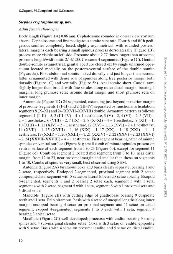
Stephos cryptospinosus sp. nov.
Adult female (holotype)
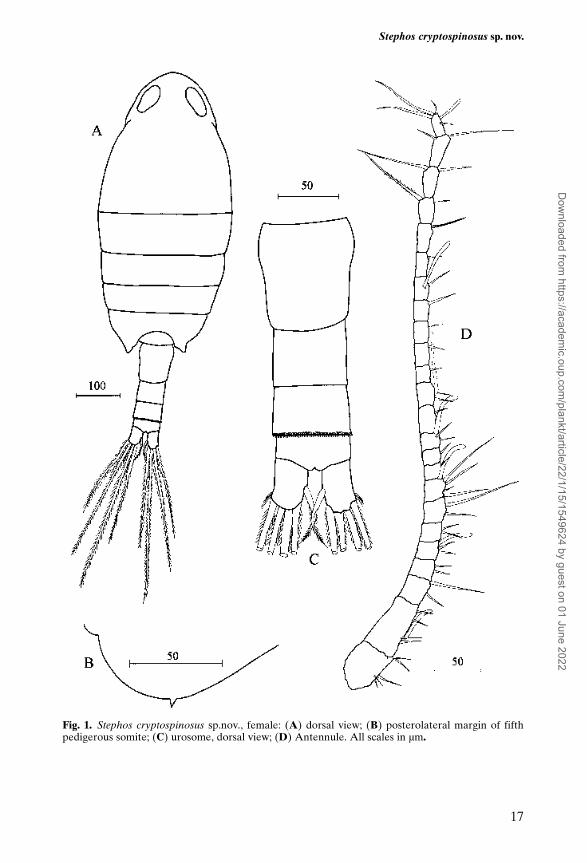
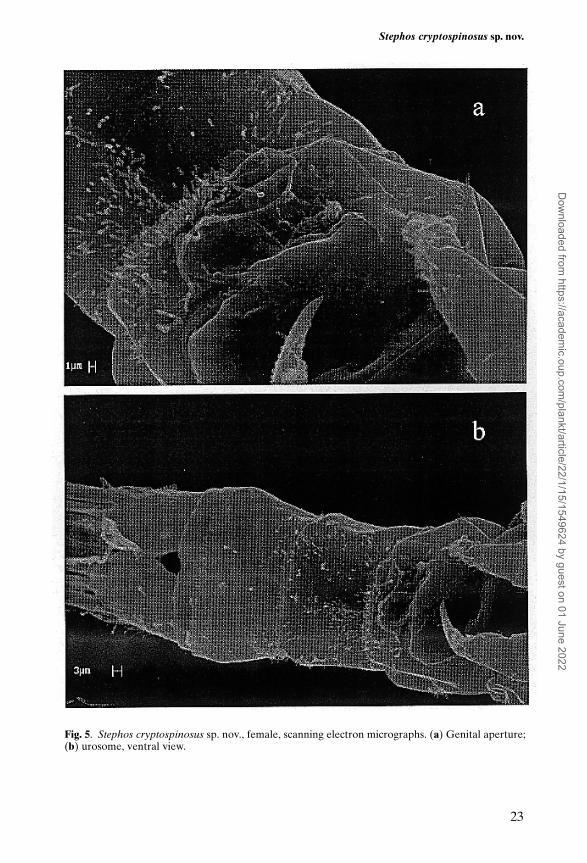
Body length (Figure 1A) 0.86 mm. Cephalosome rounded in dorsal view; rostrumabsent. Cephalosome and first pedigerous somite separate. Fourth and fifth pedi-gerous somites completely fused, slightly asymmetrical, with rounded postero-lateral margins each bearing a small spinous process dorsolaterally (Figure 1B);process more visible on left side. Prosome about 2.77 times longer than urosome;prosome length/width ratio 2.14:1.00. Urosome 4-segmented (Figure 1C). Genitaldouble-somite symmetrical; genital aperture closed off by single unarmed oper-culum located medially on the postero-ventral surface of the double somite(Figure 5a). First abdominal somite naked dorsally and just longer than second;latter ornamented with dense row of spinules along free posterior margin bothdorsally (Figure 1C) and ventrally (Figure 5b). Anal somite short. Caudal ramislightly longer than broad, with fine setules along outer distal margin, bearing 4marginal long plumose setae around distal margin and short plumose seta oninner margin.
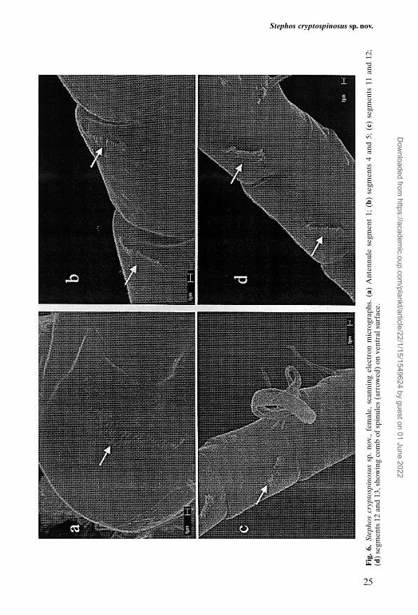
Antennule (Figure 1D) 24-segmented, extending just beyond posterior marginof prosome. Segments 1 (I–II) and 2 (III–IV) separated by functional articulation;segments 8 (X–XI) and 24 (XXVII–XXVIII) double. Armature pattern as follows:segment 1 (I–II) – 5, 2 (III–IV) – 4 + 1 aesthetasc, 3 (V) – 2, 4 (VI) – 2, 5 (VII) –2 + 1 aesthetasc, 6 (VIII) – 2, 7 (IX) – 2, 8 (X–XI) – 4 + 1 aesthetasc, 9 (XII) – 1,10 (XIII) – 1, 11 (XIV) – 2 + 1 aesthetasc, 12 (XV) – 1, 13 (XVI) – 2 + 1 aesthetasc,14 (XVII) – 1, 15 (XVIII) – 1, 16 (XIX) – 1, 17 (XX) – 1, 18 (XXI) – 1 + 1aesthetasc, 19 (XXII) – 1, 20 (XXIII) – 1, 21 (XXIV) – 2, 22 (XXV) – 2, 23 (XXVI)– 2, 24 (XXVII–XXVIII) – 4 + 1 aesthetasc. First segment bearing patch of minutespinules on ventral surface (Figure 6a); small comb of minute spinules present onventral surface of each segment from 1 to 23 (Figure 6b), except for segment 11(Figure 6c). Comb on segment 2 located mid segment; from 3 to 10, near distalmargin; from 12 to 23, near proximal margin and smaller than those on segments1 to 10. Combs of spinules very small, best observed using SEM.
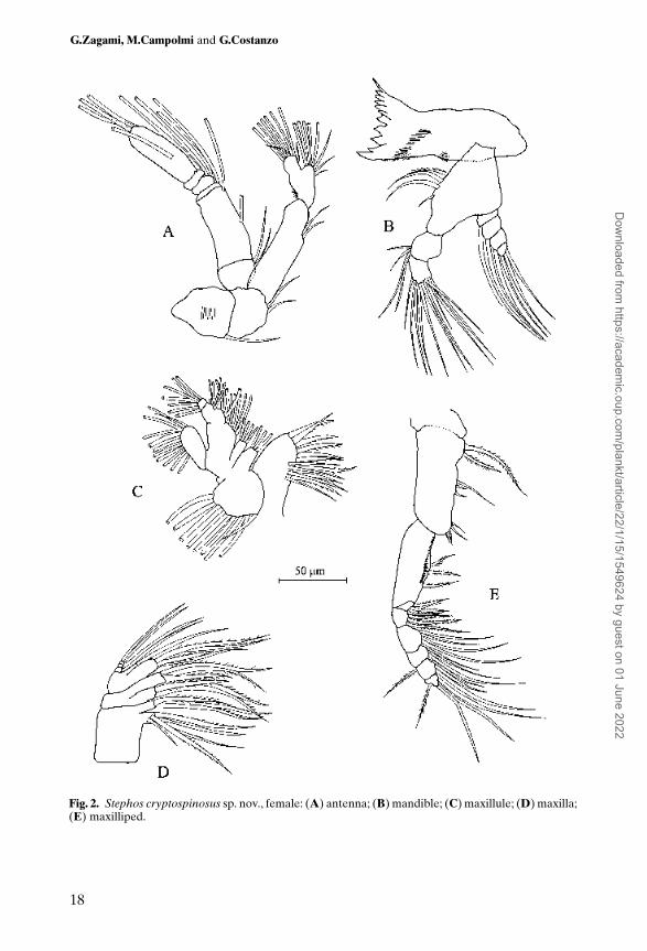
Antenna (Figure 2A) biramous: coxa and basis clearly separate, bearing 1 and2 setae, respectively. Endopod 2-segmented, proximal segment with 2 setae;compound distal segment with 8 setae on lateral lobe and 8 setae apically. Exopod6-segmented, segments 1 and 2 bearing 2 setae each, segment 3 with 1 seta,segment 4 with 2 setae, segment 5 with 1 seta, segment 6 with 1 proximal seta and3 distal setae.
Mandible (Figure 2B) with cutting edge of gnathobase bearing 9 cuspidateteeth and 1 seta. Palp biramous; basis with 4 setae of unequal lengths along innermargin; endopod bearing 4 setae on proximal segment and 11 setae on distalsegment; exopod 4-segmented, segments 1 to 3 each with 1 seta, segment 4bearing 3 apical setae.
Maxillule (Figure 2C) well developed; praecoxa with endite bearing 9 strongspines and 4 sub-marginal slender setae. Coxa with 3 setae on endite; epipoditewith 9 setae. Basis with 4 setae on proximal endite and 5 setae on distal endite.
G.Zagami, M.Campolmi and G.Costanzo
16
Dow
nloaded from https://academ
ic.oup.com/plankt/article/22/1/15/1549624 by guest on 01 June 2022
Stephos cryptospinosus sp. nov.
17
Fig. 1. Stephos cryptospinosus sp.nov., female: (A) dorsal view; (B) posterolateral margin of fifthpedigerous somite; (C) urosome, dorsal view; (D) Antennule. All scales in µm.
Dow
nloaded from https://academ
ic.oup.com/plankt/article/22/1/15/1549624 by guest on 01 June 2022
G.Zagami, M.Campolmi and G.Costanzo
18
Fig. 2. Stephos cryptospinosus sp. nov., female: (A) antenna; (B) mandible; (C) maxillule; (D) maxilla;(E) maxilliped.
Dow
nloaded from https://academ
ic.oup.com/plankt/article/22/1/15/1549624 by guest on 01 June 2022
Proximal endopod segment double, bearing proximal and distal groups of 4 setaeeach on inner margin; apical segment with 7 setae. Exopod bearing 11 plumosesetae along lateral and distal margins.
Maxilla (Figure 2D) with proximal praecoxal endite bearing 1 short and 4 longsetae, distal endite with 3 setae; both coxal endites with 1 stout and 2 slendersetae. Basis and first endopodal segment fused to form allobasis; proximal (basal)endite with 1 stout and 3 slender setae; distal endite (derived from first endopodalsegment) with 1 seta. Free endopod 3-segmented, segments 1 and 2 each bearing1 seta, segment 3 with 2 setae.
Maxilliped (Figure 2E) indistinctly 9-segmented; praecoxa and coxa apparentlyseparate. Coxa bearing groups of 2, 3 and 4 setae along inner margin represent-ing endites. Basis bearing 3 setae on inner margin and 2 setae distally, derivedfrom incorporated first endopodal segment. Basis ornamented with row of shortspinules along inner margin. Free endopod 5-segmented; first and secondsegments each bearing 4 inner marginal setae; third segment with 3 inner setae,fourth segment with 3 inner and 1 outer seta; fifth segment with 4 setae.
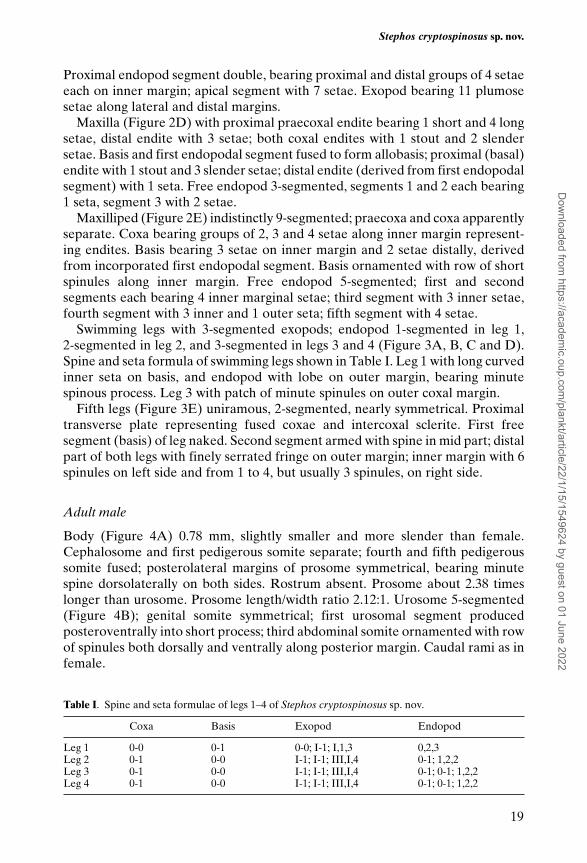
Swimming legs with 3-segmented exopods; endopod 1-segmented in leg 1, 2-segmented in leg 2, and 3-segmented in legs 3 and 4 (Figure 3A, B, C and D).Spine and seta formula of swimming legs shown in Table I. Leg 1 with long curvedinner seta on basis, and endopod with lobe on outer margin, bearing minutespinous process. Leg 3 with patch of minute spinules on outer coxal margin.
Fifth legs (Figure 3E) uniramous, 2-segmented, nearly symmetrical. Proximaltransverse plate representing fused coxae and intercoxal sclerite. First freesegment (basis) of leg naked. Second segment armed with spine in mid part; distalpart of both legs with finely serrated fringe on outer margin; inner margin with 6spinules on left side and from 1 to 4, but usually 3 spinules, on right side.
Adult male
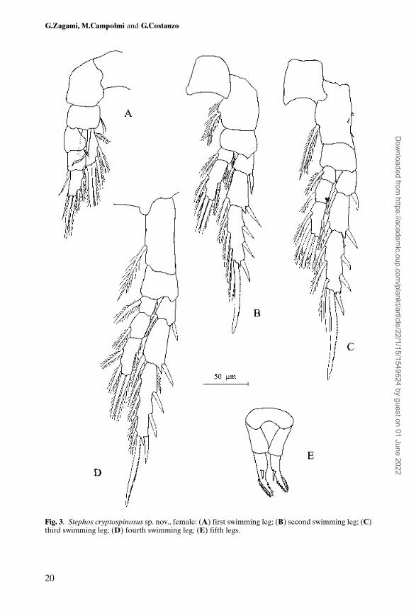
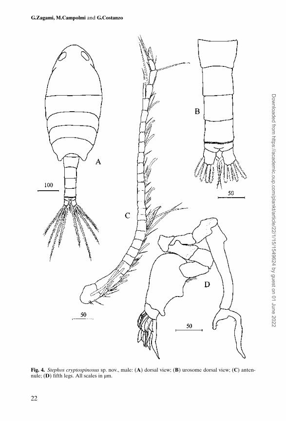
Body (Figure 4A) 0.78 mm, slightly smaller and more slender than female.Cephalosome and first pedigerous somite separate; fourth and fifth pedigeroussomite fused; posterolateral margins of prosome symmetrical, bearing minutespine dorsolaterally on both sides. Rostrum absent. Prosome about 2.38 timeslonger than urosome. Prosome length/width ratio 2.12:1. Urosome 5-segmented(Figure 4B); genital somite symmetrical; first urosomal segment producedposteroventrally into short process; third abdominal somite ornamented with rowof spinules both dorsally and ventrally along posterior margin. Caudal rami as infemale.
Stephos cryptospinosus sp. nov.
19
Table I. Spine and seta formulae of legs 1–4 of Stephos cryptospinosus sp. nov.
Coxa Basis Exopod Endopod
Leg 1 0-0 0-1 0-0; I-1; I,1,3 0,2,3Leg 2 0-1 0-0 I-1; I-1; III,I,4 0-1; 1,2,2Leg 3 0-1 0-0 I-1; I-1; III,I,4 0-1; 0-1; 1,2,2Leg 4 0-1 0-0 I-1; I-1; III,I,4 0-1; 0-1; 1,2,2
Dow
nloaded from https://academ
ic.oup.com/plankt/article/22/1/15/1549624 by guest on 01 June 2022
G.Zagami, M.Campolmi and G.Costanzo
20
Fig. 3. Stephos cryptospinosus sp. nov., female: (A) first swimming leg; (B) second swimming leg; (C)third swimming leg; (D) fourth swimming leg; (E) fifth legs.
Dow
nloaded from https://academ
ic.oup.com/plankt/article/22/1/15/1549624 by guest on 01 June 2022
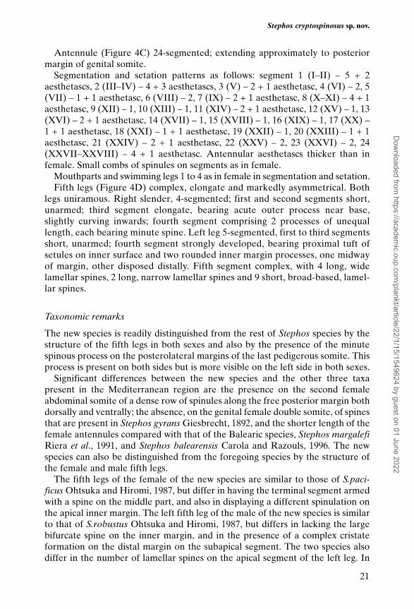
Antennule (Figure 4C) 24-segmented; extending approximately to posteriormargin of genital somite.
Segmentation and setation patterns as follows: segment 1 (I–II) – 5 + 2aesthetascs, 2 (III–IV) – 4 + 3 aesthetascs, 3 (V) – 2 + 1 aesthetasc, 4 (VI) – 2, 5(VII) – 1 + 1 aesthetasc, 6 (VIII) – 2, 7 (IX) – 2 + 1 aesthetasc, 8 (X–XI) – 4 + 1aesthetasc, 9 (XII) – 1, 10 (XIII) – 1, 11 (XIV) – 2 + 1 aesthetasc, 12 (XV) – 1, 13(XVI) – 2 + 1 aesthetasc, 14 (XVII) – 1, 15 (XVIII) – 1, 16 (XIX) – 1, 17 (XX) –1 + 1 aesthetasc, 18 (XXI) – 1 + 1 aesthetasc, 19 (XXII) – 1, 20 (XXIII) – 1 + 1aesthetasc, 21 (XXIV) – 2 + 1 aesthetasc, 22 (XXV) – 2, 23 (XXVI) – 2, 24(XXVII–XXVIII) – 4 + 1 aesthetasc. Antennular aesthetascs thicker than infemale. Small combs of spinules on segments as in female.
Mouthparts and swimming legs 1 to 4 as in female in segmentation and setation.Fifth legs (Figure 4D) complex, elongate and markedly asymmetrical. Both
legs uniramous. Right slender, 4-segmented; first and second segments short,unarmed; third segment elongate, bearing acute outer process near base,slightly curving inwards; fourth segment comprising 2 processes of unequallength, each bearing minute spine. Left leg 5-segmented, first to third segmentsshort, unarmed; fourth segment strongly developed, bearing proximal tuft ofsetules on inner surface and two rounded inner margin processes, one midwayof margin, other disposed distally. Fifth segment complex, with 4 long, widelamellar spines, 2 long, narrow lamellar spines and 9 short, broad-based, lamel-lar spines.
Taxonomic remarks
The new species is readily distinguished from the rest of Stephos species by thestructure of the fifth legs in both sexes and also by the presence of the minutespinous process on the posterolateral margins of the last pedigerous somite. Thisprocess is present on both sides but is more visible on the left side in both sexes.
Significant differences between the new species and the other three taxapresent in the Mediterranean region are the presence on the second femaleabdominal somite of a dense row of spinules along the free posterior margin bothdorsally and ventrally; the absence, on the genital female double somite, of spinesthat are present in Stephos gyrans Giesbrecht, 1892, and the shorter length of thefemale antennules compared with that of the Balearic species, Stephos margalefiRiera et al., 1991, and Stephos balearensis Carola and Razouls, 1996. The newspecies can also be distinguished from the foregoing species by the structure ofthe female and male fifth legs.
The fifth legs of the female of the new species are similar to those of S.paci-ficus Ohtsuka and Hiromi, 1987, but differ in having the terminal segment armedwith a spine on the middle part, and also in displaying a different spinulation onthe apical inner margin. The left fifth leg of the male of the new species is similarto that of S.robustus Ohtsuka and Hiromi, 1987, but differs in lacking the largebifurcate spine on the inner margin, and in the presence of a complex cristateformation on the distal margin on the subapical segment. The two species alsodiffer in the number of lamellar spines on the apical segment of the left leg. In
Stephos cryptospinosus sp. nov.
21
Dow
nloaded from https://academ
ic.oup.com/plankt/article/22/1/15/1549624 by guest on 01 June 2022
G.Zagami, M.Campolmi and G.Costanzo
22
Fig. 4. Stephos cryptospinosus sp. nov., male: (A) dorsal view; (B) urosome dorsal view; (C) anten-nule; (D) fifth legs. All scales in µm.
Dow
nloaded from https://academ
ic.oup.com/plankt/article/22/1/15/1549624 by guest on 01 June 2022
Stephos cryptospinosus sp. nov.
23
Fig. 5. Stephos cryptospinosus sp. nov., female, scanning electron micrographs. (a) Genital aperture;(b) urosome, ventral view.
Dow
nloaded from https://academ
ic.oup.com/plankt/article/22/1/15/1549624 by guest on 01 June 2022
addition, the new species can be distinguished from S.robustus by the armatureof the male antennule. Thus, the antennule segments 1 and 2 of S.robustus carryonly one aesthetasc on the second segment. In S.cryptospinosus sp. nov., theantennule segments 1 and 2 bear, respectively, two and three aesthetascs. Thenew species is further distinguished from the foregoing species by its symmetri-cal female genital double-somite and by the different urosome ornamentation inboth sexes.
In the new species, SEM observations have allowed us to identify the presenceof combs of spinules from antennule segments 1 to 23, with the exception ofsegment 11. This pattern of antennule ornamentation is very similar to that ofS.pacificus and S.robustus (Ohtsuka and Hiromi, 1987), but with some minutedifferences. In S.pacificus, combs of spinules are lacking on antennule segments4 (VI) and 11 (XIV) while in S.robustus, segments 3 (V), 4 (VI), 10 (XIII) and 11(XIV), as well as the ornamental elements of antennule segments 12 (XV) to 23(XXVI), are described as lamellar plates. However, it is possible that (i) the puta-tive lamellar plates of the antennule in the foregoing species could really besmaller combs of spinules glued together by organic detritus (as in Figure 6d),and (ii) the reported lack of combs of spinules on segment 4 (VI) of S.pacificusand on segments 3 (V), 4 (VI) and 10 (XIII) of S.robustus, could be due tomistaken observations on account of their very small size. If this were true, twodifferent lines could be hypothesized in Stephos based on the data on the orna-mentation of the antennule segments available in the literature. The first linewould display combs of spinules from antennule segments 1 (I–II) to 23 (XXVI)with the exception of 11 (XIV), while the second line would present combs ofspinules from antennule segments 12 (XV) to 21 (XXIV), as described forS.canariensis [see (Boxshall et al., 1990)]. These models could indicate phylo-genetic affinities and would have interesting implications for the interpretationof the geographic distribution of the species of Stephos.
Etymology. The specific name cryptospinosus has been chosen because the lastprosomite has a small spinous process hardly visible on both sides.
Discussion
Most S.cryptospinosus were collected during night sampling, suggesting that thishyperbenthic copepod could effect daily vertical migrations. Stephos kurilensis(Kos, 1972) and S.robustus (Ohtsuka and Hiromi, 1987) were recorded fromcoastal waters in plankton samples taken at night.
The upward migratory behaviour is a complex phenomenon related to factorssuch as feeding, reproduction, moulting, dispersal and niche diversification(Alldredge and King, 1980).
The recovery, in addition to S.cryptospinosus, of two other new hyperbenthicspecies (not yet published) belonging to the genera Stephos and Paramisophria,T.Scott, 1897, together with Metacalanus acutioperculum (Ohtsuka, 1984) andRidgewayia marki minorcaensis (Razouls and Carola, 1996), is particularly inter-esting with regard both to their biogeography and evolutionary history
G.Zagami, M.Campolmi and G.Costanzo
24
Dow
nloaded from https://academ
ic.oup.com/plankt/article/22/1/15/1549624 by guest on 01 June 2022
Stephos cryptospinosus sp. nov.
25
Fig
. 6.
Step
hos
cryp
tosp
inos
ussp
. no
v.,
fem
ale,
sca
nnin
g el
ectr
on m
icro
grap
hs.
(a)
Ant
ennu
le s
egm
ent
1; (
b) s
egm
ents
4 a
nd 5
; (c
) se
gmen
ts 1
1 an
d 12
;(d
) se
gmen
ts 1
2 an
d 13
, sho
win
g co
mb
of s
pinu
les
(arr
owed
) on
ven
tral
sur
face
.
Dow
nloaded from https://academ
ic.oup.com/plankt/article/22/1/15/1549624 by guest on 01 June 2022
(Campolmi, 1998; Campolmi et al., 1999). The species S.cryptospinosus, Stephossp. and Paramisophria sp. have morphological characters that link them phylo-genetically with S.pacificus, S.robustus (Ohtsuka and Hiromi, 1987), andParamisophria reducta (Ohtsuka et al., 1993), respectively. The latter two speciesof Stephos, together with M.acutioperculum, are distributed in the Indo-PacificOcean. Ridgewayi marki and P.reducta, on the contrary, are distributed in theWestern and Eastern areas of the Atlantic Ocean, respectively. In addition,R.marki minorcaensis has recently been recorded also from the Western Mediter-ranean (Razouls and Carola, 1996).
The separation and isolation of these species of hyperbenthic copepods as aresult of continental drift, interrupting gene flow between the ancient popu-lations, may have encouraged allopatric speciation. Following separate evolu-tionary lineages, they constitute the current hyperbenthic copepod fauna of theMediterranean.
These observations suggest that the present hyperbenthic copepod populationsof the Mediterranean possess both their own original characters that distinguishthem from the populations of the Atlantic and Indo-Pacific Oceans, and charac-ters that are common to all the populations. The strong geographical isolation ofthe foregoing hyperbenthic species, due to their particular habitat, raises someinteresting questions on their primary habitat and subsequent spread.
Acknowledgements
The authors wish to thank Profs L.Guglielmo and A.Mazzola for their help incarrying out the research program. They are very grateful to Prof. G.A.Boxshallfor the reading and constructive criticism of the manuscript. Further, they wouldlike to thank Dr M.Triscari of the Institute of Earth Sciences, Messina University,for SEM facilities.
References
Alldredge,A.L. and King,J.M. (1980) Effects of moonlight on the vertical migration patterns ofdemersal zooplankton. J. Exp. Mar. Biol. Ecol., 44, 133–156.
Boxshall,G.A., Stock,J.H. and Sánchez,E. (1990) A new species of Stephos Scott, 1892 (Copepoda:Calanoida) from an anchihaline lava pool on Lanzarote, Canary Islands. Stygologia, 5, 33–41.
Campolmi,M. (1998) Studio della comunità zooplanctonica di un bassofondo costiero mediterraneo(Stagnone di Marsala, Sicilia Occidentale). PhD thesis, Sc. Amb., University of Messina.
Campolmi,M., Costanzo,G., Crescenti,N. and Zagami,G. (1999) First record of the hyperbenthicCalanoid Copepod Metacalanus acutioperculum in the Mediterranean Sea. Fourth InternationalCrustacean Congress. Amsterdam: July 20–24, 1998.
Carola,M. and Razouls,C. (1996) Two new species of Calanoida from a marine cave of Minorca island,Mediterranean Sea: Stephos balearensis new species (Stephidae) and Paracyclopia gitana newspecies (Pseudocyclopiidae). Bull. Mar. Sci., 58, 344–352.
Fosshagen,A. (1970) Marine biological investigations in the Bahamas. 12. Stephidae (Copepoda,Calanoida ) from the Bahamas, with remarks on Stephos sinuatus Willey and S.arcticus Sars. Sarsia,41, 37–48.
Huys,R. and Boxshall,G.A. (1991) Copepod Evolution. The Ray Society, London, 468 pp. Kos,M.S. (1972) A new species of Stephos (Copepoda, Calanoida) from coastal waters of the Kuril
Islands. Crustaceana, 23, 113–118.Ohtsuka,S. (1984) Calanoid copepods collected from the near-bottom in Tanabe Bay on the Pacific
coast of the Middle Honshu, Japan. I. Arietellidae. Publ. Seto Mar. Biol. Lab., 29, 359–365.
G.Zagami, M.Campolmi and G.Costanzo
26
Dow
nloaded from https://academ
ic.oup.com/plankt/article/22/1/15/1549624 by guest on 01 June 2022
Ohtsuka,S. and Hiromi,J. (1987) Calanoid copepods collected from the near-bottom in Tanabe Bayon the Pacific coast of the Middle Honshu, Japan. III. Stephidae. Publ. Seto Mar. Biol. Lab., 32,219–232.
Ohtsuka,S., Fosshagen,A. and Iliffe,T.M. (1993) Two new species of Paramisophria (Copepoda,Calanoida, Arietellidae) from anchihaline caves on the Canary and Galápagos Islands. Sarsia, 78,57–67.
Razouls,C. and Carola,M. (1996) The presence of Ridgewayia marki minorcaensis n. ssp. in theWestern Mediterranean. Crustaceana, 69, 47–55.
Riera,T., Vives,F. and Gili,J.M. (1991) Stephos margalefi sp. nov. (Copepoda: Calanoida) from asubmarine cave of Majorca Island (Western Mediterranean). Oecol. Aquatica, 10, 317–323.
Received on January 5, 1999; accepted on 19 July, 1999
Stephos cryptospinosus sp. nov.
27
Dow
nloaded from https://academ
ic.oup.com/plankt/article/22/1/15/1549624 by guest on 01 June 2022
Related Documents