A New Potent Calmodulin Antagonist with Arylalkylamine Structure: Crystallographic, Spectroscopic and Functional Studies Veronika Harmat 1 *, Zsolt Bo ¨ cskei 2 , Ga ´bor Na ´ ray-Szabo ´ 1 , Imre Bata 2 Andrea S. Csutor 2 , Istva ´ n Hermecz 2 , Pe ´ ter Ara ´ nyi 2 , Bea ´ ta Szabo ´ 3 Ka ´ roly Liliom 3 , Bea ´ta G. Ve ´ rtessy 3 and Judit Ova ´di 3 1 Lora ´nd Eo ¨tvo ¨s University Department of Theoretical Chemistry, H-1518, Budapest 112, PO Box 32, Hungary 2 Chinoin Pharmaceuticals H-1325, Budapest PO Box 110, Hungary 3 Institute of Enzymology Biological Research Center Hungarian Academy of Sciences, H-1518, Budapest PO Box 7, Hungary An arylalkylamine-type calmodulin antagonist, N-(3,3-diphenylpropyl)- N 0 -[1-R-(3,4-bis-butoxyphenyl)ethyl]-propylene-diamine (AAA) is pre- sented and its complexes with calmodulin are characterized in solution and in the crystal. Near-UV circular dichroism spectra show that AAA binds to calmodulin with 2:1 stoichiometry in a Ca 2 -dependent manner. The crystal structure with 2:1 stoichiometry is determined to 2.64 A ˚ resol- ution. The binding of AAA causes domain closure of calmodulin similar to that obtained with trifluoperazine. Solution and crystal data indicate that each of the two AAA molecules anchors in the hydrophobic pockets of calmodulin, overlapping with two trifluoperazine sites, i.e. at a hydro- phobic pocket and an interdomain site. The two AAA molecules also interact with each other by hydrophobic forces. A competition enzymatic assay has revealed that AAA inhibits calmodulin-activated phosphodi- esterase activity at two orders of magnitude lower concentration than tri- fluoperazine. The apparent dissociation constant of AAA to calmodulin is 18 nM, which is commensurable with that of target peptides. On the basis of the crystal structure, we propose that the high-affinity binding is mainly due to a favorable entropy term, as the AAA molecule makes multiple contacts in its complex with calmodulin. # 2000 Academic Press Keywords: calmodulin antagonists; fendiline analogue; hydrophobic interactions; X-ray diffraction; circular dichroism spectroscopy *Corresponding author Introduction Calmodulin (CaM), a key player in intracellular Ca 2 -signal transduction, is a small ubiquitous pro- tein central to the regulation of several physiologi- cal processes such as muscle contraction, cell growth, cell division and secretion. CaM modu- lates the activity of several proteins in a Ca 2 - dependent manner (Klee, 1988; Crivici & Ikura, 1995). As an intracellular Ca 2 -receptor, it becomes activated upon the transient elevation of Ca 2 inside the cell (Klee, 1988). CaM consists of two sequentially and structurally highly similar domains with two Ca 2 -binding ‘‘EF-hand’’ motifs on each, and a flexible central region between the two domains (Nakayama & Kretsinger, 1994). The three-dimensional structures of apoCaM and Ca 2 - CaM have been determined by NMR and X-ray crystallography (Zhang et al., 1995; Kuboniwa et al., 1995; Finn et al., 1995; Babu et al., 1985, Kretsinger et al., 1986). Ca 2 -binding to CaM results in solvent exposure of large hydrophobic regions on both domains, which facilitates the binding to target proteins. The Ca 2 -binding domains of the target proteins differ highly in their amino acid sequences; nevertheless, all are expected to form basic amphiphilic helices when complexed with Ca 2 -CaM (O’Neil & DeGrado, 1990). In the three- dimensional structures of Ca 2 -CaM complexed E-mail address of the corresponding author: [email protected] Abbreviations used: AAA, N-(3,3-diphenylpropyl)-N 0 - [1-R-(3,4-bis-butoxyphenyl)ethyl]-propylene diamine; CaM, calmodulin; MANT-cGMP, 2 0 -(N- methylanthraniloyl)guanosine 3 0 ,5 0 -cyclic monophosphate; MLCK, myosin light chain kinase; PDE, 3 0 :5 0 -cyclic-nucleotide 5 0 -nucleotidohydrolase; PEG, polyethylene glycol; TFP, trifluoperazine; W7, N-(6- aminohexyl)-5-chloro-1-naphtalenesulfonamide. doi:10.1006/jmbi.2000.3607 available online at http://www.idealibrary.com on J. Mol. Biol. (2000) 297, 747–755 0022-2836/00/030747–9 $35.00/0 # 2000 Academic Press

Welcome message from author
This document is posted to help you gain knowledge. Please leave a comment to let me know what you think about it! Share it to your friends and learn new things together.
Transcript

doi:10.1006/jmbi.2000.3607 available online at http://www.idealibrary.com on J. Mol. Biol. (2000) 297, 747±755
A New Potent Calmodulin Antagonist withArylalkylamine Structure: Crystallographic,Spectroscopic and Functional Studies
Veronika Harmat1*, Zsolt BoÈ cskei2, GaÂbor NaÂray-Szabo 1, Imre Bata2
Andrea S. Csutor2, IstvaÂn Hermecz2, PeÂter AraÂnyi2, BeaÂta Szabo 3
KaÂroly Liliom3, BeaÂta G. VeÂrtessy3 and Judit OvaÂdi3
1LoraÂnd EoÈtvoÈs UniversityDepartment of TheoreticalChemistry, H-1518, Budapest112, PO Box 32, Hungary2Chinoin PharmaceuticalsH-1325, BudapestPO Box 110, Hungary3Institute of EnzymologyBiological Research CenterHungarian Academy ofSciences, H-1518, BudapestPO Box 7, Hungary
E-mail address of the [email protected]
Abbreviations used: AAA, N-(3,3[1-R-(3,4-bis-butoxyphenyl)ethyl]-prCaM, calmodulin; MANT-cGMP, 20methylanthraniloyl)guanosine 30,50-monophosphate; MLCK, myosin ligPDE, 30:50-cyclic-nucleotide 50-nuclepolyethylene glycol; TFP, tri¯uoperaminohexyl)-5-chloro-1-naphtalenes
0022-2836/00/030747±9 $35.00/0
An arylalkylamine-type calmodulin antagonist, N-(3,3-diphenylpropyl)-N0-[1-R-(3,4-bis-butoxyphenyl)ethyl]-propylene-diamine (AAA) is pre-sented and its complexes with calmodulin are characterized in solutionand in the crystal. Near-UV circular dichroism spectra show that AAAbinds to calmodulin with 2:1 stoichiometry in a Ca2�-dependent manner.The crystal structure with 2:1 stoichiometry is determined to 2.64 AÊ resol-ution. The binding of AAA causes domain closure of calmodulin similarto that obtained with tri¯uoperazine. Solution and crystal data indicatethat each of the two AAA molecules anchors in the hydrophobic pocketsof calmodulin, overlapping with two tri¯uoperazine sites, i.e. at a hydro-phobic pocket and an interdomain site. The two AAA molecules alsointeract with each other by hydrophobic forces. A competition enzymaticassay has revealed that AAA inhibits calmodulin-activated phosphodi-esterase activity at two orders of magnitude lower concentration than tri-¯uoperazine. The apparent dissociation constant of AAA to calmodulin is18 nM, which is commensurable with that of target peptides. On thebasis of the crystal structure, we propose that the high-af®nity binding ismainly due to a favorable entropy term, as the AAA molecule makesmultiple contacts in its complex with calmodulin.
# 2000 Academic Press
Keywords: calmodulin antagonists; fendiline analogue; hydrophobicinteractions; X-ray diffraction; circular dichroism spectroscopy
*Corresponding authorIntroduction
Calmodulin (CaM), a key player in intracellularCa2�-signal transduction, is a small ubiquitous pro-tein central to the regulation of several physiologi-cal processes such as muscle contraction, cellgrowth, cell division and secretion. CaM modu-lates the activity of several proteins in a Ca2�-dependent manner (Klee, 1988; Crivici & Ikura,
ing author:
-diphenylpropyl)-N0-opylene diamine;-(N-cyclicht chain kinase;
otidohydrolase; PEG,azine; W7, N-(6-ulfonamide.
1995). As an intracellular Ca2�-receptor, it becomesactivated upon the transient elevation of Ca2�
inside the cell (Klee, 1988). CaM consists of twosequentially and structurally highly similardomains with two Ca2�-binding ``EF-hand'' motifson each, and a ¯exible central region between thetwo domains (Nakayama & Kretsinger, 1994). Thethree-dimensional structures of apoCaM and Ca2�-CaM have been determined by NMR and X-raycrystallography (Zhang et al., 1995; Kuboniwa et al.,1995; Finn et al., 1995; Babu et al., 1985, Kretsingeret al., 1986). Ca2�-binding to CaM results in solventexposure of large hydrophobic regions on bothdomains, which facilitates the binding to targetproteins. The Ca2�-binding domains of the targetproteins differ highly in their amino acidsequences; nevertheless, all are expected to formbasic amphiphilic helices when complexed withCa2�-CaM (O'Neil & DeGrado, 1990). In the three-dimensional structures of Ca2�-CaM complexed
# 2000 Academic Press

Figure 1. Chemical structure of the arylalkylamineCaM antagonist AAA.
748 Structures of Calmodulin-Arylalkylamine Complexes
with peptides of different target enzymes the pep-tides are wrapped by the two domains of CaM,forming several contacts with the residues of thehydrophobic pockets of both domains of CaM, aswell as polar interactions with the surroundingacidic side-chains (Ikura et al., 1992; Meador et al.,1992, 1993). Beside the ¯exible Met side-chains ofthe hydrophobic pockets, the high ¯exibility of thecentral region also facilitates strong binding ofCaM to its target peptides of various sequences.The degree of domain closure of Ca2�-CaM variesamong these complexes.
Antagonists of CaM differ in both chemicalstructure and mechanism of action (Hait & Lazo,1986; Johnson & Mills, 1986; Forsen et al., 1986).Phenothiazines, arylalkylamines, naphthalenesulfo-namides, calmidazolium and felodipine are widelyused in deciphering calmodulin function(Sobieszek, 1989; Orosz et al., 1988; Tanaka &Hidaka, 1980; Gietzen et al., 1981; Johnson et al.,1986). Non-peptidic antagonists bind with fewexceptions (e.g. calmidazolium with Kd value of3 nM and compound 48/80 with Kd value of<50 nM) much weaker to CaM (Kd values in themicromolar range) than CaM-binding peptides (Kd
values in the nanomolar range; for references, seeO'Neil & DeGrado, 1990). Detailed structural infor-mation of drug binding to CaM is available for aphenothiazine (TFP) and a naphthalenesulfona-mide (W7) drug. For TFP the binding stoichi-ometry has been determined in solution. Theresults range from two to seven drug moleculesper Ca2�-CaM (Klevit et al., 1981; Dalgarno et al.,1984; Marshak et al., 1985; Jackson & Puett, 1986).The crystal structures of Ca2�-CaM-TFP complexesalso present varying stoichiometries (Vandonselaaret al., 1994; Cook et al., 1994; Vertessy et al., 1998).In the case of 1:1 CaM-TFP complex, the drug wasidenti®ed in the C-terminal hydrophobic pocket ofCaM. At the same time in the 1:4 CaM-TFP crystaltwo drug molecules bind to the hydrophobic pock-ets in each domain of CaM (cf. also the earlierNMR studies by Thulin et al., 1984), while twofurther ones bind at interdomain sites (i.e. at sitesformed by TFP1 bound in a hydrophobic pocketand side-chains from both domains). Our recent X-ray data together with the characteristic circulardichroism (CD) spectra in solution revealed thatthe binding of TFP to the C-terminal hydrophobicpocket is followed by the interaction of the seconddrug molecule in the cleft between the CaMdomains. In the CaM-2W7 complex (Osawa et al.,1998) both antagonists bind to the hydrophobicpockets. CaM can also accommodate drugs withdifferent chemical structures simultaneously, e.g.TFP and KAR-2, a new bis-indol derivative, butnot TFP and vinblastine (Vertessy et al., 1998).While the classic antagonists, like TFP or W7, exerta competitive mechanism of action, there are drugsthat bind to CaM but do not prevent the complexformation of target enzymes and CaM. Previouslywe reported that fendiline, recognized originally asa Ca2�-channel blocker, as well as another arylalk-
ylamine derivative, KHL-8430, appeared to act as``functional'' antagonists, which means that theiranti-CaM activities are expressed within the tern-ary enzyme-CaM-drug complexes (Orosz et al.,1988, 1990).
In order to understand how CaM can interactwith antagonists of highly different chemical struc-tures it is highly desirable to determine the struc-tural and functional properties of its complexeswith different types of antagonists. Arylalkylaminederivatives studied so far exhibited antagonistpotency comparable with that of TFP and naphtha-lenesulfonamide derivatives. However, a novelderivative synthesized by Chinoin Pharmaceuticalspossessed its aromatic rings separated by a long,¯exible chain in order to extend its contact surfacesin the complex to both domains on CaM, resul-ting in tighter binding. Here we present thisderivative, N-(3,3-diphenylpropyl)-N0-[1-R-(3,4-bis-butoxyphenyl)ethyl]-propylene-diamine (AAA;Figure 1), with the crystal structure of CaM com-plexed with AAA as well as the characterization ofthe complexes in solution. This combined approachreveals the structural basis for the binding af®nityof AAA. In addition, the structural and functionaleffects of AAA molecules on CaM are comparedwith that of the classical CaM antagonist, TFP.
Results and Discussion
Binding studies in solution
The binding of AAA to CaM in the Ca2�-satu-rated form was studied by near-UV CD spec-troscopy at a protein concentration of 200 mM. Thespectra measured in the absence and presence ofAAA show signi®cant changes around 250-270 nm, as illustrated in Figure 2(a). The presenceof AAA induces characteristic difference peaks at269, 262.5 and 256.5 nm (Figure 2(a), inset), indicat-ing the perturbation of benzyl moieties, whichbelong to AAA and/or phenylalanine residue(s) ofCaM. Since similar signals were detected withmelittin (Maulet & Cox, 1983) as well as with PDEpeptide (Yuan et al., 1999), it is likely that AAA isaccommodated similarly to the peptides. No sig-ni®cant alteration can be seen with apoCaM undersimilar experimental conditions (Figure 2(a), inset),indicating that the CaM-AAA interaction is highlyCa2�-dependent. Differential ellipticity at the threedifference peaks in the presence of Ca2� was

Figure 2. Near-UV CD characterization of AAA-CaMinteraction. (a) Circular dichroism spectra of 200 mMCa2�-CaM (broken line), 400 mM AAA (dotted line) andthat of their mixture (continuous line) in the 250 to350 nm wavelength range. Inset in (a) depicts the differ-ential ellipticity spectra, obtained by subtracting theCaM and AAA signals from the spectrum of the mix-ture, in the case of Ca2�-CaM (continuous line) andapoCaM (broken line). (b) Differential ellipticity at262.5 nm of the Ca2�-CaM-AAA complex as a functionof the AAA concentration, measured with 200 mM CaM.The binding curve was ®tted to the data points byassuming independent binding sites (Vertessy et al.,1998), yielding 1.99(�0.04) bound AAA per CaMmolecule.
Figure 3. AAA counteracts the near-UV differentialellipticity spectrum of the Ca2�-CaM-TFP complex. Cir-cular dichroism spectra were measured in the 250 to350 nm range and the differential ellipticity is displayedfrom 244 nm to 286 nm. Concentrations of CaM andTFP were 20 mM and 75 mM, respectively. The AAAconcentration was varied: 0 (*), 10 mM (*), 25 mM (~),and 75 mM (&). Neither TFP nor AAA have signi®cantCD signals in this wavelength range.
Structures of Calmodulin-Arylalkylamine Complexes 749
followed by varying AAA concentration. A repre-sentative titration curve presented in Figure 2(b)suggests that the association of AAA to CaMoccurs at stoichiometric concentrations as indicatedby the linear portion of the binding curve, whichreaches the saturation at nearly twice the equival-ent ligand concentration. These data, therefore,suggest that there are two well-de®ned AAAbinding sites on CaM.
Previously we demonstrated (Vertessy et al.,1998) that the binding of the ®rst TFP to CaM pro-duces a negative difference CD peak at 263 nm.A further increase in TFP concentration results in ablue shift of the negative peak with the consonantappearance of a positive peak at 270 nm (Vertessyet al., 1998). These observations together with theX-ray data obtained with the CaM-2TFP crystalsuggested that the ®rst TFP was bound to theC-terminal hydrophobic pocket, which wasfollowed by the interaction of the second TFPmolecule with an interdomain site. Therefore, thiswell-characterized system seems to be appropriateto investigate the interference in the binding ofTFP and AAA.
AAA in varying concentrations was added tothe CaM-TFP complex, which formed at 20 mMCaM and 75 mM TFP, and the alteration in thenear-UV CD spectrum was measured (Figure 3). Itshould be noted that CD signals of TFP bindingare in the same wavelength range as those charac-teristic for the CaM-AAA complex (Figure 2(a)),however, the molar extinction coef®cient of CaM-TFP is much higher, thus TFP-induced signals canbe analyzed at a tenfold lower CaM concentrationwhere AAA-induced signals are practically invis-ible. As shown in Figure 3, the addition of substoi-chiometric AAA (10 mM) decreases simultaneouslyboth the negative and positive differential ellipti-city peaks of the CaM-TFP complex. AAA at a con-centration of 25 mM almost completely abolishesboth TFP-induced peaks, suggesting that theassociation of a single AAA molecule can displacetwo molecules of TFP from CaM, which are accom-modated in the C-terminal domain as well as at aninterdomain site (Vertessy et al., 1998). The fact

Figure 4. Functional characterization of the AAA-CaM antagonism in solution. PDE activity wasmeasured ¯uorimetrically with 10 mM MANT-cGMPsubstrate as described in Experimental Procedures.(a) Concentration-dependent inhibition of CaM-activatedPDE activity by AAA (*) and TFP (&). The concen-trations of PDE and CaM were 5 nM and 20 nM,respectively. Continuous lines were obtained by ®ttingthe logistic function (see equation (1)) to the inhibitiondata, yielding IC50 values of 13(�1) nM for AAA and2.1(�0.4) mM for TFP. The pseudo-Hill coef®cients were2.1(�0.2) for AAA and 2.0(�0.5) for TFP. (b) Activationof PDE as a function of CaM concentration in theabsence (*) and presence (*) of AAA. The concen-trations of PDE and AAA were 5 nM and 100 nM,respectively. Continuous lines were obtained by ®ttingthe logistic function (see equation (2)) to the dose-response data. The calculated EC50 values and pseudo-Hill coef®cients in the absence and presence of AAAwere 15(�4) nM and 0.9(�0.1) as well as 61(�7) nM and1.5(�0.2), respectively.
750 Structures of Calmodulin-Arylalkylamine Complexes
that AAA at a near-stoichiometric ratio to CaM isable to diminish the binding of more than a two-fold excess of TFP indicates that AAA binds morestrongly to CaM than does TFP. On the basis ofthese data one can expect a signi®cant anti-CaMeffect of AAA on CaM-modulated physiologicalprocesses.
Functional studies
To test the anti-CaM potency of AAA, a ¯uori-metric PDE assay was selected, since the CaM-induced activation of PDE is well-characterizedand commonly used for testing CaM antagonism.Figure 4(a) shows the concentration-dependentinhibition of CaM-activated PDE by AAA in com-parison to the effect of TFP. In the presence of20 nM CaM, which causes about a twofold acti-vation of 5 nM PDE, AAA monotonouslydecreases the activity of PDE and virtually no CaMactivation can be detected at about 50 nM AAA.The half-maximal inhibitory concentrations ofAAA and TFP occur at 13 and 2090 nM, respect-ively, indicating that the arylalkylamine derivativeis really a powerful anti-CaM agent. The apparentKd value for AAA binding to CaM was estimatedto be 18 nM as an upper limit from the CaM acti-vation curves given in Figure 4(b), which wasdetermined in the absence and presence of 100 nMAAA as suggested by Vogel and co-workers (Yuanet al., 1999). These kinetic results together with thebinding data suggest that AAA may act as com-petitive antagonist of CaM. The fact that the af®-nity of AAA to CaM is much higher than that ofTFP suggests signi®cant differences in the contactsurfaces and/or protein conformation caused bythe association of the two compounds of differentchemical structures, despite their overlapping bind-ing sites.
Crystal packing and overall conformation ofCaM complexed with AAA
In order to localize the AAA binding sites inCa2�-CaM and identify the amino acid residuesinvolved in the protein-AAA interaction, X-raycrystallographic studies were carried out. Crystalsof space group P3221 were grown at a near 1:2CaM/AAA molar ratio. The structure is wellde®ned by the electron density apart from a fewsurface side-chains and the residues of the centrallinker region. Due to the lack of electron density,residues 1, 2, 147 and 148, and the side-chains ofresidues 6, 74-79, 81, 83, 115, 123 and 127 are miss-ing from the ®nal model. The bound AAA mol-ecules are well de®ned by electron density,although the quality of the map is weaker in thelinker region between the two aromatic moieties ofAAA molecules (Figure 5(a)). The overall confor-mation of CaM is very similar to that determinedin the CaM-4TFP complex (RMS distance of thebackbone atoms with the CaM-4TFP complexis 0.662 AÊ ).
Accommodation of AAA molecules in thecrystal structures
In the crystal structure two AAA molecules arebound to CaM. The structural similarity of the twodomains is present in the mode of binding of theantagonist (Figure 5(b) and (c)). This is especially

Figure 5. Structural details ofAAA binding in the crystal struc-ture. (a) Stereo view of (2Fo ÿ Fc)electron density map contoured atthe 1s level around the AAA bind-ing sites. Carbon atoms of theAAAs are colored magenta whileAAAs and methionine residues arelabeled. (b) Comparison of thebinding sites of AAA molecules.AAA1 (left) and AAA2 (right)(lines and circles) are shown withthe interacting protein residues(lines). The orientation of CaM ischanged between the left and rightpanels by ®tting backbone atoms ofthe two domains onto each otherconsidering the similar fold ofthose. The accommodations andconformations of the two AAAs arevery similar. (c) The backbone con-formation of CaM colored from theN-terminal to the C-terminal fromblue to red with the two boundAAA molecules. (d) Comparison of
the P3221 crystal structure (red) with the P1 structure (green). The conformation of AAA changes upon the domainmotion of CaM. AAA2 and the interacting helices of the N-terminal domain are shown with the Ca atoms of residues92 to 144 ®tted together. The molecular surface of the C-terminal hydrophobic pocket of CaM, calculated by GRASP(Nicholls et al., 1991), is shown as a chicken-wire representation (blue). The most important interactions betweenAAA and CaM, shown only for P3221, are emphasized with yellow shaded lines. The black arrow shows the changein conformation of AAA2 following the domain motion of CaM. (a) Drawn with Bobscript v2.3 (Esnouf, 1997) and(b), (c) and (d) with MOLSCRIPT v2.1 (Kraulis, 1991).
Structures of Calmodulin-Arylalkylamine Complexes 751
re¯ected in the positions and conformations of thebiphenyl-methyl moieties, which bind in thehydrophobic pockets. The RMS deviation of theAAA molecules is 0.424 AÊ for the biphenyl-methylgroups and 1.062 AÊ for all non-hydrogen atoms.One phenyl group of the biphenyl-methyl moietiessinks in the hydrophobic pocket forming contactssimilar to those of TFP and W7. AAA1 (bound atthe N-terminal hydrophobic pocket) and AAA2(bound at the C-terminal hydrophobic pocket)interact with the side-chains of residues Leu32/Leu105, Met51/Met124, Ile52/Ile125, Val55/Ala128, Phe68/Phe141 and Met71/Met144, respect-ively. AAA2 is slightly closer to the bottom of thehydrophobic pocket, forming further hydrophobicinteractions with residues Phe92, Ile100 andVal136. The positions of these phenyl groups aresimilar to that of the Trp800 indole ring at theC-terminal hydrophobic pocket of the CaM-smoothmuscle MLCK peptide complex (Meador et al.,1992; plane angle of Trp800 and AAA2 aromaticrings is 18 �) as well as to the W7 naphthalene ringat the N-terminal hydrophobic pocket (Osawa et al.,1998; plane angle of W7 and AAA1 aromatic ringsis 12 �); and overlap with the phenothiazinemoieties of the TFPs bound at intradomain sites(TFP1 and TFP4 of the CaM-4TFP complex;Vandonselaar et al., 1994).
The role of the other phenyl groups of AAAs inthe complex is to stabilize the antagonists in their
bent conformations forming strong intramolecularstacking interactions with the aromatic ring of thebis-butoxyphenyl moieties (closest distances are3.79 AÊ and 3.42 AÊ for AAA1 and AAA2, respect-ively). As illustrated in Figure 5(b), they also helpto anchor the antagonists by interactions with theedge of hydrophobic pockets (AAA1 contacts withPhe19 while AAA2 with Met109).
The bis-butoxyphenyl moieties of AAA alsoform contacts with the other domain of CaMbeside the intramolecular stacking interactions.These contacts are mainly hydrophobic. AAA1interacts with Ala88, while AAA2 with Phe12,Ala15, Val35 and Leu39. Interactions with the edgeof hydrophobic pocket of the other domain canalso be found with Val91, Phe92 and Met145 forAAA1 as well as with Leu18 and Phe19 for AAA2.The bis-butoxyphenyl moiety of AAA2 overlapswith the phenothiazine part of TFP3 in the CaM-4TFP complex. There is also an overlap betweenthe 4-butoxy group of AAA2 and the tri¯uoro-methyl group of TFP2 (interdomain site). Since thebutoxy group is very ¯exible, the real reason forthe dissociation of TFP2, in addition to TFP1, uponbinding of one AAA molecule to CaM in solutioncan be the loss of favorable hydrophobic inter-action of TFP2 with TFP1.
It is noteworthy that although the structuralsimilarity of the two CaM domains is preserved,AAA1 forms less interactions even with those resi-

Figure 6. Comparison of the AAA and TFP-bindingsites of CaM. The four TFP-binding sites, as found byVandonselaar et al. (1994), are shown as shaded ellipsesand labeled T1-T4. Considering the structural similarityof the two domains, two other sites (similar to T2 andT3) can be imagined, T5 and T6 (empty ellipses).
752 Structures of Calmodulin-Arylalkylamine Complexes
dues that are identical in the two domains,suggesting a primary role for the C-terminaldomain in the binding of AAA. In the case ofAAA2, Phe12 is also responsible for the strongerantagonist binding to the C-terminal hydrophobicpocket. It forms a side-to-side hydrophobic inter-action with the aromatic ring of the bis-butoxyphe-nyl moiety of AAA2, pushing the antagonisttowards the hydrophobic pocket of the C-terminaldomain. In contrast, there is no interaction betweenAAA1 and Ile85, which corresponds to Phe12 inthe other domain. Replacing the benzene ring inthe bis-butoxyphenyl group of AAA with anaphthalene moiety would facilitate stacking inter-action with Phe12, which could lead to thestrengthening of the antagonist binding. The proto-natable nitrogen atoms of the antagonists do notparticipate in electrostatic interactions or hydrogenbonds with CaM, but in AAA1 they form a weakintramolecular hydrogen bond (3.34 AÊ ). The AAAmolecules in the complex also form hydrophobicinteractions with each other, which leads to furtherstabilization of the structure. The 3-butoxy groupscontact each other and with the phenyl group ofthe other molecule bound at the edge of hydro-phobic pocket. There are also interactions betweenthe linker chains.
Another crystal structure was also solved for aCaM-AAA complex crystallizing in a differentspace group (referred to as structure P1, not pre-sented in detail). In this structure the degree ofdomain closure of CaM is signi®cantly differentfrom that found in the above-described CaM-2AAA structure (Figure 5(d)). Nevertheless, theaccommodations of the AAA molecules are verysimilar. The chain that links the two aromatic moi-eties of AAA seems to have an important role inbinding, as it is able to buffer slight domainmotions of CaM, ensuring that both the phenylgroup buried in the hydrophobic pocket and thebis-butoxyphenyl moiety bound at the otherdomain are able to form favorable interactionswith CaM. Figure 5(d) shows how the linker chainof AAA is able to buffer the slight domain motion.
The af®nity of AAA for CaM could be elevatedby building a connecting chain (ca a three-carbonatom involvement) between the methyl groups oftwo AAAs, which are the closest points of AAAsin the complex. The substitution of the terminalmethyl groups of the 4-butoxy groups of AAA1and AAA2 to a hydrogen donor group (e.g. aminogroup) to form salt-bridges with the nearby Glu87and Glu14 side-chains, respectively, could furtherstrengthen the binding by creating a more favor-able entropy term. These types of contacts are pre-sent in the CaM-target peptide complexes betweenAsp and Glu residues of CaM and the Arg or Lysside-chains of target proteins.
A comparison of the high-resolution structuresavailable for CaM-TFP and presently determinedfor CaM-AAA is presented in Figure 6. The TFPbinding site in the CaM-1TFP complex was ident-i®ed in the C-terminal hydrophobic pocket (Cook
et al., 1994; denoted as T1). In the CaM-4TFP crys-tal, two TFP molecules bind to the hydrophobicpockets in each domain of CaM (T1 and T4 sites)and the other two are localized at interdomainsites T2 and T3 (Vandonselaar et al., 1994). Consid-ering the structural similarity of the N and C-term-inal domains of CaM, one can imagine twoadditional binding sites for TFP (interdomain sitesT5 and T6; Figure 6). In the CaM-2TFP crystal(Vertessy et al., 1998), sites T1 and T2 are occupied.In the crystal structure of CaM-2AAA, one of theAAA molecules is anchored in the N-terminalwhile the other one is in the C-terminal hydro-phobic pocket of CaM. Both AAA binding sitesoverlap with two TFP-binding sites: one localizedin a hydrophobic pocket and one localized in aninterdomain position (T4 and T6 for AAA1 as wellas T1 and T3 for AAA2; Figure 6).
The binding af®nity and antagonistic potency ofAAA greatly exceeds that of the previouslyreported derivatives based on phenothiazine,naphthalenesulfonamide, bisindol or arylalkyla-mine structure being in the nanomolar range likethose of CaM-binding peptides, calmidazoliumand compound 48/80. The main determinant ofthe high binding af®nity of AAA is likely to be thefavorable entropy term created by the fact that thetwo AAA molecules located at the two hydro-phobic pockets make multiple contacts, not onlywith CaM, but also with each other. This feature isnot present in the CaM-4TFP or CaM-2W7 com-plex. On the other hand AAA forms mainly hydro-phobic interactions, so in the complex structure thehydrophobic regions of CaM are extended towardseach other, establishing a single hydrophobic core-like region in the interior of the complex. Thesefeatures of the complex suggest that AAA is ableto mimic, to a certain extent, the multiple attach-ment type binding of target peptides.
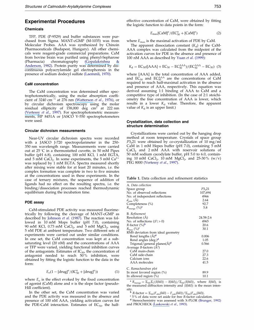
Table 1. Data collection and re®nement statistics
A. Data collectionSpace group P3221No. of observed reflections 107,694No. of independent reflections 4966dmin (AÊ ) 2.64Completeness (%) 92.7Rmerge (%)a 5.8
B. RefinementResolution (AÊ ) 24.58-2.6No. of reflections (jFj > 0) 4965R-factor (%)b 20.6Rfree (%)c 30.1RMS deviation from ideal geometry
Bond lengths (AÊ )d 0.006Bond angles (deg.)d 1.05Trigonal/general planes(AÊ )d 0.566
Average B-factors (AÊ 2)CaM main-chain 27.0CaM side-chain 27.3Calcium ions 22.6AAA molecules 41.5
C. Ramachandran plotIn most favored region (%) 89.9In allowed region (%) 10.1
a Rmerge � �hkl�ijhI(hkl)i ÿ I(hkl)ij/�hklhI(hkl)i, where I(hkl)i isthe measured diffraction intensity and hI(hkl)i is the mean inten-sity.
b R-factor � �hkljFobs(hkl) ÿ Fcalc(hkl)j/�hkljFobs(hkl)j.c 5 % of data were set aside for free R-factor calculation.d Stereochemistry was assessed with X-PLOR (Brunger, 1992)
and PROCHECK (Laskowski et al., 1993).
Structures of Calmodulin-Arylalkylamine Complexes 753
Experimental Procedures
Chemicals
TFP, PDE (P-9529) and buffer substances were pur-chased from Sigma. MANT-cGMP (M-1035) was fromMolecular Probes. AAA was synthesized by ChinoinPharmaceuticals (Budapest, Hungary). All other chemi-cals were reagent-grade commercial preparations. CaMfrom bovine brain was puri®ed using phenyl-Sepharose(Pharmacia) chromatography (Gopalakrishna &Anderson, 1982). Protein purity was determined by dis-continuous polyacrylamide gel electrophoresis in thepresence of sodium dodecyl sulfate (Laemmli, 1970).
CaM concentration
The CaM concentration was determined either spec-trophotometrically, using the molar absorption coef®-cient of 3240 cmÿ1 at 276 nm (Watterson et al., 1976), orby circular dichroism spectroscopy using the molarresidual ellipticity of 158,000 deg cm2 at 222 nm(Vertessy et al., 1997). For spectrophotometric measure-ments, HP 8451A or JASCO V-550 spectrophotometerswere used.
Circular dichroism measurements
Near-UV circular dichroism spectra were recordedwith a JASCO J-720 spectropolarimeter in the 250-350 nm wavelength range. Measurements were carriedout at 25 �C in a thermostatted cuvette, in 50 mM Pipesbuffer (pH 6.8), containing 100 mM KCl, 1 mM EGTA,and 5 mM CaCl2. In some experiments, the 5 mM Ca2�
was replaced by 1 mM EGTA. Spectra measured shortlyafter mixing were stable for at least 20 minutes, i.e. thecomplex formation was complete in two to ®ve minutesat the concentrations used in these experiments. In thecase of ternary mixtures, the sequence of addition ofligands had no effect on the resulting spectra, i.e. thebinding/dissociation processes reached thermodynamicequilibrium during the incubation time.
PDE assay
CaM-stimulated PDE activity was measured ¯uorime-trically by following the cleavage of MANT-cGMP asdescribed by Johnson et al. (1987). The reaction was fol-lowed in 10 mM Mops buffer (pH 7.0), containing90 mM KCl, 0.73 mM CaCl2, and 5 mM MgCl2, using5 nM PDE at ambient temperature. Two different sets ofexperiments were carried out under similar conditions.In one set, the CaM concentration was kept at a sub-saturating level (20 nM) and the concentrations of AAAor TFP were varied, yielding functional inhibition curvesof the antagonists. Estimates of IC50, the concentration ofantagonist needed to reach 50 % inhibition, wereobtained by ®tting the logistic function to the data in theform:
Eo�1ÿ �drug�n=�ICn50 � �drug�n�� �1�
where Eo is the effect evoked by the ®xed concentrationof agonist (CaM) alone and n is the slope factor (pseudo-Hill coef®cient).
In the other set, the CaM concentration was variedand the PDE activity was measured in the absence andpresence of 100 nM AAA, yielding activation curves forthe PDE-CaM interaction. Estimates of EC50, the half-
effective concentration of CaM, were obtained by ®ttingthe logistic function to data points in the form:
Emax�CaM�n=�ECn50 � �CaM�n� �2�
where Emax is the maximal activation of PDE by CaM.The apparent dissociation constant (Kd) of the CaM-
AAA complex was calculated from the midpoint of theactivation curves for PDE in the absence and presence of100 nM AAA as described by Yuan et al. (1999):
Kd � EC50��AAA� � EC50 ÿ ECAAA50 �=�ECAAA
50 ÿ EC50� �3�where [AAA] is the total concentration of AAA added,and EC50 and EC50
AAA are the concentrations of CaMrequired to reach half-maximal activation in the absenceand presence of AAA, respectively. This equation wasderived assuming 1:1 binding of AAA to CaM and acompetitive type of inhibition. (In the case of 2:1 stoichi-ometry the free concentration of AAA is lower, whichresults in a lower Kd value. Therefore, the apparentvalue of Kd is an upper limit.)
Crystallization, data collection andstructure determination
Crystallizations were carried out by the hanging dropmethod at room temperature. Crystals of space groupP3221 were obtained by co-crystallization of 10 mg/mlCaM in 1 mM Hepes buffer (pH 7.0), containing 5 mMCaCl2 and 2 mM AAA with reservoir solutions of50 mM sodium cacodylate buffer, pH 5.0 to 6.0, contain-ing 10 mM CaCl2, 10 mM MgCl2, and 25-30 % (w/v)PEG 8000 (Vertessy et al., 1997).

754 Structures of Calmodulin-Arylalkylamine Complexes
Re¯ections from the structure were collected from asingle-crystal at 93 K using a 300 mm diameter MarResearch image plate detector. The cryoprotectant was20 % (v/v) 2-methyl-2,4-pentanediol. Data processingwas done with the programs Denzo and Scalepack(Otwinowski, 1993) (space group P3221, isostructuralwith the CaM-TFP crystal structures, the cell parametersare: a � 40.13 AÊ , c � 173.81 AÊ , 4966 independent re¯ec-tions, Rmerge � 5.8 %, 92.7 % complete to 2.64 AÊ ). Thescaled diffraction intensities were converted to structurefactor amplitudes using the program TRUNCATE(French & Wilson, 1978) and the structure was deter-mined by molecular replacement using the AMoRe pro-gram (Navaza, 1994) of the Collaborative ComputingProject Number 4 (1994) suite, with the protein part ofthe CaM-4TFP complex (Vandonselaar et al., 1994) asmodel.
Refinement
The model building steps were carried out with the Oprogram (Jones et al., 1991) and the resulting model wasre®ned with X-PLOR, version 3.581 (Brunger, 1992).During the re®nement we used torsion angle dynamics(slow cooling from 4000 K), resolution-dependentweighting, overall anisotropic B-factor re®nement, bulksolvent correction and grouped B-factor re®nement.
At the end of the ®nal re®nement, we have R � 20.6 %and Rfree � 30.1 % (for all re¯ections) for the structure.The ®nal structure consists of 144 CaM residues, four Caions and two AAA molecules with an RMS deviation of0.006 AÊ in bond lengths and 1.05 � in bond angles, andan average temperature factor of 28.02 AÊ 2 for 1176atoms. Table 1 shows data collection and re®nement stat-istics.
Protein Data Bank accession numbers
The atomic coordinates and structure factors havebeen deposited in the RCSB Protein Data Bank (accessioncodes 1qiv and 1qiw).
Acknowledgments
This study was supported by the Hungarian NationalResearch Foundation grants OTKA T-022191 to Zs. B., F-020862, M-027852 and T-029910 to B.G.V. as well asT-025291 and 031892 to J.O. as well as by HungarianAcademy of Sciences grant AKP-98-73 3,3 to J.O. Zs.B. isgrateful for the grant Hungarian-Portuguese Scienti®cand Technological Cooperation P-1/95 to OMFB and inparticular to Dr C. Frazao. B.G.V. and K. L. are recipientsof the Bolyai JaÂnos fellowship.
The EMBL/DESY, Hamburg is acknowledged for thedata collection opportunity at beamline X11.
References
Babu, Y. S., Sack, J. S., Greenhough, T. C., Bugg, C. E.,Means, A. R. & Cook, W. J. (1985). Three-dimen-sional structure of calmodulin. Nature, 315, 37-40.
Brunger, A. T. (1992). XPLOR: A System for X-rayCrystallography and NMR Version 3, Yale University,New Haven, CT.
Collaborative Computational Project Number 4 (1994).The CCP4 suite: programs for protein crystallogra-phy. Acta Crystallog. sect. D, 50, 760-763.
Cook, W. J., Walter, L. J. & Walter, M. R. (1994). Drugbinding by calmodulin: crystal structure of a calmo-dulin-tri¯uoperazine complex. Biochemistry, 33,15259-15265.
Crivici, A. & Ikura, M. (1995). Molecular and structuralbasis of target recognition by calmodulin. Annu.Rev. Biophys. Biomol. Struct. 24, 85-115.
Dalgarno, D. C., Klevit, R. E., Levine, B. A., Scott,G. M. M., Williams, R. J. P., Gergely, J., Grabarek,Z., Leavis, P. C., Grand, R. J. A. & Drabikowski.,W. (1984). The nature of the tri¯uoperazine bindingsites on calmodulin and troponin-C. Biochim. Bio-phys. Acta, 791, 164-172.
Esnouf, R. M. (1997). An extensively modi®ed version ofMolScript that includes greatly enhanced coloringcapabilities. J. Mol. Graph. 15, 132-134.
Finn, B. E., Evenas, J., Drakenberg, T., Waltho, J. P.,Thulin, E. & Forsen, S. (1995). Calcium-inducedstructural changes and domain autonomy in calmo-dulin. Nature Struct. Biol. 2, (9), 777-783.
French, S. & Wilson, K. (1978). On the treatment ofnegative intensity observations. Acta Crystallog. sect.A, A34, 517-525.
Gietzen, K., Wutrich, A. & Bader, H. (1981). R 24571: anew powerful inhibitor of red blood cell Ca2�-trans-port ATPase and of calmodulin-regulated functions.Biochem. Biophys. Res. Commun. 101, 418-425.
Gopalakrishna, R. & Anderson, W. B. (1982). Ca2�-induced hydrophobic site on calmodulin: appli-cation for puri®cation of calmodulin by phenyl-Sepharose af®nity chromatography. Biochem. Bio-phys. Res. Commun. 104, 830-836.
Hait, W. N. & Lazo, J. S. (1986). Calmodulin: a potentialtarget for cancer chemotherapeutic agents. J. Clin.Oncol. 4, 994-1012.
Ikura, M., Clore, G. M., Gronenborn, A. M., Zhu, G.,Klee, C. B. & Bax, A. (1992). Solution structure of acalmodulin-target peptide complex by multidimen-sional NMR. Science, 256, 632-638.
Jackson, A. E. & Puett, D. (1986). Binding of tri¯uopera-zine and ¯uorene-containing compounds to calmo-dulin and adducts. Biochem. Pharmacol. 35, 4395-4400.
Johnson, J. D. & Mills, J. S. (1986). Calmodulin. Med.Res. Rev. 6, 341-363.
Johnson, J. D., Wittenauer, L. A., Thulin, E., Forsen, S. &Vogel, H. J. (1986). Localization of a felodipine(dihydropyridine) binding site on calmodulin. Bio-chemistry, 25, 2226-2231.
Johnson, J. D., Walters, J. D. & Mills, J. S. (1987). A con-tinuous ¯uorescence assay for cyclic nucleotidephosphodiesterase hydrolysis of cyclic GMP. Anal.Biochem. 162, 291-295.
Jones, T. A., Zou, J.-Y., Cowan, S. W. & Kjeldgaard, M.(1991). Improved methods for binding proteinmodels in electron density maps and the location oferrors in these models. Acta Crystallog. sect. A, 47,110-119.
Klee, C. B. (1988). Interaction of calmodulin with Ca2�
and target proteins. In Calmodulin (Cohen, P. &Klee, C. B., eds), pp. 35-56, Elsevier, New York.
Klevit, R. E., Levine, B. A. & Williams, R. J. (1981).A study of CaM and its interaction with tri¯uopera-zine by high resolution 1H NMR spectroscopy.FEBS Letters, 123, 25-29.

Structures of Calmodulin-Arylalkylamine Complexes 755
Kraulis, P. J. (1991). MOLSCRIPT: a program to produceboth detailed and schematic plots of protein struc-tures. J. Appl. Crystallog. 24, 946-950.
Kretsinger, R. H., Rudnick, S. E. & Weissman, L. J.(1986). Crystal structure of calmodulin. J. Inorg. Bio-chem. 28, 289-302.
Kuboniwa, H., Tjandra, N., Grzesiek, S., Ren, H., Klee,C. B. & Bax, A. (1995). Solution structure ofcalcium-free calmodulin. Nature Struct. Biol. 2, 768-776.
Laemmli, U. K. (1970). Cleavage of structural proteinsduring the assembly of the head of bacteriophageT4. Nature, 227, 680-688.
Laskowski, R. A., MacArthur, M. W., Moss, D. S. &Thornton, J. M. (1993). PROCHECK - a program tocheck the stereochemical quality of protein struc-tures. J. Appl. Crystallog. 26, 283-291.
Marshak, D. R., Lukas, T. J. & Watterson, D. M. (1985).Drug-protein interactions: binding of chlorproma-zine to calmodulin fragments and related calciumbinding proteins. Biochemistry, 24, 144-150.
Maulet, Y. & Cox, J. A. (1983). Structural changes inmelittin and calmodulin upon complex formationand their modulation by calcium. Biochemistry, 22,5680-5686.
Meador, W. E., Means, A. R. & Quiocho, F. A. (1992).Target enzyme recognition by calmodulin: 2. 4 AÊ
structure of a calmodulin-peptide complex. Science,257, 1251-1255.
Meador, W. E., Means, A. R. & Quiocho, F. A. (1993).Modulation of calmodulin plasticity in molecularrecognition on the basis of X-ray structures. Science,262, 1718-1721.
Nakayama, S. & Kretsinger, R. H. (1994). Evolution ofthe EF-hand family of proteins. Annu. Rev. Biophys.Biomol. Struct. 23, 473-507.
Navaza, J. (1994). AMoRe: an automated package formolecular replacement. Acta Crystallog. sect. A, 50,157-163.
Nicholls, A., Sharp, K. A. & Honig, B. (1991). Proteinfolding and association: insights from the interfacialand thermodynamic properties of hydrocarbons.Proteins: Struct. Funct. Genet. 11, 281-296.
O'Neil, K. T. & DeGrado, W. F. (1990). How calmodulinbinds its targets: sequence independent recognitionof amphiphilic alpha-helices. Trends Biochem. Sci. 15,59-64.
Orosz, F., Christova, T. Y. & OvaÂdi, J. (1988). Functionalin vitro test of calmodulin antagonism: effect ofdrugs on interaction between calmodulin and glyco-lytic enzymes. Mol. Pharmacol. 33, 678-682.
Orosz, F., Telegdi, M., Liliom, K., Solti, M., Korbonits,D. & OvaÂdi, J. (1990). Dissimilar mechanisms of
action of anti-calmodulin drugs: quantitative anal-ysis. Mol. Pharmacol. 38, 910-916.
Osawa, M., Swindells, M. B., Tanikawa, J., Tanaka, T.,Mase, T., Furuya, T. & Ikura, M. (1998). Solutionstructure of calmodulin-W-7 complex: the basis ofdiversity in molecular recognition. J. Mol. Biol. 276,165-176.
Otwinowski, Z. (1993). Data Collection and Processing(Sawyer, L., Isaacs, N. & Bailey, S., eds), SERCDaresbury Laboratory, Warrington, UK.
Sobieszek, A. (1989). Calmodulin antagonist action insmooth-muscle myosin phosphorylation. Differentmechanisms for tri¯uoreazine and calmidazoliuminhibition. Biochem. J. 262, 215-223.
Tanaka, T. & Hidaka, H. (1980). Hydrophobic regionsfunction in calmodulin-enzyme(s) interactions.J. Biol. Chem. 255, 11078-11080.
Thulin, E., Andersson, A., Drakenberg, T., Forsen, S. &Vogel, H. J. (1984). Metal ion and drug binding toproteolytic fragments of calmodulin: proteolytic,cadmium-113, and proton nuclear magnetic reson-ance studies. Biochemistry, 23, 1862-1870.
Vandonselaar, M., Hickie, R. A., Quail, J. W. &Delbaere, L. T. J. (1994). Tri¯uoperazine-inducedconformational change in Ca2�-calmodulin. NatureStruct. Biol. 1, (11), 795-801.
Vertessy, B. G., BoÈcskei, Z., Harmath, V., NaÂray-Szabo ,G. & OvaÂdi, J. (1997). Crystallization and prelimi-nary diffraction analysis of Ca2�-calmodulin-drugand apocalmodulin-drug complexes. Proteins: Struct.Funct. Genet. 28, 131-134.
Vertessy, B. G., Harmat, V., BoÈcskei, Z., NaÂray-Szabo ,G., Orosz, F. & OvaÂdi, J. (1998). Simultaneous bind-ing of drugs with different chemical structures toCa2�-calmodulin: crystallographic and spectroscopicstudies. Biochemistry, 37, 15300-15310.
Watterson, D. M., Harrelson, W. G., Keller, P. M.,Sharief, F. & Vanaman, T. C. (1976). Structural simi-larities between the Ca2�-dependent regulatory pro-teins of 30:50-cyclic nucleotide phosphodiesteraseand actomyosin ATPase. J. Biol. Chem. 251, 4501-4513.
Yuan, T., Walsh, M. P., Sutherland, C., Fabian, H. &Vogel, H. J. (1999). Calcium-dependent and -inde-pendent interactions of the calmodulin-bindingdomain of cyclic nucleotide phosphodiesterase withcalmodulin. Biochemistry, 38, 1446-1455.
Zhang, M., Tanaka, T. & Ikura, M. (1995). Calcium-induced conformational transition revealed by thesolution structure of apo calmodulin. Nature Struct.Biol. 2, (9), 758-767.
Edited by R. Huber
(Received 14 October 1999; received in revised form 9 February 2000; accepted 9 February 2000)
Related Documents