Downloaded from www.microbiologyresearch.org by IP: 54.242.161.225 On: Tue, 17 May 2016 08:24:28 A new phylogenetic lineage of Rabies virus associated with western pipistrelle bats (Pipistrellus hesperus) Richard Franka, 1 Denny G. Constantine, 2 Ivan Kuzmin, 1 Andres Velasco-Villa, 1 Serena A. Reeder, 1,3 Daniel Streicker, 1 Lillian A. Orciari, 1 Anna J. Wong, 4 Jesse D. Blanton 1 and Charles E. Rupprecht 1 Correspondence Richard Franka [email protected] 1 Centers for Disease Control and Prevention, DVRD/VRZB/Rabies, G33, 1600 Clifton Road NE, Atlanta, GA 30333, USA 2 1899 Olmo Way, Walnut Creek, CA 94598-1446, USA 3 Department of Biology and Center for Disease Ecology, Emory University, Atlanta, GA 30333, USA 4 Viral and Rickettsial Diseases Laboratory, California Department of Health Services, 850 Marina Bay Parkway, Richmond, CA 94804-6403, USA Received 6 January 2006 Accepted 20 March 2006 Bats represent the major source of human rabies cases in the New World. In the USA, most cases are associated with species that are not commonly found or reported rabid. To understand better the epidemiology and public health significance of potentially important bat species, a molecular study was performed on samples collected from naturally infected rabid western pipistrelle (Pipistrellus hesperus), eastern pipistrelle (Pipistrellus subflavus) and silver-haired bats (Lasionycteris noctivagans) from different regions of their geographical distribution in the USA. A 264 bp fragment at the 59 end of the N gene coding region was sequenced and analysed in comparison with rabies virus variants circulating within other North American mammals. Phylogenetic analysis demonstrated that P. hesperus bats maintain a unique rabies virus variant. Preliminary data also suggest that P. subflavus and Lasionycteris noctivagans may harbour two different rabies virus variants (Ps and Ln) that are likely to be maintained independently by each bat species, which recently appear to have emerged as major vectors of human disease. INTRODUCTION Despite the existence of highly effective animal rabies pre- vention and control programmes, this zoonotic disease remains a significant cause of human mortality throughout the world (WHO, 2005). Currently, most human rabies deaths are typically not related to inadequate biologicals, but rather as a result of a failure to recognize the risk of disease transmission and to seek appropriate prophylaxis following the exposure to an infected animal. A key facet to under- standing the epidemiology of rabies involves a better appre- ciation of viral variants maintained by different mammalian species. High levels of genetic and antigenic diversity of Rabies virus (RABV) have been reported in association with distinct species of mammals in the Americas (Rupprecht et al., 1991; Nadin-Davis et al., 2001; Smith, 2002; Velasco- Villa et al., 2005). Specific genetic and antigenic patterns exist among RABV lineages. Recent studies suggest that viral variants tend to be host-associated and maintained in populations predominantly through intraspecific transmis- sion, as exemplified by viral variants or biotypes found among common North American taxa, such as: domestic dogs (Canis familiaris), grey foxes (Urocyon cinereoargen- teus), eastern spotted skunks (Spilogale putorius), striped skunks (Mephitis mephitis), raccoons (Procyon lotor) and bats of several species. Worldwide, most human rabies cases are caused by rabies viruses associated with dogs. However, in the Americas, RABVs associated with bats are emerging as a dispropor- tionate source for human infection (Rupprecht et al., 1995; Messenger et al., 2002; Belotto et al., 2005). To date, bat species frequently reported rabid in the USA include the big- brown bat (Eptesicus fuscus), the Brazilian (Mexican) free- tailed bat (Tadarida brasiliensis) and in the western region, the California myotis (Myotis californicus). More limited numbers of cases are identified in the hoary bat (Lasiurus cinereus), the red bat (Lasiurus borealis) and the little-brown bat (Myotis lucifugus). Interpretation of surveillance results The GenBank/EMBL/DDBJ accession numbers for the sequences reported in this paper are DQ445308–DQ445382. 0008-1822 G 2006 Crown copyright Printed in Great Britain 2309 Journal of General Virology (2006), 87, 2309–2321 DOI 10.1099/vir.0.81822-0 Journal of General Virology (2006), 87, 2309–2321 DOI 10.1099/vir.0.81822-0

Welcome message from author
This document is posted to help you gain knowledge. Please leave a comment to let me know what you think about it! Share it to your friends and learn new things together.
Transcript

Downloaded from www.microbiologyresearch.org by
IP: 54.242.161.225
On: Tue, 17 May 2016 08:24:28
A new phylogenetic lineage of Rabies virusassociated with western pipistrelle bats(Pipistrellus hesperus)
Richard Franka,1 Denny G. Constantine,2 Ivan Kuzmin,1
Andres Velasco-Villa,1 Serena A. Reeder,1,3 Daniel Streicker,1
Lillian A. Orciari,1 Anna J. Wong,4 Jesse D. Blanton1
and Charles E. Rupprecht1
Correspondence
Richard Franka
1Centers for Disease Control and Prevention, DVRD/VRZB/Rabies, G33, 1600 Clifton RoadNE, Atlanta, GA 30333, USA
21899 Olmo Way, Walnut Creek, CA 94598-1446, USA
3Department of Biology and Center for Disease Ecology, Emory University, Atlanta, GA 30333,USA
4Viral and Rickettsial Diseases Laboratory, California Department of Health Services,850 Marina Bay Parkway, Richmond, CA 94804-6403, USA
Received 6 January 2006
Accepted 20 March 2006
Bats represent the major source of human rabies cases in the New World. In the USA, most
cases are associated with species that are not commonly found or reported rabid. To understand
better the epidemiology and public health significance of potentially important bat species,
a molecular study was performed on samples collected from naturally infected rabid western
pipistrelle (Pipistrellus hesperus), eastern pipistrelle (Pipistrellus subflavus) and silver-haired bats
(Lasionycteris noctivagans) from different regions of their geographical distribution in the USA.
A 264 bp fragment at the 59 end of the N gene coding region was sequenced and analysed
in comparison with rabies virus variants circulating within other North American mammals.
Phylogenetic analysis demonstrated that P. hesperus bats maintain a unique rabies virus variant.
Preliminary data also suggest that P. subflavus and Lasionycteris noctivagans may harbour two
different rabies virus variants (Ps and Ln) that are likely to be maintained independently by each
bat species, which recently appear to have emerged as major vectors of human disease.
INTRODUCTION
Despite the existence of highly effective animal rabies pre-vention and control programmes, this zoonotic diseaseremains a significant cause of human mortality throughoutthe world (WHO, 2005). Currently, most human rabiesdeaths are typically not related to inadequate biologicals, butrather as a result of a failure to recognize the risk of diseasetransmission and to seek appropriate prophylaxis followingthe exposure to an infected animal. A key facet to under-standing the epidemiology of rabies involves a better appre-ciation of viral variants maintained by different mammalianspecies. High levels of genetic and antigenic diversity ofRabies virus (RABV) have been reported in association withdistinct species of mammals in the Americas (Rupprechtet al., 1991; Nadin-Davis et al., 2001; Smith, 2002; Velasco-Villa et al., 2005). Specific genetic and antigenic patternsexist among RABV lineages. Recent studies suggest that viral
variants tend to be host-associated and maintained inpopulations predominantly through intraspecific transmis-sion, as exemplified by viral variants or biotypes foundamong common North American taxa, such as: domesticdogs (Canis familiaris), grey foxes (Urocyon cinereoargen-teus), eastern spotted skunks (Spilogale putorius), stripedskunks (Mephitis mephitis), raccoons (Procyon lotor) andbats of several species.
Worldwide, most human rabies cases are caused by rabiesviruses associated with dogs. However, in the Americas,RABVs associated with bats are emerging as a dispropor-tionate source for human infection (Rupprecht et al., 1995;Messenger et al., 2002; Belotto et al., 2005). To date, batspecies frequently reported rabid in the USA include the big-brown bat (Eptesicus fuscus), the Brazilian (Mexican) free-tailed bat (Tadarida brasiliensis) and in the western region,the California myotis (Myotis californicus). More limitednumbers of cases are identified in the hoary bat (Lasiuruscinereus), the red bat (Lasiurus borealis) and the little-brownbat (Myotis lucifugus). Interpretation of surveillance results
The GenBank/EMBL/DDBJ accession numbers for the sequencesreported in this paper are DQ445308–DQ445382.
0008-1822 G 2006 Crown copyright Printed in Great Britain 2309
Journal of General Virology (2006), 87, 2309–2321 DOI 10.1099/vir.0.81822-0Journal of General Virology (2006), 87, 2309–2321 DOI 10.1099/vir.0.81822-0

Downloaded from www.microbiologyresearch.org by
IP: 54.242.161.225
On: Tue, 17 May 2016 08:24:28
is complicated by the fact that certain bat species may bedifficult to identify and therefore results could be biasedbecause of misidentification. Despite the frequency ofreported rabies cases in E. fuscus and T. brasiliensis, 32 of58 human rabies deaths reported from 1958 to 2000 in theUSA were caused by RABV variants harboured by otherbat species (16 Pipistrellus subflavus, eight Lasionycterisnoctivagans, five T. brasiliensis, two Myotis californicus andone E. fuscus) (Messenger et al., 2002). Two bat speciesuncommonly submitted to state public health laboratoriesfor diagnosis and rarely found around human dwellingswere most frequently associated with human rabies cases:the silver-haired bat (Lasionycteris noctivagans) and theeastern pipistrelle (P. subflavus). The prevalence of Ln/Ps(Lasionycteris noctivagans and P. subflavus) RABV variant inrecent human rabies cases is puzzling because Lasionycterisnoctivagans and P. subflavus bats are seldom found orreported to be rabid (CDC, 1994). Since both species preferhabitats far from human dwellings, one explanation may bethat many bats die of rabies most frequently during summerin forest habitats where they are seldom observed by people.Two other hypotheses have been proposed to explainthis phenomenon: (i) increased virulence of Ln/Ps RABVrelative to other virus variants, and (ii) the existence ofunique host characteristics, such as the so-called ‘smallvector’ hypothesis (Morimoto et al., 1996; Messenger et al.,2003). The latter hypothesis describes a failure to recognizeor appreciate the significance of a bite when a small batis involved, because of the limited severity of the lesionproduced. Support of the former hypothesis has beensuggested by results from experimental data, comparinginfectivity of Ln/Ps RABV variant with those from domesticcanids (Morimoto et al., 1996; Dietzschold et al., 2000).
In North America, RABV has been detected in almost everybat species (Constantine, 1979). However, due to problemsof passive surveillance, only limited data are available forRABV variants associated with less abundant species. Severalbat species, such as Pipistrellus hesperus, are more reclusiveor restricted in distribution and rarely come into contactwith humans or domesticated animals. Relatively few sam-ples obtained from rabid P. hesperus have been available foranalysis, even though this species is relatively abundantin desert and grassland habitats of the western USA wherethey roost in rock crevices (Barbour & Davis, 1969). As thesmallest bat in the USA, P. hesperus is an excellent examplefor consideration to continue study of the ‘small vector’hypothesis, by using a molecular approach.
In the present study, RABV of P. hesperus was characterizedbased on the last 264 bp of the 59 end of the nucleoprotein(N) gene coding region. In Arizona and California, 30samples were obtained from P. hesperus bats during the years2000–2005, sequenced and compared with nine historicalsamples (collected during 1981–1997) from P. hesperus fromArizona (Flagstaff), available in GenBank. These sequenceswere also compared to more than 300 sequences of RABVcollected from different mammalian species throughout
North America. Additionally, we compared sequences ofLn/Ps RABV from Lasionycteris noctivagans and P. subflavus,to inquire if they have species-specific markers or should beconsidered as a single RABV variant.
METHODS
P. hesperus bats were collected as a part of rabies public health sur-veillance investigations in California and Arizona during 2000–2005.P. subflavus and Lasionycteris noctivagans were collected via statepublic health submissions during the period 1976–2004 from differ-ent regions of the USA (Fig. 1). Brains were removed and rabiesdiagnoses were performed by the direct fluorescent antibody test(Dean et al., 1996). In addition, another 30 samples of P. hesperus,14 samples of P. subflavus and 22 samples of Lasionycteris noctiva-gans bats were used for further characterization (Table 1). TotalRNA was extracted from infected bat brains with TRIzol reagent(Invitrogen) according to the manufacturer’s recommendations.RT-PCR was performed with primer sets designed for the rabiesvirus nucleoprotein (N): forward primer 113fw (59-GTAGGATGC-TATATGGG-39, position 1013–1029, according to the PV genome);and reverse primer 304 (Trimarchi & Smith, 2002).
Direct sequencing of the RT-PCR products was performed frompurified PCR products. We sequenced fragments of RABV N gene byusing forward primers: 1066 (59-GAGAGAAGATTCTTCAGGGA-39,position 1136–1155) and 113fw, and reverse primer: 304 (Trimarchi &Smith, 2002) (all positions are according to the PV genome, GenBankaccession no. M13215). Because of limited length of sequencesavailable in GenBank and the abundance of RABV sequences from thelast 300 nt of the N gene coding region, further comparisons werefocused on the variable 264 nt corresponding to bases 1157–1420 andaa 363–450. Use of this fragment allowed us to increase the robustnessof the analysis and provided better resolution for the detection ofpossible spillover events between species. Previous analyses showedthat this short fragment from the 59 end of the N gene generates thesame branching pattern as the whole N gene sequence (Smith et al.,1992).
A phylogenetic analysis was undertaken using more than 300 RABVsequences (only 133 unique representatives were used for the finalanalysis and are shown in the Table 1), originating from differentNorth American mammals, available from GenBank, including ninehistorical sequences from P. hesperus collected in Arizona (Table 1)and sequences from P. subflavus and Lasionycteris noctivagans availablein the CDC archival database or otherwise sequenced for this study.Sequences were edited using BioEdit software (Hall, 1999) and multi-ple alignments were built using the CLUSTAL X package (Jeanmouginet al., 1998). Duvenhage virus and European bat lyssavirus 2 (EBLV-2)were used as outgroup taxa. A neighbour-joining (NJ) analysis (p-distance model) with 1000 bootstrap replicates was performed usingthe MEGA computer program, version 2.1. (Kumar et al., 2001).The NJPLOT program from the CLUSTAL X package and the Tree-Explorer module of MEGA were used to obtain graphical output ofphylogenetic estimations. Bootstrap values of more than 70 % wereconsidered as providing support for phylogenetic grouping. Re-presentatives of particular lineages were selected for the finalphylogenetic analysis.
In addition, both maximum-likelihood (ML) and Bayesian analyseswere performed on the dataset to support or clarify results of NJanalysis. A total of 133 RABV sequences were analysed, including sevensequences from raccoon, skunk and dog RABVs that were used asoutgroup taxa. MODELTEST (Posada & Crandall, 1998) analysed 56models of evolution to determine the most appropriate selection forthe dataset. The HKY85 model (Hasegawa et al., 1985) with a gamma
2310 Journal of General Virology 87
R. Franka and others

Downloaded from www.microbiologyresearch.org by
IP: 54.242.161.225
On: Tue, 17 May 2016 08:24:28
distribution (HKY85+G) was selected and subsequently implementedfor the ML analysis in PAUP*4.0b10 (Swofford, 2002). Nucleotidefrequencies were A=0?34190, C=0?24190, G=0?20470, T=0?21150,the transition-to-transversion ratio=4?3131 and the gamma shapeparameter=0?3395.
MrBayes 3.1.1 (Ronquist & Huelsenbeck, 2003) was used for a Bayesiananalysis with the general time-reversible model incorporating bothinvariant sites and a gamma distribution (GTR+I+G) to examinethe data with a more complex model than used in the ML analysis. Twosimultaneous analyses, each with four Markov chains, were run for2 000 000 generations and sampled every 100 generations. Treesgenerated prior to the stabilization of likelihood scores were discarded,(burnin=450). The remaining trees were used to build a 50 % majorityrule consensus tree. Posterior probability values were used to assesssupport at each node (¢95=statistical support).
The deduced amino acid sequences from the consensus of particularmonophyletic clades were aligned for comparison as well. All newRABV sequences reported in this study were submitted to GenBank(Table 1).
RESULTS
The overall branching pattern of the trees constructed in ourstudy by using the NJ, ML and Bayesian methods was thesame regardless of the method used. RABV variantsharboured by North American bats formed two majorclades. One was formed by the Ph1, Ph2 and Myotis cladesand another one by Lasionycteris noctivagans, Lasiuruscinereus, Lasiurus borealis and P. subflavus. E. fuscus 1–3clades represented groups that branched differently if weused different methods.
In the NJ analysis, 79 % (31 samples) of RABV samplesoriginating from P. hesperus bats in California and Arizona(a large part of their geographical range in the USA) formeda monophyletic clade (Ph1) with high bootstrap support,
separated from other phylogenetic lineages of RABV(Fig. 2). A high level of nucleotide sequence similarity(98?7 %) within this clade originating from differentlocations was observed over the period of sample collection(1981–2004). Five samples (13 %) collected from P. hesperusin California in 2002–2005, formed a separate clade (Ph2),which was a sister to a clade consisting of Myotis species andP. hesperus with inconsistent bootstrap support (Fig. 2).Nucleotide sequence similarity within Ph2 clade was 98?8 %and within the Myotis species clade was 95?5 %. Nucleotidesequence similarity between Ph1 and Ph2 was 94?3 %,between Ph2 and the Myotis species clade was 94?3 % andbetween Ph1 and Myotis species clade was 91?7 %. Onlythree (8 %) samples collected from P. hesperus belonged toother RABV lineages (Myotis species), suggesting occasionalinfectious spillover events from Myotis species. Only four ofmore than 50 sequences (<8 %) of RABV originating fromMyotis species were found in the Ph1 clade. The P. hesperus(Ph) variants Ph1 and Ph2 shared a most recent commonancestor with a RABV variant associated with Myotis species(a RABV variant associated with different bat species fromthe genus Myotis). The separation between the Ph1 and Ph2clades was based on 11 synonymous nucleotide substitu-tions within the N gene fragment, G/A1165, C/T1237, C/T1240,G/A1264, C/A1268, T/A1282, T/C1291, T/C1297, G/A1315,A/G1324, A/C1348 and one non-synonymous substitutionT/G1205 (aa L/V379).
While closely related to each other, the viruses originatingfrom P. subflavus and Lasionycteris noctivagans separatedinto two clusters with no overlap between clusters. Themonophyletic clade of P. subflavus (Ps) included 13sequences of this species originating from differentgeographical locations including Pennsylvania, Georgia,Tennessee, Arkansas, Indiana, Virginia and Texas, collected
Fig. 1. Locations where P. hesperus
(California, Arizona), P. subflavus (Pennsy-lvania, Georgia, Tennessee, Texas, Arkansas,Indiana and Virginia) and Lasionycteris nocti-
vagans (Ontario, British Columbia, New York,Colorado, Wisconsin, Washington, Idaho,North Carolina and California) were collected(sequences from GenBank were collected inFlagstaff, AZ). Lasionycteris noctivagans isdistributed throughout the USA and southernCanada (Wilson & Ruff, 1999). Georgra-phical distribution of P. hesperus and P.
subflavus according to Wilson & Ruff (1999).The red line represents recently reportedwestward expansion of P. subflavus (Gelusoet al., 2005).
http://vir.sgmjournals.org 2311
New rabies virus lineage in western pipistrelle batsNew rabies virus lineage in western pipistrelle bats

Downloaded from www.microbiologyresearch.org by
IP: 54.242.161.225
On: Tue, 17 May 2016 08:24:28
Table 1. List of samples used
Identification no. Mammalian
species*
State County or city Year of
collection
GenBank
accession no.
Pipistrellus hesperus
AY170251Azph81 P. hesperus Arizona Coconino 1981 AY170251
AF394870Azph93 P. hesperus Arizona Mohave 1993 AF394870
AY170263Azph93 P. hesperus Arizona Maricopa 1993 AY170263
AY170261Azph95 P. hesperus Arizona Coconino 1995 AY170261
AY170252Azph96 P. hesperus Arizona Navajo 1996 AY170252
AY170254Azph96 P. hesperus Arizona Maricopa 1996 AY170254
AY170250Azph97 P. hesperus Arizona Pima 1997 AY170250
AY170256Azph97 P. hesperus Arizona Pima 1997 AY170256
AY170257Azph97 P. hesperus Arizona Pima 1997 AY170257
Cab1306ph00 P. hesperus California San Bernardino 2000 DQ445308D
Cab1308ph00 P. hesperus California San Bernardino 2000 DQ445309D
Cab1319ph00 P. hesperus California San Bernardino 2000 DQ445310D
Cab1324ph00 P. hesperus California San Bernardino 2000 DQ445311D
Cab1325ph00 P. hesperus California San Bernardino 2000 DQ445312D
Cab1326ph00 P. hesperus California San Bernardino 2000 DQ445313D
Cab1328ph00 P. hesperus California San Bernardino 2000 DQ445314D
Cab1329ph00 P. hesperus California San Bernardino 2000 DQ445315D
Cab1330ph00 P. hesperus California San Bernardino 2000 DQ445316D
Cab1332ph00 P. hesperus California San Bernardino 2000 DQ445317D
Cab1347ph01 P. hesperus California San Bernardino 2001 DQ445318D
Cab1349ph01 P. hesperus California San Bernardino 2001 DQ445319D
Cab1354ph01 P. hesperus California San Bernardino 2001 DQ445320D
Cab1355ph01 P. hesperus California San Bernardino 2001 DQ445321D
Cab1382ph01 P. hesperus California San Bernardino 2001 DQ445322D
Cab1385ph01 P. hesperus California San Bernardino 2001 DQ445323D
Cab1401ph01 P. hesperus California San Bernardino 2001 DQ445324D
Cab1402ph01 P. hesperus California San Bernardino 2001 DQ445325D
Car1406ph01 P. hesperus California Riverside 2001 DQ445326D
Car1415ph01 P. hesperus California Riverside 2001 DQ445327D
Car1416ph01 P. hesperus California Riverside 2001 DQ445328D
Car1419ph01 P. hesperus California Riverside 2001 DQ445329D
Casd1420ph01 P. hesperus California San Diego 2001 DQ445330D
Car1421ph01 P. hesperus California Riverside 2001 DQ445331D
Caaub1424ph02 P. hesperus California El Dorado 2002 DQ445332D
Ca2167ph03 P. hesperus California Los Angeles 2003 DQ445333D
Ca2149ph03 P. hesperus California Castaic 2003 DQ445334D
Ca2022ph04 P. hesperus California Encino 2004 DQ445335D
Az01ph04 P. hesperus Arizona Maricopa 2004 DQ445336D
Ca164ph05 P. hesperus California Tulare 2005 DQ445337D
Myotis speciesd
AF351837ml7alta79 M. lucifugus Alberta NA 1979 AF351837
Ny112ml83 M. lucifugus New York NA 1983 DQ445338D
Ca1563mc86 M. californicus California Shasta 1986 DQ445339D
AF394871Camc87 M. californicus California Plumas 1987 AF394871
AF351835me1bc92 M. evotis British Columbia NA 1992 AF351835
AF351836mc2bc92 M. californicus British Columbia NA 1992 AF351836
AF351839ml4bc92 M. lucifugus British Columbia NA 1992 AF351839
AF351834ml5bc92 M. lucifugus British Columbia NA 1992 AF351834
Ca2375me93 M. evotis California NA 1993 DQ445340D
Ca2373ml93 M. lucifugus California NA 1993 DQ445341D
AF351838ml6ns94 M. lucifugus Nova Scotia NA 1994 AF351838
Az3850mv96 M. velifer Arizona Show Low 1996 DQ445342D
2312 Journal of General Virology 87
R. Franka and others
Downloaded from www.microbiologyresearch.org by
IP: 54.242.161.225
On: Tue, 17 May 2016 08:24:28
AY170249Azmc96 M. californicus Arizona Pima 1996 AY170249
AY170260Azmc96 M. ciliolabrum Arizona Holbrook 1996 AY170260
AY170253Azmsp97 Myotis species Arizona Pima 1997 AY170253
AY170255Azmsp99 Myotis species Arizona Pima 1999 AY170255
AY170258Azmsp99 Myotis species Arizona Bullhead City 1999 AY170258
Eptesicus fuscus
AF351832ef34bc72 E. fuscus British Columbia NA 1972 AF351832
AF351830ef22bc88 E. fuscus British Columbia NA 1988 AF351830
AF351827ef1ont93 E. fuscus Ontario NA 1993 AF351827
AF351828ef32ont93 E. fuscus Ontario NA 1993 AF351828
Tn3307ef96 E. fuscus Tennessee Knox 1996 DQ445343D
AF351854ef72usa98 E. fuscus Connecticut NA 1998 AF351854
Lasiurus cinereus
AF351846lc2ont93 L. cinereus Ontario NA 1993 AF351846
Lasiurus borealis
Al2007lb78 L. borealis Alabama Mobile 1978 DQ445344D
Ny71lb81 L. borealis New York NA 1981 DQ445345D
AF351844lb1ont94 L. borealis Ontario NA 1994 AF351844
AF351857lb7ctusa98 L. borealis Connecticut NA 1998 AF351857
In4646lb99 L. borealis Indiana Vanderburg 1999 DQ445346D
Pipistrellus subflavus
In1071ps89 P. subflavus Indiana Vanderburg 1989 DQ445347D
AF394881ps1435ar91 P. subflavus Arkansas Houston 1991 AF394881
In3173ps92 P. subflavus Indiana Vanderburg 1992 DQ445348D
In3189ps94 P. subflavus Indiana Warrick 1994 DQ445349D
In3210ps95 P. subflavus Indiana Marion 1995 DQ445350D
Tn3315ps96 P. subflavus Tennessee Marshall 1996 DQ445351D
In3665ps97 P. subflavus Indiana Jackson 1997 DQ445352D
Pa3708ps98 P. subflavus Pennsylvania NA 1998 DQ445353D
Pa3709ps98 P. subflavus Pennsylvania NA 1998 DQ445354D
Ga4025ps98 P. subflavus Georgia Upson 1998 DQ445355D
Va4500ps99 P. subflavus Virginia NA 1999 DQ445356D
Va4502ps99 P. subflavus Virginia NA 1999 DQ445357D
Tx3775ps02 P. subflavus Texas Smith 2002 DQ445358D
Tx5512ps03 P. subflavus Texas Smith 2003 DQ445359D
Tx5168ps04 P. subflavus Texas Smith 2004 DQ445360D
Lasionycteris noctivagans
Ca806ln76 L. noctivagans California Butte 1976 DQ445361D
AF351841lan13ont80 L. noctivagans Ontario NA 1980 AF351841
Co352ln84 L. noctivagans Colorado El Paso 1984 DQ445362D
Co353ln84 L. noctivagans Colorado Boulder 1984 DQ445363D
AF394880ln2152ny84 L. noctivagans New York Westcheste 1984 AF394880
Ny2153ln85 L. noctivagans New York Albany 1985 DQ445364D
Co443ln86 L. noctivagans Colorado Pueblo 1986 DQ445365D
Co911ln87 L. noctivagans Colorado Larimer 1987 DQ445366D
AF351840lan12sask88 L. noctivagans Saskatchewan NA 1988 AF351840
Wa898ln89 L. noctivagans Washington NA 1989 DQ445367D
Co1105ln89 L. noctivagans Colorado Denver 1989 DQ445368D
In3162ln91 L. noctivagans Indiana Porter 1991 DQ445369D
AF351842lan8man92 L. noctivagans Manitoba NA 1992 AF351842
Wi2284ln93 L. noctivagans Wisconsin Ozaukee 1993 DQ445370D
AY170248ln3057az95 L. noctivagans Arizona Maricopa 1995 AY170248
Id3275ln96 L. noctivagans Idaho Boise 1996 DQ445371D
Table 1. cont.
Identification no. Mammalian
species*
State County or city Year of
collection
GenBank
accession no.
http://vir.sgmjournals.org 2313
New rabies virus lineage in western pipistrelle batsNew rabies virus lineage in western pipistrelle bats
Downloaded from www.microbiologyresearch.org by
IP: 54.242.161.225
On: Tue, 17 May 2016 08:24:28
during the time period of 1989–2004 (Table 1, Fig. 1). Thecluster of Lasionycteris noctivagans (Ln) included 24sequences of Lasionycteris noctivagans originating fromOntario, British Columbia, New York, Colorado, Wisconsin,Washington, Idaho, North Carolina and California, col-lected during the period of 1976–2004, and two sequencesfrom Myotis lucifugus originating from Alberta and BritishColumbia, collected in 1979 and 1992, respectively (Table 1,Fig. 1). The separation between the Ps and Ln clades wasbased on three conservative synonymous nucleotide sub-stitutions within the studied N gene fragment, G/A1172,A/T1184 and G/A1283 (according to the PV genome).Spillovers of the Lasiurus borealis RABV variant to P.subflavus, and spillover of the E. fuscus RABV variant toLasionycteris noctivagans were detected (Fig. 2).
Comparison of amino acid consensus sequences from cladesof P. hesperus, P. subflavus, Lasionycteris noctivagans andMyotis species revealed that most of the nucleotide muta-tions from the last 264 nt of the N coding region in theseparticular bat variants were synonymous. Moreover, 100 %amino acid similarity was found between the Ln/Ps, Myotisspecies, Ph2 and LBNA (Lasiurus borealis North America)variants. The amino acid consensus sequence of P. hesperusRABV variant (Ph1) contained a substitution of valineto leucine at position 379 aa (according to PV strain) incomparison with Myotis species, Ln/Ps, Ph2 and LBNAvariants (Fig. 3). Interestingly, amino acid consensussequences of five samples collected from P. hesperus(Ph2), branching separately from the main P. hesperus
clade (Ph1) and separately from the Myotis species clade(Fig. 2), while sharing a common ancestor, have a valine atposition 379 aa. (Fig. 3).
The trees generated by the ML (Fig. 4) and Bayesian (Fig. 5)analyses resulted in similar topologies. In terms of theoverall structure of the trees, two large clades could beidentified: one consisting of Ph1, Ph2 and Myotis species(Bayesian posterior probability=94) and another contain-ing Lasionycteris noctivagans, Lasiurus cinereus, Lasiurusborealis, P. subflavus, E. fuscus 3, D. rotundus and T. brasi-liensis (Bayesian posterior probability=99). Following thetwo large clades, a cluster of E. fuscus samples (1 and 2)joined as the basal group of bat RABVs (Bayesian posteriorprobability=99). Results from the NJ analysis (Fig. 2),however, indicated a different structure. In addition to Ph1,Ph2 and Myotis species found in the first large clade, samplesfrom E. fuscus (1, 2 and 3), D. rotundus and T. brasiliensiswere also included here, albeit with low bootstrap support.The second largest clade then consisted of Lasiurus cinereus,Lasiurus borealis, P. subflavus and Lasionycteris noctivaganswith bootstrap support at 98.
Minor differences were noted in the placement of the Ph2clade, which was basal to a clade containing both Ph1 andMyotis species in the ML analysis (Fig. 4). In the Bayesiananalysis (Fig. 5), as was also the case in the NJ analysis(Fig. 2), Ph2 was sister to the clade of Myotis species, andthat clade was then sister to Ph1. High support was found forthe Ph1 clade (bootstrap=93, posterior probability=100)in all analyses.
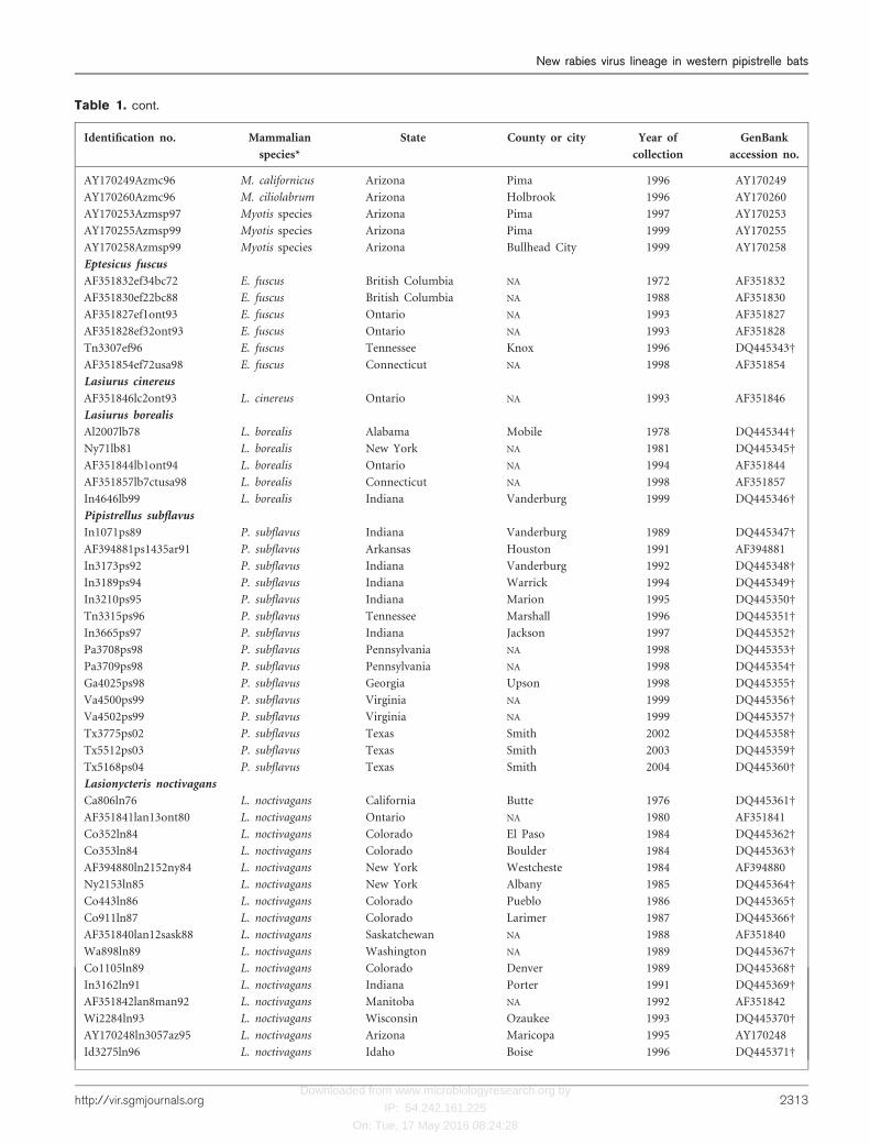
Table 1. cont.
Identification no. Mammalian
species*
State County or city Year of
collection
GenBank
accession no.
Id3277ln96 L. noctivagans Idaho Benewah 1996 DQ445372D
Co5153ln96 L. noctivagans Colorado NA 1996 DQ445373D
Wa0323ln97 L. noctivagans Washington NA 1997 DQ445374D
Wa341ln97 L. noctivagans Washington NA 1997 DQ445375D
Wa410ln97 L. noctivagans Washington NA 1997 DQ445376D
Wa580ln98 L. noctivagans Washington NA 1998 DQ445377D
Nc4249ln99 L. noctivagans North Carolina Stanly 1999 DQ445378D
Wa161ln00 L. noctivagans Washington NA 2000 DQ445379D
Co5290ln00 L. noctivagans Colorado NA 2000 DQ445380D
Wa1185ln03 L. noctivagans Washington NA 2003 DQ445381D
Wa01ln04 L. noctivagans Washington Kitsap 2004 DQ445382D
Homo sapiens
AF394882Tnhm94 Homo sapiens Tennessee Roane 1994 AF394882
Canis familiaris
AF351848cf1alta98 Canis familiaris Alberta NA 1998 AF351848
Vulpes vulpes
AF351851vv2pei93 Vulpes vulpes Prince Edward Ils. NA 1993 AF351851
*Species from which virus was collected.
DSequences generated in our study.
dMyotis species represents bats identified as Myotis species; NA, location unknown.
2314 Journal of General Virology 87
R. Franka and others

Downloaded from www.microbiologyresearch.org by
IP: 54.242.161.225
On: Tue, 17 May 2016 08:24:28
Fig. 2. Phylogenetic tree of bat and terrestriallyssaviruses using NJ analysis (p-distancemodel) of a portion of the N gene codingsequence (264 nt) with 1000 bootstrapreplicates. Groups I–IV as previously pub-lished (Nadin-Davis et al., 2001). Other con-sensus sequences: EF1 and EF2, E. fuscus
North America; EF3AZ, Eptesicus fuscus
Arizona; MCiAg, Myotis species Chile andArgentina; TbSA, Tadarida brasiliensis SouthAmerica; TBNA, Tadarida brasiliensis NorthAmerica; Dr6Mexico and Dr1-5Mexico,Desmodus rotundus Mexico; LCi, Lasiurus
species Chile; LBNA, Lasiurus borealis
North America; LCNA, Lasiurus cinereus
North America; LCCi, Lasiurus cinereus
Chile; W13MxNorthcentralSk, North Centralskunk Mexico; W11USSouthcentralSk, SouthCentral skunk USA; W12CanadaUSRaccoon,raccoon Canada and USA; W5USNorth-centralSk8498, North Central skunk USA;EBLV2, European Bat Lyssavirus 2; Sagui,rabies virus from common marmoset(Callithrix jacchus jacchus) from Brazil.Consensus sequences as in Velasco-Villaet al. (2005, 2006).
http://vir.sgmjournals.org 2315
New rabies virus lineage in western pipistrelle batsNew rabies virus lineage in western pipistrelle bats

Downloaded from www.microbiologyresearch.org by
IP: 54.242.161.225
On: Tue, 17 May 2016 08:24:28
The placement of taxa within the large clade containingLasionycteris noctivagans, Lasiurus cinereus, Lasiurus borealisand P. subflavus also differed in the three analyses. Accordingto the NJ tree (Fig. 2), a sister relationship was found betweenP. subflavus and Lasionycteris noctivagans, but bootstrapsupport was low. This clade was then sister to a cladecomprised of Lasiurus cinereus and Lasiurus borealis, with abootstrap value of 98 for the association of the four groups. Inthe ML analysis (Fig. 4), it appeared that the Lasionycterisnoctivagans, Lasiurus cinereus/borealis and P. subflavus cladeswere unresolved in relation to each other. Finally, a clade ofLasiurus cinereus and Lasiurus borealis (posterior probabil-ity=100) was sister to Lasionycteris noctivagans, which thengrouped with the P. subflavus clade in the Bayesian tree(Fig. 5). Support for the Lasionycteris noctivagans, Lasiuruscinereus, Lasiurus borealis and P. subflavus clade wasindicated by a posterior probability value of 100.
DISCUSSION
Previous phylogenetic analyses of nucleotide sequence datafrom representative RABV variants of North American batspecies, at both the N and G (glycoprotein) gene loci,identified two major clades and four principal phylogeneticgroups (I–IV), which were associated with particular batspecies (Nadin-Davis et al., 2001). Among North Americanbat RABV variants, a notable division was shown betweengroup (clade) I specimens associated with colonial, non-migratory bats (Myotis species and E. fuscus) and those ofgroup (clade) II harboured mainly by solitary, migratoryspecies (Lasiurus species and Lasionycteris noctivagans).These conclusions, however, will need to be re-evaluated,because many publications have suggested the gregariousbehaviour of Lasionycteris noctivagans (Parsons et al., 1986;Campbell et al., 1996; Mattson et al., 1996; Vonhof &Barclay, 1996; Betts, 1998). Certain Myotis species weresuggested as reservoirs, an observation often obscuredpreviously by their frequent infection with viral variants
from other Chiroptera. An additional group (III) apparentlycirculates in E. fuscus, while viruses harboured by bothMolossidae and Desmodontinae bats of Latin America forma phylogenetically distinct clade (group IV) (Nadin-Daviset al., 2001). Similar branching patterns were presented byHughes et al. (2005).
In our study RABV variants harboured by North Americanbats also formed two major clades. However, in contrastwith Nadin-Davis et al. (2001), the first clade was formed bythe Ph1, Ph2 and Myotis clades (and not by Myotis speciesand E. fuscus 1 and 2 clades) and a second one by Lasiony-cteris noctivagans, Lasiurus cinereus, Lasiurus borealis and P.subflavus. Following the two large clades, a cluster of E.fuscus (1 and 2) joined as the basal group of bat RABVs.Interestingly, the overall branching pattern of the treesconstructed in our study by using the NJ, ML and Bayesianmethods was the same without regards to the method used.The E. fuscus 3 clade represents a group that branchesdifferently if we use different methods. The differences inbranching of Myotis species and E. fuscus 1–3 clades betweentrees constructed in Nadin-Davis et al. (2001), Hughes et al.(2005), and in our study, could be explained by the differentmethods and evolution models used, and most importantlyby different outgroups and datasets used. We used sequencesof RABV collected from P. hesperus (Ph), which were notincluded in the previous publications. These new clades(Ph1 and Ph2) brought novel insights into the relationshipbetween clades, and the different branching of E. fuscus is theresult of these changes. However, the position of the E. fuscus3 clade in our NJ tree was not supported, similarly as itwasn’t supported in the NJ tree published by Nadin-Daviset al. (2001).
Our study revealed that 79 % of RABV samples originat-ing from P. hesperus bats from California and Arizona (alarge part of their natural geographical range in the USA)formed a monophyletic clade Ph1 with high bootstrap
Fig. 3. Consensus amino acid sequences of major bat rabies viruses (59 end of the N gene). Myotis, Myotis species; Ph1 andPh2, P. hesperus; Ln Ps, Lasionycteris noctivagans and P. subflavus; EF1 and EF2, Eptesicus fuscus North America; EF3AZ,Eptesicus fuscus Arizona; LCNA, Lasiurus cinereus North America; LCCi, Lasiurus cinereus Chile; LCi, Lasiurus speciesChile; LBNA, Lasiurus borealis North America; TBNA, Tadarida brasiliensis North America; TbSA, Tadarida brasiliensis SouthAmerica; DrMexico, Desmodus rotundus Mexico; DrSA, Desmodus rotundus South America; PV, Pasteur virus.
2316 Journal of General Virology 87
R. Franka and others

Downloaded from www.microbiologyresearch.org by
IP: 54.242.161.225
On: Tue, 17 May 2016 08:24:28
Fig. 4. Phylogenetic tree generated via ML analysis using the HKY85+G model for a portion of the N gene coding sequence(264 nt). Abbreviations as in Fig. 2.
http://vir.sgmjournals.org 2317
New rabies virus lineage in western pipistrelle batsNew rabies virus lineage in western pipistrelle bats
Downloaded from www.microbiologyresearch.org by
IP: 54.242.161.225
On: Tue, 17 May 2016 08:24:28
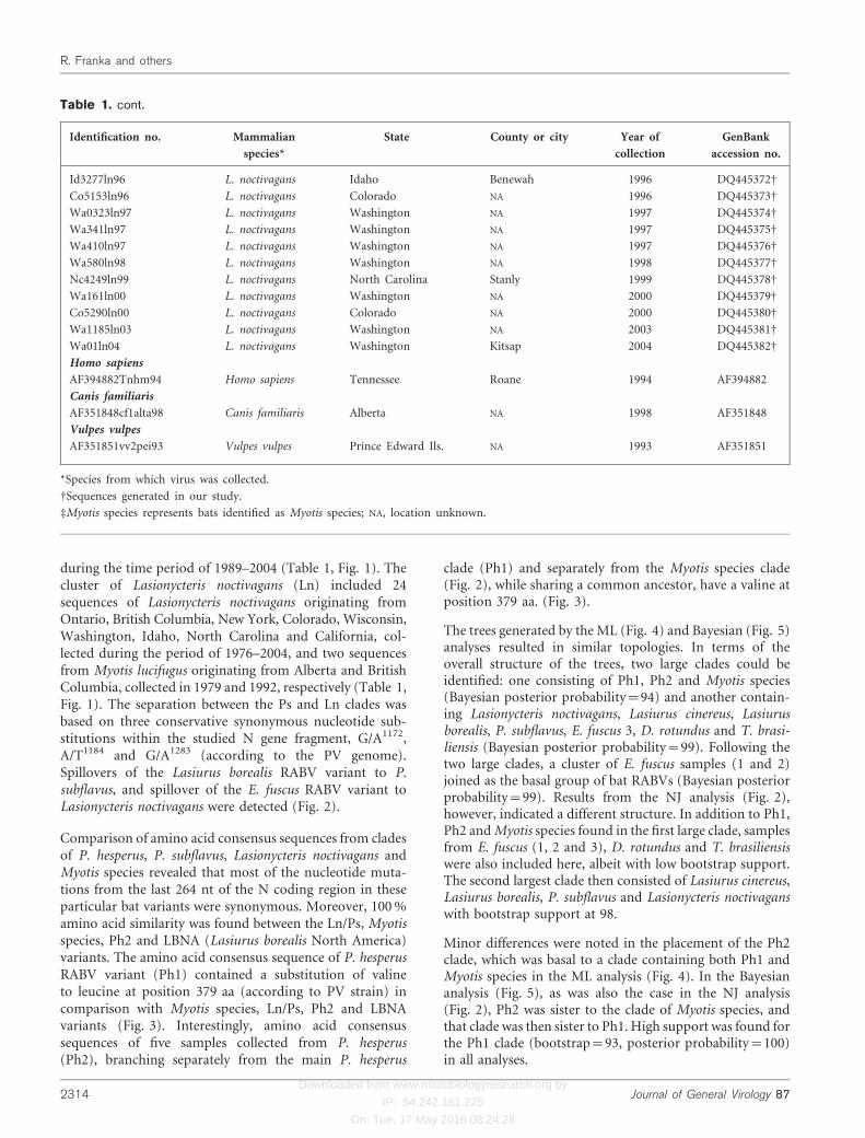
support and high posterior probabilities, separated fromother phylogenetic lineages of RABV (Fig. 2, 4 and 5).
Genetic consistency over space and time, along with thephylogenetic independence of the Ph1 clade, suggests that
Fig. 5. Phylogenetic tree generated viaBayesian analysis using the GTR+I+Gmodel for a portion of the N gene codingsequence (264 nt). Posterior probabilityvalues above 50 were shown for all majornodes (¢95=statistical support). Abbrevia-tions as in Fig. 2.
2318 Journal of General Virology 87
R. Franka and others
Downloaded from www.microbiologyresearch.org by
IP: 54.242.161.225
On: Tue, 17 May 2016 08:24:28
this RABV variant has been maintained in the P. hesperuspopulation independently. These findings raise a question ofadequacy of current passive surveillance methods and areindicative of the public health risk of the P. hesperus (Ph)RABV variant. Approximately 13 % of P. hesperus samplescollected in California (Los Angeles, El Dorado; distancebetween two sites approximately 550 km) during 2002–2005 formed a separate clade (Ph2). This clade was alsosupported by a moderate bootstrap value of 78 %. With onlyfive samples (three from the same county from 2003 to2004) representing this group, it is difficult to interpretconclusively the relationship of this clade to the Myotisspecies and P. hesperus 1 clades. However, from all threetrees (Fig. 2, 4 and 5), it is obvious that both P. hesperusRABV lineages (Ph1 and Ph2) shared a more recentcommon ancestor with a RABV variant associated with aMyotis species and their clade is separated from the cladeformed by Lasionycteris noctivagans, Lasiurus species andP. subflavus. Further analyses are needed to explain therelationship between the Ph1, Ph2 and Myotis species clades.Likely relevant to the differentiation of P. hesperus cladesPh1 and Ph2 through evolution are differences in thegeographical and ecological distributions of the P. hesperushost populations that maintain the virus of each clade.The hosts of Ph1 were collected in desert areas east of themountains (Sierra Nevada and Coastal Mt TransverseRanges: Tehachapi Mts, San Gabriel Mts and San BernardinoMts) that separate this P. hesperus population from apopulation distributed in more humid valley and coastalareas to the west. The pelage of these bats is pale grey incolour, contributing to their early subspecific distinction asP. h. hesperus. Contrastingly, the hosts of Ph2 were collectedin more western areas, where the pelage of these bats ismore brownish in colour, contributing to their early sub-specific distinction as P. h. merriami (Hatfield, 1936; Hall &Dalquest, 1950; Findley & Traut, 1970).
Only two samples collected from P. hesperus belonged toother RABV lineages (Myotis species clade), suggestingrelatively rare spillover events from Myotis species. As well,only four of more than 50 sequences of RABV collectedfrom Myotis species were included in the Ph1 clade, whichcould be explained by spillover events or by misidentifica-tion of bat species.
No spillover between Lasionycteris noctivagans, P. subflavusand P. hesperus was detected in the present study. Inaddition, spillover of the P. hesperus RABV variants intoterrestrial mammals has not been detected. In contrast,Messenger et al. (2003) reported a high prevalence ofLasionycteris noctivagans and P. subflavus variants amongterrestrial mammals in the same regions where human caseshave occurred. They suggested that increased infectivity ofthese RABV variants is responsible for relatively frequentspillover events. Our analyses showed that P. hesperusharbours RABV variants genetically distinct from the Ln andPs variants and from other RABV variants. This findingcorresponds interestingly to the taxonomic revisions of the
relationship between P. subflavus and P. hesperus, recentlypublished by Hoofer & Van Den Bussche (2003) and Hooferet al. (2006). Two bat species previously classified as mem-bers of one genus and now, on the basis of genetic classifi-cation, assigned to two different genera harbour two distinctRABV variants. New pathogenesis studies focused on theinfectivity of all bat RABV variants, together with improvedepidemiological analysis of both bat RABV prevalence andaccurate bat species identification, are necessary to evaluatethe increased infectivity hypothesis.
Data generated by phylogenetic analyses provided us withbasic information about relationships between RABVvariants harboured by North American bats. Thoughtfulinterpretation of these data, coupled with relevant epide-miological findings can lead to insights of new hypothesesthat seek to explain the frequency of human rabies casesassociated with bat RABV variants. As the smallest NorthAmerican bat P. hesperus weighs only 2–6 g, compared withthe larger P. subflavus (6–10 g) and Lasionycteris noctivagans(9–12 g). P. hesperus becomes aggressive as it developsrabies, and it engages in seemingly unprovoked attacks. Thisbehaviour may be necessary for the Ph RABV variantssurvival via transmission to other P. hesperus bats, whichare strictly solitary and may not be readily approached.Such attacks on larger bats by this diminutive butterfly sizedbat could end in its demise through retaliatory bites. Givenits occasional tendency to apparently attack people whenrabid, no human rabies cases have been linked to the PhRABV variants. However, such overt exposures may signalthe need for post-exposure prophylaxis (Constantine, 1970).Of relevant interest, a significantly greater proportion ofsmaller bats bit people than larger bats: 39 of 279 rabid batsbit people at rates ranging from 67 % of P. hesperus to only15 % of E. fuscus (Constantine, 1967). These results mightsuggest greater aggressiveness of the smaller bats or greatercare by people to avoid bites of the larger bats.
The lack of human cases linked to the Ph variants may berelated to the biology and ecology of P. hesperus. These batshave few contacts with humans and apparent difficultyinflicting deep wounds by their small teeth. Additionally,specific viral properties such as a higher level of adaptationto a principal host and a relatively lower pathogenicity forother species or limitations in virus excretion in terms ofintermittent shedding and viral dose may be operative. Thelack of spillover events to terrestrial carnivores and infre-quency of spillover to other bat species strongly supportthe hypothesis that addresses specific viral properties. Thethickness of mammalian fur could form a barrier protectingskin from penetration by the small teeth of P. hesperus, butthis argument fails to explain the absence of human casesassociated with Ph RABV variants. Moreover, terrestrialmammals contact bats primarily by the paws and mouth,where fur coverage is absent (lips, mucosae of mouthcavity). One of the most frequently reported rabid bats inCalifornia is Myotis californicus, which is also a smallbat species (weight 3?3–5?4 g). However, no human cases
http://vir.sgmjournals.org 2319
New rabies virus lineage in western pipistrelle batsNew rabies virus lineage in western pipistrelle bats
Downloaded from www.microbiologyresearch.org by
IP: 54.242.161.225
On: Tue, 17 May 2016 08:24:28
caused by this RABV variant have been reported inCalifornia, suggesting that human exposures are recognizedand treated, or these viruses fail to achieve successfulinfection. Two human cases caused by Myotis species RABVwere reported in 1984 (Pennsylvania, no history of contactwith a bat was reported) and in 1995 (Washington, rabidMyotis found in the bedroom, no known animal bite)(Messenger et al., 2002). Interestingly, most of the humanrabies cases, which had an unknown history of bite expo-sure, were caused by different RABV variants, harbouredby medium-sized bat species (14 of 16 P. subflavus variant,6–10 g; five of eight Lasionycteris noctivagans variant,9–12 g; four of five T. brasiliensis variant, 10–15 g; one ofone E. fuscus variant, 11–23 g and two of two Myotis speciesvariant, weight range according to species 3–13 g). Among15 human rabies cases reported in California since 1958,seven were acquired indigenously and all were characterizedas bat RABV variants [four Ln/Ps variant and three T.brasiliensis (Tb) variant] (Messenger et al., 2002).
Thus, given the rather diminutive P. hesperus, small bodysize alone (small vector hypothesis) may be inadequate toexplain the association between human rabies cases andcertain RABV isolates from bats. More importantly in thisregard seem to be viral properties, such as viral infectivityand relative virulence, in association with particular hostcharacteristics (e.g. receptors, body temperature, immunesystem response, etc.) coupled with host lifestyle (e.g. moresolitary versus colonial bat species).
Hughes et al. (2005) inferred from the estimated substitutionrate of the N gene that the initial branching of parental rabiesvirus in bats gave rise to the current variants associated withinfection of T. brasiliensis and D. rotundus. Compart-mentalization of RABV into lineages associated withinfection of solitary bat species (or small group-formingspecies, such as P. subflavus and Lasiurus species) and morecolonial bat species (E. fuscus, Myotis species) occurred later.Questions remain, as to the degree of sociality and thedynamics of RABV transmission in different bat species. Canwe consider bats as solitary only if individuals are solitary allyear or also those which form small (5–50 individuals)maternity colonies? There is an obvious difference betweenlarge colonies of T. brasiliensis consisting of millions ofindividuals, seasonal colonies of E. fuscus and Myotis specieswith hundreds of individuals, and maternity colonies of P.subflavus and Lasionycteris noctivagans with only tens ofindividuals. The results of Hughes et al. (2005) showed thatadaptation of RABV to colonial bat species occurred morequickly than adaptations for the more solitary species,suggesting dependence of this process on host ecology.
In this regard, one interesting result of our analysis is thatthe Ln and Ps RABV variants have circulated in two differ-ent bat species populations (P. subflavus and Lasionycterisnoctivagans) probably separately from each other andmaintain reasonably high genetic stability over relativelylong periods (Ps, 1989–1999; Ln, 1976–2004), especiallygiven the error of replication fidelity in RNA viral genomes.
The separation between Ps and Ln clades was based onthree conservative nucleotide substitutions within thestudied N gene fragment, G/A1172, A/T1184 and G/A1283
(according to the PV genome). Although all threesubstitutions were synonymous, they suggest that theseRABV variants may have circulated independently in thesehost species. Since bootstrap support for separation ofP. subflavus and Lasionycteris noctivagans clades in theNJ tree and similarly posterior probabilities in the Bayesiantree were low, further examination of the RABV variantscollected from Lasionycteris noctivagans and P. subflavuswill be necessary to understand the evolutionary relation-ships of these two clades. Taking into account the factthat 16 of 24 human rabies cases in the USA over the last50 years were associated with P. subflavus, and our recentfinding of possible independent circulation of Ln and PsRABVs in Ln and Ps populations, the finding of Geluso et al.(2005) that P. sublfavus has expanded westward in the USAto New Mexico, South Dakota and Texas in recent years(Fig. 1) should highlight a need for enhanced surveillance.
All nucleotide mutations in the highly variable 264 ntfragment of the N gene from P. hesperus (Ph2), P. subflavus,Lasionycteris noctivagans, Myotis species and LBNA weresynonymous with identical amino acid consensus sequences.The only exception was a substitution of the valine forleucine at position 379 aa (according to the PV strain), inPh1 RABV (Fig. 3). This substitution is, however, structu-rally conservative. Such findings raise a question of potentialconstraints of RABV evolution, despite differences in theestimated substitution rate between more solitary or colon-ial bat species. Comparison of other genomic regions wouldfacilitate a better understanding of differences between theseRABV lineages.
Further phylogenetic analysis of additional samples fromP. hesperus, P. subflavus and Lasionycteris noctivagans fromNorth America are needed to corroborate the resultsrevealed by our limited dataset. In particular, consideringthe extent of their distributions and their obvious role inpublic health in Canada and the USA, greater attention tothese bats should occur in Mexico and Central America.Moreover, in vitro and in vivo pathogenesis studies shouldbe conducted for a better understanding of the features ofthese particular RABV variants.
ACKNOWLEDGEMENTS
The authors thank the staff in the Viral and Rickettsial ZoonosesBranch at the CDC for their outstanding technical expertise andcontributions to this work. We also thank our colleagues from StatePublic Health Laboratories for rabies surveillance in the USA. Inparticular, we thank the California Department of Health Services,Viral and Rickettsial Disease Laboratory, for providing laboratoryspace and support. R. F. was funded by an American Societyfor Microbiology and National Centers for Infectious Diseasepost-doctoral fellowship. The findings and conclusions in this reportare those of the author(s) and do not necessarily represent the views ofthe funding agency.
2320 Journal of General Virology 87
R. Franka and others
Downloaded from www.microbiologyresearch.org by
IP: 54.242.161.225
On: Tue, 17 May 2016 08:24:28
REFERENCES
Barbour, R. W. & Davis, W. H. (1969). Bats of America. Lexington:
University of Kentucky Press.
Belotto, A., Leanes, L. F., Schneider, M. C., Tamayo, H. & Correa, E.
(2005). Overview of rabies in the Americas. Virus Res 111, 5–12.
Betts, B. J. (1998). Roosts used by maternity colonies of silver-haired
bats in northeastern Oregon. J Mammal 79, 643–650.
Campbell, L. A., Hallett, J. G. & O’Connell, M. A. (1996). Con-
servation of bats in managed forests: use of roosts by Lasionycteris
noctivagans. J Mammal 77, 976–984.
CDC (1994). Human rabies - Texas and California, 1993. Morb
Mortal Wkly Rep 43, 93–96.
Constantine, D. G. (1967). Bat rabies in the southwestern United
States. Public Health Rep 82, 867–888.
Constantine, D. G. (1970). Bats in relation to the health, welfare, and
economy of man. In Biology of Bats, vol. 2, pp. 319–449. Edited by
W. A. Wimsatt. New York: Academic Press.
Constantine, D. G. (1979). An updated list of rabies-infected bats in
North America. J Wildl Dis 15, 347–349.
Dean, D. J., Abelseth, M. K. & Atanasiu, P. (1996). The fluorescent
antibody test. In Laboratory Techniques in Rabies, 4th edn, pp. 88–95.
Edited by F. X. Meslin, M. M. Kaplan & H. Koprowski. Geneva,
Switzerland: WHO.
Dietzschold, B., Morimoto, K., Hooper, D. C., Smith, J. S.,
Rupprecht, C. E. & Koprowski, H. (2000). Genotypic and phenotypic
diversity of rabies virus variants involved in human rabies:
implications for post-exposure prophylaxis. J Hum Virol 3, 50–57.
Findley, J. S. & Traut, G. L. (1970). Geographic variation in
Pipistrellus hesperus. J Mammal 51, 741–765.
Geluso, K., Mollhagen, T. R., Tigner, J. M. & Bogan, M. A. (2005).Westward expansion of the eastern pipistrelle (Pipistrellus subflavus)
in the United States including new records from New Mexico, South
Dakota and Texas. West N Am Nat 65, 405–409.
Hall, T. A. (1999). BioEdit: a user friendly biological sequence align-
ment editor and analysis program for Windows 95/98/NT. Nucleic
Acids Symp Ser 41, 95–98.
Hall, E. R. & Dalquest, W. W. (1950). A synopsis of the American bats
of the genus Pipistrellus. Univ. Kansas Publ Mus Nat Hist 1, 591–602.
Hasegawa, M., Kishino, H. & Yano, T. (1985). Dating of the human-
ape splitting by a molecular clock of mitochondrial DNA. J Mol Evol
22, 160–174.
Hatfield, D. M. (1936). A revision of the Pipistrellus hesperus group of
bats. J Mammal 17, 257–262.
Hoofer, S. R. & Van Den Bussche, R. A. (2003). Molecular phylogenetics
of the chiropteran family Vespertilionidae. Acta Chiropt 5, S1–S63.
Hoofer, S. R., Van Den Bussche, R. A. & Horacek, I. (2006). Generic
status of American pipistrelles (Vespertilionidae) with description of
a new genus. J Mammal (in press).
Hughes, G. J., Orciari, L. A. & Rupprecht, C. E. (2005). Evolutionary
timescale of rabies virus adaptation to North American bats inferred
from the substitution rate of the nucleoprotein gene. J Gen Virol 86,
1467–1474.
Jeanmougin, F., Thompson, J. D., Gouy, M., Higgins, D. G. & Gibson,
T. J. (1998). Multiple sequence alignment with CLUSTAL X. Trends
Biochem Sci 23, 403–405.
Kumar, S., Tamura, K., Jakobsen, I. B. & Nei, M. (2001). MEGA2:
molecular evolutionary genetics analysis software. Bioinformatics 17,
1244–1245.
Mattson, T. A., Buskirk, S. W. & Stanton, N. L. (1996). Roost sites of
the silver-haired bat (Lasionycteris noctivagans) in the Black Hills,
South Dakota. Great Basin Nat 56, 247–253.
Messenger, S. L., Smith, J. S. & Rupprecht, C. E. (2002). Emerging
epidemiology of bat-associated cryptic cases of rabies in humans in
the United States. Clin Infect Dis 35, 738–747.
Messenger, S. L., Smith, J. S., Orciari, L. A., Yager, P. A. &
Rupprecht, C. E. (2003). Emerging pattern of rabies deaths and
increased viral infectivity. Emerg Infect Dis 9, 151–154.
Morimoto, K., Patel, M., Corisdeo, S., Hooper, D. C., Fu, Z. F.,
Rupprecht, C. E., Koprowski, H. & Dietzschold, B. (1996).
Characterization of a unique variant of bat rabies virus responsible
for newly emerging human cases in North America. Proc Natl Acad
Sci U S A 93, 5653–5658.
Nadin-Davis, S. A., Huang, W., Armstrong, J., Casey, G. A.,
Bahloul, C., Tordo, N. & Wandeler, A. I. (2001). Antigenic and
genetic divergence of rabies viruses from bat species indigenous to
Canada. Virus Res 74, 139–156.
Parsons, H. J., Smith, D. A. & Whittam, R. F. (1986). Maternity
colonies of silver-haired bats Lasionycteris noctivagans, in Ontario
and Saskatchewan. J Mammal 67, 598–600.
Posada, D. & Crandall, K. A. (1998). MODELTEST: testing the model
of DNA substitution. Bioinformatics 14, 817–818.
Ronquist, F. & Huelsenbeck, J. P. (2003). MrBayes 3: Bayesian
phylogenetic inference under mixed models. Bioinformatics 19, 1572–
1574.
Rupprecht, C. E., Dietzschold, B., Wunner, W. H. & Koprowski, H.
(1991). Antigenic relationships of lyssaviruses. In The Natural
History of Rabies, 2nd edn, pp. 69–100. Edited by G. M. Baer. New
York: Academic Press.
Rupprecht, C. E., Smith, J. S., Fekadu, M. & Childs, J. E. (1995). The
ascension of wildlife rabies: a cause for public health concern or
intervention? Emerg Infect Dis 1, 107–114.
Smith, J. S. (2002). Molecular epidemiology. In Rabies, pp. 79–111.
Edited by A. C. Jackson & W. H. Wunner. New York: Academic
Press.
Smith, J. S., Orciari, L. A., Yager, P. A., Seidel, H. D. & Warner, C. K.
(1992). Epidemiologic and historical relationship among 87 rabies
virus isolates as determined by limited sequence analysis. J Infect Dis
166, 296–307.
Swofford, D. L. (2002). PAUP*: phylogenetic analysis using parsimony
(*and other methods) version 4.0b10. Sunderland, MA: Sinauer
Associates.
Trimarchi, C. V. & Smith, J. S. (2002). Diagnostic evaluation. In
Rabies, pp. 307–349. Edited by A. C. Jackson & W. H. Wunner.
New York: Academic Press.
Velasco-Villa, A., Orciari, L. A., Souza, V., Juarez-Islas, V., Gomez-
Sierra, M., Castillo, A., Flisser, A. & Rupprecht, C. E. (2005).
Molecular epizootiology of rabies associated with terrestrial carni-
vores in Mexico. Virus Res 111, 13–27.
Velasco-Villa, A., Orciari, L. A., Juarez-Islas, V. & 9 other authors
(2006). Molecular diversity of rabies viruses associated with bats in
Mexico and other countries of the Americas. J Clin Microbiol 44,
1697–1710.
Vonhof, M. J. & Barclay, R. M. R. (1996). Roost-site selection and
roosting ecology of forest dwelling bats in southern British
Columbia. Can J Zool/Rev Can Zool 74, 1797–1805.
WHO (2005). WHO expert consultation on rabies. First Report,
WHO Technical Report Series 931, Geneva, Switzerland, 2004,
pp. 121. http://www.who.int/rabies/931/en/index.html.
Wilson, D. E. & Ruff, S. (1999). Family Vespertilionidae. In The
Smithsonian Book of North American Mammals, pp. 94–131.
Edited by D. E. Wilson & S. Ruff. Singapore: Smithsonian
Institution.
http://vir.sgmjournals.org 2321
New rabies virus lineage in western pipistrelle batsNew rabies virus lineage in western pipistrelle bats
Related Documents


![Essex Bat Group Newsletter 2016 Autumn · Writtle Forest (a group of more typical Essex woods). Nathusius’ pipistrelle [Photo: A Palmer] Essex Nathusius’ Pipistrelle Project We](https://static.cupdf.com/doc/110x72/5fc1334842e28310c87cfe68/essex-bat-group-newsletter-2016-writtle-forest-a-group-of-more-typical-essex-woods.jpg)