Citation: Wang, S.; Alenius, H.; El-Nezami, H.; Karisola, P. A New Look at the Effects of Engineered ZnO and TiO 2 Nanoparticles: Evidence from Transcriptomics Studies. Nanomaterials 2022, 12, 1247. https://doi.org/10.3390/ nano12081247 Academic Editor: Joachim Clement Received: 2 March 2022 Accepted: 31 March 2022 Published: 7 April 2022 Publisher’s Note: MDPI stays neutral with regard to jurisdictional claims in published maps and institutional affil- iations. Copyright: © 2022 by the authors. Licensee MDPI, Basel, Switzerland. This article is an open access article distributed under the terms and conditions of the Creative Commons Attribution (CC BY) license (https:// creativecommons.org/licenses/by/ 4.0/). nanomaterials Review A New Look at the Effects of Engineered ZnO and TiO 2 Nanoparticles: Evidence from Transcriptomics Studies Shuyuan Wang 1 , Harri Alenius 2,3 , Hani El-Nezami 1,4, * and Piia Karisola 2, * 1 School of Biological Sciences, University of Hong Kong, Hong Kong Island, Hong Kong, China; [email protected] 2 Human Microbiome Research (HUMI), Medical Faculty, University of Helsinki, 00014 Helsinki, Finland; harri.alenius@helsinki.fi 3 Institute of Environmental Medicine (IMM), Karolinska Institutet, Systems Toxicology, 171 77 Stockholm, Sweden 4 Institute of Public Health and Clinical Nutrition, University of Eastern Finland, 70211 Kuopio, Finland * Correspondence: [email protected] (H.E.-N.); piia.karisola@helsinki.fi (P.K.) Abstract: Titanium dioxide (TiO 2 ) and zinc oxide (ZnO) nanoparticles (NPs) have attracted a great deal of attention due to their excellent electrical, optical, whitening, UV-adsorbing and bactericidal properties. The extensive production and utilization of these NPs increases their chances of being released into the environment and conferring unintended biological effects upon exposure. With the increasingly prevalent use of the omics technique, new data are burgeoning which provide a global view on the overall changes induced by exposures to NPs. In this review, we provide an account of the biological effects of ZnO and TiO 2 NPs arising from transcriptomics in in vivo and in vitro studies. In addition to studies on humans and mice, we also describe findings on ecotoxicology-related species, such as Danio rerio (zebrafish), Caenorhabditis elegans (nematode) or Arabidopsis thaliana (thale cress). Based on evidence from transcriptomics studies, we discuss particle-induced biological effects, including cytotoxicity, developmental alterations and immune responses, that are dependent on both material-intrinsic and acquired/transformed properties. This review seeks to provide a holistic insight into the global changes induced by ZnO and TiO 2 NPs pertinent to human and ecotoxicology. Keywords: transcriptomics; engineered metal nanoparticles; titanium dioxide; zinc oxide; animal models (in vivo); cell cultures (in vitro); (eco)toxicology; zebrafish; C. elegans; Arabidopsis thaliana 1. Introduction The rapid development of nanotechnology holds tremendous potential for wide growth in the applications made of novel nanoparticles (NPs) for various purposes in elec- tronics, medicine, coating materials and even in personal care products (including cosmet- ics), with more coming online every day [1]. It has been estimated that over 1800 engineered nanomaterial-based products are available in the global market [2], generating a total pro- duction volume of nanomaterials of around 11 million tons worldwide [3,4]. Titanium dioxide (TiO 2 ) NPs have been extensively produced as a whitening, anti-caking and color- ing agent in various products such as paints, cosmetics and foodstuffs [5]. Zinc oxide (ZnO) NPs have excellent semiconducting, light-scattering and anti-microbial properties, which make them a suitable component for electrical and optical devices, cosmetic products and food-packaging materials [6]. At the nanoscale, NPs have a much larger surface area, which confers substantially different and usually somehow enhanced surface properties compared to their bulk-sized counterparts [7]. Although the biological and environmental effects of engineered metal-type NPs have been reviewed in the literature, the majority of published articles have discussed or summarized the responses induced by silver (Ag) NPs [8–12]. There are fewer comprehensive reviews on the impacts of the other two commonly used metal NPs, ZnO and TiO 2 [1,6,13]. Nanomaterials 2022, 12, 1247. https://doi.org/10.3390/nano12081247 https://www.mdpi.com/journal/nanomaterials

Welcome message from author
This document is posted to help you gain knowledge. Please leave a comment to let me know what you think about it! Share it to your friends and learn new things together.
Transcript

�����������������
Citation: Wang, S.; Alenius, H.;
El-Nezami, H.; Karisola, P. A New
Look at the Effects of Engineered
ZnO and TiO2 Nanoparticles:
Evidence from Transcriptomics
Studies. Nanomaterials 2022, 12, 1247.
https://doi.org/10.3390/
nano12081247
Academic Editor: Joachim Clement
Received: 2 March 2022
Accepted: 31 March 2022
Published: 7 April 2022
Publisher’s Note: MDPI stays neutral
with regard to jurisdictional claims in
published maps and institutional affil-
iations.
Copyright: © 2022 by the authors.
Licensee MDPI, Basel, Switzerland.
This article is an open access article
distributed under the terms and
conditions of the Creative Commons
Attribution (CC BY) license (https://
creativecommons.org/licenses/by/
4.0/).
nanomaterials
Review
A New Look at the Effects of Engineered ZnO and TiO2Nanoparticles: Evidence from Transcriptomics StudiesShuyuan Wang 1 , Harri Alenius 2,3 , Hani El-Nezami 1,4,* and Piia Karisola 2,*
1 School of Biological Sciences, University of Hong Kong, Hong Kong Island, Hong Kong, China;[email protected]
2 Human Microbiome Research (HUMI), Medical Faculty, University of Helsinki, 00014 Helsinki, Finland;[email protected]
3 Institute of Environmental Medicine (IMM), Karolinska Institutet, Systems Toxicology,171 77 Stockholm, Sweden
4 Institute of Public Health and Clinical Nutrition, University of Eastern Finland, 70211 Kuopio, Finland* Correspondence: [email protected] (H.E.-N.); [email protected] (P.K.)
Abstract: Titanium dioxide (TiO2) and zinc oxide (ZnO) nanoparticles (NPs) have attracted a greatdeal of attention due to their excellent electrical, optical, whitening, UV-adsorbing and bactericidalproperties. The extensive production and utilization of these NPs increases their chances of beingreleased into the environment and conferring unintended biological effects upon exposure. With theincreasingly prevalent use of the omics technique, new data are burgeoning which provide a globalview on the overall changes induced by exposures to NPs. In this review, we provide an account ofthe biological effects of ZnO and TiO2 NPs arising from transcriptomics in in vivo and in vitro studies.In addition to studies on humans and mice, we also describe findings on ecotoxicology-relatedspecies, such as Danio rerio (zebrafish), Caenorhabditis elegans (nematode) or Arabidopsis thaliana (thalecress). Based on evidence from transcriptomics studies, we discuss particle-induced biological effects,including cytotoxicity, developmental alterations and immune responses, that are dependent onboth material-intrinsic and acquired/transformed properties. This review seeks to provide a holisticinsight into the global changes induced by ZnO and TiO2 NPs pertinent to human and ecotoxicology.
Keywords: transcriptomics; engineered metal nanoparticles; titanium dioxide; zinc oxide; animalmodels (in vivo); cell cultures (in vitro); (eco)toxicology; zebrafish; C. elegans; Arabidopsis thaliana
1. Introduction
The rapid development of nanotechnology holds tremendous potential for widegrowth in the applications made of novel nanoparticles (NPs) for various purposes in elec-tronics, medicine, coating materials and even in personal care products (including cosmet-ics), with more coming online every day [1]. It has been estimated that over 1800 engineerednanomaterial-based products are available in the global market [2], generating a total pro-duction volume of nanomaterials of around 11 million tons worldwide [3,4]. Titaniumdioxide (TiO2) NPs have been extensively produced as a whitening, anti-caking and color-ing agent in various products such as paints, cosmetics and foodstuffs [5]. Zinc oxide (ZnO)NPs have excellent semiconducting, light-scattering and anti-microbial properties, whichmake them a suitable component for electrical and optical devices, cosmetic products andfood-packaging materials [6]. At the nanoscale, NPs have a much larger surface area, whichconfers substantially different and usually somehow enhanced surface properties comparedto their bulk-sized counterparts [7]. Although the biological and environmental effects ofengineered metal-type NPs have been reviewed in the literature, the majority of publishedarticles have discussed or summarized the responses induced by silver (Ag) NPs [8–12].There are fewer comprehensive reviews on the impacts of the other two commonly usedmetal NPs, ZnO and TiO2 [1,6,13].
Nanomaterials 2022, 12, 1247. https://doi.org/10.3390/nano12081247 https://www.mdpi.com/journal/nanomaterials

Nanomaterials 2022, 12, 1247 2 of 34
Downscaling of bulk materials allows NPs to gain access to biological organismsand have interactions with biomolecules, sometimes even inside the cells. This abilitysometimes makes NPs a desirable vehicle for delivering substances at the cellular level, asdemonstrated in the field of nanomedicine, but in other contexts, the enhanced penetrationmight lead to adverse effects on the living cells, as summarized by others [14,15]. However,the widely used single-endpoint measures are limited and too narrow to capture thegeneralized outcomes elicited by NPs. Conventional toxicology assays are useful forassessing the end-point effects that are evidenced in phenotypic hallmarks or systemicparameters. However, the data generated from this approach are insufficient to unravel thebiological changes occurring at the molecular level.
The systems biology approach enabled by the rapid development of omics technologiesprovides a more informative strategy that complements end-point changes with multi-leveland comprehensive molecular events upon exposures to NPs [16]. Transcriptomics, inparticular, captures changes in global gene expression patterns and strives to provide aholistic understanding of transcriptional mechanisms. Microarray, a fluorescence-basedtechnique, first emerged to enable the quantification of the differential abundance of mRNAtranscripts with predetermined sequences and predesigned oligomer probes [17]. Later inthe 2000s, the development of next-generation sequencing bloomed and is still being rapidlyupdated today. RNA-sequencing gives discrete digital read counts as a data output, and itshows enhanced performances in sensitivity, sequence resolution and result accuracy [18].It provides a high-quality measurement of gene regulation without relying on probe designand prior knowledge of genomic sequences [18]. The differentially expressed genes (DEGs)derived from either microarray or RNA-sequencing are interpreted into meaningful andbiologically relevant data via computational tools and a knowledge base of gene functionsand the associated pathways, such as Gene Ontology (GO), the Kyoto Encyclopedia ofGenes and Genomes (KEGG) and Ingenuity Pathway Analysis (IPA) [19]. A pathwayanalysis allows us to probe into the interactions between differentially regulated genes andto predict the biological pathways enriched by certain gene networking patterns [19].
In this review, we discuss the in vivo and in vitro biological effects of ZnO and TiO2NPs in the field of human and ecotoxicology, as evidenced in transcriptomics studies.
2. Synthetic and Biological Identities of ZnO and TiO2 NPs
Metal-based NPs, such as Ag, ZnO and TiO2, represent the largest proportion ofnanotechnology-derived products [2]. They are incorporated in a myriad of industrial,biomedical and personal care wares and devices, including solar cells, paints, cosmetics,clearing sprays, food additives and therapeutic agents [20,21], owing to their outstandingelectrical, plasmonic, optical and anti-microbial characteristics. Exposures to ZnO andTiO2 NPs, which are the focus of this review, are likely to occur in humans, and theconsequences of such exposures need to be addressed carefully. Despite different routesof exposures, the intrinsic properties of NPs, such as size, surface modifications anddissolution, fundamentally determine the adsorption of biomolecules onto NPs’ surfaces,thereby radically altering their acquired biological identities, cellular interactions andsubcellular localization [22,23].
2.1. Material Intrinsic Properties
The tunability of physicochemical characteristics of NPs lies at the heart of the innova-tive design of nanomaterials. Modified properties confer new or enhanced performances toachieve a wider or more efficient use of particles in different industrial sectors. Size, shape,surface chemistry and dissolution are the most-studied physicochemical properties of NPs,and they have been well demonstrated to exert significant influences on the biological ef-fects induced by NPs in various experimental set-ups. These important intrinsic propertiesare described here.

Nanomaterials 2022, 12, 1247 3 of 34
2.1.1. Size
According to the definition given by the International Organization for Standardization(ISO), NPs are classified as particles having at least one dimension falling in the rangeof 1 to 100 nm [24], which is the definition we adopted for this article. Additionally,how an NP is defined and its upper size limit rely on its specific application and field ofuse. For example, NPs utilized in pharmaceutical applications were previously definedas structures that varied in size from 10 nm to 1000 nm [25]. Nowadays, NPs as in vivodelivery vehicles in nanomedicine are often referred to as devices of less than 200 nm in size(i.e., the width of microcapillaries) to allow efficient release of the attached or encapsulatedtherapeutics [26]. Extremely small NPs (<1 nm) are able to penetrate directly across thecell membrane by passive diffusion, while bigger molecules are more readily taken up viaendocytosis mediated by specific receptors or caveolae- or clathrin-coated vesicles or viaphagocytosis [27]. The huge reduction in size increases the surface area of each particleand hence renders higher reactivity when compared with the bulk-sized equivalent. Onthe other hand, it has been suggested that the toxicity of NPs is inversely proportional tothe particle size. The high aggregation tendency of NPs also influences the actual toxicityperceived by cells or organisms. Generally, smaller particles demonstrate greater cellularinternalization and communications with biomolecules [14,15,28]. Moreover, particle sizecould affect the biopersistence, distribution and elimination of foreign matters from thebiological system. In studies on biological effects of NPs, results show that particle sizeplays a pivotal role in controlling the location of particle deposition, especially along therespiratory tract [29–31].
2.1.2. Surface Modifications
At the time of synthesis, NPs may be given distinct exterior properties with regard tosurface charge, coatings and functional groups. Modifications of these surface propertiespermit finetuning of the toxicity and behaviour of particles in biological systems. In additionto engineered alterations, the particle surface tends to be modified by the dynamic processof bio-corona formation, which consequently affects the ultimate toxicity of NPs. Surfacecharge is one of the most fundamental properties that influences particle aggregation,cellular uptake and other NP–cell interactions. A number of studies have revealed that NPscarrying positive charge, including ZnO, are more likely to penetrate through negatively-charged cell membranes and genetic materials compared to the same particle of a negativeor neutral charge, resulting in greater cytotoxic and genotoxic effects [6,32–34]. CationicNPs are also more easily recognized and removed by the immune cells [35]. In addition tocharges, the surface of NPs can be enshrouded with a layer of synthetic coating or extrafunctionalizations, such as a polyethylene glycol (PEG), amine group (-NH3) and carboxylgroup (-COOH) [36,37]. These external molecules are able to reduce particle aggregationvia the creation of steric and/or electrostatic repulsion between neighboring particles.Furthermore, they minimize protein interactions with the particles [38], thereby reducingproduction of reactive oxygen species (ROS) and lowering cytotoxicity [6]. On the otherhand, coated NPs are less recognizable by the immune cells due to the “stealth effect”,where coating materials mask the identity of NPs [36], which can lead to problems arisingfrom slower clearance and a higher bio-retention time.
2.1.3. Dissolution
A mix of dissolved metallic NPs and associated ions is produced upon particle dissolu-tion, which requires careful scrutiny of its antimicrobial capacity, cellular toxicity and otherbiological responses. The rate of dissolution is dependent on particles’ intrinsic properties,such as size, surface properties, surface area and crystallinity, and also external factors,including the pH, ionic strength and the concentration of surrounding media and storageconditions. Metal NPs exhibit different degrees of dissolution in various kinds of media.Generally, they do not dissolve readily in aqueous solutions at a neutral pH, such as in purewater or PBS [39–41]. Moreover, purely aqueous media do not reflect a realistic condition in

Nanomaterials 2022, 12, 1247 4 of 34
which NPs are in contact naturally. Biological or environmental media with a lower pH andpresence of proteins have been found to enhance the dissolution of metal NPs compared toinorganic salt solutions. For example, Ag NPs showed an increased release of Ag ions in acell culture medium (Dulbecco’s modified Eagle medium) with added fetal bovine serumthan in water [42], possibly due to the higher ionic strength and interactions of dissolvedions with cysteine and cysteine-containing proteins present in the cell medium [43,44].While TiO2 NPs showed minimal dissolution in artificial gastric juice (pH 1.5–2), ZnO NPsdissolved readily within minutes of immersion [45], which underscores the profound influ-ence of pH changes on particle dissolution during oral exposures to NPs. Bare metal NPstend to dissolve into ions more readily than capped counterparts. In addition, solubility isoften demonstrated to be inversely proportional to the particle size, as evidenced in studieson Ag, CuO, SiO2 and TiO2 NPs [46–50]. However, size does not seem to significantly affectparticle dissolution in the case of ZnO NPs [48,51,52].
2.2. Context-Dependent Properties Relevant for Humans
Upon gaining access to our body, NPs are biologically transformed and conferredwith a new identity depending on the formation and composition of another exterior layer,named the bio-corona. Both the bio-corona and inherent properties of NPs determine parti-cles’ fates in the biological system, especially in directing if and how they are recognizedby immune cells or interact with other types of somatic cells and cellular components.
2.2.1. Port of Entry
Major ZnO and TiO2 NPs exposure routes relevant for humans are (1) ingestion,(2) dermal contact and (3) inhalation. Air exposure occurs mainly under occupationalsettings during particle synthesis, handling and product manufacturing. Consumers mayalso inadvertently inhale NPs containing vaporized products, such as cleaning or cosmeticsprays. In addition, uses of nanosized ZnO and TiO2-incorporated personal care andcosmetic creams lead to particle entry via dermal contact for the general public. Lastly,ingestion contributes to the principal exposure mode of NP-containing food products, foodadditives (e.g., E171 (TiO2)) and food-packaging materials. It is worth noting that stabilityand aggregation issues are often associated with oral exposure to NPs upon contact witha multitude of biomolecules and food components and drastic changes in pH. Walczaket al. and Peters et al. have demonstrated that nanosized SiO2 and Ag aggregated intolarger particles in the gastric environment of an in vitro model that mimicked the humandigestion system [53,54]. Surprisingly, these particles reversed back to the nano-size rangewhen they entered the intestinal digestion stage, which was attributed to the shifts in pHand electrolyte concentration. More recently, Zhou et al. also reported a similar improvedstability of TiO2 and ZnO NPs in intestinal fluid under the influence of oil micelles likelypresent in digested food [55]. These pieces of evidence suggest that the characteristics andbio-reactivity of metal NPs can be altered during their passage long the gastrointestinaltract. Once NPs gain access to our body, they are first combated by the host defensemachinery. However, the unique and nanometric characteristics of NPs may undermine theeffectiveness of protective action exerted by immune cells, which invariably complicatesthe ultimate biological effects of NPs.
2.2.2. Bio-Corona
The formation of the bio-corona enshrouding the surface of NPs is a well-recognizednatural phenomenon in biological fluids. The bio-corona is thought to be the acquiredidentity of NPs in biological systems, and it changes continuously over time, during whichthere is a dynamic exchange of tightly versus loosely adsorbed corona components in thesurrounding media. The bio-corona is primarily composed of proteins, while lipids andsugars may also be present to a lower extent. Albumin, the most abundant type of proteinin blood circulation, is the dominating component of the bio-corona. The compositionof these coating biological species determines the cellular uptake mechanisms, including

Nanomaterials 2022, 12, 1247 5 of 34
adsorbed-opsonin (e.g., albumin and antibodies)-facilitated phagocytosis by immune cellsand clathrin/caveolae-dependent endocytosis by other non-specialized types of cells, asreviewed previously [23]. On the other hand, the artificial surface functionalization of NPscan significantly suppress the formation of the bio-corona and hence alter the biologicalresponses elicited by NPs. For instance, hydrophilic PEG can sterically shield NPs fromthe adsorption of opsonizing molecules in the blood and resist recognition by scavengingimmune cells [56]. Ultimately, the particle circulation time, distribution and cytotoxicitydepend on the presence and composition of the protein corona of NPs. For example, ithas been demonstrated that protein-coated ZnO NPs in serum-containing media exhibiteda lower cytotoxicity yet more extracellular ion release when compared with the sameparticles incubated in serum-free media [57]. In addition, Bianchi et al. observed thatlipopolysaccharides, a type of non-protein molecule widely present in the environmentand body, adsorbed to TiO2 NPs and markedly enhanced the pro-inflammatory signalingpathway in murine macrophages (Raw 264.7 cell line) [58].
2.2.3. Cellular Interactions and Trojan Horse Effect
NPs readily interact with cells and cellular components. Firstly, they are able topenetrate through the cell membrane and impede membrane trafficking activities [59].Alternatively, any dissolved metal ions could bind to membrane proteins or lipids, increasemembrane permeability and enhance the intracellular oxidative stress [60]. When NPssuccessfully enter the cells, the resulting cytotoxic effect can be ascribed to intracellularmetal ion release, which has been suggested to be the most pivotal factor accounting for thetoxic potential of 19 kinds of metallic NPs, including ZnO and TiO2 [61]. A phenomenoncoined as the Trojan horse effect has been proposed as the mechanism underpinning thefacilitated metal dissolution in an acidic lysosomal compartment [62], which potentiallyleads to the malfunction of intracellular proteins and enzymes via ion direct binding,enhanced build-up of oxidative stress, damage of genetic materials and mitochondrialdysfunction [59,63].
2.2.4. Subcellular Localization
Internalized NPs are transported to different subcellular compartments and laterdigested by lysosomes or removed from the cell via conventional secreting vesicles orunspecific mechanisms [64]. The intracellular trafficking routes usually start with deliveryto early endosomes, where some NPs are then transported to recycling endosomes andexocytosed, and others move inwards and fuse with the late endosome and lysosome forbiodegradation by enzymes such as lysosomal hydrolases [65]. A portion of NPs mayescape from lysosomal digestion to the cytoplasm and accumulate there. Alternatively,the undigested NP may enter the nucleus, mitochondria, endoplasmic reticulum (ER) andGolgi apparatus or leave the cells later. On the other hand, it is also possible that escapedNPs can be re-captured by the autophagic pathway and directed to lysosomal degradationagain [66].
2.3. Context-Dependent Properties Relevant for the Environment
NPs can enter air, soil and water environments via various routes during manu-facturing, transportation, usage or disposal stages [67]. In the environment, NPs un-dergo physical, chemical and biological transformations, which contribute to their alteredphysicochemical properties, yielding significantly different effects than the original ma-terials [68,69]. For example, ZnO NPs can be chemically transformed to Zn3(PO4)2 insludge and biosolids [70]. Compared to pristine nanosized ZnO, the transformed particlesexhibit a relatively higher genetic toxicity to mammalian cells due to the greater release offree Zn ions during transformation [71]. Changes in the physicochemical characteristicsof NPs can largely affect their bioavailability (i.e., the extent of uptake by organisms orcells) and toxicity [72]. Dissolution of metallic NPs, like ZnO, may decrease the persistenceof them in the environment. The transformed NPs may inhibit the growth of bacterial

Nanomaterials 2022, 12, 1247 6 of 34
strains, reduce seed germination, decrease plant growth and alter mineral nutrition andphotosynthesis [73,74]. On the other hand, transformed NPs may exhibit a decreased toxicpotential compared to the pristine form. For example, adsorption of natural organic matterwas shown to inhibit the antimicrobial activity of Ag NPs [75]. Additionally, the coexistenceof different types of NPs in the same environment can impact the behavior of each other, asevidenced in the study showing a promoting role of TiO2 NPs in the ion release of Ag NPsunder sunlight [76].
After release into the air environment, aerosolized NPs may agglomerate/aggregateand undergo a redox reaction or photolysis [77]. These reactions largely depend on theproperties of pristine NPs and air conditions, including the presence of solid particles,ultraviolet (UV) light, oxygen, ozone and other oxidants (e.g., hydroxyl, nitrate radicals andacid gases). In soils, transformations are largely regulated by soil features and components,such as the water content, texture, ionic strength, organic matter, temperature, pH andbiodiversity of organisms [78,79]. These factors can directly or indirectly influence theprocesses of sorption, aggregation, agglomeration and dissolution [80,81]. Similar to air andsoil, transformations in the aquatic environment include various physical, chemical andbiological processes, such as aggregation/agglomeration, sorption, dissolution, sulfidationand redox reactions. In reverse, NPs are shown to change community composition, diversityor activity and decrease the biomass of microbes, algae and plants in aquatics. In addition,they are able to induce mortality, malformation formations and changes in behaviour ofaquatic vertebrates [82–85].
Previous studies have reported that certain concentrations of metal NPs, includingTiO2 and ZnO, were detected in runoff from building facades, sludge from wastewatertreatment plants, rivers and sediments and soils [86,87]. A recent study demonstratedthat functional chemical groups in particulate matter with an aerodynamic diameter of≤2.5 µm (PM2.5) could attach to the surfaces of TiO2 and ZnO NPs by adsorption, leadingto changes in particle size, surface charge and functionalization [88]. Another example isthat natural organic matter could have electrosteric interactions with ZnO NPs, leading toreduced aggregation of particles [89]. On the other hand, TiO2 NPs adsorbed with hydroxylgroups in natural waters have been demonstrated to trigger further interactions with otherorganic components (e.g., humic acid) in the aquatic system and ultimately cause particleaggregation [90]. For instance, the increase of dissolution rate could result in ZnO NPsbeing more hazardous in acidic soils [91]. ZnO morphology could also be altered fromuniform nanosized spherical particles to anomalous porous particles of a much larger sizein the presence of a phosphate solution [92].
3. Transcriptomic Profiling Relevant to Human Toxicology
ZnO and TiO2 NPs may cause direct effects to somatic cells and cellular componentsonce they successfully evade from the clearance of immune cells. Beyond the cellular level,they can induce myriads of biological activities in the major exposed organs, gastrointesti-nal (GI) tract, lungs and skin. Emerging evidence shows that oxidative stress is a primaryresponse to exposures to ZnO and TiO2 NPs and/or their ions released [93–95], which canfurther result in genotoxicity due to DNA breaks [96–99] or apoptotic cell death [100–102].Such cellular stress can also cause perturbations in the immune system and induce in-flammation in various tissues [103–106]. Transcriptomic data have not only corroboratedprevious findings in conventional studies but also provide new insight into the modulatingabilities of ZnO and TiO2 NPs in the context of cell and organ homeostasis. Our overview ofthe current findings regarding the nanosized ZnO- and TiO2-induced biological processesand pathways is depicted in Figure 1.
Nanomaterials 2022, 12, 1247 7 of 34
Nanomaterials 2022, 12, x FOR PEER REVIEW 7 of 36
previous findings in conventional studies but also provide new insight into the modulat-ing abilities of ZnO and TiO2 NPs in the context of cell and organ homeostasis. Our over-view of the current findings regarding the nanosized ZnO- and TiO2-induced biological processes and pathways is depicted in Figure 1.
Figure 1. Interactions of ZnO and TiO2 nanoparticles with biological systems. Upon human-relevant exposures via ingestion, dermal contact and inhalation, ZnO and TiO2 NPs with acquired and/or transformed physicochemical identities, together with material-intrinsic properties, are able to in-duce various biological processes and pathways. Adapted from “Nanoparticle Interactions with Bi-ological Systems and Vice Versa”, by BioRender.com (2022). Retrieved from https://app.bioren-der.com/biorender-templates, accessed 25 March 2022.
A literature search was conducted on PubMed and Google Scholar using the key-words (and their combinations) “nanoparticles”, “nanomaterials”, “zinc oxide”, “titanium dioxide”, “transcriptomic”, “RNA-sequencing”, “microarray”, “whole genome expres-sion analysis”, “animal”, “cell”, “ecotoxicology”, “in vitro” and “in vivo”. Only studies containing transcriptomic data were assessed for their inclusion in the tables. For the over-view of in vitro and in vivo results, the transcriptomic method, model used, material prop-erties, exposure conditions and main transcriptomic findings are summarized in Tables 1 and 2 for nano-ZnO and nano-TiO2, respectively. We marked the main findings of publi-cations, either from the text in results/conclusions or from the DEGs/pathway data tables provided in the original publications.
Figure 1. Interactions of ZnO and TiO2 nanoparticles with biological systems. Upon human-relevantexposures via ingestion, dermal contact and inhalation, ZnO and TiO2 NPs with acquired and/ortransformed physicochemical identities, together with material-intrinsic properties, are able to inducevarious biological processes and pathways. Adapted from “Nanoparticle Interactions with BiologicalSystems and Vice Versa”, by BioRender.com (2022). Retrieved from https://app.biorender.com/biorender-templates, accessed 25 March 2022.
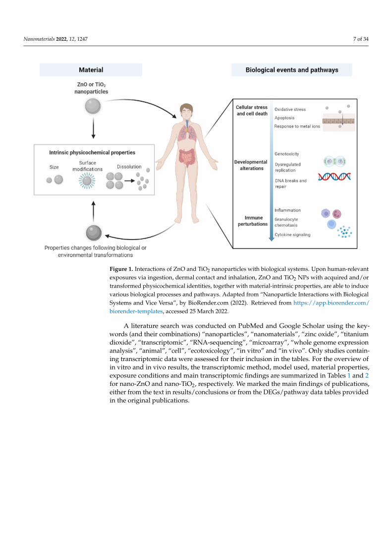
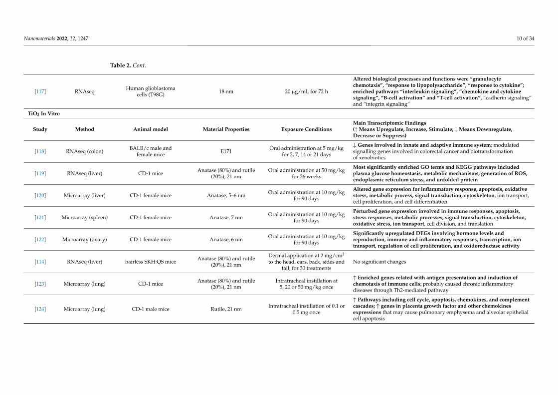
A literature search was conducted on PubMed and Google Scholar using the key-words (and their combinations) “nanoparticles”, “nanomaterials”, “zinc oxide”, “titaniumdioxide”, “transcriptomic”, “RNA-sequencing”, “microarray”, “whole genome expressionanalysis”, “animal”, “cell”, “ecotoxicology”, “in vitro” and “in vivo”. Only studies contain-ing transcriptomic data were assessed for their inclusion in the tables. For the overview ofin vitro and in vivo results, the transcriptomic method, model used, material properties,exposure conditions and main transcriptomic findings are summarized in Tables 1 and 2for nano-ZnO and nano-TiO2, respectively. We marked the main findings of publications,either from the text in results/conclusions or from the DEGs/pathway data tables providedin the original publications.

Nanomaterials 2022, 12, 1247 8 of 34
Table 1. Transcriptomic characterization of exposures to ZnO NPs in in vitro and in vivo studies.
ZnO In Vitro
Study Method Cell Model Material Properties Exposure ConditionsMain Transcriptomic Findings(↑Means Upregulate, Increase, Stimulate; ↓Means Downregulate,Decrease or Suppress)
[94] RNAseqHuman lung epithelial
carcinoma cells(A549)
Uncoated 42 nm 15 µg/mL for 1, 6 or 24 h
Enriched terms “response to metal ions”, “metallothioneins bindmetals”, “apoptosis” and “immune system” ( at 6 &24 h); ↓ moleculesrelated to DNA repair; Nrf2 pathway was predicted to be activated at6 h but repressed at 24 h
[100] Microarray
Phorbol 12-myristate13-acetate
(PMA)-differentiatedTHP-1 macrophages
Uncoated, <50 nm 2 or 8 µg/mL for 4 h
Affected genes involved in metal metabolism, transcriptionregulation, DNA binding, protein synthesis and structure; at higherdose, altered gene expression involved in inflammation, apoptosisand mitochondrial dysfunction
[107] Microarray Rat alveolar macrophages(NR8383) Uncoatad, 158 nm 4 and 17 µg/mL for 4 h
Disturbed protein synthesis/homeostasis with the eIF2 and VEGFsignaling pathways, stress response with mitochondrial dysfunction,and sirtuin signaling; ↑ metallothioneins, genes related to membranedamage sensor, lung fibrosis, and protein synthesis regulator; ↓ stressresponse mediator, cell-cycle regulator, and transcription factor
[108] Microarray Human chronic myeloidleukemia cells (K562 cell line) Uncoated, ≤40 nm 40 µg/mL for 15 h
↑ Genes involved in “response to zinc ions”, “detoxification ofinorganic compound”, and “negative regulation of growth”; ↓ genesthat regulated “immune responses”, “cell proliferation/migration”,“receptor signaling pathway via JAK-STAT” and“phosphatidylinositol 3-kinase signaling”; ↑ anti-oxidant defensesystem, mitochondrial-dependent apoptosis, and ↓ NF-κB pathway
[109] RNAseq Human skin cancer cells(A431) Uncoated, around 500 nm 150 µg/mL for 6 h
Altered gene expression for pathways in cancer, alcoholism,environmental information processing including MAPK, cytokine, TNFsignaling pathways; ↑ genes related to injured or inflamed skin, and↓ genes of apoptosis/cell cycle/cell survival
[110] MicroarrayHuman monocyte-derivedmacrophages; Jurkat T cell
leukemia derived cellUncoated, 15 nm 1 or 10 µg/mL for 6 or 24 h Affected cell death, cell growth, immune system development processes

Nanomaterials 2022, 12, 1247 9 of 34
Table 1. Cont.
ZnO In Vitro
Study Method Animal model Material Properties Exposure ConditionsMain Transcriptomic Findings(↑Means Upregulate, Increase, Stimulate; ↓Means Downregulate,Decrease or Suppress)
[105] Microarray (lung) C57BL/6J BomTacfemale mice Uncoated, 100 nm Intratracheal instillation at 11,
33 or 100 mg/kg once
Enriched pathways for cell cycle G2 to M phase DNA damagecheckpoint regulation, circadian rhythm signaling, proteinubiquitination pathway, unfolded protein response, andAMPK signaling
[111] RNAseq (liver) CD-1 male mice Around 35 nm Oral administration at 25 mg/kgfor 8 or 12 weeks
Most significantly enriched Gene Ontology (GO) and KyotoEncyclopedia of Genes and Genomes (KEGG) pathways involvedmembranes, endoplasmic reticulum stress and ROS generation
[112] RNAseq (liver) Sprague Dawley female rats Uncoated, 86.3 nm Oral administration at 100 mg/kgfor 14 consecutive days ↑ Metabolic process and metal binding in liver
[113] RNAseq (culturedskin cell) CD-1 mice Around 30 nm Mouse hair follicle stem cells were
exposed at 20 µg/mL for 12 h
Perturbed genes associated with hair follicle stem cell apoptosis anddifferentiation; altered pathways involved in cellular communicationand RNA biosynthetic processes
[114] RNAseq (liver) Hairless SKH:QS mice Uncoated, 18.2 ± 0.4 nmDermal application at 2 mg/cm2
to the head, ears, back, sides andtail, for 30 treatments
No statistically significant DEGs
Table 2. Transcriptomic characterization of exposures to TiO2 NPs in in vitro and in vivo studies.
TiO2 In Vitro
Study Method Cell Model Material Properties Exposure ConditionsMain Transcriptomic Findings(↑Means Upregulate, Increase, Stimulate; ↓Means Downregulate,Decrease or Suppress)
[115] Microarray Undifferentiated Caco-2 cells E171; Antase, 15–25 nm 1.4 µg/cm2 for 2, 4, and 24 h
E171 and TiO2 NPs ↑ genes for inflammation, immune system, transportand cancer; E171 ↑ metabolism of proteins with the insulin processingpathway; TiO2 NPs affected pathways involved in metabolism of aminoacids, prostaglandin, urea cycle, oxidative stress; two common biologicalprocesses: transport of molecules and neuronal system
[116] RNAseq Human lung epithelialcarcinoma cells (A549)
Anatase (80%) and rutile(20%), 21 nm 800 µg/mL for 24 h
↑ Genes related to inflammatory response, cell surface signaling,oxidative stress, extracellular organization, electron transport, respiratorychain complex, and metabolic processes; ↓ genes that control cell cycle,secretion and cell–cell communication
Nanomaterials 2022, 12, 1247 10 of 34
Table 2. Cont.
[117] RNAseq Human glioblastomacells (T98G) 18 nm 20 µg/mL for 72 h
Altered biological processes and functions were “granulocytechemotaxis”, “response to lipopolysaccharide”, “response to cytokine”;enriched pathways “interleukin signaling”, “chemokine and cytokinesignaling”, “B-cell activation” and “T-cell activation”, “cadherin signaling”and “integrin signaling”
TiO2 In Vitro
Study Method Animal model Material Properties Exposure ConditionsMain Transcriptomic Findings(↑Means Upregulate, Increase, Stimulate; ↓Means Downregulate,Decrease or Suppress)
[118] RNAseq (colon) BALB/c male andfemale mice E171 Oral administration at 5 mg/kg
for 2, 7, 14 or 21 days
↓ Genes involved in innate and adaptive immune system; modulatedsignalling genes involved in colorectal cancer and biotransformationof xenobiotics
[119] RNAseq (liver) CD-1 mice Anatase (80%) and rutile(20%), 21 nm
Oral administration at 50 mg/kgfor 26 weeks
Most significantly enriched GO terms and KEGG pathways includedplasma glucose homeostasis, metabolic mechanisms, generation of ROS,endoplasmic reticulum stress, and unfolded protein
[120] Microarray (liver) CD-1 female mice Anatase, 5–6 nm Oral administration at 10 mg/kgfor 90 days
Altered gene expression for inflammatory response, apoptosis, oxidativestress, metabolic process, signal transduction, cytoskeleton, ion transport,cell proliferation, and cell differentiation
[121] Microarray (spleen) CD-1 female mice Anatase, 7 nm Oral administration at 10 mg/kgfor 90 days
Perturbed gene expression involved in immune responses, apoptosis,stress responses, metabolic processes, signal transduction, cytoskeleton,oxidative stress, ion transport, cell division, and translation
[122] Microarray (ovary) CD-1 female mice Anatase, 6 nm Oral administration at 10 mg/kgfor 90 days
Significantly upregulated DEGs involving hormone levels andreproduction, immune and inflammatory responses, transcription, iontransport, regulation of cell proliferation, and oxidoreductase activity
[114] RNAseq (liver) hairless SKH:QS mice Anatase (80%) and rutile(20%), 21 nm
Dermal application at 2 mg/cm2
to the head, ears, back, sides andtail, for 30 treatments
No significant changes
[123] Microarray (lung) CD-1 mice Anatase (80%) and rutile(20%), 21 nm
Intratracheal instillation at5, 20 or 50 mg/kg once
↑ Enriched genes related with antigen presentation and induction ofchemotaxis of immune cells; probably caused chronic inflammatorydiseases through Th2-mediated pathway
[124] Microarray (lung) CD-1 male mice Rutile, 21 nm Intratracheal instillation of 0.1 or0.5 mg once
↑ Pathways including cell cycle, apoptosis, chemokines, and complementcascades; ↑ genes in placenta growth factor and other chemokinesexpressions that may cause pulmonary emphysema and alveolar epithelialcell apoptosis

Nanomaterials 2022, 12, 1247 11 of 34
Table 2. Cont.
[125] Microarray (lungand liver)
C57BL/6BomTacfemale mice
Rutile, 20 nm, coatedwith polyalcohols
Whole-body inhalation at42 mg/m3 for 11 days (1 h/day)
↑ Genes associated with acute phase, inflammation and immuneresponse; associated pathways included cytokine–cytokine receptorinteraction, metabolism, complement and coagulation cascade,hematopoeitic cell lineage, and biosynthesis of steroids
[126] Microarray (lung) CD-1 female mice Anatase, 6 nm Nasal instillation at 2.5, 5 or10 mg/kg for 90 days
↑ Genes involved in immune/inflammatory responses, apoptosis,oxidative stress, cell cycle, metabolic processes, stress responses, signaltransduction, and cell differentiation
[127] Microarray (lung) C57BL/6 female mice Rutile, 21 nm, coatedwith polyalcohols
Intratracheal instillation at18, 54 or 162 µg/mouse once
↑ Inflammatory gene expression; ↓ genes involved in ion homeostasisand muscle regulation
[128] Microarray (lung) C57BL/6J female mice
Anatase, rutile oranatase/rutile; 8, 20 and
300 nm; and hydrophobic orhydrophilic surface
modifications
Intratracheal instillation at18, 54, 162 or 486 µg/mouse once
Rutile type induced higher number of DEGs relate to inflammataion andacute phase signaling; hydrophilic surface induced higher DEGs; amongthe anatase, the smallest type showed the maximum response; anatasetypes enriched inflammatory response, response to wounding, defenseresponse, chemotaxis; high dose of anatase TiO2 affectedcytokine–cytokine receptor interaction, chemokine signalling, NOD-likereceptor signalling, p53 signalling, ataxia telangiectasia mutated signalling,and steroid metabolic process
[129] Microarray (liverand heart) C57BL/6 female mice Rutile, 21 nm, coated
with polyalcoholsIntratracheal instillation at
162 µg/mouse once
↑ Complement cascade and inflammatory processes in heart for particleopsonisation and clearance; mild changes in gene associated with acutephase responses in liver
[130] Microarray (liver) C57BL/6BomTacfemale mice
Rutile, 21 nm, coatedwith polyalcohols
Whole-body inhalation at42 mg/m3 for 10 days (1 h/day)
during gestation
Altered gene expression related to the retinoic acid signalling pathway inthe female newborn livers; associated pathways related tissuedevelopment and vitamin, mineral and lipid metabolism
[131] RNAseq (heart) Sprague Dawley female rats Anatase (80%) and rutile(20%), 21 nm
Whole-body inhalation at10 mg/m3 for 7–8 days
(4–6 h/day) during gestation
Altered pathways involved in inflammatory signaling and organismaldevelopment; ↓ protein kinase B (AKT) signaling; ↑ IL-8 signaling

Nanomaterials 2022, 12, 1247 12 of 34
3.1. ZnO and TiO2 NPs Exposure In Vitro
Depending on the physicochemical properties of ZnO and TiO2 NPs, they are ableto evoke cytotoxicity, genotoxicity and immunotoxicity in many in vitro setups. Theydemonstrate the potential of directly or indirectly interacting with the cell membrane, mito-chondria, lysosome and other organelles, leading to a disruption of cellular homeostasisand production of ROS. Despite many shared biological events, TiO2 NPs tend to be lessbioactive or toxic than ZnO NPs, as shown in some studies [45,132,133].
3.1.1. Cellular Stress and Cell Death
The spontaneous production of ROS by metal-based NPs under UV or visible lighthas been reported to trigger cell death and inflammation [59]. Once inside the cells,especially positively charged NPs readily interact with organelles such as mitochondriaand lysosomes, leading to excessive ROS generation [134]. The oxidative response toZnO NPs suppressed DNA repair processes but activated the Nrf2 pathway at an earlytime-point [94], which plays a beneficial role in protecting cells mainly against oxidativedamage and cellular dysfunction [135]. Dissolved metal ions also show a high tendency tobind with free radical-scavenging enzymes such as glutathione and superoxide dismutase,which further insults the cellular anti-oxidant capability [134]. These dynamic and counter-balancing events ultimately may direct cells to inflammation, mitochondrial dysfunctionand even to apoptotic cell death [136].
ZnO NPs have been shown to have several effects on immune responses, and es-pecially, various metal-ion-related effects are common. In transcriptomic (GO, KEGG orIPA) pathway analyses, a significant enrichment of the terms ‘Response to metal ions’ and‘Metallothioneins bind metals’ or ‘Translation’, ‘Nonsense-Mediated Decay’, ‘Apoptosis’and ‘Immune System’ were reported after 6 or 24 h-exposure to ZnO NPs [94,100,107,108],while much less data are available on TiO2 NPs. In our own studies, TiO2 NPs did notenhance oxidative stress or inflammatory responses in THP-1 monocyte-differentiatedmacrophages [133]. Metallothioneins are involved in regulating intracellular metal ionconcentration and homeostasis, and two different metallothioneins, Mt1a and Mt2A, wereamong the most upregulated genes after exposure to ZnO NPs [107]. We have noted theimportance of metallothioneins also in our own transcriptomic studies in differentiatedTHP-1 cells [133]. During the longer exposure (24 h) to Zn2+ ions, transcripts of the tricar-boxylic acid TCA (called also citric acid) cycle were shown to be significantly reduced inhuman lung epithelial carcinoma cells (A549), indicating major disruption of cellular respi-ration pathways [94]. Furthermore, with higher ZnO doses, gene expression involved ininflammation, transcription of heat-shock proteins, apoptosis and mitochondrial sufferingwere increased in PMA-differentiated THP-1 macrophages [100].
The mitochondria are the organelle responsible for energy production within thecells. At the same time, they also have a central role in apoptotic cell death. Recentdata demonstrate that besides eliciting caspase activation, mitochondrial outer mem-brane permeabilization (MOMP) engages in various pro-inflammatory signaling func-tions [137]. Doumandji et al. found that sirtuin signaling, disturbed protein synthesis withthe eIF2 signaling pathway, expression of the membrane damage sensor and stress re-sponse with mitochondrial dysfunction were the most affected functions in rat alveolarmacrophages (NR8383) after exposure to ZnO NPs [107]. Transcriptomic results fromAlsagaby et al. showed similar results, indicating that exposure to ZnO in human chronicmyeloid leukemia cells (K562 cell line) changed the anti-oxidant defense system, inducedespecially mitochondrial-dependent apoptosis (instead of necrosis) and downregulatedNF-κB pathway activities. ZnO NPs caused mitochondrial-dependent intrinsic apoptosis inK562 cells, which was probably triggered by oxidative stress-induced DNA damage [108].Both directly and indirectly Zn-induced dysregulation of the mitochondrion seems to causecell death and growth [109,110].

Nanomaterials 2022, 12, 1247 13 of 34
3.1.2. Developmental and Hereditary Modifications
Genotoxicity is associated with exposure to metal-based NPs in cells or tissues. Pos-sible mechanisms include NP/NP-released ions binding to DNA directly, NP-inducedintracellular ROS or NPs’ interactions with other nuclear proteins that are essential forDNA replication and repair [138]. As a result, DNA molecules are susceptible to dys-regulated replication, deformation and chromosomal breaks [138]. In vitro studies havedemonstrated that smaller NPs (10 nm) may enter the nuclei directly, whereas disinte-gration of the nuclear membranes during mitosis assists with the entry of bigger NPs(15–60 nm) [139,140]. Studies have reported the presence of ZnO and TiO2 NPs in thenuclei via transmission electron microscopy analysis [96,141].
Depletion of the cellular antioxidant capacity has been shown to contribute to thegenotoxicity of ZnO NPs [99]. The direct transcriptomic evidence on the genotoxicityof nanosized ZnO is sparse at the transcriptomic level. Dekkers et al. have shown thatexposure to ZnO NPs yields an increase in mRNAs with premature stop codons, whichcould reflect the increased rate of DNA damage [94]. Furthermore, the expression of DNArepair genes was reduced, including those of the base excision repair, mismatch repair,nucleotide excision repair and double-strand break repair pathways, particularly withZn ions, micro-sized Zn and nanosized ZnO. The authors concluded that these changesillustrate a progression from adaptive changes, such as metallothionein induction, to thedepletion of antioxidants (e.g., glutathione), inhibition of DNA repair and induction ofapoptosis [94].
Similarly, owing to oxidative stress, TiO2 NPs were shown to trigger DNA breaks andmicronucleus formation in skin and liver cells [96,102]. TiO2 (E171), which may containat most 50% of particles in the nano range, previously authorized as a food additive, wasno longer considered safe to consume by EFSA in 2016 [142]. In October 2021, the EUdecided to ban the use of TiO2 as a food additive, starting from early 2022 [143]. Afteroral ingestion, the absorption of TiO2 particles is low, but they can accumulate in thebody, and the genotoxicity concerns cannot be excluded. Previous studies show that E171,encompassing NPs and micro-sized particles, induces oxidative stress responses, DNAdamage and micronuclei formation in vitro, and recently, a microarray analysis of Caco-2cell indicated that E171 induced gene expression changes related to signaling, inflammation,the immune system, transport and cancer [115].
3.1.3. Changes in Immune Responses
Due to their size, NPs themselves are usually considered as non-immunogenic, mean-ing they are not able to trigger immune responses. However, depending on NP surfaceproperties and composition, they may induce versatile immune responses. The particlescan associate, bind and form aggregates with available molecules, which together maymodulate local or systemic immune responses (being immunosuppressive or immunostim-ulatory) under healthy and diseased conditions [144].
Antigen recognition and uptake: After overcoming the physical epithelial barrier, tissueinnate immune cells (such as macrophages, dendritic cells (DCs) and neutrophils) takepart in the recognition and uptake of the incoming NPs, mediated by pattern recognitionreceptors (PRRs), including toll-like receptors (TLRs), and NOD-like receptors (NLRs)on the cell surface of these phagocytes. TLRs are responsible for detecting pathogen-associated molecular patterns (PAMPs), while NLRs are for danger-associated molecularpatterns (DAMPs). Nanomaterial-associated molecular patterns (NAMPs), a new typeof molecular pattern, also emerge as one of the possible initiators for phagocytosis ofnovel nanomaterials by the host immunity, in addition to TLRs, opsonic receptors andmannose receptors [145]. The exact recognition mechanism of each type of NP dependson both material-intrinsic properties and the acquired biological identity in different flu-ids [146,147]. For instance, the components of the bio-corona may act as strong opsoninsthat aid in efficient phagocytosis via opsonic receptors [144]. Within phagocytes, NPs areintracellularly trafficked to the lysosomal compartment with a low pH, leading to biological

Nanomaterials 2022, 12, 1247 14 of 34
degradation via a superoxide/peroxynitrite-driven oxidative pathway or by enzymes suchas myeloperoxidase (MPO) and peroxidase [148–150].
Innate immune responses: Generally speaking, NPs may produce several deleteriousconsequences while interacting with the innate immunity: the suppression of phagocytosis,induction of cytokine production and activation of the inflammasome complex. NPs maysuppress the engulfment of apoptotic cells and microorganisms into the macrophage, lead-ing to a slower or even failed digestion or removal of pathogens [151,152]. Additionally,NPs can directly activate the production of cytokines (e.g., IL-1β, IL-6 and TNF-α) andcause the subsequent inflammatory responses, as seen in the effects produced by TiO2 orZnO NPs in human bronchial epithelial cells and murine astrocytes [101,153,154]. NLRP3inflammasome complexes are activated if NP-induced lysosomal damage and ruptureoccurs, accompanied by a release of inflammatory cytokine IL-1β [155–157]. Cross-talksbetween NPs and innate effector cells (e.g., macrophages and neutrophils) contribute toROS production through the activation of the nicotinamide adenine dinucleotide phos-phate (NADPH) oxidase system in inflammatory cells [158], which has been identifiedas a mechanism underpinning carbon nanotube-induced pulmonary inflammation andfibrogenesis responses [159].
The activation of innate immunity by TiO2 NPs was suggested in the study byJayaram et al. They showed that the exposure of A549 human lung epithelial cells toTiO2 (20 nm) activated intracellular ROS, specifically superoxide, along with changes inoxidative stress-related genes, which participate in inflammatory responses, cell surfacesignaling, and extracellular organization [116]. The TiO2-associated DEGs also control thecell cycle that was silenced, causing reduced mediator secretion and cell–cell communica-tion. Together, these may lead to an increased cellular resistance to oxidative metabolism,electron transport and the respiratory chain complex, and metabolic process gene functionis enriched. Their experiments suggest that TiO2 NPs adapt to oxidative stress throughtranscriptional changes over multiple generations of cells [116].
Adaptive immune responses: In some cases, NP interactions may trigger specific, adaptiveresponses. Antigen-presenting cells (APCs) like DCs process and present NPs to B or Tlymphocytes. For example, TiO2 NPs have been shown to enhance the maturation andexpression of costimulatory molecules on dendritic cellsl leading to increased proliferationof CD4+ T cells [160]. It has been hypothesized that metal NPs may function as haptenswhen they are able to conjugate with protein carriers to form larger immunogenic complexesthat trigger the elicitation of immune responses and production of antibodies (mainlyIgG) [161,162]. Furthermore, previously hidden, conformational epitopes might be revealed,which induce immune responses to the corona-forming proteins [144]. On the contrary, NPsexposure may lead to suppression of T cell proliferation after impeding the differentiationof monocytes into DCs or disturbing DC normal functions [163,164].
Based on the in vitro studies, it is reported that TiO2 NPs might compromise the in-tegrity of the blood–brain barrier and cause neuroinflammation. Exposure to noncytotoxicdoses (5 µg/mL) of TiO2 or Ag NPs had no effects on the transcriptome of T98G humanglioblastoma cells [117]. Conversely, the transcriptome of the cells exposed to 20 µg/mL ofTiO2 NPs revealed autophagy and alterations in several biological processes and molecularpathways, such as “granulocyte chemotaxis”, “response to cytokine”, “inflammation medi-ated by the chemokine and cytokine signaling pathway”, “B-cell activation” and “T-cellactivation” [117]. The results were confirmed by measuring the increased IL-8 productionfrom T98G cultures. In reverse, ZnO NPs did not cause major changes in this study [117].
3.2. ZnO and TiO2 NPs Exposure In Vivo
ZnO and TiO2 NPs may induce local changes in the function of specific organs. Further-more, due to their ultrasmall size, they are capable of transcending the organ barriers andtravelling to non-exposed organs after cellular transcytosis and systemic circulation [165].While there are already excellent reviews on findings collected from conventional toxicitystudies [1,6], the following sections focus on delineating transcriptomic-led studies that

Nanomaterials 2022, 12, 1247 15 of 34
have been conducted to reveal the local and/or systemic effects of ZnO and TiO2 NPs viathe three most-possible exposure routes: ingestion, dermal contact and inhalation.
3.2.1. Ingestion
ZnO NPs exhibit potent antimicrobial property, which renders them widely usedin food-packing materials. They can shield food substances from oxygen and moisturein order to maintain their organoleptic qualities [5]. TiO2 NPs demonstrate strong light-scattering and whitening effects and have been incorporated into food additives to enhancethe color of pastries, confectionery sweets, chewing gum, the coating of chocolates andcoffee creamer. It has been reported that nanosized TiO2 constitute up to 36% of the foodadditive E171 [166]. Oral exposures to ZnO and TiO2 NPs currently still represent oneof the most prevalent human-related exposure routes. Proquin et al. reported that micethat ingested 5 mg/kg E171 (food additive form of TiO2 NPs) daily showed a significantdownregulation of genes involved in the innate and adaptive immune system, observed asearly as on day 7 [118]. Such an immune-inhibitory effect persisted up to day 21, implyinga sustained impairment of intestinal immunity. The TiO2-induced oxidative stress responsewas also evidenced in the colonic transcriptome, mediated by the activation of MAPKgenes [118]. Furthermore, it is worth noting that the mucin-associated pathway (e.g., O-linked glycosylation) was highly upregulated in the colon [118], suggesting a stimulatoryrole of TiO2 NPs in mucus secretion.
At the same time, a number of studies have investigated the extraintestinal effects ofchronic exposure to ingested ZnO or TiO2 NPs. Hu et al. observed a TiO2-induced signifi-cant elevation of the plasma glucose concentration. The ingestion of TiO2 NPs (21 nm, 80%anatase, 20% rutile) for 26 weeks led to enrichment of the same set of genes and pathways,accompanied by the same increase in blood glucose [119]. Although it is well-recognizedthat ROS production is a possible mechanism contributing to the biological toxicity trig-gered by NPs, it is less conclusive as to what induces such an increase in the oxidant level.Based on the findings demonstrating a shared ER stress-inducing ability of ZnO and TiO2NPs, Hu et al. suggested it as a mechanism for the observed ROS excess and disruption ofblood glucose homeostasis. The liver, along with the kidney and spleen, is a major organ forsystemic distribution of orally absorbed NPs [167], supported by its physiological functionof detoxification and removal of xenobiotics. It was shown that a significant differentialregulation of genes in the liver occurred after 8 or 12 weeks of oral exposure to ZnO NPsat a dose of 25 mg/kg [111], characterized by a noticeable enrichment of genes related tothe membrane, endoplasmic reticulum stress, inflammatory responses and generation ofROS. Additionally, a 90-day subchronic oral toxicity study performed by Cui et al. showedthat 10 mg/kg of TiO2 NPs altered hepatic gene expression associated with inflammatoryresponses, apoptosis, oxidative stress, cell cycle and differentiation [120]. On the otherhand, a 14-day oral administration of ZnO NPs did not induce transcriptional changesrelated to immunity or cell cycle regulation in the rat liver [112], probably due to a shorterexposure period and the use of different particle-dispersing vehicles (e.g., 5% glucose vs.PBS). Additionally, the extraintestinal effects induced by TiO2 NPs were studied in therodent ovary and spleen. The upregulation of genes relevant to oxidative stress, inflamma-tion, ion transport and cell division regulation was common in both organs upon 90-dayoral exposures at a dose of 10 mg/kg per day [121,122]. In the mouse ovary, the expressionof 10 genes participating in hormonal production and regulation was also increased.
It has been shown that some cells lines well represent responses seen in vivo. Transcrip-tomic studies in the Caco-2 cell line revealed that E171 induced gene expression changesrelated to signaling, inflammation, the immune system, transport and cancer [115]. PureTiO2 NPs seem to affect pathways involved in the metabolism of amino acids, creatine andprostaglandin; the urea cycle; the neuronal system; the transport of small molecules (aminoacid) and oxidative stress [115]. Two biological processes, the transport of molecules andneuronal system, were shared by E171 and TiO2 NPs [115], suggesting that TiO2 NPs mighthave a route to bypass the blood–brain barrier and maybe accumulate into brain tissue.

Nanomaterials 2022, 12, 1247 16 of 34
3.2.2. Dermal Contact
Skin forms the largest barrier for our body against foreign matters and pathogens.One of the environmental stresses that we encounter almost every day is UV radiation. Sun-screen lotions, in addition to physical UV-blocking measures, are able to provide efficientprotection against the detrimental effects induced by UV, such as a photosensitive skin rashand even skin cancer. Due to their intrinsic physical properties, ZnO- and TiO2-containingsunscreen products are among the most popular type of physical UV filters that directlyscatter light and convert UV radiation to harmless infrared light and heat [168,169]. ZnOparticles can provide a broader shield against both UVA and UVB waves than TiO2 [170].Nanosized ZnO and TiO2, compared to their bulk-sized counterparts, confer the sunscreencream a transparent and light-weight appearance. On the other hand, these ultrasmall par-ticles have aroused great attention due to their penetration ability across the skin epidermis.Many investigations regarding their local and distant biological effects are still underway.
ZnO NPs are able to accumulate at the hair follicle after topical administration. Ge et al.revealed that 30 nm ZnO NPs caused transcriptional perturbations in cultured mousehair follicle stem cells and highly enriched genes and pathways related to the regulationof cell communication, apoptosis, cell proliferation/differentiation, RNA synthesis andprocessing [113]. Beyond skin, the effect of dermal ZnO and TiO2 NPs in other organs afterpossible penetration is still under further investigation. Based on the hepatic transcriptomeof mice repeatedly exposed (30 treatments) to ZnO and TiO2 NPs on skin, there were no orvery few DEGs found in their livers, despite a low-level presence of elemental titanium,probably due to chronic ingestion during daily grooming and licking [114].
We have studied the effects of ZnO exposure during the sensitization or during thechallenge of pre-sensitized contact in an allergic individual in a CHS mouse model (datato be published). The skin-swelling effect of the CHS response was markedly inhibitedupon topical administration of ZnO NPs during the challenge phase. We saw a significantreduction of local inflammatory cells infiltration and a full abrogation of global innate andadaptive immune responses in the skin transcriptome, suggesting a beneficial effect evokedby ZnO NPs as strong immunosuppressive substances.
3.2.3. Inhalation
The production volume of ZnO and TiO2 NPs increases exponentially, reaching thou-sands of tons of nano-enabled goods produced per year [171]. The inhalation of these NPsduring particle synthesis and product manufacturing presents exposure risks to workers,administrative officers and cleaners in the nanotechnology field. Particularly, the circu-lation of air-borne or surface-settled NPs are promoted during daily clean-up and whenstaff motility increases [172]. Consumers, such as users of cleaning or sunscreen sprays,may also encounter pulmonary exposure to NP-carrying aerosol, when particles comeclose to the breathing zone. A number of conventional toxicity testing studies have beencarried out regarding the toxic potential of inhaled NPs in lungs. Complementary evidencedrawn from transcriptomics-oriented studies further unraveled the biological changesinduced by ZnO and TiO2 NPs in lungs, which are otherwise missing in the traditionalend-point studies.
Effects of ZnO exposure in lungs: Plenty of studies have shown that ZnO NPs areable to induce increases in inflammatory cells (e.g., macrophages, neutrophils), cytokines,metallothionein expression and oxidative stress markers (e.g., heme oxygenase-1, SOD andMDA) in BAL fluid or lung tissues [173–179]. To date, however, there has only been onetranscriptomics-led inhalation study on ZnO NPs that has been published. Hadrup et al.measured the levels of DNA strand breaks in BAL fluid and lung and liver tissues by acomet assay [105]. In BAL fluid, increased levels of DNA strand breaks were observedonly for coated ZnO at a low-dose and long time after the exposure. In lung tissue, DNAstrand breaks were observed for both uncoated and coated ZnO NPs at day 28, whereas noincreased levels of DNA strand breaks were observed in liver tissue. These DNA changesin lungs were accompanied with gene expression changes related to unfolded protein

Nanomaterials 2022, 12, 1247 17 of 34
responses and the cell cycle G2 to M phase transition during DNA damage checkpointregulation [105]. In our lab, we performed transcriptomic profiling of a single pulmonaryexposure to ZnO NPs via oropharyngeal aspiration in mice, and we followed the lungtranscriptome at days 1, 7, and 28 post-exposure (data to be published). We found thatZnO NPs significantly induced the highest number of DEGs on day 1 in lungs, leadingto canonical pathways of hypercytokinemia, granulocyte diapedesis, cell-cycle controland interferon signaling. In addition, an unfolded protein response and NRF2-mediatedoxidative stress were also the major events induced by inhaled ZnO NPs.
Effects of TiO2 exposure in lungs: Inflammatory responses in the lungs characterizedby the induction of chemotaxis, cytokine signaling and complement cascade pathwayshave been commonly found in a series of inhalation studies on TiO2 NPs, regardless ofthe chosen route of administration (whole-body inhalation, nasal instillation or intratra-cheal instillation) or duration of exposure [123–126]. In addition, it is worth noting thatTiO2 NPs were able to induce changes in smooth muscle function in lungs. Husain et al.provided transcriptomic evidence pointing out that even a very low dose (18 µg permouse, corresponding to 1.5 working days based on Danish occupational exposure level)of TiO2 NPs, although it did not induce infiltration of inflammatory cells in BAL fluid, wasretained in the lungs and significantly downregulated genes related to muscle develop-ment/contraction [127] even on 28 days post-exposure. They postulated that the retentionof TiO2 NPs over time may undermine lung muscle contraction activities via impedingair movement, and as a result, it might lead to the development of lung diseases such aschronic obstructive pulmonary disease and pulmonary fibrosis [127].
Structure- or coating-specific effects have also been studied with inhaled TiO2 NPs.Rahman et al. compared the genome-wide alterations induced by different structures(anatase vs rutile) and coatings (hydrophilic vs hydrophobic) of TiO2 NPs in mouselungs [128]. They reported that rutile-structured TiO2 was a more potent inducer oftranscriptional perturbations than anatase TiO2, as supported by much higher numbers ofDEGs in the liver, regardless of dosages. Among the rutile, it was shown that a hydrophilicmodification imparted greater inflammogenicity compared to the hydrophobic type, asevidenced in their pathway analyses. Nonetheless, both anatase and rutile TiO2 inducedDEGs associated with inflammatory responses such as cytokine/chemokine signaling, IL-17signaling, granulocyte adhesion and diapedesis, probably via pathogen pattern recognitionmechanisms, including TLR- (for rutile) and NLR-signaling (for anatase). As alluded toearlier, the recognition of NPs can be achieved via TLRs or NLRs, where the former istriggered by bacterial components such as the lipopolysaccharide and peptidoglycan orviral DNA [180], while the latter is initiated by endogenous molecules, including high-mobility group box 1 proteins and heat shock proteins that are released during cellularstress or cell death [181]. Kinaret et al. demonstrated that amination of TiO2-NPs led tothe strongest inflammation in the airways of mice, while PEGylation substantially inhib-ited pulmonary toxicity, supported by the transcriptomic profiles of nanomaterials of anidentical composition but different coatings [182].
A chronic inflammation-related gene expression pattern was still observed a few weeksafter one single exposure to TiO2 NPs [123,124,126,128], along with evidence drawn fromthe histopathological analysis post-exposure. In particular, Chen et al. reported that a singleinstillation of 0.1, 0.5 or 1 mg of TiO2 NPs induced expression of placenta growth factor(PGF) and other chemokines such as CXCL1, CXCL5 and CCL3 in mouse lungs, mimickingan emphysema-like condition (a type of chronic obstructive pulmonary diseases) [124]. Onthe other hand, Li et al. and Halappanavar et al. sought to recognize the potential effectsof inhaled anatase TiO2 NPs in lungs after repeated exposure. Treatment with 10 mg/kgof nano-TiO2 led to over 500 DEGs after a 90-day consecutive nasal instillation, with themajority being upregulated and involved in immune responses, apoptosis, oxidative stress,metabolic processes, signal transduction and the cell cycle [126]. Additionally, five daysafter an 11-day repeated whole body inhalation of 42.4 ± 2.9 mg of rutile TiO2/m3, it led to

Nanomaterials 2022, 12, 1247 18 of 34
increases in the expression of gene sets sharing similar functions in the lungs, includingacute inflammation, complement cascade, and cytokine/chemokine signaling [125].
Effects of NPs in distal organs: In addition to the local effect, efforts have also beenput into studying the potential transcriptomic alterations in distal organs. A previousbio-distribution study on inhaled TiO2 NPs showed that they were able to translocate tothe brain via the nasal cavity [183]. Husain et al. examined TiO2 NPs’ influences on thetranscriptome profiles of heart and liver tissues 24 h or 28 days after a single intratrachealinstillation of TiO2. Although a larger amount of TiO2 was detected in the liver than theheart, only 63 DEGs were found in the liver, and no specific pathway was enriched, whichwas in stark contrast to around 500 DEGs present in the heart that significantly perturbedpathways associated with activation of the complement cascade (seven DEGs, includingcomplement factors D and 3), acute phase signaling and inflammatory processes [129].All transcriptional changes were reversed back to the baseline level when observed atthe day 28 post-exposure time-point, suggesting an efficient resolution of TiO2-inducedinflammatory responses in the heart. It is important to note that Husain et al. also foundout that the lectin pathway (microbes-initiated) may be the most possible mechanismexplaining the complement activation triggered by TiO2 NPs in the cardiovascular system.The complement system encompasses a family of proteins that, when activated, opsonizeforeign matters such as NPs, pathogens and damaged cells to direct phagocytosis andrecruit more inflammatory cells to the site of activation [184].
Lastly, we found two transcriptomic studies that have been carried out to shed lighton the developmental toxicity induced by TiO2 NPs in mouse newborns’ livers and hearts,caused during the whole-body inhalation of the same particles in their mother [130,131].In the study of Jackson et al., female offspring livers showed an altered gene expressionrelated to retinoic acid signaling, while the same gene sets were not responsive to suchexposure in male offspring [130]. In the heart of progeny that experienced prenatal maternalexposure to TiO2 NPs, the canonical pathways associated with inflammatory signaling andorganismal development were the most significantly enriched. In particular, they foundincreased expression of the lymphotoxin beta receptor gene (a member of tumor necrosisfactor receptors), upregulation of IL-8 signaling and downregulation of the inhibitor ofnuclear factor kappa-B kinase subunit alpha (IKK-α). Taken together, it was suggestedthat increased activation of NF-κB and IL-8 pathways may be the mechanism for theTiO2-induced inflammation in fetal hearts [131].
4. Transcriptomic Studies in Environmental Toxicology
The extensive production and utilization of engineered ZnO and TiO2 NPs increasetheir chances of being released into the environment and confer unintended biologicaleffects in different environmental organisms upon exposure. It is of high relevance to alsounderstand the most-updated transcriptomic findings on such exposure across representa-tive ecotoxicology species, such as Danio rerio, Caenorhabditis elegans and Arabidopsis thaliana.Our overview of the existing findings (listed in Table 3) regarding the effects of ZnO andTiO2 NPs on biological pathways and functions in those representative species is depictedin Figure 2.
The same literature search strategy as mentioned in Section 3 was conducted to screenstudies on the effect of ZnO or TiO2 NPs on the transcriptome of ecotoxicology-relevantspecies, with an addition of the keywords “ecotoxicology”, “Danio rerio”, “Caenorhabditiselegans” and “Arabidopsis thaliana”. The eligible studies are listed in Table 3.

Nanomaterials 2022, 12, 1247 19 of 34
Table 3. Transcriptomic characterization of exposures to ZnO and TiO2 NPs in ecotoxicology-related models.
ZnO
Study Method Ecotox Model Material Properties Exposure ConditionsMain Transcriptomic Findings(↑ Means Upregulate, Increase, Stimulate; ↓ Means Downregulate, Decreaseor Suppress)
[185] Microarray Zebrafish <50 nm 4.8 mg/L for 96 hMainly affected nucleic acid metabolism via altering nucleic acid binding;enriched KEGG pathways included “cell cycle”, “DNA replication”, and“homologous recombination”
[186] Microarray Zebrafish Uncoated, 20–30 nm 0.01, 0.1, 1 or 10 mg/L for96 h post-fertilization
↑ Genes for inflammation and the immune system; toxicological pathwaysincluded cytokine-cytokine receptor interactions and the intestinal immunenetwork for IgA production
[187] Microarray Zebrafish larva Uncoated, 10–30 nm 1 or 4 µmol/L for 72 hpost-fertilization
↑ Cell differentiation and pathways associated with the immune system; ↑several key genes involved in cancer cell signaling
[188] Microarray Caenorhabditis elegans Pristine, phosphatized orsulfidized, 30 nm
0.7 mg/L (ZnO), 7.5 mg/L(pZnO) and 7.5 mg/L
(sZnO) for 48 h
Induced DEGs related to metal responsive genes; enriched pathways forprotein biosynthesis (Aminoacyl-tRNA biosynthesis) and associated withdetoxification (ABC transporters) were shared among pristine and one or bothtransformed ZnO NPs
[189] Microarray (roots) Arabidopsis thaliana Uncoated, <100 nm 100 mg/L for 7 days
Mainly perturbed genes involved in stress responses to abiotic (oxidative,salt, water deprivation) and biotic (wounding and defense to pathogens)stimuli; ↑ genes involved in cellular metal ion homeostasis and transport, andenzymes against oxidative stress; ↓ genes related to cell organization andbiogenesis, translation, nucleosome assembly and microtubule-based process
[190] Microarray Arabidopsis thaliana Uncoated, 20 nm 4 mg/L for 7 days
↑ Genes for stress responses (e.g., to salt, osmotic stress or water deprivation),responses to pathogens, oxidative stress, transcription factors, and transporters; ↓genes involved in cell organization and biogenesis, nucleic acid metabolism,ribosomal proteins, cell wall modification and cell growth
TiO2
Study Method Ecotox Model Material Properties Exposure ConditionsMain Transcriptomic Findings(↑ Means Upregulate, Increase, Stimulate; ↓ Means Downregulate, Decreaseor Suppress)
[191] Microarray Zebrafish embryos Anatase, 25 nm Microinjections of 8.5 ng/gInterfered pathways related to circadian rhythm, cell signaling throughkinase-related activities, trafficking of Golgi vesicles, immune function,and exocytosis

Nanomaterials 2022, 12, 1247 20 of 34
Table 3. Cont.
[192] Microarray (ovary) Zebrafish Anatase, <25 nm 0.1 and 1 mg/L for13 weeks
Perturbed expresssion of genes involved in proteolysis, oxidative stressregulation, metabolism, insulin signaling, apoptosis and oocyte maturation; ↑genes associated with protein degradation or ROS production
[193] Microarray Caenorhabditis elegans Anatase, 32 nm 200 µg/mL for 72 h Affected genes involved in metal binding/detoxification, fertility andreproduction, worm growth, body morphogenesis, and neuronal function
[194] Microarray Caenorhabditis elegans
Anatase (83%) and rutile(17%), 34.1 nm; anatase:
5.9–16.2 nm; rutile:12.6–68.9 nm
0.01, 0.1, 1 and 10 mg/Lfor 24 h
Altered regulation of anti-oxidant system, stress resistance regulator andembryonic development; anatase type greatly influenced metabolic pathwayswhereas rutile particles significantly affected developmental processes
[189] Microarray (root) Arabidopsis thaliana Anatase (80%) and rutile(20%), 21 nm 100 mg/L for 7 days Mild changes, primarily responses to biotic and abiotic stimuli
[195] Microarray Arabidopsis thalianagerminants
Anatase (80%) and rutile(20%), 21 nm 500 mg/L for 12 days
↑ Genes related to metabolic processes (DNA metabolism, hormonemetabolism, triterpenoid biosynthesis and photosynthesis, indole glucosinolatemetabolism, tryptophan catabolism), root development and cell differentiation,ion transport, and redox reaction; ↓ genes related to respiratory burst,responses to stress, hypoxia, and immune responses

Nanomaterials 2022, 12, 1247 21 of 34Nanomaterials 2022, 12, x FOR PEER REVIEW 20 of 36
Figure 2. NP-driven environmental effects studied in different ecotoxicology-related models. Ara-bidopsis thaliana plants, C. elegans nematodes and Danio rerio zebrafish represent soil and aquatic species that are conventionally used for ecological toxicity assessment. Transcriptomic analyses yield DEGs, which identify the major affected pathways and the associated biological functions. Created with BioRender.com (https://app.biorender.com, accessed 25 March 2022).
The same literature search strategy as mentioned in Section 3 was conducted to screen studies on the effect of ZnO or TiO2 NPs on the transcriptome of ecotoxicology-relevant species, with an addi-tion of the keywords “ecotoxicology”, “Danio rerio”, “Caenorhabditis elegans” and “Arabidopsis thali-ana”. The eligible studies are listed in Table 3.
Figure 2. NP-driven environmental effects studied in different ecotoxicology-related models. Ara-bidopsis thaliana plants, C. elegans nematodes and Danio rerio zebrafish represent soil and aquaticspecies that are conventionally used for ecological toxicity assessment. Transcriptomic analyses yieldDEGs, which identify the major affected pathways and the associated biological functions. Createdwith BioRender.com (https://app.biorender.com, accessed 25 March 2022).
4.1. Danio rerio
Zebrafish (Danio rerio) are commonly used as model animals for testing NP toxicityand biocompatibility, also in high-throughput acute toxicity studies, and for evaluatingtheir value in nanotoxicity assessment [196–200]. Zebrafish are small, transparent, lowcost and easy to maintain, with rapid embryogenesis and continuous reproduction [201].Estimated environmental concentrations of nanosized TiO2 and ZnO in aquatic ecosystemsrange from 0.0007 to 0.0245 mg/mL and 76 µg/L to ≤2 mg/L, respectively [202,203].Zebrafish can be used to test different NPs and other agents efficiently via multiple routesof exposure, including directly in the water, which is especially relevant for environmentaltoxicology applications. The combination of a well-established zebrafish model organism,and species-specific oligo microarray platform (Agilent, Santa Clara, CA, USA) and RSEQstrategies provide tools for studies on the molecular mechanisms underlying the adaptiveresponse of fish to NPs, and they aid in the identification of NP-specific genes and signalingpathways in fish.
ZnO effects in zebrafish adults: Several studies have reported that ZnO NPs are toxic tozebrafish [201,204,205]. Zhu et al. demonstrated that ZnO NPs induced a concentration-dependent decrease in hatching rates [204]. In addition, previous studies have investigatedoxidative stress induced by ZnO NPs in aquatic ecosystems, but their toxicity mechanismsand specific gene biomarkers have remained unknown [206–208]. Hou et al. showed thatZnO NPs mainly affected nucleic acid metabolism in the nucleus via alterations in nucleicacid binding [185]. They exposed zebrafish to eight concentrations of ZnO (1–128 mg/L)and found 1434 ZnO-specific DEGs using Agilent zebrafish (V3) gene expression microar-rays. KEGG analyses classified the DEGs to the genotoxicity-related pathways “cell cycle”,“Fanconi anemia”, “DNA replication” and “homologous recombination”. Germline inacti-vation of any of the Fanconi anemia genes causes the disease Fanconi anemia, which leadsto bone marrow failure and predisposition to cancer. Hou et al. suggested that based ontheir pathway results, in addition to double-strand breaks in DNA, NP exposure may alsolead to DNA cross-link damage in D. rerio. DNA cross-link damage can block the formationof the DNA replication fork and disturb the replication process. The DNA cross-link repairprocess is relatively complex, and the Fanconi anemia pathway is one effective repairmethod. Together, these impairments in DNA synthesis and repair can disrupt mitosis orchromosomes by mechanical or chemical binding, leading to enhanced genotoxicity andinterference of cell cycle checkpoint functions and the production of mitochondrial ROS.

Nanomaterials 2022, 12, 1247 22 of 34
ZnO effects in zebrafish embryos and larvae: Furthermore, pericardial edema and mal-formations were observed in ZnO NP-exposed embryos [204]. Choi et al. investigated thedevelopmental toxicity of ZnO NPs (0.01, 0.1, 1 and 10 mg/L) to embryonic and larvalzebrafish [186]. The principal malformations induced by ZnO NPs were pericardial edemaand yolk-sac edema. Gene-expression profiling using microarrays demonstrated 689 DEGsfollowing exposure to ZnO NPs, where six of them were associated with inflammation, andthe immune system responded specifically to ZnO NPs. Therefore, ZnO NPs may affectgenes related to inflammation and the immune system, resulting in a yolk-sac edema andpericardia edema in the embryonic/larval developmental stages. These results assist inelucidating the mechanisms of toxicity of ZnO NPs during the development of zebrafish.The size-dependent ecotoxicity has also been observed in ZnO NP studies. Kim et al.compared the toxicity of a sublethal concentration of ZnO NPs (LC10, 1 µmol/L) andZnSO4 (LC30, 38 µmol/L) in zebrafish after 72 h of exposure [187]. They did not find anyDEGs for ZnO NPs after transcriptomics studied in microarray analyses when comparedto a non-exposed control group, while ZnSO4 yielded hundreds of DEGs. In reverse,according to the identification of lethal doses, ZnO NPs were more toxic than ZnSO4 tozebrafish larvae. This discrepancy was explained by the physical properties of the ZnONPs, which may have been responsible for the increased zebrafish larvae mortality due tothe mouth-gaping behavior, which leads to an increased uptake of ZnO NPs [209]. It hasbeen reported that zebrafish mortality was more increased by exposure to ZnO NPs thanby bulk ZnO [210], which could indicate that the particle size could be the most importantfactor controlling ZnO NP toxicity to hatched larvae. When the authors compared the DEGsthat were downregulated at the ZnSO4 LC10 relative to at the ZnO NP LC10, they foundcancer cell differentiation; endocytic transport and genes such as the epidermal growthfactor receptor (EGFR), V-Ki-ras2 Kirsten rat sarcoma viral oncogene homolog (KRAS), andphosphoinositide-3-kinase regulatory subunit 6 (PIK3R6) genes were upregulated in ZnO.These data suggest that ZnO NPs induce cell differentiation and pathways associated withthe immune system and activate several key genes involved in cancer cell signaling.
TiO2 effects in zebrafish: TiO2 NPs are usually perceived as non-toxic, especially insome short-term studies, and they have already been widely used in many products andapplications. The ultimate release of TiO2 NPs into the aquatic environment can act as a sinkfor engineered NPs, while their long-term impact on the environment and human health isstill a concern and deserves more research efforts. Jovanovic et al. micro-injected TiO2 NPsinto zebrafish embryos to determine the potential for NPs to change the expression of genesinvolved in the cross-talk of the nervous and immune systems [191]. After 96 h of follow-uptime, 2380 DEGs were induced, which interfered mainly with four areas of organismal andimmune functions, including the circadian rhythm, cell signaling through kinase-relatedactivities, exocytosis and trafficking of Golgi vesicles [191]. Altogether, Jovanovic et al.concluded that TiO2 NPs might cause deregulation of broad physiological and behavioraleffects, such as period homolog 2 (Per2) and cryptochromes (1a, 1b and DASH), by thecircadian system in aquatic animals [191]. Furthermore, reproductive toxicity has beenshown in the chronic exposure of zebrafish to a low dose of nano-TiO2 (0.1 mg/L) [192],where a reduced cumulative number of zebrafish eggs was found after 13 weeks of TiO2NPs exposure. The TiO2-accumulated fish ovaries suggest a likely penetration of these NPsinto the ovary, presumably via blood circulation. In addition, Wang et al. found DEGs thatwere involved in proteolysis, oxidative stress regulation, metabolism, insulin signalingand apoptosis and oocyte maturation, explaining multi-faceted modes of action for TiO2NPs-mediated disruption of reproduction. In particular, the growth and maturation of stageI follicles were inhibited [192], presumably by altering the expression of several regulatorsthat are critical to this stage of folliculogenesis. At the same time, several up-regulatedgenes were associated with protein degradation or ROS production, which may reflectthe stress responses of ovaries that were dealing with TiO2 NPs. All in all, nanosizedTiO2 in aquatic organisms may lead to alterations in population dynamics and the aquatic

Nanomaterials 2022, 12, 1247 23 of 34
ecosystem balance, and thus, it warrants careful scrutiny on toxicity assessment, especiallyin its long-term impact.
4.2. Caenorhabditis elegans
In addition to zebrafish, Caenorhabditis elegans can also be used as a representativeanimal species for ecotoxicological study of the major trophic levels [211,212]. C. elegans isa type of nematodes that is abundantly found in the liquid phase of soil or aquatic media,and it feeds on microorganisms in the environment. Furthermore, C. elegans has as shortlife cycle and is easy to grow and handle in the laboratory, which renders it one of themost used in vivo models for toxicological studies, as shown in the toxicity tests of heavymetals in the sediment habitat from a molecular to individual level (e.g., development,reproduction and behaviour) [213–216]. Acute exposure (within 24 h), prolonged exposure(from the L1 phase of larvae to mature adults; 72 h) or chronic exposure (from adult day 1to day 10) has been chosen to examine the potential toxicity of specific substances at variousconcentrations. Acute high-level exposures of ZnO and TiO2 NPs in C. elegans have beenshown to cause detrimental effects at both the lethal and sublethal endpoints, includingincreased mortality, growth inhibition, impairment of reproduction, decreased locomotionor alterations in gene expression [217–221].
ZnO effects in C. elegans: Although prolonged or chronic incubation with ZnO NPsdo not significantly affect the mortality, growth or reproduction of C. elegans [222–224],these particles pose harmful effects with regard to other sublethal markers, includingdecreased locomotion behaviour (evaluated by body bend and head thrash), increasedROS production and reduced ATP levels [222–224]. These previous findings suggest thatZnO and TiO2 NPs are associated with oxidative stress and metabolic and locomotivetoxicities in C. elegans. Reduced ATP levels can be caused by additional energy costsinduced by stress responses (detoxification and antioxidant defense mechanisms) or bydirect metabolic inhibition by metals or metal ions [225]. Starnes et al. exposed C. elegansto a sublethal dose of particulate ZnO NPs or ionic ZnSO4 salt for 48 h and revealedthat dissolution of ZnO, regardless of forms, may be one of the drivers for the observedshared gene expression pattern, mostly enriched by metal responsive, innate immunityand lysosome pathway-related genes [188]. Nonetheless, there were unique pathwaysassociated with nanosized ZnO only, including amino acid synthesis and metabolismand detoxification processes (ABC transporters). They also demonstrated that althoughtransformed ZnO NPs (phosphatized or sulfurized) drastically reduced the mortalitycompared to the pristine form, phosphatized ZnO induced the highest number of uniqueGO terms related to spermatogenesis, meiotic cell cycle and sex determination. Thesefindings suggest that phosphatized ZnO NPs are likely to exhibit distinct toxic mechanismsaffecting the reproductive system in C. elegans [188].
TiO2 effects in C. elegans: TiO2 NPs have been shown to cause detrimental effects inC. elegans larvae and adults. Hu et al. were the first to reveal that TiO2 NPs have access to thesubcellular compartment of C. elegans neurons and consequently negatively affect the axonalgrowth, as evidenced in phenotypes of shorter axons and inhibited locomotion behavior ina worm thrashing assay [193]. They also explored the cellular mechanisms involved in TiO2NPs toxicity via a microarray. Consistent with the macroscopic observations, various DEGsrelated to neuronal function were found. Furthermore, although anatase TiO2 NPs did notaffect larva’s body length as opposed to the rutile type, these particles enriched DEGs thatexhibit multiple functions related to metal binding or detoxification, fertility, worm growthand body morphogenesis, such as mtl-2, nhr-257 and clec-70 genes, which are involvedin both metal stress responses and worm growth or reproduction [193]. Similarly, thedifferential regulation of genes related to stress responses, detoxification, metal binding andreproduction have been observed in another microarray study of the effects of TiO2 NPs inC. elegans, including glutathione-S-transferase, stress resistance regulator and cytochromeP450 [194]. Furthermore, their study showed that anatase TiO2 NPs exhibited a greater

Nanomaterials 2022, 12, 1247 24 of 34
potential to affect the metabolic pathways than the rutile, while the latter was a strongerinducer of changes in developmental processes [194].
4.3. Arabidopsis thaliana
It is generally acknowledged that metallic NP can cause phytotoxicity in variousterrestrial plant species, such as wheat, rice and corn [226–228]. Arabidopsis thaliana is anideal test plant species for toxicity screening of terrestrially relevant substances, owning toits quick germination process and relatively short life span [229]. Additionally, it exhibitsa high sensitivity to potential toxicants, owing to the relatively larger surface area-to-volume ratio of its small seed [230]. Physiological parameters such as decreased biomass,reduced root length, delayed see germination speed, altered nutrient transport and loweredchlorophyll content have been associated with the plant’s response to stress induced bymetal NPs (therefore, also identified as phytotoxins) [231–235]. For example, ZnO NPscould impede the chlorophyll synthesis process in wheat leaves and induce size-dependentinhibition of seed germination in A. thaliana [226,236]. A couple of modes of action at thebiochemical, molecular and morphological levels have been investigated regarding thephytotoxicity of ZnO and TiO2, such as ZnO or TiO2 NPs-induced genotoxic chromosomalaberrations or DNA breaks [237,238] and lipid peroxidation via ROS production in anonion and A. thaliana [239,240]. Although studies by toxicogenomic profiling of A. thalianaremain very limited, it was the first plant to have had its genome sequenced in 2000 [241].
ZnO effects in A. thaliana: Microarrays were conducted to analyze and compare thegene regulation in A. thaliana roots upon a seven-day treatment with ZnO or TiO2 NPs [189].Overall, it was shown that ZnO NPs were able to induce a much greater number ofDEGs and associated diverse biological processes, indicating a severe phytotoxic roleof ZnO NPs in contrast to mild changes (mainly responses to biotic and abiotic stimuli)in global gene expression triggered by TiO2 NPs. Specifically, ZnO NPs significantlyreduced the transcription of genes involved in cell organization, biogenesis, translation,nucleosome assembly and microtubule-based process, suggesting the capability of ZnONPs in altering cell structure, cell division activity and DNA packaging. Additionally,nanosized ZnO exposure downregulated the genes coded for ribosomal proteins mainlyinvolved in electron transport and energy pathways, possibly leading to a reduction inoverall protein biosynthesis of the whole plant. On the other hand, adaptive responsesto salt, wound, metal ion, oxidative and osmotic stresses, along with defense againstpathogens, were strongly upregulated upon ZnO NPs incubation. A later study conductedby the same group elucidated that released Zn ions largely contributed to the toxic effectobserved in A. thaliana roots based on the similarity in transcriptomic profiles of rootsamples exposed to ZnO NPs versus ionic ZnSO4 [190].
TiO2 effects in A. thaliana: In addition to studying the roots of mature A. thaliana, Tum-buru et al. provided a phenotypic and transcriptional understanding of the alterationsinduced by a 12-day TiO2 NPs treatment in the germinants of A. thaliana [195]. Interestingly,they showed that TiO2 NPs were able to enhance germination, evidenced by an increasedpercentage of seeds exhibiting hypocotyls and cotyledons and a greater number of ger-minants with fully grown leaves, which agrees with another study showing promotedgerminant growth in wheat after exposure to TiO2 NPs [242]. Overall, there were muchmore upregulated than downregulated DEGs induced by TiO2 NPs [195]. Despite theprimary upregulation of genes participating in oxidative and osmotic stresses, an array ofmetabolic processes, including DNA, protein and phytohormone metabolism; tetrapyrrolesynthesis in the chloroplast and photosynthesis, were notably upregulated as well, whichwere believed to synergistically facilitate root growth and development, cell organizationand cell differentiation. For example, significant increases in the expression abundanceof cell wall proteins (e.g., arabinogalactan-proteins) were noted. The authors suggestedthat photocatalytic TiO2 NPs may promote A. thaliana seedling growth and developmentvia elevating the expression of genes that have key roles in photosynthesis and hormonal

Nanomaterials 2022, 12, 1247 25 of 34
metabolism [195]. A potential beneficial role of TiO2 NPs in the growth of A. thalianaremains to be further explored.
5. Conclusions
With the increasingly prevalent use of omics techniques, new data are burgeoningto provide a global view on the overall changes induced by exposure to ZnO and TiO2NPs. The variable results reported on particles with different characteristics, concentrationsand exposure routes have sometimes yielded even opposite outcomes at different organs.Transcriptomic studies enable us to better track, compare and conclude the shared andparticle-specific effects. These findings are essential for the optimization of “safety bydesign” of NPs for wider uses in human society. It is noticeable that transcriptomics dataon diseased in vivo models regarding the gut, skin and lungs are still missing. Researchon contrasting the differential effects of ZnO or TiO2 NPs in healthy versus immuno-compromised organisms is warranted for evaluating the risk of exposures for vulnerablepopulations. The transcriptomic approach offers a more detailed view of the cellular andorganismal responses after NP exposures in in vitro and in vivo studies. It advances thetoxicological understanding and sheds light on the local-oriented and distant effects oforiginal, distributed or transformed NPs in organisms and also in the environment.
Author Contributions: Conceptualization, S.W., P.K., H.E.-N.; Visualization, S.W., P.K.; Writing—OriginalDraft, S.W., P.K.; Writing—Review & Editing, S.W., H.E.-N., P.K., H.A.; Supervision, H.E.-N., P.K.;Project Administration, H.E.-N., P.K. All authors have read and agreed to the published version ofthe manuscript.
Funding: The study was supported by a grant from the Academy of Finland (decision 307768). Openaccess funded by Helsinki University Library.
Institutional Review Board Statement: Not applicable.
Informed Consent Statement: Not applicable.
Data Availability Statement: No new data were created in this study. Data sharing is not possible inthis article.
Conflicts of Interest: The authors declare no conflict of interest.
References1. Shi, H.; Magaye, R.; Castranova, V.; Zhao, J. Titanium dioxide nanoparticles: A review of current toxicological data. Part. Fibre
Toxicol. 2013, 10, 15. [CrossRef] [PubMed]2. Vance, M.E.; Kuiken, T.; Vejerano, E.P.; McGinnis, S.P.; Hochella, M.F., Jr.; Rejeski, D.; Hull, M.S. Nanotechnology in the real world:
Redeveloping the nanomaterial consumer products inventory. Beilstein J. Nanotechnol. 2015, 6, 1769–1780. [CrossRef]3. European Commission. Types and Uses of Nanomaterials, Including Safety Aspects; Commission Staff Working Paper SWD 288 Final.
Brussels, Belgium, 2012. Available online: https://eur-lex.europa.eu/LexUriServ/LexUriServ.do?uri=SWD:2012:0288:FIN:EN:PDF (accessed on 1 March 2022).
4. World Health Organization. WHO Guidelines on Protecting Workers from Potential Risks of Manufactured Nanomaterials; World HealthOrganization: Geneva, Switzerland, 2017.
5. Lamas, B.; Martins Breyner, N.; Houdeau, E. Impacts of foodborne inorganic nanoparticles on the gut microbiota-immune axis:Potential consequences for host health. Part. Fibre Toxicol. 2020, 17, 19. [CrossRef]
6. Canta, M.; Cauda, V. The investigation of the parameters affecting the ZnO nanoparticles cytotoxicity behaviour: A tutorialreview. Biomater. Sci. 2020, 8, 6157–6174. [CrossRef]
7. Schins, R.P.; Lightbody, J.H.; Borm, P.J.; Shi, T.; Donaldson, K.; Stone, V. Inflammatory effects of coarse and fine particulate matterin relation to chemical and biological constituents. Toxicol. Appl. Pharmacol. 2004, 195, 1–11. [CrossRef] [PubMed]
8. Ferdous, Z.; Nemmar, A. Health impact of silver nanoparticles: A review of the biodistribution and toxicity following variousroutes of exposure. Int. J. Mol. Sci. 2020, 21, 2375. [CrossRef] [PubMed]
9. Li, Y.; Cummins, E. Hazard characterization of silver nanoparticles for human exposure routes. J. Environ. Sci. Health Part A 2020,55, 704–725. [CrossRef]
10. Akter, M.; Sikder, M.T.; Rahman, M.M.; Ullah, A.A.; Hossain, K.F.B.; Banik, S.; Hosokawa, T.; Saito, T.; Kurasaki, M. A systematicreview on silver nanoparticles-induced cytotoxicity: Physicochemical properties and perspectives. J. Adv. Res. 2018, 9, 1–16.[CrossRef]

Nanomaterials 2022, 12, 1247 26 of 34
11. Maurer, L.; Meyer, J. A systematic review of evidence for silver nanoparticle-induced mitochondrial toxicity. Environ. Sci. Nano2016, 3, 311–322. [CrossRef]
12. Marin, S.; Mihail Vlasceanu, G.; Elena Tiplea, R.; Raluca Bucur, I.; Lemnaru, M.; Minodora Marin, M.; Mihai Grumezescu, A.Applications and toxicity of silver nanoparticles: A recent review. Curr. Top. Med. Chem. 2015, 15, 1596–1604. [CrossRef] [PubMed]
13. Llop, J.; Estrela-Lopis, I.; Ziolo, R.F.; González, A.; Fleddermann, J.; Dorn, M.; Vallejo, V.G.; Simon-Vazquez, R.; Donath, E.; Mao, Z.Uptake, biological fate, and toxicity of metal oxide nanoparticles. Part. Part. Syst. Charact. 2014, 31, 24–35. [CrossRef]
14. Aillon, K.L.; Xie, Y.; El-Gendy, N.; Berkland, C.J.; Forrest, M.L. Effects of nanomaterial physicochemical properties on in vivotoxicity. Adv. Drug Deliv. Rev. 2009, 61, 457–466. [CrossRef] [PubMed]
15. Lewinski, N.; Colvin, V.; Drezek, R. Cytotoxicity of nanoparticles. Small 2008, 4, 26–49. [CrossRef] [PubMed]16. Sturla, S.J.; Boobis, A.R.; FitzGerald, R.E.; Hoeng, J.; Kavlock, R.J.; Schirmer, K.; Whelan, M.; Wilks, M.F.; Peitsch, M.C. Systems
toxicology: From basic research to risk assessment. Chem. Res. Toxicol. 2014, 27, 314–329. [CrossRef] [PubMed]17. Schena, M.; Shalon, D.; Davis, R.W.; Brown, P.O. Quantitative monitoring of gene expression patterns with a complementary
DNA microarray. Science 1995, 270, 467–470. [CrossRef]18. Kukurba, K.R.; Montgomery, S.B. RNA sequencing and analysis. Cold Spring Harb. Protoc. 2015, 2015, pdb.top084970.19. Khatri, P.; Sirota, M.; Butte, A.J. Ten years of pathway analysis: Current approaches and outstanding challenges. PLoS Comput.
Biol. 2012, 8, e1002375. [CrossRef] [PubMed]20. Sengul, A.B.; Asmatulu, E. Toxicity of metal and metal oxide nanoparticles: A review. Environ. Chem. Lett. 2020, 18, 1659–1683.
[CrossRef]21. Dash, K.K.; Deka, P.; Bangar, S.P.; Chaudhary, V.; Trif, M.; Rusu, A. Applications of Inorganic Nanoparticles in Food Packaging: A
Comprehensive Review. Polymers 2022, 14, 521. [CrossRef] [PubMed]22. Monopoli, M.P.; Åberg, C.; Salvati, A.; Dawson, K.A. Biomolecular coronas provide the biological identity of nanosized materials.
Nat. Nanotechnol. 2012, 7, 779–786. [CrossRef] [PubMed]23. Behzadi, S.; Serpooshan, V.; Tao, W.; Hamaly, M.A.; Alkawareek, M.Y.; Dreaden, E.C.; Brown, D.; Alkilany, A.M.; Farokhzad, O.C.;
Mahmoudi, M. Cellular uptake of nanoparticles: Journey inside the cell. Chem. Soc. Rev. 2017, 46, 4218–4244. [CrossRef] [PubMed]24. British Standards Institution. Publicly Available Specification (PAS) 136: 2007; Terminology for Nanomaterials; British Standards
Institution: London, UK, 2007.25. Kreuter, J. Encyclopaedia of Pharmaceutical Technology; Marcel Dekker Inc.: New York, NY, USA, 1994; pp. 165–190.26. Singh, R.; Lillard, J.W., Jr. Nanoparticle-based targeted drug delivery. Exp. Mol. Pathol. 2009, 86, 215–223. [CrossRef] [PubMed]27. Krug, H.F.; Wick, P. Nanotoxicology: An interdisciplinary challenge. Angew. Chem. Int. Ed. 2011, 50, 1260–1278. [CrossRef]
[PubMed]28. Nel, A.; Xia, T.; Mädler, L.; Li, N. Toxic potential of materials at the nanolevel. Science 2006, 311, 622–627. [CrossRef]29. Asgharian, B.; Price, O.T. Deposition of ultrafine (nano) particles in the human lung. Inhal. Toxicol. 2007, 19, 1045–1054. [CrossRef]
[PubMed]30. Geiser, M.; Kreyling, W.G. Deposition and biokinetics of inhaled nanoparticles. Part. Fibre Toxicol. 2010, 7, 2. [CrossRef] [PubMed]31. Praphawatvet, T.; Peters, J.I.; Williams, R.O., III. Inhaled nanoparticles—An updated review. Int. J. Pharm. 2020, 587, 119671.
[CrossRef] [PubMed]32. Mitchell, M.J.; Billingsley, M.M.; Haley, R.M.; Wechsler, M.E.; Peppas, N.A.; Langer, R. Engineering precision nanoparticles for
drug delivery. Nat. Rev. Drug Discov. 2021, 20, 101–124. [CrossRef] [PubMed]33. Navya, P.; Daima, H.K. Rational engineering of physicochemical properties of nanomaterials for biomedical applications with
nanotoxicological perspectives. Nano Converg. 2016, 3, 1. [CrossRef] [PubMed]34. Arvizo, R.R.; Miranda, O.R.; Thompson, M.A.; Pabelick, C.M.; Bhattacharya, R.; Robertson, J.D.; Rotello, V.M.; Prakash, Y.;
Mukherjee, P. Effect of nanoparticle surface charge at the plasma membrane and beyond. Nano Lett. 2010, 10, 2543–2548.[CrossRef]
35. Blanco, E.; Shen, H.; Ferrari, M. Principles of nanoparticle design for overcoming biological barriers to drug delivery. Nat.Biotechnol. 2015, 33, 941–951. [CrossRef] [PubMed]
36. Sun, H.; Jiang, C.; Wu, L.; Bai, X.; Zhai, S. Cytotoxicity-related bioeffects induced by nanoparticles: The role of surface chemistry.Front. Bioeng. Biotechnol. 2019, 7, 414. [CrossRef]
37. Guerrini, L.; Alvarez-Puebla, R.A.; Pazos-Perez, N. Surface modifications of nanoparticles for stability in biological fluids.Materials 2018, 11, 1154. [CrossRef] [PubMed]
38. Luo, M.; Shen, C.; Feltis, B.N.; Martin, L.L.; Hughes, A.E.; Wright, P.F.; Turney, T.W. Reducing ZnO nanoparticle cytotoxicity bysurface modification. Nanoscale 2014, 6, 5791–5798. [CrossRef]
39. Kittler, S.; Greulich, C.; Diendorf, J.; Koller, M.; Epple, M. Toxicity of silver nanoparticles increases during storage because of slowdissolution under release of silver ions. Chem. Mater. 2010, 22, 4548–4554. [CrossRef]
40. Jeong, J.; Kim, S.-H.; Lee, S.; Lee, D.-K.; Han, Y.; Jeon, S.; Cho, W.-S. Differential contribution of constituent metal ions to thecytotoxic effects of fast-dissolving metal-oxide nanoparticles. Front. Pharmacol. 2018, 9, 15. [CrossRef]
41. Avramescu, M.-L.; Chénier, M.; Palaniyandi, S.; Rasmussen, P.E. Dissolution behavior of metal oxide nanomaterials in cell culturemedium versus distilled water. J. Nanopart. Res. 2020, 22, 222. [CrossRef]
42. Hansen, U.; Thünemann, A.F. Characterization of silver nanoparticles in cell culture medium containing fetal bovine serum.Langmuir 2015, 31, 6842–6852. [CrossRef] [PubMed]

Nanomaterials 2022, 12, 1247 27 of 34
43. Huynh, K.A.; Chen, K.L. Aggregation kinetics of citrate and polyvinylpyrrolidone coated silver nanoparticles in monovalent anddivalent electrolyte solutions. Environ. Sci. Technol. 2011, 45, 5564–5571. [CrossRef] [PubMed]
44. Zook, J.M.; Long, S.E.; Cleveland, D.; Geronimo, C.L.A.; MacCuspie, R.I. Measuring silver nanoparticle dissolution in complexbiological and environmental matrices using UV–visible absorbance. Anal. Bioanal. Chem. 2011, 401, 1993–2002. [CrossRef]
45. Cho, W.-S.; Kang, B.-C.; Lee, J.K.; Jeong, J.; Che, J.-H.; Seok, S.H. Comparative absorption, distribution, and excretion of titaniumdioxide and zinc oxide nanoparticles after repeated oral administration. Part. Fibre Toxicol. 2013, 10, 9. [CrossRef]
46. Zhang, W.; Yao, Y.; Sullivan, N.; Chen, Y. Modeling the primary size effects of citrate-coated silver nanoparticles on their ionrelease kinetics. Environ. Sci. Technol. 2011, 45, 4422–4428. [CrossRef]
47. Ma, R.; Levard, C.; Marinakos, S.M.; Cheng, Y.; Liu, J.; Michel, F.M.; Brown, G.E., Jr.; Lowry, G.V. Size-controlled dissolution oforganic-coated silver nanoparticles. Environ. Sci. Technol. 2012, 46, 752–759. [CrossRef] [PubMed]
48. Mortimer, M.; Kasemets, K.; Kahru, A. Toxicity of ZnO and CuO nanoparticles to ciliated protozoa Tetrahymena thermophila.Toxicology 2010, 269, 182–189. [CrossRef] [PubMed]
49. Roelofs, F.; Vogelsberger, W. Dissolution kinetics of synthetic amorphous silica in biological-like media and its theoreticaldescription. J. Phys. Chem. B 2004, 108, 11308–11316. [CrossRef]
50. Schmidt, J.; Vogelsberger, W. Dissolution kinetics of titanium dioxide nanoparticles: The observation of an unusual kinetic sizeeffect. J. Phys. Chem. B 2006, 110, 3955–3963. [CrossRef] [PubMed]
51. Franklin, N.M.; Rogers, N.J.; Apte, S.C.; Batley, G.E.; Gadd, G.E.; Casey, P.S. Comparative toxicity of nanoparticulate ZnO, bulkZnO, and ZnCl2 to a freshwater microalga (Pseudokirchneriella subcapitata): The importance of particle solubility. Environ. Sci.Technol. 2007, 41, 8484–8490. [CrossRef] [PubMed]
52. Li, M.; Zhu, L.; Lin, D. Toxicity of ZnO nanoparticles to Escherichia coli: Mechanism and the influence of medium components.Environ. Sci. Technol. 2011, 45, 1977–1983. [CrossRef] [PubMed]
53. Peters, R.; Kramer, E.; Oomen, A.G.; Herrera Rivera, Z.E.; Oegema, G.; Tromp, P.C.; Fokkink, R.; Rietveld, A.; Marvin, H.J.;Weigel, S. Presence of nano-sized silica during in vitro digestion of foods containing silica as a food additive. ACS Nano 2012, 6,2441–2451. [CrossRef]
54. Walczak, A.P.; Fokkink, R.; Peters, R.; Tromp, P.; Herrera Rivera, Z.E.; Rietjens, I.M.; Hendriksen, P.J.; Bouwmeester, H. Behaviourof silver nanoparticles and silver ions in an in vitro human gastrointestinal digestion model. Nanotoxicology 2012, 7, 1198–1210.[CrossRef] [PubMed]
55. Zhou, P.; Guo, M.; Cui, X. Effect of food on orally-ingested titanium dioxide and zinc oxide nanoparticle behaviors in simulateddigestive tract. Chemosphere 2021, 268, 128843. [CrossRef] [PubMed]
56. Couvreur, P.; Vauthier, C. Nanotechnology: Intelligent design to treat complex disease. Pharm. Res. 2006, 23, 1417–1450. [CrossRef][PubMed]
57. Hsiao, I.-L.; Huang, Y.-J. Effects of serum on cytotoxicity of nano-and micro-sized ZnO particles. J. Nanopart. Res. 2013, 15, 1829.[CrossRef] [PubMed]
58. Bianchi, M.G.; Allegri, M.; Chiu, M.; Costa, A.L.; Blosi, M.; Ortelli, S.; Bussolati, O.; Bergamaschi, E. Lipopolysaccharide adsorbedto the bio-Corona of TiO2 nanoparticles powerfully activates selected pro-inflammatory transduction pathways. Front. Immunol.2017, 8, 866. [CrossRef]
59. Buchman, J.T.; Hudson-Smith, N.V.; Landy, K.M.; Haynes, C.L. Understanding nanoparticle toxicity mechanisms to informredesign strategies to reduce environmental impact. Acc. Chem. Res. 2019, 52, 1632–1642. [CrossRef] [PubMed]
60. Applerot, G.; Lellouche, J.; Lipovsky, A.; Nitzan, Y.; Lubart, R.; Gedanken, A.; Banin, E. Understanding the antibacterialmechanism of CuO nanoparticles: Revealing the route of induced oxidative stress. Small 2012, 8, 3326–3337. [CrossRef] [PubMed]
61. Horie, M.; Fujita, K.; Kato, H.; Endoh, S.; Nishio, K.; Komaba, L.K.; Nakamura, A.; Miyauchi, A.; Kinugasa, S.; Hagihara, Y.Association of the physical and chemical properties and the cytotoxicity of metal oxidenanoparticles: Metal ion release, adsorptionability and specific surface area. Metallomics 2012, 4, 350–360. [CrossRef] [PubMed]
62. Sabella, S.; Carney, R.P.; Brunetti, V.; Malvindi, M.A.; Al-Juffali, N.; Vecchio, G.; Janes, S.M.; Bakr, O.M.; Cingolani, R.; Stellacci,F. A general mechanism for intracellular toxicity of metal-containing nanoparticles. Nanoscale 2014, 6, 7052–7061. [CrossRef][PubMed]
63. Song, W.; Zhang, J.; Guo, J.; Zhang, J.; Ding, F.; Li, L.; Sun, Z. Role of the dissolved zinc ion and reactive oxygen species incytotoxicity of ZnO nanoparticles. Toxicol. Lett. 2010, 199, 389–397. [CrossRef] [PubMed]
64. Rauch, J.; Kolch, W.; Laurent, S.; Mahmoudi, M. Big signals from small particles: Regulation of cell signaling pathways bynanoparticles. Chem. Rev. 2013, 113, 3391–3406. [CrossRef] [PubMed]
65. Chou, L.Y.; Ming, K.; Chan, W.C. Strategies for the intracellular delivery of nanoparticles. Chem. Soc. Rev. 2011, 40, 233–245.[CrossRef] [PubMed]
66. Ma, X.; Wu, Y.; Jin, S.; Tian, Y.; Zhang, X.; Zhao, Y.; Yu, L.; Liang, X.-J. Gold nanoparticles induce autophagosome accumulationthrough size-dependent nanoparticle uptake and lysosome impairment. ACS Nano 2011, 5, 8629–8639. [CrossRef]
67. National Nanotechnology Initiative. Strategy for Nanotechnology-Related Environmental; Health, and Safety Research, NationalScience and Technology Council: Washington, DC, USA, 2008. Available online: https://obamawhitehouse.archives.gov/galleries/NSTC/NNI_EHS_Research_Strategy.pdf (accessed on 1 March 2022).

Nanomaterials 2022, 12, 1247 28 of 34
68. Abbas, Q.; Yousaf, B.; Ali, M.U.; Munir, M.A.M.; El-Naggar, A.; Rinklebe, J.; Naushad, M. Transformation pathways and fate ofengineered nanoparticles (ENPs) in distinct interactive environmental compartments: A review. Environ. Int. 2020, 138, 105646.[CrossRef] [PubMed]
69. Amde, M.; Liu, J.-f.; Tan, Z.-Q.; Bekana, D. Transformation and bioavailability of metal oxide nanoparticles in aquatic andterrestrial environments. A review. Environ. Pollut. 2017, 230, 250–267. [CrossRef] [PubMed]
70. Lombi, E.; Donner, E.; Tavakkoli, E.; Turney, T.W.; Naidu, R.; Miller, B.W.; Scheckel, K.G. Fate of zinc oxide nanoparticles duringanaerobic digestion of wastewater and post-treatment processing of sewage sludge. Environ. Sci. Technol. 2012, 46, 9089–9096.[CrossRef] [PubMed]
71. Wang, M.M.; Wang, Y.C.; Wang, X.N.; Liu, Y.; Zhang, H.; Zhang, J.W.; Huang, Q.; Chen, S.P.; Hei, T.K.; Wu, L.J. Mutagenicity ofZnO nanoparticles in mammalian cells: Role of physicochemical transformations under the aging process. Nanotoxicology 2015, 9,972–982. [CrossRef] [PubMed]
72. Lead, J.R.; Batley, G.E.; Alvarez, P.J.; Croteau, M.N.; Handy, R.D.; McLaughlin, M.J.; Judy, J.D.; Schirmer, K. Nanomaterials inthe environment: Behavior, fate, bioavailability, and effects—An updated review. Environ. Toxicol. Chem. 2018, 37, 2029–2063.[CrossRef]
73. Liu, W.; Zeb, A.; Lian, J.; Wu, J.; Xiong, H.; Tang, J.; Zheng, S. Interactions of metal-based nanoparticles (MBNPs) and metal-oxidenanoparticles (MONPs) with crop plants: A critical review of research progress and prospects. Environ. Rev. 2020, 28, 294–310.[CrossRef]
74. Rastogi, A.; Zivcak, M.; Sytar, O.; Kalaji, H.M.; He, X.; Mbarki, S.; Brestic, M. Impact of metal and metal oxide nanoparticles onplant: A critical review. Front. Chem. 2017, 5, 78. [CrossRef] [PubMed]
75. Fabrega, J.; Fawcett, S.R.; Renshaw, J.C.; Lead, J.R. Silver nanoparticle impact on bacterial growth: Effect of pH, concentration,and organic matter. Environ. Sci. Technol. 2009, 43, 7285–7290. [CrossRef] [PubMed]
76. Zhang, Y.; Qiang, L.; Yuan, Y.; Wu, W.; Sun, B.; Zhu, L. Impacts of titanium dioxide nanoparticles on transformation of silvernanoparticles in aquatic environments. Environ. Sci.: Nano 2018, 5, 1191–1199. [CrossRef]
77. Ding, Y.; Kuhlbusch, T.A.; Van Tongeren, M.; Jiménez, A.S.; Tuinman, I.; Chen, R.; Alvarez, I.L.; Mikolajczyk, U.; Nickel, C.;Meyer, J. Airborne engineered nanomaterials in the workplace—a review of release and worker exposure during nanomaterialproduction and handling processes. J. Hazard. Mater. 2017, 322, 17–28. [CrossRef] [PubMed]
78. Li, M.; Wang, P.; Dang, F.; Zhou, D.-M. The transformation and fate of silver nanoparticles in paddy soil: Effects of soil organicmatter and redox conditions. Environ. Sci. Nano 2017, 4, 919–928. [CrossRef]
79. Sun, W.; Dou, F.; Li, C.; Ma, X.; Ma, L.Q. Impacts of metallic nanoparticles and transformed products on soil health. Crit. Rev.Environ. Sci. Technol. 2021, 51, 973–1002. [CrossRef]
80. Milani, N.; Hettiarachchi, G.M.; Kirby, J.K.; Beak, D.G.; Stacey, S.P.; McLaughlin, M.J. Fate of zinc oxide nanoparticles coated ontomacronutrient fertilizers in an alkaline calcareous soil. PLoS ONE 2015, 10, e0126275. [CrossRef] [PubMed]
81. Milani, N.; McLaughlin, M.J.; Stacey, S.P.; Kirby, J.K.; Hettiarachchi, G.M.; Beak, D.G.; Cornelis, G. Dissolution kinetics ofmacronutrient fertilizers coated with manufactured zinc oxide nanoparticles. J. Agric. Food Chem. 2012, 60, 3991–3998. [CrossRef]
82. Lapresta-Fernández, A.; Fernández, A.; Blasco, J. Nanoecotoxicity effects of engineered silver and gold nanoparticles in aquaticorganisms. TrAC Trends Anal. Chem. 2012, 32, 40–59. [CrossRef]
83. Khoshnamvand, M.; Hao, Z.; Fadare, O.O.; Hanachi, P.; Chen, Y.; Liu, J. Toxicity of biosynthesized silver nanoparticles to aquaticorganisms of different trophic levels. Chemosphere 2020, 258, 127346. [CrossRef]
84. Matranga, V.; Corsi, I. Toxic effects of engineered nanoparticles in the marine environment: Model organisms and molecularapproaches. Mar. Environ. Res. 2012, 76, 32–40. [CrossRef] [PubMed]
85. Xue, J.Y.; Li, X.; Sun, M.Z.; Wang, Y.P.; Wu, M.; Zhang, C.Y.; Wang, Y.N.; Liu, B.; Zhang, Y.S.; Zhao, X. An assessment of the impactof SiO2 nanoparticles of different sizes on the rest/wake behavior and the developmental profile of zebrafish larvae. Small 2013,9, 3161–3168. [CrossRef] [PubMed]
86. Markus, A.A.; Parsons, J.R.; Roex, E.W.; de Voogt, P.; Laane, R.W. Modelling the Release, Transport and fate of engineerednanoparticles in the aquatic environment—A review. Rev. Environ. Contam. Toxicol. 2016, 243, 53–87.
87. Gottschalk, F.; Sun, T.; Nowack, B. Environmental concentrations of engineered nanomaterials: Review of modeling and analyticalstudies. Environ. Pollut. 2013, 181, 287–300. [CrossRef] [PubMed]
88. Baysal, A.; Saygin, H.; Ustabasi, G.S. Interaction of PM2.5 airborne particulates with ZnO and TiO2 nanoparticles and their effecton bacteria. Environ. Monit. Assess. 2018, 190, 34. [CrossRef]
89. Jiang, X.; Tong, M.; Kim, H. Influence of natural organic matter on the transport and deposition of zinc oxide nanoparticles insaturated porous media. J. Colloid Interface Sci. 2012, 386, 34–43. [CrossRef] [PubMed]
90. Zhu, M.; Wang, H.; Keller, A.A.; Wang, T.; Li, F. The effect of humic acid on the aggregation of titanium dioxide nanoparticlesunder different pH and ionic strengths. Sci. Total Environ. 2014, 487, 375–380. [CrossRef] [PubMed]
91. Waalewijn-Kool, P.; Ortiz, M.D.; Lofts, S.; van Gestel, C.A. The effect of pH on the toxicity of zinc oxide nanoparticles to Folsomiacandida in amended field soil. Environ. Toxicol. Chem. 2013, 32, 2349–2355. [CrossRef]
92. Lv, J.; Zhang, S.; Luo, L.; Han, W.; Zhang, J.; Yang, K.; Christie, P. Dissolution and microstructural transformation of ZnOnanoparticles under the influence of phosphate. Environ. Sci. Technol. 2012, 46, 7215–7221. [CrossRef]
93. Kao, Y.-Y.; Chen, Y.-C.; Cheng, T.-J.; Chiung, Y.-M.; Liu, P.-S. Zinc oxide nanoparticles interfere with zinc ion homeostasis to causecytotoxicity. Toxicol. Sci. 2012, 125, 462–472. [CrossRef] [PubMed]

Nanomaterials 2022, 12, 1247 29 of 34
94. Dekkers, S.; Williams, T.D.; Zhang, J.; Zhou, J.A.; Vandebriel, R.J.; De La Fonteyne, L.J.; Gremmer, E.R.; He, S.; Guggenheim, E.J.;Lynch, I. Multi-omics approaches confirm metal ions mediate the main toxicological pathways of metal-bearing nanoparticles inlung epithelial A549 cells. Environ. Sci. Nano 2018, 5, 1506–1517. [CrossRef]
95. Shukla, R.K.; Kumar, A.; Pandey, A.K.; Singh, S.S.; Dhawan, A. Titanium dioxide nanoparticles induce oxidative stress-mediatedapoptosis in human keratinocyte cells. J. Biomed. Nanotechnol. 2011, 7, 100–101. [CrossRef] [PubMed]
96. Shukla, R.K.; Sharma, V.; Pandey, A.K.; Singh, S.; Sultana, S.; Dhawan, A. ROS-mediated genotoxicity induced by titaniumdioxide nanoparticles in human epidermal cells. Toxicol. Vitr. 2011, 25, 231–241. [CrossRef] [PubMed]
97. Saquib, Q.; Al-Khedhairy, A.A.; Siddiqui, M.A.; Abou-Tarboush, F.M.; Azam, A.; Musarrat, J. Titanium dioxide nanoparticlesinduced cytotoxicity, oxidative stress and DNA damage in human amnion epithelial (WISH) cells. Toxicol. Vitr. 2012, 26, 351–361.[CrossRef] [PubMed]
98. Sharma, V.; Singh, P.; Pandey, A.K.; Dhawan, A. Induction of oxidative stress, DNA damage and apoptosis in mouse liver aftersub-acute oral exposure to zinc oxide nanoparticles. Mutat. Res. /Genet. Toxicol. Environ. Mutagenesis 2012, 745, 84–91. [CrossRef]
99. Sharma, V.; Anderson, D.; Dhawan, A. Zinc oxide nanoparticles induce oxidative DNA damage and ROS-triggered mitochondriamediated apoptosis in human liver cells (HepG2). Apoptosis 2012, 17, 852–870. [CrossRef] [PubMed]
100. Safar, R.; Doumandji, Z.; Saidou, T.; Ferrari, L.; Nahle, S.; Rihn, B.H.; Joubert, O. Cytotoxicity and global transcriptional responsesinduced by zinc oxide nanoparticles NM 110 in PMA-differentiated THP-1 cells. Toxicol. Lett. 2019, 308, 65–73. [CrossRef][PubMed]
101. Song, W.-J.; Jeong, M.-S.; Choi, D.-M.; Kim, K.-N.; Wie, M.-B. Zinc oxide nanoparticles induce autophagy and apoptosis viaoxidative injury and pro-inflammatory cytokines in primary astrocyte cultures. Nanomaterials 2019, 9, 1043. [CrossRef]
102. Shukla, R.K.; Kumar, A.; Gurbani, D.; Pandey, A.K.; Singh, S.; Dhawan, A. TiO2 nanoparticles induce oxidative DNA damage andapoptosis in human liver cells. Nanotoxicology 2013, 7, 48–60. [CrossRef] [PubMed]
103. Sun, Q.; Tan, D.; Ze, Y.; Sang, X.; Liu, X.; Gui, S.; Cheng, Z.; Cheng, J.; Hu, R.; Gao, G. Pulmotoxicological effects caused bylong-term titanium dioxide nanoparticles exposure in mice. J. Hazard. Mater. 2012, 235, 47–53. [CrossRef] [PubMed]
104. Cao, X.; Han, Y.; Gu, M.; Du, H.; Song, M.; Zhu, X.; Ma, G.; Pan, C.; Wang, W.; Zhao, E. Food Additives: Foodborne TitaniumDioxide Nanoparticles Induce Stronger Adverse Effects in Obese Mice than Non-Obese Mice: Gut Microbiota Dysbiosis, ColonicInflammation, and Proteome Alterations (Small 36/2020). Small 2020, 16, 2070199. [CrossRef]
105. Hadrup, N.; Rahmani, F.; Jacobsen, N.R.; Saber, A.T.; Jackson, P.; Bengtson, S.; Williams, A.; Wallin, H.; Halappanavar, S.; Vogel,U. Acute phase response and inflammation following pulmonary exposure to low doses of zinc oxide nanoparticles in mice.Nanotoxicology 2019, 13, 1275–1292. [CrossRef] [PubMed]
106. Chia, S.L.; Tay, C.Y.; Setyawati, M.I.; Leong, D.T. Biomimicry 3D gastrointestinal spheroid platform for the assessment of toxicityand inflammatory effects of zinc oxide nanoparticles. Small 2015, 11, 702–712. [CrossRef] [PubMed]
107. Doumandji, Z.; Safar, R.; Lovera-Leroux, M.; Nahle, S.; Cassidy, H.; Matallanas, D.; Rihn, B.; Ferrari, L.; Joubert, O. Protein andlipid homeostasis altered in rat macrophages after exposure to metallic oxide nanoparticles. Cell Biol. Toxicol. 2020, 36, 65–82.[CrossRef] [PubMed]
108. Alsagaby, S.A.; Vijayakumar, R.; Premanathan, M.; Mickymaray, S.; Alturaiki, W.; Al-Baradie, R.S.; AlGhamdi, S.; Aziz, M.A.;Alhumaydhi, F.A.; Alzahrani, F.A. Transcriptomics-Based Characterization of the Toxicity of ZnO Nanoparticles Against ChronicMyeloid Leukemia Cells. Int. J. Nanomed. 2020, 15, 7901–7921. [CrossRef] [PubMed]
109. Jevapatarakul, D.; Jiraroj, T.; Payungporn, S.; Chavalit, T.; Khamwut, A.; T-Thienprasert, N.P. Utilization of Cratoxylumformosum crude extract for synthesis of ZnO nanosheets: Characterization, biological activities and effects on gene expression ofnonmelanoma skin cancer cell. Biomed. Pharmacother. 2020, 130, 110552. [CrossRef]
110. Tuomela, S.; Autio, R.; Buerki-Thurnherr, T.; Arslan, O.; Kunzmann, A.; Andersson-Willman, B.; Wick, P.; Mathur, S.; Scheynius,A.; Krug, H.F. Gene expression profiling of immune-competent human cells exposed to engineered zinc oxide or titanium dioxidenanoparticles. PLoS ONE 2013, 8, e68415. [CrossRef]
111. Hu, H.; Guo, Q.; Fan, X.; Wei, X.; Yang, D.; Zhang, B.; Liu, J.; Wu, Q.; Oh, Y.; Feng, Y. Molecular mechanisms underlying zincoxide nanoparticle induced insulin resistance in mice. Nanotoxicology 2020, 14, 59–76. [CrossRef] [PubMed]
112. Yu, J.; Choi, S.-J. Particle Size and Biological Fate of ZnO Do Not Cause Acute Toxicity, but Affect Toxicokinetics and GeneExpression Profiles in the Rat Livers after Oral Administration. Int. J. Mol. Sci. 2021, 22, 1698. [CrossRef] [PubMed]
113. Ge, W.; Zhao, Y.; Lai, F.-N.; Liu, J.-C.; Sun, Y.-C.; Wang, J.-J.; Cheng, S.-F.; Zhang, X.-F.; Sun, L.-L.; Li, L. Cutaneous appliednano-ZnO reduce the ability of hair follicle stem cells to differentiate. Nanotoxicology 2017, 11, 465–474. [CrossRef] [PubMed]
114. Osmond-McLeod, M.J.; Oytam, Y.; Rowe, A.; Sobhanmanesh, F.; Greenoak, G.; Kirby, J.; McInnes, E.F.; McCall, M.J. Long-termexposure to commercially available sunscreens containing nanoparticles of TiO2 and ZnO revealed no biological impact in ahairless mouse model. Part. Fibre Toxicol. 2015, 13, 44. [CrossRef]
115. Proquin, H.; Jonkhout, M.C.; Jetten, M.J.; van Loveren, H.; de Kok, T.M.; Briedé, J.J. Transcriptome changes in undifferentiatedCaco-2 cells exposed to food-grade titanium dioxide (E171): Contribution of the nano-and micro-sized particles. Sci. Rep. 2019,9, 18287. [CrossRef] [PubMed]
116. Jayaram, D.T.; Kumar, A.; Kippner, L.E.; Ho, P.-Y.; Kemp, M.L.; Fan, Y.; Payne, C.K. TiO2 nanoparticles generate superoxide andalter gene expression in human lung cells. RSC Adv. 2019, 9, 25039–25047. [CrossRef] [PubMed]
117. Fuster, E.; Candela, H.; Estévez, J.; Vilanova, E.; Sogorb, M.A. Titanium Dioxide, but Not Zinc Oxide, Nanoparticles CauseSE-Vere Transcriptomic Alterations in t98g Human Glioblastoma Cells. Int. J. Mol. Sci. 2021, 22, 2084. [CrossRef] [PubMed]

Nanomaterials 2022, 12, 1247 30 of 34
118. Proquin, H.; Jetten, M.J.; Jonkhout, M.C.; Garduño-Balderas, L.G.; Briedé, J.J.; de Kok, T.M.; van Loveren, H.; Chirino, Y.I.Transcriptomics analysis reveals new insights in E171-induced molecular alterations in a mouse model of colon cancer. Sci. Rep.2018, 8, 9738. [CrossRef] [PubMed]
119. Hu, H.; Li, L.; Guo, Q.; Zong, H.; Yan, Y.; Yin, Y.; Wang, Y.; Oh, Y.; Feng, Y.; Wu, Q. RNA sequencing analysis shows thattitanium dioxide nanoparticles induce endoplasmic reticulum stress, which has a central role in mediating plasma glucose inmice. Nanotoxicology 2018, 12, 341–356. [CrossRef] [PubMed]
120. Cui, Y.; Liu, H.; Ze, Y.; Zengli, Z.; Hu, Y.; Cheng, Z.; Cheng, J.; Hu, R.; Gao, G.; Wang, L. Gene expression in liver injury caused bylong-term exposure to titanium dioxide nanoparticles in mice. Toxicol. Sci. 2012, 128, 171–185. [CrossRef] [PubMed]
121. Sheng, L.; Wang, L.; Sang, X.; Zhao, X.; Hong, J.; Cheng, S.; Yu, X.; Liu, D.; Xu, B.; Hu, R. Nano-sized titanium dioxide-inducedsplenic toxicity: A biological pathway explored using microarray technology. J. Hazard. Mater. 2014, 278, 180–188. [CrossRef][PubMed]
122. Gao, G.; Ze, Y.; Li, B.; Zhao, X.; Zhang, T.; Sheng, L.; Hu, R.; Gui, S.; Sang, X.; Sun, Q. Ovarian dysfunction and gene-expressedcharacteristics of female mice caused by long-term exposure to titanium dioxide nanoparticles. J. Hazard. Mater. 2012, 243, 19–27.[CrossRef]
123. Park, E.-J.; Yoon, J.; Choi, K.; Yi, J.; Park, K. Induction of chronic inflammation in mice treated with titanium dioxide nanoparticlesby intratracheal instillation. Toxicology 2009, 260, 37–46. [CrossRef] [PubMed]
124. Chen, H.W.; Su, S.F.; Chien, C.T.; Lin, W.H.; Yu, S.L.; Chou, C.C.; Chen, J.J.; Yang, P.C.; Chen, H.W.; Su, S.F. Titanium dioxidenanoparticles induce emphysema-like lung injury in mice. FASEB J. 2006, 20, 2393–2395. [CrossRef]
125. Halappanavar, S.; Jackson, P.; Williams, A.; Jensen, K.A.; Hougaard, K.S.; Vogel, U.; Yauk, C.L.; Wallin, H. Pulmonary response tosurface-coated nanotitanium dioxide particles includes induction of acute phase response genes, inflammatory cascades, andchanges in microRNAs: A toxicogenomic study. Environ. Mol. Mutagenesis 2011, 52, 425–439. [CrossRef]
126. Li, B.; Ze, Y.; Sun, Q.; Zhang, T.; Sang, X.; Cui, Y.; Wang, X.; Gui, S.; Tan, D.; Zhu, M. Molecular mechanisms of nanosized titaniumdioxide–Induced pulmonary injury in mice. PLoS ONE 2013, 8, e55563. [CrossRef]
127. Husain, M.; Saber, A.T.; Guo, C.; Jacobsen, N.R.; Jensen, K.A.; Yauk, C.L.; Williams, A.; Vogel, U.; Wallin, H.; Halappanavar, S.Pulmonary instillation of low doses of titanium dioxide nanoparticles in mice leads to particle retention and gene expressionchanges in the absence of inflammation. Toxicol. Appl. Pharmacol. 2013, 269, 250–262. [CrossRef]
128. Rahman, L.; Wu, D.; Johnston, M.; Williams, A.; Halappanavar, S. Toxicogenomics analysis of mouse lung responses followingexposure to titanium dioxide nanomaterials reveal their disease potential at high doses. Mutagenesis 2017, 32, 59–76. [CrossRef][PubMed]
129. Husain, M.; Wu, D.; Saber, A.T.; Decan, N.; Jacobsen, N.R.; Williams, A.; Yauk, C.L.; Wallin, H.; Vogel, U.; Halappanavar, S.Intratracheally instilled titanium dioxide nanoparticles translocate to heart and liver and activate complement cascade in theheart of C57BL/6 mice. Nanotoxicology 2015, 9, 1013–1022. [CrossRef] [PubMed]
130. Jackson, P.; Halappanavar, S.; Hougaard, K.S.; Williams, A.; Madsen, A.M.; Lamson, J.S.; Andersen, O.; Yauk, C.; Wallin, H.; Vogel, U.Maternal inhalation of surface-coated nanosized titanium dioxide (UV-Titan) in C57BL/6 mice: Effects in prenatally exposedoffspring on hepatic DNA damage and gene expression. Nanotoxicology 2013, 7, 85–96. [CrossRef] [PubMed]
131. Stapleton, P.; Hathaway, Q.; Nichols, C.; Abukabda, A.; Pinti, M.; Shepherd, D.; McBride, C.; Yi, J.; Castranova, V.; Hollander,J. Maternal engineered nanomaterial inhalation during gestation alters the fetal transcriptome. Part. Fibre Toxicol. 2018, 15, 3.[CrossRef] [PubMed]
132. Wang, M.; Yang, Q.; Long, J.; Ding, Y.; Zou, X.; Liao, G.; Cao, Y. A comparative study of toxicity of TiO2, ZnO, and Ag nanoparticlesto human aortic smooth-muscle cells. Int. J. Nanomed. 2018, 13, 8037. [CrossRef]
133. Poon, W.-L.; Alenius, H.; Ndika, J.; Fortino, V.; Kolhinen, V.; Mešceriakovas, A.; Wang, M.; Greco, D.; Lähde, A.; Jokiniemi, J.Nano-sized zinc oxide and silver, but not titanium dioxide, induce innate and adaptive immunity and antiviral response indifferentiated THP-1 cells. Nanotoxicology 2017, 11, 936–951. [CrossRef] [PubMed]
134. Monteiro-Riviere, N.A.; Tran, C.L. Nanotoxicology: Characterization, Dosing and Health Effects; CRC Press: Boca Raton, FL, USA, 2007.135. Ikehata, H.; Yamamoto, M. Roles of the KEAP1-NRF2 system in mammalian skin exposed to UV radiation. Toxicol. Appl.
Pharmacol. 2018, 360, 69–77. [CrossRef]136. López-Armada, M.J.; Riveiro-Naveira, R.R.; Vaamonde-García, C.; Valcárcel-Ares, M.N. Mitochondrial dysfunction and the
inflammatory response. Mitochondrion 2013, 13, 106–118. [CrossRef] [PubMed]137. Bock, F.J.; Tait, S.W. Mitochondria as multifaceted regulators of cell death. Nat. Rev. Mol. Cell Biol. 2020, 21, 85–100. [CrossRef]
[PubMed]138. Magdolenova, Z.; Collins, A.; Kumar, A.; Dhawan, A.; Stone, V.; Dusinska, M. Mechanisms of genotoxicity. A review of in vitro
and in vivo studies with engineered nanoparticles. Nanotoxicology 2014, 8, 233–278. [CrossRef] [PubMed]139. Singh, N.; Manshian, B.; Jenkins, G.J.; Griffiths, S.M.; Williams, P.M.; Maffeis, T.G.; Wright, C.J.; Doak, S.H. NanoGenotoxicology:
The DNA damaging potential of engineered nanomaterials. Biomaterials 2009, 30, 3891–3914. [CrossRef] [PubMed]140. Liang, X.-J.; Chen, C.; Zhao, Y.; Jia, L.; Wang, P.C. Biopharmaceutics and therapeutic potential of engineered nanomaterials. Curr.
Drug Metab. 2008, 9, 697–709. [CrossRef] [PubMed]141. Hackenberg, S.; Scherzed, A.; Technau, A.; Kessler, M.; Froelich, K.; Ginzkey, C.; Koehler, C.; Burghartz, M.; Hagen, R.; Kleinsasser,
N. Cytotoxic, genotoxic and pro-inflammatory effects of zinc oxide nanoparticles in human nasal mucosa cells in vitro. Toxicol.Vitr. 2011, 25, 657–663. [CrossRef] [PubMed]

Nanomaterials 2022, 12, 1247 31 of 34
142. EU Commission. Amending Annex VI to Regulation (EC) No 1223/2009 of the european parliament and of the council oncosmetic products. Off. J. Eur. Union 2016, 106, 4.
143. European Commission. Re-Evaluation. Available online: https://ec.europa.eu/food/safety/food-improvement-agents/additives/re-evaluation_en#ecl-inpage-779 (accessed on 9 November 2021).
144. Farrera, C.; Fadeel, B. It takes two to tango: Understanding the interactions between engineered nanomaterials and the immunesystem. Eur. J. Pharm. Biopharm. 2015, 95, 3–12. [CrossRef] [PubMed]
145. Fadeel, B. Clear and present danger? Engineered nanoparticles and the immune system. Swiss Med. Wkly. 2012, 142, w13609.[CrossRef]
146. Mukherjee, S.P.; Bondarenko, O.; Kohonen, P.; Andón, F.T.; Brzicová, T.; Gessner, I.; Mathur, S.; Bottini, M.; Calligari, P.; Stella, L.Macrophage sensing of single-walled carbon nanotubes via Toll-like receptors. Sci. Rep. 2018, 8, 1115. [CrossRef] [PubMed]
147. Turabekova, M.; Rasulev, B.; Theodore, M.; Jackman, J.; Leszczynska, D.; Leszczynski, J. Immunotoxicity of nanoparticles: Acomputational study suggests that CNTs and C 60 fullerenes might be recognized as pathogens by Toll-like receptors. Nanoscale2014, 6, 3488–3495. [CrossRef]
148. Kagan, V.E.; Kapralov, A.A.; St. Croix, C.M.; Watkins, S.C.; Kisin, E.R.; Kotchey, G.P.; Balasubramanian, K.; Vlasova, I.I.; Yu, J.;Kim, K. Lung macrophages “digest” carbon nanotubes using a superoxide/peroxynitrite oxidative pathway. ACS Nano 2014, 8,5610–5621. [CrossRef]
149. Kagan, V.E.; Konduru, N.V.; Feng, W.; Allen, B.L.; Conroy, J.; Volkov, Y.; Vlasova, I.I.; Belikova, N.A.; Yanamala, N.; Kapralov, A.Carbon nanotubes degraded by neutrophil myeloperoxidase induce less pulmonary inflammation. Nat. Nanotechnol. 2010, 5,354–359. [CrossRef] [PubMed]
150. Kotchey, G.P.; Zhao, Y.; Kagan, V.E.; Star, A. Peroxidase-mediated biodegradation of carbon nanotubes in vitro and in vivo. Adv.Drug Deliv. Rev. 2013, 65, 1921–1932. [CrossRef] [PubMed]
151. Witasp, E.; Shvedova, A.A.; Kagan, V.E.; Fadeel, B. Single-walled carbon nanotubes impair human macrophage engulfment ofapoptotic cell corpses. Inhal. Toxicol. 2009, 21, 131–136. [CrossRef] [PubMed]
152. Lundborg, M.; Dahlén, S.-E.; Johard, U.; Gerde, P.; Jarstrand, C.; Camner, P.; Låstbom, L. Aggregates of ultrafine particles impairphagocytosis of microorganisms by human alveolar macrophages. Environ. Res. 2006, 100, 197–204. [CrossRef] [PubMed]
153. Val, S.; Hussain, S.; Boland, S.; Hamel, R.; Baeza-Squiban, A.; Marano, F. Carbon black and titanium dioxide nanoparticles inducepro-inflammatory responses in bronchial epithelial cells: Need for multiparametric evaluation due to adsorption artifacts. Inhal.Toxicol. 2009, 21, 115–122. [CrossRef] [PubMed]
154. Liang, X.; Zhang, D.; Liu, W.; Yan, Y.; Zhou, F.; Wu, W.; Yan, Z. Reactive oxygen species trigger NF-κB-mediated NLRP3inflammasome activation induced by zinc oxide nanoparticles in A549 cells. Toxicol. Ind. Health 2017, 33, 737–745. [CrossRef][PubMed]
155. Yazdi, A.S.; Guarda, G.; Riteau, N.; Drexler, S.K.; Tardivel, A.; Couillin, I.; Tschopp, J. Nanoparticles activate the NLR pyrindomain containing 3 (Nlrp3) inflammasome and cause pulmonary inflammation through release of IL-1α and IL-1β. Proc. Natl.Acad. Sci. USA 2010, 107, 19449–19454. [CrossRef] [PubMed]
156. Yang, M.; Flavin, K.; Kopf, I.; Radics, G.; Hearnden, C.H.; McManus, G.J.; Moran, B.; Villalta-Cerdas, A.; Echegoyen, L.A.;Giordani, S. Functionalization of carbon nanoparticles modulates inflammatory cell recruitment and NLRP3 inflammasomeactivation. Small 2013, 9, 4194–4206. [CrossRef] [PubMed]
157. Peeters, P.M.; Perkins, T.N.; Wouters, E.F.; Mossman, B.T.; Reynaert, N.L. Silica induces NLRP3 inflammasome activation inhuman lung epithelial cells. Part. Fibre Toxicol. 2013, 10, 1–11. [CrossRef]
158. Bonner, J.C.; Brown, J.M. Interaction of Nanomaterials with the Immune System; Springer: Berlin/Heidelberg, Germany, 2020.159. Shvedova, A.; Kisin, E.; Murray, A.; Kommineni, C.; Castranova, V.; Fadeel, B.; Kagan, V. Increased accumulation of neutrophils
and decreased fibrosis in the lung of NADPH oxidase-deficient C57BL/6 mice exposed to carbon nanotubes. Toxicol. Appl.Pharmacol. 2008, 231, 235–240. [CrossRef] [PubMed]
160. Schanen, B.C.; Karakoti, A.S.; Seal, S.; Drake , D.R., III; Warren, W.L.; Self, W.T. Exposure to titanium dioxide nanomaterialsprovokes inflammation of an in vitro human immune construct. ACS Nano 2009, 3, 2523–2532. [CrossRef] [PubMed]
161. Boraschi, D.; Costantino, L.; Italiani, P. Interaction of nanoparticles with immunocompetent cells: Nanosafety considerations.Nanomedicine 2012, 7, 121–131. [CrossRef] [PubMed]
162. Zolnik, B.S.; González-Fernández, Á.; Sadrieh, N.; Dobrovolskaia, M.A. Minireview: Nanoparticles and the immune system.Endocrinology 2010, 151, 458–465. [CrossRef] [PubMed]
163. Laverny, G.; Casset, A.; Purohit, A.; Schaeffer, E.; Spiegelhalter, C.; de Blay, F.; Pons, F. Immunomodulatory properties ofmulti-walled carbon nanotubes in peripheral blood mononuclear cells from healthy subjects and allergic patients. Toxicol. Lett.2013, 217, 91–101. [CrossRef]
164. Tkach, A.V.; Shurin, G.V.; Shurin, M.R.; Kisin, E.R.; Murray, A.R.; Young, S.-H.; Star, A.; Fadeel, B.; Kagan, V.E.; Shvedova, A.A.Direct effects of carbon nanotubes on dendritic cells induce immune suppression upon pulmonary exposure. ACS Nano 2011, 5,5755–5762. [CrossRef]
165. Wijnhoven, S.W.; Peijnenburg, W.J.; Herberts, C.A.; Hagens, W.I.; Oomen, A.G.; Heugens, E.H.; Roszek, B.; Bisschops, J.; Gosens,I.; Van De Meent, D. Nano-silver–a review of available data and knowledge gaps in human and environmental risk assessment.Nanotoxicology 2009, 3, 109–138. [CrossRef]

Nanomaterials 2022, 12, 1247 32 of 34
166. Weir, A.; Westerhoff, P.; Fabricius, L.; Hristovski, K.; Von Goetz, N. Titanium dioxide nanoparticles in food and personal careproducts. Environ. Sci. Technol. 2012, 46, 2242–2250. [CrossRef] [PubMed]
167. Almeida, J.P.M.; Chen, A.L.; Foster, A.; Drezek, R. In vivo biodistribution of nanoparticles. Nanomedicine 2011, 6, 815–835.[CrossRef] [PubMed]
168. Ghamsari, M.S.; Alamdari, S.; Han, W.; Park, H.-H. Impact of nanostructured thin ZnO film in ultraviolet protection. Int. J.Nanomed. 2017, 12, 207. [CrossRef] [PubMed]
169. Saito, M. Antibacterial, deodorizing, and UV absorbing materials obtained with zinc oxide (ZnO) coated fabrics. J. Coat. Fabr.1993, 23, 150–164. [CrossRef]
170. Schneider, S.L.; Lim, H.W. A review of inorganic UV filters zinc oxide and titanium dioxide. Photodermatol. Photoimmunol.Photomed. 2019, 35, 442–446. [CrossRef]
171. Landsiedel, R.; Ma-Hock, L.; Kroll, A.; Hahn, D.; Schnekenburger, J.; Wiench, K.; Wohlleben, W. Testing metal-oxide nanomaterialsfor human safety. Adv. Mater. 2010, 22, 2601–2627. [CrossRef] [PubMed]
172. Maynard, A.D.; Baron, P.A.; Foley, M.; Shvedova, A.A.; Kisin, E.R.; Castranova, V. Exposure to carbon nanotube material: Aerosolrelease during the handling of unrefined single-walled carbon nanotube material. J. Toxicol. Environ. Health Part A 2004, 67,87–107. [CrossRef]
173. Adamcakova-Dodd, A.; Stebounova, L.V.; Kim, J.S.; Vorrink, S.U.; Ault, A.P.; T O’Shaughnessy, P.; Grassian, V.H.; Thorne, P.S.Toxicity assessment of zinc oxide nanoparticles using sub-acute and sub-chronic murine inhalation models. Part. Fibre Toxicol.2014, 11, 1–15. [CrossRef]
174. Morimoto, Y.; Izumi, H.; Yoshiura, Y.; Tomonaga, T.; Oyabu, T.; Myojo, T.; Kawai, K.; Yatera, K.; Shimada, M.; Kubo, M. Evaluationof pulmonary toxicity of zinc oxide nanoparticles following inhalation and intratracheal instillation. Int. J. Mol. Sci. 2016, 17, 1241.[CrossRef]
175. Chen, J.-K.; Ho, C.-C.; Chang, H.; Lin, J.-F.; Yang, C.S.; Tsai, M.-H.; Tsai, H.-T.; Lin, P. Particulate nature of inhaled zinc oxidenanoparticles determines systemic effects and mechanisms of pulmonary inflammation in mice. Nanotoxicology 2015, 9, 43–53.[CrossRef] [PubMed]
176. Huang, K.-L.; Lee, Y.-H.; Chen, H.-I.; Liao, H.-S.; Chiang, B.-L.; Cheng, T.-J. Zinc oxide nanoparticles induce eosinophilic airwayinflammation in mice. J. Hazard. Mater. 2015, 297, 304–312. [CrossRef]
177. Xia, T.; Zhao, Y.; Sager, T.; George, S.; Pokhrel, S.; Li, N.; Schoenfeld, D.; Meng, H.; Lin, S.; Wang, X. Decreased dissolution ofZnO by iron doping yields nanoparticles with reduced toxicity in the rodent lung and zebrafish embryos. ACS Nano 2011, 5,1223–1235. [CrossRef] [PubMed]
178. Jiang, X.; Tang, Q.; Zhang, J.; Wang, H.; Bai, L.; Meng, P.; Qin, X.; Xu, G.; Bose, D.D.; Wang, B. Autophagy-dependent release ofzinc ions is critical for acute lung injury triggered by zinc oxide nanoparticles. Nanotoxicology 2018, 12, 1068–1091. [CrossRef]
179. Cho, W.-S.; Duffin, R.; Howie, S.E.; Scotton, C.J.; Wallace, W.A.; MacNee, W.; Bradley, M.; Megson, I.L.; Donaldson, K. Progressivesevere lung injury by zinc oxide nanoparticles; the role of Zn2+ dissolution inside lysosomes. Part. Fibre Toxicol. 2011, 8, 27.[CrossRef]
180. Underhill, D.M.; Goodridge, H.S. Information processing during phagocytosis. Nat. Rev. Immunol. 2012, 12, 492–502. [CrossRef]181. Seong, S.-Y.; Matzinger, P. Hydrophobicity: An ancient damage-associated molecular pattern that initiates innate immune
responses. Nat. Rev. Immunol. 2004, 4, 469–478. [CrossRef] [PubMed]182. Kinaret, P.A.; Ndika, J.; Ilves, M.; Wolff, H.; Vales, G.; Norppa, H.; Savolainen, K.; Skoog, T.; Kere, J.; Moya, S. Toxicogenomic
Profiling of 28 Nanomaterials in Mouse Airways. Adv. Sci. 2021, 8, 2004588. [CrossRef]183. Song, B.; Liu, J.; Feng, X.; Wei, L.; Shao, L. A review on potential neurotoxicity of titanium dioxide nanoparticles. Nanoscale Res.
Lett. 2015, 10, 342. [CrossRef]184. Murphy, K.; Weaver, C. Janeway’s Immunobiology; Garland science: New York, NY, USA, 2016.185. Hou, J.; Liu, H.; Wang, L.; Duan, L.; Li, S.; Wang, X. Molecular toxicity of metal oxide nanoparticles in Danio rerio. Environ. Sci.
Technol. 2018, 52, 7996–8004. [CrossRef] [PubMed]186. Choi, J.S.; Kim, R.-O.; Yoon, S.; Kim, W.-K. Developmental toxicity of zinc oxide nanoparticles to zebrafish (Danio rerio): A
transcriptomic analysis. PLoS ONE 2016, 11, e0160763. [CrossRef] [PubMed]187. Kim, R.-O.; Choi, J.S.; Kim, B.-C.; Kim, W.-K. Comparative analysis of transcriptional profile changes in larval zebrafish exposed
to zinc oxide nanoparticles and zinc sulfate. Bull. Environ. Contam. Toxicol. 2017, 98, 183–189. [CrossRef]188. Starnes, D.; Unrine, J.; Chen, C.; Lichtenberg, S.; Starnes, C.; Svendsen, C.; Kille, P.; Morgan, J.; Baddar, Z.E.; Spear, A.
Toxicogenomic responses of Caenorhabditis elegans to pristine and transformed zinc oxide nanoparticles. Environ. Pollut. 2019,247, 917–926. [CrossRef] [PubMed]
189. Landa, P.; Vankova, R.; Andrlova, J.; Hodek, J.; Marsik, P.; Storchova, H.; White, J.C.; Vanek, T. Nanoparticle-specific changesin Arabidopsis thaliana gene expression after exposure to ZnO, TiO2, and fullerene soot. J. Hazard. Mater. 2012, 241, 55–62.[CrossRef]
190. Landa, P.; Prerostova, S.; Petrova, S.; Knirsch, V.; Vankova, R.; Vanek, T. The transcriptomic response of Arabidopsis thalianato zinc oxide: A comparison of the impact of nanoparticle, bulk, and ionic zinc. Environ. Sci. Technol. 2015, 49, 14537–14545.[CrossRef] [PubMed]
191. Jovanovic, B.; Ji, T.; Palic, D. Gene expression of zebrafish embryos exposed to titanium dioxide nanoparticles and hydroxylatedfullerenes. Ecotoxicol. Environ. Saf. 2011, 74, 1518–1525. [CrossRef] [PubMed]

Nanomaterials 2022, 12, 1247 33 of 34
192. Wang, J.; Zhu, X.; Zhang, X.; Zhao, Z.; Liu, H.; George, R.; Wilson-Rawls, J.; Chang, Y.; Chen, Y. Disruption of zebrafish (Daniorerio) reproduction upon chronic exposure to TiO2 nanoparticles. Chemosphere 2011, 83, 461–467. [CrossRef] [PubMed]
193. Hu, C.-C.; Wu, G.-H.; Hua, T.-E.; Wagner, O.I.; Yen, T.-J. Uptake of TiO2 nanoparticles into C. elegans neurons negatively affectsaxonal growth and worm locomotion behavior. ACS Appl. Mater. Interfaces 2018, 10, 8485–8495. [CrossRef]
194. Rocheleau, S.; Arbour, M.; Elias, M.; Sunahara, G.I.; Masson, L. Toxicogenomic effects of nano-and bulk-TiO2 particles in the soilnematode Caenorhabditis elegans. Nanotoxicology 2015, 9, 502–512. [CrossRef]
195. Tumburu, L.; Andersen, C.P.; Rygiewicz, P.T.; Reichman, J.R. Phenotypic and genomic responses to titanium dioxide and ceriumoxide nanoparticles in Arabidopsis germinants. Environ. Toxicol. Chem. 2015, 34, 70–83. [CrossRef]
196. Felix, L.C.; Ortega, V.A.; Ede, J.D.; Goss, G.G. Physicochemical characteristics of polymer-coated metal-oxide nanoparticles andtheir toxicological effects on zebrafish (Danio rerio) development. Environ. Sci. Technol. 2013, 47, 6589–6596. [CrossRef] [PubMed]
197. Wang, J.; Zhu, X.; Chen, Y.; Chang, Y. Application of embryonic and adult zebrafish for nanotoxicity assessment. In Nanotoxicity;Springer: Berlin/Heidelberg, Germany, 2012; pp. 317–329.
198. Parng, C. In vivo zebrafish assays for toxicity testing. Curr. Opin. Drug Discov. Dev. 2005, 8, 100–106.199. Parng, C.; Seng, W.L.; Semino, C.; McGrath, P. Zebrafish: A preclinical model for drug screening. Assay Drug Dev. Technol. 2002, 1,
41–48. [CrossRef]200. Rubinstein, A.L. Zebrafish assays for drug toxicity screening. Expert Opin. Drug Metab. Toxicol. 2006, 2, 231–240. [CrossRef]
[PubMed]201. Zhao, X.; Wang, S.; Wu, Y.; You, H.; Lv, L. Acute ZnO nanoparticles exposure induces developmental toxicity, oxidative stress and
DNA damage in embryo-larval zebrafish. Aquat. Toxicol. 2013, 136, 49–59. [CrossRef] [PubMed]202. Luo, Z.; Qiu, Z.; Chen, Z.; Du Laing, G.; Liu, A.; Yan, C. Impact of TiO2 and ZnO nanoparticles at predicted environmentally
relevant concentrations on ammonia-oxidizing bacteria cultures under ammonia oxidation. Environ. Sci. Pollut. Res. 2015, 22,2891–2899. [CrossRef] [PubMed]
203. Mueller, N.C.; Nowack, B. Exposure modeling of engineered nanoparticles in the environment. Environ. Sci. Technol. 2008, 42,4447–4453. [CrossRef]
204. Zhu, X.; Zhu, L.; Duan, Z.; Qi, R.; Li, Y.; Lang, Y. Comparative toxicity of several metal oxide nanoparticle aqueous suspensions toZebrafish (Danio rerio) early developmental stage. J. Environ. Sci. Health Part A 2008, 43, 278–284. [CrossRef]
205. Zhu, X.; Wang, J.; Zhang, X.; Chang, Y.; Chen, Y. The impact of ZnO nanoparticle aggregates on the embryonic development ofzebrafish (Danio rerio). Nanotechnology 2009, 20, 195103. [CrossRef]
206. Hao, L.; Chen, L.; Hao, J.; Zhong, N. Bioaccumulation and sub-acute toxicity of zinc oxide nanoparticles in juvenile carp (Cyprinuscarpio): A comparative study with its bulk counterparts. Ecotoxicol. Environ. Saf. 2013, 91, 52–60. [CrossRef] [PubMed]
207. Mehinto, A.C.; Prucha, M.S.; Colli-Dula, R.C.; Kroll, K.J.; Lavelle, C.M.; Barber, D.S.; Vulpe, C.D.; Denslow, N.D. Gene networksand toxicity pathways induced by acute cadmium exposure in adult largemouth bass (Micropterus salmoides). Aquat. Toxicol.2014, 152, 186–194. [CrossRef] [PubMed]
208. Chen, D.; Zhang, D.; Jimmy, C.Y.; Chan, K.M. Effects of Cu2O nanoparticle and CuCl2 on zebrafish larvae and a liver cell-line.Aquat. Toxicol. 2011, 105, 344–354. [CrossRef]
209. Zhou, Z.; Son, J.; Harper, B.; Zhou, Z.; Harper, S. Influence of surface chemical properties on the toxicity of engineered zinc oxidenanoparticles to embryonic zebrafish. Beilstein J. Nanotechnol. 2015, 6, 1568–1579. [CrossRef] [PubMed]
210. Xiong, D.; Fang, T.; Yu, L.; Sima, X.; Zhu, W. Effects of nano-scale TiO2, ZnO and their bulk counterparts on zebrafish: Acutetoxicity, oxidative stress and oxidative damage. Sci. Total Environ. 2011, 409, 1444–1452. [CrossRef]
211. Wu, Q.; Qu, Y.; Li, X.; Wang, D. Chromium exhibits adverse effects at environmental relevant concentrations in chronic toxicityassay system of nematode Caenorhabditis elegans. Chemosphere 2012, 87, 1281–1287. [CrossRef] [PubMed]
212. Zhang, H.; He, X.; Zhang, Z.; Zhang, P.; Li, Y.; Ma, Y.; Kuang, Y.; Zhao, Y.; Chai, Z. Nano-CeO2 exhibits adverse effects atenvironmental relevant concentrations. Environ. Sci. Technol. 2011, 45, 3725–3730. [CrossRef] [PubMed]
213. Kumar, R.; Pradhan, A.; Khan, F.A.; Lindström, P.; Ragnvaldsson, D.; Ivarsson, P.; Olsson, P.-E.; Jass, J. Comparative analysis ofstress induced gene expression in Caenorhabditis elegans following exposure to environmental and lab reconstituted complexmetal mixture. PLoS ONE 2015, 10, e0132896. [CrossRef] [PubMed]
214. Tejeda-Benitez, L.; Flegal, R.; Odigie, K.; Olivero-Verbel, J. Pollution by metals and toxicity assessment using Caenorhabditiselegans in sediments from the Magdalena River, Colombia. Environ. Pollut. 2016, 212, 238–250. [CrossRef] [PubMed]
215. Yu, S.; Rui, Q.; Cai, T.; Wu, Q.; Li, Y.; Wang, D. Close association of intestinal autofluorescence with the formation of severeoxidative damage in intestine of nematodes chronically exposed to Al2O3-nanoparticle. Environ. Toxicol. Pharmacol. 2011, 32,233–241. [CrossRef] [PubMed]
216. Guo, Y.; Yang, Y.; Wang, D. Induction of reproductive deficits in nematode Caenorhabditis elegans exposed to metals at differentdevelopmental stages. Reprod. Toxicol. 2009, 28, 90–95. [CrossRef]
217. Wang, H.; Wick, R.L.; Xing, B. Toxicity of nanoparticulate and bulk ZnO, Al2O3 and TiO2 to the nematode Caenorhabditis elegans.Environ. Pollut. 2009, 157, 1171–1177. [CrossRef] [PubMed]
218. Ma, H.; Bertsch, P.M.; Glenn, T.C.; Kabengi, N.J.; Williams, P.L. Toxicity of manufactured zinc oxide nanoparticles in the nematodeCaenorhabditis elegans. Environ. Toxicol. Chem. Int. J. 2009, 28, 1324–1330. [CrossRef] [PubMed]

Nanomaterials 2022, 12, 1247 34 of 34
219. Roh, J.-Y.; Park, Y.-K.; Park, K.; Choi, J. Ecotoxicological investigation of CeO2 and TiO2 nanoparticles on the soil nematodeCaenorhabditis elegans using gene expression, growth, fertility, and survival as endpoints. Environ. Toxicol. Pharmacol. 2010, 29,167–172. [CrossRef]
220. Khare, P.; Sonane, M.; Pandey, R.; Ali, S.; Gupta, K.C.; Satish, A. Adverse effects of TiO2 and ZnO nanoparticles in soil nematode,Caenorhabditis elegans. J. Biomed. Nanotechnol. 2011, 7, 116–117. [CrossRef]
221. Ma, H.; Kabengi, N.; Bertsch, P.; Unrine, J.; Glenn, T.; Williams, P. Comparative phototoxicity of nanoparticulate and bulk ZnO toa free-living nematode Caenorhabditis elegans: The importance of illumination mode and primary particle size. Environ. Pollut.2011, 159, 1473–1480. [CrossRef] [PubMed]
222. Huang, C.-W.; Li, S.-W.; Liao, V.H.-C. Chronic ZnO-NPs exposure at environmentally relevant concentrations results in metabolicand locomotive toxicities in Caenorhabditis elegans. Environ. Pollut. 2017, 220, 1456–1464. [CrossRef]
223. Huang, C.-W.; Li, S.-W.; Liao, V.H.-C. Long-term sediment exposure to ZnO nanoparticles induces oxidative stress in Caenorhab-ditis elegans. Environ. Sci. Nano 2019, 6, 2602–2614. [CrossRef]
224. Wu, Q.; Nouara, A.; Li, Y.; Zhang, M.; Wang, W.; Tang, M.; Ye, B.; Ding, J.; Wang, D. Comparison of toxicities from threemetal oxide nanoparticles at environmental relevant concentrations in nematode Caenorhabditis elegans. Chemosphere 2013, 90,1123–1131. [CrossRef]
225. McLaggan, D.; Amezaga, M.R.; Petra, E.; Frost, A.; Duff, E.I.; Rhind, S.M.; Fowler, P.A.; Glover, L.A.; Lagido, C. Impact ofsublethal levels of environmental pollutants found in sewage sludge on a novel Caenorhabditis elegans model biosensor. PLoSONE 2012, 7, e46503. [CrossRef]
226. Dimkpa, C.O.; McLean, J.E.; Latta, D.E.; Manangón, E.; Britt, D.W.; Johnson, W.P.; Boyanov, M.I.; Anderson, A.J. CuO and ZnOnanoparticles: Phytotoxicity, metal speciation, and induction of oxidative stress in sand-grown wheat. J. Nanoparticle Res. 2012,14, 1125. [CrossRef]
227. Mirzajani, F.; Askari, H.; Hamzelou, S.; Farzaneh, M.; Ghassempour, A. Effect of silver nanoparticles on Oryza sativa L. and itsrhizosphere bacteria. Ecotoxicol. Environ. Saf. 2013, 88, 48–54. [CrossRef] [PubMed]
228. Zhao, L.; Peng, B.; Hernandez-Viezcas, J.A.; Rico, C.; Sun, Y.; Peralta-Videa, J.R.; Tang, X.; Niu, G.; Jin, L.; Varela-Ramirez, A. Stressresponse and tolerance of Zea mays to CeO2 nanoparticles: Cross talk among H2O2, heat shock protein, and lipid peroxidation.ACS Nano 2012, 6, 9615–9622. [CrossRef]
229. Sunahara, G.I.; Renoux, A.Y.; Thellen, C.; Gaudet, C.L.; Pilon, A. Environmental Analysis of Contaminated Sites; John Wiley & Sons:Hoboken, NJ, USA, 2002.
230. Pennacchio, M.; Jefferson, L.V.; Havens, K. Arabidopsis thaliana: A new test species for phytotoxic bioassays. J. Chem. Ecol. 2005,31, 1877–1885. [CrossRef]
231. Asli, S.; Neumann, P.M. Colloidal suspensions of clay or titanium dioxide nanoparticles can inhibit leaf growth and transpirationvia physical effects on root water transport. Plant Cell Environ. 2009, 32, 577–584. [CrossRef] [PubMed]
232. Kim, S.; Lee, S.; Lee, I. Alteration of phytotoxicity and oxidant stress potential by metal oxide nanoparticles in Cucumis sativus.Water Air Soil Pollut. 2012, 223, 2799–2806. [CrossRef]
233. Feng, Y.; Cui, X.; He, S.; Dong, G.; Chen, M.; Wang, J.; Lin, X. The role of metal nanoparticles in influencing arbuscular mycorrhizalfungi effects on plant growth. Environ. Sci. Technol. 2013, 47, 9496–9504. [CrossRef] [PubMed]
234. Yoon, S.-J.; Kwak, J.I.; Lee, W.-M.; Holden, P.A.; An, Y.-J. Zinc oxide nanoparticles delay soybean development: A standard soilmicrocosm study. Ecotoxicol. Environ. Saf. 2014, 100, 131–137. [CrossRef] [PubMed]
235. Servin, A.D.; Morales, M.I.; Castillo-Michel, H.; Hernandez-Viezcas, J.A.; Munoz, B.; Zhao, L.; Nunez, J.E.; Peralta-Videa, J.R.;Gardea-Torresdey, J.L. Synchrotron verification of TiO2 accumulation in cucumber fruit: A possible pathway of TiO2 nanoparticletransfer from soil into the food chain. Environ. Sci. Technol. 2013, 47, 11592–11598. [CrossRef]
236. Lee, C.W.; Mahendra, S.; Zodrow, K.; Li, D.; Tsai, Y.C.; Braam, J.; Alvarez, P.J. Developmental phytotoxicity of metal oxidenanoparticles to Arabidopsis thaliana. Environ. Toxicol. Chem. Int. J. 2010, 29, 669–675. [CrossRef] [PubMed]
237. Kumari, M.; Khan, S.S.; Pakrashi, S.; Mukherjee, A.; Chandrasekaran, N. Cytogenetic and genotoxic effects of zinc oxidenanoparticles on root cells of Allium cepa. J. Hazard. Mater. 2011, 190, 613–621. [CrossRef] [PubMed]
238. Ghosh, M.; Bandyopadhyay, M.; Mukherjee, A. Genotoxicity of titanium dioxide (TiO2) nanoparticles at two trophic levels: Plantand human lymphocytes. Chemosphere 2010, 81, 1253–1262. [CrossRef] [PubMed]
239. Ma, C.; Chhikara, S.; Xing, B.; Musante, C.; White, J.C.; Dhankher, O.P. Physiological and molecular response of Arabidopsisthaliana (L.) to nanoparticle cerium and indium oxide exposure. ACS Sustain. Chem. Eng. 2013, 1, 768–778. [CrossRef]
240. Khan, A.R.; Wakeel, A.; Muhammad, N.; Liu, B.; Wu, M.; Liu, Y.; Ali, I.; Zaidi, S.H.R.; Azhar, W.; Song, G. Involvement of ethylenesignaling in zinc oxide nanoparticle-mediated biochemical changes in Arabidopsis thaliana leaves. Environ. Sci. Nano 2019, 6,341–355. [CrossRef]
241. Kaul, S.; Koo, H.L.; Jenkins, J.; Rizzo, M.; Rooney, T.; Tallon, L.J.; Feldblyum, T.; Nierman, W.; Benito, M.-I.; Lin, X. Analysis of thegenome sequence of the flowering plant Arabidopsis thaliana. Nature 2000, 408, 796–815.
242. Feizi, H.; Moghaddam, P.R.; Shahtahmassebi, N.; Fotovat, A. Impact of bulk and nanosized titanium dioxide (TiO2) on wheatseed germination and seedling growth. Biol. Trace Elem. Res. 2012, 146, 101–106. [CrossRef] [PubMed]
Related Documents