A New Ligand for Immunoglobulin G Subdomains by Screening of a Synthetic Peptide Library Antonio Verdoliva, [b] Daniela Marasco, [a] Antonia De Capua, [c] Angela Saporito, [a] Piero Bellofiore, [b] Vincenzo Manfredi, [b] Roberto Fattorusso, [c] Carlo Pedone, [a] and Menotti Ruvo* [a] Introduction Antibodies are among the most used classes of proteins in re- search, diagnostic, and clinical applications. They are a group of bifunctional glycoproteins with unique structural features (for a comprehensive review on antibody structure, see ref. [1]), and they play a central role in the regulation and func- tioning of the immune system of all mammals. Due to their re- markable properties, immunoglobulins are routinely used in biochemical and biological research as analytical reagents for the qualitative and quantitative determination of molecules in a variety of assays and as biotherapeutic molecules. In fact, both polyclonal (&&please define IVIG&&, hyperimmune im- munoglobulin G (IgG) [2] ) and monoclonal antibodies have become the basis for standard therapies in a number of malig- nancies. [3–5] The rising need for highly purified immunoglobu- lins, as well as for other biotherapeutic agents, as injectable drugs has been accompanied by the exponential growth of regulatory restrictions applied to their production and this has led to the pursuit of new solutions in order to reduce times and costs. Although antibodies of the G class can be conveniently puri- fied by affinity chromatography by using immobilized protei- n A or G, even on large scale, [6–10] the use of synthetic ligands would be worthwhile, [11] since, as well as being less expensive, they would pose fewer problems from both the stability and regulatory points of view and with regard to the possibility of their sanitation and regeneration under very stringent condi- tions. A number of synthetic derivatives, mainly for the G class, have been proposed for this purpose, including amino acids, [12] thiols, [13] dyes, [14] triazine-based ligands, [15] modified pep- tides, [16–20] and peptides. [21–22] Due to the multifunctional nature of the antibody molecule, ligands could also be effective as therapeutic agents, according to their recognition sites. In fact, antigen:antibody complexes can activate a wide range of bio- logical responses that promote their elimination or destruction. The principal antibody ligands for the primary regulation (acti- vation) of the immune response and clearance mechanisms (inflammatory reactions) are cell-surface receptors for the Fc region of antibodies. [23, 24] Antibodies from all classes bind and activate a number of corresponding Fc receptors (FcRs). [25–27] &&ok?&& They are glycoproteins expressed on hemato- poietic cells, with activating (ITAM) or inhibiting (ITIM) intracy- toplasmatic domains whose combinations can produce activa- tion or inhibition of immune system regulatory pathways, with a strong influence on autoimmune reactions and cancer, a stronger influence than antibody-mediated enhancement of viral infections, allergic reactions, and asthma. [25–27] Thus, the position of many FcRs as a gateway to both cellular and hu- moral aspects of the immune cascade makes them attractive [a] && D. Marasco, && A. Saporito, && C. Pedone, Dr. M. Ruvo Istituto di Biostrutture e Bioimmagini del CNR, Sezione Biostrutture Via Mezzocannone 16, 80134, Napoli (Italy) Fax: (+ 39) 081-551-4305 E-mail: [email protected] [b] &&title?&& A. Verdoliva, && P. Bellofiore, && V. Manfredi TECNOGEN S.C.p.A. LocalitȤ La Fagianeria 81015, Piana Di Monte Verna, Caserta (Italy) [c] && A. De Capua, && R. Fattorusso FacoltȤ di Scienze Ambientali, Seconda UniversitȤ di Napoli Via Vivaldi 43, 81100 Caserta (Italy) Supporting Information for this article is available on the WWW under http://www.chembiochem.org or from the author. By screening a synthetic peptide library of general formula (NH 2 - Cys 1 -X 2 -X 3 -X 4 ) 2 -Lys-Gly-OH, a disulfide-bridged cyclic peptide, where X 2 -X 3 -X 4 is the tripeptide Phe-His-His, has been selected as a ligand for immunoglobulin G (IgG). The peptide, after a prelimi- nary chromatographic characterization, has proved useful as a new affinity ligand for the purification of polyclonal as well as monoclonal antibodies from biological fluids, with recovery yields of up to 90 % (90 % purity). The ligand is able to bind antibody fragments containing both Fab and Fc from different antibody isotypes, a fact suggesting the presence of at least two different antibody-binding sites. While the recognition site on Fab is un- known, comparative binding studies with Fc, in association with the striking similarities of the peptide (named Fc-receptor mimet- ic, FcRM) with a region of the human FcgRIII receptor, strongly indicate that the peptide could recognize a short amino acid stretch of the lower hinge region, which has a key role in autoim- mune disease triggering. The unique properties make the ligand attractive for both the purification of antibody fragments and as a lead for the generation of Fc or Fc-receptor antagonists. ChemBioChem 2005, 6, 1 – 13 DOI: 10.1002/cbic.200400368 # 2005 Wiley-VCH Verlag GmbH & Co. KGaA, Weinheim 1

Welcome message from author
This document is posted to help you gain knowledge. Please leave a comment to let me know what you think about it! Share it to your friends and learn new things together.
Transcript
A New Ligand for Immunoglobulin GSubdomains by Screening of a SyntheticPeptide LibraryAntonio Verdoliva,[b] Daniela Marasco,[a] Antonia De Capua,[c] Angela Saporito,[a]
Piero Bellofiore,[b] Vincenzo Manfredi,[b] Roberto Fattorusso,[c] Carlo Pedone,[a]
and Menotti Ruvo*[a]
Introduction
Antibodies are among the most used classes of proteins in re-search, diagnostic, and clinical applications. They are a groupof bifunctional glycoproteins with unique structural features(for a comprehensive review on antibody structure, seeref. [1]), and they play a central role in the regulation and func-tioning of the immune system of all mammals. Due to their re-markable properties, immunoglobulins are routinely used inbiochemical and biological research as analytical reagents forthe qualitative and quantitative determination of molecules ina variety of assays and as biotherapeutic molecules. In fact,both polyclonal (&&please define IVIG&&, hyperimmune im-munoglobulin G (IgG)[2]) and monoclonal antibodies havebecome the basis for standard therapies in a number of malig-nancies.[3–5] The rising need for highly purified immunoglobu-lins, as well as for other biotherapeutic agents, as injectabledrugs has been accompanied by the exponential growth ofregulatory restrictions applied to their production and this hasled to the pursuit of new solutions in order to reduce timesand costs.
Although antibodies of the G class can be conveniently puri-fied by affinity chromatography by using immobilized protei-n A or G, even on large scale,[6–10] the use of synthetic ligandswould be worthwhile,[11] since, as well as being less expensive,they would pose fewer problems from both the stability andregulatory points of view and with regard to the possibility oftheir sanitation and regeneration under very stringent condi-tions. A number of synthetic derivatives, mainly for the G class,have been proposed for this purpose, including amino acids,[12]
thiols,[13] dyes,[14] triazine-based ligands,[15] modified pep-tides,[16–20] and peptides.[21–22] Due to the multifunctional nature
of the antibody molecule, ligands could also be effective astherapeutic agents, according to their recognition sites. In fact,antigen:antibody complexes can activate a wide range of bio-logical responses that promote their elimination or destruction.The principal antibody ligands for the primary regulation (acti-vation) of the immune response and clearance mechanisms(inflammatory reactions) are cell-surface receptors for the Fcregion of antibodies.[23, 24] Antibodies from all classes bind andactivate a number of corresponding Fc receptors (FcRs).[25–27]
&&ok?&& They are glycoproteins expressed on hemato-poietic cells, with activating (ITAM) or inhibiting (ITIM) intracy-toplasmatic domains whose combinations can produce activa-tion or inhibition of immune system regulatory pathways, witha strong influence on autoimmune reactions and cancer, astronger influence than antibody-mediated enhancement ofviral infections, allergic reactions, and asthma.[25–27] Thus, theposition of many FcRs as a gateway to both cellular and hu-moral aspects of the immune cascade makes them attractive
[a] && D. Marasco, && A. Saporito, && C. Pedone, Dr. M. RuvoIstituto di Biostrutture e Bioimmagini del CNR, Sezione BiostruttureVia Mezzocannone 16, 80134, Napoli (Italy)Fax: (+ 39) 081-551-4305E-mail : [email protected]
[b] &&title ?&& A. Verdoliva, && P. Bellofiore, && V. ManfrediTECNOGEN S.C.p.A. Localit� La Fagianeria81015, Piana Di Monte Verna, Caserta (Italy)
[c] && A. De Capua, && R. FattorussoFacolt� di Scienze Ambientali, Seconda Universit� di NapoliVia Vivaldi 43, 81100 Caserta (Italy)
Supporting Information for this article is available on the WWW underhttp ://www.chembiochem.org or from the author.
By screening a synthetic peptide library of general formula (NH2-Cys1-X2-X3-X4)2-Lys-Gly-OH, a disulfide-bridged cyclic peptide,where X2-X3-X4 is the tripeptide Phe-His-His, has been selected asa ligand for immunoglobulin G (IgG). The peptide, after a prelimi-nary chromatographic characterization, has proved useful as anew affinity ligand for the purification of polyclonal as well asmonoclonal antibodies from biological fluids, with recovery yieldsof up to 90 % (90 % purity). The ligand is able to bind antibodyfragments containing both Fab and Fc from different antibodyisotypes, a fact suggesting the presence of at least two different
antibody-binding sites. While the recognition site on Fab is un-known, comparative binding studies with Fc, in association withthe striking similarities of the peptide (named Fc-receptor mimet-ic, FcRM) with a region of the human FcgRIII receptor, stronglyindicate that the peptide could recognize a short amino acidstretch of the lower hinge region, which has a key role in autoim-mune disease triggering. The unique properties make the ligandattractive for both the purification of antibody fragments and asa lead for the generation of Fc or Fc-receptor antagonists.
ChemBioChem 2005, 6, 1 – 13 DOI: 10.1002/cbic.200400368 � 2005 Wiley-VCH Verlag GmbH & Co. KGaA, Weinheim 1
targets for therapies based on antibody/receptor antago-nists.[28] For IgGs, at least four distinct class of receptors havebeen described, FcgRI, FcgRII, FcgRIII, and FcRn,[23–25, 27, 29] whichhave been largely functionally and structurally character-ized.[30–36] While FcRn binds on the same site as that recognizedby protein A/G,[32] the FcgRI–III receptors contain two or threeextracellular Ig-like domains, which bind Fc with different affin-ities across the CH2 domain and the highly flexible hingeregion.[30, 31, 33–36] Binding determinants on both antibodies andreceptors have been precisely mapped by crystallographicstudies on Fc:FcR complexes, as well as on a number of isolat-ed Fc units and receptors,[30–36] and some peptide-based antag-onists have been investigated and proposed for both IgE andIgG.[28, 37–39] Nevertheless, none of these has been used as an af-finity ligand or has yet been proposed as a possible therapeu-tic agent.
To identify new conformationally restrained ligands for anti-bodies of the G class, we have synthesized, characterized, andscreened a cyclic dimeric peptide library.[40–43] The dimericstructures, selected to increase the molecular surface and atthe same time to simplify the synthesis work, have been pro-duced by using a lysine residue as a branching unit and havebeen cyclized by formation of a disulfide bridge between twocysteines at the N-terminal endsof each monomer. Throughthe screening, we have identified a peptide that is able to binda large variety of both monoclonal and polyclonal IgGs andfragments thereof; the peptide has been named Fc-receptormimetic (FcRM). The new molecule has been fully investigatedby ELISA, affinity chromatography, and NMR spectroscopy inorder to characterize its recognition properties and to defineits structure and conformation properties in solution.
Experimental Section
Materials : HPLC columns were from Phenomenex (Torrance, CA).The MALDI-TOF Voyager DE mass spectrometer was from AppliedBiosystems (Monza, Italy). Polypropylene syringes (8 mL) endowedwith filtration septa were from Alltech SpA (Sedriano, Italy). 9-Fluo-renylmethoxycarbonyl-glycine-4-hydroxymethylphenoxyacetic(Fmoc-Gly-HMP) derivatized polystyrene resin (PS) for solid-phasepeptide synthesis was purchased from Novabiochem (Laufelfingen,Switzerland), while all Fmoc-derivatized amino acids (purity >
99 %) were from Inbios (Pozzuoli, Italy) and Chem-Impex (WoodDale, IL). HPLC-grade dichloromethane (DCM), N-methylpyrrolidone(NMP), methanol, trifluoroacetic acid (TFA), diethyl ether, water,and acetonitrile (ACN) &&ok?&& were from LabScan (Dublin,Ireland). Reagents used as scavengers during cleavage of peptidesfrom resin, such as phenol, thioanisol, and triisopropylsilane, the ni-trocellulose membrane (usually 9 � 12 cm), all anti-IgGs, anti-Fc per-oxidase, anti-Fab peroxidase, anti-Ig peroxidase, the recombinantsoluble mouse tumor necrosis factor (TNF) receptor (TNFR), papain,pepsin, and all other chemicals for library screening and for otherassays were purchased from Sigma–Aldrich (Milan, Italy), unlessotherwise stated. 0.50 m stock solutions of all protected residueswere prepared by dissolving 2.5 mmol of each amino acid in5.0 mL of dry N,N-dimethylformamide (DMF). The solutions werethen stored at �20 8C until used. The prepacked recombinant pro-tein A/Sepharose Fast Flow (rPA/SFF) and HiTrap Desalting columnswere from Amersham-Biosciences (Uppsala, Sweden). 3 m Emphaze
Biosupport medium with azalactone groups was purchased fromPierce (Rockford, IL). The BIO-DOT apparatus was from Bio-Rad,(Milan, Italy). Deuterated solvents were from Isotec Inc. (Milwaukee,WI). Monoclonal antibodies 7H3, 4E10, 9B11, and ST2146 andKaptiv-GY columns were from Tecnogen (Piana di MonteVerna, CE,Italy).
Synthesis of peptide libraries :Synthesis of the dimeric tripeptide li-brary (Cys1-X2-X3-X4)2-Lys-Gly (mother library): Libraries were synthe-sized manually by applying the portioning–mixing method[44, 45]
and using 8 mL polypropylene reaction vessels endowed with fil-tration septa. The synthesis was performed from Fmoc-Lys(Fmoc)-Gly-HMP-PS resin (200 mg) previously prepared by coupling Fmoc-Lys(Fmoc)-OH to PS-HMP resin (120 mg; substitution =0.99 mmoles g�1). The resin was dispensed into 18 tubes after sus-pension in a mixture of DMF/DCM (1:1). Resins (around 6 mmoleach) were washed 3 times with of DMF/DCM (1:1; 1.0 mL) and2 times with dry DMF (1.0 mL). After Fmoc deprotection (15 min,30 % piperidine in DMF (1.0 mL)) and DMF washes, a differentamino acid was coupled to each resin (30 min, RT) by using a 5-fold excess. Amino acids (0.50 m in DMF stock solutions) were acti-vated in situ with &&please define PyBOP&& (PyBOP) in DCM(1 equiv) and diisopropylethylamine (DIEA; 2 equiv). 18 natural l-amino acids were used, excluding cysteine and tryptophan toavoid oxidation side reactions. All of the resins were recombinedand, after Fmoc deprotection and washes, were again split into18 equal samples. The coupling with the 18 amino acids and themix–split procedure were repeated. The tubes were then labeledand the contents were separately coupled again with the 18 aminoacids, deprotected with piperidine, and finally coupled with Fmoc-l-Cys(Trt)-OH (Trt = trityl = triphenylmethyl ; 5-fold excess, 30 min,RT). Once the the Fmoc groups were removed, all resins werewashed with DMF (2.0 mL, 3 � ), DCM (2.0 mL, 3 � ), MeOH (2.0 mL,3 � ), and Et2O (2.0 mL, 3 � ), then dried under vacuum for 20 min.
Since three positions were randomized by using 18 buildingblocks, theoretically 5832 different dimeric peptides were produced(183), arranged in 18 separated sublibraries, each containing324 different molecules. The dimeric peptides were cleaved fromthe solid support by treatment with TFA/H2O/thioanisol/phenol/ethandithiol mixture (86:3:3:4.5:1.5, v/v; 900 mL &&ok?&& pertube) for 5 h at RT in the same reaction vessels as were used forthe solid-phase synthesis. The resins were filtered off and the pep-tides precipitated in cold Et2O (5.0 mL). The white precipitateswere washed once with diethyl ether, dissolved in 50 % ACN and0.1 % TFA, and lyophilized. Products were repeatedly lyophilizeduntil no thiol odor was detected. The dimeric peptide pools weresubsequently cyclized by dissolving them at a concentration of0.1 mg mL�1 in 50 mm NH4HCO3 (pH 8.5) and stirring for 72 h. Allsolutions were then acidified to pH 2 with concentrated HCl,frozen, and lyophilized.
Resynthesis of the sublibrary (Cys1-Phe2-X3-X4)2-Lys-Gly : This subli-brary, selected in the first screening round, was resynthesized asdescribed before for the mother library on a scale of approximately100 mmol &&ok?&& from Fmoc-Lys(Fmoc)-Gly-PS resin(200 mg). The procedure was repeated until the first mix–split step,then the tubes were labeled and the resins were coupled with the18 different Fmoc-protected amino acids. After removal of theFmoc groups, Fmoc-Phe-OH and Fmoc-Cys(Trt)-OH were subse-quently coupled to all resins as described. The resins were driedand the libraries were cleaved and cyclized following the proce-dures described earlier.
2 � 2005 Wiley-VCH Verlag GmbH & Co. KGaA, Weinheim www.chembiochem.org ChemBioChem 2005, 6, 1 – 13
M. Ruvo et al.
Resynthesis of sublibrary (Cys1-Phe2-His3-X4)2-Lys-Gly : This library,composed of 18 single peptides, was prepared by performing aparallel synthesis of the molecules on a 5 mmol per peptide scale.The peptides were cleaved from the resins, cyclized, and character-ized in the open and cyclic forms by RP-HPLC and MALDI-TOFmass spectrometry. Single cyclic peptides were purified by semi-preparative RP-HPLC (5 mg aliquots) on a 25 � 1.0 cm ID RP18 Jupi-ter column (Phenomenex) by applying a gradient of 5!60 % ofACN with 0.1 % TFA over 30 min. The fractions corresponding tothe main peaks were collected and lyophilized. Characterizationwas achieved by RP-HPLC and MALDI-TOF mass spectrometry. Byfollowing similar procedures, monomeric Phe-His-His-Gly peptide(LIN) and dimeric noncyclic FcRM (NC-FcRM, Phe-His-His)2-Lys-Gly)were also prepared.
Characterization of peptide libraries : The mother library wascharacterized by pool amino acid analysis following hydrolysis ofpeptide mixtures as reported elsewhere.[48] Less complex peptidemixtures (18 components) were also analyzed by RP-HPLC (125 �4.6 mm C8 Zorbax column, linear gradients from 3!80 % ACNwith 0.1 % TFA over 25 min, flow 1 mL min�1), by also checking (insome cases) the shifts of retention times observed upon disulfide-bridge formation,[46–48] and by MALDI-TOF mass spectrometry. Allsingle peptides were produced by using the same procedures asfor the synthesis of the libraries; the products were fully character-ized in terms of purity, molecular weight, and disulfide-bridge for-mation.
After cyclization, the solutions were acidified up to pH�4 and re-peatedly lyophilized. The resulting peptide mixtures were redis-solved in dimethylsulfoxide (DMSO) to obtain stock solutions at aconcentration of 5 mg mL�1.
Screening of libraries by ELISA-like assay : Peptide libraries at aconcentration of 5 mg mL�1 in DMSO were diluted in 50 mm
NH4HCO3 at pH 8.5 (coating buffer) to a final concentration of50 mg mL�1. These solutions (100 mL) were dispensed into the platewells in duplicate, while some wells were filled with coating bufferonly (100 mL, blanks). The plate was left overnight at 4 8C and thenwashed 3 times with phosphate-buffered saline (PBS) to removethe unbound material. Blocking solution (3 % bovine serum albu-min (BSA) in PBS; 200 mL per well) was added, then the plate wasincubated for 2 h at 37 8C. The plate was washed 3 times with PBSand a 20 mg mL�1 solution of the 7H3 monoclonal antibody, direct-ed against TNF receptor I, was dispensed into separate wells. Afterincubation for 1 h at 37 8C, the plate was washed 6 times with PBScontaining Tween 0.05 % (PBS-T) with 0.5 % BSA. A goat anti-mousehorseradish peroxidase (GAM-HRP) solution in PBS-T with 0.5 %BSAat 1000-fold dilution was prepared and added to the wells, thenthe plate was incubated again for 1 h at 37 8C. After 6 washes withPBS-T containing 0.5 % BSA, freshly prepared o-phenylendiammine(OPD) solution (100 mL per well, 4 mm in citrate buffer at pH 5.0)with catalytic amounts of H2O2 was added and the plate was left inthe dark for 15 min to allow color development. After addition of3.0 m H2SO4 (25 mL per well), the absorbance at 492 nm in all wellswas determined by using a microplate reader. Data, obtained asoptical density (OD) values in each well, were elaborated, with du-plicates averaged and corresponding blank lines subtracted. Datawere reported as bar plots. All assays were carried out at leasttwice and the results expressed as an average.
Screening of libraries by nitrocellulose adsorption : Pieces of ni-trocellulose membrane of proper size were wetted with 100 mm
tris(hydroxymethyl)aminomethane (Tris) containing 150 mm NaClat pH 7 (binding buffer) and allowed to soak for 10 min. The Bio-
Dot apparatus was assembled as described by manufacturer; then,after placing the membrane on it, 100 mm Tris containing 150 mm
NaCl at pH 7.5 (100 mL) was applied to all 96 sample wells by usinga multichannel pipette. The buffer was allowed to filter throughthe membrane by gravity flow (30–40 min), then a slight vacuumwas applied to complete removal of the solution. In the same way,library aliquots (100 mL) were applied. After rapid washing withbuffer, the membrane was removed from the apparatus and cut inorder to obtain only the piece containing samples (usually 3 �8 cm). Sample positions were lightly marked with a pencil for label-ing. The membrane was incubated for 1 h in binding buffer con-taining 3 % BSA (blocking buffer), then rapidly (1 min) washed withbinding buffer. The membrane was incubated with the antibodytarget diluted in binding/blocking buffer at a final concentration of30 mg mL�1 for 2 h. The membrane was washed 3 times for 5 minin binding buffer, then it was incubated for 2 h with the alkalinephosphatase (AP) labeled anti-mouse IgG (secondary antibody) so-lution diluted 1:5000 in binding/blocking buffer. After the mem-brane was washed 3 times in binding buffer containing 0.5 % BSA(binding/blocking buffer), it was incubated with the color substratesolution (3-ethyl benzothiazoline-6-sulphonic acid (ABTS) solution(45 mL) and phosphate buffer (35 mL), thereby allowing color devel-opment to proceed in the dark. When the desired intensity spotshad developed, the reaction was stopped by washing the mem-brane several times with redistilled water and then drying it onpaper towels.
ELISA : Dose-dependent assays were carried out as described inthe previous section, by using different peptide-coating (1, 10, and50 mg mL�1) and antibody (0–20 mg mL�1) concentrations. In a fur-ther experiment, three different monoclonal antibodies, 7H3, 4E10,and 9B11, were tested, all against the TNF receptor. A competitionexperiment with increasing amounts of TNF receptor (5–250 ng mL�1) was carried out by immobilizing FcRM at 5 mg mL�1
with a constant concentration of 7H3 of 5 mg mL�1. Incubationtimes, washings, secondary antibody concentrations, and detectionconditions were the same as those reported for the binding experi-ments.
Binding experiments with Protein A purified polyclonal antibodieswere performed as described for the screening assays by using thecorresponding anti-antibody antibodies and with a peptide-coatingconcentration of 1 mg mL�1. Sera from rabbit, rat, mouse, and goatwere used in these experiments.
For the mouse- and human-IgG determination in the crude materi-al and the bound and unbound fractions, polystyrene microtiterplates were incubated overnight at 4 8C or for 2 h at room temper-ature, in a humid covered box, with a solution of anti-Ig (100 mLper well) in PBS (5–10 mg mL�1). After 5 washings with the PBS solu-tion, the wells were saturated with PBS (200 mL) containing 3 % (w/v) dried milk (PBS-M) for 1 h at room temperature, to block the un-coated plastic surface. Plates were washed again with PBS-T andfilled with standard immunoglobulins to a concentration in therange of 5–0.01 mg mL�1, and with crude, unbound, and bound ma-terials at varying concentrations, previously diluted with PBS con-taining 0.5 % (w/v) BSA (PBS-BSA). After 1 h incubation, the plateswere washed 5 times with PBS-T. For antibody detection, wellswere filled with a horseradish peroxidase labeled anti-Ig solution(100 mL) diluted 1:1000 with PBS-M. The plates were left for 1 h atroom temperature, washed 5 times with PBS-T, and then filled withchromogenic substrate solution consisting of 1 mg mL�1 2,2’-azino-bis(3-ethylbenzthiazoline-6-sulfonic acid) (Sigma) in 0.10 m sodiumcitrate phosphate buffer (pH 5.0) containing 5 mm hydrogen per-
ChemBioChem 2005, 6, 1 – 13 www.chembiochem.org � 2005 Wiley-VCH Verlag GmbH & Co. KGaA, Weinheim 3
Antibody-Binding Peptide
oxide. The absorbance at 405 nm was determined with a mode-l 3550 EIA microplate reader (Bio-Rad).
Similarly, with 7H3 only, comparative binding and inhibition assayswere carried out by using FcRM, NC-FcRM, and the monomeric LIN.As we were unable to directly adsorb the monomeric peptide ontothe ELISA wells, the binding experiment was performed by usingcoated keyhole limpet hemocyanin (KLH) conjugated peptides.KLH–FcRM and KLH–NC-FcRM were coated at a fixed concentrationof 20 mg mL�1, whereas KLH–LIN was coated at 10 mg mL�1 (the mo-lecular weight of monomeric LIN is about half that of the dimericpeptides). 7H3 was used at a range of concentrations of 1–50 mg mL�1 total protein. The competition assay with free peptides(at a range of concentrations of 0.1–100 mg mL�1 for competitorsand a fixed concentration of 4 mg mL�1 for 7H3) was carried out toobtain an estimation of analogues relative affinities toward 7H3.Conjugation of the peptides to the carrier was carried out by usingthe same molar ratio of peptide:protein under the same condi-tions. To assess binding specificity, we also set up competitionassays by using 7H3-derived Fc and Fab fragments. Fragmentswere prepared as described below and, after extensive dialysis,used as competitors in a range of concentrations of 0–180 mg mL�1. Detection was carried out by using anti-Fc and anti-Fab secondary antibodies in Fab and Fc competition, respectively.Inhibition of 7H3:FcRM interaction was carried out by using immo-bilized peptide (1 mg, about 0.8 nmol) and a constant concentra-tion of 7H3 (approximately 2.7 pmol). Soluble mouse TNFR (0–250 ng, 0–7 pmol) was used as a competitor.
Affinity resin preparation : The FcRM peptide was coupled to anEmphaze matrix (polyacrylamide/azalactone-activated gel), as rec-ommended by the manufacturers’ protocols. Peptide (5.0 mg) wasdissolved in 200 mm NaHCO3 containing 600 mm sodium citrate(pH 8.0, 5.0 mL) and incubated with dry preactivated matrix(130 mg, corresponding to 1.0 mL). The suspension was incubatedfor several hours at room temperature under gentle agitation andthe the extent of peptide incorporation was monitored by RP-HPLC analysis at different times. The coupling yield was always >
90 %. After washing with 100 mm Tris (pH 8.5) to deactivate residu-al active groups, the resins were finally packed into a 100 � 6.6 mmi.d. glass column.
Affinity purification : Samples were desalted on a G25 column, dia-lyzed or diluted 1:4 (v/v) with the starting buffer, and loaded ontothe FcRM/Emphaze column (1.0 mL) equilibrated at a flow rate of0.50 mL min�1 with the selected buffer. After elution of unboundmaterial, the eluent was changed to 100 mm acetic acid (pH 2.7) toelute the adsorbed material. Bound fractions were immediatelyneutralized with a few drops of 1.0 m Tris (pH 9.5) and character-ized by ELISA, UV analysis, sodium dodecylsulfate (SDS) PAGE,Western blotting, and gel-permeation analysis in order to deter-mine IgG recovery, the purity, and the binding properties of FcRMpeptide for IgG and fragments. In a preliminary experiment, puri-fied 7H3 monoclonal IgG (5.0 mg) at a concentration of1.0 mg mL�1 was loaded onto a FcRM/Emphaze column (1.0 mL) ata flow rate of 0.50 mL min�1 in 25 mm sodium phosphate buffer(pH 7.0). Bound fractions were characterized in terms of IgG recov-ery by UV analysis by using e= 13.4 cm�1. &&ok?&& The sameexperiment was performed with 25 mm Tris (pH 7.5) and 25 mm &
&please define Bis-Tris&& (Bis-Tris, pH 6.5) as binding buffers, toinvestigate the dependence of the loading capacities on the load-ing buffer and pH value. Further affinity experiments were carriedout with other monoclonal and polyclonal immunoglobulins frombiological fluids. To this aim, cellular supernatants of monoclonal7H3 and ST2146 and samples of human serum were used. Mono-
clonal supernatants were first subjected to a desalting step on aG25 column, then samples (5–10 mL, corresponding to about 4–6 mg of IgG) were loaded at a flow rate of 0.50 mL min�1 andeluted as described above. Serum (0.50 mL) was diluted 5-fold withbuffer and applied to the column at the same flow rate. 25 mm
Bis-Tris (pH 6.5) was used as the binding buffer throughout all theexperiments with sera and supernatants. The purity and concentra-tion of the recovered fractions were assessed by ELISA, SDS PAGE,Western blotting, and gel filtration analysis. Similar protocols werefollowed to those for the FcRM affinity experiments of antibodyfragments, again using 25 mm Bis-Tris (pH 6.5) as the runningbuffer. Bound and unbound fractions were collected and analyzedby &&please define SEC&& (SEC) HPLC, SDS PAGE, and Westernblotting to identify retained and unretained IgG fragments. Similarprocedures were used for affinity purification of human serum ona Kaptiv-GY column (1.0 mL), as recommended by the manufactur-ers’ protocols.[17]
Protein A purification of antibodies and fragments thereof was car-ried out on a rPA/SFF column (1.0 mL) as recommended by themanufacturers’ protocols. Retained antibodies were eluted by low-ering the pH value with 0.10 m Gly to pH 2.7 and were neutralizedwith 1.0 m Tris (pH 9.5). All fractions derived from the Kaptiv andprotein A purifications were again characterized by ELISA, SDSPAGE, Western blotting, and gel-permeation analysis.
SDS PAGE analysis : Characterization of the bound fractions fromthe affinity columns was performed by SDS PAGE analysis undernonreducing conditions, on a 12 % and 4–20 % gradient gel (Bio-Rad, Hercules, CA) of acrylamide/bisacrylamide solution. About7 mg of total proteins were analyzed by performing the electropho-retic runs on the Mini-Protean II apparatus (Bio-Rad), following themanufacturers’ instructions. Detection of the protein bands wasperformed with the Brilliant Blue Coomassie R-250 (Merck) stainingmethod, and the degree of purity was determined by electronicscanning and densitometric analysis of the gel with the IMAGEPRO-PLUS software.
IgG fragmentation : The three mouse monoclonal antibodies 7H3(IgG1), 9B11 (IgG2a), and ST2146 (IgG2b) were subjected to frag-mentation with papain and pepsin according to their different sus-ceptibilities toward the proteolytic enzymes.[46, 47] 7H3 and 9B11 (4–6 mg mL�1) were incubated with papain in presence of 5.0 mm cys-teine (reducing conditions) and 2.0 mm ethylenediamine tetraace-tate (EDTA) in 50 mm sodium phosphate (pH 7.0) at 37 8C by usingan enzyme/IgG ratio of 1:100 (w/w) for IgG1 and 1:200 (w/w) forIgG2a. The reactions, monitored by SEC-HPLC on a SuperdexHR200 10/30 column (Amersham), were complete in 2–4 h and theenzyme was then inactivated by addition of 10 mm iodoacetamideto avoid further degradation. ST2146 was digested in absence ofreducing agent. In this case, papain was preactivated with 10 mm
cysteine in 50 mm sodium phosphate containing 2.0 mm EDTA(pH 7.0) for 30 min at 37 8C, and the reducing agent was rapidly re-moved by a desalting step in the same buffer. The antibody(6 mg mL�1) was then incubated in 50 mm sodium phosphate con-taining 2.0 mm EDTA (pH 7.0) at 37 8C with an enzyme/IgG ratio of1:200, while the progression of the reaction was monitored by SECHPLC. After 3 h, the reaction was stopped as previously described.In the same way, a second papain digestion was also carried outon 7H3, with the reaction time lengthened to 15 h. Pepsin diges-tion of 7H3 and 9B11 was carried out according to the protocol ofParham.[46] The two antibodies (4–6 mg mL�1) were incubated withpepsin at 37 8C with an enzyme/IgG ratio of 1:50 (w/w) in 0.10 m
sodium citrate at pH 3.5 and 4.2, respectively. The reactions weremonitored for 8–10 h by SEC HPLC until the formation of the de-
4 � 2005 Wiley-VCH Verlag GmbH & Co. KGaA, Weinheim www.chembiochem.org ChemBioChem 2005, 6, 1 – 13
M. Ruvo et al.
sired products (3 h for 9B11 and 10 h for 7H3) and they were thenstopped by raising the pH value to 7.5–8.0 with 1.0 m Tris (pH 9.5).7H3 Fab and Fc were produced as described above by preactivat-ed papain treatment and purified by a two-step procedure withprotein A to remove Fc and size-exclusion chromatography to sep-arate Fab and F(ab)’2. Fragments were dialyzed and quantified byusing a Bio-rad kit.
Western-blotting analysis : Standard human immunoglobulins(IgG, IgM, and IgA) and bound and unbound materials (2 mg oftotal proteins) were run on a SDS PAGE gel, as previously de-scribed, and transferred to a nitrocellulose filter by the electroblot-ting method. After protein transfer, the filter was incubated over-night at 4 8C in 100 mm Tris with 0.15 m NaCl and containing 5 %dried milk (blocking buffer B1). After being washed 5 times withB1, the membrane was incubated for 1.5 h at RT with goat anti-hu-man Ig-HRP diluted 1:1000 with B1. The membrane was then leftfor 1 h at room temperature, washed 3 times with water and thensoaked with a chromogenic substrate solution consisting of0.7 mg mL�1 3,3’-diaminobenzidine and 0.17 mg mL�1 urea hydro-gen peroxide in 60 mm Tris (Sigma-Aldrich, Milan, Italy). This sub-strate produces an intense brown-black precipitate at the site ofenzyme binding.
Gel filtration analysis : Gel permeation analysis was performed byusing a Superdex HR 10/30 GF column (300 � 10 mm, Amersham,Milan, Italy) equilibrated at a flow-rate of 0.75 mL min�1 with PBSand 10 mm NaN3 (pH 6.8) with monitoring of the effluent at280 nm. About 400 mg of total protein deriving from crude and un-bound material or 150 mg of standard or FcRM/Emphaze affinitycolumn purified immunoglobulins were filtered (0.22 mm) and ap-plied to the column.
NMR analysis : The peptide FcRM was dissolved in a H2O/DMSO-d6
mixture (500 mL, 20:80 v/v) at a concentration of 2.0 mm. NMR ex-periments were acquired at 25 8C by using a 600 MHz Varian Inovaspectrometer. All 2D spectra were recorded by the States–Haber-korn method and water suppression was obtained by the Waterga-te PFG technique. Spin-system identification and assignment of in-dividual peptide resonances were carried out by using a combina-tion of 2D-TOCSY[48] and DQF-COSY[49] spectra. Mixing times for 2D-NOESY experiments were set at 50, 100, 150, 200, 250, and 300 msto determine NOE build-up rates, which were found to be linearup to 250 ms. 2D-TOCSY experiments were recorded with mixingtimes of 30 and 70 ms and 2D-ROESY[50] spectra were recordedwith a mixing time of 150 ms. The data were apodized with asquare sine window function and zero-filled to 1 K in f1 prior toFourier transformation. Chemical shifts were referenced to residualDMSO (d= 2.49 ppm) and measurements of coupling constantswere obtained from 1D and DQF-COSY spectra. Data were trans-formed with the standard Varian software and processed with theXEASY program.[51] Experimental distance restraints for structurecalculations were derived from cross-peak intensities in NOESYspectra at 250 ms. NOESY cross-peaks were manually integrated byusing the XEASY program and converted into upper distance con-straints by using the CALIBA module of the DYANA program.[52]
Distance constraints were then used by the GRIDSEARCH moduleto generate a set of allowable dihedral angles, and the structurecalculation was carried out with the macro ANNEAL by using thetorsion-angle dynamics. The 20 structures with the lowest targetfunctions were selected. The analysis modules of the MOLMOL pro-gram were used for structure analysis.
Results
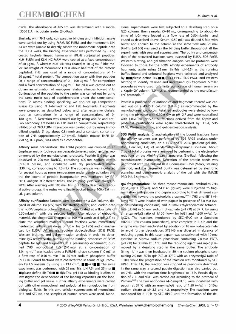
A cyclic dimeric tripeptide library (Scheme 1 A) composed of18 natural amino acids has been prepared by solid-phase pep-
tide synthesis,[40–43] by applying the mix-split method.[44, 45] Afteron-resin assembly, cleavage, and lyophilization, an average of5.5 mg of the sublibraries were obtained. Peptide pools werecyclized by spontaneous oxidation of the two N-terminal cys-teine residues[40–43] in a slightly alkaline aqueous solution buf-fered with ammonium bicarbonate[41] to ensure partial salt re-moval over lyophilization. The library contained a theoreticaltotal number of 183 = 5832 different peptides, arranged in18 different subpools, each theoretically containing 324 cyclicpeptides. The pools, given the high complexity, were charac-terized only by amino acid analysis,[42, 43] with comparison ofthe data obtained with those expected by an equimolar distri-bution of all components. Data deriving from this analysiswere highly consistent with the expected values. Less complexpeptide mixtures composed of 18 molecules, preparedthroughout library deconvolution, were submitted to a moreextensive characterization by reversed-phase (RP) HPLC analy-sis, MALDI-TOF mass spectrometry, and amino acid analysis.[42]
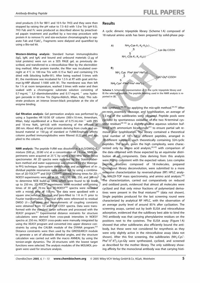
The characterization, carried out comparatively on reducedand oxidized pools, evidenced that almost all molecules werecyclized and that only minor fractions of polymerized deriva-tives were present in the final mixtures[42] (data not shown).Single peptides produced for the last screening round werecharacterized by analytical RP HPLC, with the observation ofan average purity level of around 85 % after cyclization. Thescreening assays, carried out by both ELISA and nitrocelluloseadsorption, evidenced that the sublibrary best able to bind the7H3 antibody was that carrying phenylalanine residues on thepositions next to the cysteines. The ELISA assay (Figure 1 A)showed that other sublibraries also efficiently bound the anti-body, but these were not considered for resynthesis as theywere only slightly active in the nitrocellulose assay (data notshown). After this first screening, the sublibraries (NH2-Cys1-Phe2-X3-X4)2-Lys-Gly were synthesized, cyclized, and screenedas described for the mother library. The only sublibrary show-ing affinity for the monoclonal antibody was that carrying histi-
Scheme 1. Schematic representation of A) the cyclic tripeptide library andB) the selected peptide. The peptide labeling used in the NMR analysis is re-ported in (B).
ChemBioChem 2005, 6, 1 – 13 www.chembiochem.org � 2005 Wiley-VCH Verlag GmbH & Co. KGaA, Weinheim 5
Antibody-Binding Peptide
dine on the third position (Figure 1 B) and the nitrocelluloseassay confirmed this outcome (data not shown). After the
18 single peptides were produced and cyclized, they were sub-mitted to screening. At least 5 different peptides, carrying His,Leu, Asn, Gly, or Phe in position 4, (Figure 1 C), bound the 7H3antibody, but the His peptide was definitely the most active.The nitrocellulose counterproof was not carried out in this lastcase. The cyclic peptide (NH2-Cys1-Phe2-His3-His4)2-Lys-Gly(Scheme 1 B), named FcRM, was then selected as the bestunder these conditions and submitted to further characteriza-tion.
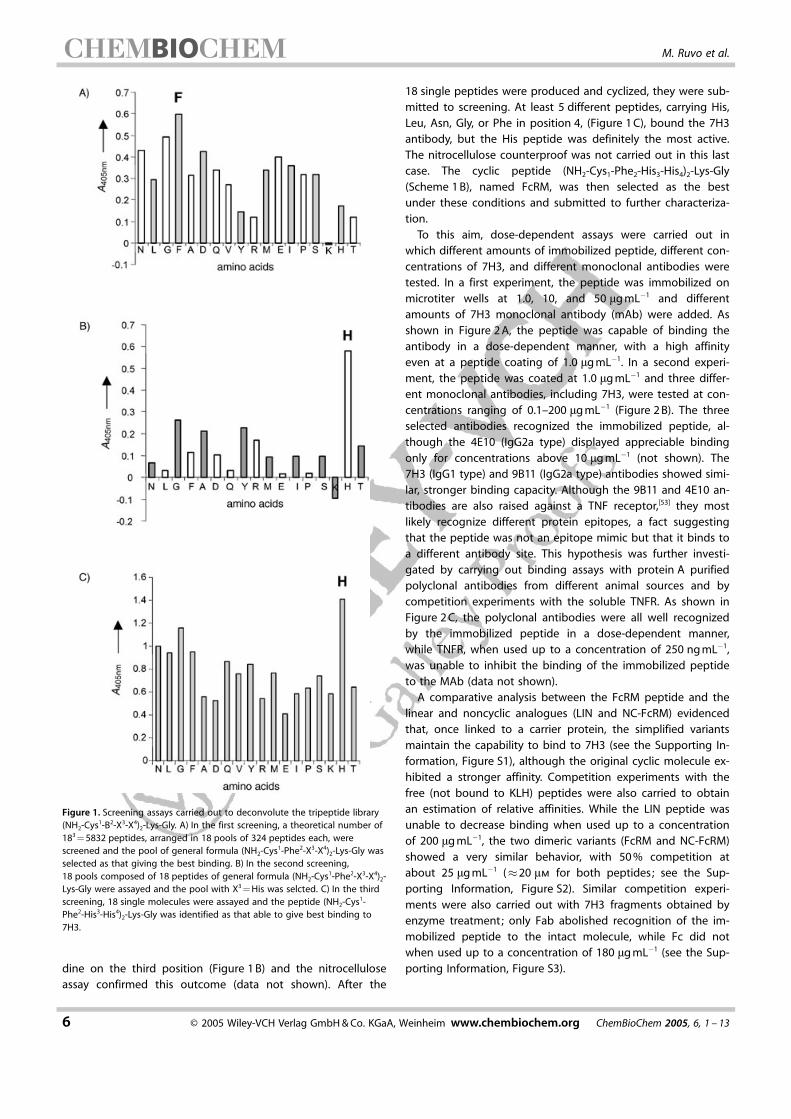
To this aim, dose-dependent assays were carried out inwhich different amounts of immobilized peptide, different con-centrations of 7H3, and different monoclonal antibodies weretested. In a first experiment, the peptide was immobilized onmicrotiter wells at 1.0, 10, and 50 mg mL�1 and differentamounts of 7H3 monoclonal antibody (mAb) were added. Asshown in Figure 2 A, the peptide was capable of binding theantibody in a dose-dependent manner, with a high affinityeven at a peptide coating of 1.0 mg mL�1. In a second experi-ment, the peptide was coated at 1.0 mg mL�1 and three differ-ent monoclonal antibodies, including 7H3, were tested at con-centrations ranging of 0.1–200 mg mL�1 (Figure 2 B). The threeselected antibodies recognized the immobilized peptide, al-though the 4E10 (IgG2a type) displayed appreciable bindingonly for concentrations above 10 mg mL�1 (not shown). The7H3 (IgG1 type) and 9B11 (IgG2a type) antibodies showed simi-lar, stronger binding capacity. Although the 9B11 and 4E10 an-tibodies are also raised against a TNF receptor,[53] they mostlikely recognize different protein epitopes, a fact suggestingthat the peptide was not an epitope mimic but that it binds toa different antibody site. This hypothesis was further investi-gated by carrying out binding assays with protein A purifiedpolyclonal antibodies from different animal sources and bycompetition experiments with the soluble TNFR. As shown inFigure 2 C, the polyclonal antibodies were all well recognizedby the immobilized peptide in a dose-dependent manner,while TNFR, when used up to a concentration of 250 ng mL�1,was unable to inhibit the binding of the immobilized peptideto the MAb (data not shown).
A comparative analysis between the FcRM peptide and thelinear and noncyclic analogues (LIN and NC-FcRM) evidencedthat, once linked to a carrier protein, the simplified variantsmaintain the capability to bind to 7H3 (see the Supporting In-formation, Figure S1), although the original cyclic molecule ex-hibited a stronger affinity. Competition experiments with thefree (not bound to KLH) peptides were also carried to obtainan estimation of relative affinities. While the LIN peptide wasunable to decrease binding when used up to a concentrationof 200 mg mL�1, the two dimeric variants (FcRM and NC-FcRM)showed a very similar behavior, with 50 % competition atabout 25 mg mL�1 (�20 mm for both peptides; see the Sup-porting Information, Figure S2). Similar competition experi-ments were also carried out with 7H3 fragments obtained byenzyme treatment; only Fab abolished recognition of the im-mobilized peptide to the intact molecule, while Fc did notwhen used up to a concentration of 180 mg mL�1 (see the Sup-porting Information, Figure S3).
Figure 1. Screening assays carried out to deconvolute the tripeptide library(NH2-Cys1-B2-X3-X4)2-Lys-Gly. A) In the first screening, a theoretical number of183 = 5832 peptides, arranged in 18 pools of 324 peptides each, werescreened and the pool of general formula (NH2-Cys1-Phe2-X3-X4)2-Lys-Gly wasselected as that giving the best binding. B) In the second screening,18 pools composed of 18 peptides of general formula (NH2-Cys1-Phe2-X3-X4)2-Lys-Gly were assayed and the pool with X3 = His was selcted. C) In the thirdscreening, 18 single molecules were assayed and the peptide (NH2-Cys1-Phe2-His3-His4)2-Lys-Gly was identified as that able to give best binding to7H3.
6 � 2005 Wiley-VCH Verlag GmbH & Co. KGaA, Weinheim www.chembiochem.org ChemBioChem 2005, 6, 1 – 13
M. Ruvo et al.
To evaluate the affinity properties of FcRM in chromatogra-phy applications, the peptide was immobilized on a solid sup-port. Preliminary experiments were carried out by loading frac-tions of pure 7H3 monoclonal IgG (5.0 mg) in 25 mm sodiumphosphate (pH 7.0) buffer onto a FcRM/Emphaze column(1.0 mL). After elution of the unretained material, bound IgGwas recovered by changing the eluent to 100 mm acetic acid(pH 2.7). Analysis of the bound fractions showed that about50 % of the loaded IgG was retained, thereby demonstratingthe effectiveness of FcRM to bind IgG even when covalentlybound on a solid surface. The same experiment, when repeat-ed with 25 mm Tris (pH 7.5) and 25 mm Bis-Tris (pH 6.5) asbinding buffers, showed that the highest recovery was ob-tained with the Bis-Tris buffer at pH 6.5 (>70 %), while only aminor fraction (30 %) of 7H3 was retained with the pH 7.5buffer. Subsequent experiments were then performed by usingthis buffer system.
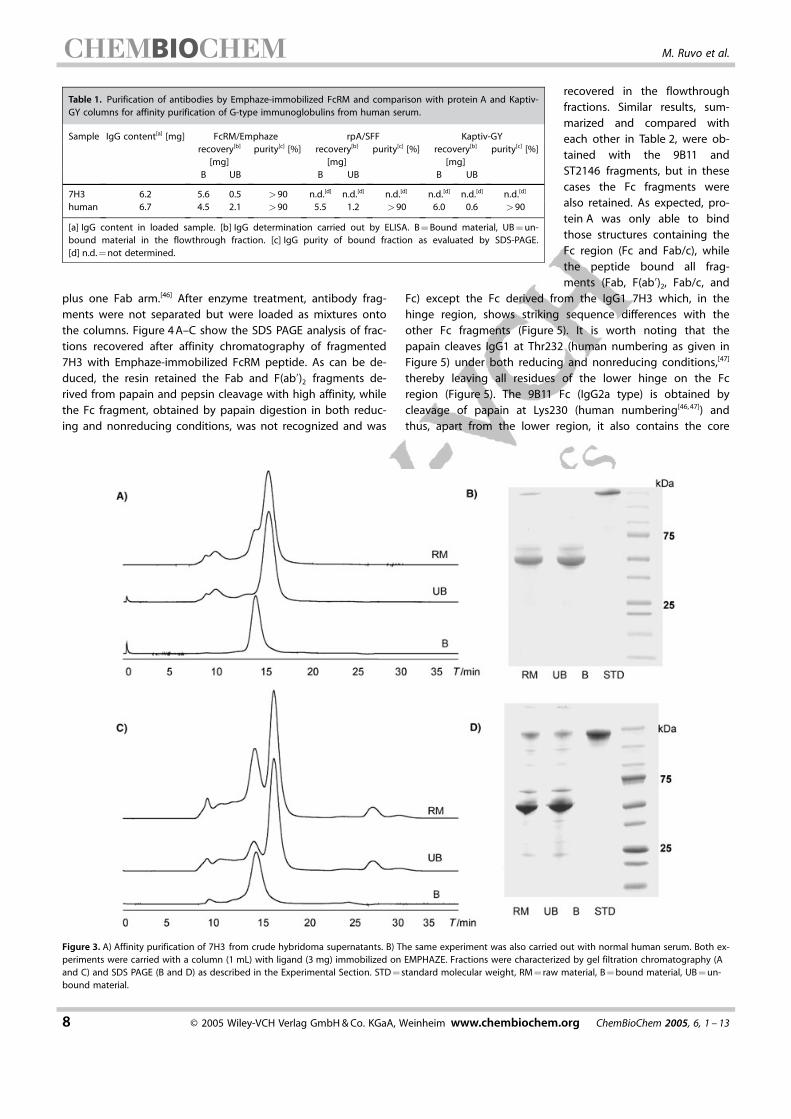
The capacity and selectivity of the FcRM ligand to purify im-munoglobulins were further assessed in affinity chromatogra-phy experiments on IgG-containing biological fluids. In a firstpurification, cellular supernatant of monoclonal 7H3 (5–10 mL,corresponding to about 6–7 mg of IgG; produced as previouslydescribed[54]) was loaded onto the FcRM/Emphaze column(1.0 mL) and processed as reported in the Experimental Sec-tion. Subsequently, human serum (0.50 mL) was diluted fivetimes with the starting buffer (25 mm Bis-Tris, pH 6.5) and ap-plied to the column. The results, expressed as IgG recoveryand purity, are shown in Table 1 and compared with data ob-tained by performing human-serum purifications on protein Aand Kaptiv-GY columns (1.0 mL).[17] The purity of the retainedIgGs was very high (>90 %), as determined by SEC HPLC andSDS PAGE (Figure 3 A–D), while other matrix components wererevealed only in the flowthrough fraction, along with the re-maining antibodies. The identification of IgA and IgM in thebound material of the human-serum purification was only pos-sible by Western-blotting analysis (data not shown), a result in-dicating a much lower specificity of FcRM for these antibodyclasses. The amounts of recovered IgG, determined by ELISAassays, ranged between 4.5 (hIgG) and 5.6 mg (mIgG1; Table 1)and corresponded to 67–90 % of the loaded immunoglobulins.
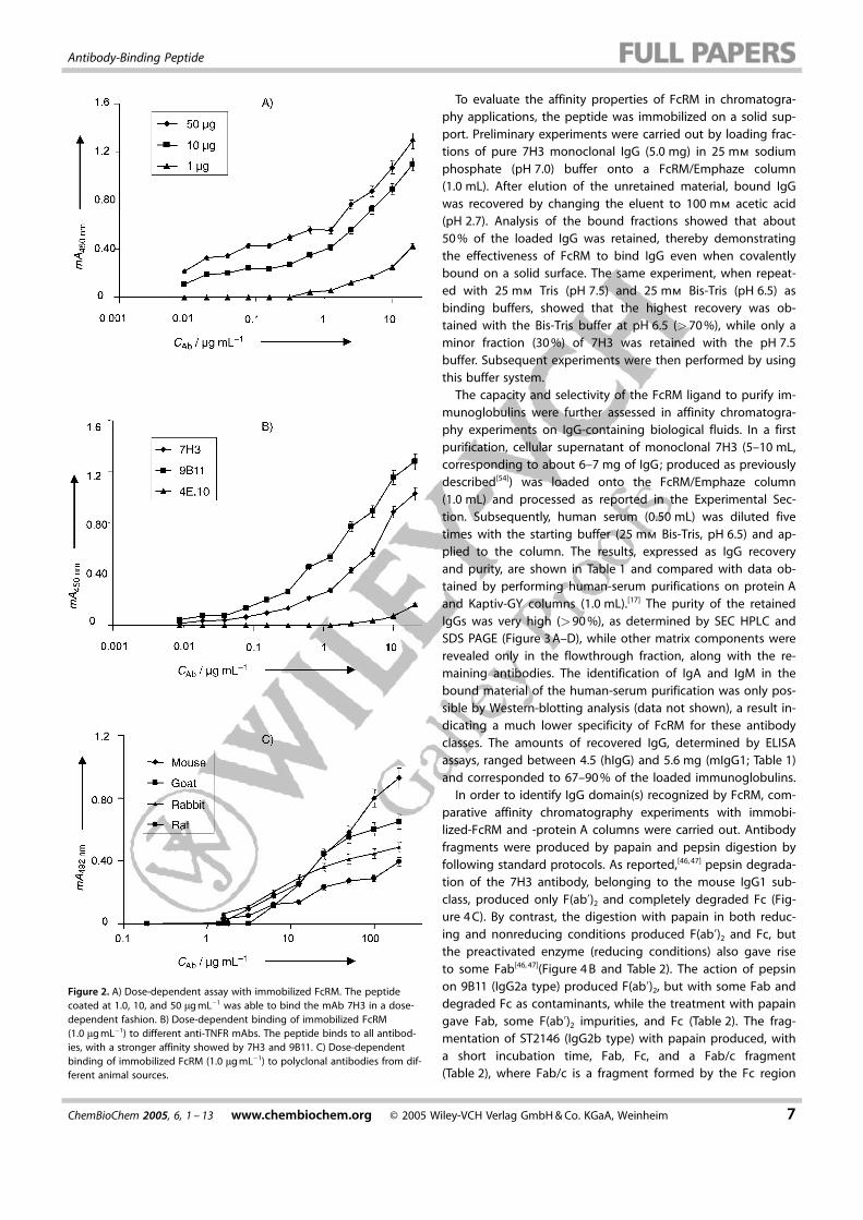
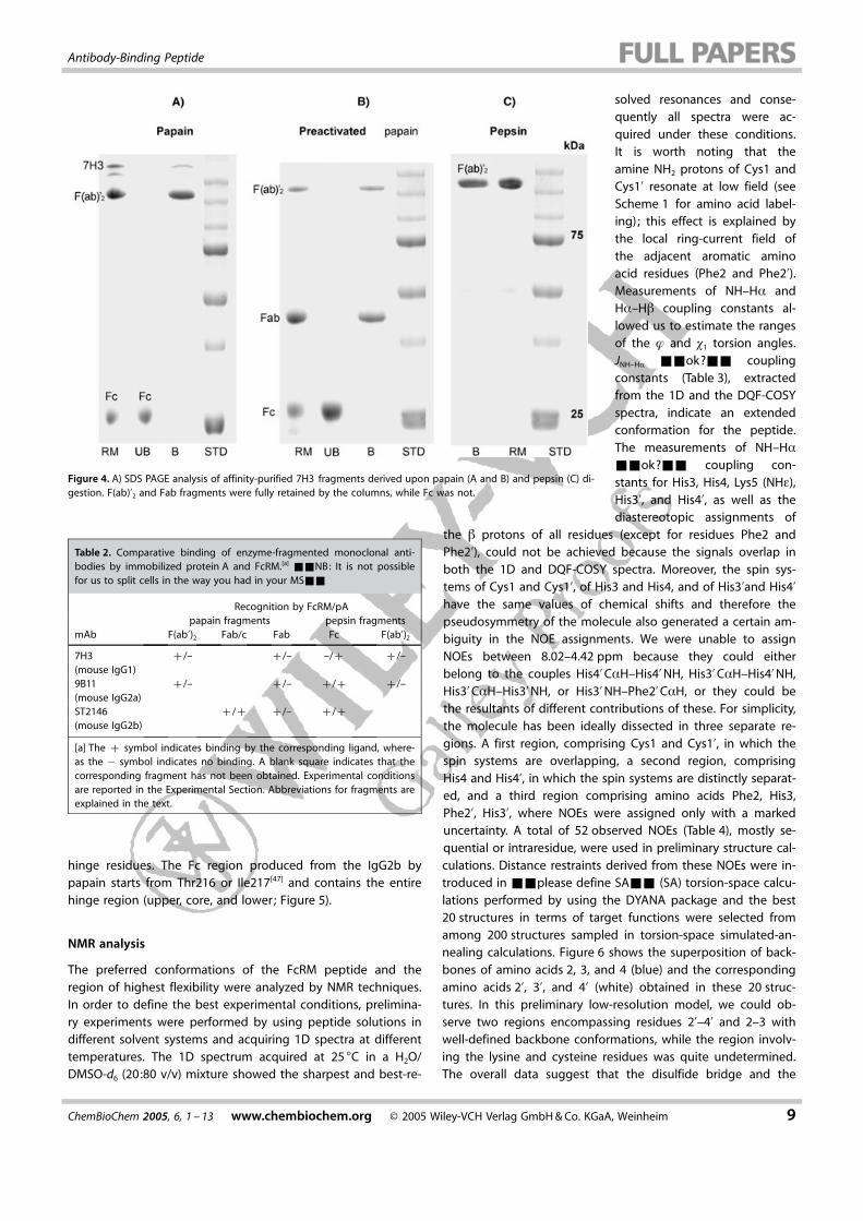
In order to identify IgG domain(s) recognized by FcRM, com-parative affinity chromatography experiments with immobi-lized-FcRM and -protein A columns were carried out. Antibodyfragments were produced by papain and pepsin digestion byfollowing standard protocols. As reported,[46, 47] pepsin degrada-tion of the 7H3 antibody, belonging to the mouse IgG1 sub-class, produced only F(ab’)2 and completely degraded Fc (Fig-ure 4 C). By contrast, the digestion with papain in both reduc-ing and nonreducing conditions produced F(ab’)2 and Fc, butthe preactivated enzyme (reducing conditions) also gave riseto some Fab[46, 47](Figure 4 B and Table 2). The action of pepsinon 9B11 (IgG2a type) produced F(ab’)2, but with some Fab anddegraded Fc as contaminants, while the treatment with papaingave Fab, some F(ab’)2 impurities, and Fc (Table 2). The frag-mentation of ST2146 (IgG2b type) with papain produced, witha short incubation time, Fab, Fc, and a Fab/c fragment(Table 2), where Fab/c is a fragment formed by the Fc region
Figure 2. A) Dose-dependent assay with immobilized FcRM. The peptidecoated at 1.0, 10, and 50 mg mL�1 was able to bind the mAb 7H3 in a dose-dependent fashion. B) Dose-dependent binding of immobilized FcRM(1.0 mg mL�1) to different anti-TNFR mAbs. The peptide binds to all antibod-ies, with a stronger affinity showed by 7H3 and 9B11. C) Dose-dependentbinding of immobilized FcRM (1.0 mg mL�1) to polyclonal antibodies from dif-ferent animal sources.
ChemBioChem 2005, 6, 1 – 13 www.chembiochem.org � 2005 Wiley-VCH Verlag GmbH & Co. KGaA, Weinheim 7
Antibody-Binding Peptide
plus one Fab arm.[46] After enzyme treatment, antibody frag-ments were not separated but were loaded as mixtures ontothe columns. Figure 4 A–C show the SDS PAGE analysis of frac-tions recovered after affinity chromatography of fragmented7H3 with Emphaze-immobilized FcRM peptide. As can be de-duced, the resin retained the Fab and F(ab’)2 fragments de-rived from papain and pepsin cleavage with high affinity, whilethe Fc fragment, obtained by papain digestion in both reduc-ing and nonreducing conditions, was not recognized and was
recovered in the flowthroughfractions. Similar results, sum-marized and compared witheach other in Table 2, were ob-tained with the 9B11 andST2146 fragments, but in thesecases the Fc fragments werealso retained. As expected, pro-tein A was only able to bindthose structures containing theFc region (Fc and Fab/c), whilethe peptide bound all frag-ments (Fab, F(ab’)2, Fab/c, and
Fc) except the Fc derived from the IgG1 7H3 which, in thehinge region, shows striking sequence differences with theother Fc fragments (Figure 5). It is worth noting that thepapain cleaves IgG1 at Thr232 (human numbering as given inFigure 5) under both reducing and nonreducing conditions,[47]
thereby leaving all residues of the lower hinge on the Fcregion (Figure 5). The 9B11 Fc (IgG2a type) is obtained bycleavage of papain at Lys230 (human numbering[46, 47]) andthus, apart from the lower region, it also contains the core
Table 1. Purification of antibodies by Emphaze-immobilized FcRM and comparison with protein A and Kaptiv-GY columns for affinity purification of G-type immunoglobulins from human serum.
Sample IgG content[a] [mg] FcRM/Emphaze rpA/SFF Kaptiv-GYrecovery[b]
[mg]purity[c] [%] recovery[b]
[mg]purity[c] [%] recovery[b]
[mg]purity[c] [%]
B UB B UB B UB
7H3 6.2 5.6 0.5 >90 n.d.[d] n.d.[d] n.d.[d] n.d.[d] n.d.[d] n.d.[d]
human 6.7 4.5 2.1 >90 5.5 1.2 >90 6.0 0.6 >90
[a] IgG content in loaded sample. [b] IgG determination carried out by ELISA. B = Bound material, UB = un-bound material in the flowthrough fraction. [c] IgG purity of bound fraction as evaluated by SDS-PAGE.[d] n.d. = not determined.
Figure 3. A) Affinity purification of 7H3 from crude hybridoma supernatants. B) The same experiment was also carried out with normal human serum. Both ex-periments were carried with a column (1 mL) with ligand (3 mg) immobilized on EMPHAZE. Fractions were characterized by gel filtration chromatography (Aand C) and SDS PAGE (B and D) as described in the Experimental Section. STD = standard molecular weight, RM = raw material, B = bound material, UB = un-bound material.
8 � 2005 Wiley-VCH Verlag GmbH & Co. KGaA, Weinheim www.chembiochem.org ChemBioChem 2005, 6, 1 – 13
M. Ruvo et al.
hinge residues. The Fc region produced from the IgG2b bypapain starts from Thr216 or Ile217[47] and contains the entirehinge region (upper, core, and lower; Figure 5).
NMR analysis
The preferred conformations of the FcRM peptide and theregion of highest flexibility were analyzed by NMR techniques.In order to define the best experimental conditions, prelimina-ry experiments were performed by using peptide solutions indifferent solvent systems and acquiring 1D spectra at differenttemperatures. The 1D spectrum acquired at 25 8C in a H2O/DMSO-d6 (20:80 v/v) mixture showed the sharpest and best-re-
solved resonances and conse-quently all spectra were ac-quired under these conditions.It is worth noting that theamine NH2 protons of Cys1 andCys1’ resonate at low field (seeScheme 1 for amino acid label-ing); this effect is explained bythe local ring-current field ofthe adjacent aromatic aminoacid residues (Phe2 and Phe2’).Measurements of NH–Ha andHa–Hb coupling constants al-lowed us to estimate the rangesof the f and c1 torsion angles.JNH–Ha &&ok?&& couplingconstants (Table 3), extractedfrom the 1D and the DQF-COSYspectra, indicate an extendedconformation for the peptide.The measurements of NH–Ha
&&ok?&& coupling con-stants for His3, His4, Lys5 (NHe),His3’, and His4’, as well as thediastereotopic assignments of
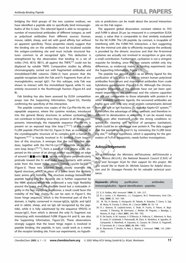
the b protons of all residues (except for residues Phe2 andPhe2’), could not be achieved because the signals overlap inboth the 1D and DQF-COSY spectra. Moreover, the spin sys-tems of Cys1 and Cys1’, of His3 and His4, and of His3’and His4’have the same values of chemical shifts and therefore thepseudosymmetry of the molecule also generated a certain am-biguity in the NOE assignments. We were unable to assignNOEs between 8.02–4.42 ppm because they could eitherbelong to the couples His4’CaH–His4’NH, His3’CaH–His4’NH,His3’CaH–His3’NH, or His3’NH–Phe2’CaH, or they could bethe resultants of different contributions of these. For simplicity,the molecule has been ideally dissected in three separate re-gions. A first region, comprising Cys1 and Cys1’, in which thespin systems are overlapping, a second region, comprisingHis4 and His4’, in which the spin systems are distinctly separat-ed, and a third region comprising amino acids Phe2, His3,Phe2’, His3’, where NOEs were assigned only with a markeduncertainty. A total of 52 observed NOEs (Table 4), mostly se-quential or intraresidue, were used in preliminary structure cal-culations. Distance restraints derived from these NOEs were in-troduced in &&please define SA&& (SA) torsion-space calcu-lations performed by using the DYANA package and the best20 structures in terms of target functions were selected fromamong 200 structures sampled in torsion-space simulated-an-nealing calculations. Figure 6 shows the superposition of back-bones of amino acids 2, 3, and 4 (blue) and the correspondingamino acids 2’, 3’, and 4’ (white) obtained in these 20 struc-tures. In this preliminary low-resolution model, we could ob-serve two regions encompassing residues 2’–4’ and 2–3 withwell-defined backbone conformations, while the region involv-ing the lysine and cysteine residues was quite undetermined.The overall data suggest that the disulfide bridge and the
Figure 4. A) SDS PAGE analysis of affinity-purified 7H3 fragments derived upon papain (A and B) and pepsin (C) di-gestion. F(ab)’2 and Fab fragments were fully retained by the columns, while Fc was not.
Table 2. Comparative binding of enzyme-fragmented monoclonal anti-bodies by immobilized protein A and FcRM.[a]
&&NB: It is not possiblefor us to split cells in the way you had in your MS&&
Recognition by FcRM/pApapain fragments pepsin fragments
mAb F(ab’)2 Fab/c Fab Fc F(ab’)2
7H3 + /– + /– –/ + + /–(mouse IgG1)9B11 + /– + /– + / + + /–(mouse IgG2a)ST2146 + / + + /– + / +
(mouse IgG2b)
[a] The + symbol indicates binding by the corresponding ligand, where-as the � symbol indicates no binding. A blank square indicates that thecorresponding fragment has not been obtained. Experimental conditionsare reported in the Experimental Section. Abbreviations for fragments areexplained in the text.
ChemBioChem 2005, 6, 1 – 13 www.chembiochem.org � 2005 Wiley-VCH Verlag GmbH & Co. KGaA, Weinheim 9
Antibody-Binding Peptide
lysine side chain (Figure 6) are characterized by a marked flexi-bility and work as a sort of hinge around which both copies ofthe rigid Phe-His-His tripeptide can freely fluctuate.
Discussion
By screening a peptide library of general formula (NH2-Cys1-X2-X3-X4)2-Lys-Gly, where the molecules have been cyclized by
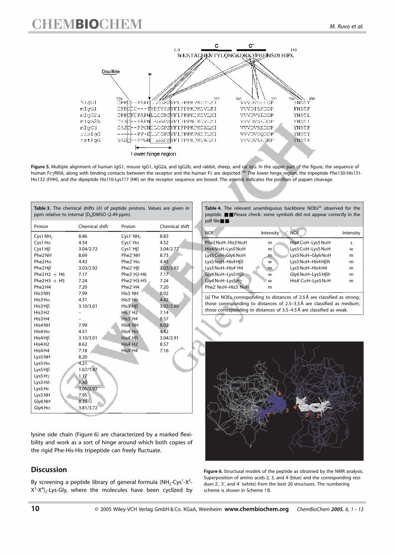
Figure 5. Multiple alignment of human IgG1, mouse IgG1, IgG2a, and IgG2b, and rabbit, sheep, and rat IgG. In the upper part of the figure, the sequence ofhuman FcgRIIIA, along with binding contacts between the receptor and the human Fc are depicted.[36] The lower hinge region, the tripeptide Phe130-His131-His132 (FHH), and the dipeptide His116-Lys117 (HK) on the receptor sequence are boxed. The asterisk indicates the position of papain cleavage.
Table 3. The chemical shifts (d) of peptide protons. Values are given inppm relative to internal [D6]DMSO (2.49 ppm).
Proton Chemical shift Proton Chemical shift
Cys1 NH2 8.86 Cys1’NH2 8.83Cys1 Ha 4.54 Cys1’Ha 4.52Cys1 Hb 3.04/2.72 Cys1’Hb 3.04/2.72Phe2 NH 8.69 Phe2’NH 8.73Phe2 Ha 4.43 Phe2’Ha 4.43Phe2 Hb 3.03/2.92 Phe2’Hb 3.03/2.92Phe2 H2 + H6 7.17 Phe2’H2-H6 7.17Phe2 H3 + H5 7.24 Phe2’H3-H5 7.24Phe2 H4 7.20 Phe2’H4 7.20His3 NH 7.99 His3’NH 8.02His3 Ha 4.51 His3’Ha 4.42His3 Hb 3.10/3.01 His3’Hb 3.02/2.90His3 H2 – His3’H2 7.14His3 H4 – His3’H4 8.57His4 NH 7.99 His4’NH 8.02His4 Ha 4.51 His4’Ha 4.42His4 Hb 3.10/3.01 His4’Hb 3.04/2.91His4 H2 8.62 His4’H2 8.57His4 H4 7.18 His4’H4 7.16Lys5 NH 8.20Lys5 Ha 4.21Lys5 Hb 1.67/1.47Lys5 Hg 1.17Lys5 Hd 1.30Lys5 He 3.06/2.92Lys5 NH 7.95Gly6 NH 8.35Gly6 Ha 3.81/3.72
Table 4. The relevant unambiguous backbone NOEs[a] observed for thepeptide. &&Please check: some symbols did not appear correctly in thepdf file&&
NOE Intensity NOE Intensity
Phe2 NaH–His3 NaH m His4 CaH–Lys5 NaH sHis4 NaH–Lys5 NaH m Lys5 CaH–Lys5 NaH wLys5 CaH–Gly6 NaH m Lys5 NaH–Gly6 NaH mLys5 NaH–His4 Hbl w Lys5 NaH–His4 Hbh mLys5 NaH–His4’H4 m Lys5 NaH–His4 H4 mGly6 NaH–Lys5 Hbl w Gly6 NaH–Lys5 Hbh mGly6 NaH–Lys5 Hg w His4’CaH–Lys5 NeH mPhe2’NaH–His3’NaH m
[a] The NOEs corresponding to distances of 2.5 � are classified as strong;those corresponding to distances of 2.5–3.5 � are classified as medium;those corresponding to distances of 3.5–4.5 � are classified as weak.
Figure 6. Structural models of the peptide as obtained by the NMR analysis.Superposition of amino acids 2, 3, and 4 (blue) and the corresponding resi-dues 2’, 3’, and 4’ (white) from the best 20 structures. The numberingscheme is shown in Scheme 1 B.
10 � 2005 Wiley-VCH Verlag GmbH & Co. KGaA, Weinheim www.chembiochem.org ChemBioChem 2005, 6, 1 – 13
M. Ruvo et al.
bridging the thiol groups of the two cysteine residues, wehave identified a peptide able to specifically bind immunoglo-bulins of the G class. The selected ligand is able to recognize anumber of monoclonal antibodies of different isotypes, as wellas polyclonal antibodies from different sources (human,mouse, rabbit, sheep, and rat) with good affinity but withoutan apparent specificity. These properties clearly suggest thatthe binding site on the antibodies must be localized outsidethe antigen-combining site and must include structural fea-tures common to all recognized IgGs. This deduction isstrengthened by the observation that binding to a set ofmAbs (7H3, 4E10, 9B11), all against the TNFR,[53] could not bedisplaced by soluble TNFR. Comparative studies by affinitychromatography carried out with fragmented antibodies andimmobilized-FcRM columns (Table 2) have proven that thepeptide recognizes both the Fab and Fc fragments from all im-munoglobulins, except IgG1. For this subtype, only Fab wasshown to interact with the immobilized ligand, while Fc wasentirely recovered in the flowthrough fraction (Figure 4 A andB).
Fab binding has also been assessed by ELISA competitionassays (see the Supporting Information, Figure S3), therebyconfirming the specificity of this interaction.
The peptide contains two copies of the Cys-Phe-His-His tet-rapeptide sequence, where the cysteine residues, introducedinto the general library structures to achieve cyclization, donot contribute to binding since they present in all library com-ponents. Interestingly, the tripeptide sequence Phe-His-His ispresent on an exposed loop (the loop C’[30, 31]) of the humanFcgRIII peptide (Phe130–His132, Figure 5) that, as evidenced inthe crystallographic structure of its complex with a human Fcfragment,[30, 31] is heavily involved in Fc binding. From inspec-tion of this structure, it emerges that the two histidine resi-dues, together with the His116–Lys117 dipeptide on an adja-cent loop (loop C[30, 31]), form a patch of four amino acids, dis-persed on the corner of an almost perfect square with an edgeof around 5 &&units?&&, whose side chains perpendicularlyprotrude toward the Fc and make many contacts with aminoacids from the lower hinge region, namely Lys234–Ser239[30]
(Figure 5). These two adjacent loops closely resemble theligand structure, which, in place of a lysine bears the similarlybasic amino acid histidine. The structure analogy between thepeptide ligand and the receptor site is further supported bythe NMR analysis, which has evidenced a very high flexibilityaround the lysine and the disulfide bond but a noticeable ri-gidity in the two tripeptide backbones, a result could favor themobility of the side chains. As can be seen in Figure 5, thelower hinge sequence, which is the N-terminal part of the Fcdomain, is highly conserved in mouse IgG2a, IgG2b, and IgG3and in rabbit, sheep, and rat IgG (all recognized by the pep-tide), while it is fully substituted by the sequence VPEVSS inmouse IgG1, from which is derived the only Fc fragment notinteracting with immobilized FcRM (Figure 4 A and B; see alsothe Supporting Information, Figure S3). These observationsstrongly suggest that the lower hinge region is involved inpeptide binding; the peptide, in turn, could work as a mimicof the receptor binding site. From our experiments, no hypoth-
esis or predictions can be made about the second interactionsite on the Fab region.
The apparent global dissociation constant relative to 7H3and FcRM is about 20 mm (as measured in a competition ELISAassay), a value that is comparable to that similarly evaluatedfor the NC-FcRM. The LIN peptide, by contrast, is incapable ofinterfering with the FcRM-7H3 interaction, a fact suggestingthat the minimal unit able to efficiently recognize the antibodyis provided by the dimeric structure and that the N-terminalcysteines are actually not involved in recognition or give onlya small contribution. Furthermore, cyclization is not a stringentrequisite for binding, since the two variants exhibit only tinydifferences, as evidenced mainly in binding experiments (seethe Supporting Information, Figure S1).
The peptide has proved useful as an affinity ligand for thepurification of IgG, since it is able to extract human polyclonalantibodies from serum and monoclonal antibodies from crudehybridoma supernatants (Figure 3 A–D). Although the chroma-tographic properties of the peptide have not yet been opti-mized (experiments are underway) and the column capacitiesare still not comparable to those attainable with protein A orother available synthetic ligands, the recovered antibodies arehighly pure with only very small protein contaminants derivedfrom the IgM or IgA fractions. As with the Kaptiv-GY system,[17]
FcRM offers the advantage of being synthetic and therefore in-different to denaturation or unfolding. It can be reused manytimes, even after treatment under the strong conditions re-quired for cleaning and removal of pyrogens (sanitation),which could be an advantage over protein A. The data suggestthat the peptide could bind Fc by mimicking the FcgRIII bind-ing site,[30] but this hypothesis, which is appealing for the gen-eration of Fc/FcR antagonists, needs further investigations.
Acknowledgements
We acknowledge the Ministero dell’Istruzione, dell’Universit� edella Ricerca (M.I.U.R.), the National Research Council (C.N.R.) ofItaly, and Tecnogen SCpA for their support for this project. Wealso would like to thank Dr. Michele Saviano for helpful discus-sion and Dr. Giuseppe Perretta for his valuable technical assis-tance.
Keywords: affinity purification · antibodies ·immunoglobulins · ligand identification · peptides
[1] E. A. Padlan, Mol. Immunol. 1994, 31, 169 – 217.[2] R. J. Lories, J. A. Maertens, J. L. Ceuppens, W. E. Peetermans, Acta Clin.
Belg. 2000, 55, 163 – 169.[3] M. Toi, H. Bando, S. Horiguchi, M. Takada, A. Kataoka, T. Ueno, S. Saji,
M. Muta, N. Funata, S. Ohno, Br. J. Cancer 2004, 90, 10 – 14.[4] D. J. Slamon, B. Leyland-Jones, S. Shak, H. Fuchs, V. Paton, A. Baja-
monde, T. Fleming, W. Eiermann, J. Wolter, M. Pegram, J. Baselga, L.Norton, N. Engl. J. Med. 2001, 344, 783 – 792.
[5] R. De Santis, A. M. Inastasi, V. D’Alessio, A. Pelliccia, C. Albertoni, A. Rosi,B. Leoni, R. Lindstedt, F. Petronzelli, M. Dani, A. Verdoliva, A. Ippolito, N.Campanile, V. Manfredi, A. Esposito, G. Cassani, M. Chinol, G. Paganelli,P. Carminati, Br. J. Cancer 2003, 88, 996 – 1003.
[6] B. Akerstrom, T. Brodin, K. Reis, L. Bjorck, J. Immunol. 1985, 135, 2589 –2592.
ChemBioChem 2005, 6, 1 – 13 www.chembiochem.org � 2005 Wiley-VCH Verlag GmbH & Co. KGaA, Weinheim 11
Antibody-Binding Peptide
[7] M. A. Godfrey, P. Kwasowski, R. Clift, V. Marks, J. Immunol. Methods1993, 160, 97 – 105.
[8] J. Balthasar, H. L. Fung, J. Pharm. Sci. 1995, 84, 2 – 6.[9] S. F. Chou, C. Y. Chen, Hybridoma 2001, 20, 59 – 62.
[10] Z. Yan, J. Huang, J. Chromatogr. B 2000, 738, 149 – 154.[11] K. Huse, H.-J. Bçhme, G. H. Scholz, J. Biochem. Biophys. Methods 2002,
51, 217 – 231.[12] S. M. A. Bueno, K. Haupt, M. A. Vijayalakshmi, J. Chromatogr. B 1995,
667, 57 – 67.[13] E. Boschetti, J. Biochem. Biophys. Methods 2001, 49, 361 – 389.[14] C. R. Lowe, J. C. Pearson, Methods Enzymol. 1984, 104, 97 – 113.[15] R. Li, V. Dowd, D. J. Stewart, S. J. Burton, C. R. Lowe, Nat. Biotechnol.
1998, 16, 190 – 195.[16] G. Fassina, M. Ruvo, G. Palombo, A. Verdoliva, M. Marino, J. Biochem.
Biophys. Methods 2001, 49, 481 – 490.[17] A. Verdoliva, F. Pannone, M. Rossi, S. Catello, V. Manfredi, J. Immunol.
Methods 2002, 271, 77 – 88.[18] G. Fassina, A. Verdoliva, G. Palombo, M. Ruvo, G. Cassani, J. Mol. Recog-
nit. 1998, 11, 128 – 133.[19] G. Palombo, A. Verdoliva, G. Fassina, J. Chromatogr. B 1998, 715, 137 –
145.[20] A. Verdoliva, G. Basile, G. Fassina, J. Chromatogr. B 2000, 749, 233 – 242.[21] E. E. Idusogie, L. G. Presta, H. Santoro-Gazzano, K. Totpal, P. Y. Wong, M.
Ultsch, Y. G. Meng, M. G. Mullkerrin, J. Immunol. 2000, 164, 4178 – 4184.[22] W. L. DeLano, M. H. Ultsch, A. M. de Vos, J. A. Wells, Science 2000, 287,
1279 – 1283.[23] T. Takai, Nat. Rev. Immunol. 2002, 2, 580 – 592.[24] T. Takai, A. Nakamura, K. Akiyama, Curr. Drug Targets: Immune Endocr.
Metab. Disord. 2003, 3, 187 – 197.[25] S. Radaev, P. Sun, Mol. Immunol. 2001, 38, 1073 – 1083.[26] B. J. Sutton, R. L. Beavil, A. Beavil, &&Br. Med. Bull. ok?&& 2000, 56,
1004 – 1018.[27] H. M. Dijstelbloem, J. G. J. van de Winkel, C. G. M. Kallenberg, Trends Im-
munol. 2001, 22, 510 – 516.[28] P. Sondermann, V. Oosthuizen, Immunol. Lett. 2002, 82, 51 – 56.[29] R. Jefferis, J. Lund, Immunol. Lett. 2002, 82, 57 – 65.[30] P. Sondermann, R. Huber, V. Oosthuizen, U. Jacob, Nature 2000, 406,
267 – 273.[31] P. Sondermann, J. Kaiser, U. Jacob, J. Mol. Biol. 2001, 309, 737 – 749.[32] W. L. Martin, A. P. West, Jr. , L. Gan, P. J. Bjorkman, Mol. Cell 2001, 7, 867 –
877.[33] K. Kato, C. Sautes-Fridman, W. Yamada, K. Kobayashi, S. Uchiyama, H.
Kim, J. Enokizono, A. Galinha, Y. Kobayashi, W. H. Fridman, Y. Arata, I.Shimada, J. Mol. Biol. 2000, 295, 213 – 224.
[34] S. C. Garman, J. P. Kinet, T. S. Jardetzky, Cell 1998, 95, 951 – 961.[35] S. C. Garman, B. A. Wurzburg, S. S. Tarchevskaya, J. P. Kinet, T. S. Jardetz-
ky, Nature 2000, 406, 259 – 266.[36] A. B. Herr, E. R. Ballister, P. J. Bjorkman, Nature 2003, 423, 614 – 620.[37] L. J. Rigby, H. Trist, J. Snider, M. D. Hulett, P. M. Hogarth, V. C. Epa, Allergy
2000, 55, 609 – 619.[38] G. R. Nakamura, M. E. Reynolds, Y. M. Chen, M. A. Starovasnik, H. B.
Lowman, Proc. Natl. Acad. Sci. USA 2002, 99, 1303 – 1308.[39] G. R. Nakamura, M. A. Starovasnik, M. E. Reynolds, H. B. Lowman, Bio-
chemistry 2001, 40, 9828 – 9835.[40] G. Fassina, P. Scardino, M. Ruvo, P. Fucile, P. Amodeo, G. Cassani in Pep-
tides (Ed. : H. Maia), ESCOM, Leiden, 1994, pp. 489 – 490.[41] M. Marino, N. Campanile, A. Ippolito, A. Scarallo, M. Ruvo, G. Fassina in
Peptides (Eds. : S. Bajusz, F. Hudecz), Akad�miai Kiad�, Budapest, 1998,pp. 776 – 777.
[42] M. Ruvo, G. Fassina in Combinatorial Chemistry and Technologies : Princi-ples, Methods and Applications (Eds. : S. Miertus, G. Fassina), MarcelDekker, New York, 1999, pp. 7 – 21.
[43] M. Ruvo, P. Scardino, G. Cassani, G. Fassina, Pept. Protein Lett. 1994, 1,187 – 192.
[44] K. S. Lam, S. E. Salmon, E. M. Hersh, V. J. Hruby, W. M. Kazmierski, R. J.Knapp, Nature 1991, 354, 82 – 84.
[45] A. Furka, M. Sebestyen, M. Asgedom, G. Dibo, Int. J. Pept. Protein Res.1991, 37, 487 – 493.
[46] P. Parham, J. Immunol. 1983, 131, 2895 – 2902.[47] Y. Yamaguchi, H. Kim, K. Kato, K. Masuda, I. Shimada, Y. Arata, J. Immu-
nol. Methods 1995, 181, 259 – 267.[48] C. Greisinger, G. Otting, K. W�thrich, R. R. Ernst, J. Am. Chem. Soc. 1988,
110, 7870 – 7872.[49] M. Rance, O. W. Sørensen, G. Bodenhausen, G. Wagner, R. R. Ernst, K.
W�thrich, Biochem. Biophys. Res. Commun. 1983, 117, 479 – 485.[50] A. A. Bothener-By, R. L. Stephens, J. Lee, C. D. Warren, R. W. Jeanloz, J.
Am. Chem. Soc. 1984, 106, 811 – 813.[51] C. Bartels, T. Xia, M. Billeter, K. W�thrich, J. Biomol. NMR 1995, 5, 1 – 10.[52] P. G�ntert, W. Braun, K. W�thrich, J. Mol. Biol. 1991, 217, 517 – 530.[53] A. Corti, L. Bagnasco, G. Cassani, Lymphokine Cytokine Res. 1994, 13,
183 – 190.[54] M. Marino, A. Corti, A. Ippolito, G. Cassani, G. Fassina, Biotechnol.
Bioeng. 1997, 54, 17 – 25.
Received: October 14, 2004Revised: March 3, 2005Published online on && &&, 2005
12 � 2005 Wiley-VCH Verlag GmbH & Co. KGaA, Weinheim www.chembiochem.org ChemBioChem 2005, 6, 1 – 13
M. Ruvo et al.

ARTICLES
A. Verdoliva, D. Marasco, A. De Capua,A. Saporito, P. Bellofiore, V. Manfredi,R. Fattorusso, C. Pedone, M. Ruvo*
&& –&&
A New Ligand for Immunoglobulin GSubdomains by Screening of aSynthetic Peptide Library
A ring that binds : A cyclic dimeric tri-peptide (1) has been selected from asynthetic combinatorial library as an af-finity ligand for G-type immunoglobu-lins of different animal species. A pre-
liminary chromatographic characteriza-tion is provided, along with an indica-tion of one of the potential bindingsites on immunoglobulin G.
ChemBioChem 2005, 6, 1 – 13 www.chembiochem.org � 2005 Wiley-VCH Verlag GmbH & Co. KGaA, Weinheim 13
Related Documents