ACOUSTICS 2017 Page 1 of 11 A NEW CLUE TO INFRASOUND – EXPERIMENTAL EVIDENCE SUPPORTING OSMOTIC BASELINE STABILISATION IN THE EAR Eric LePage (1,2), Paul Avan (2) (1) PO Box 2564, Mount Claremont, W.A. 6010, Australia (2) Laboratory of Neurosensory Biophysics, UMR INSERM 1107, School of Medicine, University of Auvergne, Clermont-Ferrand, France ABSTRACT This work examines what defines the low-frequency limit of audibility and concludes that because of the way the cochlea has evolved, the definition of infrasound needs to be extended down to zero frequency, viz. static pres- sures. Auditory frequency analysis is usually modelled using the two-chamber model of von Békésy in which the middle chamber serves no obvious mechanical function. Scala media (part of the endolymphatic duct) is, however, associated with cochlear homeostasis and evident regulation of the transverse position of the basilar membrane. This tiny, sandwiched, vessel sometimes develops enough osmotic pressure to rupture its membranes. A hy- pothesis is developed that its central function may be to routinely vary its internal pressure to slowly counterbal- ance atmospheric (ambient) pressure variation delivered to the perilymph, keeping the basilar membrane in the static position required for optimal hearing sensitivity. Two key missing pieces of evidence are needed to support this theory: 1) that cochlear fluid pressures vary in ways influenced by endocochlear potential, and 2) the now well-documented water channels (AQP5s) lining this endolymphatic chamber are gated to control the flow of water down the documented osmotic gradient. We here present the first direct evidence from micropuncture pressure measurements, accessed through of the round-window membrane of living rodents. When appropriately invoked, the data reveal behaviours consistent with aquaporin gating as well as pressure release. It follows that individual susceptibility to infrasound may result from loss of the stabilisation afforded by an ancient form of hydraulic assist. 1 INTRODUCTION Infrasound is that class of pressure variations of lower frequency than the lowest audible limit of hearing (<20Hz). It has always been assumed that the ear takes care of infrasound such that acousticians and physiologists need only to be concerned with acoustic waves within the auditory passband. The recent need to study the human response to infrasound means dealing with the perplexing situation in which very-low-frequency noise constitutes a signal. The object of this study queries what is the physiological nature of the filtering process which determines whether an oscillatory pressure variation should be treated as belonging within the auditory passband, or not? A previous attempt to address this question included examining the nature of the tonotopic organisation as rep- resented by the frequency-place map in the cochlea(Eric L LePage 2003). For most mammals the high-frequency end of the cochlea is spatially organised with octave separations represented by equal distances along the basilar membrane. By contrast, at the low-frequency end of the cochlea, particularly for the larger mammals, this rela- tionship breaks down and it is apparent that there is a distinct difference in the way low-frequency sounds are processed. In the evolutionary stream since therians (Vater, Meng, and Fox 2004; Manley 2013), most auditory receptors have been able to sense low-frequencies, but high-frequency reception occurred with the middle ear and elongation of the cochlear partition. The dominant term in the equation for the tonotopy (Greenwood 1990) describes the high frequency end, while the primordial low-frequency response is paradoxically represented by an end-correction. Moreover, in that equation the difference between the high- and low-frequency ranges sepa- rates at around 1kHz yet there is no sign of class difference between the low-frequency end of the spectrum and what is now being termed infrasound. Study of the gross morphology of the mammalian cochlea, suggests that the essential difference may have its origin in how high frequency- and very low frequency sounds enter the cochlea. Contrary to the predominant view that all frequencies enter via the middle ear, there is an argument, that evolutionary adaptation will preserve any working system, and exapt its function to serve another need (Gould and Vrba 1982), e.g. the ears of fishes are fluid-coupled to their environment using a variety of conduits (Schulz- Mirbach et al. 2013). It is argued that in mammals the entry of low-frequency pressure may have been preserved as a fluid conduit (viz. the cochlear aqueduct) and that its continuing function is to compensate the sensory hair cell operating points for changes in ambient pressure. High-frequency information is mostly air-borne and so the later evolutionary adaptation to living on land required the evolution impedance-matching devices such as the middle and external ears (Christensen-Dalsgaard and Manley 2013). This possible arrangement might make more sense of the ‘warping’ of the map at some mid-frequency crossover. To some extent in sensing our low Paper Not Peer Reviewed

Welcome message from author
This document is posted to help you gain knowledge. Please leave a comment to let me know what you think about it! Share it to your friends and learn new things together.
Transcript

ACOUSTICS 2017 Page 1 of 11
A NEW CLUE TO INFRASOUND – EXPERIMENTAL EVIDENCE SUPPORTING OSMOTIC BASELINE STABILISATION IN THE EAR
Eric LePage (1,2), Paul Avan (2)
(1) PO Box 2564, Mount Claremont, W.A. 6010, Australia (2) Laboratory of Neurosensory Biophysics, UMR INSERM 1107,
School of Medicine, University of Auvergne, Clermont-Ferrand, France
ABSTRACT
This work examines what defines the low-frequency limit of audibility and concludes that because of the way the cochlea has evolved, the definition of infrasound needs to be extended down to zero frequency, viz. static pres-sures. Auditory frequency analysis is usually modelled using the two-chamber model of von Békésy in which the middle chamber serves no obvious mechanical function. Scala media (part of the endolymphatic duct) is, however, associated with cochlear homeostasis and evident regulation of the transverse position of the basilar membrane. This tiny, sandwiched, vessel sometimes develops enough osmotic pressure to rupture its membranes. A hy-pothesis is developed that its central function may be to routinely vary its internal pressure to slowly counterbal-ance atmospheric (ambient) pressure variation delivered to the perilymph, keeping the basilar membrane in the static position required for optimal hearing sensitivity. Two key missing pieces of evidence are needed to support this theory: 1) that cochlear fluid pressures vary in ways influenced by endocochlear potential, and 2) the now well-documented water channels (AQP5s) lining this endolymphatic chamber are gated to control the flow of water down the documented osmotic gradient. We here present the first direct evidence from micropuncture pressure measurements, accessed through of the round-window membrane of living rodents. When appropriately invoked, the data reveal behaviours consistent with aquaporin gating as well as pressure release. It follows that individual susceptibility to infrasound may result from loss of the stabilisation afforded by an ancient form of hydraulic assist.
1 INTRODUCTION
Infrasound is that class of pressure variations of lower frequency than the lowest audible limit of hearing (<20Hz). It has always been assumed that the ear takes care of infrasound such that acousticians and physiologists need only to be concerned with acoustic waves within the auditory passband. The recent need to study the human response to infrasound means dealing with the perplexing situation in which very-low-frequency noise constitutes a signal. The object of this study queries what is the physiological nature of the filtering process which determines whether an oscillatory pressure variation should be treated as belonging within the auditory passband, or not?
A previous attempt to address this question included examining the nature of the tonotopic organisation as rep-resented by the frequency-place map in the cochlea(Eric L LePage 2003). For most mammals the high-frequency end of the cochlea is spatially organised with octave separations represented by equal distances along the basilar membrane. By contrast, at the low-frequency end of the cochlea, particularly for the larger mammals, this rela-tionship breaks down and it is apparent that there is a distinct difference in the way low-frequency sounds are processed. In the evolutionary stream since therians (Vater, Meng, and Fox 2004; Manley 2013), most auditory receptors have been able to sense low-frequencies, but high-frequency reception occurred with the middle ear and elongation of the cochlear partition. The dominant term in the equation for the tonotopy (Greenwood 1990) describes the high frequency end, while the primordial low-frequency response is paradoxically represented by an end-correction. Moreover, in that equation the difference between the high- and low-frequency ranges sepa-rates at around 1kHz yet there is no sign of class difference between the low-frequency end of the spectrum and what is now being termed infrasound. Study of the gross morphology of the mammalian cochlea, suggests that the essential difference may have its origin in how high frequency- and very low frequency sounds enter the cochlea. Contrary to the predominant view that all frequencies enter via the middle ear, there is an argument, that evolutionary adaptation will preserve any working system, and exapt its function to serve another need (Gould and Vrba 1982), e.g. the ears of fishes are fluid-coupled to their environment using a variety of conduits (Schulz-Mirbach et al. 2013). It is argued that in mammals the entry of low-frequency pressure may have been preserved as a fluid conduit (viz. the cochlear aqueduct) and that its continuing function is to compensate the sensory hair cell operating points for changes in ambient pressure. High-frequency information is mostly air-borne and so the later evolutionary adaptation to living on land required the evolution impedance-matching devices such as the middle and external ears (Christensen-Dalsgaard and Manley 2013). This possible arrangement might make more sense of the ‘warping’ of the map at some mid-frequency crossover. To some extent in sensing our low
Paper Not Peer Reviewed

Proceedings of ACOUSTICS 2017 19-22 November 2017,
Perth, Australia
Page 2 of 11 ACOUSTICS 2017
frequency environment we may be using our “fish-ears”. Refining this hypothesis, the infrasound discussion needs to encompass zero frequency (static pressures) for physiological reasons. This extended definition con-siders slow atmospheric (ambient) pressure variations which are often many orders of magnitude larger than acoustic waveforms (Figure 1).
How does the ear cope with infrasound? It normally appears to achieve the high-pass filtering of infrasound by equalisation -- in the same manner of the familiar pressure equalisation across the eardrum, but this time within the fluids of the mammalian inner ear. Historic evidence exists that the endolymphatic system evolved to acquire hearing on a planet with an atmosphere and deep oceans with abundant food. The existence of the endolymphatic duct has always been an enigma (Gates 2006) because most studies of its function have produced confusing results. This status may be because the management of infrasound has been assumed as automatic, not some function which needed to be incorporated into ear function. Just because we do not normally hear infrasound, does not necessarily mean that the ear does not compensate for it. The audibility of infrasound by some people may mean that, rather than being supersensitive to some low-frequency component, their ears lack an otherwise-normal capacity to cancel it. The sensory hair cells of the inner ear lie along the basilar membrane which is sandwiched in-between the peri-lymph compartment and the endolymph compartment (see Figure 2). The perilymph compartment is open to ambient and slow internal pressure changes conveyed via the cochlear aqueduct. By comparison, the endolymph is contained within a tiny closed system known as the membranous labyrinth. Studies of the evolution of the ear show that two things happened in higher mammals, 1) the bone containing the sensory apparatus became sub-stantially thicker (Clack et al. 2003) forming a rigid vault, and 2) the lateral support structures for the basilar membrane also became bone (Raphael and Altschuler 2003), channelling infrasound pressure variations and dragging the basilar membrane with it. It is argued that at the same time, the endolymphatic duct (ED) was formed to counterbalance perilymph pressures. Both ends are essentially sealed but its internal pressure is controlled osmotically (Klis and Smoorenburg 1994; Meyer-Zum-Gottesberge et al. 2000). Because the total lymph volume within the vault is fixed, when the ED expands it displaces perilymph back into cranium. Thus the tiny volume of the ED can internally develop high pressures to counterbalance infrasound as the need arises – such as a change in the weather or a reduction in aircraft cabin pressure (Mirza and Richardson 2005). Any pressure imbalance therefore will influence the mean transverse position of the basilar membrane, the operating points of the hair cells, the gain of the cochlear amplifier and in turn hearing sensitivity. There are osmotic pressure gradients between endolymph and perilymph and also along the endolymphatic duct (Bosher and Warren 1971)(Klis and Smoorenburg 1994). Importantly, within the epithelial lining of the endolymphatic duct are two specialised sets of
Figure 1 All pressures have dimensions ML-1T-2. Shown is a comparison of the ranges, common units and typical pressures within the domains of acoustics, biological and environmental pressures. Most acoustical discussion assumes that just be-cause we do not normally sense infrasound (covering most examples shown on the right side of the figure), that (to develop acoustical sensitivity) our ears do not need to register it, track it and continuously compensate for it. The case is made that this is the central function of the enigmatic endolymphatic system evolved since the early vertebrates 200 million years ago.
Proceedings of ACOUSTICS 2017 19-22 November 2017, Perth, Australia
ACOUSTICS 2017 Page 3 of 11
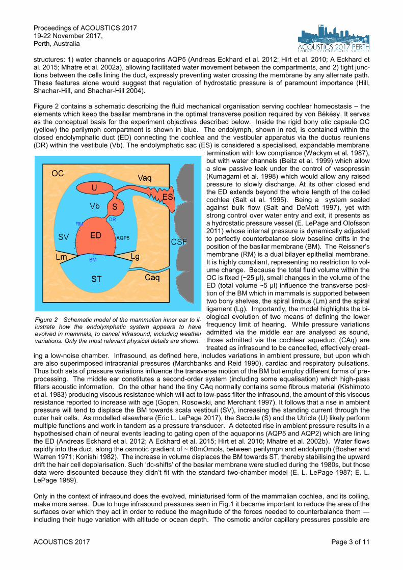
structures: 1) water channels or aquaporins AQP5 (Andreas Eckhard et al. 2012; Hirt et al. 2010; A Eckhard et al. 2015; Mhatre et al. 2002a), allowing facilitated water movement between the compartments, and 2) tight junc-tions between the cells lining the duct, expressly preventing water crossing the membrane by any alternate path. These features alone would suggest that regulation of hydrostatic pressure is of paramount importance (Hill, Shachar-Hill, and Shachar-Hill 2004). Figure 2 contains a schematic describing the fluid mechanical organisation serving cochlear homeostasis – the elements which keep the basilar membrane in the optimal transverse position required by von Békésy. It serves as the conceptual basis for the experiment objectives described below. Inside the rigid bony otic capsule OC (yellow) the perilymph compartment is shown in blue. The endolymph, shown in red, is contained within the closed endolymphatic duct (ED) connecting the cochlea and the vestibular apparatus via the ductus reuniens (DR) within the vestibule (Vb). The endolymphatic sac (ES) is considered a specialised, expandable membrane
termination with low compliance (Wackym et al. 1987), but with water channels (Beitz et al. 1999) which allow a slow passive leak under the control of vasopressin (Kumagami et al. 1998) which would allow any raised pressure to slowly discharge. At its other closed end the ED extends beyond the whole length of the coiled cochlea (Salt et al. 1995). Being a system sealed against bulk flow (Salt and DeMott 1997), yet with strong control over water entry and exit, it presents as a hydrostatic pressure vessel (E. LePage and Olofsson 2011) whose internal pressure is dynamically adjusted to perfectly counterbalance slow baseline drifts in the position of the basilar membrane (BM). The Reissner’s membrane (RM) is a dual bilayer epithelial membrane. It is highly compliant, representing no restriction to vol-ume change. Because the total fluid volume within the OC is fixed (~25 µl), small changes in the volume of the ED (total volume ~5 µl) influence the transverse posi-tion of the BM which in mammals is supported between two bony shelves, the spiral limbus (Lm) and the spiral ligament (Lg). Importantly, the model highlights the bi-ological evolution of two means of defining the lower frequency limit of hearing. While pressure variations admitted via the middle ear are analysed as sound, those admitted via the cochlear aqueduct (CAq) are treated as infrasound to be cancelled, effectively creat-
ing a low-noise chamber. Infrasound, as defined here, includes variations in ambient pressure, but upon which are also superimposed intracranial pressures (Marchbanks and Reid 1990), cardiac and respiratory pulsations. Thus both sets of pressure variations influence the transverse motion of the BM but employ different forms of pre-processing. The middle ear constitutes a second-order system (including some equalisation) which high-pass filters acoustic information. On the other hand the tiny CAq normally contains some fibrous material (Kishimoto et al. 1983) producing viscous resistance which will act to low-pass filter the infrasound, the amount of this viscous resistance reported to increase with age (Gopen, Rosowski, and Merchant 1997). It follows that a rise in ambient pressure will tend to displace the BM towards scala vestibuli (SV), increasing the standing current through the outer hair cells. As modelled elsewhere (Eric L. LePage 2017), the Saccule (S) and the Utricle (U) likely perform multiple functions and work in tandem as a pressure transducer. A detected rise in ambient pressure results in a hypothesised chain of neural events leading to gating open of the aquaporins (AQP5 and AQP2) which are lining the ED (Andreas Eckhard et al. 2012; A Eckhard et al. 2015; Hirt et al. 2010; Mhatre et al. 2002b). Water flows rapidly into the duct, along the osmotic gradient of ~ 60mOmols, between perilymph and endolymph (Bosher and Warren 1971; Konishi 1982). The increase in volume displaces the BM towards ST, thereby stabilising the upward drift the hair cell depolarisation. Such ‘dc-shifts’ of the basilar membrane were studied during the 1980s, but those data were discounted because they didn’t fit with the standard two-chamber model (E. L. LePage 1987; E. L. LePage 1989).
Only in the context of infrasound does the evolved, miniaturised form of the mammalian cochlea, and its coiling, make more sense. Due to huge infrasound pressures seen in Fig.1 it became important to reduce the area of the surfaces over which they act in order to reduce the magnitude of the forces needed to counterbalance them ― including their huge variation with altitude or ocean depth. The osmotic and/or capillary pressures possible are
Figure 2 Schematic model of the mammalian inner ear to il-lustrate how the endolymphatic system appears to have evolved in mammals, to cancel infrasound, including weather variations. Only the most relevant physical details are shown.

Proceedings of ACOUSTICS 2017 19-22 November 2017,
Perth, Australia
Page 4 of 11 ACOUSTICS 2017
large, as is exemplified in the pressures in the roots of tall trees (Singh 2016). So the ranges of the two opposing forces can be matched by the evolutionary reduction in size of the sensory apparatus. The model suggests the need for two new lines of experimentation: 1) the search for a central control element taking input from a pressure transducer, and 2) an effector-element controlling water flow from perilymph into the endolymphatic duct. Re-cently it has been shown using models, that 1) mammalian otoliths may be control elements regulating endolym-phatic pressure (LePage, 2017) and that 2) the aquaporins AQP5s lining the ED exhibit two forms of water gating -- switching and graded (Janosi and Ceccarelli 2013). We here present the first experimental evidence from the ears of living rodents that 1) pressures in the cochlear chambers are not necessarily low, and 2) such gating behaviours do exist, observed in fluid pressure measure-ments made through the round window. There are very distinct temporal characteristics associated with what appears to be the opening of water channels. It is difficult to account for these data based on experimental artifacts, nor measurement device behaviours. Moreover these behaviours occur with rise in pressure with hy-poxia and result in demonstrated pressure release. The efficiency of this slow pressure equalisation system will thus affect how precisely the ear counterbalances infrasound.
2 METHODS
Pressure measurements were made using the WPI-900A micropuncture system using glass micropipettes. In principle the device uses a micropi-pette filled with 2M KCl and inserted into the measurement vessel con-taining normal serum. The interface between the two concentrations at the tip has an electrical resistance and that resistance varies with pres-sure at the tip. A rise in pressure within the vessel causes a very steep rise in electrode resistance and vice versa. The device servo-controls the back pressure of the micropipette to keep the tip electrical resistance constant for pressure variations up to 300Hz, well in excess of infra-sound frequencies. Depending on configuration the device has a linear transduction range typically ±300 mmHg. It was designed and built before the general introduction of SI units for pressure and the use of mmHg has been retained for this project. The micropipettes are pulled from borosilicate glass blanks 1.5mm in diameter, using a Narishige P200 micropipette puller. After filling with 2MKCl their electrical and mechanical characteristics are adjusted under a light microscope to give a bevelled tip diameter of 2 to 3 µm and in a cylindrical glass calibration chamber filled with normal saline an electrical resistance of 2 to 4 MΩ. Pres-sure calibration was achieved in two ways: by pressurising the sealed calibration chamber with a 60cc syringe measuring with a water manometer calibrated in mmHg (same as the device readout), or by using the device itself which operates by applying a pressure to the reverse-end of the micropipette via a special micropipette holder providing both pressure and electrical connection. Details examination of the device revealed 1) the need to supply fixed pressure rails with an external pump and smoothed pressure lines, 2) the relationship of the linear range of the device relative to those pressure rails analogous to the voltage rails for an electronic amplifier, 3) hysteresis in its characteristics, 4) consequences of leaks in any of the tubing, the 5) lack of an external readout in this circa 1980 design to indicate that the phase-lock device is indeed phase-locked and actually tracking pres-sure changes at the tip of the micropipette. Without this control previous users may have mistaken low-pressure readings as valid records. As an extra precaution, local changes in ambient pressure were generated, the brief transient confirming that the record was indeed registering pressure. 6) A serious mistake is possible, employing a procedure commonly used in micropipette recording of single neurons -- that of withdrawing the pipette and trying a different point of entry. Only one penetration of the round window is afforded in each preparation, because the hole from the previous penetration serves as a very effective leak in the chamber. 7) Likewise breaking the tip of the micropipette while penetrating the tough basilar membrane to enter the endolymphatic duct is fatal for the experiment. Repeat checking of micropipette characteristics after each experiment reveals that any tip break may result in a jagged tip with a diameter of 30 µm or more. 8) the presence of air bubbles within any micropipette is fatal for any experiment and techniques were developed to minimise this possibility. 9) the atmospheric pres-sure was routinely recorded at the beginning of each experiment, 10) latterly, dynamic variations in atmospheric pressure were monitored with an Android I9300 device. Rodents (Mongolian gerbils, Javier laboratories, France) 50 to 70g of either sex, raised in low-noise conditions were used for living preparations in which other parameters recorded including the tip potential, indicating whether the tip was in perilymph or endolymph, plus the round window potential mostly used to monitor animal condition. Ambient sounds were also recorded. All 4 signals were recorded using a Cambridge Electronic Design 4-channel
Figure 3 Schematic for micropuncture
pressure measurements in cochlear fluids

Proceedings of ACOUSTICS 2017 19-22 November 2017, Perth, Australia
ACOUSTICS 2017 Page 5 of 11
1401 system, using Spike2 v7 software, in which the sound and cochlear microphonic were sampled each at 50kHz, while the slower micropipette pressures and tip potential were sampled at 1kHz. Preparations were anaesthetised with a mix of Ketamine 60mg/kg and Xylazine 6mg/kg. The anaesthetic dose was repeated every half hour initially, or whenever the stable respiration rate increased. A stereotaxic frame was specially designed and 3-D printed for holding the cranium stationary. For consistency only the right ear was used. The preparations were routinely fitted with a cannula sealed to the cranium, pre-filled with normal saline, for manipulating CSF pressure using a water column. This also served as an extra check on the calibration of micropipettes located in scala tympani. The micropipettes were mounted in a WPI MEH6RF10 micropipette holder in a Narishige MM-3 micromanipulator, for capturing both pressure and potential. More latterly, a specially-designed holder was 3-D printed with rear illumination using laser light (532nm wavelength) to aid visualisation through a Leica binocular microscope of micropipette tip penetration and sealing with the round window membrane and of the basilar mem-brane. Subsequent data analysis and display is carried out using MATLAB™ 2017a.
3 RESULTS
The four-year series of investiga-tions fall into two categories, those extensively concerned with the performance of the WPI-900A micropipette “servo-null”, pres-sure-measurement system, and those using the 2µm probe tip to penetrate the round-window membrane to observe of pres-sures in scala tympani plus sub-sequent penetration of the basilar membrane to access pressures in scala media, i.e. the cochlear-end of the endolymphatic duct (Long 3rd and Morizono 1987; Bӧhmer 1993; Takeuchi, Takeda, and Saito 1990; Yoshida and Lowry 1984). The controls on the equipment were necessary due to the sparsity of manufacturer infor-mation, particularly about the capacity and speed of the device to track pressure variations as the equipment has been used extensively to measure pressures in vessels in lung, kidney, arterioles and plants. It has never been reported that an additional limitation existed in using the equipment for cochlear measurements, particularly if an objective was to observe fluid pressure changes while using loud sound to produce fatigue. The servo device works by matching the pressure at the tip of the micropipette with the pressure at the back end of the pipette. The matching process cannot be achieved using direct current to measure the tip resistance because the pipettes act as diodes. It must use alternating current and the frequency of this is factory-fixed to be 1kHz, which is in the middle of the auditory frequency range. Accordingly sound cannot be used to monitor the condition of the prep-aration without causing considerable other uncertainty. The second level of uncertainty is the fact that in the pressure matching process the device is injecting pressures so that any measured behaviour must be interpreted in the light of the response of the “most sensitive mechano-receptor known” responding to the external servo, with what is being realised is a very sophisticated pressure regulation system of its own. The other channels recorded provide some controls over the experiment, knowing the normal behaviours of the endocochlear potential. Entry of the tip into the endolymph immediately brings a rise of the tip potential to a typical value of typically +90mV confirming location. However, Figure 4 shows how this co-varies with the measured pressure. Bench calibration of the device is essentially linear with pressure, except as the pressure approaches the pump and vacuum supply rails (-150,+250) mmHg [(-20,+33) kPa] supplied to the equipment to control the pipette back pressure. Figure 5 shows a second pair of time records showing the covariation of the mechanical and electrical parameters at the tip of the micropipette. Pressure in scala media is seen to rise with decline in the electric driving potential (the EP). In both cases the data exhibit immediate declines from normal potential and a departure from the start-ingsetpoint of total pressure in the chamber. A confusing feature is that the micropipette will not register slight differences in pressure between the chambers which will exist across the less compliant basilar membrane (see discussion).
Figure 4 Two examples of micropipette simultaneous records of pressure Psm and endocochlear potential (EP) from scala media, showing a reversible response to the diuretic furosemide which not only affects aquaporin behaviour in the kidney, but also aquaporins in the cochlea. Pressures which may appear high in acoustic terms are nevertheless only a fraction of atmospheric pressure.
Proceedings of ACOUSTICS 2017 19-22 November 2017,
Perth, Australia
Page 6 of 11 ACOUSTICS 2017
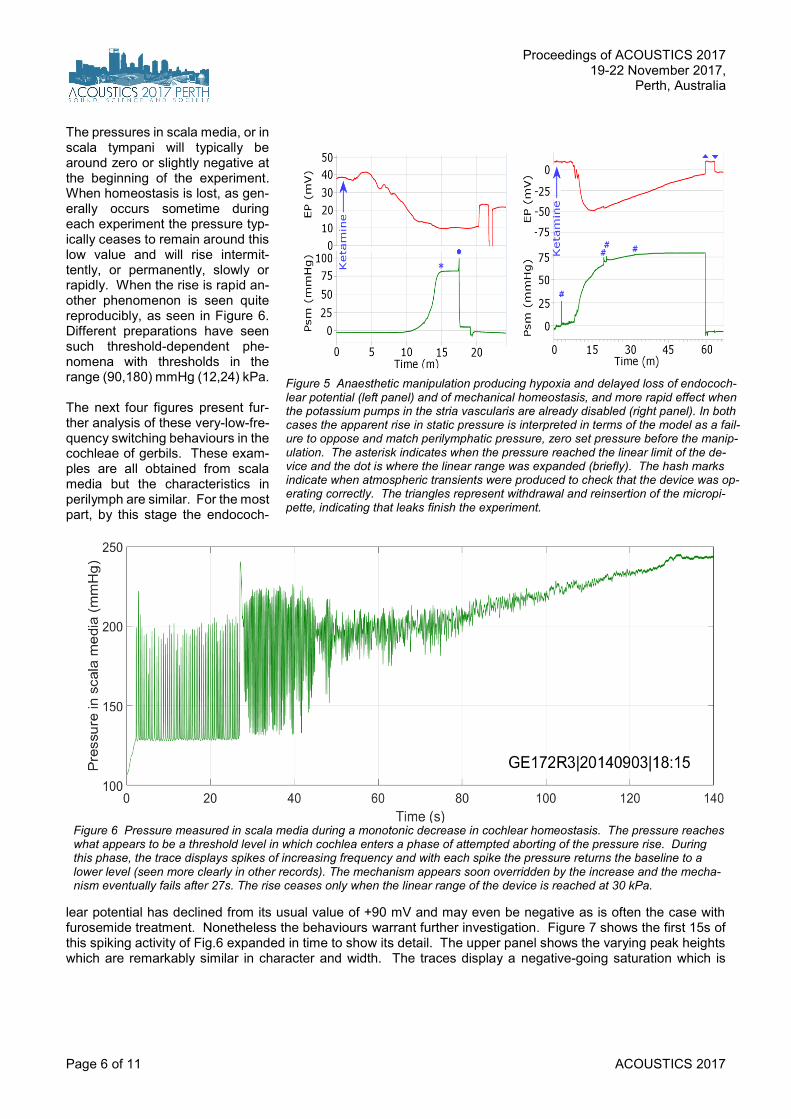
The pressures in scala media, or in scala tympani will typically be around zero or slightly negative at the beginning of the experiment. When homeostasis is lost, as gen-erally occurs sometime during each experiment the pressure typ-ically ceases to remain around this low value and will rise intermit-tently, or permanently, slowly or rapidly. When the rise is rapid an-other phenomenon is seen quite reproducibly, as seen in Figure 6. Different preparations have seen such threshold-dependent phe-nomena with thresholds in the range (90,180) mmHg (12,24) kPa. The next four figures present fur-ther analysis of these very-low-fre-quency switching behaviours in the cochleae of gerbils. These exam-ples are all obtained from scala media but the characteristics in perilymph are similar. For the most part, by this stage the endococh-
lear potential has declined from its usual value of +90 mV and may even be negative as is often the case with furosemide treatment. Nonetheless the behaviours warrant further investigation. Figure 7 shows the first 15s of this spiking activity of Fig.6 expanded in time to show its detail. The upper panel shows the varying peak heights which are remarkably similar in character and width. The traces display a negative-going saturation which is
Figure 5 Anaesthetic manipulation producing hypoxia and delayed loss of endococh-lear potential (left panel) and of mechanical homeostasis, and more rapid effect when the potassium pumps in the stria vascularis are already disabled (right panel). In both cases the apparent rise in static pressure is interpreted in terms of the model as a fail-ure to oppose and match perilymphatic pressure, zero set pressure before the manip-ulation. The asterisk indicates when the pressure reached the linear limit of the de-vice and the dot is where the linear range was expanded (briefly). The hash marks indicate when atmospheric transients were produced to check that the device was op-erating correctly. The triangles represent withdrawal and reinsertion of the micropi-pette, indicating that leaks finish the experiment.
Figure 6 Pressure measured in scala media during a monotonic decrease in cochlear homeostasis. The pressure reaches what appears to be a threshold level in which cochlea enters a phase of attempted aborting of the pressure rise. During this phase, the trace displays spikes of increasing frequency and with each spike the pressure returns the baseline to a lower level (seen more clearly in other records). The mechanism appears soon overridden by the increase and the mecha-nism eventually fails after 27s. The rise ceases only when the linear range of the device is reached at 30 kPa.

Proceedings of ACOUSTICS 2017 19-22 November 2017, Perth, Australia
ACOUSTICS 2017 Page 7 of 11
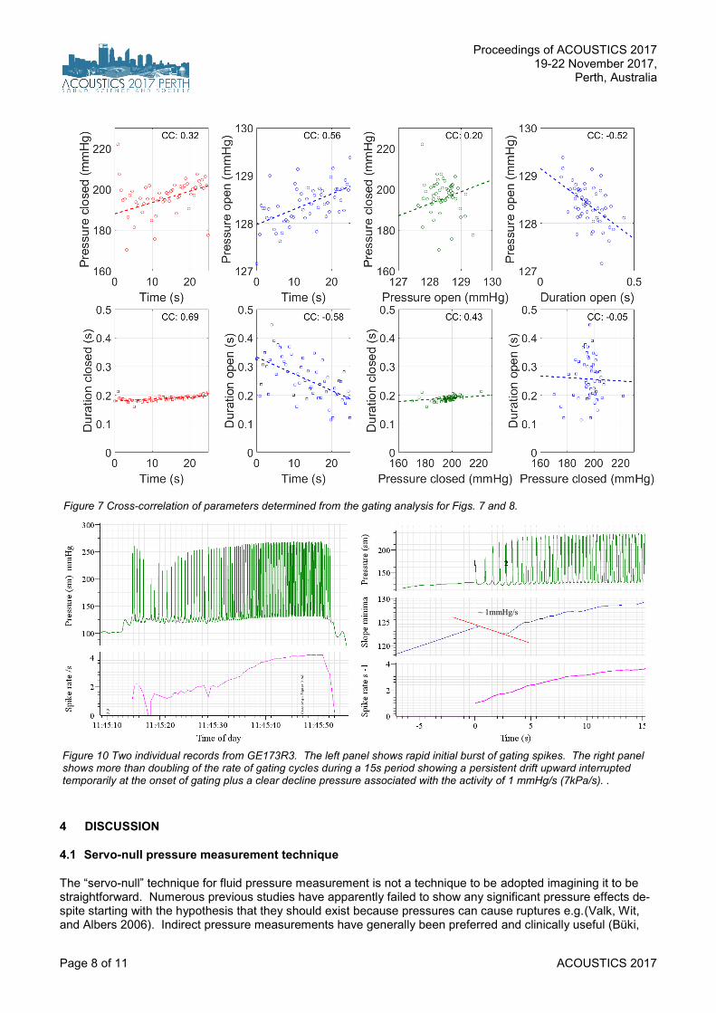
consistent at between +128 to +129 mmHg. Val-ues below the median value of 129mmHg are shown in red and these red saturated values are expanded in the lower panel to reveal that they all appear to be slightly different. The widths of these saturated regions appear to be lower fol-lowing in time after lower level peaks. These sat-urations are consistent with the notion of a valve being opened at a very consistent trigger level. If the tip of the probe is inside a vessel being pres-surised, the pressure registered would be ex-pected to rise when the valve is closed, to be re-leased when the valve is opened. So these re-producible, spiking behaviours are suggestive of valve action. The noisiness of the plateaus is of interest. Their varying plateau heights suggest the valve itself exhibits some his history-depend-ence. Another interpretation is that instead of one valve, there are very many such valves work-ing in concert, responding to a common trigger signal, such as the rate of change of pressure. Figure 8 is the result of the observation that these “open plateaus” are not symmetric in time. There is a much sharper “corner” as the pressure enters the falling phase as the valve is opening. The sudden changes in gradient were quantified by taking the first and second time-derivatives of the pressure waveform. Remarkably the second de-rivative is extremely reproducible across all such trains. The top panel shows a prominent sharp peak. These segments are aligned exactly with the peak and averaged. This reveals a burst of high frequencies suggestive of gating character the moment the valve is opened or possibly re-flecting the energetics required to open the valve. This waveform is low- and high-pass filtered (fil-ter delays taken into account) to confirm that the “high” frequency burst is very precisely timed with the instant of valve-opening. A caveat is added to this conclusion that whether the valves are opening or closing depends upon which side of the valve the micropipette is located and that can-not yet be determined with certainty. The statistics of these gating characteristics are examined further in Figure 9 to see if there may be any cross-dependence between the observed parameters. The strongest feature seems to be that once the gating begins, the duration of valve closure is nearly constant with time, while the du-ration remaining open decreases with time. The other interesting, if not significant effect is an ap-parent decline with time the valve stays open. Gating characteristics from another preparation are shown in Figure 10 suggesting an initial burst with pressure release temporarily eliminating the trigger.
Figure 7 Details of spiking phenomenon in the pressure waveform of Fig.6 suggestive of containment of the pressure rise. In the upper trace, parts of the trace below the pressure median value coloured in red are expanded vertically in the lower plot. The trace is suggestive of the operation of a valve (or valves). When it is open the pressure is contained around 129mmHg (17 kPa), when it is closed the pres-sure is free to rise as shown.
Figure 8 To examine the apparent gating characteristics, the second time-derivative of the pressure waveform is taken (upper trace) and found to be remarkably consistent, more so than the spikes them-selves. After aligning the peaks, averaging the waveform and appro-priately filtering above and below 20 Hz (second panel) it is appreci-ated that a higher frequency burst is aligned in time with the opening of the valve (third panel). It suggests that a transient energetic pro-cess is implicated in the opening.
Proceedings of ACOUSTICS 2017 19-22 November 2017,
Perth, Australia
Page 8 of 11 ACOUSTICS 2017
4 DISCUSSION
4.1 Servo-null pressure measurement technique
The “servo-null” technique for fluid pressure measurement is not a technique to be adopted imagining it to be straightforward. Numerous previous studies have apparently failed to show any significant pressure effects de-spite starting with the hypothesis that they should exist because pressures can cause ruptures e.g.(Valk, Wit, and Albers 2006). Indirect pressure measurements have generally been preferred and clinically useful (Büki,
Figure 7 Cross-correlation of parameters determined from the gating analysis for Figs. 7 and 8.
Figure 10 Two individual records from GE173R3. The left panel shows rapid initial burst of gating spikes. The right panel shows more than doubling of the rate of gating cycles during a 15s period showing a persistent drift upward interrupted temporarily at the onset of gating plus a clear decline pressure associated with the activity of 1 mmHg/s (7kPa/s). .
~ 1mmHg/s

Proceedings of ACOUSTICS 2017 19-22 November 2017, Perth, Australia
ACOUSTICS 2017 Page 9 of 11
Giraudet, and Avan 2009). However, they fall far short of shedding light on the hypothesis being tested in this study. An ideal technique might be to use a pressure-sensitive dye, yet these appear considerably too insensi-tive for this application and have not been tested. Although bench calibration provided some confidence in the linearity of the technique, testing in vivo presented considerable challenges because of uncertainty with interac-tions with the current injection for the tip resistance measurement interfering with normal electromechanical phe-nomena within the cochlea. Likewise, sound could not be used to check the condition of the preparation for the same reason. The pressure measurement could be interrupted to do such a check, but thereafter continuity with phase-lock status which is essential could not be assured. On the other hand, manipulating cranial pres-sure, plus the ambient pressure transients did offer good controls that the device was operating correctly.
4.2 Condition of the preparation
An “industry standard” for such experiments is that the data be subject to the demonstration of high hearing sen-sitivity and sharp tuning. Although this was difficult to maintain in these preparations, this traditional criterion is itself invalidated by the success with the approach. The hypothesis is to test if there is a narrow range or “sweet-spot” for basilar membrane transverse position which obtains when cochlear homeostasis is continu-ously maintained by the osmoregulation. This experiment was designed to produce direct mechanical evidence for such a mechanism which is itself stabilising the basilar membrane and hair cell operating points. The experi-ment needed to monitor displacements outside of this narrow range to test the hypothesis. While this is very early days in this line of investigation, it appears to have succeeded with this objective: The indication that a molecular mechanism may exist to counter-balance infrasound ironically becomes the conceptual starting point assumed in von Békésy’s two-chamber model. It follows that infrasound, and other forms of displacement bias such as loud low-frequency air-pressure pulsations (Salt and Hullar 2010) will have a more profound effect upon hearing sensitivity, and indeed upon vestibular sensation (Merchant, Adams, and Nadol Jr 2005), if an individ-ual’s baseline stabilisation is compromised by e.g. connexin malformation (Andreas Eckhard et al. 2012). The loss of such stabilisation explains previous observations of anomalous “dc-shifts” in the motion of the basilar membrane in guinea pigs in response to tones of 110dB SPL (E. L. LePage 1989) this experiment later re-peated (Flock and Flock 2000) showing such displacements were likely due to endolymphatic hydrops.
5 CONCLUSIONS
It is premature to connect these pressure spikes with the action of aquaporins. The fact that the behaviour co-varies with the endocochlear potential is encouraging, as is the noisiness of the trace which suggests that many such “gates” could be working in concert, yet exhibiting varying levels of capture. The demonstration of new kinds of hydromechanical responses from within the mammalian cochlea, which as seen here are below the au-dible frequency range, suggests a new line of investigation to elucidate an underlying homeostatic mechanism which normally nulls infrasound sensation yet may be scarcely perceptible. Such a mechanism has long been anticipated based on its molecular biological indicators but has been awaiting a form of physical confirmation.
ACKNOWLEDGEMENTS The authors thank the considerable support given to the project by Drs. T. Mom, S. Laurent, F. Giraudet, Y. Pavier, A. Bascoul, M. LePage, and technical assistance from Echodia, WPI and CED companies, and our families. ELeP thanks The University of Western Australia for library support.
REFERENCES Beitz, E., H. Kumagami, P. Krippeit-Drews, J. P. Ruppersberg, and J. E. Schultz. 1999. “Expression Pattern of
Aquaporin Water Channels in the Inner Ear of the Rat: The Molecular Basis for a Water Regulation System in the Endolymphatic Sac.” Hearing Research 132 (1). Elsevier: 76–84.
Bosher, SK, and RL Warren. 1971. “A Study of the Electrochemistry and Osmotic Relationships of the Cochlear Fluids in the Neonatal Rat at the Time of the Development of the Endocochlear Potential.” The Journal of Physiology 212 (3). Physiological Soc: 739–61.
Büki, B, F Giraudet, and P Avan. 2009. “Non-Invasive Measurements of Intralabyrinthine Pressure Changes by Electrocochleography and Otoacoustic Emissions.” Hearing Research 251 (1). Elsevier: 51–59.
Bӧhmer, A. 1993. “Hydrostatic Pressure in the Inner Ear Fluid Compartments and Its Effects on Inner Ear Func-tion.” Acta Oto-Laryngologica 113 (S507). Informa UK Ltd UK: 5–24.
Christensen-Dalsgaard, Jakob, and Geoffrey A Manley. 2013. “The Malleable Middle Ear: An Underappreciated Player in the Evolution of Hearing in Vertebrates.” In Insights from Comparative Hearing Research, 157–91. Springer.
Clack, Jennifer A, Per E Ahlberg, SM Finney, P Dominguez Alonso, and others. 2003. “A Uniquely Specialized Ear in a Very Early Tetrapod.” Nature 425 (6953). Nature Publishing Group: 65.

Proceedings of ACOUSTICS 2017 19-22 November 2017,
Perth, Australia
Page 10 of 11 ACOUSTICS 2017
Eckhard, A, A Dos Santos, W Liu, M Bassiouni, H Arnold, C Gleiser, B Hirt, et al. 2015. “Regulation of the Peri-lymphatic-Endolymphatic Water Shunt in the Cochlea by Membrane Translocation of Aquaporin-5.” Pflügers Archiv-European Journal of Physiology 467 (12). Springer: 2571–88.
Eckhard, Andreas, Corinna Gleiser, Heinz Arnold, Helge Rask-Andersen, Hidetaka Kumagami, Marcus Müller, Bernhard Hirt, and Hubert Lӧwenheim. 2012. “Water Channel Proteins in the Inner Ear and Their Link to Hearing Impairment and Deafness.” Molecular Aspects of Medicine 33 (5). Elsevier: 612–37.
Flock, A., and B. Flock. 2000. “Hydrops in the Cochlea Can Be Induced by Sound as Well as by Static Pres-sure.” Hear Res 150 (1-2): 175–88.
Gates, George A. 2006. “Meniere’s Disease Review 2005.” Journal of the American Academy of Audiology 17 (1). American Academy of Audiology: 16–26.
Gopen, Q., J. J. Rosowski, and S. N. Merchant. 1997. “Anatomy of the Normal Human Cochlear Aqueduct with Functional Implications.” Hearing Research 107 (1-2). Elsevier: 9–22.
Gould, Stephen Jay, and Elisabeth S Vrba. 1982. “Exaptation—a Missing Term in the Science of Form.” Paleo-biology 8 (1): 4–15.
Greenwood, Donald D. 1990. “A Cochlear Frequency-Position Function for Several species—29 Years Later.” The Journal of the Acoustical Society of America 87: 2592.
Hill, A E, B Shachar-Hill, and Y Shachar-Hill. 2004. “What Are Aquaporins For?” The Journal of Membrane Biol-ogy 197 (1): 1–32.
Hirt, B, ZH Penkova, A Eckhard, Wei Liu, Helge Rask-Andersen, M Müller, and H Lӧwenheim. 2010. “The Sub-cellular Distribution of Aquaporin 5 in the Cochlea Reveals a Water Shunt at the Perilymph-Endolymph Bar-rier.” Neuroscience 168 (4). Elsevier: 957–70.
Janosi, Lorant, and Matteo Ceccarelli. 2013. “The Gating Mechanism of the Human Aquaporin 5 Revealed by Molecular Dynamics Simulations.” PloS One 8 (4): e59897. doi:10.1371/journal.pone.0059897.
Kishimoto, S, K Nagahara, U Fisch, and N Dillier. 1983. “Inner Ear Pressure Measurements. Effects of Obstruc-tion of the Cochlear Aqueduct and Endolymphatic Duct on the Perilymphatic Pressure.” Otolaryngologic Clin-ics of North America 16 (1): 21.
Klis, S. F., and G. F. Smoorenburg. 1994. “Osmotically Induced Pressure Difference in the Cochlea and Its Ef-fect on Cochlear Potentials.” Hear Res 75 (1-2): 114–20.
Konishi, Teruzo. 1982. “Ion and Water Control in Cochlear Endolymph.” American Journal of Otolaryngology 3 (6): 434.
Kumagami, H., H. Loewenheim, E. Beitz, K. Wild, H. Schwartz, K. Yamashita, J. Schultz, J. Paysan, H. P. Zen-ner, and J. P. Ruppersberg. 1998. “The Effect of Anti-Diuretic Hormone on the Endolymphatic Sac of the In-ner Ear.” Pflügers Archiv European Journal of Physiology 436 (6). Springer: 970–75.
LePage, E. L. 1987. “Frequency-Dependent Self-Induced Bias of the Basilar Membrane and Its Potential for Controlling Sensitivity and Tuning in the Mammalian Cochlea.” The Journal of the Acoustical Society of America 82: 139–54.
———. 1989. “Functional Role of the Olivo-Cochlear Bundle: A Motor Unit Control System in the Mammalian Cochlea.” Hearing Research 38 (3). Elsevier: 177–98.
LePage, E., and Å. Olofsson. 2011. “Modeling Scala Media as a Pressure Vessel.” In AIP Conference Proceed-ings, What Fire Is in Mine Ears: Progress in Auditory Mechanics, 1403:212, eds C. Shera and E.Olson.
LePage, Eric L. 2003. “The Mammalian Cochlear Map Is Optimally Warped.” The Journal of the Acoustical Soci-ety of America 114 (2). ASA: 896–906.
LePage, Eric L. 2017. “A Role for the Otoliths in the Mechanics of Cochlear Homeostasis?” In Proceedings of the Mechanics of Hearing Workshop, Brock University, Ontario, Canada, June 18-24, 2017, edited by C. Ber-gevin and S. Puria.
Long 3rd, CH, and T. Morizono. 1987. “Hydrostatic Pressure Measurements of Endolymph and Perilymph in a Guinea Pig Model of Endolymphatic Hydrops.” Otolaryngology-Head and Neck Surgery: Official Journal of American Academy of Otolaryngology-Head and Neck Surgery 96 (1): 83.
Manley, Geoffrey A. 2013. “Mosaic Evolution of the Mammalian Auditory Periphery.” In Basic Aspects of Hear-ing, 3–9. Springer.
Marchbanks, RJ, and A. Reid. 1990. “Cochlear and Cerebrospinal Fluid Pressure: Their Inter-Relationship and Control Mechanisms.” British Journal of Audiology 24 (3). Informa UK Ltd UK: 179–87.
Merchant, Saumil N, Joe C Adams, and Joseph B Nadol Jr. 2005. “Pathophysiology of Meniere’s Syndrome: Are Symptoms Caused by Endolymphatic Hydrops?” Otology \& Neurotology 26 (1). LWW: 74–81.
Meyer-Zum-Gottesberge, A, O Schubert, V Balz, and S Tsujikawa. 2000. “Aquaporins in the Inner Ear.” Meniere’s Disease 1999-Update. Kugler Publications, 161.
Mhatre, Anand N, Ryan E Stern, Jiang Li, and Anil K Lalwani. 2002a. “Aquaporin 4 Expression in the Mamma-lian Inner Ear and Its Role in Hearing.” Biochemical and Biophysical Research Communications 297 (4): 987–96.

Proceedings of ACOUSTICS 2017 19-22 November 2017, Perth, Australia
ACOUSTICS 2017 Page 11 of 11
———. 2002b. “Aquaporin 4 Expression in the Mammalian Inner Ear and Its Role in Hearing.” Biochemical and Biophysical Research Communications 297 (4): 987–96.
Mirza, S, and H Richardson. 2005. “Otic Barotrauma from Air Travel.” Journal of Laryngology \& Otology 119 (05). Cambridge Univ Press: 366–70.
Raphael, Y., and R. A. Altschuler. 2003. “Structure and Innervation of the Cochlea.” Brain Research Bulletin 60 (5). Elsevier: 397–422.
Salt, A. N., M. M. Henson, S. L. Gewalt, A. W. Keating, J. E. DeMott, and O. W. Jr. Henson. 1995. “Detection and Quantification of Endolymphatic Hydrops in the Guinea Pig Cochlea by Magnetic Resonance Micros-copy.” Hear Res 88 (1-2): 79–86.
Salt, A. N., and J. DeMott. 1997. “Longitudinal Endolymph Flow Associated with Acute Volume Increase in the Guinea Pig Cochlea.” Hearing Research 107 (1). Elsevier: 29–40.
Salt, A. N., and T. E. Hullar. 2010. “Manuscript: Responses of the Ear to Low Frequency Sounds, Infrasound and Wind Turbines.” Hearing Research 268 (1). Elsevier: 12–21.
Schulz-Mirbach, Tanja, Martin Heß, Brian D Metscher, and Friedrich Ladich. 2013. “A Unique Swim Bladder-Inner Ear Connection in a Teleost Fish Revealed by a Combined High-Resolution Microtomographic and Three-Dimensional Histological Study.” BMC Biology 11 (1). BioMed Central: 75.
Singh, Sanjay. 2016. “Root Pressure: Getting to the Root of Pressure.” In Progress in Botany 77, 105–50. Springer.
Takeuchi, S., T. Takeda, and H. Saito. 1990. “Pressure Relationship between Perilymph and Endolymph in Guinea Pigs.” Acta Otolaryngol 109 (1-2): 93–100.
Valk, WL, HP Wit, and FWJ Albers. 2006. “Rupture of Reissner’s Membrane during Acute Endolymphatic Hy-drops in the Guinea Pig: A Model for Ménière’s Disease?” Acta Oto-Laryngologica 126 (10). Informa UK Ltd UK: 1030–35.
Vater, Marianne, Jin Meng, and Richard C Fox. 2004. “Hearing Organ Evolution and Specialization: Early and Later Mammals.” In Evolution of the Vertebrate Auditory System, 256–88. Springer.
Wackym, P. A., U. Friberg, D. Bagger-Sjoback, F. H. Jr. Linthicum, I. Friedmann, and H. Rask-Andersen. 1987. “Human Endolymphatic Sac: Possible Mechanisms of Pressure Regulation.” J Laryngol Otol 101 (8): 768–79. Yoshida, Masufumi, and Louis D Lowry. 1984. “Hydrostatic Pressure Measurement of Endolymph and Peri-lymph in the Guinea Pig Cochlea.” American Journal of Otolaryngology 5 (3). Elsevier: 159–65.
Related Documents