Purdue University Purdue e-Pubs Weldon School of Biomedical Engineering Faculty Publications Weldon School of Biomedical Engineering 2006 A new biomechanical head injury criterion Charles F. Babbs Purdue University, [email protected] Follow this and additional works at: hp://docs.lib.purdue.edu/bmepubs Part of the Biomedical Engineering and Bioengineering Commons is document has been made available through Purdue e-Pubs, a service of the Purdue University Libraries. Please contact [email protected] for additional information. Recommended Citation Babbs, Charles F., "A new biomechanical head injury criterion" (2006). Weldon School of Biomedical Engineering Faculty Publications. Paper 42. hp://docs.lib.purdue.edu/bmepubs/42

Welcome message from author
This document is posted to help you gain knowledge. Please leave a comment to let me know what you think about it! Share it to your friends and learn new things together.
Transcript

Purdue UniversityPurdue e-PubsWeldon School of Biomedical Engineering FacultyPublications Weldon School of Biomedical Engineering
2006
A new biomechanical head injury criterionCharles F. BabbsPurdue University, [email protected]
Follow this and additional works at: http://docs.lib.purdue.edu/bmepubs
Part of the Biomedical Engineering and Bioengineering Commons
This document has been made available through Purdue e-Pubs, a service of the Purdue University Libraries. Please contact [email protected] foradditional information.
Recommended CitationBabbs, Charles F., "A new biomechanical head injury criterion" (2006). Weldon School of Biomedical Engineering Faculty Publications.Paper 42.http://docs.lib.purdue.edu/bmepubs/42

A NEW BIOMECHANICAL HEAD INJURY CRITERION
CHARLS F. BABBS, MD, PhD
Department of Basic Medical Sciences, 1246 Lynn Hall,
Purdue University, West Lafayette, IN 47907-1246 USA
Abstract
This paper presents a new analysis of the physics of closed head injury caused by intense
acceleration of the head. At rest a 1 cm gap filled with cerebrospinal fluid (CSF) separates
the human brain from the skull. During impact whole head acceleration induces artificial
gravity within the skull. Because its density differs slightly from that of CSF, the brain
accelerates, strikes the inner aspect of the rigid skull, and undergoes viscoelastic
deformation. Analytical methods for a lumped parameter model of the brain predict
internal brain motions that correlate well with published high-speed photographic studies.
The same methods predict a truncated hyperbolic strength-duration curve for impacts that
produce a given critical compressive strain. A family of such curves exists for different
critical strains. Each truncated hyperbolic curve defines a head injury criterion (HIC) or
concussive threshold, which is little changed by small offsetting corrections for curvature
of the brain and for viscous damping. Such curves predict results of experimental studies
of closed head injury, known limits for safe versus dangerous falls, and the relative
resistance of smaller versus larger animals to acceleration of the head. The underlying
theory provides improved understanding of closed head injury and better guidance to
designers of protective equipment and to those extrapolating research results from animals
to man.
Keywords: acceleration, biomechanics, brain, concussion, deformation, diffuse axonal
injury, HIC, impact, shear, strain, threshold, tolerance

1. Introduction
Head injuries are common consequences of auto accidents, falls, direct blows to the head,
bicycle accidents, fights, and participation in contact sports1, 2
. Nearly two million persons
with head injury are medically attended and over 300,000 are hospitalized annually in the
USA 2. One third of these injuries are severe, leading to prolonged coma, permanent
neurological impairment, or death3-6
. The majority of head injuries in man2, 6, 7
and in
experimental animal models8, 9
are mild or moderate, and are produced by acceleration of
the head only—without skull fracture9. Such non-penetrating head injuries result in
transient loss of consciousness and usually no permanent neurological deficits. However,
repeated mild head injuries (concussions) can and do lead to lasting decrements in problem
solving ability, memory, and personality, as well as to anatomical lesions in the brain10
.
This paper addresses the question of how to specify the threshold between safe and
dangerous levels of head acceleration in situations such as contact sports, vehicle crashes,
and falls. This question arises for designers protective headgear, seat restraints, car and
aircraft interiors, and the like, who must rely on head injury criteria11-16
that specify
physical parameters of harmful blows, such as critical energy, impulse (force x time)11, 17
or acceleration16, 18
. Such criteria help designers create equipment, rules, and operating
procedures that keep the intensity of impacts to the head below the concussive threshold.
It is clear that there must be a tradeoff between the amplitude and the duration of
acceleration required to produce closed head injury. Very brief intense accelerations
produce the same change in head velocity, as do more moderate, prolonged accelerations.
Thus one needs to generate a strength-duration curve describing harmful levels of
acceleration as a function of impact duration (Figure 1). Points above the curve are
considered dangerous; points below the curve are considered tolerable, if not completely
safe. The farther a point is from the curve on either side, the safer or more dangerous the
corresponding impact.
The classical head injury criterion of this type was generated by fitting a power function to
impact tolerance data from animals and a limited number of humans for whom measured
accelerations were available in the year 197116
. It is widely referenced and has become
known as "the" head injury criterion or HIC, although there are alternative versions11-16
.
The formula for the classical Versace HIC16
is a2.5
= 1000, where the variable, a, is time
averaged external acceleration of the head in units of Gs, the variable, , is the duration of
head acceleration in seconds, and value 1000 is a constant in units of G2.5
-sec that defines
the concussive threshold. This particular head injury criterion specifies a strength-duration
curve of the form,
4.0t
16a ,
which is plotted in Figure 1.
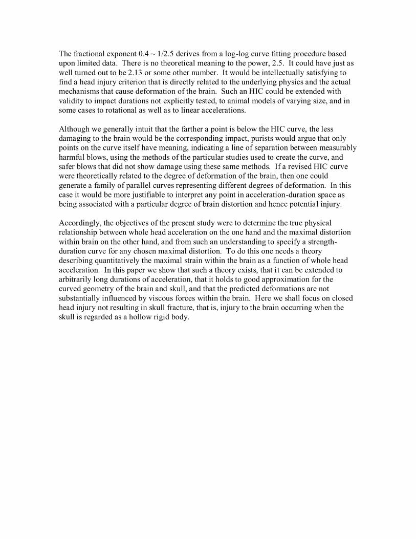
The fractional exponent 0.4 ~ 1/2.5 derives from a log-log curve fitting procedure based
upon limited data. There is no theoretical meaning to the power, 2.5. It could have just as
well turned out to be 2.13 or some other number. It would be intellectually satisfying to
find a head injury criterion that is directly related to the underlying physics and the actual
mechanisms that cause deformation of the brain. Such an HIC could be extended with
validity to impact durations not explicitly tested, to animal models of varying size, and in
some cases to rotational as well as to linear accelerations.
Although we generally intuit that the farther a point is below the HIC curve, the less
damaging to the brain would be the corresponding impact, purists would argue that only
points on the curve itself have meaning, indicating a line of separation between measurably
harmful blows, using the methods of the particular studies used to create the curve, and
safer blows that did not show damage using these same methods. If a revised HIC curve
were theoretically related to the degree of deformation of the brain, then one could
generate a family of parallel curves representing different degrees of deformation. In this
case it would be more justifiable to interpret any point in acceleration-duration space as
being associated with a particular degree of brain distortion and hence potential injury.
Accordingly, the objectives of the present study were to determine the true physical
relationship between whole head acceleration on the one hand and the maximal distortion
within brain on the other hand, and from such an understanding to specify a strength-
duration curve for any chosen maximal distortion. To do this one needs a theory
describing quantitatively the maximal strain within the brain as a function of whole head
acceleration. In this paper we show that such a theory exists, that it can be extended to
arbitrarily long durations of acceleration, that it holds to good approximation for the
curved geometry of the brain and skull, and that the predicted deformations are not
substantially influenced by viscous forces within the brain. Here we shall focus on closed
head injury not resulting in skull fracture, that is, injury to the brain occurring when the
skull is regarded as a hollow rigid body.

2. Theory and Methods
2.0 Approach
Harmful blunt impacts can last for a wide range of durations from about 2 to 2000 msec16
.
Subsequent motion of the brain inside the skull lasts several seconds and is rarely seen.
Only a few studies, using high-speed photography through a transparent plastic calvarium
or high speed fluoroscopy of implanted radiodense pellets 8, 19-21
, have examined the actual
motion of the brain during closed head injury in animals. Fortunately mathematical
analysis and modeling of the skull and brain in response to known pulses of head
acceleration allow one to study a wide variety of conditions that are difficult, impossible,
or unethical to reproduce in animals or humans22
. This mathematical approach is ideal for
developing an improved head injury criterion.
A person standing in a rising elevator feels a tug of artifical gravity as the elevator
accelerates upward. Within the internal frame of reference of the elevator, the passenger
experiences acceleration toward the floor of the elevator that is equal and opposite to the
upward acceleration of the elevator in the shaft23
. Acceleration of this local frame of
reference produces artificial gravity, which by all possible experiments conducted within
the elevator is indistinguishable from ordinary gravity. The effect of whole head
acceleration on the brain is similar to the effect of acceleration of a rising elevator on its
occupants. In this case the skull is analogous to the walls of the elevator, and the brain is
analogous to a passenger in the elevator. However, since the brain is surrounded by and
suspended in clear aqueous cerebrospinal fluid (CSF), the acceleration felt by the brain is
attenuated by about 95 percent, since the brain weighs only about 5% as much in water as
it does in air22
. Nevertheless, considerable isovolumic deformation of the brain (which is
quite soft) can take place, as the whole head is accelerated.
The nature of brain deformation during closed head injury has been revealed using
analytical methods and computer models22
, coupled with remarkable experiments
conducted years ago in animals fitted with transparent skull caps and studied with high
speed photography19, 21
. During brief, intense impacts the brain’s velocity toward the skull
increases almost instantaneously (within 1/50’s of a second or less) to a velocity of tens of
centimeters per second along the axis of the blow. Within another 10 msec or so the brain
traverses the CSF gap and crashes into the rigid skull. The impact initiates a strain wave
that traverses the brain substance at a much higher velocity, near 300 cm/sec, causing
compression in one dimension and elongation in the other two perpendicular dimensions.
A body of literature24-26
suggests indirectly that it is probably the lateral expansion rather
than the compression itself that damages brain substance, causing lysis of axons and
damage to capillary blood vessels with micro-hemorrhaging or bruising. These are the
classical lesions of closed head injury3, 4, 9, 27, 28
. Here we describe analytically the motion
of a soft, viscoelastic body such as the brain, as it is accelerates toward a rigid wall such as
the skull, deforms, and then rebounds. In turn, we define the intensity and duration of
acceleration associated with a given degree of maximal strain.

2.1 Brain models
The present analysis is done with strict adherence to Newton's Laws of Motion, together
with published values for the material properties of the brain. In addition we adopt
simplified representations of the geometry of the skull, subarachnoid space, and cranial
cavity. We regard the brain as a uniform viscoelastic mass, suspended in water density
cerebrospinal fluid (CSF) and residing in an internal frame of reference within the rigid
skull. Definitions of symbols used in the analysis are provided in Table 1.
For simplicity, we shall consider the brain to be of homogenous density and elasticity, in
keeping with experimental observations 29, 30
. We also assume that the brain is isovolumic
(i.e. Poisson's ratio = 0.5), so that neither brain volume or CSF volume changes during or
immediately after impact. Viscoelastic properties of brain have been rather well studied
over the past 30 years, and consensus values for Young's modulus of elasticity and for an
analogously defined energy loss modulus can be gleaned from the literature (Table 2).
Despite rather large variability in published values, these data provide a basis for a
standard model of a typical brain that is sufficient for present purposes. Brain density data
are more consistent. They are summarized in Table 3. For the following theoretical work
we take a typical value of brain stiffness as 10000 Pa and a typical value of brain density as
1046 kg/m3. Water density is 1000 kg/m
3. The maximal internal fronto-occipital diameter
of the human skull is taken as 18 cm.
2.2 Acceleration in internal vs. external frames of reference
In this problem it is helpful to distinguish clearly two frames of reference for motion. The
first is the internal frame of reference within the skull. This frame of reference is
important because it is the relative motion of brain with respect to the skull that is
responsible for closed head injury. The second is the external frame of reference in which
the head, neck, and body move. This frame of reference is important because an external
blow or fall causes movement of the whole head in this frame. Acceleration of the rigid
skull by an amount a(t) produces a corresponding artificial gravity within the skull of -a(t).
Because the brain is suspended in aqueous CSF, its buoyancy must be accounted. If 1 is
CSF density, 2 is brain density, and V2 is brain volume, then the force of gravity on the
brain is )t(aV22 , and the buoyancy of the brain is )t(aV21 . Applying
Newton's second law of motion (Force = mass x acceleration) to the whole brain and
ignoring drag#,
)t(gV)t(aV 22212 , (1)
# It is reasonable to ignore the effects of viscous drag on the brain from CSF water, because the distance
traveled by the brain through CSF is short and brain speeds relative to the skull are limited over this distance.

where g(t) is the acceleration of the brain with respect to the skull in the direction parallel
to whole head acceleration. In turn,
)t(a)t(g
2
12
(2)
in the internal frame of reference of the skull. This acceleration is substantially less than
the acceleration, a(t), of the head as a whole in magnitude and opposite in direction. If
brain and CSF density were perfectly matched, the acceleration of the brain with respect to
the skull would be zero. In reality, brain density is approximately 1.05 and CSF density is
approximately 1.0029, 31, 32
(Table 3), so internal brain acceleration is about 5 percent of
external head acceleration.
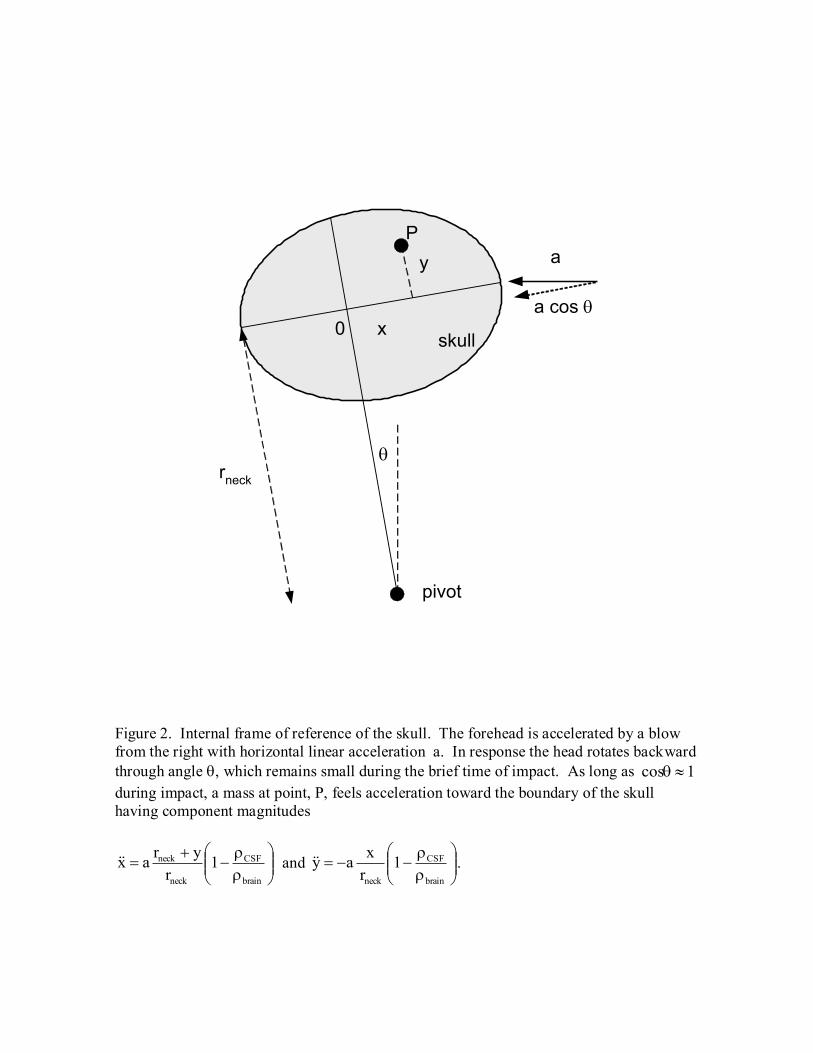
In cases of rotational accelerationof the skull (Figure 2) we have for internal coordinates x
and y and neck radius rneck , defined in Figure 2 the local components of internal
acceleration
neck
neckx
r
yr)t(g)t,y,x(g and
neck
yr
x)t(g)t,y,x(g (3)
This two-dimensional acceleration is considered in detail in reference 22, which shows that
the major component of brain motion is in the x direction. Here we consider a simplified
one dimension case of linear brain acceleration along the axis of the blow, when x, y, and
the distance moved by the head during the actual blow are small with respect to rneck.
2.3 Three ranges of acceleration duration
To further reduce the problem, it is helpful to consider three ranges of duration for whole
head acceleration. These can be termed short, intermediate, and long duration impacts.
Short duration accelerations terminate while the brain is moving toward the skull and
before it has hit the inner wall of the skull. Intermediate duration accelerations terminate
after the leading edge of the brain has reached the skull but before the induced strain wave
has traveled one full brain diameter along the axis of acceleration. During this time the
brain becomes increasingly compressed against the side of contact. Long duration
accelerations last even longer than this time of maximal compression. They pin the brain
against the skull and, as long as acceleration persists, prevent recoil of the brain toward the
opposite side of the skull. Recoil can occur after termination of long duration
accelerations.
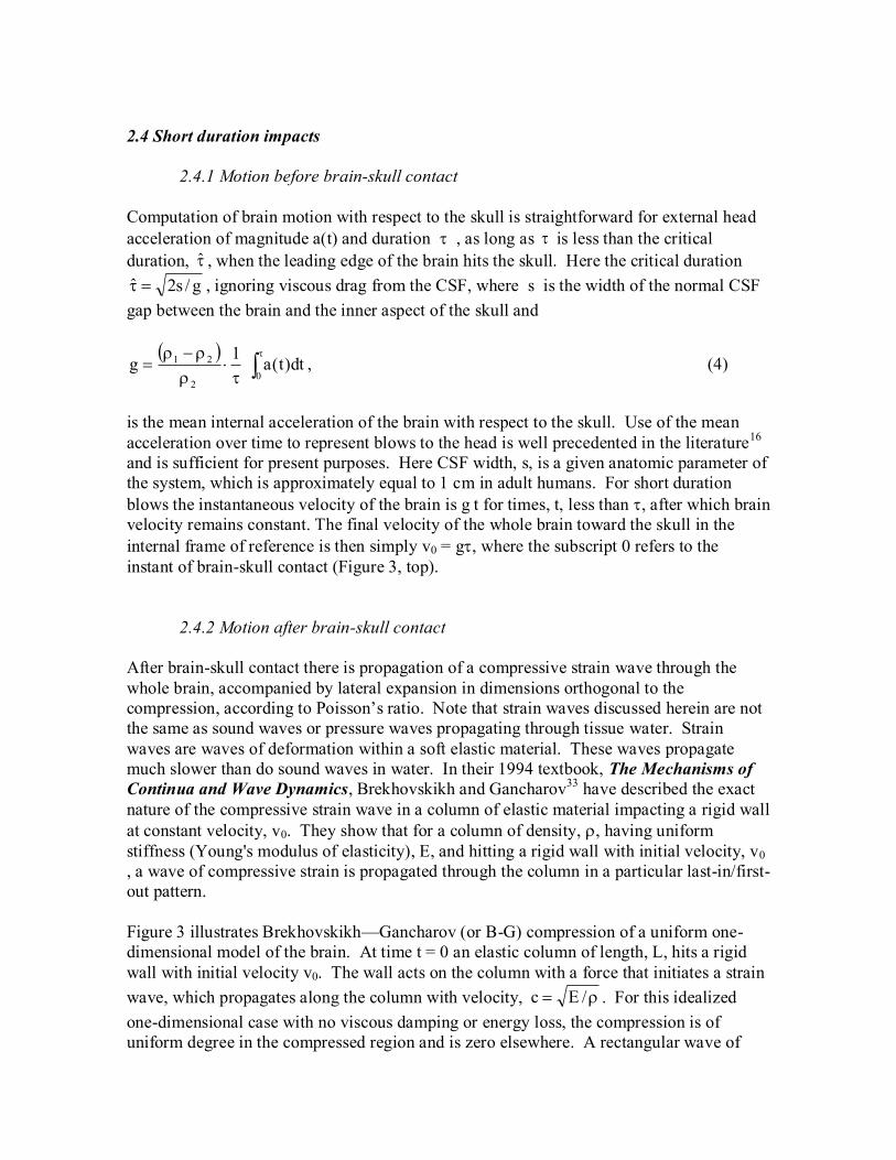
2.4 Short duration impacts
2.4.1 Motion before brain-skull contact
Computation of brain motion with respect to the skull is straightforward for external head
acceleration of magnitude a(t) and duration , as long as is less than the critical
duration, , when the leading edge of the brain hits the skull. Here the critical duration
g/s2ˆ , ignoring viscous drag from the CSF, where s is the width of the normal CSF
gap between the brain and the inner aspect of the skull and
dt)t(a
1g
02
21
, (4)
is the mean internal acceleration of the brain with respect to the skull. Use of the mean
acceleration over time to represent blows to the head is well precedented in the literature16
and is sufficient for present purposes. Here CSF width, s, is a given anatomic parameter of
the system, which is approximately equal to 1 cm in adult humans. For short duration
blows the instantaneous velocity of the brain is g t for times, t, less than , after which brain
velocity remains constant. The final velocity of the whole brain toward the skull in the
internal frame of reference is then simply v0 = g, where the subscript 0 refers to the
instant of brain-skull contact (Figure 3, top).
2.4.2 Motion after brain-skull contact
After brain-skull contact there is propagation of a compressive strain wave through the
whole brain, accompanied by lateral expansion in dimensions orthogonal to the
compression, according to Poisson’s ratio. Note that strain waves discussed herein are not
the same as sound waves or pressure waves propagating through tissue water. Strain
waves are waves of deformation within a soft elastic material. These waves propagate
much slower than do sound waves in water. In their 1994 textbook, The Mechanisms of
Continua and Wave Dynamics, Brekhovskikh and Gancharov33
have described the exact
nature of the compressive strain wave in a column of elastic material impacting a rigid wall
at constant velocity, v0. They show that for a column of density, , having uniform
stiffness (Young's modulus of elasticity), E, and hitting a rigid wall with initial velocity, v0
, a wave of compressive strain is propagated through the column in a particular last-in/first-
out pattern.
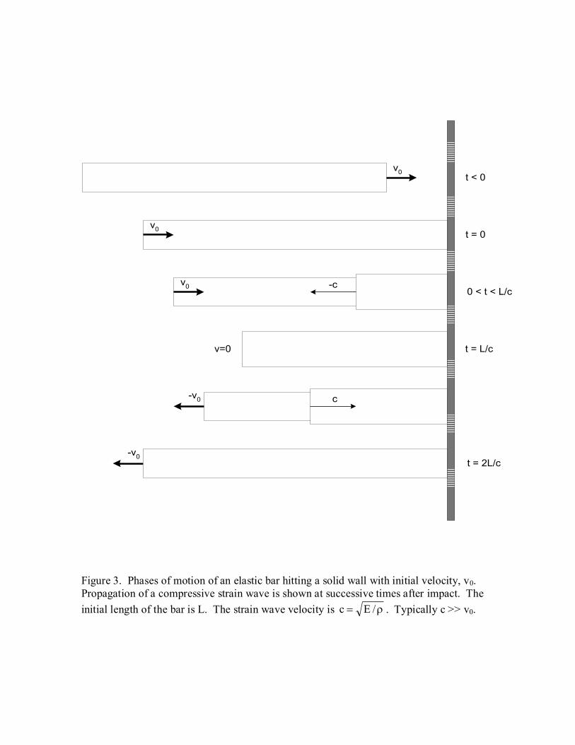
Figure 3 illustrates Brekhovskikh—Gancharov (or B-G) compression of a uniform one-
dimensional model of the brain. At time t = 0 an elastic column of length, L, hits a rigid
wall with initial velocity v0. The wall acts on the column with a force that initiates a strain
wave, which propagates along the column with velocity, /Ec . For this idealized
one-dimensional case with no viscous damping or energy loss, the compression is of
uniform degree in the compressed region and is zero elsewhere. A rectangular wave of

compression and lateral expansion travels from the wall toward the free end of the column.
At time t = L/c the entire column is uniformly compressed. The amount of compressive
strain is simply v0/c. Because we can compute v0 = g, as just described, it is a simple
matter to find the value of compressive strain, v0/c. Moreover, if the brain material is
isovolumic during deformation (Poisson's ratio = 0.5), then the expansive strain
perpendicular to v0 is equal to 0.5v0/c.
Thereafter, for times, t, approximately in the range L/c < t < 2L/c there is recoil, in reverse
order, beginning with the free end. For times t > 2L/c the entire column recoils with
velocity v0. In this idealized case, our brain model, having suffered a "coup" would drift at
velocity -v0 toward the other side of the skull where it would undergo a mirror image
deformation or "contrecoup". Without energy loss due to damping, the cycle would be
repeated indefinitely.**
This one-dimensional analytical model captures several essential
aspects of brain motion in closed head injury.
A recent paper utilizing two dimensional finite element models of the brain confirms the
essential correctness of the Brekhovskikh—Gancharov analysis of short duration blows to
ellipsoidal head and brain models22
. Compressive strain waves propagate through the
entire brain, just as outlined in Figure 3. This predicted motion also is remarkably similar
to that observed directly through clear plastic (Lexan) calvaria in rare experimental studies
of monkeys subjected to controlled head injury19, 21
. In Sections 2.5 through 2.9 we extend
this analysis to longer duration impacts, to viscoelastic brain material, and to non-
rectangular geometries. It is helpful first to specify the strength duration curve for a simple
B-G elastic body in response to short-duration impacts.
**
In the absence of damping it is easy to show conservation of energy at maximum compression, when axial
brain length is diminished by amount L. The stored energy at maximum compression is 2)L(k2
1U ,
where L
EAk is the spring constant for the elastic material of length L and cross section A, and
ELvL
c
vL 0
0 at maximum compression. Hence, 2
0
22
0 vm2
1
E
Lv
L
EA
2
1U
, which is the kinetic
energy of the original bar of total mass, m, moving at v0 toward the wall. A similar approach shows
conservation of energy at all times during a cycle of impact, compression, and recoil.
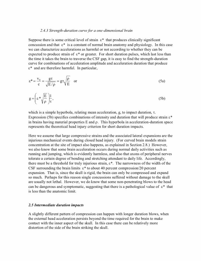
2.4.3 Strength-duration curve for a one-dimensional brain
Suppose there is some critical level of strain * that produces clinically significant
concussion and that * is a constant of normal brain anatomy and physiology. In this case
we can characterize accelerations as harmful or not according to whether they can be
expected to produce strain of * or greater. For short duration pulses, which last less than
the time it takes the brain to traverse the CSF gap, it is easy to find the strength-duration
curve for combinations of acceleration amplitude and acceleration duration that produce
* and are therefore harmful. In particular,
Eg
/E
g
c
v* 0
or (5a)
1E*g , (5b)
which is a simple hyperbola, relating mean acceleration, g, to impact duration, .
Expression (5b) specifies combinations of intensity and duration that will produce strain *
in brains having material properties E and . This hyperbola in acceleration-duration space
represents the theoretical head injury criterion for short duration impacts.
Here we assume that large compressive strains and the associated lateral expansions are the
injurious mechanical events during closed head injury. (For curved brain models strain
concentration at the site of impact also happens, as explained in Section 2.8.) However,
we also know that some brain acceleration occurs during normal daily activities such as
running and jumping, which is evidently harmless, and also that axons of peripheral nerves
tolerate a certain degree of bending and stretching attendant to daily life. Accordingly,
there must be a threshold for truly injurious strain, *. The narrowness of the width of the
CSF surrounding the brain limits * to about 40 percent compression/20 percent
expansion. That is, since the skull is rigid, the brain can only be compressed and expand
so much. Perhaps for this reason single concussions suffered without damage to the skull
are usually not lethal. However, we do know that some non-penetrating blows to the head
can be dangerous and symptomatic, suggesting that there is a pathological value of * that
is less than the anatomic limit.
2.5 Intermediate duration impacts
A slightly different pattern of compression can happen with longer duration blows, when
the external head acceleration persists beyond the time required for the brain to make
contact with the inner aspect of the skull. In this case there can be relatively more
distortion of the side of the brain striking the skull.

2.5.1 Stress and strain
When the duration of acceleration extends beyond first contact, one can use the principle
of superposition for strain waves to find the net stress and strain at the contact point33
. The
net strain is the sum of the strain produced by an elastic rod hitting the wall at v0 with no
gravity (case 1, as in Figure 3) and the strain produced by the added force built up as a
result of gravity (case 2) after the brain contacts the skull. One can visualize case 2 by
imagining a person who gently places a cube of gelatin on a table and then lets go,
allowing it to sag toward an equilibrium shape under 1G. From Brekhovskikh—
Gancharov analysis we know that, in response to any force on the bottom surface of the
cube, a strain wave will travel through the gelatin with velocity /Ec . Now consider
the one dimensional brain model. If the strain wave of distortion travels distance,
ˆtcx , out from the wall, then the weight of the collapsed brain tissue on the wall,
that is the extra force for artificial gravity g > 0, is Axg. In turn, the total contact stress at
post contact time, ˆt , is that caused by the initial velocity, as in case 1 for short duration
impacts, plus the extra stress caused by gravity persisting after time , or
g)ˆt(cc
vE
A
gxA
c
vE 00
c
(6a)
for post contact times )ˆt(gvc/Lˆt 0 . Noting that 2c/E , we have
gˆtc
E
c
vE 0
c . (6b)
The strain at the point of contact, which is the maximal strain in this weighted system
during propagation of the outbound strain wave across brain length, L, is given by contact
stress divided by Young’ modulus, E, or
ˆc
g
c
v0max (7)
for accelerations of duration where )ˆ(gvc/Lˆˆ0 . This is the
maximum strain in the model during propagation of the outbound strain wave under g. To
specify the time frame for (7) in terms of given system parameters, we note for CSF width,
s, we have g/s2ˆ and ˆgv0 . So the contact strain in (7) for intermediate duration
impacts happens for approximate durations gc/Lg/s2g/s2 .

2.5.2 the extended strength duration curve
During the outbound strain wave for a critical harmful strain, * , we have from
Equation (7) ˆggv*c 0 . But ˆgv0 under the prevailing artificial gravity, g ,
and so the strength-duration curve for a critical harmful strain, * , can be written to good
approximation by g*c , or
*cg , for all gc/Lˆ . (8)
Therefore, the strength-duration curve for the concussive threshold remains hyperbolic for
both short and intermediate duration impacts!
It is helpful to specify the upper bound of impulse duration in (8)
~g~c/Lˆ~ (9a)
in terms of the dimensions and material properties of the brain. To do this we note that for
any particular threshold, *, we have
~g~*c . (9b)
For the no-drag case the CSF thickness
2~g~
2
1s . (9c)
Combining (9a), (9b), and (9c) to eliminate the variables andg~ , we have
*
s2
*1
L
E*
s2
*1
L
c
1~ . (10)
Expression (10) gives the extent of the hyperbolic region of the strength duration curve in
terms of fundamental geometric and mechanical properties of the brain model.
Thus the strength-duration curve during outbound propagation of the strain wave
( ~ˆ ) is an extension of the hyperbolic strength-duration curve for short impact
durations ˆ . This is a helpful, simplifying result. However, for intermediate duration
impacts the degree of compressive strain is not uniform across the length, L, of the brain.
Because of the added effect of persistent acceleration, there is instead a gradient of
compressive strain, which is greatest at the wall and diminishes with distance from the
wall. For the purpose of defining harmful impacts, we are interested in the maximal
distortion, *, anywhere in the brain. Accordingly, to generate a head injury criterion it is

sufficient to use the maximal strain at the point of contact as a measure of undesirable
distortion.
2.6 Long duration impacts
For durations of acceleration greater than ~ , as defined in (10), the brain remains pinned
against the rigid skull under the force of g. There are small oscillations in strain associated
with internal reflection of the strain wave. The maximal strain experienced by the brain at
any time remains the same as that given by (7) for t = ~ , even for indefinitely long internal
acceleration. Hence we can sketch the entire strength-duration curve for times from near
zero to infinity by combining expressions (8) and (10), that is,
*cg for ~0 ,
~
*cg for ~ . (11a)
Recall that lower case g represents time-averaged acceleration of the brain surrounded by
CSF within the skull. If we use the variable a() to represent mean acceleration of the head
with respect to an external frame of reference over impulse duration, , then using
Equation (2) we have 221 /)(ag . In terms of external acceleration of the head,
which is the usual way of specifying an HIC,
*c20
*c)(a
21
2 for ~0 ,
and
~
*c20~
*c)(a
21
2 for ~ (11b)
2.7 Estimating the effect of viscous damping
Brekhovskikh—Gancharov theory assumes that the elastic rod impacting a rigid surface in
Figure 3 is purely elastic. Now let us regard the elastic rod as being composed of parallel
spring and damper elements connected in series (a Voight/Maxwell body). Assume that
the various dampers in series act as an equivalent damper with viscous loss modulus, D,
cross sectional area A, and length L. D is a material property of brain tissue, which is
defined analogously to Young’s modulus of elasticity, with a value on the order of 200
kg/m/s22
. The total work required to compress the equivalent damper may be estimated as
average force exerted by the equivalent damper, multiplied by the distance moved. Since
the velocity of tissue diminishes from v0 to zero during compression, let us estimate

average force as one half v0 multiplied by the damping constant, = DA/L. Then for short
duration impacts the product of average force and distance, namely LL
DAv
2
10 , equals
the energy absorbed by the damper. Now suppose that the strain at the time of maximum
compression is uniform along length, L. (Finite element models show that it diminishes
slightly farther from the wall, because of damper induced slowing of rod velocity. Still,
uniform strain at maximal crunch is a useful approximation.) Then, from conservation of
energy we must have that the original kinetic energy minus the energy absorbed by the
damper equals total energy stored in the springs at maximal compression, or
2maxmax0
2
0 LL
EA
2
1L
L
DAv
2
1LvA
2
1 . (12)
This is a quadratic equation in , which we can solve to determine how a small amount of
damping, D, influences maximal strain max.
In particular,
2
2
0
2
0max
02
maxc
v
E
v
LE
vD
(13)
For D = 0, we have c
v0max , as before. For nonzero damping moduli, D, strain at
maximal crunch depends on the ratio L
v
E
D 0 . For brain, experimental data22
suggest D/E
~ 200/10000 = 0.02. For threshold concussive blows in humans the ratio
v0/L ~ 4; that is, the brain is traveling at about 4 brain diameters, about 70 cm, or less each
second when it hits the skull. For small D/E the quadratic (13) can be well approximated
as
04.0c
v
L
v
E
D
2
1
E
v 00
2
0max
(14)
Thus strain is slightly less with realistic viscous damping. The purely elastic B-G model of
brain, for which = v0/c , gives a modest overestimate of maximal strain.
2.8 Curved, higher-dimensional geometry
Another difference between the simple B-G model and real brains is the presence of curved
geometry. In the present impact problem the essence of the difference is that the leading
edge of the brain is not flat, but has a rounded nose. It is this nose that strikes the skull
first. The contact area of the nose is smaller than the mid-level cross section of the brain.
This situation is depicted for a spheroid impacting a hard flat surface in Figure 4(a). In this

case the spheroid can be modeled as cylindrical core, surrounded by a collar, as shown in
Figure 4(b). The core cylinder has volume Vcore , and the peripheral collar, which never
makes contact with the impacting surface, but which carries momentum and kinetic
energy, has volume, Vcollar.
Consider the time of maximum compression of the core against the wall. Ignoring the
shear energy in the collar, which can be shown to be quite small, we can analyze this
system at maximal compression using the relationship
Initial kinetic energy = strain energy per unit volume x core volume,
or
core
2
max
2
0collarcore VE2
1vVV
2
1 . (15)
In (15) we assume that at maximal compression there is uniform strain, max , throughout
the core only, as suggested in Figure 3, that the collar does not cushion the blow, and that
collar volume is substantially smaller than core volume. Solving for maximal strain using 2c/E , we have
core
collar0
core
collar0max
V
V
2
11
c
v
V
V1
c
v . (16)
The maximal strain for a short duration impact is increased by a modest factor equal to
about half the ratio of collar to core volumes.
2.9 A complete, discontinuous HIC and strength-duration curve
Combining the above considerations we notice that the effect of damping causes the strain
to be slightly overestimated by the elastic B-G expression max = v0/c , while the effect of
curvature causes the strain to be slightly underestimated by max = v0/c. That is, if we add
viscous damping there should be a little less maximal compression at the point of contact,
but if we add curvature there should be a little more. Hence we have offsetting errors, and
after consideration of the complexities, the simple one-dimensional B-G model holds up
rather well. To a good approximation the strength duration curve defining a head injury
criterion for any selected critical maximal strain * is still given by Equations (11a) and
(11b) for a simple elastic brain model. The strength duration curve is hyperbolic up to a
duration of ~ , after which it is constant.

3. Comparison with biological data
3.0 Critical strain
To compare the proposed hyperbolic head injury criterion with experimental data, it is
necessary to specify a critical level of strain, , that is presumed to be injurious. For
example, suppose that the critical compressive strain 3.0* . This value implies that a
30% intracranial compression, accompanied by 15 percent orthogonal stretch for Poisson's
ratio = 0.5, occurs during blows at the concussive threshold. The critical positive strain for
stretch injury has been relatively well established by experiments carried out in isolated
nerves. For example, functional or morphological impairment occurs in squid axons after
12 percent stretch 25
, in guinea pig optic nerves after 15 percent stretch 34
, and in sciatic
nerves of frogs after 18 percent stretch 26
. Using Poisson’s ratio = 0.5 these experimental
values of positive stretch would correspond to compressive strains in whole brain of 0.24,
0.30, and 0.36, with a mean value of 0.30. Hence, as a working value, it is indeed
reasonable to suppose that 3.0* .
3.1 Comparison with experimental strength-duration curves
Using this value for critical strain one can compare the proposed truncated hyperbolic HIC
with clinical and experimental data describing the concussion threshold in humans and
animals. Figure 5 shows the summary clinical data, redrawn from Versace16
, as open
triangles in acceleration-duration space. Also shown as open circles are data of Yarnell35
from experimental whiplash injury in a primate model. The curved line represents the
proposed truncated hyperbolic HIC as given in Equation (11b). The truncated hyperbolic
function fits both short and long duration data. It is also is based upon the underlying
physics of the problem, rather than being purely descriptive.
A separate analysis of concussion threshold data in monkeys was done by Ommaya12
, who
found that any short duration blow in monkeys producing an angular head velocity of
greater than 250 radians/sec produces physiological concussion. Taking the neck radius
for a monkey as about 7 cm and applying Equation (3), the linear external head
acceleration associated with 250 radians/sec head and neck rotational velocity is
sec
m5.17
sec
radian250x
radian
m07.0vta head
at threshold external head velocity. Since internal acceleration of the brain with respect to
the skull is about 5 percent of external head acceleration, given the weight of the brain in
cerebrospinal fluid, then
sec
m875.0
sec
m5.1705.0vbrain

would be the velocity of the brain approaching the skull. According to B-G theory, this
internal brain velocity would produce a critical compressive strain of
28.0sec/m1.3
sec/m875.0
c
v* brain ,
with a corresponding stretch of 0.14 in orthogonal dimensions. This value is very close to
the mean value of 3.0* obtained from animal experiments in isolated nerves.
3.2. Comparison with known injurious and safe accelerations in humans
Estimation of the range of safe head accelerations can provide another point of validation.
Adams 27
noted that severe diffuse axonal injury in humans, happening as a result of falls,
occurs only after falls from substantially greater than a person's own height—for example,
from a ladder, bridge, elevator shaft, or even a mountain! As before, we expect from
theory that a given acceleration-time product, creating a given change in whole head
velocity v, will produce a given maximal strain in the brain. For falls from a particular
height, we can estimate v as the velocity of a falling body at the Earth's surface from a
particular height, h, which is hg2v . For a grown man standing approximately 2
meters high this works out to be about 6 m/sec. Then, accounting for the weight of brain
floating in CSF, such a fall would produce about 10% compressive strain or 5% elongation
strain, and would be relatively safe from the point of view of experimental studies of
stretched neurons26, 34, 36
.
In another study of safe accelerations, reviewed by Margulies and Thibault18
, peak
rotational acceleration and angular velocity following sub-concussive blows to the heads of
volunteer boxers were recorded with specially instrumented helmets. These blows
produced changes in rotational velocity of the head of 25 rad/sec. For an effective radius
of the neck of about 0.2 meters/radian in humans, the linear v is 0.2 x 25 = 5 m/sec,
essentially the same value as for safe falls above.
3.3 Comparison with direct observations of brain motion
Although rare and difficult to reproduce today, direct observations of the motion of the
brain within the skull during closed head injury in primates have been made after surgical
replacement of the calvarium with a transparent plastic material19, 21
. Gosch and coworkers
were able to take high speed photographs through a Lexan calvarium of a strain wave
passing through the brain of an anesthetized Rhesus monkey subjected to blunt impact21
.
They found that "The cerebral mass maintains momentum of acceleration in relation to the
skull, which results in concomitant compression of the intracranial contents". A maximal
compressive strain of 20 percent is directly observable from published photographs of the
brain surface21
. The pattern of compression is similar to that shown in Figure 3 for
compressive strain waves. Moreover, from the images taken 1/200 sec apart one can

estimate the speed of travel of the compression wave at approximately 20 mm/5 msec = 4
m/sec. From theory just presented and published values of Young's modulus of brain, one
would expect the compression wave velocity to be about 100010000E = 3.1
m/sec, which is in reasonable agreement with observation, given the large variability in
measured values of Young’s modulus, E, for brain (Table 2). Thus the simple B-G model
and the associated family of truncated hyperbolas (Figure 6) are able to predict and
synthesize much diverse and hard-to-obtain experimental data in the field of head injury.
4. Discussion
The whole head acceleration required to produce a given maximal strain within the brain
can be can be described by a truncated hyperbola when plotted as a function of duration.
There is a separate hyperbola for each level maximal strain or brain deformation. The
resulting family of curves (Figure 6) represents a reasonable, approximate solution to an
open problem in biomedical engineering: how to predict deformation of the brain caused
by a given blow to the head.
This problem has been hard to study in the laboratory because brain deformation happens
over a short period of time in a moving frame of reference and is concealed from view.
Studies of actual brain motion in instrumented human volunteers are ethically and
practically impossible. Pathological studies of threshold or mild concussions are
impossible, because the patients recover. Human autopsy data are available only for lethal
concussions, which rarely occur under conditions in which the acceleration and duration
can be known. (Impulses can be estimated, however, for falls, as discussed in Section 4.)
Fortunately, analysis of the physics involved can produce theoretical insights that can be
validated with available published data.
The exact shape of the strength-duration curve for threshold head injury matters in the
making of policy decisions and in the design of protective gear and equipment. For
example, if one assumes the traditional HIC (a2.5
= 1000) as a guide, shortening a long
duration impact from 200 msec to 100 msec would make the impact substantially safer.
However, the discontinuous SD curve of Figure 5 is absolutely flat for durations greater
than about 90 msec. Another example relates to the safety of heading a soccer ball. By
changing the inflation pressure of the ball it is possible, for example, to convert a 4 G, 10
msec ball-head impact into a 2 G, 20 msec impact37
. Under the traditional HIC concept, in
which mean acceleration is raised to the 2.5th
power and duration is raised only to the first
power, halving intensity and simultaneously doubling duration would make an impact only
about 35 percent as dangerous as the original one. However, if the strength-duration curve
is actually hyperbolic for the short durations of head-ball contact, then there is no
advantage in safety with regard to acceleration injury of the brain.
A head injury criterion based upon an underlying theory is more reassuring than one based
solely on curve fitting from limited animal experiments. Theory also suggests that there is
a family of tolerance criteria (nested hyperbolas in Figure 6), which are related to different
levels of maximal strain and different levels of brain damage. This more biophysical

approach allows extrapolation to situations quite different from those in which the original
data for a curve fit were collected. Examples include brains with greatly different
dimensions and different CSF thicknesses, as well as blows with substantial rotational as
well as linear acceleration.
For example, Ommaya and coworkers12
have suggested that the acceleration required to
produce concussion, i.e. the tolerance level, should vary as the physical scale of the animal.
They suggest that larger animals are more vulnerable to head injury than are smaller ones.
The existence of such a relationship is important in extrapolating results from animal
models to humans. In its simplest embodiment B-G theory predicts that the critical strain
for a given head acceleration is independent of brain size and is determined by two scale-
independent material properties of brain, namely stiffness and density, so that
/Ev0max . However, the scale of the animal, in particular brain length, L, does
factor into the HIC in a subtle way. The point of truncation of the hyperbola is different
for smaller animals for two reasons. First, the time for the brain to traverse a smaller CSF
gap, s, is less for smaller animals. Second, the time required for an outbound strain wave
to traverse the brain under persistent acceleration is also less.
Thus the critical duration, ~ , (Equation 10) is itself dependent on body size, because it is
related to CSF width and to brain width. For example, if CSF width in a mouse is one
tenth that in a human, and the brain width is 1/18th
that of a human, then the SD curve for a
mouse is greatly altered, as shown in Figure 7. Appreciation of such subtleties may be
important in interpreting results from small animal models of head injury, which are most
commonly studied today. Results in small animal models might underestimate the risk of
concussion if extrapolated to humans. The present analysis and the associated family of
biomechanical head injury criteria provide a means of predicting the maximal local
deformation of the brain for any particular blow to the head, specified in terms of its mean
acceleration and duration, the stiffness and density of brain tissue, and also indirectly, in
terms of the size of the head.

Figures and Legends
0
20
40
60
80
100
120
140
0 0.1 0.2 0.3
Impact duration (sec)
Me
an
he
ad
acce
lera
tio
n (
G)
Figure 1: Typical strength-duration curve defining a concussive threshold.
0 x
y
P
pivot
a
a cos
rneck
skull
Figure 2. Internal frame of reference of the skull. The forehead is accelerated by a blow
from the right with horizontal linear acceleration a. In response the head rotates backward
through angle , which remains small during the brief time of impact. As long as 1cos
during impact, a mass at point, P, feels acceleration toward the boundary of the skull
having component magnitudes
brain
CSF
neck
neck 1r
yrax and
brain
CSF
neck
1r
xay .
-v0
v0
v0
v0
t < 0
t = 0
0 < t < L/c
t = L/c
t = 2L/c
-c
v=0
c-v0
Figure 3. Phases of motion of an elastic bar hitting a solid wall with initial velocity, v0.
Propagation of a compressive strain wave is shown at successive times after impact. The
initial length of the bar is L. The strain wave velocity is /Ec . Typically c >> v0.

Collar
CoreCore
(b)(a)
Figure 4: Curved brain impacting a concave surface of somewhat greater radius of
curvature. (a) smooth ellipsoidal model (b) cylindrical core and collar model. Acceleration
is from the bottom, as in a rising elevator.

0
100
200
300
400
500
600
700
0 0.05 0.1 0.15 0.2
Impact duration (sec)
Me
an
he
ad
acce
lera
tio
n (
G)
Theory
Versace data
Yarnell data
Figure 5: Comparison of present theory with experimental data in humans16
and in
monkeys35
describing the threshold for concussive injury. The neck length of monkeys
(radius for angular acceleration) is taken as 7 cm. Theoretical curve is a plot of Equation
(11b) for the concussive threshold in the form of a truncated hyperbola for an adult human
model (* = 0.30, s = 1 cm, c = 3.1 m/sec, L = 18 cm).

0
100
200
300
400
500
0 0.05 0.1 0.15
Impact duration (sec)
Me
an
he
ad
acce
lera
tio
n (
G)
Critical strain = 0.1
Critical strain = 0.2
Critical strain = 0.3
Critical strain = 0.4
Figure 6: A family of truncated hyperbolic strength-duration curves for four presumed
levels of critical injurious compressive strain.

0
100
200
300
400
500
600
700
0 0.05 0.1 0.15
Impact duration (sec)
Me
an
he
ad
acce
lera
tio
n (
G)
Mouse
Man
Figure 7: Head injury criteria of mice and men. Differing acceleration tolerances of large
and small animals are explained at longer duration pulses by a difference in scale. For
mouse brain length L = 1 cm and CSF width s = 0.1 cm; for man L = 18 cm and CSF width
= 1 cm.

Tables
Table 1. Nomenclature
A cross sectional area of brain model
a(t) brief, forceful acceleration of whole head due to external force
c strain wave propagation velocity through brain
D damping or loss modulus of brain tissue
E Young's modulus of elasticity of brain tissue
local strain in a model of the brain
* a threshold harmful compressive strain
g average acceleration of brain toward skull during impact based upon weight
of brain in CSF water
k spring constant of a dx length column of elastic material, namely dx/AEk
L length of column of elastic material in brain model along the axis
of linear acceleration
damping constant of a dx length column of elastic material, namely dx/AD
Poisson's ratio
CSF mass density of cerebrospinal fluid
mass density of brain
R, r neck radius
s width of fluid filled gap between brain at rest and inner aspect of skull
i.e. the distance traveled by the brain through CSF before brain-skull contact
duration of acceleration impulse
duration of acceleration impulse ending at the instant the brain strikes the skull
t time

angle of brain rotation about a pivot point near the base of the neck
angular velocity
angular acceleration
v velocity
v0 velocity of brain toward skull at instant of brain-skull impact
V volume
x longitudinal distance along axis of initial acceleration
y transverse distance perpendicular to the x-axis

Table 2. Viscoelastic properties of brain: measured values of Young's modulus of
elasticity (E) and damping modulus (D) of brain near 1 Hz with 0 to 20% compressive
strain.
E (Pa)# D (Pa-sec)
#, * Investigator Year
10,000 6 Ommaya38
1968
3,000 22 Fallenstein39
1969
15,000 -- Metz40
1970
17,000 770 Galford41
1970
22,000 200 Shuck42
1972
11,000 -- Sahay43
1992
8,000 180 Donnelley44
1997
5,000 -- Miller30
1997
3,000 -- Miller45
2000
8,000 400 Babbs22
2005
10,200
6,200
260
290
Mean
SD
# Representative median value for each study. These studies include a wide variability of
biological samples and test techniques (relaxation, pure shear, compression, various
loading rates, magnitude of applied strains, temperatures, post mortem changes in samples,
or the use of pre-conditioning trials to establish repeatable results).
*Converted from shear moduli when necessary using E 3G46, 47
. The damping modulus,
D, is defined analogously to Young's modulus, namely the damping coefficient = DA/L
for a block of viscoelastic material of cross sectional area A and length, L, parallel to the
direction of compression or extension. The damping modulus, D, represents viscous losses
in the material.

Table 3. Measured values of brain density ()
(g/cm3) Investigator Year
1.044# Shigeno
29 1982
1.044* Shigeno29
1982
1.040 Duck32
1990
1.044 DiResta31
1991
1.056 Babbs22
2005
1.046 Mean
# Gray matter
* White matter

References
1. Gurdjian ES. Head injury from antiquity to the present with special reference to
penetrating head wounds. Springfield, IL: Charles C Thomas, 1973:24-25, 54-57,
72-75.
2. Kraus JF. Epidemiology of head injury. In: Cooper PR, ed. Head Injury. Baltimore:
Williams and Wilkins, 1993:pp. 1, 4-5,14,15, and 20-25.
3. Adams JH, Graham DI, Scott G, Parker LS, Doyle D. Brain damage in fatal non-
missile head injury. J Clin Pathol 1980; 33:1132-45.
4. Graham DI, Adams JH, Gennarelli TA. Pathology of brain damage in head injury.
In: Cooper PR, ed. Head Injury. Balitmore: Williams & Wilkins, 1993:91-113.
5. Gennarelli TA. Cerebral Concussion and Diffuse Brain Injuries. In: Cooper PR, ed.
Head Injury. Baltimore: Williams & Wilkins, 1993:137-158.
6. Strich S. Shearing of nerve fibers as a cause of brain damage due to head injury: a
pathological study of twenty cases. The Lancet 1961; 2:443-448.
7. Dacey RG, Vollmer D, Dikmen S. Mild Head Injury. In: Cooper PR, ed. Head
Injury. Baltimore: Williams and Wilkins, 1993:pp. 159-165 and 172-181.
8. Ommaya AK. Experimental head Injury in the monkey. In: Walker AE, ed. Head
Injury. Philadelphia: J B lippincott Company, 1966:260-275.
9. Adams JH, Graham DI, Gennarelli TA. Head injury in man and experimental
animals: neuropathology. Acta Neurochir Suppl (Wien) 1983; 32:15-30.
10. Barth JT, Macciocchi SN, Giordani B, Rimel R, Jane JA, Boll TJ.
Neuropsychological sequelae of minor head injury. Neurosurgery 1983; 13:529-33.
11. Hirsch AE. Current problems in head protection, Head Injury Conference,
University of Chicago, 1966. J B Lippincott Company.
12. Ommaya AK, Fisch F, Mahone RM, P C, F L. Comparative tolerances for cerebral
concussion by head impact and whiplash injury in primates. Biomechanics
1993:265-274.
13. Lighthall JW, Melvin JW, K U. Toward a biomechanical criterion for functional
brain injury. Biomechanics 1993:621-627.
14. Lockett FJ. Biomechanics justification for empirical head tolerance criteria. J
Biomech 1985; 18:217-24.
15. Margulies SS, Thibault LE. An Analytical Model of Traumatic Diffuse Brain
Injury. Biomechanical of Engineering 1989; 111:241-249.
16. Versace J. A review of the severity index, Proceedings of the 15th Stapp car crash
conference, New York, 1971.
17. Kornhauser M, Lawton RW. Impact tolerance of mammals. In: Ely LD, ed.
Proceedings of the fourth AFBMD/STL symposium advances in ballistic missile
and space technology. Vol. 3. Oxford: Permagon Press, 1961:368-394.
18. Margulies SS, Thibault LE. A proposed tolerance criterion for diffuse axonal injury
in man. Biomechanics 1992; 25:917-923.
19. Pudenz RH, Shelden CH. The lucite calvarium-a method for direct observation of
the brain. Journal of Neurosurgery 1946; 3:487-505.
20. Gosch HH, Gooding E, Schneider RC. Distortion and displacement of the brain in
experimental head injuries. Surg Forum 1969; 20:425-6.

21. Gosch HH, Gooding E, Schneider RC. The lexan calvarium for the study of
cerebral responses to acute trauma. J Trauma 1970; 10:370-6.
22. Babbs CF. Brain motion and deformation during closed head injury in the presence
of cerebrospinal fluid. Journal of Mechanics in Medicine and Biology 2005; 5:277-
306.
23. Paris A. Subtle is the Lord -- The Science and the Life of Albert Einstein. Oxford
and New York: Oxford University Press, 1882:552.
24. Shi R, Pryor JD. Pathological changes of isolated spinal cord axons in response to
mechanical stretch. Neuroscience 2002; 110:765-77.
25. Galbraith JA, Thibault LE, Matteson DR. Mechanical and electrical responses of
the squid giant axon to simple elongation. J Biomech Eng 1993; 115:13-22.
26. Gray JAB, Ritchie JM. Effects of stretch on single myelinated nerve fibers. J
Physiol 1954:84-99.
27. Adams JH, Doyle D, Ford I, Gennarelli TA, Graham DI, McLellan DR. Diffuse
axonal injury in head injury: definition, diagnosis and grading. Histopathology
1989; 15:49-59.
28. Gennarelli TA, Thibault LE, Adams JH, Graham DI, Thompson CJ, Marcincin RP.
Diffuse axonal injury and traumatic coma in the primate. Ann Neurol 1982;
12:564-74.
29. Shigeno T, Brock M, Shigeno S, Fritschka E, Cervos-Navarro J. The determination
of brain water content: microgravimetry versus drying-weighing method. J
Neurosurg 1982; 57:99-107.
30. Miller K, Chinzei K. Constitutive modelling of brain tissue: experiment and theory.
J Biomech 1997; 30:1115-21.
31. DiResta G, Lee J, Arbit E. Measurement of brain tissue specific gravity using
pycnometry. Neuroscience 1991; 39:245-251.
32. Duck FA. Physical Properties of Tissue -- A Comprehensive Reference Book.
London: Academic Press, 1990:226-245.
33. Brekhovskikh LM, Goncharov V. Mechanics of Continua and Wave Dynamics.
Berlin: Springer-Verlag, 1994.
34. Bain AC, Meaney DF. Thresholds for mechanical injury to the in vivo white
matter. 43rd Stapp car crash conference proceedings 1999:295-302.
35. Yarnell P, Ommaya AK. Experimental cerebral concussion in the rhesus monkey.
Bull N Y Acad Med 1969; 45:39-45.
36. Meaney DF, Smith DH, Shreiber DI, et al. Biomechanical analysis of experimental
diffuse axonal injury. J Neurotrauma 1995; 12:689-94.
37. Babbs CF. Biomechanics of heading a soccer ball: implications for player safety.
ScientificWorldJournal 2001; 1:281-322.
38. Ommaya AK, Faas F, Yarnell P. Whiplash injury and brain damage: an
experimental study. Jama 1968; 204:285-9.
39. Fallenstein GT, Hulce VD, Melvin JW. Dynamic mechanical properties of human
brain tissue. Biomechanics 1969; 2.
40. Metz H, McElhaney J, Ommaya AK. A comparison of the elasticity of live, dead,
and fixed brain tissue. Biomechanics 1970; 3:453-458.
41. Galford JE, McElhaney JH. A viscoelastic study of scalp, brain, and dura. J
Biomech 1970; 3:211-21.

42. Shuck LZ, Advani SH. Rheological Response of Human Brain Tissue in Shear.
Journal of basic engineering 1972; 94:905-911.
43. Sahay KB, Mehrotra R, Sachdeva U, K BA. Elastomechanical characterization of
brain tissues. Biomechanics 1992; 25:319-326.
44. Donnelly BR, Medige J. Shear properties of human brain tissue. J Biomech Eng
1997; 119:423-32.
45. Miller K, Chinzei K, Orssengo G, Bednarz P. Mechanical properties of brain tissue
in-vivo: experiment and computer simulation. J Biomech 2000; 33:1369-76.
46. Muvdi BB, McNabb JW. Engineering mechanics of materials. New York:
Springer-Verlag, 1991.
47. Fung YC. Biomechanics : mechanical properties of living tissues. New York:
Springer-Verlag, 1981:23-57.
Related Documents