Applied Surface Science 257 (2011) 10514–10519 Contents lists available at ScienceDirect Applied Surface Science jou rn al h om epa g e: www.elsevier.com/locate/apsusc A new approach to immobilize poly(vinyl alcohol) on poly(dimethylsiloxane) resulting in low protein adsorption Leandro B. Carneiro a , Jacqueline Ferreira b , Marcos J.L. Santos c , Johny P. Monteiro a , Emerson M. Girotto a,∗ a Universidade Estadual de Maringá, Av. Colombo 5790, 87020 - 900 Maringá, Paraná, Brazil b Instituto de Química e Geociências, Universidade Federal de Pelotas, Pelotas, RS, Brazil c Instituto de Química, Universidade Federal do Rio Grande do Sul, 91501–970, Porto Alegre, RS, Brazil a r t i c l e i n f o Article history: Received 17 May 2011 Received in revised form 13 June 2011 Accepted 6 July 2011 Available online 12 July 2011 Keywords: Polydimethylsiloxane PDMS PVA Protein adsorption Microfluidics Biosensors a b s t r a c t The hydrophobic characteristics of PDMS and non-specific protein adsorption are major drawbacks for its application in biosensing. Here we have combined surface oxidation by plasma and chemical binding of polyvinyl alcohol (PVA) to obtain long-term stability of hydrophilic PDMS surfaces. Mer- captopropyltrimethoxisilane and aminopropyltrimethoxisilane were used as adhesives between the plasma-oxidized PDMS surface and the PVA, immobilized at room temperature. This approach has allowed for fast, uniform, and very stable modification of the PDMS surface, which maintained a hydrophilic character for as long as 30 days. In addition, the modified hydrophilic surface presented minimized protein adsorption when compared to pristine PDMS. The results obtained in this work are important contributions to the growing field of integrated microfluidic biosensors. © 2011 Elsevier B.V. All rights reserved. 1. Introduction The need to determine trace amounts of analyte in blood and other biological fluids has become increasingly evident in areas such as cancer diagnosis, allergy testing, forensic medicine, the screening of transfusion blood for viral and other contaminants, and environmental monitoring [1–4]. Many research groups have devoted their efforts to the study and development of techniques to allow ultra high sensitivity approaches such as Surface Plasmon Resonance (SPR), microcan- tilevers, and semiconductor nanowires for monitoring biological binding events [1]. The advent of microfluidics provided a great contribution to those efforts. Since the first microfluidic devices were manufactured in the 1970s [5], due to very interesting prop- erties such as the very small amount of analyte needed and the short analysis time [6], much has been improved in their application for the development of efficient and highly sensitive biosensors [7]. Hence, microfluidic devices have been widely applied in analyti- cal systems, biomedical devices, chemistry tools, and biochemistry systems [8,9]. ∗ Corresponding author. Tel.: +55 44 3011 3653; fax: +55 44 3011 4152. E-mail address: [email protected] (E.M. Girotto). Integrated microfluidic biosensors present many advantages, enabling sensor miniaturization and multiplex analysis [10]. Despite the short history of research on this approach, most studies have contributed to the understanding and controlling of param- eters related to microfluidics and their applications, mainly in sensing devices. Assay miniaturization allows the measurement of different analytes simultaneously using the same small amount of sample and an expressive reduction in assay performance times, offers the possibility of “point-of-care” testing and many other practical benefits. The glass and silicon etching are the main conventional methods used to fabricate microfluidic devices [11]. However, the fabrication of microfluidics in polydimethylsiloxane (PDMS) by soft lithog- raphy provides much faster and less expensive routes than the conventional methods [12–14]. PDMS is by the far the most used polymer in microfluidics. Properties such as low reactivity, opti- cal transparency in the visible region, durability, flexibility, and the possibility of assembling microstructures made this material very popular for integration with biosensors. However, the hydrophobic character of PDMS is a barrier to be overcome before its efficient application in systems involving liquid water transport [15,16]. One major problem related to the high hydrophobicity of PDMS is the high adsorption of protein-like molecule on the surface [17]. Aiming to overcome this issue, chemical modification of the PDMS surface is a very interesting approach to obtain hydrophilic and 0169-4332/$ – see front matter © 2011 Elsevier B.V. All rights reserved. doi:10.1016/j.apsusc.2011.07.031

Welcome message from author
This document is posted to help you gain knowledge. Please leave a comment to let me know what you think about it! Share it to your friends and learn new things together.
Transcript
Ar
LEa
b
c
a
ARRAA
KPPPPMB
1
ossa
aatbcweatHcs
0d
Applied Surface Science 257 (2011) 10514– 10519
Contents lists available at ScienceDirect
Applied Surface Science
jou rn al h om epa g e: www.elsev ier .com/ locate /apsusc
new approach to immobilize poly(vinyl alcohol) on poly(dimethylsiloxane)esulting in low protein adsorption
eandro B. Carneiroa, Jacqueline Ferreirab, Marcos J.L. Santosc, Johny P. Monteiroa,merson M. Girottoa,∗
Universidade Estadual de Maringá, Av. Colombo 5790, 87020 - 900 Maringá, Paraná, BrazilInstituto de Química e Geociências, Universidade Federal de Pelotas, Pelotas, RS, BrazilInstituto de Química, Universidade Federal do Rio Grande do Sul, 91501–970, Porto Alegre, RS, Brazil
r t i c l e i n f o
rticle history:eceived 17 May 2011eceived in revised form 13 June 2011ccepted 6 July 2011vailable online 12 July 2011
a b s t r a c t
The hydrophobic characteristics of PDMS and non-specific protein adsorption are major drawbacksfor its application in biosensing. Here we have combined surface oxidation by plasma and chemicalbinding of polyvinyl alcohol (PVA) to obtain long-term stability of hydrophilic PDMS surfaces. Mer-captopropyltrimethoxisilane and aminopropyltrimethoxisilane were used as adhesives between theplasma-oxidized PDMS surface and the PVA, immobilized at room temperature. This approach hasallowed for fast, uniform, and very stable modification of the PDMS surface, which maintained a
eywords:olydimethylsiloxaneDMSVArotein adsorptionicrofluidics
hydrophilic character for as long as 30 days. In addition, the modified hydrophilic surface presentedminimized protein adsorption when compared to pristine PDMS. The results obtained in this work areimportant contributions to the growing field of integrated microfluidic biosensors.
© 2011 Elsevier B.V. All rights reserved.
iosensors
. Introduction
The need to determine trace amounts of analyte in blood andther biological fluids has become increasingly evident in areasuch as cancer diagnosis, allergy testing, forensic medicine, thecreening of transfusion blood for viral and other contaminants,nd environmental monitoring [1–4].
Many research groups have devoted their efforts to the studynd development of techniques to allow ultra high sensitivitypproaches such as Surface Plasmon Resonance (SPR), microcan-ilevers, and semiconductor nanowires for monitoring biologicalinding events [1]. The advent of microfluidics provided a greatontribution to those efforts. Since the first microfluidic devicesere manufactured in the 1970s [5], due to very interesting prop-
rties such as the very small amount of analyte needed and the shortnalysis time [6], much has been improved in their application forhe development of efficient and highly sensitive biosensors [7].ence, microfluidic devices have been widely applied in analyti-
al systems, biomedical devices, chemistry tools, and biochemistryystems [8,9].∗ Corresponding author. Tel.: +55 44 3011 3653; fax: +55 44 3011 4152.E-mail address: [email protected] (E.M. Girotto).
169-4332/$ – see front matter © 2011 Elsevier B.V. All rights reserved.oi:10.1016/j.apsusc.2011.07.031
Integrated microfluidic biosensors present many advantages,enabling sensor miniaturization and multiplex analysis [10].Despite the short history of research on this approach, most studieshave contributed to the understanding and controlling of param-eters related to microfluidics and their applications, mainly insensing devices. Assay miniaturization allows the measurement ofdifferent analytes simultaneously using the same small amount ofsample and an expressive reduction in assay performance times,offers the possibility of “point-of-care” testing and many otherpractical benefits.
The glass and silicon etching are the main conventional methodsused to fabricate microfluidic devices [11]. However, the fabricationof microfluidics in polydimethylsiloxane (PDMS) by soft lithog-raphy provides much faster and less expensive routes than theconventional methods [12–14]. PDMS is by the far the most usedpolymer in microfluidics. Properties such as low reactivity, opti-cal transparency in the visible region, durability, flexibility, and thepossibility of assembling microstructures made this material verypopular for integration with biosensors. However, the hydrophobiccharacter of PDMS is a barrier to be overcome before its efficientapplication in systems involving liquid water transport [15,16].
One major problem related to the high hydrophobicity of PDMSis the high adsorption of protein-like molecule on the surface [17].Aiming to overcome this issue, chemical modification of the PDMSsurface is a very interesting approach to obtain hydrophilic and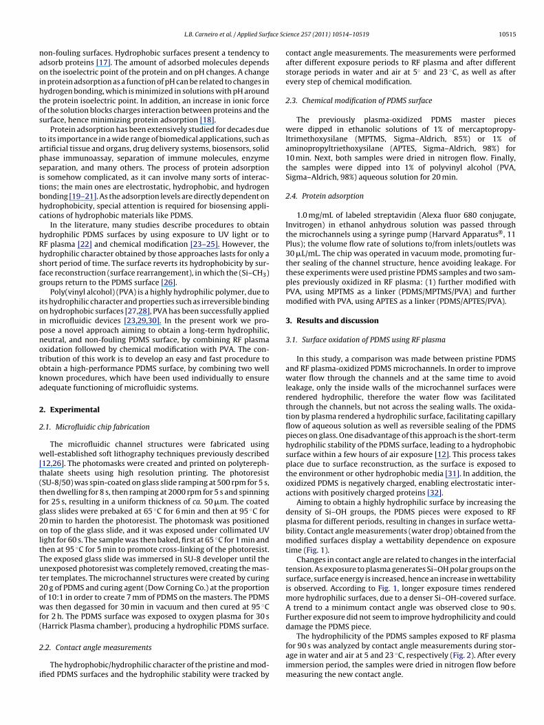
ace Sc
naoihtos
tapsitbhc
hRhsfg
ioipnotoka
2
2
w[t(tfg2oltTut2owf(
2
i
L.B. Carneiro et al. / Applied Surf
on-fouling surfaces. Hydrophobic surfaces present a tendency todsorb proteins [17]. The amount of adsorbed molecules dependsn the isoelectric point of the protein and on pH changes. A changen protein adsorption as a function of pH can be related to changes inydrogen bonding, which is minimized in solutions with pH aroundhe protein isoelectric point. In addition, an increase in ionic forcef the solution blocks charges interaction between proteins and theurface, hence minimizing protein adsorption [18].
Protein adsorption has been extensively studied for decades dueo its importance in a wide range of biomedical applications, such asrtificial tissue and organs, drug delivery systems, biosensors, solidhase immunoassay, separation of immune molecules, enzymeeparation, and many others. The process of protein adsorptions somehow complicated, as it can involve many sorts of interac-ions; the main ones are electrostatic, hydrophobic, and hydrogenonding [19–21]. As the adsorption levels are directly dependent onydrophobicity, special attention is required for biosensing appli-ations of hydrophobic materials like PDMS.
In the literature, many studies describe procedures to obtainydrophilic PDMS surfaces by using exposure to UV light or toF plasma [22] and chemical modification [23–25]. However, theydrophilic character obtained by those approaches lasts for only ahort period of time. The surface reverts its hydrophobicity by sur-ace reconstruction (surface rearrangement), in which the (Si–CH3)roups return to the PDMS surface [26].
Poly(vinyl alcohol) (PVA) is a highly hydrophilic polymer, due tots hydrophilic character and properties such as irreversible bindingn hydrophobic surfaces [27,28], PVA has been successfully appliedn microfluidic devices [23,29,30]. In the present work we pro-ose a novel approach aiming to obtain a long-term hydrophilic,eutral, and non-fouling PDMS surface, by combining RF plasmaxidation followed by chemical modification with PVA. The con-ribution of this work is to develop an easy and fast procedure tobtain a high-performance PDMS surface, by combining two wellnown procedures, which have been used individually to ensuredequate functioning of microfluidic systems.
. Experimental
.1. Microfluidic chip fabrication
The microfluidic channel structures were fabricated usingell-established soft lithography techniques previously described
12,26]. The photomasks were created and printed on polytereph-halate sheets using high resolution printing. The photoresistSU-8/50) was spin-coated on glass slide ramping at 500 rpm for 5 s,hen dwelling for 8 s, then ramping at 2000 rpm for 5 s and spinningor 25 s, resulting in a uniform thickness of ca. 50 �m. The coatedlass slides were prebaked at 65 ◦C for 6 min and then at 95 ◦C for0 min to harden the photoresist. The photomask was positionedn top of the glass slide, and it was exposed under collimated UVight for 60 s. The sample was then baked, first at 65 ◦C for 1 min andhen at 95 ◦C for 5 min to promote cross-linking of the photoresist.he exposed glass slide was immersed in SU-8 developer until thenexposed photoresist was completely removed, creating the mas-er templates. The microchannel structures were created by curing0 g of PDMS and curing agent (Dow Corning Co.) at the proportionf 10:1 in order to create 7 mm of PDMS on the masters. The PDMSas then degassed for 30 min in vacuum and then cured at 95 ◦C
or 2 h. The PDMS surface was exposed to oxygen plasma for 30 sHarrick Plasma chamber), producing a hydrophilic PDMS surface.
.2. Contact angle measurements
The hydrophobic/hydrophilic character of the pristine and mod-fied PDMS surfaces and the hydrophilic stability were tracked by
ience 257 (2011) 10514– 10519 10515
contact angle measurements. The measurements were performedafter different exposure periods to RF plasma and after differentstorage periods in water and air at 5◦ and 23 ◦C, as well as afterevery step of chemical modification.
2.3. Chemical modification of PDMS surface
The previously plasma-oxidized PDMS master pieceswere dipped in ethanolic solutions of 1% of mercaptopropy-ltrimethoxysilane (MPTMS, Sigma–Aldrich, 85%) or 1% ofaminopropyltriethoxysilane (APTES, Sigma–Aldrich, 98%) for10 min. Next, both samples were dried in nitrogen flow. Finally,the samples were dipped into 1% of polyvinyl alcohol (PVA,Sigma–Aldrich, 98%) aqueous solution for 20 min.
2.4. Protein adsorption
1.0 mg/mL of labeled streptavidin (Alexa fluor 680 conjugate,Invitrogen) in ethanol anhydrous solution was passed throughthe microchannels using a syringe pump (Harvard Apparatus®, 11Plus); the volume flow rate of solutions to/from inlets/outlets was30 �L/mL. The chip was operated in vacuum mode, promoting fur-ther sealing of the channel structure, hence avoiding leakage. Forthese experiments were used pristine PDMS samples and two sam-ples previously oxidized in RF plasma: (1) further modified withPVA, using MPTMS as a linker (PDMS/MPTMS/PVA) and furthermodified with PVA, using APTES as a linker (PDMS/APTES/PVA).
3. Results and discussion
3.1. Surface oxidation of PDMS using RF plasma
In this study, a comparison was made between pristine PDMSand RF plasma-oxidized PDMS microchannels. In order to improvewater flow through the channels and at the same time to avoidleakage, only the inside walls of the microchannel surfaces wererendered hydrophilic, therefore the water flow was facilitatedthrough the channels, but not across the sealing walls. The oxida-tion by plasma rendered a hydrophilic surface, facilitating capillaryflow of aqueous solution as well as reversible sealing of the PDMSpieces on glass. One disadvantage of this approach is the short-termhydrophilic stability of the PDMS surface, leading to a hydrophobicsurface within a few hours of air exposure [12]. This process takesplace due to surface reconstruction, as the surface is exposed tothe environment or other hydrophobic media [31]. In addition, theoxidized PDMS is negatively charged, enabling electrostatic inter-actions with positively charged proteins [32].
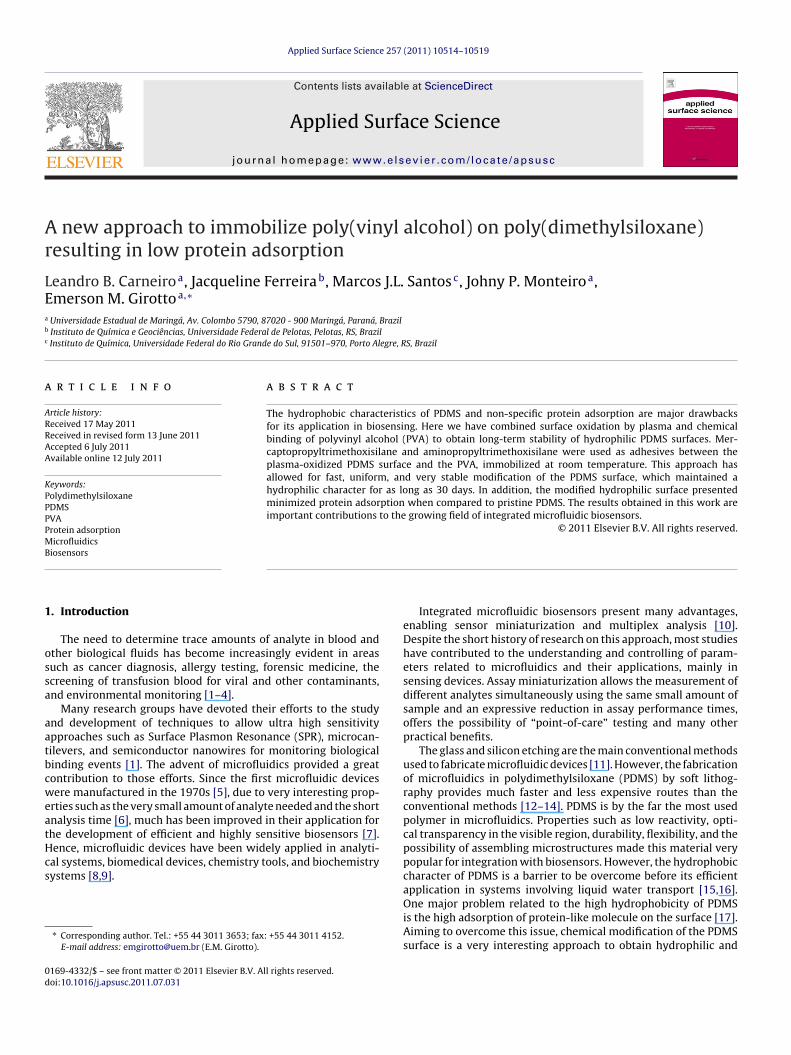
Aiming to obtain a highly hydrophilic surface by increasing thedensity of Si–OH groups, the PDMS pieces were exposed to RFplasma for different periods, resulting in changes in surface wetta-bility. Contact angle measurements (water drop) obtained from themodified surfaces display a wettability dependence on exposuretime (Fig. 1).
Changes in contact angle are related to changes in the interfacialtension. As exposure to plasma generates Si–OH polar groups on thesurface, surface energy is increased, hence an increase in wettabilityis observed. According to Fig. 1, longer exposure times renderedmore hydrophilic surfaces, due to a denser Si–OH-covered surface.A trend to a minimum contact angle was observed close to 90 s.Further exposure did not seem to improve hydrophilicity and coulddamage the PDMS piece.
The hydrophilicity of the PDMS samples exposed to RF plasma
for 90 s was analyzed by contact angle measurements during stor-age in water and air at 5 and 23 ◦C, respectively (Fig. 2). After everyimmersion period, the samples were dried in nitrogen flow beforemeasuring the new contact angle.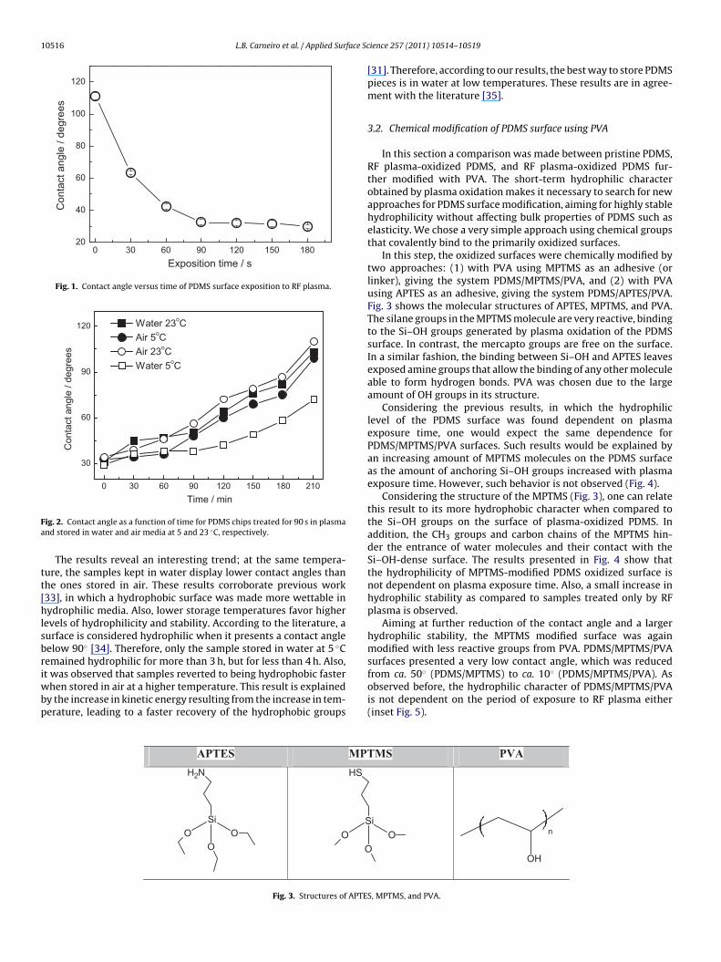
10516 L.B. Carneiro et al. / Applied Surface Sc
180150120906030020
40
60
80
100
120
Con
tact
ang
le /
degr
ees
Exposition time / s
Fig. 1. Contact angle versus time of PDMS surface exposition to RF plasma.
2101801501209060300
30
60
90
120
Con
tact
ang
le /
degr
ees
Time / mi n
Water 23oC Air 5oC Air 23oC Water 5oC
Fa
tt[hlsbriwbp
ig. 2. Contact angle as a function of time for PDMS chips treated for 90 s in plasmand stored in water and air media at 5 and 23 ◦C, respectively.
The results reveal an interesting trend; at the same tempera-ure, the samples kept in water display lower contact angles thanhe ones stored in air. These results corroborate previous work33], in which a hydrophobic surface was made more wettable inydrophilic media. Also, lower storage temperatures favor higher
evels of hydrophilicity and stability. According to the literature, aurface is considered hydrophilic when it presents a contact angleelow 90◦ [34]. Therefore, only the sample stored in water at 5 ◦Cemained hydrophilic for more than 3 h, but for less than 4 h. Also,
t was observed that samples reverted to being hydrophobic fasterhen stored in air at a higher temperature. This result is explainedy the increase in kinetic energy resulting from the increase in tem-erature, leading to a faster recovery of the hydrophobic groups
APTES MPT
SiO
OO
NH2
SO
O
HS
Fig. 3. Structures of APTE
ience 257 (2011) 10514– 10519
[31]. Therefore, according to our results, the best way to store PDMSpieces is in water at low temperatures. These results are in agree-ment with the literature [35].
3.2. Chemical modification of PDMS surface using PVA
In this section a comparison was made between pristine PDMS,RF plasma-oxidized PDMS, and RF plasma-oxidized PDMS fur-ther modified with PVA. The short-term hydrophilic characterobtained by plasma oxidation makes it necessary to search for newapproaches for PDMS surface modification, aiming for highly stablehydrophilicity without affecting bulk properties of PDMS such aselasticity. We chose a very simple approach using chemical groupsthat covalently bind to the primarily oxidized surfaces.
In this step, the oxidized surfaces were chemically modified bytwo approaches: (1) with PVA using MPTMS as an adhesive (orlinker), giving the system PDMS/MPTMS/PVA, and (2) with PVAusing APTES as an adhesive, giving the system PDMS/APTES/PVA.Fig. 3 shows the molecular structures of APTES, MPTMS, and PVA.The silane groups in the MPTMS molecule are very reactive, bindingto the Si–OH groups generated by plasma oxidation of the PDMSsurface. In contrast, the mercapto groups are free on the surface.In a similar fashion, the binding between Si–OH and APTES leavesexposed amine groups that allow the binding of any other moleculeable to form hydrogen bonds. PVA was chosen due to the largeamount of OH groups in its structure.
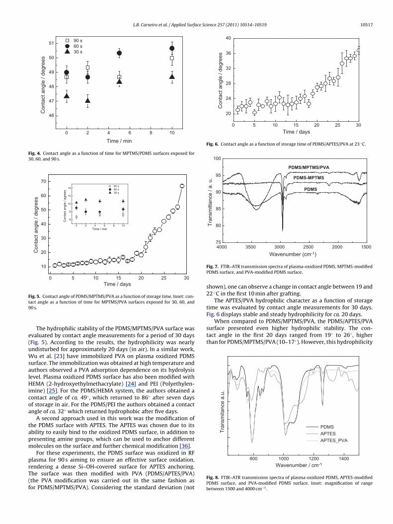
Considering the previous results, in which the hydrophiliclevel of the PDMS surface was found dependent on plasmaexposure time, one would expect the same dependence forPDMS/MPTMS/PVA surfaces. Such results would be explained byan increasing amount of MPTMS molecules on the PDMS surfaceas the amount of anchoring Si–OH groups increased with plasmaexposure time. However, such behavior is not observed (Fig. 4).
Considering the structure of the MPTMS (Fig. 3), one can relatethis result to its more hydrophobic character when compared tothe Si–OH groups on the surface of plasma-oxidized PDMS. Inaddition, the CH3 groups and carbon chains of the MPTMS hin-der the entrance of water molecules and their contact with theSi–OH-dense surface. The results presented in Fig. 4 show thatthe hydrophilicity of MPTMS-modified PDMS oxidized surface isnot dependent on plasma exposure time. Also, a small increase inhydrophilic stability as compared to samples treated only by RFplasma is observed.
Aiming at further reduction of the contact angle and a largerhydrophilic stability, the MPTMS modified surface was againmodified with less reactive groups from PVA. PDMS/MPTMS/PVAsurfaces presented a very low contact angle, which was reduced
from ca. 50◦ (PDMS/MPTMS) to ca. 10◦ (PDMS/MPTMS/PVA). Asobserved before, the hydrophilic character of PDMS/MPTMS/PVAis not dependent on the period of exposure to RF plasma either(inset Fig. 5).MS PVA
iO
OH
n
S, MPTMS, and PVA.
L.B. Carneiro et al. / Applied Surface Science 257 (2011) 10514– 10519 10517
1086420
46
47
48
49
50
51C
onta
ct a
ngle
/ de
gree
s
Time / min
90 s 60 s 30 s
Fig. 4. Contact angle as a function of time for MPTMS/PDMS surfaces exposed for30, 60, and 90 s.
302520151050
10
20
30
40
50
60
70
1086420
6
8
10
12
14
Con
tact
ang
le /
agre
es
Time / min
90 s 60 s 30 s
Con
tact
ang
le /
degr
ees
Time / days
Fig. 5. Contact angle of PDMS/MPTMS/PVA as a function of storage time. Inset: con-t9
e(uWsalHicoa
tapm
prT(f
302520151050
20
24
28
32
36
40
Con
tact
ang
le /
degr
ees
Time / days
Fig. 6. Contact angle as a function of storage time of PDMS/APTES/PVA at 23 ◦C.
15002000250030003500400075
80
85
90
95
100
PDMS/MPTMS/PVA
PDMS
Tra
nsm
ittan
ce /
a. u
.
Wavenumber (cm-1 )
PDMS-MPTMS
When compared to PDMS/MPTMS/PVA, the PDMS/APTES/PVAsurface presented even higher hydrophilic stability. The con-tact angle in the first 20 days ranged from 19◦ to 26◦, higherthan for PDMS/MPTMS/PVA (10–17◦). However, this hydrophilicity
140012001000800
Tran
smita
nce
a.u.
Wavenumber / cm-1
PDMS APTES APTES_PVA
act angle as a function of time for MPTMS/PVA surfaces exposed for 30, 60, and0 s.
The hydrophilic stability of the PDMS/MPTMS/PVA surface wasvaluated by contact angle measurements for a period of 30 daysFig. 5). According to the results, the hydrophilicity was nearlyndisturbed for approximately 20 days (in air). In a similar work,u et al. [23] have immobilized PVA on plasma oxidized PDMS
urface. The immobilization was obtained at high temperature anduthors observed a PVA adsorption dependence on its hydrolysisevel. Plasma oxidized PDMS surface has also been modified withEMA (2-hydroxyethylmethacrylate) [24] and PEI (Polyethylen-
mine) [25]. For the PDMS/HEMA system, the authors obtained aontact angle of ca. 49◦, which returned to 86◦ after seven daysf storage in air. For the PDMS/PEI the authors obtained a contactngle of ca. 32◦ which returned hydrophobic after five days.
A second approach used in this work was the modification ofhe PDMS surface with APTES. The APTES was chosen due to itsbility to easily bind to the oxidized PDMS surface, in addition toresenting amine groups, which can be used to anchor differentolecules on the surface and further chemical modification [36].For these experiments, the PDMS surface was oxidized in RF
lasma for 90 s aiming to ensure an effective surface oxidation,endering a dense Si–OH-covered surface for APTES anchoring.
he surface was then modified with PVA (PDMS/APTES/PVA)the PVA modification was carried out in the same fashion asor PDMS/MPTMS/PVA). Considering the standard deviation (notFig. 7. FTIR–ATR transmission spectra of plasma-oxidized PDMS, MPTMS-modifiedPDMS surface, and PVA-modified PDMS surface.
shown), one can observe a change in contact angle between 19 and22 ◦C in the first 10 min after grafting.
The APTES/PVA hydrophilic character as a function of storagetime was evaluated by contact angle measurements for 30 days.Fig. 6 displays stable and steady hydrophilicity for ca. 20 days.
Fig. 8. FTIR–ATR transmission spectra of plasma-oxidized PDMS, APTES-modifiedPDMS surface, and PVA-modified PDMS surface. Inset: magnification of rangebetween 1500 and 4000 cm−1.
10518 L.B. Carneiro et al. / Applied Surface Science 257 (2011) 10514– 10519
F S, (B)o
rPsssdcsth
3
bt[im[sscgvt3t
btarba3
ab3ac1wt
3
p
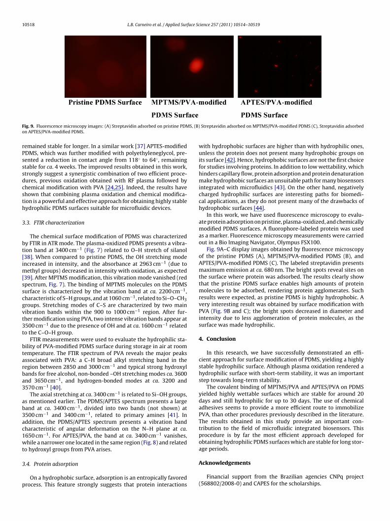
ig. 9. Fluorescence microscopy images: (A) Streptavidin adsorbed on pristine PDMn APTES/PVA-modified PDMS.
emained stable for longer. In a similar work [37] APTES-modifiedDMS, which was further modified with polyethyleneglycol, pre-ented a reduction in contact angle from 118◦ to 64◦, remainingtable for ca. 4 weeks. The improved results obtained in this work,trongly suggest a synergistic combination of two efficient proce-ures, previous oxidation obtained with RF plasma followed byhemical modification with PVA [24,25]. Indeed, the results havehown that combining plasma oxidation and chemical modifica-ion is a powerful and effective approach for obtaining highly stableydrophilic PDMS surfaces suitable for microfluidic devices.
.3. FTIR characterization
The chemical surface modification of PDMS was characterizedy FTIR in ATR mode. The plasma-oxidized PDMS presents a vibra-ion band at 3400 cm−1 (Fig. 7) related to O–H stretch of silanol38]. When compared to pristine PDMS, the OH stretching modencreased in intensity, and the absorbance at 2963 cm−1 (due to
ethyl groups) decreased in intensity with oxidation, as expected39]. After MPTMS modification, this vibration mode vanished (redpectrum, Fig. 7). The binding of MPTMS molecules on the PDMSurface is characterized by the vibration band at ca. 2200 cm−1,haracteristic of S–H groups, and at 1060 cm−1, related to Si–O–CH3roups. Stretching modes of C–S are characterized by two mainibration bands within the 900 to 1000 cm−1 region. After fur-her modification using PVA, two intense vibration bands appear at500 cm−1 due to the presence of OH and at ca. 1600 cm−1 relatedo the C–O–H group.
FTIR measurements were used to evaluate the hydrophilic sta-ility of PVA-modified PDMS surface during storage in air at roomemperature. The FTIR spectrum of PVA reveals the major peaksssociated with PVA: a C–H broad alkyl stretching band in theegion between 2850 and 3000 cm−1 and typical strong hydroxylands for free alcohol, non-bonded –OH stretching modes ca. 3600nd 3650 cm−1, and hydrogen-bonded modes at ca. 3200 and570 cm−1 [40].
The axial stretching at ca. 3400 cm−1 is related to Si–OH groups,s mentioned earlier. The PDMS/APTES spectrum presents a largeand at ca. 3400 cm−1, divided into two bands (not shown) at500 cm−1 and 3400 cm−1, related to primary amines [41]. Inddition, the PDMS/APTES spectrum presents a vibration bandharacteristic of angular deformation on the N–H plane at ca.650 cm−1. For APTES/PVA, the band at ca. 3400 cm−1 vanishes,hile a narrower one located in the same region (Fig. 8) and related
o hydroxyl groups from PVA arises.
.4. Protein adsorption
On a hydrophobic surface, adsorption is an entropically favoredrocess. This feature strongly suggests that protein interactions
Streptavidin adsorbed on MPTMS/PVA-modified PDMS (C), Streptavidin adsorbed
with hydrophobic surfaces are higher than with hydrophilic ones,unless the protein does not present many hydrophobic groups onits surface [42]. Hence, hydrophobic surfaces are not the first choicefor studies involving proteins. In addition to low wettability, whichhinders capillary flow, protein adsorption and protein denaturationmake hydrophobic surfaces an unsuitable path for many biosensorsintegrated with microfluidics [43]. On the other hand, negativelycharged hydrophilic surfaces are interesting paths for biomedi-cal applications, as they do not present many of the drawbacks ofhydrophobic surfaces [44].
In this work, we have used fluorescence microscopy to evalu-ate protein adsorption on pristine, plasma-oxidized, and chemicallymodified PDMS surfaces. A fluorophore-labeled protein was usedas a marker. Fluorescence microscopy measurements were carriedout in a Bio Imaging Navigator, Olympus FSX100.
Fig. 9A–C display images obtained by fluorescence microscopyof the pristine PDMS (A), MPTMS/PVA-modified PDMS (B), andAPTES/PVA-modified PDMS (C). The labeled streptavidin presentsmaximum emission at ca. 680 nm. The bright spots reveal sites onthe surface where protein was adsorbed. The results clearly showthat the pristine PDMS surface enables high amounts of proteinmolecules to be adsorbed, rendering protein agglomerates. Suchresults were expected, as pristine PDMS is highly hydrophobic. Avery interesting result was obtained by surface modification withPVA (Fig. 9B and C); the bright spots decreased in diameter andintensity due to less agglomeration of protein molecules, as thesurface was made hydrophilic.
4. Conclusion
In this research, we have successfully demonstrated an effi-cient approach for surface modification of PDMS, yielding a highlystable hydrophilic surface. Although plasma oxidation rendered ahydrophilic surface with short-term stability, it was an importantstep towards long-term stability.
The covalent binding of MPTMS/PVA and APTES/PVA on PDMSyielded highly wettable surfaces which are stable for around 20days and still hydrophilic for up to 30 days. The use of chemicaladhesives seems to provide a more efficient route to immobilizePVA, than other procedures previously described in the literature.The results obtained in this study provide an important con-tribution to the field of microfluidic integrated biosensors. Thisprocedure is by far the most efficient approach developed forobtaining hydrophilic PDMS surfaces which are stable for long stor-age periods.
Acknowledgements
Financial support from the Brazilian agencies CNPq project(568802/2008-0) and CAPES for the scholarships.
ace Sc
R
[
[
[
[
[
[[
[
[
[
[
[
[
[
[
[
[
[
[
[
[
[
[
[
[
[
[
[[
[
[
[
[
L.B. Carneiro et al. / Applied Surf
eferences
[1] E.C. Nice, B. Catimel, Instrumental biosensors: new perspectives for the analysisof biomolecular interactions, BioEssays 21 (1999) 339–352.
[2] J. Ferreira, M.J.L. Santos, M.M. Rahman, A.G. Brolo, G. Reuven, D. Sinton, E.M.Girotto, Attomolar protein detection using in-hole surface plasmon resonance,J. Am. Chem. Soc. 131 (2009) 436–437.
[3] M. Fan, A.G. Brolo, Silver nanoparticles self assembly as SERS substrates withnear single molecule detection limit, Phys. Chem. Chem. Phys. 11 (2009)7381–7389.
[4] J. Ferreira, E.M. Girotto, Optical pH sensitive material based on bro-mophenol blue-doped polypyrrole, Sens. Actuators B-Chem. 137 (2009)426–431.
[5] S.C. Terry, J.H. Jerman, J.B. Angell, IEEE Trans. Electron. Devices 26 (1979) 1880.[6] A.J. deMello, Control and detection of chemical reactions in microfluidic sys-
tems, Nature 442 (2006) 394–402.[7] A. Manz, N. Graber, H.M. Widmer, Miniaturized total chemical-analysis sys-
tems – a novel concept for chemical sensing, Sens. Actuators B 1 (1990)244–245.
[8] B. Zheng, L.S. Roach, R.F. Ismagilov, Screening of protein crystallization condi-tions on a microfluidic chip using nanoliter-size droplets, J. Am. Chem. Soc. 125(2003) 11170–11171.
[9] V. Srinivasan, V.K. Pamula, R.B. Fair, Droplet-based microfluidic lab-on-a-chipfor glucose detection, Anal. Chim. Acta 507 (2004) 145–150.
10] P.R.H. Stark, A.E. Halleck, D.N. Larson, Short order nanohole arrays in metals forhighly sensitive probing of local indices of refraction as the basis for a highlymultiplexed biosensor technology, Methods 37 (2005) 37.
11] N.L. Jeon, S.K.W. Dertinger, D.T. Chiu, I.S. Choi, A.D. Stroock, G.M. Whitesides,Generation of solution and surface gradients using microfluidic systems, Lang-muir 16 (2000) 8311–8316.
12] J.C. McDonald, G.M. Whitesides, Poly(dimethyldiloxane) as a material for fab-ricating microfluidic devices, Acc. Chem. Res. 35 (2002) 491–499.
13] J.C. McDonald, D.C. Duffy, J.R. Anderson, D.T. Chiu, H.K. Wu, O.J.A. Schueller, G.M.Whitesides, Fabrication of microfluidic systems in poly(dimethylsiloxane),Electrophoresis 21 (2000) 27–40.
14] G.M. Whitesides, The origins and the futures of microfluidics, Nature 442 (2006)368–373.
15] D.Q. Xiao, T. Van Le, M.J. Wirth, Anal. Chem. 76 (2004) 2055–2061.16] A. Theodorakakos, T. Ous, M. Gavaises, J.M. Nouri, N. Nikolopoulos, H. Yanagi-
hara, Dynamics of water droplets detached from porous surfaces of relevanceto PEM fuel cells, J. Colloid Interface Sci. 300 (2006) 673–687.
17] V. Linder, E. Verpoorte, W. Thormann, N.F. de Rooij, H. Sigrist, Surfacebiopassivation of replicated poly(dimethylsiloxane) microfluidic channels andapplication to heterogeneous immunoreaction with on-chip fluorescencedetection, Anal. Chem. 73 (2001) 4181.
18] L. Xu, V. Vadillo-Rodriguez, B.E. Logan, Residence time, loading force, pH, andionic strength affect adhesion forces between colloids and biopolymer-coatedsurfaces, Langmuir 21 (2005) 7491.
19] V.N Vasilets, A.V. Kuznetsov, V.I. Sevastianov, Vacuum ultraviolet treatmentof polyethylene to change surface properties and characteristics of proteinadsorption, J. Biomed. Mater. Res. A 64A (2004) 428–435.
20] R.G. Chapman, E. Ostuni, S. Takayama, R.E. Holmlin, L. Yan, G.M. Whitesides,Surveying for surfaces that resist the adsorption of proteins, J. Am. Chem. Soc.122 (2000) 8303–8304.
21] H.R. Byon, H.C. Choi, Network single-walled carbon nanotube-field effect tran-
sistors (SWNT-FETs) with increased Schottky contact area for highly sensitivebiosensor applications, J. Am. Chem. Soc. 128 (2006) 2188–2189.22] M.K. Chaudhury, G.M. Whitesides, Direct measurement of interfacial interac-tions between semispherical lenses and flat sheets of poly(dimethylsiloxane)and their chemical derivatives, Langmuir 7 (1991) 1013–1025.
[
[
ience 257 (2011) 10514– 10519 10519
23] D. Wu, Y. Luo, X. Zhou, Z. Dai, B. Lin, Multilayer poly(vinyl alcohol)-adsorbedcoating on poly(dimethylsiloxane) microfluidic chips for biopolymer separa-tion, Electrophoresis 26 (1) (2005) 211–218.
24] C. Khan-Malek, D. Bodas, Fabrication of long-term hydrophilic surfaces ofpoly(dimethyl siloxane) using 2-hydroxy ethyl methacrylate, Sens. ActuatorsB 120 (2007) 719–723.
25] N. Maheshwari, A. Kottantharayil, M. Kumar, S. Mukherji, Long term hydrophiliccoating on poly(dimethylsiloxane) substrates for microfluidic applications,Appl. Surf. Sci. 257 (2010) 451–457.
26] C.D. Duffy, J.C. Mcdonald, O.J.A. Schueller, G.M. Whitesides, Rapid prototyp-ing of microfluidic systems in poly(dimethylsiloxane), Anal. Chem. 70 (1998)4974–4984.
27] M. Kozlov, M. Quarmyne, W. Chen, T.J. McCarthy, Adsorption of poly(vinyl alco-hol) onto hydrophobic substrates. A general approach for hydrophiling andchemically activating surfaces, Macromolecules 36 (2003) 6054–6059.
28] D.A. Barrett, M.S. Hartshorne, M.A. Hussain, P.N. Shaw, M.C. Davies, Resistanceto nonspecific protein adsorption by poly(vinyl alcohol) thin films adsorbed toa poly(styrene) support matrix studied using surface plasmon resonance, Anal.Chem. 73 (2001) 5232–5239.
29] M. Ludwig, D. Belder, Coated microfluidic devices for improved chiral separa-tion in microchip electrophoresis, Electrophoresis 24 (2003) 2481–2486.
30] D. Belder, A. Deege, F. Kohler, M. Ludwig, Poly(vinyl alcohol)-coated microflu-idic devices for high-perfomance microchip electrophoresis, Electrophoresis23 (2002) 3567–3573.
31] S.R. Holmes-Farley, R.H. Reamey, R. Nuzzo, T.J. McCarthy, G.M. Whitesides,Reconstruction of the interface of oxidatively functionalized polyethylene andderivatives, Langmuir 3 (1987) 799–815.
32] E. Ostuni, B.A. Grzybowski, M. Mrksich, C.S. Roberts, G.M. Whitesides, Adsorp-tion of proteins to hydrophobic sites on mixed self-assembled monolayers,Langmuir 19 (2003) 1861–1872.
33] D.W. Branch, B.C. Wheeler, G.J. Brewer, D.E. Leckband, Long-term stability ofgrafted polyethylene glycol surfaces for use with microstamped substrates inneuronal cell culture, Biomaterials 22 (2001) 1035–1047.
34] A. Marmur, Soft contact: measurement and interpretation of contact angles,Soft Matter 2 (2006) 12–17.
35] R.A. Lawton, C.R. Price, A.F. Runge, W.J. Doherty III, S.S. Saavedra, Air plasmatreatment of submicron thick PDMS polymer films: effect of oxidation timeand storage conditions, Colloids Surf. A 253 (2005) 213.
36] S. Bhattacharya, A. Datta, J.M. Berg, S. Gangopadhyay, Studies on surface wet-tability of poly(dimethyl) siloxane (PDMS) and glass under oxygen-plasmatreatment and correlation with bond strength, J. Microelectromech. Syst. 14(2005) 590–597.
37] A.J. Wang, J.J. Feng, J. Fan, J. Chromatogr. A 1192 (2008) 173–179.38] L.K. Paiva, Introduction to Spectroscopy, 3rd ed., Brooks/Cole Publishing Com-
pany, California, EUA, 2001.39] F. Garbassi, M. Morra, E. Occhiello, Polymer Surfaces, John Wiley & Sons, New
York, 1994.40] H.S. Mansur, R.L. Orefice, A.A.P. Mansur, Characterization of
poly(vinylalcohol)/poly(ethylene glycol) hydrogels and PVA-derived hybridsby small-angle X-ray scattering and FTIR spectroscopy, Polymer 45 (2004)7193–7202.
41] R.M. Silverstein, F.X. Webster, D.J. Kiemle, Identificac ão Espectrométrica deCompostos Orgânicos ED, 7th ed, LTC, Rio de Janeiro, 2006.
42] P. Roach, D. Farrar, C. Perry, Interpretation of protein adsorption: surface-induced conformational changes, J. Am. Chem. Soc. 127 (2005) 8168–8173.
43] P. Wagner, Immobilization strategies for biological scanning probe microscopy,FEBS Lett. 430 (1998) 112–115.
44] M. Karlsson, J. Ekeroth, H. Elwing, U. Carlsson, Reduction of irreversible proteinadsorption on solid surfaces by protein engineering for increased stability, TheJ. Biol. Chem. 27 (2005) 25558.
Related Documents


![Impact of the poly(propylene oxide)-b-poly(dimethylsiloxane ......[15]. Byczyński et al. reported PURs coatings, based on different soft segments. With the use of α,ω-dihydroxy-](https://static.cupdf.com/doc/110x72/60ffb46eeaacf422803d8ca3/impact-of-the-polypropylene-oxide-b-polydimethylsiloxane-15-byczyski.jpg)