Restorative Neurology and Neuroscience 30 (2012) 407–418 DOI 10.3233/RNN-2012-120224 IOS Press 407 A neurally-interfaced hand prosthesis tuned inter-hemispheric communication G. Di Pino a,b,1 , C. Porcaro c,d,1,∗ , M. Tombini a , G. Assenza a , G. Pellegrino a , F. Tecchio c,e,2 and P.M. Rossini e,f ,2 a Department of Neurology, Campus Bio-Medico University, Rome, Italy b Biomedical Robotics and Biomicrosystems Lab., University Campus Bio-Medico, Rome, Italy c LET’s – ISTC – CNR, Ospedale Fatebenefratelli, Isola Tiberina, Rome, Italy d Institute of Neuroscience, Newcastle University, Medical School, Newcastle upon Tyne, UK e Casa di Cura S. Raffaele, Cassino and IRCCS S. Raffaele-Pisana, Rome, Italy f Institute of Neurology, Catholic University, Rome, Italy Abstract. Purpose: This work investigates how a direct bidirectional connection between brain and hand prosthesis modifies the bi-hemispheric sensorimotor system devoted to the movement control of the lost limb. Hand prostheses are often unable to satisfy users’ expectations, mostly due to the poor performance of their interfacing system. Neural Interfaces implanted inside nerves of the stump offer the advantage of using the bidirectional neural pathways ‘naturally’ dispatching signals to control proper hand actions and feed-back sensations. Learning to control a neurally-interfaced hand prosthesis and decode sensory information was previously observed to reduce the inter-hemispheric asymmetry of cortical motor maps and the clinical symptoms of phantom limb syndrome. Methods: Electroencephalographic (EEG) data was analysed using Functional Source Separation (FSS), a semi-blind method that incorporates prior knowledge about the signal of interest into data decomposition to give access to cortical patch activities. Results: Bi-hemispheric cortices showed normalization of their activity (topographical and spectral patterns) and of functional connectivity between homologous hand controlling areas, during the delivery of the motor command to the cybernetic prosthesis. Conclusions: The re-establishment of central-peripheral communication with the lost limb induced by a neurally-interfaced hand prosthesis produces beneficial plastic reorganization, not only restructuring contralateral directly-connected control areas, but also their functional balance within the bi-hemispheric system necessary for motor control. Keywords: Functional source separation, hand prosthesis, neural interface, inter-hemispheric coherence, neurorehabilitation 1. Introduction Human beings are able to interact with the environ- ment through several modalities, which involves neural signals passing through the nerves, muscle activity 1 These authors contributed equally to this work as first author. 2 These authors contributed equally to this work as last author. ∗ Corresponding author: Dr. Camillo Porcaro, LET’S-ISTC- CNR, Department of Clinical Neuroscience, Fatebenefratelli Hospital – Isola Tiberina, 00186 Rome, Italy. E-mail: [email protected]. and the integrity of cutaneous/proprioceptive sensory organs. Brain Machine Interfaces (BMI) bypass the above steps and allow motor signals to be picked up directly from the nervous system and relayed to an external electronic or robotic effector which physically performs the subject’s motor intention and plans. In addition to facilitating extraction of motor volition, bidirectional BMIs also create the capacity to man- age neural signal flows in both afferent and efferent directions. Hand/finger manipulation is a very complex activ- ity performed almost exclusively by humans and a 0922-6028/12/$27.50 © 2012 – IOS Press and the authors. All rights reserved

Welcome message from author
This document is posted to help you gain knowledge. Please leave a comment to let me know what you think about it! Share it to your friends and learn new things together.
Transcript
Restorative Neurology and Neuroscience 30 (2012) 407–418DOI 10.3233/RNN-2012-120224IOS Press
407
A neurally-interfaced hand prosthesis tunedinter-hemispheric communication
G. Di Pinoa,b,1, C. Porcaroc,d,1,∗, M. Tombinia, G. Assenzaa, G. Pellegrinoa,F. Tecchioc,e,2 and P.M. Rossinie,f,2aDepartment of Neurology, Campus Bio-Medico University, Rome, ItalybBiomedical Robotics and Biomicrosystems Lab., University Campus Bio-Medico, Rome, ItalycLET’s – ISTC – CNR, Ospedale Fatebenefratelli, Isola Tiberina, Rome, ItalydInstitute of Neuroscience, Newcastle University, Medical School, Newcastle upon Tyne, UKeCasa di Cura S. Raffaele, Cassino and IRCCS S. Raffaele-Pisana, Rome, Italyf Institute of Neurology, Catholic University, Rome, Italy
Abstract. Purpose: This work investigates how a direct bidirectional connection between brain and hand prosthesis modifiesthe bi-hemispheric sensorimotor system devoted to the movement control of the lost limb. Hand prostheses are often unableto satisfy users’ expectations, mostly due to the poor performance of their interfacing system. Neural Interfaces implantedinside nerves of the stump offer the advantage of using the bidirectional neural pathways ‘naturally’ dispatching signals tocontrol proper hand actions and feed-back sensations. Learning to control a neurally-interfaced hand prosthesis and decodesensory information was previously observed to reduce the inter-hemispheric asymmetry of cortical motor maps and the clinicalsymptoms of phantom limb syndrome.Methods: Electroencephalographic (EEG) data was analysed using Functional Source Separation (FSS), a semi-blind methodthat incorporates prior knowledge about the signal of interest into data decomposition to give access to cortical patch activities.Results: Bi-hemispheric cortices showed normalization of their activity (topographical and spectral patterns) and of functionalconnectivity between homologous hand controlling areas, during the delivery of the motor command to the cybernetic prosthesis.Conclusions: The re-establishment of central-peripheral communication with the lost limb induced by a neurally-interfacedhand prosthesis produces beneficial plastic reorganization, not only restructuring contralateral directly-connected control areas,but also their functional balance within the bi-hemispheric system necessary for motor control.
Keywords: Functional source separation, hand prosthesis, neural interface, inter-hemispheric coherence, neurorehabilitation
1. Introduction
Human beings are able to interact with the environ-ment through several modalities, which involves neuralsignals passing through the nerves, muscle activity
1These authors contributed equally to this work as first author.2These authors contributed equally to this work as last author.∗Corresponding author: Dr. Camillo Porcaro, LET’S-ISTC-
CNR, Department of Clinical Neuroscience, FatebenefratelliHospital – Isola Tiberina, 00186 Rome, Italy. E-mail:[email protected].
and the integrity of cutaneous/proprioceptive sensoryorgans. Brain Machine Interfaces (BMI) bypass theabove steps and allow motor signals to be picked updirectly from the nervous system and relayed to anexternal electronic or robotic effector which physicallyperforms the subject’s motor intention and plans. Inaddition to facilitating extraction of motor volition,bidirectional BMIs also create the capacity to man-age neural signal flows in both afferent and efferentdirections.
Hand/finger manipulation is a very complex activ-ity performed almost exclusively by humans and a
0922-6028/12/$27.50 © 2012 – IOS Press and the authors. All rights reserved
408 G. Di Pino et al. / A neurally-interfaced hand prosthesis
few primates. Although prosthetics was one of thefirst applications for artificial human-like hands, engi-neers and roboticists that developed multi-fingeredhumanoid robots and mechatronic hand prostheses,have not so far been able to achieve the same manip-ulative ability owned by the human hand (Carrozzaet al., 2006; Zollo et al., 2007). However, many effortswith significant progresses have been achieved in thisdirection.
To date, what the market is able to offer to upper limbamputees still suffers a series of limitations relatedto their mechanics, control system and embeddedproprioceptive and exteroceptive sensors. However,a fundamental limitation afflicting their performanceis the poverty of the user-device interplay achievableby present interfacing systems (Micera et al., 2006).Hence, the control of anthropomorphic hand prosthe-ses represents a promising field for the application ofneural interfaces.
At the amputation site all afferent and efferentnerves, originally devoted to the lost segment, areinterrupted. The consequent deprivation of periph-eral inputs and actuators results in retrograde changesthat affect not only the peripheral nervous systembut also central structures including the motor andsomatosensory cortices. In fact, following amputationthe deafferented cortical areas become responsive toinputs from the parts of the body that are representedadjacent in the Penfield homunculus (Kaas, 1991;Merzenich et al., 1984; Pons et al., 1991; Ramachan-dran et al., 1992).
While a wide literature indicates the adaptivepotential of brain plasticity, mainly in conditions ofcongenital deficits (for reviews on blind people seePascual-Leone et al., 2005; Sadato, 2005, Gilbert andWalsh, 2004), experience in amputee suggests that thetraumatic event prevents adaptive cortical changes. Infact, the aberrant counterpart of plastic phenomenaprobably represents the pathophysiological substrateof a disabling and painful experience called phan-tom limb pain (PLP), that afflicts the majority ofamputees and is widely considered as a maladaptivecorrelate of neuroplasticity (Flor et al., 1995; Flor et al.,2006). Therefore it seems reasonable to assume thatthe amount and characteristics of PLP are some ofthe indicators of harmful cortical reorganization. For acomprehensive review of neuroplasticity in amputeessee (Di Pino et al., 2009).
Commonly the ability possessed by the majority ofamputees to move the phantom of the lost limb at their
will was considered as motor imagery. Recently it hasbeen shown that after amputation the ability to exe-cute a movement with the affected limb is maintainedby the amputees’ brain (Raffin et al., 2011), despite theabsence of a peripheral effector. In fact motor imagina-tion and execution differentially activate cerebral areaswith a significantly greater activation in primary motorand sensory cortices during execution, while imagina-tion is associated with greater parietal and occipitallobe activity (Raffin et al., 2012). Even though thenervous system is altered by the amputation, resid-ual neural patterns appropriate for the lost limb maystill be activated when suitably stimulated (Mercieret al., 2006; Reilly et al., 2006). Those pathways aretherefore a possible target of central or peripheral neu-ral implants to restore a direct and relatively ‘natural’channel for data exchanging.
Multielectrode arrays implanted inside peripheralnerves represent an optimal compromise between theselectivity of exchanged data and the invasivenessof the required surgery. Their fundamental ability tointeract with a small number of nerve fascicles andto exchange information bidirectionally (Micera andNavarro, 2009; Micera et al., 2008; Navarro et al.,2005) make such interface appropriate to enhance neu-roplastic phenomena (Di Pino et al., 2009).
A positive correlation between use of a nerve-interfaced hand prosthesis and the reduction of aberrantbrain plasticity has already been described by ourgroup using TMS cortical mapping and PLP reductionassessment (Rossini et al., 2010), and by analysis ofevent-related EEG synchronization/desynchronization(Tombini et al., 2012).
The present study explores whether and how a directconnection between the brain and a hand prosthesis viaa neural implant modifies bi-hemispheric EEG activ-ity in primary sensorimotor cortical areas controllingmovements of the lost limb. In particular we applya Functional Sources Separation (FSS) comparativeanalysis of the EEG pattern recorded during the intentof voluntary moving the lost limb phantom, collectedbefore and after the implant period.
EEG and MEG signals are linear mixtures of sourceactivities, and are often analysed using the blindsource separation (BSS) technique. These algorithmsestimate the complete source time-courses based onstatistical properties of the generated signal, withouttaking into account the physical nature of the gener-ating phenomenon. Thus, BSS procedures use onlyinformation contained in the waveform of original
G. Di Pino et al. / A neurally-interfaced hand prosthesis 409
signals, a convenient property for neurophysiologi-cal techniques (EEG and MEG) which provide themost informative time–frequency signal from the intacthuman brain. BSS techniques, in particular indepen-dent component analysis (ICA) algorithms, have beensuccessfully applied to EEG and MEG data (Barbatiet al., 2004; Makeig et al., 2004; Medaglia et al., 2009;Porcaro et al., 2011; Porcaro et al., 2006). The aimof such techniques is to decompose in a ‘blind’ fash-ion (i.e., without making specific assumptions) thesources underlying the EEG and MEG recorded signals(for a comprehensive review on BSS see: Hyvarinenet al., 2001; Cichocki and Amari, 2002; Tecchio et al.,2007b). Our group developed a new semi-BSS methodcalled Functional Source Separation (FSS) (Barbatiet al., 2006; Tecchio et al., 2007b), an extension of ICAthat incorporates prior information into the decompo-sition, resulting in a single component which containsa hallmark of the source of interest (for this reason ithas been denominated semi blind method). The aimof FSS is to enhance the separation of relevant signalsby exploiting some a priori knowledge (i.e., spectralcharacteristics or evoked activity property) withoutlosing the advantages of using only information con-tained in original signal waveforms. In other terms,FSS allows the identification and analysis of specificneuronal pools on the bases of their functional proper-ties instead and independently of their spatial position.This is an extremely helpful feature especially in thosecases where cerebral plastic changes have altered thelocation of brain functions with respect to standardanatomical landmarks typical of healthy people.
2. Methods
2.1. Subject and prosthesis control
A brief description is given in the following section,while a complete report on the clinical and technicalaspects of this case can be found elsewhere (Rossiniet al., 2010).
A 26 year-old right-handed male (PP) was recruitedtwo years after a left arm trans-radial amputation.His previous medical history was unremarkable. Thestudy was approved by the local Ethics Committee andthe assigned office of Italian Ministry of Health, andan informed consent was signed by the patient. P.P.referred to a moderate phantom limb syndrome andperceived his phantom limb as if ‘. . . the missing hand
is still attached to the stump and tightly fastened by abelt without any forearm’.
The subject had been implanted with four longi-tudinal intra-fascicular electrode arrays (tf-LIFE4s)(Hoffmann and Kock, 2005) in the Ulnar and Mediannerves of his stump for four weeks, providing a totalamount of 32 bidirectional contacts.
Before the implantation the subject was trained fortwo weeks in dispatching the motor commands toperform movements with his phantom hand, withoutactivating stump muscles.
Three movements of a virtual left hand were shownin videos: power grip, pinch grip and flexion of thelittle finger. The actions were exploited because theyrepresent the range of movements controlled by theimplanted nerves: median fibres for the pinch, ulnarfor little finger flexion, and both for the power grip.
During the four experiment weeks after electrodeimplantation, PP was required to perform the hand griptasks triggered by the same videos, while recordingneural signals.
Signals from tf-LIFE4s, biceps and triceps EMGelectrodes were simultaneously recorded using a48 kHz sampling rate. The mean, rectified signals werecalculated for bins of width 1000 samples.
Each prosthesis movement type was triggered bythe signal level of a single channel with the bestsignal-to-noise ratio. Channels were coupled with gripsdepending on their anatomo-functional location (i.e.,channels from the median nerve for power or pinchgrip, channels from the ulnar for little finger flex-ion). To avoid interference from muscle activity orenvironmental noise, only rectified values greater than3 �V in a time window ranging from 5 to 20 ms wereused.
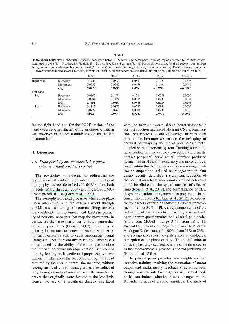
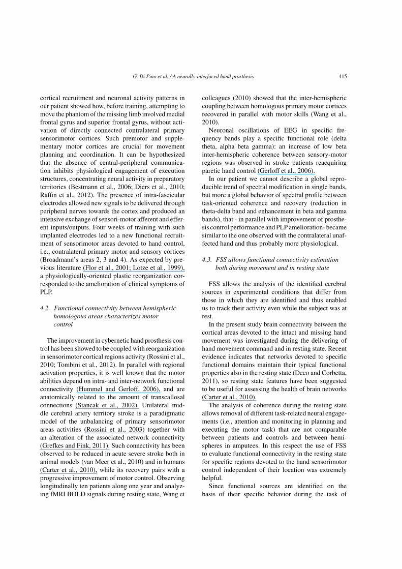
Together with visual feedback from the prosthesisposed in front of him, PP received sensory feedbackvia suprathreshold electrical pulses delivered throughthe implanted electrodes, triggered by the experimenter(Fig. 1). Stimulus trains (0.3 s train of 70 Hz, 10 �A,10 �s pulses) were delivered after each burst of efferentactivity recorded by tf-LIFE4 s. The channels used toselect when and to provide the stimulation were thesame, and were established according to the performedaction (i.e., channels into the median nerve for poweror pinch grip, channels into the ulnar for little fingerflexion, channels into both nerves for the power grip).In all cases, the subject described discrete tactile (non-proprioceptive) sensations in the fascicular projectionterritories that corresponded to the stimulated nerves.

410 G. Di Pino et al. / A neurally-interfaced hand prosthesis
Fig. 1. Block scheme of the online control of the prosthetic device.
2.2. EEG recordings
EEG signals were recorded from the scalp duringa handgrip task including periods lasting 5 secondsof active control of motor commands dispatched tothe hand (movement), intermingled with 5 s periodsof relaxation (recovery). The study of left phantomhand movement was executed at two times: i) afterpre-implant training the day before surgery (PRE),ii) at the end of four weeks of intensive training tocontrol the hand prosthesis using implanted LIFEs(POST). Thirty-two electrodes (scalp sites definedaccording to the international 10–20 electrode sys-tem) were mounted on an elastic cap and a binauralreference was used. Skin/electrode impedances werekept below 5 k�. Recordings were carried out usinga time constant of 0.1 s. EEG data were sampled at1024 Hz (pre-sampling analogical filter 0.48–256 Hz,BrainAmp System).
2.3. Data analysis
The first step of the analysis we have employedis a semiautomatic independent component analysis(ICA)-based procedure to identify and remove cardiacand/or ocular artifacts without rejecting the contami-nated epochs (Barbati et al., 2004).
The functional source separation (FSS) procedure(Porcaro et al., 2009; Porcaro et al., 2010; Tecchio
et al., 2007b) was applied to identify the cortical neu-ral networks devoted to hand movement. As in theICA approach, FSS starts from an additive hiddensource model of the type x = As, where x representsthe observed EEG data, s are the underlying unknownsources and A is the source-sensor coupling matrixto be estimated. Additional information to a stan-dard ICA model is used to bias the decompositionalgorithm towards solutions that satisfy physiologi-cal assumptions. A modified (with respect to standardICA) contrast function is defined:
F = J + λRFS (1)
where J is the statistical index normally used in ICAand RFS accounts for the a-priori information exploitedto identify the source. Using the weighting parameterλ it is possible to adjust the relative weight of thesetwo aspects. In this study, λ was chosen to be equalto 1000 in all cases, as detailed in Porcaro and col-leagues (Porcaro et al., 2008). Briefly, λ was chosento both minimize computational time and maximizeRFS . This scheme allows the possibility of extract-ing only one component that maximizes the functionalbehavior in agreement with the functional constraint.In the present work, a maximization of the differencein Power (Power Spectral Density, PSD) between restand task was used.
G. Di Pino et al. / A neurally-interfaced hand prosthesis 411
2.4. Primary sensorimotor functional source(FS hand)
The functional constraint exploited mu rhythm(8–25 Hz) reactivity (Pfurtscheller and Lopes da Silva,1999) that occurs in contralateral sensorimotor areasduring uni-manual motor tasks, by requiring maxi-mal variation of spectral power in alpha and betabands between the period of prosthesis control andrest. The ad-hoc functional constraint R was thus asfollows:
R(FSα+β) =
∑
α+β
PSD(FSα+β)Task − ∑
α+β
PSD(FSα+β)Rest
∑
α+β
PSD(FSα+β)Rest(2)
with the Power Spectrum Density (PSD) during Taskestimated in the 5 sec windows of each movement trialand at Rest in the 5 sec windows preceding each trial.� + � frequency band included 8 to 25 Hz (Barbatiet al., 2008; Porcaro et al., 2011).
2.5. Functional source behavior
Cortical activity of areas devoted to hand control wasevaluated by time-frequency modulations of FS powerspectral density during the motor task as compared tothe resting state baseline. Twenty single trials from thesource were chosen in the time interval of −5 to 5 s,where time 0 is the movement go signal. They wereconvolved by a Morlet wavelet (setting equal to 7 theconstant which defines the compromise between timeand frequency resolution), and the squared absolutevalues of the convolution over trials were averaged. Foreach frequency, the time course of power modulationwas represented as the percentage of the mean of thebaseline period (–5 to 0 s). The cortical recruitmentsand the time frequency behavior were compared beforeand after neural implant.
Significant power changes from Rest to Move-ment Imagination (Task) periods were assessed usinga resampling bootstrap technique thresholded atP = 0.05, while non-significant changes were set to 0.The above procedure was applied for the PRE, POSTand on the right hand movement.
2.6. Functional source localization
In order to investigate cortical recruitment occur-ring during the intention of moving of the (intact)right hand and (phantom–prosthetic) left hand before
(PRE) and after training with the implanted neu-ral electrodes (POST), the FSs identified by FSSwere submitted to a source localization algorithm(sLORETA - (Pascual-Marqui, 2002)) implementedin CURRY 6 (Neuroscan, Hamburg, Germany,http://www.neuroscan.com/). sLORETA was per-formed for each source using a regular grid with aspacing of 3 mm throughout the brain region and a fourshell spherical head model. The results were projected
onto the template brain of the Montreal NeurologicalInstitute (MNI) within CURRY.
2.7. Functional connectivity between homologoushand control areas
The functional connectivity between the hemi-spheric homologous primary cortical circuits wasestimated by spectral coherence between the activitiesof the areas devoted to the control of the contralateralhand. Instead of anatomical criteria, we preferred toselect the region on a functional basis, thus we consid-ered the areas activated during the right and left motortasks. Being interested in primary circuits, in the caseof the left affected hand we chose the region of interest(RoI) corresponding to the FS in the POST session, i.e.,the primary contralateral right RoI, since the FS in thePRE-session involved a bilateral network prevailing inthe ipsi-lateral left sensorimotor areas. A standard FFTapproach (Welch technique and Hanning windowing)was applied in the same time windows used for PSDestimates:
∣∣Cxy(f )
∣∣2 =
∣∣Sxy(f )
∣∣2
Sx(f )Sy(f )(3)
with f the frequency bin, x and y the FS right hand andFS left hand, Sxy the cross spectrum between x andy, Sx and Sy the spectra of x and y respectively. Themodulus of coherence takes a dimensional values inthe [0, 1] range (0 indicates no relationship). The 95%confidence limit is estimated according to (Halliday
et al., 1995) by (1 − 0.05)1
(L−1) , where L is the numberof trials used to estimate the coherence function.
412 G. Di Pino et al. / A neurally-interfaced hand prosthesis
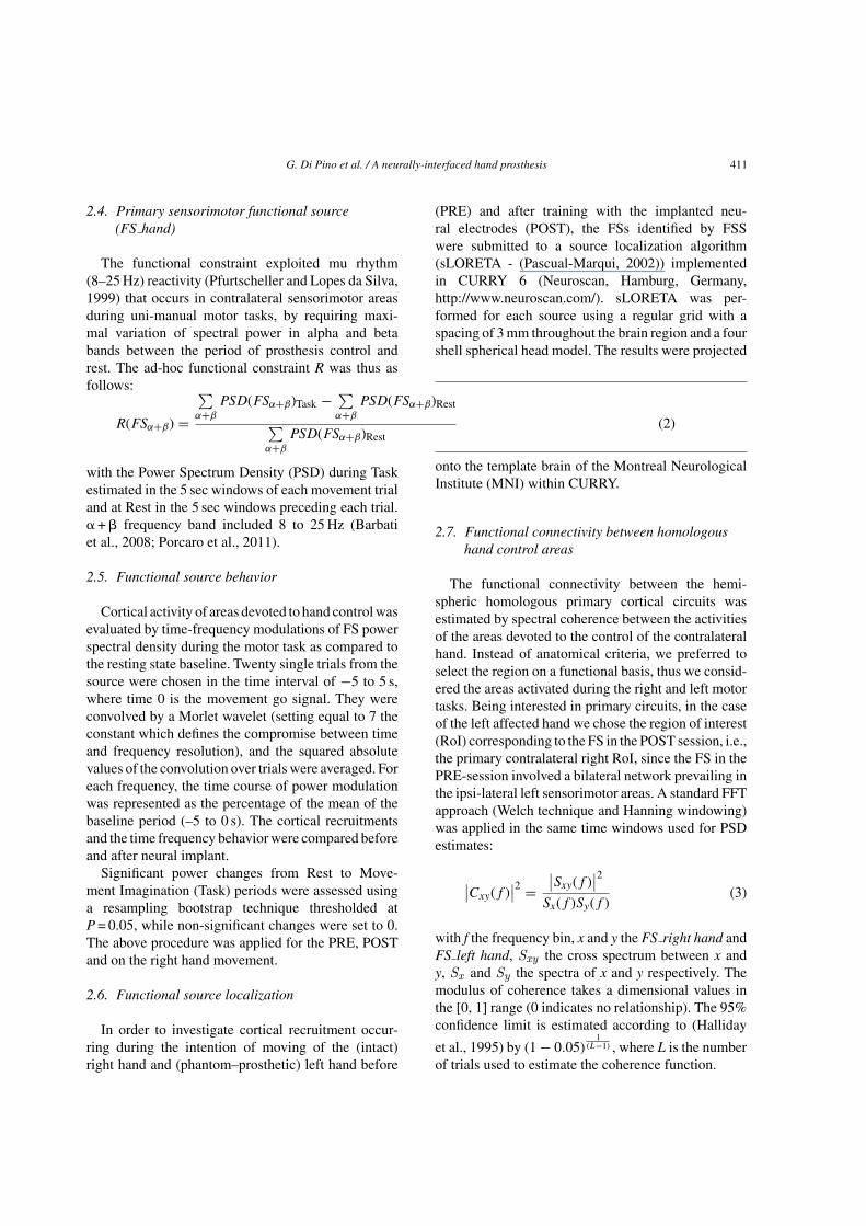
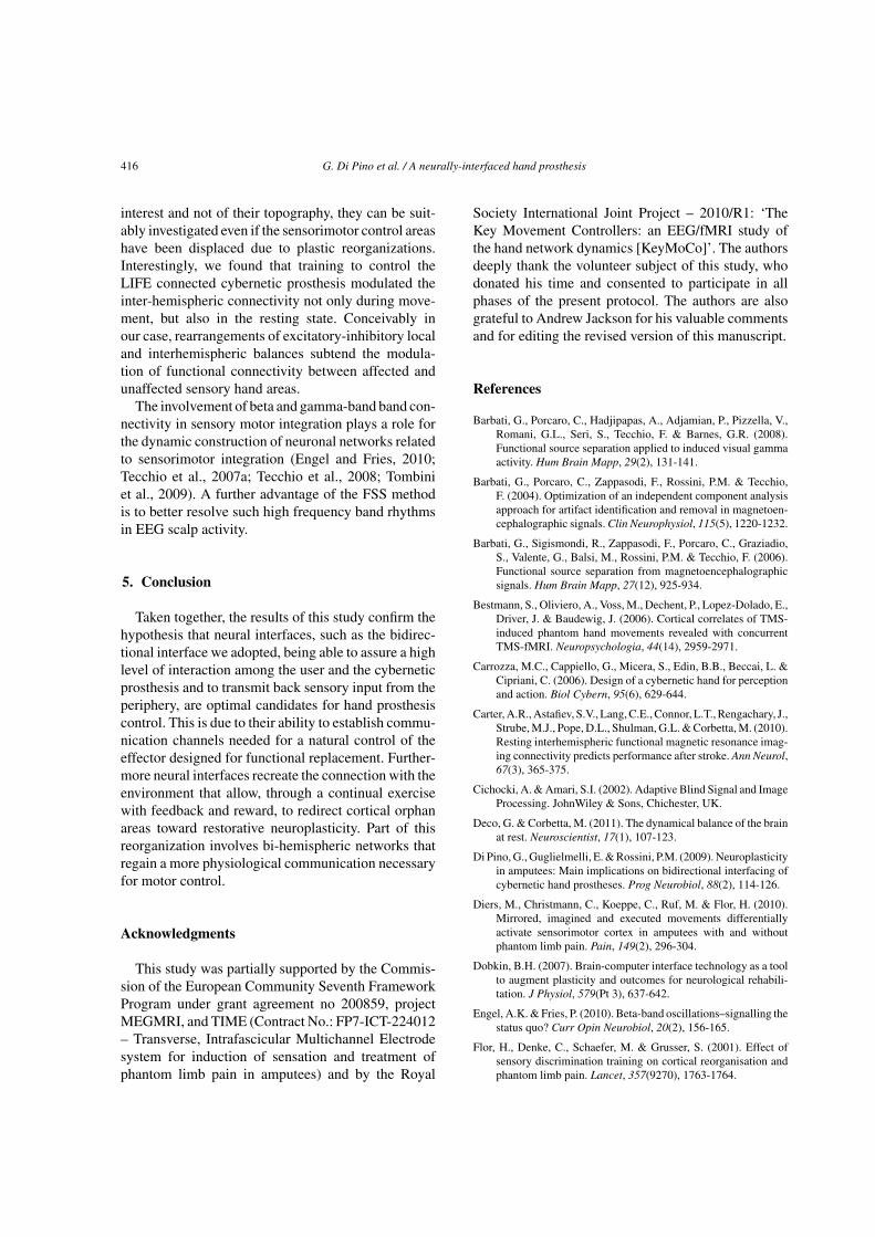
Fig. 2. FSs’ cerebral recruitment and time-frequency behavior. From Left: solutions of the sLORETA model localization algorithm for the FSsrelated to right hand control (Up Left), to the missing limb phantom control before LIFE implant (PRE – Middle Left) and at the end of 4weeks-long training with LIFEs implanted (POST – Bottom left) are shown superimposed on the MNI brain template in coronal, sagittal andaxial sections. Right: Time frequency representation of each FS activity, 0 correspond to the motor Task initiation. The color code representssignificant changes in average power (across epochs) as a function of time and frequency. Values for a given frequency bin are expressed aspercentage of the Rest period. Power change significance was assessed by using a bootstrap technique and thresholded at p = 0.05; non-significantchanges were set to 0. Note that FS left hand in PRE condition revealed a much wider cortical recruitment than for the right hand (see results).
3. Results
3.1. Movement of the right hand
Right hand movement recruited well segregatedcontra-lateral left sensorimotor areas (Fig. 2 – firstrow). In particular, source localization shows a clearactivation of the primary sensory and motor areas (leftPostcentral Gyrus – BA2, BA3 and Precentral Gyrus -
BA4). The analysis of time frequency behavior exhib-ited a clear response that was stronger in the first 2seconds and covered the whole mu band including bothalpha and beta frequencies, becoming concentrated inthe alpha band for the 2–5 s period.
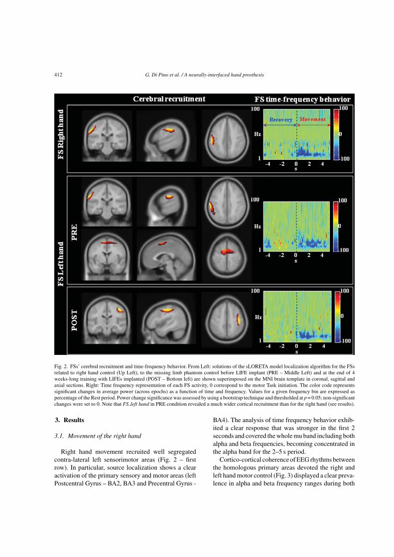
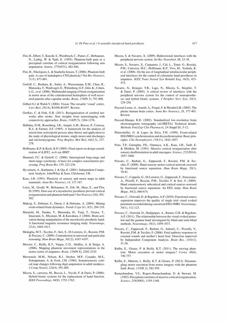
Cortico-cortical coherence of EEG rhythms betweenthe homologous primary areas devoted the right andleft hand motor control (Fig. 3) displayed a clear preva-lence in alpha and beta frequency ranges during both
G. Di Pino et al. / A neurally-interfaced hand prosthesis 413
Fig. 3. Homologous hand areas’ coherence. Top: solutions of thesLORETA model localization algorithm (coronal and sagittal view)for the cerebral recruited hand areas (see methods) superimposedon the MNI brain template. Bottom: Cortico-cortical coherencebetween the cerebral recruited areas during recovery (blue line) andmovement (red line) periods. Horizontal line indicates the confidencelimit (see Materials and Methods section). (Colours are visible in theonline version of the article; http://dx.doi.org/10.3233/RNN-2012-120224)
movement and recovery periods. A reduction in delta-theta and an enhancement in beta-gamma bands wereobserved during movement with respect to recovery(Table 1, Fig. 3).
3.2. Movement of the left cybernetic handprosthesis
Delivery of motor commands for the phantom of theleft amputated limb before the LIFEs implant recruitedareas in the ipsilateral primary sensory and motor areas(left Postcentral Gyrus – BA2, BA3 and PrecentralGyrus - BA4, Fig. 2 – second row top) and bilateral pre-motor and supplementary motor cortex (left and rightBA6, Fig. 2 – second row bottom). At this stage no con-tralateral primary motor cortex activity was found. Theanalysis of time frequency behavior evidenced activ-ity in alpha and beta bands lasting for the whole taskduration with no time-specificity.
The cerebral recruitment during the intent to movethe phantom of the left amputated limb changedmarkedly after the four-weeks of prosthesis motorcontrol training with implanted LIFEs. Cortical recruit-ment became almost symmetrical with respect to righthand movements, with selective involvement of contra-lateral sensorimotor cortex (right Postcentral Gyrus –BA2, BA3 and Precentral Gyrus - BA4, Fig. 2 – thirdrow). The time frequency behaviour regained evolv-ing properties similar to those for right hand control:stronger in the first 2 seconds involving the whole alphaand beta frequencies, while weaker and more concen-trated in alpha band in the 2–5 s period (Fig. 2 – thirdrow right).
Before LIFE implant, the cortico-cortical coherencebetween the hemispheric homologous primary sen-sorimotor cortices (Fig. 3) was present only at lowfrequencies in recovery periods. During movement areduction in all frequency bands occurred, except inthe delta band where coherence was enhanced. Afterfour weeks of training with LIFE implants communi-cating with the cybernetic prosthesis, a similar patternto right hand movement was observed: although atlower levels, inter-hemispheric communication pre-vailed in delta and beta bands during recovery periods.During movement, a delta-theta reduction and a betaband increase relative to recovery periods were found(Table 1, Fig. 3).
The reduction in delta and enhancement in beta andgamma bands during active movement is evident both
414 G. Di Pino et al. / A neurally-interfaced hand prosthesis
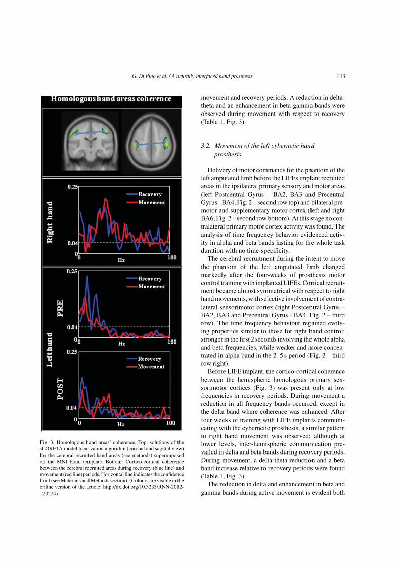
Table 1
Homologous hand areas’ coherence. Spectral coherence between FS activity of hemispheric primary regions devoted to the hand controlintegrated in delta [1, 4] Hz, theta [5, 7], alpha [8, 12], beta [13, 32] and gamma [33, 48] Hz bands normalized by the frequency bin numbersduring motor command dispatched to each hand (Movement) and during intermingled resting periods (Recovery). The difference between the
two conditions is also shown (Recovery-Movement, Diff). Band coherences are calculated integrating only significant values (p > 0.04)
Delta Theta Alpha Beta Gamma
Right hand Recovery 0.1446 0.0530 0.0557 0.1221 0.0597Movement 0.0732 0.0340 0.0476 0.1401 0.0940Diff 0.0714 0.0190 0.0081 −0.0180 −0.0343
Left handPre Recovery 0.0692 0.1474 0.1211 0.0778 0.0000
Movement 0.0984 0.1174 0.0705 0.0293 0.0000Diff −0.0292 0.0300 0.0506 0.0485 0.0000
Post Recovery 0.1115 0.0877 0.0227 0.0154 0.0000Movement 0.0732 0.0260 0.0000 0.0290 0.0076Diff 0.0383 0.0617 0.0227 −0.0136 −0.0076
for the right hand and for the POST-session of thehand cybernetic prosthesis, while an opposite patternwas observed in the pre-training session for the leftphantom hand.
4. Discussion
4.1. Brain plasticity due to neutrally-interfacedcybernetic hand prosthesis control
The possibility of inducing or redirecting theorganization of cortical and subcortical functionaltopography has been described with fMRI studies, bothin acute (Maruishi et al., 2004) and in chronic EMG-driven prosthesis use (Lotze et al., 1999).
The neurophysiological processes which take placewhen interacting with the external world througha BMI, such as tuning of neuronal firing towardsthe constraints of movement, and Hebbian plastic-ity of neuronal networks that map the movements incortex, are the same that underlie motor neuroreha-bilitation procedures (Dobkin, 2007). Thus it is ofprimary importance to better understand whether ornot an interface is able to cause appropriate neuralchanges that benefit restorative plasticity. This processis facilitated by the ability of the interface to closethe user-action-environment-perception-user controlloop by feeding back tactile and proprioceptive sen-sations. Furthermore, the reduction of cognitive loadrequired by the user to control the machine, withoutforcing artificial control strategies, can be achievedonly through a natural interface with the muscles ornerves that originally were devoted to the lost limb.Hence, the use of a prosthesis directly interfaced
with the nervous system should better compensatefor lost function and avoid aberrant CNS reorganiza-tion. Nevertheless, to our knowledge, there is scantdata in the literature concerning the reshaping ofcerebral pathways by the use of prostheses directlycoupled with the nervous system. Training for robotichand control and for sensory perception via a multi-contact peripheral nerve neural interface producednormalization of the somatosensory and motor corticalorganization that had previously been rearranged fol-lowing amputation-induced neurodegeneration. Ourgroup recently described a significant reduction ofthe cortical area from which motor evoked potentialscould be elicited in the spared muscles of affectedlimb (Rossini et al., 2010), and normalization of EEGdesynchronization during movement preparation in thesensorimotor areas (Tombini et al., 2012). Moreover,the four weeks of training induced a clinical improve-ment of about 30% of PLP, an epiphenomenon of theredirection of aberrant cortical plasticity, assessed withopen answer questionnaires and clinical pain scales(short form McGill – range 0–45- from 18 to 11,Present Pain Inventory – range 0–5- from 3 to 2, VisualAnalogue Scale – range 0–100%- from 38% to 23%),and a progressive return towards a more physiologicalperception of the phantom hand. The modification ofcortical plasticity occurred over the same time-courseas the improvement in prosthesis control performance(Rossini et al., 2010).
The present paper provides new insights on howintensive training involving the restoration of motoroutput and multisensory feedback (i.e., stimulationthrough a neural interface together with visual feed-back) can induce adaptive plastic changes in theRolandic cortices of chronic amputees. The study of
G. Di Pino et al. / A neurally-interfaced hand prosthesis 415
cortical recruitment and neuronal activity patterns inour patient showed how, before training, attempting tomove the phantom of the missing limb involved medialfrontal gyrus and superior frontal gyrus, without acti-vation of directly connected contralateral primarysensorimotor cortices. Such premotor and supple-mentary motor cortices are crucial for movementplanning and coordination. It can be hypothesizedthat the absence of central-peripheral communica-tion inhibits physiological engagement of executionstructures, concentrating neural activity in preparatoryterritories (Bestmann et al., 2006; Diers et al., 2010;Raffin et al., 2012). The presence of intra-fascicularelectrodes allowed new signals to be delivered throughperipheral nerves towards the cortex and produced anintensive exchange of sensori-motor afferent and effer-ent inputs/outputs. Four weeks of training with suchimplanted electrodes led to a new functional recruit-ment of sensorimotor areas devoted to hand control,i.e., contralateral primary motor and sensory cortices(Broadmann’s areas 2, 3 and 4). As expected by pre-vious literature (Flor et al., 2001; Lotze et al., 1999),a physiologically-oriented plastic reorganization cor-responded to the amelioration of clinical symptoms ofPLP.
4.2. Functional connectivity between hemispherichomologous areas characterizes motorcontrol
The improvement in cybernetic hand prosthesis con-trol has been showed to be coupled with reorganizationin sensorimotor cortical regions activity (Rossini et al.,2010; Tombini et al., 2012). In parallel with regionalactivation properties, it is well known that the motorabilities depend on intra- and inter-network functionalconnectivity (Hummel and Gerloff, 2006), and areanatomically related to the amount of transcallosalconnections (Stancak et al., 2002). Unilateral mid-dle cerebral artery territory stroke is a paradigmaticmodel of the unbalancing of primary sensorimotorareas activities (Rossini et al., 2003) together withan alteration of the associated network connectivity(Grefkes and Fink, 2011). Such connectivity has beenobserved to be reduced in acute severe stroke both inanimal models (van Meer et al., 2010) and in humans(Carter et al., 2010), while its recovery pairs with aprogressive improvement of motor control. Observinglongitudinally ten patients along one year and analyz-ing fMRI BOLD signals during resting state, Wang et
colleagues (2010) showed that the inter-hemisphericcoupling between homologous primary motor corticesrecovered in parallel with motor skills (Wang et al.,2010).
Neuronal oscillations of EEG in specific fre-quency bands play a specific functional role (deltatheta, alpha beta gamma): an increase of low betainter-hemispheric coherence between sensory-motorregions was observed in stroke patients reacquiringparetic hand control (Gerloff et al., 2006).
In our patient we cannot describe a global repro-ducible trend of spectral modification in single bands,but more a global behavior of spectral profile betweentask-oriented coherence and recovery (reduction intheta-delta band and enhancement in beta and gammabands), that - in parallel with improvement of prosthe-sis control performance and PLP amelioration- becamesimilar to the one observed with the contralateral unaf-fected hand and thus probably more physiological.
4.3. FSS allows functional connectivity estimationboth during movement and in resting state
FSS allows the analysis of the identified cerebralsources in experimental conditions that differ fromthose in which they are identified and thus enabledus to track their activity even while the subject was atrest.
In the present study brain connectivity between thecortical areas devoted to the intact and missing handmovement was investigated during the delivering ofhand movement command and in resting state. Recentevidence indicates that networks devoted to specificfunctional domains maintain their typical functionalproperties also in the resting state (Deco and Corbetta,2011), so resting state features have been suggestedto be useful for assessing the health of brain networks(Carter et al., 2010).
The analysis of coherence during the resting stateallows removal of different task-related neural engage-ments (i.e., attention and monitoring in planning andexecuting the motor task) that are not comparablebetween patients and controls and between hemi-spheres in amputees. In this respect the use of FSSto evaluate functional connectivity in the resting statefor specific regions devoted to the hand sensorimotorcontrol independent of their location was extremelyhelpful.
Since functional sources are identified on thebasis of their specific behavior during the task of
416 G. Di Pino et al. / A neurally-interfaced hand prosthesis
interest and not of their topography, they can be suit-ably investigated even if the sensorimotor control areashave been displaced due to plastic reorganizations.Interestingly, we found that training to control theLIFE connected cybernetic prosthesis modulated theinter-hemispheric connectivity not only during move-ment, but also in the resting state. Conceivably inour case, rearrangements of excitatory-inhibitory localand interhemispheric balances subtend the modula-tion of functional connectivity between affected andunaffected sensory hand areas.
The involvement of beta and gamma-band band con-nectivity in sensory motor integration plays a role forthe dynamic construction of neuronal networks relatedto sensorimotor integration (Engel and Fries, 2010;Tecchio et al., 2007a; Tecchio et al., 2008; Tombiniet al., 2009). A further advantage of the FSS methodis to better resolve such high frequency band rhythmsin EEG scalp activity.
5. Conclusion
Taken together, the results of this study confirm thehypothesis that neural interfaces, such as the bidirec-tional interface we adopted, being able to assure a highlevel of interaction among the user and the cyberneticprosthesis and to transmit back sensory input from theperiphery, are optimal candidates for hand prosthesiscontrol. This is due to their ability to establish commu-nication channels needed for a natural control of theeffector designed for functional replacement. Further-more neural interfaces recreate the connection with theenvironment that allow, through a continual exercisewith feedback and reward, to redirect cortical orphanareas toward restorative neuroplasticity. Part of thisreorganization involves bi-hemispheric networks thatregain a more physiological communication necessaryfor motor control.
Acknowledgments
This study was partially supported by the Commis-sion of the European Community Seventh FrameworkProgram under grant agreement no 200859, projectMEGMRI, and TIME (Contract No.: FP7-ICT-224012– Transverse, Intrafascicular Multichannel Electrodesystem for induction of sensation and treatment ofphantom limb pain in amputees) and by the Royal
Society International Joint Project – 2010/R1: ‘TheKey Movement Controllers: an EEG/fMRI study ofthe hand network dynamics [KeyMoCo]’. The authorsdeeply thank the volunteer subject of this study, whodonated his time and consented to participate in allphases of the present protocol. The authors are alsograteful to Andrew Jackson for his valuable commentsand for editing the revised version of this manuscript.
References
Barbati, G., Porcaro, C., Hadjipapas, A., Adjamian, P., Pizzella, V.,Romani, G.L., Seri, S., Tecchio, F. & Barnes, G.R. (2008).Functional source separation applied to induced visual gammaactivity. Hum Brain Mapp, 29(2), 131-141.
Barbati, G., Porcaro, C., Zappasodi, F., Rossini, P.M. & Tecchio,F. (2004). Optimization of an independent component analysisapproach for artifact identification and removal in magnetoen-cephalographic signals. Clin Neurophysiol, 115(5), 1220-1232.
Barbati, G., Sigismondi, R., Zappasodi, F., Porcaro, C., Graziadio,S., Valente, G., Balsi, M., Rossini, P.M. & Tecchio, F. (2006).Functional source separation from magnetoencephalographicsignals. Hum Brain Mapp, 27(12), 925-934.
Bestmann, S., Oliviero, A., Voss, M., Dechent, P., Lopez-Dolado, E.,Driver, J. & Baudewig, J. (2006). Cortical correlates of TMS-induced phantom hand movements revealed with concurrentTMS-fMRI. Neuropsychologia, 44(14), 2959-2971.
Carrozza, M.C., Cappiello, G., Micera, S., Edin, B.B., Beccai, L. &Cipriani, C. (2006). Design of a cybernetic hand for perceptionand action. Biol Cybern, 95(6), 629-644.
Carter, A.R., Astafiev, S.V., Lang, C.E., Connor, L.T., Rengachary, J.,Strube, M.J., Pope, D.L., Shulman, G.L. & Corbetta, M. (2010).Resting interhemispheric functional magnetic resonance imag-ing connectivity predicts performance after stroke. Ann Neurol,67(3), 365-375.
Cichocki, A. & Amari, S.I. (2002). Adaptive Blind Signal and ImageProcessing. JohnWiley & Sons, Chichester, UK.
Deco, G. & Corbetta, M. (2011). The dynamical balance of the brainat rest. Neuroscientist, 17(1), 107-123.
Di Pino, G., Guglielmelli, E. & Rossini, P.M. (2009). Neuroplasticityin amputees: Main implications on bidirectional interfacing ofcybernetic hand prostheses. Prog Neurobiol, 88(2), 114-126.
Diers, M., Christmann, C., Koeppe, C., Ruf, M. & Flor, H. (2010).Mirrored, imagined and executed movements differentiallyactivate sensorimotor cortex in amputees with and withoutphantom limb pain. Pain, 149(2), 296-304.
Dobkin, B.H. (2007). Brain-computer interface technology as a toolto augment plasticity and outcomes for neurological rehabili-tation. J Physiol, 579(Pt 3), 637-642.
Engel, A.K. & Fries, P. (2010). Beta-band oscillations–signalling thestatus quo? Curr Opin Neurobiol, 20(2), 156-165.
Flor, H., Denke, C., Schaefer, M. & Grusser, S. (2001). Effect ofsensory discrimination training on cortical reorganisation andphantom limb pain. Lancet, 357(9270), 1763-1764.
G. Di Pino et al. / A neurally-interfaced hand prosthesis 417
Flor, H., Elbert, T., Knecht, S., Wienbruch, C., Pantev, C., Birbaumer,N., Larbig, W. & Taub, E. (1995). Phantom-limb pain as aperceptual correlate of cortical reorganization following armamputation. Nature, 375(6531), 482-484.
Flor, H., Nikolajsen, L. & Staehelin Jensen, T. (2006). Phantom limbpain: A case of maladaptive CNS plasticity? Nat Rev Neurosci,7(11), 873-881.
Gerloff, C., Bushara, K., Sailer, A., Wassermann, E.M., Chen, R.,Matsuoka, T., Waldvogel, D., Wittenberg, G.F., Ishii, K., Cohen,L.G., et al. (2006). Multimodal imaging of brain reorganizationin motor areas of the contralesional hemisphere of well recov-ered patients after capsular stroke. Brain, 129(Pt 3), 791-808.
Gilbert S.J. & Walsh V. (2004). Vision: The versatile ‘visual’ cortex.Curr Biol, 29(14), R1056-R1057. Review.
Grefkes, C. & Fink, G.R. (2011). Reorganization of cerebral net-works after stroke: New insights from neuroimaging withconnectivity approaches. Brain, 134(Pt 5), 1264-1276.
Halliday, D.M., Rosenberg, J.R., Amjad, A.M., Breeze, P., Conway,B.A. & Farmer, S.F. (1995). A framework for the analysis ofmixed time series/point process data–theory and application tothe study of physiological tremor, single motor unit dischargesand electromyograms. Prog Biophys Mol Biol, 64(2-3), 237-278.
Hoffmann, K.P. & Kock, K.P. (2005). Final report on design consid-eration of tLIFE2. tech rep IBMT.
Hummel, F.C. & Gerloff, C. (2006). Interregional long-range andshort-range synchrony: A basis for complex sensorimotor pro-cessing. Prog Brain Res 159, 223-236.
Hyvarinen, A., Karhunen, J. & Oja, E. (2001). Independent Compo-nent Analysis. JohnWiley & Sons, Chichester, UK.
Kaas, J.H. (1991). Plasticity of sensory and motor maps in adultmammals. Annu Rev Neurosci, 14, 137-167.
Lotze, M., Grodd, W., Birbaumer, N., Erb, M., Huse, E., and Flor,H (1999). Does use of a myoelectric prosthesis prevent corticalreorganization and phantom limb pain? Nat Neurosci 2(6), 501-502.
Makeig, S., Debener, S., Onton, J. & Delorme, A. (2004). Miningevent-related brain dynamics. Trends Cogn Sci, 8(5), 204-210.
Maruishi, M., Tanaka, Y., Muranaka, H., Tsuji, T., Ozawa, Y.,Imaizumi, S., Miyatani, M. & Kawahara, J. (2004). Brain acti-vation during manipulation of the myoelectric prosthetic hand:A functional magnetic resonance imaging study. Neuroimage,21(4), 1604-1611.
Medaglia, M.T., Tecchio, F., Seri, S., Di Lorenzo, G., Rossini, P.M.& Porcaro, C. (2009). Contradiction in universal and particularreasoning. Hum Brain Mapp, 30(12), 4187-4197.
Mercier, C., Reilly, K.T., Vargas, C.D., Aballea, A. & Sirigu, A.(2006). Mapping phantom movement representations in themotor cortex of amputees. Brain, 129(Pt 8), 2202-2210.
Merzenich, M.M., Nelson, R.J., Stryker, M.P., Cynader, M.S.,Schoppmann, A. & Zook, J.M. (1984). Somatosensory corti-cal map changes following digit amputation in adult monkeys.J Comp Neurol, 224(4), 591-605.
Micera, S., carrozza, M., Beccai, L., Vecchi, F. & Dario, P. (2006).Hybrid bionic systems for the replacement of hand function.IEEE Proceedings, 94(9), 1752-1762.
Micera, S. & Navarro, X. (2009). Bidirectional interfaces with theperipheral nervous system. Int Rev Neurobiol, 86, 23-38.
Micera, S., Navarro, X., Carpaneto, J., Citi, L., Tonet, O., Rossini,P.M., Carrozza, M.C., Hoffmann, K.P., Vivo, M., Yoshida, K.et al. (2008). On the use of longitudinal intrafascicular periph-eral interfaces for the control of cybernetic hand prostheses inamputees. IEEE Trans Neural Syst Rehabil Eng, 16(5), 453-472.
Navarro, X., Krueger, T.B., Lago, N., Micera, S., Stieglitz, T.& Dario, P. (2005). A critical review of interfaces with theperipheral nervous system for the control of neuroprosthe-ses and hybrid bionic systems. J Peripher Nerv Syst, 10(3),229-258.
Pascual-Leone, A., Amedi, A., Fregni, F. & Merabet LB. (2005). Theplastic human brain cortex. Annu Rev Neurosci, 28, 377-401.Review.
Pascual-Marqui, R.D. (2002). Standardized low-resolution brainelectromagnetic tomography (sLORETA): Technical details.Methods Find Exp Clin Pharmacol, 24 (Suppl D), 5-12.
Pfurtscheller, G. & Lopes da Silva, F.H. (1999). Event-relatedEEG/MEG synchronization and desynchronization: Basic prin-ciples. Clin Neurophysiol, 110(11), 1842-1857.
Pons, T.P., Garraghty, P.E., Ommaya, A.K., Kaas, J.H., Taub, E.& Mishkin, M. (1991). Massive cortical reorganization aftersensory deafferentation in adult macaques. Science, 252(5014),1857-1860.
Porcaro, C., Barbati, G., Zappasodi, F., Rossini, P.M. & Tec-chio, F. (2008). Hand sensory-motor cortical network assessedby functional source separation. Hum Brain Mapp, 29(1),70-81.
Porcaro, C., Coppola, G., Di Lorenzo, G., Zappasodi, F., Siracusano,A., Pierelli, F., Rossini, P.M., Tecchio, F. & Seri, S. (2009).Hand somatosensory subcortical and cortical sources assessedby functional source separation: An EEG study. Hum BrainMapp, 30(2), 660-674.
Porcaro, C., Ostwald, D. & Bagshaw, A.P. (2010). Functional sourceseparation improves the quality of single trial visual evokedpotentials recorded during concurrent EEG-fMRI. Neuroimage,50(1), 112-123.
Porcaro, C., Ostwald, D., Hadjipapas, A., Barnes, G.R. & Bagshaw,A.P. (2011). The relationship between the visual evoked poten-tial and the gamma band investigated by blind and semi-blindmethods. Neuroimage, 56(3), 1059-1071.
Porcaro, C., Zappasodi, F., Barbati, G., Salustri, C., Pizzella, V.,Rossini, P.M. & Tecchio, F. (2006). Fetal auditory responses toexternal sounds and mother’s heart beat: Detection improvedby Independent Component Analysis. Brain Res, 1101(1),51-58.
Raffin, E., Giraux, P. & Reilly, K.T. (2011). The moving phan-tom: Motor execution or motor imagery? Cortex 48(6),746-757.
Raffin, E., Mattout, J., Reilly, K.T. & Giraux, P. (2012). Disentan-gling motor execution from motor imagery with the phantomlimb. Brain, 135(Pt 2), 582-595.
Ramachandran, V.S., Rogers-Ramachandran, D. & Stewart, M.(1992). Perceptual correlates of massive cortical reorganization.Science, 258(5085), 1159-1160.
418 G. Di Pino et al. / A neurally-interfaced hand prosthesis
Reilly, K.T., Mercier, C., Schieber, M.H. & Sirigu, A. (2006). Per-sistent hand motor commands in the amputees’ brain. Brain,129(Pt 8), 2211-2223.
Rossini, P.M., Calautti, C., Pauri, F. & Baron, J.C. (2003). Post-stroke plastic reorganisation in the adult brain. Lancet Neurol,2(8), 493-502.
Rossini, P.M., Micera, S., Benvenuto, A., Carpaneto, J., Cavallo, G.,Citi, L., Cipriani, C., Denaro, L., Denaro, V., Di Pino, G., et al.(2010). Double nerve intraneural interface implant on a humanamputee for robotic hand control. Clin Neurophysiol, 121(5),777-783.
Sadato, N. (2005). How the blind “see” Braille: Lessons fromfunctional magnetic resonance imaging. Neuroscientist, 11,577-582. Review.
Stancak, A., Lucking, C.H. & Kristeva-Feige, R. (2002). The sizeof corpus callosum and functional connectivities of corticalregions in finger and shoulder movements. Brain Res CognBrain Res, 13(1), 61-74.
Tecchio, F., Graziadio, S., Barbati, G., Sigismondi, R., Zappasodi,F., Porcaro, C., Valente, G., Balsi, M. & Rossini, P.M. (2007a).Somatosensory dynamic gamma-band synchrony: A neuralcode of sensorimotor dexterity. Neuroimage, 35(1), 185-193.
Tecchio, F., Porcaro, C., Barbati, G. & Zappasodi, F. (2007b).Functional source separation and hand cortical representationfor a brain-computer interface feature extraction. J Physiol,580(Pt 3), 703-721.
Tecchio, F., Zappasodi, F., Porcaro, C., Barbati, G., Assenza,G., Salustri, C. & Rossini, P.M. (2008). High-gamma band
activity of primary hand cortical areas: A sensorimotor feed-back efficiency index. Neuroimage, 40(1), 256-264.
Tombini, M., Rigosa, J., Zappasodi, F., Porcaro, C., Citi, L.,Carpaneto, J., Rossini, P.M. & Micera, S. (2012). Combinedanalysis of cortical (EEG) and nerve stump signals improvesrobotic hand control. Neurorehabil Neural Repair, 26(3), 275-281.
Tombini, M., Zappasodi, F., Zollo, L., Pellegrino, G., Cavallo, G.,Tecchio, F., Guglielmelli, E. & Rossini, P.M. (2009). Brainactivity preceding a 2D manual catching task. Neuroimage,47(4), 1735-1746.
van Meer, M.P., van der Marel, K., Wang, K., Otte, W.M., ElBouazati, S., Roeling, T.A., Viergever, M.A., Berkelbach vander Sprenkel, J.W. & Dijkhuizen, R.M. (2010). Recovery ofsensorimotor function after experimental stroke correlates withrestoration of resting-state interhemispheric functional connec-tivity. J Neurosci, 30(11), 3964-3972.
Wang, L., Yu, C., Chen, H., Qin, W., He, Y., Fan, F., Zhang, Y., Wang,M., Li, K., Zang, Y., et al. (2010). Dynamic functional reorga-nization of the motor execution network after stroke. Brain,133(Pt 4), 1224-1238.
Zollo, L., Roccella, S., Guglielmelli, E., Carrozza, M.C. & Dario,P. (2007). Biomechatronic design and control of an anthropo-morphic artificial hand for prosthetic and robotic applications.Paper presented at: IEEE/ASME Transactions on Mechatron-ics, 12(4), 418-429.
Related Documents