EMBO open A multifunctional serine protease primes the malaria parasite for red blood cell invasion This is an open-access article distributed under the terms of the Creative Commons Attribution License, which permits distribution, and reproduction in any medium, provided the original author and source are credited. This license does not permit commercial exploitation or the creation of derivative works without specific permission. Konstantinos Koussis 1 , Chrislaine Withers-Martinez 1 , Sharon Yeoh 1 , Matthew Child 1 , Fiona Hackett 1 , Ellen Knuepfer 1 , Luiz Juliano 2 , Ute Woehlbier 3 , Hermann Bujard 3 and Michael J Blackman 1, * 1 Division of Parasitology, National Institute for Medical Research, London, UK, 2 Department of Biophysics, Escola Paulista de Medicina, Universidade Federal de Sa ˜o Paulo, Sa ˜o Paulo, Brazil and 3 Zentrum fuer Molekulare Biologie Heidelberg (ZMBH), Universitaet Heidelberg, Heidelberg, Germany The malaria parasite Plasmodium falciparum replicates within an intraerythrocytic parasitophorous vacuole (PV). Rupture of the host cell allows release (egress) of daughter merozoites, which invade fresh erythrocytes. We previously showed that a subtilisin-like protease called PfSUB1 regulates egress by being discharged into the PV in the final stages of merozoite development to proteoly- tically modify the SERA family of papain-like proteins. Here, we report that PfSUB1 has a further role in ‘priming’ the merozoite prior to invasion. The major protein com- plex on the merozoite surface comprises three proteins called merozoite surface protein 1 (MSP1), MSP6 and MSP7. We show that just before egress, all undergo pro- teolytic maturation by PfSUB1. Inhibition of PfSUB1 activ- ity results in the accumulation of unprocessed MSPs on the merozoite surface, and erythrocyte invasion is signifi- cantly reduced. We propose that PfSUB1 is a multifunc- tional processing protease with an essential role in both egress of the malaria merozoite and remodelling of its surface in preparation for erythrocyte invasion. The EMBO Journal (2009) 28, 725–735. doi:10.1038/ emboj.2009.22; Published online 12 February 2009 Subject Categories: microbiology & pathogens; molecular biology of disease Keywords: malaria; merozoite; MSP; PfSUB1; Plasmodium falciparum Introduction Transmission of Plasmodium spp., the protozoan parasite that causes malaria, occurs following the bite of an infected Anopheline mosquito. Injected sporozoites migrate to the liver where they invade hepatocytes and replicate within a parasitophorous vacuole (PV) to yield a liver-stage schizont containing several thousand merozoites per cell. In a process called egress, the schizont then ruptures to release the merozoites, which enter the bloodstream and invade erythro- cytes. This initiates the asexual erythrocytic cycle, responsi- ble for the clinical manifestations of the disease. At each round of subsequent intraerythrocytic growth, further mitotic replication takes place, also inside a PV, producing 16–32 daughter merozoites, which egress to invade fresh erythro- cytes and perpetuate the cycle. Developing malaria merozoites, including those of the most dangerous form, Plasmodium falciparum, express a number of merozoite surface proteins (MSPs). MSP1 is a major malaria vaccine candidate antigen (reviewed by Vekemans and Ballou, 2008), and is indispensable in the parasite life cycle (O’Donnell et al, 2000; Drew et al, 2004). MSP1 is synthesized as an B195-kDa glycosyl phosphatidy- linositol-anchored precursor that assembles as a complex with two peripheral membrane proteins called MSP6 and MSP7 (Holder et al, 1985; Stafford et al, 1996; Pachebat et al, 2001; Trucco et al, 2001; Kauth et al, 2003, 2006). The MSP1/ 6/7 complex is abundant, uniformly coats the merozoite surface and is involved in the receptor-mediated interactions that initiate erythrocyte invasion (Su et al, 1993; Goel et al, 2003; Li et al, 2004). Just prior to egress, all three MSPs undergo a series of precise ‘primary’ proteolytic cleavage events (Freeman and Holder, 1983; Lyon et al, 1986, 1987; Holder et al, 1987; McBride and Heidrich, 1987; Stafford et al, 1994; Pachebat et al, 2007). The major cleavage products remain non-covalently associated on the surface of the re- leased merozoite, until the complex is finally shed at the point of erythrocyte invasion in an essential secondary processing step by the action of a membrane-bound parasite protease called PfSUB2 (Harris et al, 2005). The specific nature of the primary proteolysis and the positional conser- vation of the cleavage sites in MSP1 orthologues across the Plasmodium genus (reviewed by Blackman, 2000) suggest that primary processing is important for the function of the MSP1/6/7 complex and for merozoite viability. However the protease(s) responsible for primary processing is unknown. Parasite protease activity is required for blood-stage egress in P. falciparum (Delplace et al, 1988; Salmon et al, 2001; Wickham et al, 2003; Gelhaus et al, 2005), but the mechan- isms by which egress is regulated are only just beginning to become clear. We recently showed that, just prior to egress, an essential subtilisin-like serine protease called PfSUB1 is discharged from specialized merozoite organelles called exo- nemes into the PV space (Yeoh et al, 2007). There, PfSUB1 mediates the proteolytic maturation of members of a family of abundant, papain-like putative proteases called SERA, Received: 28 October 2008; accepted: 14 January 2009; published online: 12 February 2009 *Corresponding author. Division of Parasitology, National Institute for Medical Research, Mill Hill, London NW7 1AA, UK. Tel.: þ 44 208 816 2127; Fax: þ 44 208 816 2730; E-mail: [email protected] The EMBO Journal (2009) 28, 725–735 | & 2009 European Molecular Biology Organization | Some Rights Reserved 0261-4189/09 www.embojournal.org & 2009 European Molecular Biology Organization The EMBO Journal VOL 28 | NO 6 | 2009 EMBO THE EMBO JOURNAL THE EMBO JOURNAL 725

Welcome message from author
This document is posted to help you gain knowledge. Please leave a comment to let me know what you think about it! Share it to your friends and learn new things together.
Transcript
EMBOopen
A multifunctional serine protease primes themalaria parasite for red blood cell invasion
This is an open-access article distributed under the terms of the Creative Commons Attribution License, which permitsdistribution,andreproduction inanymedium,provided theoriginalauthorandsourceare credited.This licensedoesnotpermit commercial exploitation or the creation of derivative works without specific permission.
Konstantinos Koussis1,Chrislaine Withers-Martinez1,Sharon Yeoh1, Matthew Child1,Fiona Hackett1, Ellen Knuepfer1,Luiz Juliano2, Ute Woehlbier3,Hermann Bujard3 andMichael J Blackman1,*1Division of Parasitology, National Institute for Medical Research,London, UK, 2Department of Biophysics, Escola Paulista de Medicina,Universidade Federal de Sao Paulo, Sao Paulo, Brazil and 3Zentrum fuerMolekulare Biologie Heidelberg (ZMBH), Universitaet Heidelberg,Heidelberg, Germany
The malaria parasite Plasmodium falciparum replicates
within an intraerythrocytic parasitophorous vacuole
(PV). Rupture of the host cell allows release (egress) of
daughter merozoites, which invade fresh erythrocytes. We
previously showed that a subtilisin-like protease called
PfSUB1 regulates egress by being discharged into the PV
in the final stages of merozoite development to proteoly-
tically modify the SERA family of papain-like proteins.
Here, we report that PfSUB1 has a further role in ‘priming’
the merozoite prior to invasion. The major protein com-
plex on the merozoite surface comprises three proteins
called merozoite surface protein 1 (MSP1), MSP6 and
MSP7. We show that just before egress, all undergo pro-
teolytic maturation by PfSUB1. Inhibition of PfSUB1 activ-
ity results in the accumulation of unprocessed MSPs on
the merozoite surface, and erythrocyte invasion is signifi-
cantly reduced. We propose that PfSUB1 is a multifunc-
tional processing protease with an essential role in both
egress of the malaria merozoite and remodelling of its
surface in preparation for erythrocyte invasion.
The EMBO Journal (2009) 28, 725–735. doi:10.1038/
emboj.2009.22; Published online 12 February 2009
Subject Categories: microbiology & pathogens; molecular
biology of disease
Keywords: malaria; merozoite; MSP; PfSUB1; Plasmodium
falciparum
Introduction
Transmission of Plasmodium spp., the protozoan parasite
that causes malaria, occurs following the bite of an infected
Anopheline mosquito. Injected sporozoites migrate to the
liver where they invade hepatocytes and replicate within a
parasitophorous vacuole (PV) to yield a liver-stage schizont
containing several thousand merozoites per cell. In a process
called egress, the schizont then ruptures to release the
merozoites, which enter the bloodstream and invade erythro-
cytes. This initiates the asexual erythrocytic cycle, responsi-
ble for the clinical manifestations of the disease. At each
round of subsequent intraerythrocytic growth, further mitotic
replication takes place, also inside a PV, producing 16–32
daughter merozoites, which egress to invade fresh erythro-
cytes and perpetuate the cycle.
Developing malaria merozoites, including those of the
most dangerous form, Plasmodium falciparum, express a
number of merozoite surface proteins (MSPs). MSP1 is a
major malaria vaccine candidate antigen (reviewed by
Vekemans and Ballou, 2008), and is indispensable in the
parasite life cycle (O’Donnell et al, 2000; Drew et al, 2004).
MSP1 is synthesized as an B195-kDa glycosyl phosphatidy-
linositol-anchored precursor that assembles as a complex
with two peripheral membrane proteins called MSP6 and
MSP7 (Holder et al, 1985; Stafford et al, 1996; Pachebat et al,
2001; Trucco et al, 2001; Kauth et al, 2003, 2006). The MSP1/
6/7 complex is abundant, uniformly coats the merozoite
surface and is involved in the receptor-mediated interactions
that initiate erythrocyte invasion (Su et al, 1993; Goel et al,
2003; Li et al, 2004). Just prior to egress, all three MSPs
undergo a series of precise ‘primary’ proteolytic cleavage
events (Freeman and Holder, 1983; Lyon et al, 1986, 1987;
Holder et al, 1987; McBride and Heidrich, 1987; Stafford et al,
1994; Pachebat et al, 2007). The major cleavage products
remain non-covalently associated on the surface of the re-
leased merozoite, until the complex is finally shed at the
point of erythrocyte invasion in an essential secondary
processing step by the action of a membrane-bound parasite
protease called PfSUB2 (Harris et al, 2005). The specific
nature of the primary proteolysis and the positional conser-
vation of the cleavage sites in MSP1 orthologues across the
Plasmodium genus (reviewed by Blackman, 2000) suggest
that primary processing is important for the function of the
MSP1/6/7 complex and for merozoite viability. However the
protease(s) responsible for primary processing is unknown.
Parasite protease activity is required for blood-stage egress
in P. falciparum (Delplace et al, 1988; Salmon et al, 2001;
Wickham et al, 2003; Gelhaus et al, 2005), but the mechan-
isms by which egress is regulated are only just beginning to
become clear. We recently showed that, just prior to egress,
an essential subtilisin-like serine protease called PfSUB1 is
discharged from specialized merozoite organelles called exo-
nemes into the PV space (Yeoh et al, 2007). There, PfSUB1
mediates the proteolytic maturation of members of a family
of abundant, papain-like putative proteases called SERA,Received: 28 October 2008; accepted: 14 January 2009; publishedonline: 12 February 2009
*Corresponding author. Division of Parasitology, National Institute forMedical Research, Mill Hill, London NW7 1AA, UK.Tel.: þ 44 208 816 2127; Fax: þ 44 208 816 2730;E-mail: [email protected]
The EMBO Journal (2009) 28, 725–735 | & 2009 European Molecular Biology Organization | Some Rights Reserved 0261-4189/09
www.embojournal.org
&2009 European Molecular Biology Organization The EMBO Journal VOL 28 | NO 6 | 2009
EMBO
THE
EMBOJOURNAL
THE
EMBOJOURNAL
725
previously implicated in egress (Pang et al, 1999; Aly and
Matuschewski, 2005). Selective inhibition of PfSUB1, with
either a natural compound called MRT12113 (Yeoh et al,
2007) or covalently modifying chloroisocoumarins (Arastu-
Kapur et al, 2008) prevents SERA maturation and blocks
egress. This indicates a role for PfSUB1 in triggering egress,
probably through activation of the SERA enzymes. We also
noticed in our study that, at relatively low concentrations of
MRT12113, egress occurred but the released merozoites were
defective in invasion (Yeoh et al, 2007). This implied an
additional role for PfSUB1 in merozoite maturation.
Here, we report that PfSUB1 is indeed required for the
development of invasive merozoites. We show that, upon its
release into the PV, PfSUB1 directly mediates the primary
proteolytic processing of MSP1, MSP6 and MSP7. Inhibition
of PfSUB1-mediated processing of MSP1 abrogates invasion
by released merozoites, indicating a crucial role in merozoite
viability. Our new insights provide a simple model to explain
the strict temporal and spatial regulation of the primary and
secondary processing steps to which the MSP1/6/7 complex
is subjected, and show that PfSUB1 is a multifunctional
processing protease of the malarial PV that regulates both
egress and proteolytic remodelling of the developing mero-
zoite in preparation for its release from the confines of the
infected cell.
Results
Characterization of PfSUB1 substrate specificity
Maturation of SERA5 by PfSUB1 involves cleavage at three
positions, referred to as site 1 (386EIKAEkTEDDD395 in the
3D7 SERA5 sequence), site 2 (882IIFGQkDTAGS891) and an
allele-specific sequence called site 3 (196TVRGDkTEPIS205)
(Yeoh et al, 2007). A comparison of blood stage-expressed
SERA family members showed that features of the site 1 and 2
cleavage sites—which flank the central papain-like putative
catalytic domain found in all SERAs—were conserved. We
then showed that both SERA4 and SERA6, two other abun-
dant members of the SERA family, were also processed by
PfSUB1 at schizont rupture. Taken together with analyses of a
limited series of synthetic peptides (Withers-Martinez et al,
2002), these results suggested a putative consensus PfSUB1
recognition motif of Ile/Leu/Val/Thr/Phe-Xaa-Gly/Ala-Paa
(not Leu)kXaa (where Xaa is any amino-acid residue and
Paa tends to be a polar residue). We also noticed a tendency
for acidic residues as well as Ser or Thr at one or more of the
proximal five positions on the prime side of the scissile bond.
To seek support for this prediction and further dissect the
sequence requirements for recognition by PfSUB1, we synthe-
sized several fluorogenic peptides based on the internal
PfSUB1 sequence 215LVSADkNIDI223 at which autocatalytic
cleavage occurs to remove the PfSUB1 propeptide during
maturation of the protease in the malarial secretory system
(Blackman et al, 2002). After confirmation that it was cleaved
as expected by recombinant PfSUB1 at the Asp–Asn bond
(Supplementary Figure S1), peptide Abz-LVSADNIDIQ-
EDDnp was compared with a series of similar peptides that
differed only by substitution of individual residues with Ala
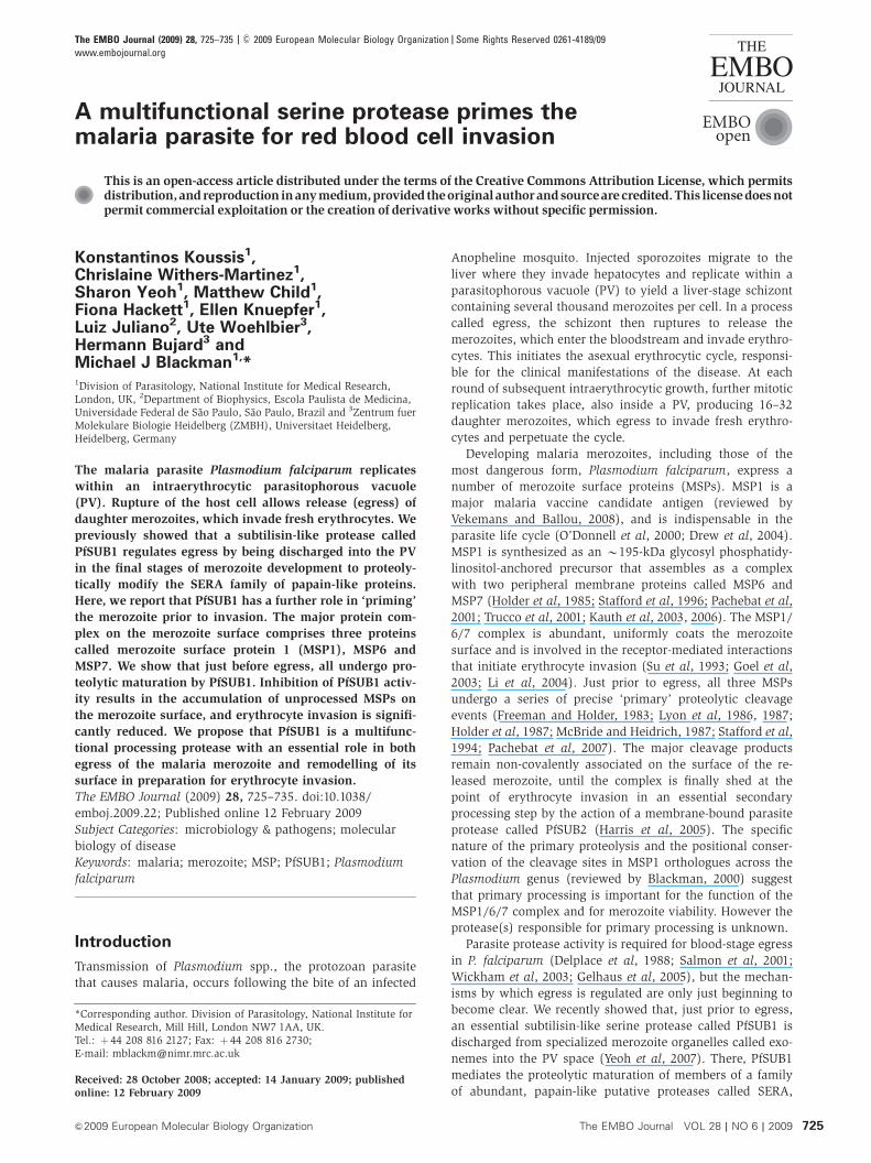
(Figure 1A). This showed that, of the non-prime side posi-
tions examined, substitution of the Val at P4 had the greatest
effect on substrate cleavage efficiency. This is consistent with
the predicted hydrophobic nature of the PfSUB1 S4 pocket
(Withers-Martinez et al, 2002), and the known importance of
the P4 residue in substrate recognition by subtilases generally
(Siezen and Leunissen, 1997). In addition, the P5 Leu and P30
Asp positions were found to be relatively important for
PfSUB1 cleavage. We next investigated the importance of
the P1 position, using a further set of peptides that varied
only at this position (Figure 1B). Although replacement with
Ser or Ala had no significant effect on cleavage, peptides
containing Glu, Arg, Asn, Tyr, Gly or His at P1 were cleaved
with intermediate efficiency, Pro at P1 was cleaved poorly,
and a peptide with Leu at P1 was not cleaved. Finally, the
importance of the P2 position was examined in a similar
Figure 1 Analysis of PfSUB1 substrate specificity. (A) Alaninesubstitution scanning of fluorogenic peptide Abz-LVSADNIDIQ-EDDnp. The parent peptide sequence is shown (inset), with thecleavage site arrowed. The x-axis indicates residue positions thatwere individually substituted with Ala. (B) P1 scanning of the sameparent peptide Abz-LVSADNIDIQ-EDDnp. The x-axis indicates thevarious amino-acid substitutions made at the P1 position. (C) P2scanning of Abz-LVSADNIDIQ-EDDnp. The x-axis indicates thevarious amino-acid substitutions made at P2. In each plot, barsindicate the efficiency of cleavage by recombinant PfSUB1,expressed as the ratio of activity against the substituted peptide,versus activity against the parent peptide (indicated in each case bya grey bar with a value of 1). Ratios are expressed as relative kcat/Km
values. In all cases, each value represents the mean of eightseparate determinations. Error bars, standard deviation values.
PfSUB1 matures malaria parasite surface proteinsK Koussis et al
The EMBO Journal VOL 28 | NO 6 | 2009 &2009 European Molecular Biology Organization726
manner (Figure 1C). This showed that, of the amino-acid
residues assessed, only Ala and Gly could be accommodated
at P2, with an apparent preference for Gly; substitution with
any of nine alternative residues resulted in peptides that were
completely refractory to cleavage by PfSUB1.
These results support the consensus motif described
above, confirming the importance of the P4 and—in particu-
lar—the P2 subsites in substrate cleavage by PfSUB1. The
data also provide experimental evidence for a role in sub-
strate recognition of both the P5 position and of acidic
residues on the prime side of the cleavage site.
The PfSUB1 consensus substrate recognition motif
resembles all known MSP primary processing sites
Armed with the above information, we searched for addi-
tional physiological PfSUB1 substrates that might explain the
observed effects of inhibitor MRT12113 on the invasive
capacity of released merozoites. We reasoned that, upon its
release into the PV to exert an effect upon the SERAs just
prior to schizont rupture, PfSUB1 should also have access to
other PV proteins, including surface-resident merozoite pro-
teins that are exposed to the PV lumen. Extensive work over
the last 25 years has shown that several MSPs undergo
dramatic proteolytic modification just prior to schizont rup-
ture. The best-characterized examples of these are MSP1 and
two peripheral membrane proteins, MSP6 and MSP7
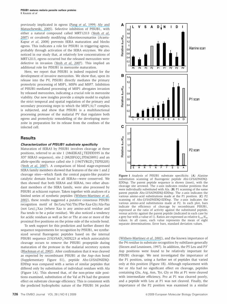
(Figure 2). MSP1, which exists in two major allelic forms in
P. falciparum, is converted to four polypeptide fragments of
B83, 38, 30 and 42 kDa (referred to as MSP183, MSP138,
MSP130 and MSP142, respectively). MSP6 and MSP7, which
are also dimorphic proteins and both of which are non-
covalently associated with MSP1 on the merozoite surface,
are also cleaved at internal sites; in each case the C-terminal
products remain bound to the MSP1 complex, whereas the
fate of the N-terminal fragments is unknown. Most of the
cleavage sites have been mapped by N-terminal sequencing
of endogenous cleavage products (Supplementary Figure S2).
As shown in Table I, comparison of all these MSP processing
sites with the internal PfSUB1 processing site, the known
SERA5 processing sites, and the predicted analogous sites in
SERA4 and SERA6, revealed a pattern strikingly consistent
with the PfSUB1 consensus recognition motif described
above. Most conspicuously, every site has either Ala or Gly
at the P2 position, and all have Leu, Ile, Val or Thr at P4. In
addition, none of the sites contains Pro or Leu at P1, and on
the prime side of the scissile bond most exhibit an acidic
tendency and/or a preference for Ser and Thr residues.
Interestingly, cleavage at the MSP719 site does not occur in
forms of MSP7 that contain a Lys substitution of Gly193, the
residue at the P2 position in those MSP7 alleles that are
processed at this site (Pachebat et al, 2007). Collectively,
these observations raised the strong possibility that MSP1,
MSP6 and MSP7 processing are all directly mediated by
PfSUB1 upon its release into the PV.
Recombinant PfSUB1 mediates correct primary
processing of parasite-derived MSP1, MSP6 and MSP7
To test the above conjecture, we developed an in vitro assay
to assess the capacity of recombinant PfSUB1 (rPfSUB1) to
convert MSP1, MSP6 and MSP7 precursors to species resem-
bling those on naturally released mature merozoites. Our
assay took advantage of the fact that biosynthesis of all three
precursor proteins initiates at around the beginning of schi-
zont development, whereas primary processing takes place
only at the end of this process, just prior to merozoite egress.
Mid-stage P. falciparum schizonts were treated with a cocktail
Figure 2 Proteolytic processing of the MSP1/6/7 complex. Biosynthesis of all three MSPs begins at the start of schizogony (nuclear division).As indicated, the complex is a stoichiometric assembly of the various components, except that the MSP6 precursor (yellow) binds as a tetramer(Kauth et al, 2006). Primary processing converts it to MSP636. The MSP7 precursor undergoes an early processing step during secretorytransport (Pachebat et al, 2007) to produce MSP733 (green), the form that binds to the MSP1 precursor (brown). Primary processing convertsMSP733 to both MSP722 and MSP719. The various MSP1 processing products are numbered for clarity. Secondary processing, which occursfollowing egress and probably at the point of invasion, involves further cleavage within MSP142 to produce MSP133 and MSP119. The latter iscarried into the newly invaded erythrocyte on the merozoite surface (to form a so-called ring-stage parasite), whereas MSP133 is shed alongwith the remaining MSP1/6/7 complex. Secondary processing is mediated by a membrane-bound protease called PfSUB2, whereas theprotease(s) responsible for primary processing has been hitherto unknown (indicated by a question mark).
PfSUB1 matures malaria parasite surface proteinsK Koussis et al
&2009 European Molecular Biology Organization The EMBO Journal VOL 28 | NO 6 | 2009 727
of protease inhibitors to inactivate endogenous proteases,
including PfSUB1, as completely as possible. The parasites
were then released from their host cells using saponin, which
disrupts the erythrocyte and PV membrane (but not the
parasite plasma membrane), and were finally washed to
remove the protease inhibitors. Western blot showed that
these preparations contained, as expected, predominantly
full-length forms of all three MSPs (Figure 3, all ‘START’
lanes). Incubation with rPfSUB1 resulted in rapid conversion
of these to smaller processed fragments indistinguishable
from those present in the extracts of highly mature schizonts
(harvested at around the point of egress) or purified naturally
released merozoites (Figure 3A–E). Some low-level conver-
sion to these processing fragments occurred upon prolonged
incubation in the absence of added rPfSUB1, but this could be
completely blocked by the presence of either MRT12113 (not
shown, but see below) or recombinant PfSUB1 prodomain
(Figure 4), another selective inhibitor of PfSUB1 (Jean et al,
2003), suggesting that it was mediated by residual endogen-
ous PfSUB1 that had not been fully inactivated by protease
inhibitor treatment of the schizonts. These data support our
hypothesis that PfSUB1 regulates primary processing of all
three components of the MSP1/6/7 complex.
Recombinant PfSUB1 correctly processes recombinant
MSP1, MSP6 and MSP7 and peptides based on
processing sites
To confirm that the MSP processing observed in parasite
extracts was directly mediated by PfSUB1, and was not the
result of activation by PfSUB1 of a distinct protease, we next
examined the effects of rPfSUB1 on recombinant MSPs.
Preliminary experiments (Supplementary Figure S3) showed
that addition of rPfSUB1 to full-length recombinant MSP1
resulted in conversion to just 4–5 major fragments, consistent
with cleavage at a limited number of internal sites. Attempts
to define these sites by N-terminal sequencing proved un-
successful due to the limited amounts of available protein.
Accordingly, we turned to the use of a previously described
recombinant heterodimer comprising the N- and C-terminal
‘halves’ of MSP1 (Kauth et al, 2003). Although this approach
did not allow the analysis of cleavage at the MSP130–MSP138
junction, this protein was available in relatively large
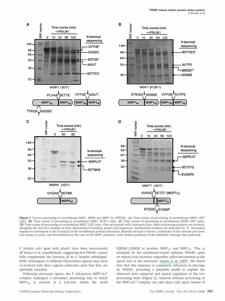
amounts in a pure form. As shown in Figure 5, addition of
purified rPfSUB1 to this, or to full-length recombinant MSP6
or MSP7, resulted in all cases in conversion to products of
approximately the expected size. N-terminal sequencing of
the processed products confirmed cleavage at the expected
sites. These included the 3D7 MSP719 site, confirming that
PfSUB1 can cleave this sequence that possesses a Thr at P4
(see Table I). In addition, the analysis identified the only two
previously unknown MSP1 primary processing sites:
TTKGAkSAQSG, a fourth site that lies at the beginning of a
repetitive sequence within the 3D7 form of MSP183 and so
could not previously be precisely defined (Stafford et al,
1994); and KTEGQkSDNSE, at the N terminus of the FCB-1
MSP130. The latter lies within a polymorphic region of MSP1
and hence is different from the analogous site in the 3D7
allelic form; nonetheless, it is positionally conserved in the
MSP1 primary sequence (Supplementary Figure S4). Both
sites are completely consistent with the putative PfSUB1
consensus recognition motif established above. These results
showed that PfSUB1 can directly mediate correct processing
of all three MSPs, without the involvement of another
protease.
On the basis of the above, we predicted that peptides based
on the processing sites would be good substrates for rPfSUB1.
Accordingly, decapeptides based on some of the MSP proces-
sing sites were examined for susceptibility to cleavage by
rPfSUB1. All were correctly cleaved only at the expected bond
(Supplementary Figure S5; Supplementary Table S1), con-
firming the capacity of PfSUB1 to correctly target these
sequences. A comparative analysis of all these peptides,
plus similar peptides based on the known and predicted
SERA4, SERA5 and SERA6 processing sites (Yeoh et al,
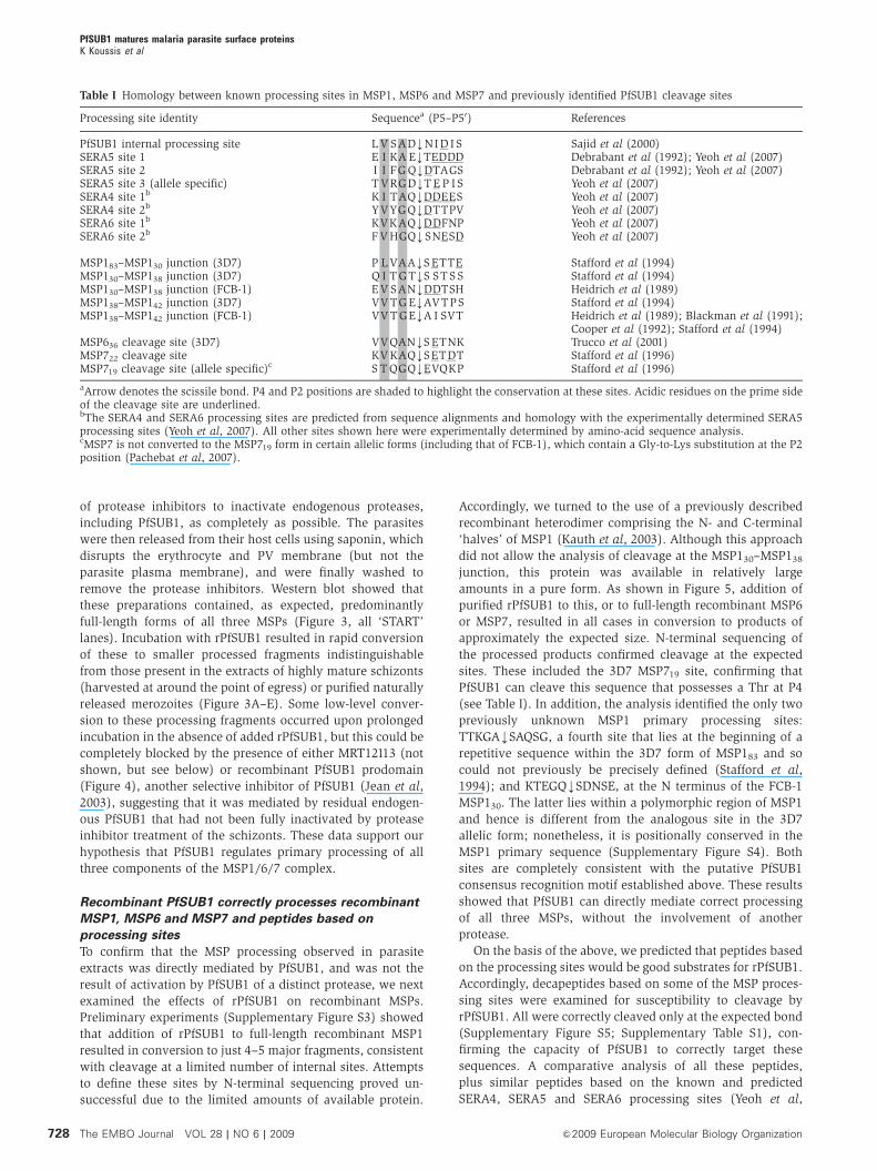
Table I Homology between known processing sites in MSP1, MSP6 and MSP7 and previously identified PfSUB1 cleavage sites
Processing site identity Sequencea (P5–P50) References
PfSUB1 internal processing site L V SADkNI D I S Sajid et al (2000)SERA5 site 1 E I KA EkTEDDD Debrabant et al (1992); Yeoh et al (2007)SERA5 site 2 I I FG QkDTAGS Debrabant et al (1992); Yeoh et al (2007)SERA5 site 3 (allele specific) T VRGDkT E P I S Yeoh et al (2007)SERA4 site 1b K I TAQkDDEES Yeoh et al (2007)SERA4 site 2b YVYGQkDTTPV Yeoh et al (2007)SERA6 site 1b KVKAQkDDFNP Yeoh et al (2007)SERA6 site 2b F V HGQkSNESD Yeoh et al (2007)
MSP183–MSP130 junction (3D7) P L VAAkS ETTE Stafford et al (1994)MSP130–MSP138 junction (3D7) Q I T GTkS ST S S Stafford et al (1994)MSP130–MSP138 junction (FCB-1) E V SANkDDTSH Heidrich et al (1989)MSP138–MSP142 junction (3D7) VV TG EkAVTPS Stafford et al (1994)MSP138–MSP142 junction (FCB-1) VV T GEkA I SVT Heidrich et al (1989); Blackman et al (1991);
Cooper et al (1992); Stafford et al (1994)MSP636 cleavage site (3D7) VVQANkS ETNK Trucco et al (2001)MSP722 cleavage site KV KAQkSETDT Stafford et al (1996)MSP719 cleavage site (allele specific)c S T QGQkEVQKP Stafford et al (1996)
aArrow denotes the scissile bond. P4 and P2 positions are shaded to highlight the conservation at these sites. Acidic residues on the prime sideof the cleavage site are underlined.bThe SERA4 and SERA6 processing sites are predicted from sequence alignments and homology with the experimentally determined SERA5processing sites (Yeoh et al, 2007). All other sites shown here were experimentally determined by amino-acid sequence analysis.cMSP7 is not converted to the MSP719 form in certain allelic forms (including that of FCB-1), which contain a Gly-to-Lys substitution at the P2position (Pachebat et al, 2007).
PfSUB1 matures malaria parasite surface proteinsK Koussis et al
The EMBO Journal VOL 28 | NO 6 | 2009 &2009 European Molecular Biology Organization728
2007), indicated substantial differences in turnover rates, but
identified the most efficiently cleaved peptide as that based
on SERA4 site 1, Ac-KITAQDDEES-OH (Supplementary Figure
S6). This site is particularly rich in prime side acidic residues,
consistent with a preference for cleavage at such sites.
In contrast to the results with peptides based on the
primary processing sites, a peptide based on the secondary
MSP1 processing site known to be targeted by PfSUB2
(FQDMLkNISQH) was completely refractory to cleavage by
rPfSUB1 (data not shown). This was not surprising, given our
evidence that PfSUB1 cannot cleave on the C-terminal side of
a Leu residue.
Inhibition of parasite PfSUB1 activity interferes
with MSP1 primary processing and results in
invasive-defective merozoites
Identification of PfSUB1 as the enzyme responsible for pri-
mary processing of the MSP1/6/7 complex suggested that the
previously observed effects of MRT12113 on invasion might
be a result of impaired MSP processing on released mero-
zoites. To seek evidence that MRT12113 inhibits MSP proces-
sing under in vitro culture conditions, we first examined the
effects of treating parasites with 150mM MRT12113 during the
final stages of schizont maturation. Detailed dose–response
experiments have shown that at this concentration of
MRT12113, egress is only partly blocked (Yeoh et al, 2007),
enabling examination of proteins released into culture super-
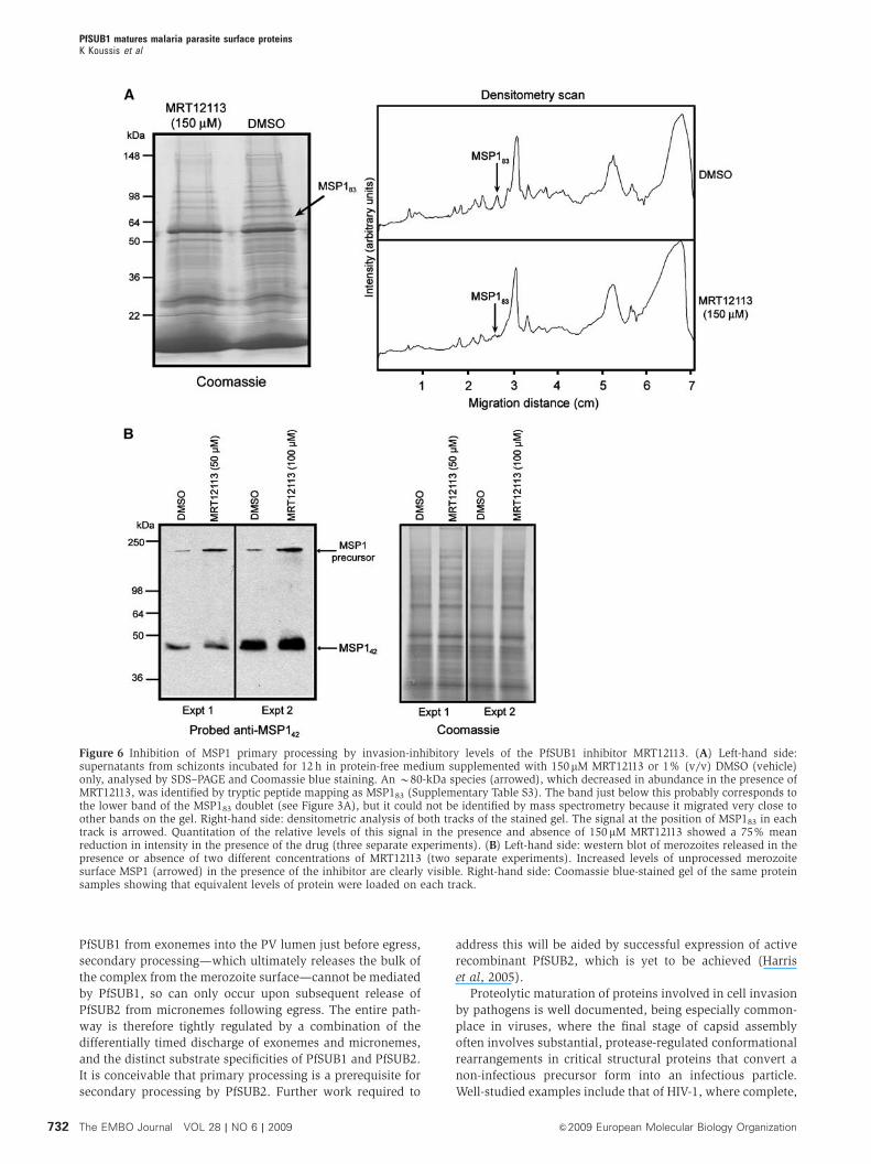
natants upon egress. Examination of the protein profile of the
culture supernatants revealed that, as well as the effects on
SERA5 processing noted previously, a species of B80 kDa
was significantly reduced in abundance in the presence of
MRT12113 (Figure 6A). Western blot (not shown) and tryptic
peptide mapping (Supplementary Table S3) identified this as
MSP183. Shedding of this protein normally occurs upon
secondary processing of the MSP1/6/7 complex by PfSUB2
following egress (Figure 2), but as MRT12113 does not
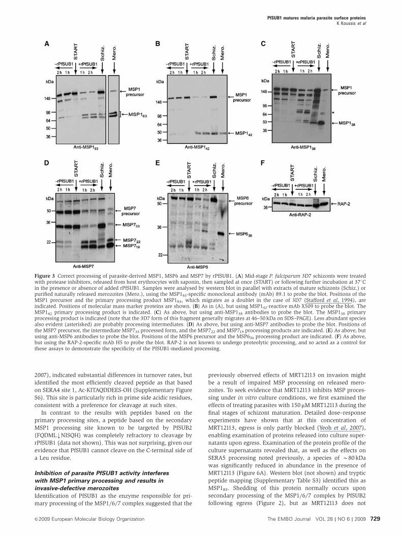
Figure 3 Correct processing of parasite-derived MSP1, MSP6 and MSP7 by rPfSUB1. (A) Mid-stage P. falciparum 3D7 schizonts were treatedwith protease inhibitors, released from host erythrocytes with saponin, then sampled at once (START) or following further incubation at 371Cin the presence or absence of added rPfSUB1. Samples were analysed by western blot in parallel with extracts of mature schizonts (Schiz.) orpurified naturally released merozoites (Mero.), using the MSP183-specific monoclonal antibody (mAb) 89.1 to probe the blot. Positions of theMSP1 precursor and the primary processing product MSP183, which migrates as a doublet in the case of 3D7 (Stafford et al, 1994), areindicated. Positions of molecular mass marker proteins are shown. (B) As in (A), but using MSP142-reactive mAb X509 to probe the blot. TheMSP142 primary processing product is indicated. (C) As above, but using anti-MSP138 antibodies to probe the blot. The MSP138 primaryprocessing product is indicated (note that the 3D7 form of this fragment generally migrates at 46–50 kDa on SDS–PAGE). Less abundant speciesalso evident (asterisked) are probably processing intermediates. (D) As above, but using anti-MSP7 antibodies to probe the blot. Positions ofthe MSP7 precursor, the intermediate MSP733 processed form, and the MSP722 and MSP719 processing products are indicated. (E) As above, butusing anti-MSP6 antibodies to probe the blot. Positions of the MSP6 precursor and the MSP636 processing product are indicated. (F) As above,but using the RAP-2-specific mAb H5 to probe the blot. RAP-2 is not known to undergo proteolytic processing, and so acted as a control forthese assays to demonstrate the specificity of the PfSUB1-mediated processing.
PfSUB1 matures malaria parasite surface proteinsK Koussis et al
&2009 European Molecular Biology Organization The EMBO Journal VOL 28 | NO 6 | 2009 729
directly inhibit PfSUB2 activity (Yeoh et al, 2007) the reduc-
tion in MSP183 production must result from a defect in
primary processing. This demonstrates that MRT12113 can
indeed interfere with MSP processing by PfSUB1 under
culture conditions.
The ED50 of MRT12113 against egress is B180 mM, whereas
its activity against invasion is much higher (ED50 B25 mM).
We have previously postulated (Yeoh et al, 2007) that this
apparent discrepancy may be a result of both the unusual
abundance of the SERA proteins and fundamental differences
between the requirements for egress and invasion.
Specifically, if complete inhibition of SERA maturation is
needed to block egress, schizonts in which this is only
partially blocked would not exhibit a rupture defect. On the
other hand, if efficient or complete processing of MSPs is
required for invasion, even modest inhibition of MSP1/6/7
maturation might prevent invasion. To address the possibility
of a link between invasion and MSP processing, merozoites
released in the presence of egress-permissive but invasion-
inhibitory levels of MRT12113 (50–100 mM) were analysed by
western blot. Figure 6B shows that although only trace
amounts of MSP1 precursor were detectable in control natu-
rally released merozoites (see also Figure 3), those released in
the presence of MRT12113 possessed significantly higher
levels of unprocessed MSP1. These results support our hy-
pothesis that the invasion defect produced by MRT12113 is a
direct result of its inhibition of MSP maturation. Correct and
complete proteolytic maturation of the MSP1/6/7 complex by
PfSUB1 may be a prerequisite for invasion.
Discussion
It has long been recognized that the final stages of intracel-
lular development of the P. falciparum merozoite are accom-
panied by extensive proteolytic modification of MSPs.
Particular interest has focused on the MSP1/6/7 complex
because of its abundance, its essential nature and its involve-
ment in the initial interactions between the merozoite and its
host erythrocyte. We have now shown that all primary
processing events to which this complex is subjected are
directly mediated by PfSUB1, a serine protease released into
the PV lumen just prior to egress. Together with our earlier
findings, our work implicates PfSUB1 not only in regulation
of egress, but also in preparing the merozoite for release from
the host cell. Our results provide the opportunity to dissect
the consequences of MSP processing and its functional sig-
nificance for erythrocyte invasion. Moreover, they explain
how maturation of the MSP1/6/7 complex is temporally
regulated.
Our peptide scanning analysis, together with our identifi-
cation of MSP1, MSP6 and MSP7 as authentic macromolecu-
lar substrates for PfSUB1, has expanded our picture of the
substrate specificity of PfSUB1 (Supplementary Figure S7). As
well as substantiating the importance of Gly or Ala at the P2
subsite, our results confirm the preference for acidic residues,
Ser or Thr on the prime side of the scissile bond and allow us
to confidently add Thr to the list of resides that can be
accommodated at P4. It is worth noting that Thr and Phe
are the only other residues apart from Ile, Val and Leu that are
observed at the P4 position of the predicted site 1 and site 2
processing sites in those blood-stage SERA family members
(SERA1, 2, 3, 7 and 9) that have not been experimentally
demonstrated to be PfSUB1 substrates (Yeoh et al, 2007),
supporting our prediction that all these SERAs are likely
processed by PfSUB1. The role of the P5 position in substrate
recognition by PfSUB1 remains unclear; the alanine scanning
data suggest that it may be important, and consistent with
this we have yet to observe Ala at P5 in any of the physio-
logical cleavage sites identified to date. PfSUB1 has ortholo-
gues in all Plasmodium species examined, and the active-site
cleft residues predicted to be involved in substrate recogni-
tion by the protease are highly conserved (Withers-Martinez
et al, 2004). The few MSP1 primary processing sites that have
been mapped in other species (reviewed by Blackman, 2000)
are very similar to those in P. falciparum. However, none
is identical. Attempts to perform an allelic replacement of the
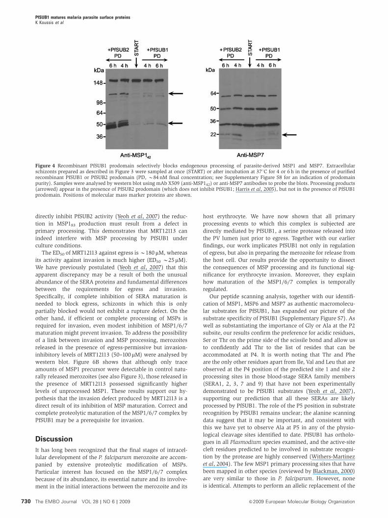
Figure 4 Recombinant PfSUB1 prodomain selectively blocks endogenous processing of parasite-derived MSP1 and MSP7. Extracellularschizonts prepared as described in Figure 3 were sampled at once (START) or after incubation at 371C for 4 or 6 h in the presence of purifiedrecombinant PfSUB1 or PfSUB2 prodomain (PD, B84 nM final concentration; see Supplementary Figure S8 for an indication of prodomainpurity). Samples were analysed by western blot using mAb X509 (anti-MSP142) or anti-MSP7 antibodies to probe the blots. Processing products(arrowed) appear in the presence of PfSUB2 prodomain (which does not inhibit PfSUB1; Harris et al, 2005), but not in the presence of PfSUB1prodomain. Positions of molecular mass marker proteins are shown.
PfSUB1 matures malaria parasite surface proteinsK Koussis et al
The EMBO Journal VOL 28 | NO 6 | 2009 &2009 European Molecular Biology Organization730
P. berghei sub1 gene with pfsub1 have been unsuccessful
(R Tewari et al, unpublished), suggesting that PfSUB1 cannot
fully complement the function of its P. berghei orthologue.
SUB1 orthologues in different Plasmodium species may have
co-evolved with their cognate substrates such that they are
optimally matched.
Following merozoite egress, the P. falciparum MSP1/6/7
complex undergoes a secondary processing step in which
MSP142 is cleaved at a Leu-Asn within the motif
FQDMLkNISQH to produce MSP119 and MSP133. This is
mediated by the membrane-bound subtilase PfSUB2 upon
its release from secretory organelles called micronemes at the
apical end of the merozoite (Harris et al, 2005). We found
here that this sequence is completely refractory to cleavage
by PfSUB1, providing a plausible model to explain the
observed strict temporal and spatial regulation of the two
processing steps (Figure 2); whereas primary processing of
the MSP1/6/7 complex can take place only upon release of
Figure 5 Correct processing of recombinant MSP1, MSP6 and MSP7 by rPfSUB1. (A) Time course of processing of recombinant MSP1 (3D7type). (B) Time course of processing of recombinant MSP1 (FCB-1 type). (C) Time course of processing of recombinant MSP6 (3D7 type).(D) Time course of processing of recombinant MSP7 (3D7 type). Gels are stained with Coomassie blue. Major processing products are arrowed,alongside the first five residues of their determined N-terminal amino-acid sequences. Unidentified residues are indicated by ‘X’. Asteriskedsequences correspond to the N termini of the recombinant protein precursors. Beneath each gel is shown a schematic of the relevant precursor(not drawn to scale, and heterodimers in the case of the MSP1 proteins), with relative positions of the identified cleavage sites indicated.
PfSUB1 matures malaria parasite surface proteinsK Koussis et al
&2009 European Molecular Biology Organization The EMBO Journal VOL 28 | NO 6 | 2009 731
PfSUB1 from exonemes into the PV lumen just before egress,
secondary processing—which ultimately releases the bulk of
the complex from the merozoite surface—cannot be mediated
by PfSUB1, so can only occur upon subsequent release of
PfSUB2 from micronemes following egress. The entire path-
way is therefore tightly regulated by a combination of the
differentially timed discharge of exonemes and micronemes,
and the distinct substrate specificities of PfSUB1 and PfSUB2.
It is conceivable that primary processing is a prerequisite for
secondary processing by PfSUB2. Further work required to
address this will be aided by successful expression of active
recombinant PfSUB2, which is yet to be achieved (Harris
et al, 2005).
Proteolytic maturation of proteins involved in cell invasion
by pathogens is well documented, being especially common-
place in viruses, where the final stage of capsid assembly
often involves substantial, protease-regulated conformational
rearrangements in critical structural proteins that convert a
non-infectious precursor form into an infectious particle.
Well-studied examples include that of HIV-1, where complete,
Figure 6 Inhibition of MSP1 primary processing by invasion-inhibitory levels of the PfSUB1 inhibitor MRT12113. (A) Left-hand side:supernatants from schizonts incubated for 12 h in protein-free medium supplemented with 150mM MRT12113 or 1% (v/v) DMSO (vehicle)only, analysed by SDS–PAGE and Coomassie blue staining. An B80-kDa species (arrowed), which decreased in abundance in the presence ofMRT12113, was identified by tryptic peptide mapping as MSP183 (Supplementary Table S3). The band just below this probably corresponds tothe lower band of the MSP183 doublet (see Figure 3A), but it could not be identified by mass spectrometry because it migrated very close toother bands on the gel. Right-hand side: densitometric analysis of both tracks of the stained gel. The signal at the position of MSP183 in eachtrack is arrowed. Quantitation of the relative levels of this signal in the presence and absence of 150 mM MRT12113 showed a 75% meanreduction in intensity in the presence of the drug (three separate experiments). (B) Left-hand side: western blot of merozoites released in thepresence or absence of two different concentrations of MRT12113 (two separate experiments). Increased levels of unprocessed merozoitesurface MSP1 (arrowed) in the presence of the inhibitor are clearly visible. Right-hand side: Coomassie blue-stained gel of the same proteinsamples showing that equivalent levels of protein were loaded on each track.
PfSUB1 matures malaria parasite surface proteinsK Koussis et al
The EMBO Journal VOL 28 | NO 6 | 2009 &2009 European Molecular Biology Organization732
ordered and accurate cleavage of the Gag polyprotein by the
virus-encoded aspartic protease during capsid assembly and
budding is strictly required for virus infectivity (Kaplan et al,
1993; Pettit et al, 1994; Wiegers et al, 1998). Proteolytic
modification of the MSP1/6/7 complex by PfSUB1 may
represent an analogous process, functionally activating im-
portant merozoite surface molecules and so effectively prim-
ing the parasite for invasion. Although caution is required in
the interpretation of experiments relying on the use of a small
compound inhibitor of only modest potency (raising the
possibility of off-target effects), our data also suggest that—
similar to the situation with HIV Gag—even partial inhibition
of MSP processing may impact on invasion efficiency. The
different susceptibility to PfSUB1 of peptides based on the
various MSP and SERA cleavage sites may also have analo-
gies with Gag processing. Assuming that it reflects differences
in cleavage efficiency in the context of the intact proteins—a
postulate supported by our previous observations that clea-
vage at SERA5 site 1 occurs faster than cleavage at site 2, a
distinction shared by peptide substrates based on these sites
(Yeoh et al, 2007)—it implies a degree of kinetic regulation of
PfSUB1-mediated processing. The relative rates of proteolysis
at different sites, both within and between individual protein
substrates, may be important. In the moments just before
egress, for example, SERA processing may need to occur
more rapidly than processing of the MSPs, as it likely forms
the initial step in a multistage cascade that needs to be
completed for egress to proceed.
It is informative to contrast our results with aspects of
sporozoite invasion. Sporozoites are injected by the mosquito
vector into the host dermis, from where they migrate the
often considerable distance to the liver. During this journey,
the sporozoite can either non-productively penetrate and
migrate straight through tissue cells, or alternatively can
productively invade them with the formation of a PV (Mota
et al, 2001). Productive invasion is associated with proteolytic
cleavage of the major sporozoite surface circumsporozoite
protein. Cleavage is markedly upregulated upon interaction
with the highly sulphated heparin sulphate proteoglycans
expressed by host hepatocytes, but not by contact with low-
sulphated forms expressed by other cell types (Coppi et al,
2007). Thus, as it is important that the sporozoite does not
engage productively with the many non-permissive cells it
comes into contact with during its journey between the
dermis and the liver, it becomes infection-competent only
upon reaching the appropriate cells, hepatocytes. Merozoites
are released directly into the bloodstream, where new host
cells are immediately available, but in contrast to the spor-
ozoite the merozoite needs to productively invade without
delay, as its invasive half-life is extremely short and in
immune or semi-immune hosts it is immediately exposed to
high levels of serum antibodies that can block invasion. This
may explain why it is advantageous for the merozoite to be
‘prepared’ for invasion during the moments leading up to
egress, rather than after it.
MSP1 is essential to blood-stage parasite viability, and the
MSP1/6/7 complex has been shown to have a function in the
initial stages of interaction between merozoite and host
erythrocyte. The molecular details of this role remain ob-
scure. It is likely that processing of at least some of the sites
examined here is essential, although in cases where poly-
morphism results in allele-specific cleavage—as in the case of
the MSP719 site—it seems reasonable to assume that proces-
sing at this site is not critical for merozoite viability. The next
challenge will be to identify the key cleavage events in the
MSP1/6/7 complex, and future work will attempt to dissect
the importance of individual processing events for parasite
viability. In addition, we now have the tools—rPfSUB1 plus
soluble recombinant MSPs—to use in vitro approaches to
recapitulate and characterize conformational changes that
may be induced in MSPs by processing, and to investigate
their functional significance.
New antimalarial drugs are urgently required in the face of
increasing resistance to available therapeutics, and our findings
have implications for the potential of PfSUB1 as a drug target.
We now know that PfSUB1 has multiple physiological sub-
strates, most of which are cleaved at more than one site. We
would therefore predict that the capacity for simultaneous co-
evolution of the protease and its substrates in response to
protease inhibitor-based drugs is minimal, limiting the emer-
gence of parasites resistant to anti-PfSUB1 drugs. Furthermore,
the fact that both MSP1 and SERAs are also expressed in liver
stages (Szarfman et al, 1988; Tarun et al, 2008) makes it likely
that PfSUB1 has a function in development and egress of liver-
stage merozoites. As well as preventing blood-stage parasite
replication, drugs targeting PfSUB1 might therefore also block
the parasite life cycle at an early, non-symptomatic stage in
progression of the infection.
Materials and methods
Parasite cultureMaintenance in vitro of asexual blood stages of P. falciparum clone3D7, purification of schizonts and merozoites, treatment withcompound MRT12113 and identification by tryptic peptide finger-printing of parasite proteins released into serum-free culturemedium were as described (Blackman, 1994; Yeoh et al, 2007).Densitometric analysis of Coomassie blue-stained, SDS–PAGE-fractionated proteins was performed using ImageJ (http://rsb.info.nih.gov/ij/index.html).
Peptide substrate assaysInternally quenched fluorogenic peptides of the general structureAbz-(Xaa)n-Q-EDDnp (where Abz is ortho-aminobenzoic acid,EDDnp is N-(ethylenediamine)-2,4-dinitrophenyl amide and Xaaare natural amino acids followed by a glutamine [Q]) weresynthesized as described (Portaro et al, 2000; Melo et al, 2001).Concentrations of stock solutions in DMSO were calculated bycolorimetric determination of the 2,4-dinitrophenyl group(e365¼17 300 M�1 cm�1) and then adjusted to 1 mM and stored inthe dark at �201C. Unlabelled N-acetylated decapeptides based onMSP and SERA processing sites were synthesized using standardFmoc chemistry, and stock solutions (80 mM in DMSO) storedat �201C.
Working solutions of all peptides were prepared by dilution intoPfSUB1 digestion buffer (50 mM Tris–HCl pH 7.6, 15 mM CaCl2,25 mM (3-[(3cholamidopropyl)-dimethylammonio] propanesulpho-nate) (CHAPS)). For relative kcat/Km determination of hydrolysis offluorogenic peptides, samples (195ml) of peptide solution atconcentrations of 10, 5, 2.5 and 1.25mM (ooKm) were dispensedinto white 96-well microplates (FluoroNunc, NUNC) and supple-mented with purified rPfSUB1 (B40 nM). Hydrolysis was mon-itored at room temperature using a Varian Cary Eclipse fluorescencespectrophotometer by measuring the increase in fluorescence ofAbz (using excitation and emission wavelengths of 320 and 420 nm,respectively). Once a steady-state initial rate was determined,reactions were driven to completion by the addition of pronase andfluorescence values converted to moles of substrate hydrolysedas described (Blackman et al, 2002). For comparing cleavageefficiencies of unlabelled peptides, mixtures containing equalconcentrations (B2 mM) of two peptides were supplemented with
PfSUB1 matures malaria parasite surface proteinsK Koussis et al
&2009 European Molecular Biology Organization The EMBO Journal VOL 28 | NO 6 | 2009 733
rPfSUB1 (B40 nM) and initial rates of cleavage were determined byanalytical RP-HPLC as described (Withers-Martinez et al, 2002).Under these conditions, the relative initial rates of hydrolysis of thetwo peptides are directly proportional to their relative kcat/Km
values. To determine which of the collection of unlabelled peptideswas most efficiently cleaved, a ‘league table’-type approach wasused. Peptides were initially randomly compared pairwise as above,and the most rapidly cleaved of each pair was selected. These werethen compared pairwise with each other, and the best cleaved ofeach pair was again selected. This process was repeated until themost rapidly cleaved peptide of the entire set was identified. Fordetermination of cleavage specificity by rPfSUB1, partially digestedpeptides were fractionated by RP-HPLC and digestion products wereidentified by electrospray mass spectrometry as described (Black-man et al, 2002; Withers-Martinez et al, 2002).
Parasite MSP processing assaysPurified mid-stage 3D7 schizonts were washed in protein-free RPMI1640 medium and resuspended in phosphate-buffered saline (PBS)containing a cocktail of protease inhibitors (listed in SupplementaryTable S2). After incubation on ice for 10 min, the parasitesuspension was supplemented with 0.1 volume of 1.5% (w/v)saponin in PBS to disrupt the erythrocyte and PV membranes.Released parasites were washed, resuspended in 25 mM HEPES pH6.5, 15 mM CaCl2 and frozen in aliquots at �701C. For processingassays, aliquots were divided into two and supplemented witheither purified rPfSUB1 or an equal volume of control buffer (10 mMTris–HCl pH 7.6, 150 mM NaCl and 10% glycerol). Samples wereincubated at 371C and reactions were stopped by boiling in SDSsample buffer prior to western blot analysis.
Antibodies and western blotWestern blots were carried out as described (Jean et al, 2003),probing with mAb 89.1 to detect MSP183 (Holder et al, 1985), mAbX509 to detect MSP142 (Blackman et al, 1991), or a rabbit anti-MSP138 (Woehlbier et al, 2006). To detect MSP6 and 7, blots wereprobed with rabbit antibodies to MSP6 (Kauth et al, 2006) or MSP7
(Pachebat et al, 2007). The anti-RAP-2 mAb H5 has been described(Harris et al, 2005).
Recombinant proteins and N-terminal sequencingRecombinant PfSUB1 and PfSUB2 prodomains were expressed ininsect cells or Escherichia coli, respectively, and purified andquantified as described (Withers-Martinez et al, 2002; Jean et al,2003; Harris et al, 2005). Production and purification of recombi-nant full-length MSP1 (MSP-1D corresponding to the 3D7 allelicform and MSP-1F to the FCB-1 type), recombinant MSP1 hetero-dimer (a reconstituted complex corresponding to MSP183/30
complexed with MSP138/42) for both allelic forms, as well asrecombinant MSP6 and MSP7, have been described (Epp et al, 2003;Kauth et al, 2003, 2006).
Recombinant MSPs in 50 mM Tris–HCl pH 7.6, 15 mM CaCl2were supplemented with rPfSUB1 or control buffer as above,incubated at 371C and sampled over a period of 2 h. Samples wereanalysed by SDS–PAGE and staining with Coomassie blue. For N-terminal sequencing, SDS–PAGE fractionated proteins were trans-ferred to polyvinylidene difluoride membrane and Edman degrada-tion performed at the Protein and Nucleic Acid Chemistry Facility,University of Cambridge, UK.
Supplementary dataSupplementary data are available at The EMBO Journal Online(http://www.embojournal.org).
Acknowledgements
This study was supported by the Medical Research Council, UK. InBrazil, LJ was supported by the Fundacao de Amparo a Pesquisasdo Estado de Sao Paulo (FAPESP) and Conselho Nacional deDesenvolvimento Cientifico e Tecnologico (CNPq). This study ispart of the activities of the BioMalPar European Network ofExcellence supported by a European grant (LSHP-CT-2004-503578)from the Priority 1 ‘Life Sciences, Genomics and Biotechnology forHealth’ in the sixth Framework Programme.
References
Aly AS, Matuschewski K (2005) A malarial cysteine protease isnecessary for Plasmodium sporozoite egress from oocysts. J ExpMed 202: 225–230
Arastu-Kapur S, Ponder EL, Fonovic UP, Yeoh S, Yuan F, Fonovic M,Grainger M, Phillips CI, Powers JC, Bogyo M (2008) Identificationof proteases that regulate erythrocyte rupture by the malariaparasite Plasmodium falciparum. Nat Chem Biol 4: 203–213
Blackman MJ (1994) Purification of Plasmodium falciparum mer-ozoites for analysis of the processing of merozoite surfaceprotein-1. Methods Cell Biol 45: 213–220
Blackman MJ (2000) Proteases involved in erythrocyte invasion bythe malaria parasite: function and potential as chemotherapeutictargets. Curr Drug Targets 1: 59–83
Blackman MJ, Corrie JE, Croney JC, Kelly G, Eccleston JF, JamesonDM (2002) Structural and biochemical characterization of afluorogenic rhodamine-labeled malarial protease substrate.Biochemistry 41: 12244–12252
Blackman MJ, Whittle H, Holder AA (1991) Processing of thePlasmodium falciparum major merozoite surface protein-1: iden-tification of a 33-kilodalton secondary processing product whichis shed prior to erythrocyte invasion. Mol Biochem Parasitol 49:35–44
Cooper JA, Cooper LT, Saul AJ (1992) Mapping of the regionpredominantly recognized by antibodies to the Plasmodiumfalciparum merozoite surface antigen MSA 1. Mol BiochemParasitol 51: 301–312
Coppi A, Tewari R, Bishop JR, Bennett BL, Lawrence R, Esko JD,Billker O, Sinnis P (2007) Heparan sulfate proteoglycans provide asignal to Plasmodium sporozoites to stop migrating and produc-tively invade host cells. Cell Host Microbe 2: 316–327
Debrabant A, Maes P, Delplace P, Dubremetz JF, Tartar A, Camus D(1992) Intramolecular mapping of Plasmodium falciparum P126proteolytic fragments by N-terminal amino acid sequencing.Mol Biochem Parasitol 53: 89–95
Delplace P, Bhatia A, Cagnard M, Camus D, Colombet G, DebrabantA, Dubremetz JF, Dubreuil N, Prensier G, Fortier B, Hao A,Weber J, Vernes A (1988) Protein p126: a parasitophorousvacuole antigen associated with the release of Plasmodiumfalciparum merozoites. Biol Cell 64: 215–221
Drew DR, O’Donnell RA, Smith BJ, Crabb BS (2004) A commoncross-species function for the double epidermal growth factor-likemodules of the highly divergent Plasmodium surface proteinsMSP-1 and MSP-8. J Biol Chem 279: 20147–20153
Epp C, Kauth CW, Bujard H, Lutz R (2003) Expression and purifica-tion of Plasmodium falciparum MSP-1(42): a malaria vaccinecandidate. J Chromatogr B Analyt Technol Biomed Life Sci 786:61–72
Freeman RR, Holder AA (1983) Surface antigens of malaria mer-ozoites. A high molecular weight precursor is processed to an83 000 mol wt form expressed on the surface of Plasmodiumfalciparum merozoites. J Exp Med 158: 1647–1653
Gelhaus C, Vicik R, Schirmeister T, Leippe M (2005) Blocking effectof a biotinylated protease inhibitor on the egress of Plasmodiumfalciparum merozoites from infected red blood cells. Biol Chem386: 499–502
Goel VK, Li X, Chen H, Liu SC, Chishti AH, Oh SS (2003) Band 3 is ahost receptor binding merozoite surface protein 1 during thePlasmodium falciparum invasion of erythrocytes. Proc NatlAcad Sci USA 100: 5164–5169
Harris PK, Yeoh S, Dluzewski AR, O’Donnell RA, Withers-MartinezC, Hackett F, Bannister LH, Mitchell GH, Blackman MJ (2005)Molecular identification of a malaria merozoite surface sheddase.PLoS Pathog 1: 241–251
Heidrich HG, Miettinen-Baumann A, Eckerskorn C, Lottspeich F(1989) The N-terminal amino acid sequences of the Plasmodiumfalciparum (FCB1) merozoite surface antigens of 42 and 36kilodalton, both derived from the 185-195-kilodalton precursor.Mol Biochem Parasitol 34: 147–154
PfSUB1 matures malaria parasite surface proteinsK Koussis et al
The EMBO Journal VOL 28 | NO 6 | 2009 &2009 European Molecular Biology Organization734
Holder AA, Lockyer MJ, Odink KG, Sandhu JS, Riveros-Moreno V,Nicholls SC, Hillman Y, Davey LS, Tizard ML, Schwarz RT,Freeman RR (1985) Primary structure of the precursor to thethree major surface antigens of Plasmodium falciparum mero-zoites. Nature 317: 270–273
Holder AA, Sandhu JS, Hillman Y, Davey LS, Nicholls SC, Cooper H,Lockyer MJ (1987) Processing of the precursor to the majormerozoite surface antigens of Plasmodium falciparum.Parasitology 94 (Part 2): 199–208
Jean L, Hackett F, Martin SR, Blackman MJ (2003) Functionalcharacterisation of the propeptide of Plasmodium falciparumsubtilisin-like protease-1. J Biol Chem 278: 28572–28579
Kaplan AH, Zack JA, Knigge M, Paul DA, Kempf DJ, Norbeck DW,Swanstrom R (1993) Partial inhibition of the human immuno-deficiency virus type 1 protease results in aberrant virus assemblyand the formation of noninfectious particles. J Virol 67:4050–4055
Kauth CW, Epp C, Bujard H, Lutz R (2003) The merozoite surfaceprotein 1 complex of human malaria parasite Plasmodium falci-parum: interactions and arrangements of subunits. J Biol Chem278: 22257–22264
Kauth CW, Woehlbier U, Kern M, Mekonnen Z, Lutz R, Mucke N,Langowski J, Bujard H (2006) Interactions between merozoitesurface proteins 1, 6, and 7 of the malaria parasite Plasmodiumfalciparum. J Biol Chem 281: 31517–31527
Li X, Chen H, Oo TH, Daly TM, Bergman LW, Liu SC, Chishti AH, OhSS (2004) A co-ligand complex anchors Plasmodium falciparummerozoites to the erythrocyte invasion receptor band 3. J BiolChem 279: 5765–5771
Lyon JA, Geller RH, Haynes JD, Chulay JD, Weber JL (1986) Epitopemap and processing scheme for the 195 000-dalton surfaceglycoprotein of Plasmodium falciparum merozoites deducedfrom cloned overlapping segments of the gene. Proc Natl AcadSci USA 83: 2989–2993
Lyon JA, Haynes JD, Diggs CL, Chulay JD, Haidaris CG, Pratt-Rossiter J (1987) Monoclonal antibody characterization of the195-kilodalton major surface glycoprotein of Plasmodium falci-parum malaria schizonts and merozoites: identification of addi-tional processed products and a serotype-restricted repetitiveepitope. J Immunol 138: 895–901
McBride JS, Heidrich HG (1987) Fragments of the polymorphic Mr185 000 glycoprotein from the surface of isolated Plasmodiumfalciparum merozoites form an antigenic complex. Mol BiochemParasitol 23: 71–84
Melo RL, Alves LC, Del Nery E, Juliano L, Juliano MA (2001)Synthesis and hydrolysis by cysteine and serine proteases ofshort internally quenched fluorogenic peptides. Anal Biochem293: 71–77
Mota MM, Pradel G, Vanderberg JP, Hafalla JC, Frevert U,Nussenzweig RS, Nussenzweig V, Rodriguez A (2001) Migrationof Plasmodium sporozoites through cells before infection. Science291: 141–144
O’Donnell RA, Saul A, Cowman AF, Crabb BS (2000) Functionalconservation of the malaria vaccine antigen MSP-119 acrossdistantly related Plasmodium species. Nat Med 6: 91–95
Pachebat JA, Kadekoppala M, Grainger M, Dluzewski AR,Gunaratne RS, Scott-Finnigan TJ, Ogun SA, Ling IT, BannisterLH, Taylor HM, Mitchell GH, Holder AA (2007) Extensive pro-teolytic processing of the malaria parasite merozoite surfaceprotein 7 during biosynthesis and parasite release from erythro-cytes. Mol Biochem Parasitol 151: 59–69
Pachebat JA, Ling IT, Grainger M, Trucco C, Howell S, Fernandez-Reyes D, Gunaratne R, Holder AA (2001) The 22 kDa componentof the protein complex on the surface of Plasmodium falciparummerozoites is derived from a larger precursor, merozoite surfaceprotein 7. Mol Biochem Parasitol 117: 83–89
Pang XL, Mitamura T, Horii T (1999) Antibodies reactive with the N-terminal domain of Plasmodium falciparum serine repeat antigeninhibit cell proliferation by agglutinating merozoites and schi-zonts. Infect Immun 67: 1821–1827
Pettit SC, Moody MD, Wehbie RS, Kaplan AH, Nantermet PV, KleinCA, Swanstrom R (1994) The p2 domain of human immunode-ficiency virus type 1 Gag regulates sequential proteolytic proces-sing and is required to produce fully infectious virions. J Virol 68:8017–8027
Portaro FC, Santos AB, Cezari MH, Juliano MA, Juliano L, Carmona E(2000) Probing the specificity of cysteine proteinases at subsitesremote from the active site: analysis of P4, P3, P20 and P30
variations in extended substrates. Biochem J 347 (Part 1): 123–129Sajid M, Withers-Martinez C, Blackman MJ (2000) Maturation and
specificity of Plasmodium falciparum subtilisin-like protease-1, amalaria merozoite subtilisin-like serine protease. J Biol Chem 275:631–641
Salmon BL, Oksman A, Goldberg DE (2001) Malaria parasite exitfrom the host erythrocyte: a two-step process requiring extraer-ythrocytic proteolysis. Proc Natl Acad Sci USA 98: 271–276
Siezen RJ, Leunissen JA (1997) Subtilases: the superfamily ofsubtilisin-like serine proteases. Protein Sci 6: 501–523
Stafford WH, Blackman MJ, Harris A, Shai S, Grainger M, HolderAA (1994) N-terminal amino acid sequence of the Plasmodiumfalciparum merozoite surface protein-1 polypeptides. MolBiochem Parasitol 66: 157–160
Stafford WH, Gunder B, Harris A, Heidrich HG, Holder AA,Blackman MJ (1996) A 22 kDa protein associated with thePlasmodium falciparum merozoite surface protein-1 complex.Mol Biochem Parasitol 80: 159–169
Su S, Sanadi AR, Ifon E, Davidson EA (1993) A monoclonalantibody capable of blocking the binding of Pf200 (MSA-1) tohuman erythrocytes and inhibiting the invasion of Plasmodiumfalciparum merozoites into human erythrocytes. J Immunol 151:2309–2317
Szarfman A, Walliker D, McBride JS, Lyon JA, Quakyi IA, Carter R(1988) Allelic forms of gp195, a major blood-stage antigen ofPlasmodium falciparum, are expressed in liver stages. J Exp Med167: 231–236
Tarun AS, Peng X, Dumpit RF, Ogata Y, Silva-Rivera H, Camargo N,Daly TM, Bergman LW, Kappe SH (2008) A combined transcrip-tome and proteome survey of malaria parasite liver stages. ProcNatl Acad Sci USA 105: 305–310
Trucco C, Fernandez-Reyes D, Howell S, Stafford WH, Scott-Finnigan TJ, Grainger M, Ogun SA, Taylor WR, Holder AA(2001) The merozoite surface protein 6 gene codes for a 36 kDaprotein associated with the Plasmodium falciparum merozoitesurface protein-1 complex. Mol Biochem Parasitol 112: 91–101
Vekemans J, Ballou WR (2008) Plasmodium falciparum malariavaccines in development. Expert Rev Vaccines 7: 223–240
Wickham ME, Culvenor JG, Cowman AF (2003) Selective inhibitionof a two-step egress of malaria parasites from the host erythro-cyte. J Biol Chem 278: 37658–37663
Wiegers K, Rutter G, Kottler H, Tessmer U, Hohenberg H, KrausslichHG (1998) Sequential steps in human immunodeficiency virusparticle maturation revealed by alterations of individual Gagpolyprotein cleavage sites. J Virol 72: 2846–2854
Withers-Martinez C, Jean L, Blackman MJ (2004) Subtilisin-likeproteases of the malaria parasite. Mol Microbiol 53: 55–63
Withers-Martinez C, Saldanha JW, Ely B, Hackett F, O’Connor T,Blackman MJ (2002) Expression of recombinant Plasmodiumfalciparum subtilisin-like protease-1 in insect cells: characteriza-tion, comparison with the parasite protease, and homologymodelling. J Biol Chem 277: 29698–29709
Woehlbier U, Epp C, Kauth CW, Lutz R, Long CA, Coulibaly B,Kouyate B, Arevalo-Herrera M, Herrera S, Bujard H (2006)Analysis of antibodies directed against merozoite surface protein1 of the human malaria parasite Plasmodium falciparum. InfectImmun 74: 1313–1322
Yeoh S, O’Donnell RA, Koussis K, Dluzewski AR, Ansell KH,Osborne SA, Hackett F, Withers-Martinez C, Mitchell GH,Bannister LH, Bryans JS, Kettleborough CA, Blackman MJ(2007) Subcellular discharge of a serine protease mediates releaseof invasive malaria parasites from host erythrocytes. Cell 131:1072–1083
The EMBO Journal is published by NaturePublishing Group on behalf of European
Molecular Biology Organization. This article is licensedunder a Creative Commons Attribution-Noncommercial-No Derivative Works 3.0 Licence. [http://creativecommons.org/licenses/by-nc-nd/3.0]
PfSUB1 matures malaria parasite surface proteinsK Koussis et al
&2009 European Molecular Biology Organization The EMBO Journal VOL 28 | NO 6 | 2009 735
Related Documents