A model of mucopolysaccharidosis IIIB (San¢lippo syndrome type IIIB): N-acetyl- a- D-glucosaminidase de¢ciency in Schipperke dogs N. M. ELLINWOOD 1,2 , P. WANG 2 , T. SKEEN 4,8 , N. J. H. SHARP 4,9 , M. CESTA 5 , S. DECKER 5 , N. J. EDWARDS 6 , I. BUBLOT 6,10 , J. N. THOMPSON 7 , W. BUSH 2,11 , E. HARDAM 1 , M. E. HASKINS 1 and U. GIGER 2 * 1 Department of Pathobiology, 2 Department of Clinical Studies, School of Veterinary Medicine and 3 Department of Anatomy and Cell Biology, School of Dentistry, University of Pennsylvania, Philadelphia, Pennsylvania; 4 Department of Clinical Sciences and 5 Department of Molecular Biomedical Sciences, North Carolina State University, Raleigh, North Carolina; 6 Capital Region Veterinary Medical Specialties, Altamont, New York; 7 Department of Pediatrics, University of Alabama at Birmingham, Birmingham, Alabama, USA. Current addresses: 8 Carolina Veterinary Specialists, Greensboro, North Carolina, USA; 9 Animal Critical Care Group of Vancouver, Burnaby, British Columbia, Canada; 10 De¤partment des Carnivores Domestiques, Ecole Nationale Ve¤te¤rinaire de Lyon, Marcy l’Etoile, France; 11 Veterinary Referral Assoc. Inc., Gaithersburg, Maryland, USA. *Correspondence: Department of Clinical Studies, School of Veterinary Medicine, University of Pennsylvania, 3850 Spruce Street, Philadelphia, PA 19104-6010, USA. E-mail: [email protected] MS received 03.03.03 Accepted 01.05.03 Summary: Mucopolysaccharidosis III (MPS III) is characterized by lysosomal accumulation of the glycosaminoglycan (GAG) heparan sulphate (HS). In humans, the disease manifests in early childhood, and is characterized by a progressive central neuropathy leading to death in the second decade. This disease has also been described in mice (MPS IIIA and IIIB), dogs (MPS IIIA), emus (MPS IIIB) and goats (MPS IIID). We now report on dogs with naturally occurring MPS IIIB, detailing the clinical signs, diagnosis, histopathology, tissue enzymology and substrate levels. Two 3-year-old Schipperke dogs were evaluated for tremors and episodes of stumbling. Examination of the animals found signs consistent with cerebellar disease including dysmetria, hind limb ataxia and a wide-based stance with truncal swaying. There were mildly dystrophic corneas and small peripheral foci of retinal degeneration. Magnetic resonance imaging of the brain and skeletal radiographs were normal. Intracytoplasmic J. Inherit. Metab. Dis. 26 (2003) 489^504 # SSIEM and Kluwer Academic Publishers. Printed in the Netherlands 489

Welcome message from author
This document is posted to help you gain knowledge. Please leave a comment to let me know what you think about it! Share it to your friends and learn new things together.
Transcript

A model of mucopolysaccharidosis IIIB(San¢lippo syndrome type IIIB):N-acetyl-a-D-glucosaminidase de¢ciencyin Schipperke dogsN. M. ELLINWOOD1,2, P. WANG2, T. SKEEN4,8, N. J. H. SHARP4,9,M. CESTA5, S. DECKER5, N. J. EDWARDS6, I. BUBLOT6,10,J. N. THOMPSON7, W. BUSH2,11, E. HARDAM1, M. E. HASKINS1
and U. GIGER2*1Department of Pathobiology, 2Department of Clinical Studies, School of VeterinaryMedicine and 3Department of Anatomy and Cell Biology, School of Dentistry,University of Pennsylvania, Philadelphia, Pennsylvania; 4Department of ClinicalSciences and 5Department of Molecular Biomedical Sciences, North Carolina StateUniversity, Raleigh, North Carolina; 6Capital Region Veterinary MedicalSpecialties, Altamont, New York; 7Department of Pediatrics, University of Alabamaat Birmingham, Birmingham, Alabama, USA. Current addresses: 8CarolinaVeterinary Specialists, Greensboro, North Carolina, USA; 9Animal Critical CareGroup of Vancouver, Burnaby, British Columbia, Canada; 10De¤ partment desCarnivores Domestiques, Ecole Nationale Ve¤ te¤ rinaire de Lyon, Marcy l’Etoile,France; 11Veterinary Referral Assoc. Inc., Gaithersburg, Maryland, USA.
*Correspondence: Department of Clinical Studies, School of Veterinary Medicine,University of Pennsylvania, 3850 Spruce Street, Philadelphia, PA 19104-6010, USA.E-mail: [email protected]
MS received 03.03.03 Accepted 01.05.03
Summary: Mucopolysaccharidosis III (MPS III) is characterized by lysosomalaccumulation of the glycosaminoglycan (GAG) heparan sulphate (HS). Inhumans, the disease manifests in early childhood, and is characterized by aprogressive central neuropathy leading to death in the second decade. Thisdisease has also been described in mice (MPS IIIA and IIIB), dogs (MPS IIIA),emus (MPS IIIB) and goats (MPS IIID). We now report on dogs with naturallyoccurring MPS IIIB, detailing the clinical signs, diagnosis, histopathology, tissueenzymology and substrate levels. Two 3-year-old Schipperke dogs wereevaluated for tremors and episodes of stumbling. Examination of the animalsfound signs consistent with cerebellar disease including dysmetria, hind limbataxia and awide-based stancewith truncal swaying. Thereweremildly dystrophiccorneas and small peripheral foci of retinal degeneration. Magnetic resonanceimaging of the brain and skeletal radiographs were normal. Intracytoplasmic
J. Inherit.Metab.Dis. 26 (2003) 489^504# SSIEMandKluwerAcademic Publishers. Printed in theNetherlands
489

granules were found in the white cells of peripheral blood and cerebral spinal fluid,and in myeloid lineages in bone marrow. Electrophoresis of urinary GAGsindicated the presence of HS, while assays of cultured fibroblasts foundN-acetyl-a-D-glucosaminidase (Naglu) activity of between 4.3% and 9.2% ofnormal. Owing to neurological deterioration, both dogs were euthanized, andpost-mortem examinations were performed. Biochemical studies of liver andkidney from both animals demonstrated profound deficiency of Naglu activityand abnormally high GAG levels. Pathology of the brain included severecerebellar atrophy, Purkinje cell loss, and cytoplasmic vacuolation in neuronsand perithelial cells throughout the central nervous system. Pedigree analysesand Naglu levels of family members supported an autosomal recessive modeof inheritance. Using an obligate heterozygote, a breeding colony has been estab-lished to aid in understanding the pathogenesis of MPS IIIB and testing ofpotential therapies.
Mucopolysaccharidosis IIIB (MPS IIIB, Sanfilippo syndrome type IIIB, McKusick252920) is one of a group of autosomal recessive diseases caused by loss of functionof one of the four lysosomal enzymes involved in heparan sulphate (HS) catabolism.The cause of MPS IIIB is deficient activity of the lysosomal glycosidaseN-acetyl-a-D-glucosaminidase (Naglu, EC 3.2.1.50). In humans, loss of normalNaglu activity results in HS and glyosphingolipid accumulation, and is characterizedclinically as a childhood-onset (3��6 years of age) progressive neuropathy of thecentral nervous system (CNS), leading to death usually in the second decade of life(Neufeld and Muenzer 2001). Of the four enzymopathies seen in humans (designatedA to D), MPS IIIA and IIIB are the most common (Poorthuis et al 1999). The inci-dence of MPS III overall has been estimated to be 1 in 73 000 live births (Meikleet al 1999). The pathological lesions seen throughout the CNS primarily affectneurons and macrophage-like cells. As part of the clinical course, severe behaviouralabnormalities may be seen, which can include sleep disturbance, hyperactivity andaggressive behaviour.
There is no speci¢c primary therapy for this condition; however, the study of ani-mal models may prove bene¢cial. To date, animal models exist for all forms ofMPS III except type IIIC. Extant animal models include the wirehaired Dachshundand New Zealand Huntaway dog, both with MPS IIIA (Aronovich et al 2000;Fischer et al 1998; Jolly et al 2000; Yogalingam et al 2002); a spontaneous mousemodel of MPS IIIA (Bhattacharyya et al 2001 Bhaumik et al 1999); a knockoutmouse model of MPS IIIB (Li et al 1999); an emu model of MPS IIIB (Aronovichet al 2001; Bermudez et al 1995, 1997; Freischutz et al 1997; Giger et al 1997);and a caprine model of MPS IIID (Cavanagh et al 1995; Friderici et al 1995;Thompson et al 1992). In this report, we detail the ¢ndings of two Schipperke dogswith MPS IIIB, the ¢rst spontaneous form of MPS IIIB seen in a nonhumanmammal. An o¡spring from one of these animals was used in the foundation of aresearch colony for the study of canine MPS IIIB.
490 Ellinwood et al
J. Inherit. Metab. Dis. 26 (2003)

METHODS
Animals
Dog 1 was a neutered male Schipperke fromNewYork State. The dog was the productof a breeding between half-siblings, with the dog having a grandsire common to bothmaternal and paternal lines. At 3.7 years of age, after a fall down stairs, the animalwas presented by his owners to their veterinarian. Before this time, the animalhad an unremarkable history involving only routine veterinary care, with the excep-tion that 5 months prior to the fall the animal was noted to have a change in haircoat colour from the normal black to a deep auburn, and mental dullness. Uponpresentation, complaints included mental dullness, ataxia, lethargy and anorexia.At this time the owners pursued a referral consultation. The referral veterinariansconsidered there to be marked cerebral and cerebellar involvement. An inheritedmetabolic condition was among their differential diagnoses, and urine samples wereevaluated at the Metabolic Genetic Screening Laboratory at the Veterinary Hospitalof the University of Pennsylvania (VHUP), at which time increased heparan sulphatewas found, leading to a working diagnosis of MPS III. At the age 5.3 years the dogwas examined by the Section ofMedical Genetics at VHUP. Physical and neurologicalexamination revealed a poor body condition, mental dullness and ataxia, withhypermetria, truncal swaying and fine whole-body and head tremors. Posturalreactions ranged from absent to normal to slightly increased, and segmental reflexeswere normal to slightly decreased. The disease was considered diffuse and central,with a prominent cerebellar component. Ophthamological examination indicatedminimal corneal dystrophy and 2��6 small areas of mild peripheral retinaldegeneration. A menace reflex was absent in both eyes, but this was consideredto be due to the CNS component and not the observed and slight ophthalmologicalabnormalities. Radiographs indicated a normal skeletal system, an enlarged liverthat extended beyond the costo-condral arches, and a paucity of retroperitoneal fat.A complete blood cell count was normal with the exception of azurophilic granulesin many lymphocytes. Serum biochemistry showed only a slight elevation of bloodurea nitrogen (BUN; 47mg/dl, normal 5��25). At 5.6 years of age, the dog was againseen by the Section of Medical Genetics at VHUP. At this time there was a mild(II/VI) systolic heart murmur. Neurological signs had progressed, with a worseningof postural reactions, which were delayed and exaggerated. Because of the poorprognosis, the patient was euthanized at the request of the owners, and a completenecropsy was conducted. Examination of cerebrospinal fluid (CSF) detectedcytoplasmic granules in WBC. Samples were processed for both routine histopatho-logy and electron microscopy. Unfixed tissues were also taken and cultured or frozen.Dog 2 was a sexually intact female Schipperke from North Carolina. This dog was
the product of nonconsanguineous breeding as assessed using a four-generationpedigree. Dog 2 was reported by the owners to have been normal and healthy untilpresented by them to the local veterinarian at 3 years of age with complaints of ahead tilt. Shortly after the onset of the head tilt, there was a mating between dog2 and an unrelated Schipperke in the household. The pregnancy went to term,
Canine MPS IIIB (San¢lippo syndrome type IIIB) 491
J. Inherit. Metab. Dis. 26 (2003)

and four healthy pups were subsequently whelped and weaned. During the preg-nancy and weaning period, the owners noticed in dog 2 a progressive general ataxia,characterized by a high stepping gait and a propensity to fall. Vestibular episodesoccurred several times a day characterized by opisthotonos lasting for approxi-mately 20 seconds. At the age of 3.5 years the owners presented the dog to theNeurology Service at the Veterinary Teaching Hospital of the North Carolina StateUniversity. The animal was tetraparetic, with a wide-based stance and a head tiltto either direction. The dog fell to either side, had an intention tremor, washypermetric, and had exaggerated postural reactions, bilaterally decreased menaceresponse and vertical nystagmus. Diagnostics included serum biochemistry, a com-plete blood cell count, urinalysis, MRI and CSF analysis. Abnormalities includedelevated BUN (55mg/dl, normal 7��31) and serum potassium (6.2mmol/L, normal3.8��5.6), thrombocytosis (579 000/ml, normal 181 000��350 000), lymphocytosis(6216/ml, normal 1000��5000) and eosinophilia (1156/ml, normal 100��750).Urinalysis was consistent with a mild bacterial urinary tract infection. Althoughtotal nucleated cell count in the CSF was normal, cytospin examination identi¢edmononuclear cells with many variably sized dark-staining cytoplasmic granules.This last ¢nding prompted a provisional diagnosis of galactosialidosis, a lysosomalstorage disease previously reported in Schipperkes (Knowles et al 1993). Samplesincluding urine and whole blood for metabolic studies, a bone marrow aspiratefor cytology and culture, and a skin biopsy for ¢broblast culture were taken whenthe dog was 3.8 years of age and sent to the Section of Medical Genetics at VHUP.The analysis of these samples indicated that the patient had granulations in periph-eral blood lymphocytes; granulation in the stromal cells, macrophages, lymphocytesand plasma cells of the bone marrow; and increased urinary heparan sulphate. Assayof cultured skin ¢broblasts identi¢ed a marked Naglu de¢ciency (see below). At 4.2years of age the neurological signs had progressed and the owners elected euthanasia,followed by a complete necropsy. Samples were processed for both routinehistopathology and electron microscopy. Un¢xed tissues were also taken for furtherbiochemical analyses.
In addition to the patients described above, samples were also analysed from thefour pups that dog 2 had whelped, as well as the sire. One of these pups, a presump-tive obligate carrier, was bred to two mixed-breed bitches in order to found abreeding colony to further study this condition. Carriers were selected on the basisof Naglu activity in cultured ¢broblasts. One male and two females carriers eachfrom the two resultant litters were kept, and F1 intercross breedings were conductedto produce a¡ecteds.
Biochemical analysis
All the biochemical tissue analyses were performed on samples stored at � 80�C,with the exception of cultured fibroblasts.
Enzyme analysis: Tissues assayed included fibroblasts from the two clinicallyaffected dogs and from a healthy unrelated dog; white blood cells (WBC) from
492 Ellinwood et al
J. Inherit. Metab. Dis. 26 (2003)
dog 2, four of her pups, the sire of the litter, and a normal control; and fresh frozensamples of liver, spleen, and kidney cortex and medulla from the two affected ani-mals and four age-matched normal beagles (three female, one male). Tissues werehomogenized in the appropriate buffers and supernatants were assayed for enzymeactivity. Protein concentration was determined using a kit (Bio-Rad Protein Assaykit, Bio-Rad Laboratories, Hercules, CA), based on the assay of Bradford (1976).Activities were calculated as amount of substrate released/h permg protein, andare also reported as percentage of control activity. All tissue samples were assayedfor Naglu activity (Marsh and Fensom 1985). Cultured fibroblasts and organ tissuesamples were further assayed for b-glucuronidase (b-gluc, EC 3.2.1.31) and totalb-hexosaminidase (tb-hex, EC 3.2.1.52) activities (Sammarco et al 2000). Culturedfibroblasts were assayed as previously described (Li et al 1999; Thompson andNowakowski 1991; Wenger and Williams 1991) for activities of heparin sulph-amidase (SGSH; EC 3.10.1.1), acetyl-CoA:a-glucosaminide N-acetyltransferase(GMAT; EC 2.3.1.3), N-acetylglucosamine 6-sulphatase (GMS; EC 3.1.6.14),a-L-iduronidase (a-idua, EC 3.2.1.76), arylsulphatase B (AsB, EC 3.1.6.12),a-mannosidase (a-mann, EC 3.2.1.24), b-galactosidase (b-gal, EC 3.2.1.23) anda-neuraminidase (a-neur, EC 3.2.1.18).
Glycosaminoglycan analysis: The analysis of urine glycosaminoglycans (GAGs)was conducted as previously described (Fischer et al 1998). Preliminary screeningof urine was by a toluidine blue spot test (Berry and Spinanger 1960), which wasperformed on urine of clinically a¡ected dogs and normal controls. Qualitative analy-sis of the urinary GAG pattern was done by cellulose acetate electrophoresis (Wessler1968). The identity of the urine GAGwas con¢rmed by digestion using chondroitinaseABC and AC (Taniguchi et al 1975). Liver, spleen, and kidney cortex and medullaGAG content was measured on the two a¡ected animals and four age-matched nor-mal beagles (three female, one male) and was performed as previously described(Sammarco et al 2000). The assay is based, with modi¢cations, on the assay ofBj˛rnsson (1993), which relies on the speci¢c precipitation by Alcian blue of sulphatedGAGs in a low-pH, high-concentration salt solution. The GAG content is reported asmg sulphated GAG/mg protein.
Evaluation of pathology
Post-mortem samples were processed for routine histopathology and electronmicroscopy immediately after euthanasia. Euthanasia was performed by intra-venous administration of 80mg/kg of sodium pentobarbital (VeterinaryLaboratories, Inc., Lenexa, KS, USA) in accordance with the American VeterinaryMedical Association guidelines, and tissue samples were frozen immediately on dryice. Samples for light microscopy were collected and fixed in buffered 10% formal-in, paraffin-embedded, sectioned and stained with haematoxylin and eosin, periodicacid��Schiff reagent (with and without diastase digestion), Luxol fast blue, andtoluidine blue. Specimens for electron microscopy were immersion fixed in Trump’suniversal fixative (McDowell and Trump 1976) and were post-fixed in osmiumtetroxide. Semi-thin sections were stained with toluidine blue and basic fuchsin.
Canine MPS IIIB (San¢lippo syndrome type IIIB) 493
J. Inherit. Metab. Dis. 26 (2003)
Pedigree analysis
Pedigrees for animals 1 and 2, as issued by the American Kennel Club, were suppliedby the owners. Further analysis utilized an online database of Schipperke pedigrees(http://www.bonchien.com/schipsearch.html).
RESULTS
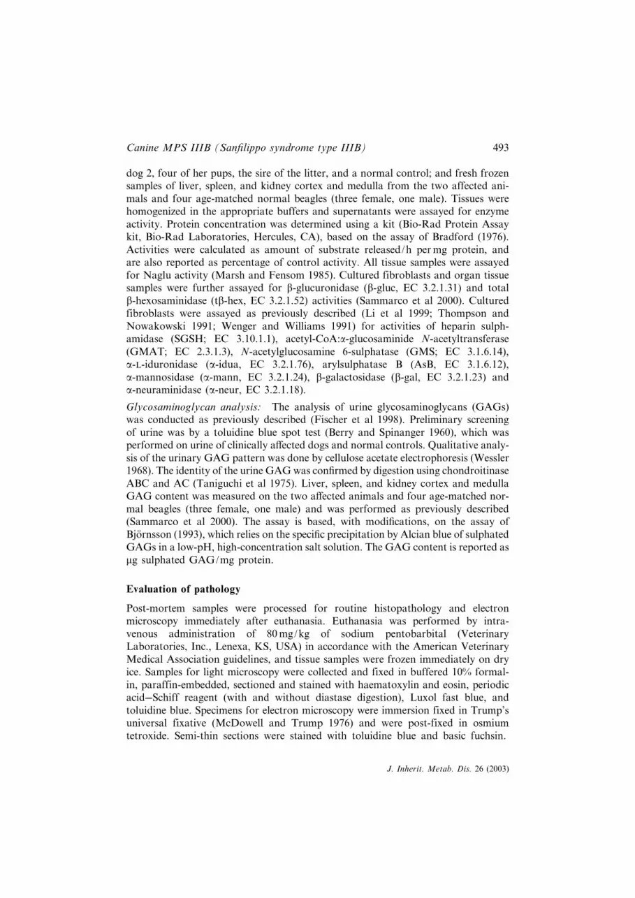
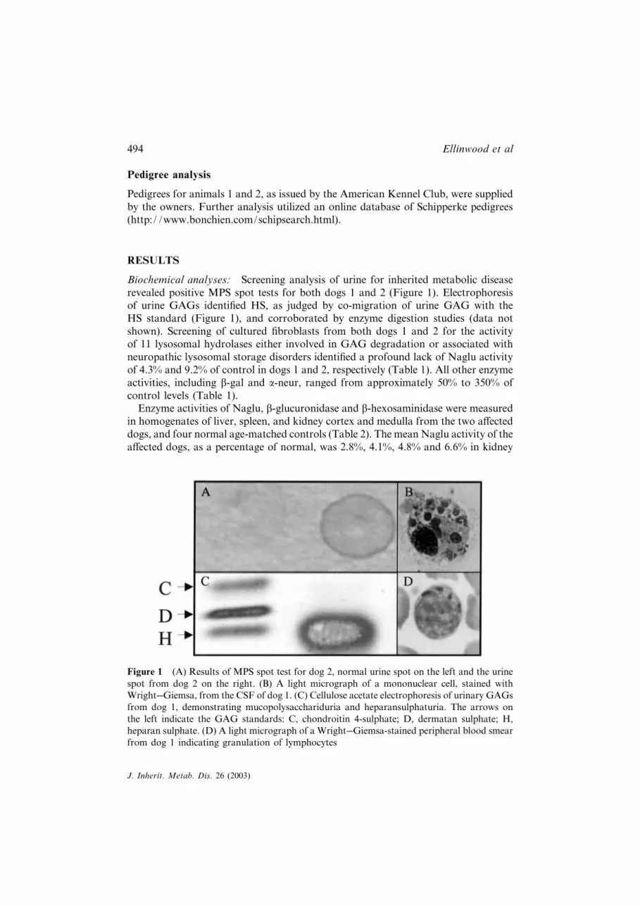
Biochemical analyses: Screening analysis of urine for inherited metabolic diseaserevealed positive MPS spot tests for both dogs 1 and 2 (Figure 1). Electrophoresisof urine GAGs identi¢ed HS, as judged by co-migration of urine GAG with theHS standard (Figure 1), and corroborated by enzyme digestion studies (data notshown). Screening of cultured ¢broblasts from both dogs 1 and 2 for the activityof 11 lysosomal hydrolases either involved in GAG degradation or associated withneuropathic lysosomal storage disorders identi¢ed a profound lack of Naglu activityof 4.3% and 9.2% of control in dogs 1 and 2, respectively (Table 1). All other enzymeactivities, including b-gal and a-neur, ranged from approximately 50% to 350% ofcontrol levels (Table 1).
Enzyme activities of Naglu, b-glucuronidase and b-hexosaminidase were measuredin homogenates of liver, spleen, and kidney cortex and medulla from the two a¡ecteddogs, and four normal age-matched controls (Table 2). The mean Naglu activity of thea¡ected dogs, as a percentage of normal, was 2.8%, 4.1%, 4.8% and 6.6% in kidney
Figure 1 (A) Results of MPS spot test for dog 2, normal urine spot on the left and the urinespot from dog 2 on the right. (B) A light micrograph of a mononuclear cell, stained withWright��Giemsa, from the CSF of dog 1. (C) Cellulose acetate electrophoresis of urinary GAGsfrom dog 1, demonstrating mucopolysacchariduria and heparansulphaturia. The arrows onthe left indicate the GAG standards: C, chondroitin 4-sulphate; D, dermatan sulphate; H,heparan sulphate. (D) A light micrograph of a Wright��Giemsa-stained peripheral blood smearfrom dog 1 indicating granulation of lymphocytes
494 Ellinwood et al
J. Inherit. Metab. Dis. 26 (2003)
Table1
Resultsof¢broblastenzymeassays
Dog1
Dog2
Enzyme
Disease
Controlc
%Normal
Controlc
%Normal
a-L-Idu
ronida
sea
MPS
I16
.214
.611
117
.415
.411
3Heparin
sulpha
midaseb
MPS
IIIA
695
208
334
330
208
159
N-A
cetyl-a-
D-glucosaminidasea
MPS
IIIB
0.34
8.0
4.3
0.61
6.7
9.2
Acetyl-CoA
:a-glucosaminide
N-acetyltransferase
bMPS
IIIC
1280
026
000
49.0
2740
026
000
105
N-A
cetylglucosamine6-sulpha
tase
cMPS
IIID
3.2
1.3
250
4.5
1.3
350
Arylsulph
ataseBd
MPS
VI
57.5
31.4
183
57.5
31.4
183
b-Glucuronida
sea
MPS
VII
71.5
111
64.4
75.3
46.6
162
b-Galactosida
sea
GM
1ga
nglio
sido
sis
130
143
90.4
260.1
165
157
b-Hexosam
inidase(total
activity)a
GM
2ga
nglio
sido
sis
150
0185
081
.547
541
811
4a-Man
nosida
sea
a-Man
nosdosis
142
132
107
226
162
139
a-Neuraminidasea
Sialidosis
1211
110
9.1
9.7
94a nmol
4-methy
lumberlliferon
e/hpermgprotein
bcpm/h
permgprotein
c mgN-acetylglucosamine/hpermgprotein
dnm
olnitrocatecho
l/hpermgprotein
e Con
trol
valueforan
unrelatedno
rmal
dog
Canine MPS IIIB (San¢lippo syndrome type IIIB) 495
J. Inherit. Metab. Dis. 26 (2003)

Table2
Organ
enzymeactivity
Kidney
medulla
Kidney
cortex
Liver
Spleen
b-Glucuronida
sea
A¡e
cted
b73
094
068
035
0Normal
(�stan
dard
deviation)
c28
0�11
046
0�12
026
0�52
360�37
A¡e
cted
as%
ofno
rmal
260
210
270
97
b-Hexosam
inidase(total
activity)a
A¡e
cted
b14
000
1700
013
000
440
0Normal
(�stan
dard
deviation)
c280
0�43
0380
0�110
0340
0�100
0310
0�140
0A¡e
cted
as%
ofno
rmal
520
430
390
140
N-A
cetyl-a-
D-glucosaminidasea
A¡e
cted
b0.32
0.40
0.35
0.21
Normal
(�stan
dard
deviation)
c11
�2.4
9.7�2.6
7.3�0.97
3.2�1.1
A¡e
cted
as%
ofno
rmal
2.8
4.1
4.8
6.6
a nmol
4-methy
lumberlliferon
e/hpermgprotein
bMeanof
values
fordo
gs1an
d2
c Meanof
values
for4age-matched
norm
alcontrolbeagle
dogs
(3femalean
d1male)
496 Ellinwood et al
J. Inherit. Metab. Dis. 26 (2003)

cortex, kidney medulla, liver and spleen, respectively (Table 2). The activities ofreference enzymes tended to be increased in the a¡ected dogs.
Total sulphated GAGs were measured in the liver, spleen, and kidney cortex andkidney medulla (Figure 2). Marked elevations of GAGs were seen in the liver(� 11-fold increase), kidney cortex (� 8-fold increase) and kidney medulla (� 7-foldincrease) in the a¡ected dogs. The values of GAGs from the spleen either were notincreased (dog 2) or were only marginally increased (dog 1) relative to the ¢ndingsin other tissues. Values of tissue GAGs were consistently higher in dog 1 relativeto dog 2, which may be related to age at euthanasia (5.6 versus 4.2 years of age,respectively).
Evaluation of pathology: Pathological ¢ndings were similar in both dogs andincluded grossly enlarged lateral ventricles (dog 2) and atrophied cerebellum.Histotologically, there was severe di¡use Purkinje cell loss, and the remaining Purkinjecells contained ¢ne cytoplasmic granules, were mildly swollen and were often dis-placed within the granular layer. There was variable, moderate to marked, di¡usethinning of the molecular and granular layers. Neuronal and ependymal cellvacuolation in the spinal cord was di¡use and moderate, with severe lesions inthe ventral horn. There was evidence of focal arachnoid proliferation (dog 1). Therewas di¡use, mild to severe, neuronal and glial cell vacuolation in the cerebrumand brainstem, which included cells of the spinal nucleus of the trigeminal nerve,
Figure 2 Total sulphated GAG values from the two MPS IIIB-a¡ected dogs reported as mgGAG/mg protein. The GAGs were assayed in liver, kidney cortex and kidney medulla, usingan Alcian blue dye precipitation method. Normal values were derived using 4 age-matchednormal beagles (3 females, 1 male). Error bars on normal values represent one standarddeviation
Canine MPS IIIB (San¢lippo syndrome type IIIB) 497
J. Inherit. Metab. Dis. 26 (2003)

Figure 3 Photomicrographs of the histopathology of cytoplasmic storage in brain, kidney andliver of dogs 1 and 2. (I) (�200) and (II) (�500) are stained with toluidine blue and showthe intracytoplasmic accumulation of acid GAGs in neurons of the midbrain of dog 2. (III)(�100) and (IV) (�400) show intracellular storage in the spinal nucleus of the trigeminal nerveof dog 1 stained with PAS. (III) shows distended neuronal cell bodies throughout the section.(IV) shows lighter-staining PAS-positive storage material (small arrow) compressing thedarker-staining Nissl substance around the nucleus (arrowhead). Storage vacuoles of perikarialand perithelial microglial cells are evident (large arrows). (V) and (VI) are photomicrographsof semi-thin sections of liver (V, �200) and kidney (VI, �200) from dog 1, stained with basicfuchsin, indicating severe hepatocellular and renal tubular epithelial accumulation of storagematerial. (VII) High magni¢cation (�500) of a neuronal cell body from the cerebrum ofdog 2, stained with Luxol fast blue, indicating probable glycosphingolipid storage material.Magni¢cations given are the original magni¢cations, reproduced at 80%
498 Ellinwood et al
J. Inherit. Metab. Dis. 26 (2003)

vestibular nuclei, cerebral cortex, hippocampus, hypothalamus, thalamus, andcaudate nucleus. Cytoplasmic vacuolation of perikarial and perithelial microglial cellswas evident, suggesting accumulation of GAGs. The spinal nucleus of the trigeminalnerve contained periodic acid��Schi¡ (PAS)-positive storage material in the cytoplasmof neurons (Figure 3).
Vacuoles were also evident in perikarial and perithelial microglial cells. Intracellularneuronal accumulation of toluidine blue- and Luxol fast blue-positive material is con-sistent with storage of GAGs and glycosphingolipids (Figure 3). While speci¢cimmunohistochemical staining was not conducted, this toluidine blue- and Luxol fastblue-positive staining pattern of storage material in the brain would not be inconsist-ent with the intracellular accumulation of HS and the glycosphingolipid GM2
ganglioside, the latter being known to accumulate in the brain of MPS III patients(Jones et al 1997; Wallace et al 1966) and in animal models of MPS I and IIID (Joneset al 1998; Shull et al 1984).
Hepatic accumulation of storage material was severe, located both in periportal andmidzonal hepatocytes as well as in Kup¡er cells (Figure 3). The kidney showed evi-dence of segmental accumulation of storage material (Figure 3) and moderate di¡usechronic pyelitis (dog 1). Mild accumulation of storage material, particularly inmacrophages, was noted in nearly every tissue examined, but also included theganglion cells of the retina, the bronchiolar epithelial cells, the ductal epithelialand islet cells of the pancreas, and the myenteric plexuses of the small and large
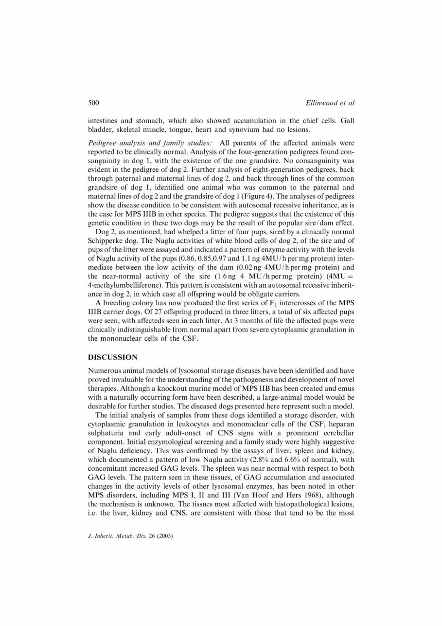
Figure 4 A pedigree showing all common ancestry found in eight-generation pedigrees of thesire and dam of dog 2 and the common grandsire of dog 1. The animal at the top centerof the pedigree was the only individual common to the pedigrees of all three animals. Thecommon grandsire of dog 1 and the sire and dam of dog 2 are shown as carriers (half-shadedsymbols), to re£ect their status as putative carrier and obligate carriers, respectively. The indi-viduals are intentionally shown using the sex-unknown symbol (diamond), and carriers arenot speci¢cally identi¢ed to maintain the anonymity of the pedigree
Canine MPS IIIB (San¢lippo syndrome type IIIB) 499
J. Inherit. Metab. Dis. 26 (2003)
intestines and stomach, which also showed accumulation in the chief cells. Gallbladder, skeletal muscle, tongue, heart and synovium had no lesions.
Pedigree analysis and family studies: All parents of the a¡ected animals werereported to be clinically normal. Analysis of the four-generation pedigrees found con-sanguinity in dog 1, with the existence of the one grandsire. No consanguinity wasevident in the pedigree of dog 2. Further analysis of eight-generation pedigrees, backthrough paternal and maternal lines of dog 2, and back through lines of the commongrandsire of dog 1, identi¢ed one animal who was common to the paternal andmaternal lines of dog 2 and the grandsire of dog 1 (Figure 4). The analyses of pedigreesshow the disease condition to be consistent with autosomal recessive inheritance, as isthe case for MPS IIIB in other species. The pedigree suggests that the existence of thisgenetic condition in these two dogs may be the result of the popular sire/dam e¡ect.
Dog 2, as mentioned, had whelped a litter of four pups, sired by a clinically normalSchipperke dog. The Naglu activities of white blood cells of dog 2, of the sire and ofpups of the litter were assayed and indicated a pattern of enzyme activity with the levelsof Naglu activity of the pups (0.86, 0.85,0.97 and 1.1 ng 4MU/h permg protein) inter-mediate between the low activity of the dam (0.02 ng 4MU/h permg protein) andthe near-normal activity of the sire (1.6 ng 4 MU/hpermg protein) (4MU¼
4-methylumbelliferone). This pattern is consistent with an autosomal recessive inherit-ance in dog 2, in which case all o¡spring would be obligate carriers.
A breeding colony has now produced the ¢rst series of F1 intercrosses of the MPSIIIB carrier dogs. Of 27 o¡spring produced in three litters, a total of six a¡ected pupswere seen, with a¡ecteds seen in each litter. At 3 months of life the a¡ected pups wereclinically indistinguishable from normal apart from severe cytoplasmic granulation inthe mononuclear cells of the CSF.
DISCUSSION
Numerous animal models of lysosomal storage diseases have been identified and haveproved invaluable for the understanding of the pathogenesis and development of noveltherapies. Although a knockout murine model of MPS IIB has been created and emuswith a naturally occurring form have been described, a large-animal model would bedesirable for further studies. The diseased dogs presented here represent such a model.
The initial analysis of samples from these dogs identi¢ed a storage disorder, withcytoplasmic granulation in leukocytes and mononuclear cells of the CSF, heparansulphaturia and early adult-onset of CNS signs with a prominent cerebellarcomponent. Initial enzymological screening and a family study were highly suggestiveof Naglu de¢ciency. This was con¢rmed by the assays of liver, spleen and kidney,which documented a pattern of low Naglu activity (2.8% and 6.6% of normal), withconcomitant increased GAG levels. The spleen was near normal with respect to bothGAG levels. The pattern seen in these tissues, of GAG accumulation and associatedchanges in the activity levels of other lysosomal enzymes, has been noted in otherMPS disorders, including MPS I, II and III (Van Hoof and Hers 1968), althoughthe mechanism is unknown. The tissues most a¡ected with histopathological lesions,i.e. the liver, kidney and CNS, are consistent with those that tend to be the most
500 Ellinwood et al
J. Inherit. Metab. Dis. 26 (2003)

a¡ected tissues in human cases of MPS III as well as in animal models (Bhaumik et al1999; Fischer et al 1998; Jolly et al 2000; Jones et al 1997; 1998; Li et al 1999; Wallaceet al 1966). The presence in the CNS of neurons that stain positive with Luxol fast blueor PAS with diastase treatment is indicative of storage of polar lipids or carbo-hydrate-containing vicinal glycal groups and saturated lipids, respectively (Lhotka1953; Pearse 1961, 1968). These staining properties would not be inconsistent withthe storage of GM2 and GM3 gangliosides, which are known to accumulate inthe neurons of humans with MPS III as well as in animal models of MPS III (Had¢eldet al 1980; Jones et al 1997, 1998; Li et al 1999; Wallace et al 1966).
While many of the CNS lesions are strikingly similar to those seen in human MPSIII, such as the storage in neurons of glycosphingolipids and the vacuolation ofmicroglial and perithelial cells, the most impressive ¢nding was the near-completeloss of Purkinje cells. Reports describing the neuropathological lesions of MPSIII in human patients are few and vary with respect to detail; they describe a broadrange of involvement of the cerebellum, ranging from an early report of MPS III(type unknown) in two siblings, aged 2 and 4 years, with marked decreases in thenumber of Purkinje and granular cells (Wallace et al 1966), to a case of MPS IIIBin a 17-year-old patient in which little evidence of cerebellar lesions was found(Had¢eld et al 1980), with more typical ¢ndings being occasional loss of Purkinjecells, ballooning of some remaining Purkinje cells and frequent expansion of theirmolecular layer dendrites with storage material (Dekaban and Constantopoulos 1977;Ghatak et al 1977; Jones et al 1997; Kriel et al 1978). The most likely explanation ofthe severity of the cerebellar signs of the canine form of MPS IIIB may be that itis a species peculiarity of HS accumulation seen in the dog. This conclusion is borneout by ¢ndings in the canine models of MPS IIIA, which have primarily cerebellarsigns and which show severe, either local or widespread, loss of Purkinje cells (Fischeret al 1998; Jolly et al 2000).
Analysis of the pedigree of the two animals reported in this study suggests the exist-ence of a popular sire/dam e¡ect in the Schipperke breed with an individual up to eightgenerations back that may have carried the mutant allele. A previous report of alysosomal storage disease in Schipperkes exists, detailing a clinical and pathologicalcondition that in every way mirrors the cases presented here, wherein the authorsposited a diagnosis of galactosialidosis (Knowles et al 1993), which unfortunatelycould not be con¢rmed with the tissues available. Given the extraordinary similarityof these cases, their occurrence in the same breed of dog, and the incomplete aeti-ological diagnosis of the former case, we speculate that the earlier report was in facta case of MPS IIIB.
The importance of animal models for the study of rare disorders has prompted us tofound a breeding colony to better study the pathogenesis of and treatments for MPSIIIB. Canine models of MPS I and VII have long been important tools in understand-ing pathological processes and evaluating treatments, and recent work in canine MPSVII has shown the dramatic potential of gene therapy (Ponder et al 2002). Thoroughanalyses of the ¢rst pups are underway. Areas of particular interest for study willinclude neuronal pathogenesis in general and the mechanism of Purkinje cell lossin particular, and evaluations of any of a number of potential therapeutic approaches
Canine MPS IIIB (San¢lippo syndrome type IIIB) 501
J. Inherit. Metab. Dis. 26 (2003)
including gene therapy, brain targeted therapy, stem cell therapy and substratereduction therapy.
ACKNOWLEDGEMENTS
The authors thank the owners of the animals presented here for allowing completeevaluations of their companion animals. Additionally we wish to acknowledge UlanaProcuik, for assistance with cell culture and Drs Polly Foreman, Gina Barone andLindsay Tomlinson, for assistance with neurological assessments, necropsies andhistopathological evaluation. Support for the work was provided by the NationalMPS Society, Inc., and the National Institutes of Health (RR007063 and RR002512).
REFERENCES
Aronovich EL, Carmichael KP, Morizono H, et al (2000) Canine heparan sulfate sulfamidaseand the molecular pathology underlying San¢lippo syndrome type A in Dachshunds.Genomics 68: 80��84.
Aronovich EL, Johnston JM, Wang P, Giger U, Whitley CB (2001) Molecular basis ofmucopolysaccharidosis type IIIB in emu (Dromaius novaehollandiae): an avian model ofSan¢lippo syndrome type B. Genomics 74: 299��305.
Bermudez AJ, Johnson GC, Vanier MT, et al (1995) Gangliosidosis in emus (Dromaiusnovaehollandiae). Avian Dis 39: 292��303.
Bermudez AJ, Freischutz B, Yu RK, et al (1997) Heritability and biochemistry of gangliosidosisin emus (Dromaius novaehollandiae). Avian Dis 41: 838��849.
Berry HK, Spinanger J (1960) A paper spot test useful in study of Hurler’s syndrome. J LabClinMed 55: 136��138.
Bhattacharyya R, Gliddon B, Beccari T, Hopwood JJ, Stanley P (2001) A novel missensemutation in lysosomal sulfamidase is the basis of MPS III A in a spontaneous mouse mutant.Glycobiology 11: 99��103.
Bhaumik M, Muller VJ, Rozaklis T, et al (1999) A mouse model for mucopolysaccharidosistype III A (San¢lippo syndrome). Glycobiology 9: 1389��1396.
Bjornsson S (1993) Simultaneous preparation and quantitation of proteoglycans by precipi-tation with alcian blue. Anal Biochem 210: 282��291.
BradfordMM (1976) A rapid and sensitive method for the quantitation of microgram quantitiesof protein utilizing the principle of protein��dye binding. Anal Biochem 72: 248��254.
Cavanagh KT, Leipprandt JR, Jones MZ, Friderici K (1995) Molecular defect of caprineN-acetylglucosamine-6-sulphatase de¢ciency. A single base substitution creates a stop codonin the 50-region of the coding sequence. J Inherit Metab Dis 18: 96.
Dekaban AS, Constantopoulos G (1977) Mucopolysaccharidosis type I, II, IIIA and V. Patho-logical and biochemical abnormalities in the neural and mesenchymal elements of the brain.Acta Neuropathol (Berl) 39: 1��7.
Fischer A, Carmichael KP, Munnell JF, et al (1998) Sulfamidase de¢ciency in a family ofDachshunds: a canine model of mucopolysaccharidosis IIIA (San¢lippo A). Pediatr Res44: 74��82.
Freischutz B, Tokuda A, Ariga T, Bermudez AJ, Yu RK (1997) Unusual gangliosidosis in emu(Dromaius novaehollandiae). J Neurochem 68: 2070��2078.
Friderici K, Cavanagh KT, Leipprandt JR, et al (1995) Cloning and sequence analysis ofcaprine N-acetylglucosamine 6-sulfatase cDNA. Biochim Biophys Acta 1271: 369��373.
502 Ellinwood et al
J. Inherit. Metab. Dis. 26 (2003)

Ghatak NR, Fleming DF, Hinman A (1977) Neuropathology of San¢lippo syndrome. AnnNeurol 2: 161��166.
Giger U, Shivaprasad H, Wang P, Jezyk P, Patterson D, Bradley G (1997)Mucopolysaccharidosis type IIIB (San¢lippo B syndrome) in emus. Vet Pathol 34: 473[Abstract].
Had¢eld MG, Ghatak NR, Nakoneczna I, et al (1980) Pathologic ¢ndings inmucopolysaccharidosis type IIIB (San¢lippo’s syndrome B). Arch Neurol 37: 645��650.
Jolly RD, Allan FJ, Collett MG, Rozaklis T, Muller VJ, Hopwood JJ (2000)Mucopolysaccharidosis IIIA (San¢lippo syndrome) in a New Zealand Huntaway dog withataxia NZ Vet J 48: 144��148.
Jones MZ, Alroy J, Rutledge JC, et al (1997) Human mucopolysaccharidosis IIID: clinical,biochemical morphological and immunohistochemical characteristics. J Neuropathol ExpNeurol 56: 1158��1167.
Jones MZ, Alroy J, Boyer PJ, et al (1998) Caprine mucopolysaccharidosis-IIID: clinical,biochemical, morphological and immunohistochemical characteristics. J Neuropathol ExpNeurol 57: 148��157.
Knowles K, Alroy J, Castagnaro M, Raghavan SS, Jakowski RM, Freden GO (1993)Adult-onset lysosomal storage disease in a Schipperke dog: clinical, morphological and bio-chemical studies. Acta Neuropathol (Berl) 86: 306��312.
Kriel RL, Hauser WA, Sung JH, Posalaky Z (1978) Neuroanatomical and electro-encephalographic correlations in San¢lippo syndrome type A. Arch Neurol 35: 838��843.
Lhotka JF (1953) Some histochemical observations on periodic acidPamino-acid mechanisms.Nature 171: 1123��1124.
Li HH, Yu WH, Rozengurt N, et al (1999) Mouse model of San¢lippo syndrome type B pro-duced by targeted disruption of the gene encoding alpha-N-acetylglucosaminidase. Proc NatlAcad Sci USA 96: 14505��14510.
Marsh J, Fensom AH (1985) 4-Methylumbelliferyl alpha-N-acetylglucosaminidase activity fordiagnosis of San¢lippo B disease. Clin Genet 27: 258��262.
McDowell EM, Trump BF (1976) Histologic ¢xatives suitable for diagnostic light and electronmicroscopy. Arch Pathol Lab Med 100: 405��414.
Meikle PJ, Hopwood JJ, Clague AE, Carey WF (1999) Prevalence of lysosomal storagedisorders. J Am Med Assoc 281: 249��254.
Neufeld EF, Muenzer J (2001) The mucopolysaccharidoses. In Scriver CR, Beaudet AL, SlyWS, Valle D, eds.; Childs B, Kinzler KW, Vogelstein B, assoc. eds. TheMetabolic andMole-cular Bases of Inherited Disease, 8th edn. New York: McGraw-Hill, 3421��3452.
Pearse AGE (1961) The plasmalemmal reaction. In Pearse AGE, ed. Histochemistry, Theor-etical and Applied. Boston: Little Brown, 347��365.
Pearse AGE (1968) Lipids, lipoproteins and proteolipids. In Pearse AGE, ed. Histochemistry,Theoretical and Applied. Boston: Little Brown, 398��446.
Ponder KP, Melniczek JR, Xu L, et al (2002) Therapeutic neonatal hepatic gene therapy inmucopolysaccharidosis VII dogs. Proc Natl Acad Sci USA 99: 13102��13107.
Poorthuis BJ, Wevers RA, Kleijer WJ, et al (1999) The frequency of lysosomal storage diseasesin The Netherlands. Hum Genet 105: 151��156.
Sammarco C,Weil M, Just C, et al (2000) E¡ects of bone marrow transplantation on the cardio-vascular abnormalities in canine mucopolysaccharidosis VII. Bone Marrow Transplant 25:1289��1297.
Shull RM, Helman RG, Spellacy E, Constantopoulos G, Munger RJ, Neufeld EF (1984) Mor-phologic and biochemical studies of canine mucopolysaccharidosis I. Am J Pathol 114:487��495.
Taniguchi N, Koizumi S, Masaki K, Kobayashi Y (1975) Diagnosis of genetic muco-polysaccharidoses: electrophoretic and enzymatic characterization of urinary glycosamino-glycans. Biochem Med 14: 241��249.
Canine MPS IIIB (San¢lippo syndrome type IIIB) 503
J. Inherit. Metab. Dis. 26 (2003)

Thompson JN, Nowakowski RW (1991) Enzymatic diagnosis of selected muco-polysaccharidosis: Hunter, Morquio type A. and San¢lippo types A, B, C, and D, andprocedures for measurement of 35SO4-glycosaminoglycans. In Hommes F, ed. Techniquesin Diagnostic Human Biochemical Genetics: A Laboratory Manual. New York: Wiley-Liss,567��586.
Thompson JN, Jones MZ, Dawson G, Hu¡man PS (1992) N-acetylglucosamine 6-sulphatasede¢ciency in a Nubian goat: a model of San¢lippo syndrome type D (mucopolysaccharidosisIIID). J Inherit Metab Dis 15: 760��768.
Van Hoof F, Hers HG (1968) The abnormalities of lysosomal enzymes in mucopoly-saccharidoses. Eur J Biochem 7: 34��44.
Wallace BJ, Kaplan D, Adachi M, Schneck L, Volk BW (1966) Mucopolysaccharidosis type 3.Morphologic and biochemical studies of two siblings with San¢lippo syndrome. Arch Pathol82: 462��473.
Wenger DA, Williams C (1991) Screening for lysosomal disorders. In Hommes F, ed. Tech-niques in Diagnostic Human Biochemical Genetics: A Laboratory Manual. New York:Wiley-Liss, 587��617.
Wessler E (1968) Analytical and preparative separation of acidic glycosaminoglycans by elec-trophoresis in barium acetate. Anal Biochem 26: 439��444.
Yogalingam G, Pollard T, Gliddon B, Jolly RD, Hopwood JJ (2002) Identi¢cation of amutation causing mucopolysaccharidosis type IIIA in New Zealand Huntaway dogs.Genomics 79: 150��153.
504 Ellinwood et al
J. Inherit. Metab. Dis. 26 (2003)
Related Documents