RESEARCH ARTICLE RESEARCH ARTICLE A Melanoma Cell State Distinction Influences Sensitivity to MAPK Pathway Inhibitors David J. Konieczkowski 1,2 , Cory M. Johannessen 1,2 , Omar Abudayyeh 1 , Jong Wook Kim 1,2 , Zachary A. Cooper 7,8 , Adriano Piris 3 , Dennie T. Frederick 4 , Michal Barzily-Rokni 1 , Ravid Straussman 1 , Rizwan Haq 5,6 , David E. Fisher 5,6 , Jill P. Mesirov 1 , William C. Hahn 1,2 , Keith T. Flaherty 5 , Jennifer A. Wargo 7,8 , Pablo Tamayo 1 , and Levi A. Garraway 1,2 on July 1, 2018. © 2014 American Association for Cancer Research. cancerdiscovery.aacrjournals.org Downloaded from Published OnlineFirst April 25, 2014; DOI: 10.1158/2159-8290.CD-13-0424

Welcome message from author
This document is posted to help you gain knowledge. Please leave a comment to let me know what you think about it! Share it to your friends and learn new things together.
Transcript

RESEARCH ARTICLE RESEARCH ARTICLE
A Melanoma Cell State Distinction Infl uences Sensitivity to MAPK Pathway Inhibitors David J. Konieczkowski 1 , 2 , Cory M. Johannessen 1 , 2 , Omar Abudayyeh 1 , Jong Wook Kim 1 , 2 , Zachary A. Cooper 7 , 8 , Adriano Piris 3 , Dennie T. Frederick 4 , Michal Barzily-Rokni 1 , Ravid Straussman 1 , Rizwan Haq 5 , 6 , David E. Fisher 5 , 6 , Jill P. Mesirov 1 , William C. Hahn 1 , 2 , Keith T. Flaherty 5 , Jennifer A. Wargo 7 , 8 , Pablo Tamayo 1 , and Levi A. Garraway 1 , 2
on July 1, 2018. © 2014 American Association for Cancer Research. cancerdiscovery.aacrjournals.org Downloaded from
Published OnlineFirst April 25, 2014; DOI: 10.1158/2159-8290.CD-13-0424
JULY 2014�CANCER DISCOVERY | 817
ABSTRACT Most melanomas harbor oncogenic BRAF V600 mutations, which constitutively acti-
vate the MAPK pathway. Although MAPK pathway inhibitors show clinical benefi t
in BRAF V600 -mutant melanoma, it remains incompletely understood why 10% to 20% of patients fail to
respond. Here, we show that RAF inhibitor–sensitive and inhibitor-resistant BRAF V600 -mutant melano-
mas display distinct transcriptional profi les. Whereas most drug-sensitive cell lines and patient biopsies
showed high expression and activity of the melanocytic lineage transcription factor MITF, intrinsically
resistant cell lines and biopsies displayed low MITF expression but higher levels of NF-κB signaling and
the receptor tyrosine kinase AXL. In vitro , these MITF-low/NF-κB–high melanomas were resistant to
inhibition of RAF and MEK, singly or in combination, and ERK. Moreover, in cell lines, NF-κB activation
antagonized MITF expression and induced both resistance marker genes and drug resistance. Thus,
distinct cell states characterized by MITF or NF-κB activity may infl uence intrinsic resistance to MAPK
pathway inhibitors in BRAF V600 -mutant melanoma.
SIGNIFICANCE: Although most BRAF V600 -mutant melanomas are sensitive to RAF and/or MEK inhibi-
tors, a subset fails to respond to such treatment. This study characterizes a transcriptional cell state
distinction linked to MITF and NF-κB that may modulate intrinsic sensitivity of melanomas to MAPK
pathway inhibitors. Cancer Discov; 4(7); 816–27. ©2014 AACR.
Authors’ Affi liations: 1 Broad Institute of Harvard and MIT, Cambridge; 2 Department of Medical Oncology, Dana-Farber Cancer Institute; Divi-sions of 3 Dermatopathology and 4 Surgical Oncology, 5 Massachusetts Gen-eral Hospital Cancer Center, and 6 Dermatology and Cutaneous Biology Research Center, Massachusetts General Hospital, Boston, Massachu-setts; Departments of 7 Surgical Oncology, and 8 Genomic Medicine, The University of Texas MD Anderson Cancer Center, Houston, Texas
Note: Supplementary data for this article are available at Cancer Discovery Online (http://cancerdiscovery.aacrjournals.org/).
D.J. Konieczkowski and C.M. Johannessen contributed equally to this work.
Corresponding Author: Levi A. Garraway, Department of Medical Oncol-ogy, Dana-Farber Cancer Institute, 450 Brookline Avenue, Boston, MA 02215. Phone: 617-632-6689; Fax: 617-582-7880; E-mail: [email protected]
doi: 10.1158/2159-8290.CD-13-0424
©2014 American Association for Cancer Research.
INTRODUCTION Mutations affecting codon 600 of the serine/threonine
kinase BRAF are among the most highly recurrent genetic
aberrations in melanoma ( 1, 2 ). These mutations activate the
downstream kinases MEK and ERK within the MAPK pathway,
leading to enhanced cellular proliferation and survival. The
discovery that BRAF V600 mutations predict sensitivity to MAPK
pathway inhibitors ( 3 ) revolutionized therapeutic approaches
to melanoma. MAPK pathway inhibitors—including the RAF
inhibitors (RAFi) vemurafenib and dabrafenib and the MEK
inhibitor (MEKi) trametinib—achieve clinical benefi t in 80% to
90% of patients with BRAF V600 -mutant melanoma ( 4–6 ). How-
ever, among patients whose tumors respond to MAPK pathway
inhibitors, relapse is universal (acquired resistance). Moreover,
10% to 20% of patients never achieve meaningful response to
therapy (innate or intrinsic resistance).
Recent studies have characterized numerous mechanisms of
acquired resistance to MAPK pathway inhibitors in BRAF V600 -
mutant melanoma. These include activating mutations in
the downstream kinase MEK ( 7–9 ) as well as acquisition of
an inhibitor-resistant BRAF splice variant ( 10 ). Alternative
MAP3K proteins [e.g., CRAF ( 11 ) or COT ( 12 )] are also able to
reengage the MAPK pathway in the presence of BRAF inhibi-
tion. Similarly, upstream of RAF proteins, activation of RAS
signaling [e.g., by mutation ( 13 ), NF1 loss ( 14, 15 ), or relief of
negative feedback ( 16 )] confers RAFi resistance. Cumulatively,
these studies have most commonly converged upon reactiva-
tion of the MAPK pathway as a common effector of many
mechanisms of acquired resistance.
In contrast, fewer studies have directly queried intrinsic
resistance to RAF inhibition in melanoma. Two recent stud-
ies examined stromal contributions to intrinsic resistance.
This work identifi ed stromal secretion of hepatocyte growth
factor (HGF ), activation of the receptor tyrosine kinase MET,
and subsequent MAPK pathway reactivation as a mechanism
of intrinsic MAPK pathway inhibitor resistance in melanoma
( 17, 18 ). It remains largely unknown, however, whether cell-
autonomous differences might also contribute to the intrin-
sic resistance phenotype. Therefore, we sought to elucidate
molecular features that might mediate intrinsic resistance to
RAF/MEK inhibition in BRAF V600 -mutant melanoma.
RESULTS We hypothesized that cell-autonomous differences, such as
distinct gene expression programs, might partially account for
why some melanomas display intrinsic resistance to MAPK
pathway inhibitors. To test this hypothesis, we examined 29
BRAF V600 -mutant melanoma cell lines from the Cancer Cell
Line Encyclopedia (CCLE; ref. 19 ) for which gene expression
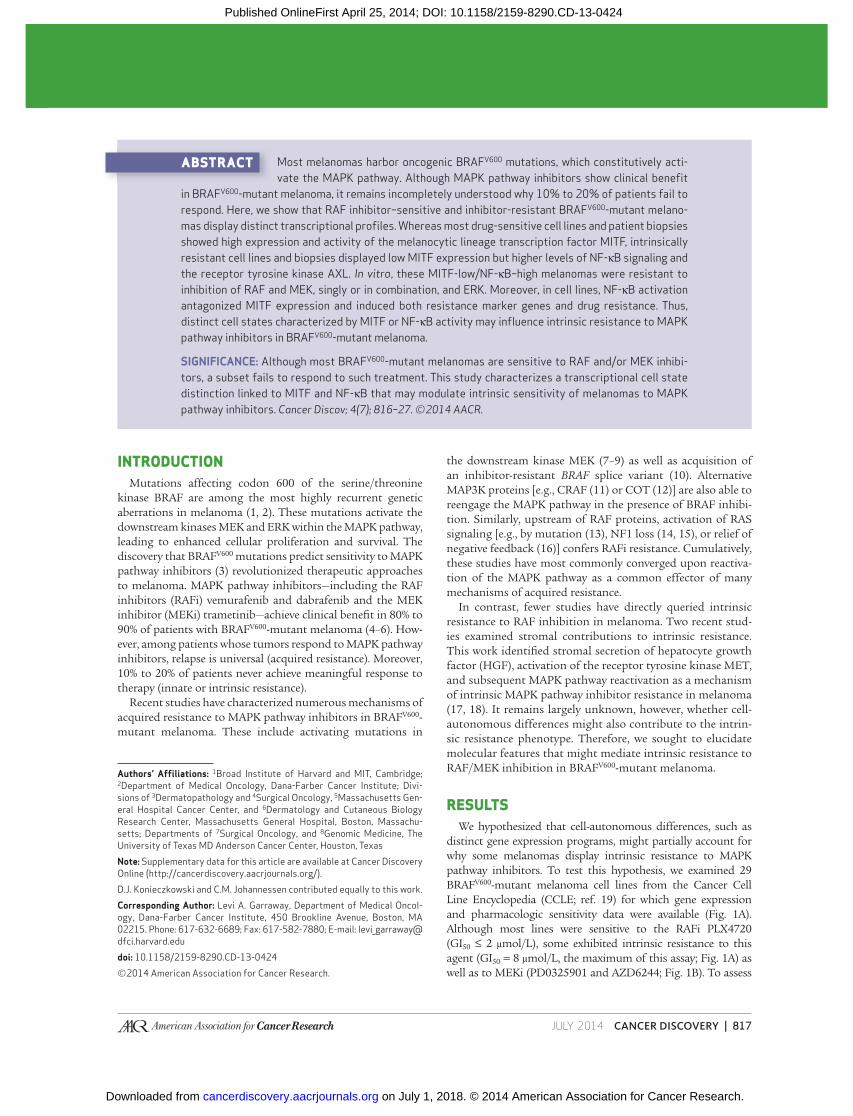
and pharmacologic sensitivity data were available ( Fig. 1A ).
Although most lines were sensitive to the RAFi PLX4720
(GI 50 ≤ 2 μmol/L ), some exhibited intrinsic resistance to this
agent (GI 50 = 8 μmol/L, the maximum of this assay; Fig. 1A ) as
well as to MEKi (PD0325901 and AZD6244; Fig. 1B ). To assess
on July 1, 2018. © 2014 American Association for Cancer Research. cancerdiscovery.aacrjournals.org Downloaded from
Published OnlineFirst April 25, 2014; DOI: 10.1158/2159-8290.CD-13-0424
818 | CANCER DISCOVERY�JULY 2014 www.aacrjournals.org
Konieczkowski et al.RESEARCH ARTICLE
whether differences in transcriptional programs existed between
intrinsically sensitive and resistant lines, we identifi ed genes
whose expression across the cell lines was strongly correlated
or anticorrelated with their PLX4720 GI 50 values. MITF , which
encodes a melanocyte lineage regulatory transcription factor
and melanoma oncogene ( 20 ), emerged as the single gene best
correlated with sensitivity to PLX4720 (Supplementary Fig. S1A
and Supplementary Table S1). Although MITF was strongly
expressed in the majority of drug-sensitive lines, it was poorly
expressed in the resistant lines ( Fig. 1B and Supplementary
Fig. S2). Because MITF transcriptional activity can be regulated
separately from MITF expression levels, we also measured MITF
function by querying expression of MITF target genes PMEL ,
TYRP1 , and MLANA , as well as a global transcriptional signature
of MITF activity ( 21 ). These too were poorly expressed in the
resistant lines, implying a reduction not only in expression but
also in activity of MITF ( Fig. 1B , Supplementary Fig. S2, and
Supplementary Table S1). Instead, drug-resistant BRAF V600 -
mutant melanoma cell lines displayed multiple expression
signatures of NF-κB activation, suggesting elevated NF-κB tran-
scriptional activity ( Fig. 1B ). Correspondingly, levels of phos-
phorylated, activated RELA (an NF-κB transcription factor; ref.
22 ) were increased in the majority of resistant lines relative to
the sensitive lines (Supplementary Fig. S2). Resistant lines also
expressed individual marker genes such as AXL , TPM1 , NRP1 ,
and CDH13 ( Fig. 1B and Supplementary Fig. S2) that were not
observed in the sensitive lines. These genes have not been pre-
viously characterized as MITF- or NF-κB–associated, but they
nominated additional features that might be associated with
the MITF-low/NF-κB–high transcriptional state.
Because prosurvival signaling through NF-κB has previously
been associated with resistance to cytotoxic therapies, such as
doxorubicin ( 23 ), we sought to assess whether the NF-κB–high
state was associated with generalized drug resistance. However,
among 24 targeted and cytotoxic anticancer drugs, differential
MITF expression levels were strongly correlated with differ-
ential sensitivity only to the four MAPK inhibitors (MAPKi )
tested (Supplementary Fig. S3). This fi nding suggests that the
MITF-low/NF-κB–high transcriptional state may pertain spe-
cifi cally to resistance to MAPK pathway inhibition.
To verify that the MITF/NF-κB class distinction was repro-
ducibly associated with differential sensitivity to MAPK path-
way inhibition, we examined a collection of patient-derived
BRAF V600 -mutant melanoma short-term cultures for which gene
expression data, but not pharmacologic sensitivity data, were
available. As in other datasets, we identifi ed reciprocity between
MITF and NF-κB levels across this collection ( Fig. 1C and Sup-
plementary Fig. S4). This relationship was evident for MITF
itself as well as for both the MITF transcriptional signature as a
whole ( Fig. 1C ) and individual MITF target genes ( Fig. 1C and
Supplementary Fig. S4), confi rming that MITF transcriptional
activity varied together with MITF expression across these
Figure 1. Association of expression classes with differential MAPK pathway inhibitor sensitivity in BRAF V600 -mutant melanoma in vitro . A, sensitiv-ity to PLX4720 (RAFi) across a collection of BRAF V600 -mutant melanoma cell lines. B, relationship between PLX4720 sensitivity and MITF-high versus NF-κB–high classes. C, transcriptional class distinction in BRAF V600 -mutant melanoma short-term cultures (STC). Red arrows identify cell lines predicted to be drug sensitive; blue arrows identify cell lines predicted to be drug resistant. D, relationship in short-term cultures between expression class and MAPK pathway inhibitor sensitivity. Graph shows GI 50 values relative to median GI 50 of the sensitive short-term cultures. PLX4720, RAFi; AZD6244, MEKi; VTX11E, ERK inhibitor (ERKi).
PL
X4
72
0 IC
(μm
ol/L
)
Re
sis
tan
ce
ma
rke
rs
Rela
tive
sensitiv
ity
Sig
.S
ig.
Ge
ne
sG
en
es
Se
nsitiv
ity
ma
rke
rs
Re
sis
tan
ce
ma
rke
rs
Sig
.S
ig.
Ge
ne
sG
en
es
Se
nsitiv
ity
ma
rke
rs
29 BRAFV600-mutant melanoma cell linesA
B
C
D29 BRAFV600-mutant melanoma cell lines
29 BRAFV600-mutant melanoma STCs
More drug-resistant
More drug-resistant
Predicted
drug-resistantPredicted drug-sensitive
More drug-sensitive
More drug-sensitive
WM
-88
WM
-88
WM
-98
3B
Ma
lme
-3M
UA
CC
-62
A-3
75
ME
L-H
OIG
R-3
7H
T-1
44
CO
LO
-67
9
RV
H-4
21
UA
CC
-25
7S
K-M
EL
-5W
M1
79
9G
-36
1M
DA
-MB
-43
5S
WM
-26
6-4
K0
29
AX
WM
-79
3C
OL
O 7
41
Hs 9
39
.TC
32
SK
-ME
L-2
4L
OX
IM
VI
Hs 6
95
TA
20
58
WM
-11
5H
s-2
94
TR
PM
I-7
95
1
IGR
-39
WM
-19
42
M-9
70
10
9M
-00
02
16
WM
-32
15
WM
-19
76
WM
-34
82
WM
-45
1L
uW
M-3
72
7M
-99
08
02
WM
-33
81
AW
M-3
21
8M
-00
09
21
WM
-35
26
M-0
10
32
2
WM
-31
63
WM
-36
27
WM
-18
62
WM
-32
59
WM
-98
4W
M-3
29
7W
M-1
72
0W
M-3
45
7
WM
-85
3.2
WM
-32
28
WM
-17
45
WM
-19
30
WM
-17
16
WM
-18
52
WM
-32
43
NC
I.A
5
UA
CC
-62
A-3
75
Ma
lme
-3M
ME
L-H
O
SK
-ME
L-5
WM
-98
3B
CO
LO
-67
9
RV
H-4
21
IGR
-37
UA
CC
-25
7
WM
17
99
G-3
61
HT-
14
4
WM
-26
6-4
Hs 9
39
.T
K0
29
AX
C3
2
MD
A-M
B-4
35
S
CO
LO
74
1
SK
-ME
L-2
4
Hs 6
95
T
Hs 2
94
T
IGR
-39
LO
X IM
VI
RP
MI-
79
51
WM
-11
5
WM
-79
3
–2 +2
MITF_GENE
0.842 <1e–04
0.931
PLX4720
AZD6244
PLX+AZD
VTX11E
WM
-186
2
WM
-194
2
WM
-197
6
WM
-321
5
WM
-348
2
WM
-372
7
WM
-451
Lu
WM
-174
5
WM
-185
2
WM
-193
0
25
20
15
10
5
0
<1e–040.904 <1e–04
0.691 <1e–040.509 0.00350.402 0.0257
–0.390 0.0013–0.445 1e–04–0.384 0.0014–0.280 0.0311
–0.371 0.0020
0.567 0.00120.539 0.00170.465 0.01280.437 0.0102
<1e–04
<1e–04
<1e–04<1e–04
<1e–04
2e–04
5e–040.031
0.001
2e–04
Matching score
(Index) P value
(Index)
Matching score
P value
0.7440.617
0.716
–0.290–0.371–0.435–0.470
–0.526–0.545
–0.561
MLANA_GENE
PMEL_GENETYRP1_GENE
AXL_GENE
TPM1_GENE
NRP1_GENE
CDH13_GENE
SEKI_LPS
HINATA_NFKB_FB
TIAN_TNF_NFKB
PLX4720
PD0325901AZD6244
MITF_GENEMLANA_GENE
PMEL_GENETYRP1_GENE
Yokoyama_MITF
CDH13_GENENRP1_GENETPM1_GENE
AXL_GENE
HINATA_NFKB_FB
TIAN_TNF_NFKBSEKI_LPS
Yokoyama_MITF
Z score
–2
Drug
sensitivity
+2Z score
A2
05
8
9
8
7
6
5
4
3
2
1
0
on July 1, 2018. © 2014 American Association for Cancer Research. cancerdiscovery.aacrjournals.org Downloaded from
Published OnlineFirst April 25, 2014; DOI: 10.1158/2159-8290.CD-13-0424
JULY 2014�CANCER DISCOVERY | 819
Melanoma Cell State Affects Intrinsic MAPK Inhibitor Resistance RESEARCH ARTICLE
samples. Correspondingly, both NF-κB–related gene expression
levels (e.g., AXL; Fig. 1C and Supplementary Fig. S4) and tran-
scriptional signatures of NF-κB pathway activity ( Fig. 1C ) varied
across the collection in a manner similar to that of the initial
melanoma cell line panel. Similarly, phosphorylated, activated
levels of the NF-κB transcription factor RELA (Supplementary
Fig. S4) also segregated inversely with MITF activity.
Next, we performed pharmacologic growth inhibition
studies on seven MITF-high and three NF-κB–high short-
term cultures. As predicted, all seven MITF-high/NF-κB–low
short-term cultures were sensitive to RAF and MEK inhibi-
tion, whereas each of the three MITF-low/NF-κB–high short-
term cultures was resistant to these agents ( Fig. 1D ). These
fi ndings supported the premise that the MITF-low/NF-κB–
high transcriptional signature correlated with resistance to
MAPK pathway inhibition in BRAF V600 -mutant melanoma.
We next sought to assess whether this intrinsic resistance
phenotype might derive from incomplete MAPK pathway
inhibition, as opposed to indifference to MAPK pathway
inhibition in these cells. First, we noted that MITF-low/
NF-κB–high short-term cultures ( Fig. 1D ) and cell lines (Sup-
plementary Fig. S5) showed resistance not only to single-agent
RAF and MEK inhibition, but also to combined RAF–MEK
inhibition and ERK inhibition (VTX11E), suggesting that
intrinsic resistance extends to multiple levels of the RAF–
MEK–ERK signaling cascade. Moreover, the resistance phe-
notype was not clearly attributable to incomplete MAPK
pathway suppression by these agents, because the reduction
of phosphorylation of both ERK and the ERK substrate FRA1
in resistant lines following exposure to RAFi, MEKi, and ERK
inhibitors (ERKi ) was comparable with that observed in sensi-
tive lines (Supplementary Fig. S6). Unique among the tested
resistant lines, RPMI-7951 cells maintain MAPK pathway
activity even in the setting of inhibitor treatment (Supplemen-
tary Fig. S6); however, this line also harbors amplifi cation of
MAP3K8 / COT , which is suffi cient to reactivate ERK signaling
following inhibition of RAF and/or MEK in this cell line ( 12 ).
Consistent with prior literature ( 24, 25 ), the ERKi VTX11E did
not inhibit phosphorylation of ERK itself. Nonetheless, the mag-
nitude of phosphorylated FRA1 (pFRA1) suppression by VTX11E
was comparable with that induced by the ERKi SCH772984 ( 25 ),
which inhibits both ERK activity and ERK phosphorylation (Sup-
plementary Fig. S7A). VTX11E and SCH772984 also showed a
similar spectrum of sensitivity across a panel of BRAF V600 -mutant
melanoma cell lines (Supplementary Figs. S4A and S7B).
In addition to showing comparable biochemical responses
to MAPK pathway inhibition, sensitive and resistant short-
term cultures (Supplementary Fig. S8) and cell lines (Sup-
plementary Fig. S9) displayed no clear differences in basal
phosphorylation levels of ERK or FRA1 or cell lines, imply-
ing that differential basal MAPK pathway activity does not
account for differential response to MAPK pathway inhibitors.
Together, these fi ndings suggest that a molecularly defi ned
subset of MITF-low/NF-κB–high BRAF V600 -mutant melano-
mas may exhibit indifference to MAPK pathway inhibition.
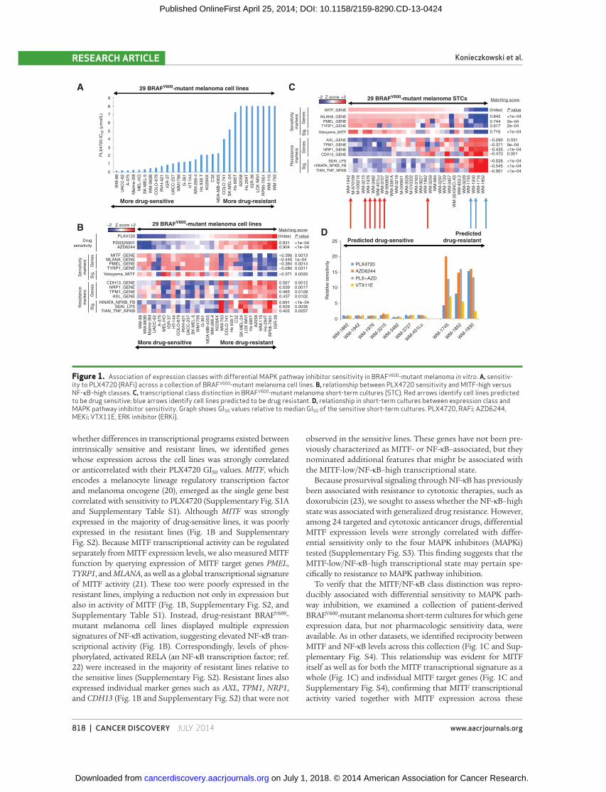
To confi rm that the apparent reciprocity between MITF
and NF-κB expression signatures was also evident in vivo , we
examined a collection of primary and metastatic BRAF V600 -
mutant melanomas from The Cancer Genome Atlas (https://
tcga-data.nci.nih.gov/tcga/). In this dataset, we also observed
anticorrelation between expression of MITF (and its activ-
ity, as measured by target genes and signature) and NF-κB
activation (as measured by single-gene markers as well as
an expression signature of NF-κB activity; Fig. 2A ). Thus,
the transcriptional class distinction that segregated MAPK
pathway inhibitor–sensitive and –resistant cell lines was also
discernible in melanoma tumors.
This reciprocity between MITF and NF-κB transcriptional pro-
fi les was reminiscent of prior transcriptional ( 26 ) and histopatho-
logic ( 27 ) evidence for a two-class distinction in melanoma.
These distinct gene expression programs, however, have not pre-
viously been linked to resistance to vemurafenib or dabrafenib/
trametinib in BRAF V600 -mutant melanomas. Extending these
prior results, our fi ndings suggest that this transcriptional class
distinction may have a previously unrecognized association with
differential susceptibility to MAPK pathway inhibition.
To assess whether the resistance phenotype linked to
this class distinction in vitro was also evident in melanoma
tumors, we examined biopsy specimens from patients with
metastatic BRAF V600 -mutant melanoma. Samples were
obtained before treatment with MAPK pathway inhibitors;
after biopsy, patients received combined RAFi–MEKi ther-
apy. Using AXL expression as a readout of the NF-κB–high
cellular state ( Figs. 1B and C and 2A and Supplementary
Figs. S2 and S4), we stratifi ed the cohort into MITF-high/
NF-κB–low ( n = 4) and MITF-low/NF-κB–high ( n = 8) groups
on the basis of immunohistochemistry ( Fig. 2B and Supple-
mentary Table S2). Immunocytochemistry on known MITF-
positive and AXL-positive cell lines confi rmed the sensitivity
and specifi city of this method (Supplementary Fig. S10).
Progression-free survival following dabrafenib/trametinib
therapy was signifi cantly shorter in the MITF-low/NF-κB–
high group relative to the MITF-high/NF-κB–low group
(median 5.0 months vs. 14.5 months; P = 0.0313, two-tailed t
test; Fig. 2C ). This fi nding is consistent with a possible thera-
peutic relevance of this two-class distinction in melanoma.
Among the individual features reproducibly associated with
the resistance state was the expression of the AXL receptor
tyrosine kinase. AXL has been previously identifi ed as a media-
tor of acquired resistance to PLX4720 in BRAF V600 -mutant
melanoma ( 12 ) and to lapatinib in EGFR-mutant lung cancer
( 28 ). Therefore, we queried whether the intrinsic resistance
phenotype in some BRAF V600 -mutant melanomas might simply
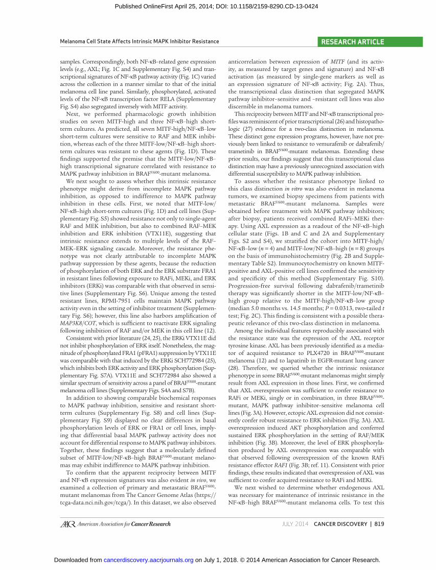
result from AXL expression in those lines. First, we confi rmed
that AXL overexpression was suffi cient to confer resistance to
RAFi or MEKi, singly or in combination, in three BRAF V600 -
mutant, MAPK pathway inhibitor–sensitive melanoma cell
lines ( Fig. 3A ). However, ectopic AXL expression did not consist-
ently confer robust resistance to ERK inhibition ( Fig. 3A ). AXL
overexpression induced AKT phosphorylation and conferred
sustained ERK phosphorylation in the setting of RAF/MEK
inhibition ( Fig. 3B ). Moreover, the level of ERK phosphoryla-
tion produced by AXL overexpression was comparable with
that observed following overexpression of the known RAFi
resistance effector RAF1 ( Fig. 3B ; ref. 11 ). Consistent with prior
fi ndings, these results indicated that overexpression of AXL was
suffi cient to confer acquired resistance to RAFi and MEKi.
We next wished to determine whether endogenous AXL
was necessary for maintenance of intrinsic resistance in the
NF-κB–high BRAF V600 -mutant melanoma cells. To test this
on July 1, 2018. © 2014 American Association for Cancer Research. cancerdiscovery.aacrjournals.org Downloaded from
Published OnlineFirst April 25, 2014; DOI: 10.1158/2159-8290.CD-13-0424

820 | CANCER DISCOVERY�JULY 2014 www.aacrjournals.org
Konieczkowski et al.RESEARCH ARTICLE
hypothesis, we used three small-molecule AXL inhibitors
[R428 ( 29 ), XL184 ( 30 ), and XL880 ( 28 , 31 )]. To confi rm the
pharmacologic effects of these compounds, we exposed A-375
melanoma cells engineered to overexpress AXL to each drug
in vitro . All three compounds abrogated AXL-mediated induc-
tion of AKT phosphorylation and rescue of ERK phosphor-
ylation ( Fig. 3C ) but had no effect on pAKT or pERK levels in
A-375 or other sensitive cell lines in the absence of exogenous
AXL expression (Supplementary Fig. S11).
Next, we assessed the effects of these small molecules on
intrinsically resistant melanoma cell lines that express endog-
enous AXL. In contrast to the setting of ectopic AXL expression,
we observed no effect of the AXL inhibitors on pAKT or pERK
levels in the intrinsically resistant lines, either at baseline or
following treatment with PLX4720 ( Fig. 3D ). (AXL inhibitors
also did not alter pERK or pAKT levels in intrinsically sensitive
lines; Supplementary Fig. S11.) Similarly, treatment with AXL
inhibitors did not alter the PLX4720 GI 50 values of any of the
intrinsically resistant melanoma lines ( Fig. 3E ). Comparable
results were observed following knockdown of AXL using three
independent shRNAs (the most effective of nine tested; Supple-
mentary Figs. S12 and S13). Here, we noted that a single AXL
shRNA (575) modulated PLX4720 GI 50 values; although this
fi nding could be due to marginally more effective knockdown
with this shRNA, the failure of the other AXL shRNAs and the
small-molecule AXL inhibitors to reproduce this phenotype
makes it more likely that this modulation stems from off-target
shRNA effects. Altogether, our results suggest that AXL expres-
sion may be suffi cient to confer acquired resistance to MAPK
pathway inhibitors, but may not be necessary for maintenance
of resistance. Although AXL expression may nonetheless con-
tribute to the intrinsic resistant phenotype in these melanoma
cells, AXL does not seem to be the sole or limiting resistance
effector in this setting. We therefore returned to the broader
differences in transcriptional state that exist among lines with
differential sensitivity to MAPK pathway inhibitors.
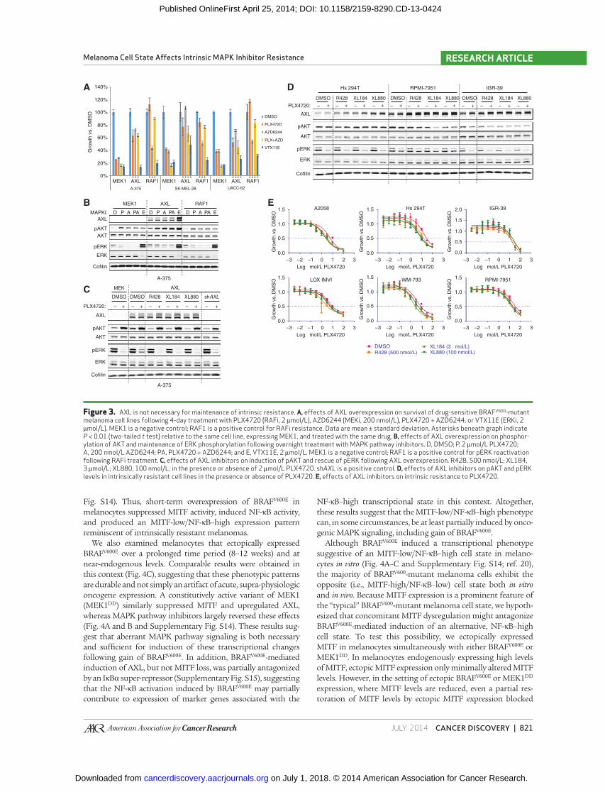
The foregoing experiments raised the possibility that distinct
melanoma cell states characterized by specifi c transcriptional
profi les might underpin intrinsic resistance to MAPK path-
way inhibition in BRAF V600 -mutant melanoma. One possible
explanation for the existence of these transcriptional states is
that they might arise from distinct precursor cells. To test
this possibility, we analyzed whether the transcriptional states
could both be established from immortalized primary human
melanocytes. At baseline, immortalized melanocytes expressed
high levels of both MITF and its target genes, reminiscent of
MITF-high/NF-κB–low melanoma cell lines. In addition, expres-
sion of NF-κB–associated signatures and marker genes was low
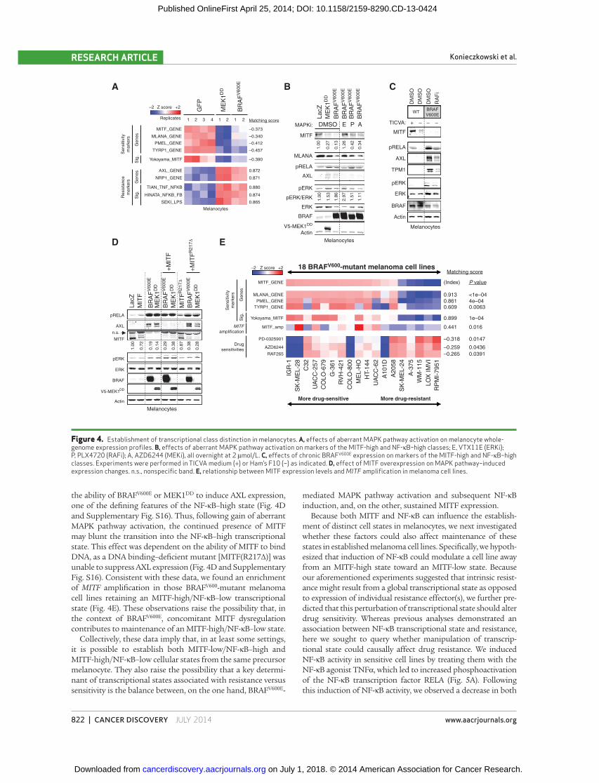
in these melanocytes ( Fig. 4A and B ). As expected, introduction
of BRAF V600E into immortalized melanocytes augmented ERK
phosphorylation ( Fig. 4B and Supplementary Fig. S14). Consist-
ent with prior observations ( 20 ), ectopic BRAF V600E also abro-
gated MITF expression [at both the transcriptional ( Fig. 4A )
and protein levels ( Fig. 4B and Supplementary Fig. S14)] and
MITF transcriptional activity (as measured by an expression
signature of MITF activity as well as levels of individual MITF
target genes; Fig. 4A and B and Supplementary Fig. S14). Sur-
prisingly, expression of BRAF V600E also induced NF-κB path-
way activation, as measured by expression of NF-κB–driven
transcriptional signatures ( Fig. 4A ), RELA phosphorylation
( Fig. 4B and Supplementary Fig. S14), and expression of mark-
ers such as AXL and NRP1 ( Fig. 4A and B and Supplementary
Figure 2. MITF-low/NF-κB–high transcriptional class is present and associated with resistance to MAPK pathway inhibition in human tumors. A, transcriptional class distinction in BRAF V600 -mutant melanoma tumor samples. B, examples of AXL and MITF staining in pretreat-ment melanoma biopsies (magni-fi cation, ×40). C, comparison of progression-free survival between MITF-positive/AXL-negative and MITF-negative/AXL-positive classes. *, P = 0.0313, two-tailed t test.
–2 +2Z score
MITF_GENE
113 BRAFV600-mutant melanoma tumorsA
B C
PMEL_GENE
MLANA_GENE
TYRP1_GENE
Yokoyama_MITF
NRP1_GENE
AXL_GENE
CDH13_GENE
SEKI_LPS
*
25
20
15
10
5
0
MITF
MIT
F(+)
AXL(–)
MIT
F(–)
AXL(+)
MITF+/AXL– MITF–/AXL+
AXL
Pro
gre
ssio
n-f
ree
su
rviv
al (m
o)
Re
sis
tan
ce
ma
rke
rs
Sig
.S
ig.
Ge
ne
sG
en
es
Se
nsitiv
ity
ma
rke
rs
(Index)
0.906
0.863
0.855
0.995
–0.505
–0.546
–0.552
–0.354 0.0426
1e–04
<1e–04
<1e–04
<1e–04
<1e–04
<1e–04
<1e–04
Matching score
P value
on July 1, 2018. © 2014 American Association for Cancer Research. cancerdiscovery.aacrjournals.org Downloaded from
Published OnlineFirst April 25, 2014; DOI: 10.1158/2159-8290.CD-13-0424
JULY 2014�CANCER DISCOVERY | 821
Melanoma Cell State Affects Intrinsic MAPK Inhibitor Resistance RESEARCH ARTICLE
Figure 3. AXL is not necessary for maintenance of intrinsic resistance. A, effects of AXL overexpression on survival of drug-sensitive BRAF V600 -mutant melanoma cell lines following 4-day treatment with PLX4720 (RAFi, 2 μmol/L), AZD6244 (MEKi, 200 nmol/L), PLX4720 + AZD6244, or VTX11E (ERKi, 2 μmol/L). MEK1 is a negative control; RAF1 is a positive control for RAFi resistance. Data are mean ± standard deviation. Asterisks beneath graph indicate P < 0.01 (two-tailed t test) relative to the same cell line, expressing MEK1, and treated with the same drug. B, effects of AXL overexpression on phosphor-ylation of AKT and maintenance of ERK phosphorylation following overnight treatment with MAPK pathway inhibitors. D, DMSO; P, 2 μmol/L PLX4720; A, 200 nmol/L AZD6244; PA, PLX4720 + AZD6244; and E, VTX11E, 2 μmol/L. MEK1 is a negative control; RAF1 is a positive control for pERK reactivation following RAFi treatment. C, effects of AXL inhibitors on induction of pAKT and rescue of pERK following AXL overexpression. R428, 500 nmol/L; XL184, 3 μmol/L; XL880, 100 nmol/L; in the presence or absence of 2 μmol/L PLX4720. sh AXL is a positive control. D, effects of AXL inhibitors on pAKT and pERK levels in intrinsically resistant cell lines in the presence or absence of PLX4720. E, effects of AXL inhibitors on intrinsic resistance to PLX4720.
MEK1
Gro
wth
vs. D
MS
O
Gro
wth
vs. D
MS
O
Gro
wth
vs. D
MS
O
Gro
wth
vs. D
MS
O
A
B
C
E
D120%
140%
100%
80%
60%
40%
20%
0%
A-375 SK-MEL-28 UACC-62
AXL*** * ** * ** * *** * *** * ***
RAF1 MEK1 AXL RAF1 MEK1 AXL RAF1
MEK1
MAPKi:
PLX4720:
AXL
AXL
pAKT
pAKT
pERK
ERK
Cofilin
AKT
AKT
pERK
ERK
Cofilin
A-375
A-375
D P A PA E
MEK
DMSO
– + +– +– +– +– +–
DMSO R428 XL184 XL880 shAXL
AXL
D P PAA ED P PAA E
AXL RAF1
DMSO
PLX4720
AZD6244
PLX+AZD
VTX11E
A2058 Hs 294T IGR-39
WM-793LOX IMVI
1.5
1.0
0.5
0.0
2.0
1.5
1.0
0.5
0.0
1.5
1.0
0.5
0.0
Gro
wth
vs. D
MS
O
Gro
wth
vs. D
MS
O
1.5
1.0
0.5
0.0
Log µmol/L PLX4720
–3 –2 –1 0 1 2 3
Gro
wth
vs. D
MS
O1.5
1.0
0.5
0.0
Log µmol/L PLX4720
–3 –2 –1 0 1 2 3
–3 –2 –1
Log µmol/L PLX4720
0 1 2 3
DMSOR428 (500 nmol/L) XL880 (100 nmol/L)
XL184 (3 µmol/L)
1.5 RPMI-7951
1.0
0.5
0.0
Log µmol/L PLX4720
–3 –2 –1 0 1 2 3
Log µmol/L PLX4720
–3 –2 –1 0 1 2 3
–3 –2 –1
Log µmol/L PLX4720
0 1 2 3
Hs 294T RPMI-7951 IGR-39
PLX4720:
AXL
pAKT
AKT
pERK
ERK
Cofilin
DMSO
– + – + – + – + – + – + – + – + – + – + – + – +
R428 XL184 XL880 DMSO R428 XL184 XL880 DMSO R428 XL184 XL880
Fig. S14). Thus, short-term overexpression of BRAF V600E in
melanocytes suppressed MITF activity, induced NF-κB activity,
and produced an MITF-low/NF-κB–high expression pattern
reminiscent of intrinsically resistant melanomas.
We also examined melanocytes that ectopically expressed
BRAF V600E over a prolonged time period (8–12 weeks) and at
near-endogenous levels. Comparable results were obtained in
this context ( Fig. 4C ), suggesting that these phenotypic patterns
are durable and not simply an artifact of acute, supra-physiologic
oncogene expression. A constitutively active variant of MEK1
(MEK1 DD ) similarly suppressed MITF and upregulated AXL,
whereas MAPK pathway inhibitors largely reversed these effects
( Fig. 4A and B and Supplementary Fig. S14). These results sug-
gest that aberrant MAPK pathway signaling is both necessary
and suffi cient for induction of these transcriptional changes
following gain of BRAF V600E . In addition, BRAF V600E -mediated
induction of AXL, but not MITF loss, was partially antagonized
by an IκBα super-repressor (Supplementary Fig. S15), suggesting
that the NF-κB activation induced by BRAF V600E may partially
contribute to expression of marker genes associated with the
NF-κB–high transcriptional state in this context. Altogether,
these results suggest that the MITF-low/NF-κB–high phenotype
can, in some circumstances, be at least partially induced by onco-
genic MAPK signaling, including gain of BRAF V600E .
Although BRAF V600E induced a transcriptional phenotype
suggestive of an MITF-low/NF-κB–high cell state in melano-
cytes in vitro ( Fig. 4A–C and Supplementary Fig. S14; ref. 20 ),
the majority of BRAF V600 -mutant melanoma cells exhibit the
opposite (i.e., MITF-high/NF-κB–low) cell state both in vitro
and in vivo . Because MITF expression is a prominent feature of
the “typical” BRAF V600 -mutant melanoma cell state, we hypoth-
esized that concomitant MITF dysregulation might antagonize
BRAF V600E -mediated induction of an alternative, NF-κB–high
cell state. To test this possibility, we ectopically expressed
MITF in melanocytes simultaneously with either BRAF V600E or
MEK1 DD . In melanocytes endogenously expressing high levels
of MITF, ectopic MITF expression only minimally altered MITF
levels. However, in the setting of ectopic BRAF V600E or MEK1 DD
expression, where MITF levels are reduced, even a partial res-
toration of MITF levels by ectopic MITF expression blocked
on July 1, 2018. © 2014 American Association for Cancer Research. cancerdiscovery.aacrjournals.org Downloaded from
Published OnlineFirst April 25, 2014; DOI: 10.1158/2159-8290.CD-13-0424
822 | CANCER DISCOVERY�JULY 2014 www.aacrjournals.org
Konieczkowski et al.RESEARCH ARTICLE
the ability of BRAF V600E or MEK1 DD to induce AXL expression,
one of the defi ning features of the NF-κB–high state ( Fig. 4D
and Supplementary Fig. S16). Thus, following gain of aberrant
MAPK pathway activation, the continued presence of MITF
may blunt the transition into the NF-κB–high transcriptional
state. This effect was dependent on the ability of MITF to bind
DNA, as a DNA binding–defi cient mutant [MITF(R217Δ)] was
unable to suppress AXL expression ( Fig. 4D and Supplementary
Fig. S16). Consistent with these data, we found an enrichment
of MITF amplifi cation in those BRAF V600 -mutant melanoma
cell lines retaining an MITF-high/NF-κB–low transcriptional
state ( Fig. 4E ). These observations raise the possibility that, in
the context of BRAF V600E , concomitant MITF dysregulation
contributes to maintenance of an MITF-high/NF-κB–low state.
Collectively, these data imply that, in at least some settings,
it is possible to establish both MITF-low/NF-κB–high and
MITF-high/NF-κB–low cellular states from the same precursor
melanocyte. They also raise the possibility that a key determi-
nant of transcriptional states associated with resistance versus
sensitivity is the balance between, on the one hand, BRAF V600E -
mediated MAPK pathway activation and subsequent NF-κB
induction, and, on the other, sustained MITF expression.
Because both MITF and NF-κB can infl uence the establish-
ment of distinct cell states in melanocytes, we next investigated
whether these factors could also affect maintenance of these
states in established melanoma cell lines. Specifi cally, we hypoth-
esized that induction of NF-κB could modulate a cell line away
from an MITF-high state toward an MITF-low state. Because
our aforementioned experiments suggested that intrinsic resist-
ance might result from a global transcriptional state as opposed
to expression of individual resistance effector(s), we further pre-
dicted that this perturbation of transcriptional state should alter
drug sensitivity. Whereas previous analyses demonstrated an
association between NF-κB transcriptional state and resistance,
here we sought to query whether manipulation of transcrip-
tional state could causally affect drug resistance. We induced
NF-κB activity in sensitive cell lines by treating them with the
NF-κB agonist TNFα, which led to increased phosphoactivation
of the NF-κB transcription factor RELA ( Fig. 5A ). Following
this induction of NF-κB activity, we observed a decrease in both
Figure 4. Establishment of transcriptional class distinction in melanocytes. A, effects of aberrant MAPK pathway activation on melanocyte whole-genome expression profi les. B, effects of aberrant MAPK pathway activation on markers of the MITF-high and NF-κB–high classes; E, VTX11E (ERKi); P, PLX4720 (RAFi); A, AZD6244 (MEKi), all overnight at 2 μmol/L. C, effects of chronic BRAF V600E expression on markers of the MITF-high and NF-κB–high classes. Experiments were performed in TICVA medium (+) or Ham’s F10 (−) as indicated. D, effect of MITF overexpression on MAPK pathway–induced expression changes. n.s., nonspecifi c band. E, relationship between MITF expression levels and MITF amplifi cation in melanoma cell lines.
MITF_GENE
A B C
D
Replicates
GF
P
Genes
1.0
0
0.7
2
0.1
9
0.1
4
0.2
9
0.3
8
0.8
7
0.3
8
0.2
8
Sig
.S
ig.
Resis
tance
mark
ers
Sensitiv
ity
mark
ers
Genes
Sig
.
Sensitiv
ity
mark
ers
Genes
ME
K1
DD
BR
AF
V6
00
E
BR
AF
V6
00
E
ME
K1
DD
ME
K1
DD
ME
K1
DD
IGR
-1
C32
UA
CC
-25
7
CO
LO
-67
9
G-3
61
RV
H-4
21
CO
LO
-80
0
ME
L-H
O
HT-
14
4
UA
CC
-62
A1
01
D
A2
05
8
A-3
75
WM
-11
5
LO
X I
MV
I
RP
MI-
79
51
SK
-ME
L-2
4
SK
-ME
L-2
8
BR
AF
V6
00
E
MIT
FR
21
7Δ
+MIT
FR
21
7Δ
+M
ITF
BR
AF
V6
00
E
MIT
F
La
cZ
1 2 3 4 1 2 1 2
–0.373
MelanocytesD
MS
O
DM
SO
DM
SO
RA
Fi
MITF
WTBRAF
V600E
pRELA
AXL
TPM1
pERK
ERK
BRAF
Actin
Melanocytes
TICVA: – – –+
–0.340
–0.412
–0.457
–0.390
0.872
0.871
0.880
0.874
0.865
Matching score
–2 Z score +2
–2 Z score +2
MLANA_GENE
PMEL_GENE
TYRP1_GENE
Yokoyama_MITF
pRELA
pERK
ERK
BRAF
V5-MEK1
Actin
AXL
n.s.
MITF
AXL_GENE
NRP1_GENE
TIAN_TNF_NFKB
HINATA_NFKB_FB
SEKI_LPS
MITF_GENE
MLANA_GENE
PMEL_GENE
TYRP1_GENE
Yokoyama_MITF
MITF_amp
PD-0325901
MITFamplification
Drug
sensitivitiesAZD6244
RAF265
Melanocytes
18 BRAFV600-mutant melanoma cell lines
0.9130.8610.609
0.899
0.441
–0.318
–0.259
–0.265 0.0391
0.0436
0.0147
0.016
1e–04
0.00634e–04<1e–04
Matching score
(Index) P value
More drug-resistantMore drug-sensitive
E
DMSO E AP
1.0
01.0
0
1.5
3
1.8
6
2.9
7
1.5
1
1.1
1
0.2
7
0.1
3
1.2
6
0.4
2
0.3
4
BR
AF
V6
00
E
ME
K1
DD
BR
AF
V6
00
E
BR
AF
V6
00
E
BR
AF
V6
00
E
La
cZ
MITF
MLANA
pRELA
AXL
pERK
pERK/ERK
ERK
Melanocytes
BRAF
V5-MEK1DD
Actin
MAPKi:
on July 1, 2018. © 2014 American Association for Cancer Research. cancerdiscovery.aacrjournals.org Downloaded from
Published OnlineFirst April 25, 2014; DOI: 10.1158/2159-8290.CD-13-0424
JULY 2014�CANCER DISCOVERY | 823
Melanoma Cell State Affects Intrinsic MAPK Inhibitor Resistance RESEARCH ARTICLE
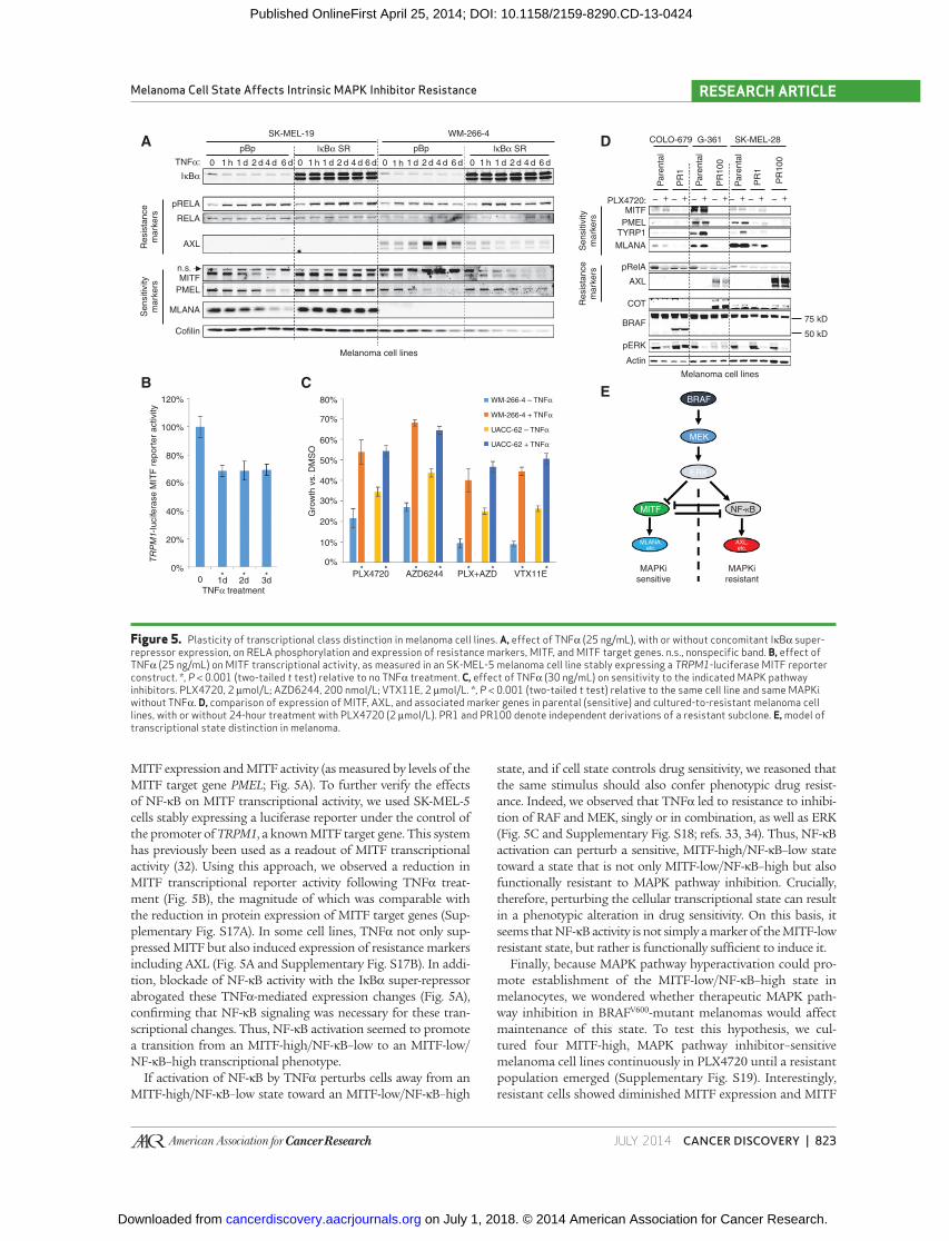
MITF expression and MITF activity (as measured by levels of the
MITF target gene PMEL ; Fig. 5A ). To further verify the effects
of NF-κB on MITF transcriptional activity, we used SK-MEL-5
cells stably expressing a luciferase reporter under the control of
the promoter of TRPM1 , a known MITF target gene. This system
has previously been used as a readout of MITF transcriptional
activity ( 32 ). Using this approach, we observed a reduction in
MITF transcriptional reporter activity following TNFα treat-
ment ( Fig. 5B ), the magnitude of which was comparable with
the reduction in protein expression of MITF target genes (Sup-
plementary Fig. S17A). In some cell lines, TNFα not only sup-
pressed MITF but also induced expression of resistance markers
including AXL ( Fig. 5A and Supplementary Fig. S17B). In addi-
tion, blockade of NF-κB activity with the IκBα super-repressor
abrogated these TNFα-mediated expression changes ( Fig. 5A ),
confi rming that NF-κB signaling was necessary for these tran-
scriptional changes. Thus, NF-κB activation seemed to promote
a transition from an MITF-high/NF-κB–low to an MITF-low/
NF-κB–high transcriptional phenotype.
If activation of NF-κB by TNFα perturbs cells away from an
MITF-high/NF-κB–low state toward an MITF-low/NF-κB–high
state, and if cell state controls drug sensitivity, we reasoned that
the same stimulus should also confer phenotypic drug resist-
ance. Indeed, we observed that TNFα led to resistance to inhibi-
tion of RAF and MEK, singly or in combination, as well as ERK
( Fig. 5C and Supplementary Fig. S18; refs. 33, 34 ). Thus, NF-κB
activation can perturb a sensitive, MITF-high/NF-κB–low state
toward a state that is not only MITF-low/NF-κB–high but also
functionally resistant to MAPK pathway inhibition. Crucially,
therefore, perturbing the cellular transcriptional state can result
in a phenotypic alteration in drug sensitivity. On this basis, it
seems that NF-κB activity is not simply a marker of the MITF-low
resistant state, but rather is functionally suffi cient to induce it.
Finally, because MAPK pathway hyperactivation could pro-
mote establishment of the MITF-low/NF-κB–high state in
melanocytes, we wondered whether therapeutic MAPK path-
way inhibition in BRAF V600 -mutant melanomas would affect
maintenance of this state. To test this hypothesis, we cul-
tured four MITF-high, MAPK pathway inhibitor–sensitive
melanoma cell lines continuously in PLX4720 until a resistant
population emerged (Supplementary Fig. S19). Interestingly,
resistant cells showed diminished MITF expression and MITF
Figure 5. Plasticity of transcriptional class distinction in melanoma cell lines. A, effect of TNFα (25 ng/mL), with or without concomitant IκBα super-repressor expression, on RELA phosphorylation and expression of resistance markers, MITF, and MITF target genes. n.s., nonspecifi c band. B, effect of TNFα (25 ng/mL) on MITF transcriptional activity, as measured in an SK-MEL-5 melanoma cell line stably expressing a TRPM1 -luciferase MITF reporter construct. *, P < 0.001 (two-tailed t test) relative to no TNFα treatment. C, effect of TNFα (30 ng/mL) on sensitivity to the indicated MAPK pathway inhibitors. PLX4720, 2 μmol/L; AZD6244, 200 nmol/L; VTX11E, 2 μmol/L. *, P < 0.001 (two-tailed t test) relative to the same cell line and same MAPKi without TNFα. D, comparison of expression of MITF, AXL, and associated marker genes in parental (sensitive) and cultured-to-resistant melanoma cell lines, with or without 24-hour treatment with PLX4720 (2 μmol/L). PR1 and PR100 denote independent derivations of a resistant subclone. E, model of transcriptional state distinction in melanoma.
SK-MEL-19A D
ECB
pRELAMITF
PLX4720: – + – + – + – + – + – + – +
COLO-679
PMELTYRP1
MLANA
pRelA
AXL
COT
BRAF
pERK
Actin
Melanoma cell lines
75 kD
50 kD
RELA
AXL
PMEL
Cofilin
BRAF
MEK
ERK
NF-KB
MAPKi
sensitive
MAPKi
resistant
MITF
MLANA,etc.
AXL,etc.
WM-266-4 – TNFα
TNFα treatment
WM-266-4 + TNFα
UACC-62 – TNFα
UACC-62 + TNFα
80%
70%
60%
50%
40%
30%
20%
10%
0%
120%
100%
80%
60%
40%
20%
0%
0 1d* PLX4720
**AZD6244 PLX+AZD
** **VTX11E
**
2d*
3d*
Melanoma cell lines
MLANA
MITFn.s.
WM-266-4
pBp
0 1 h 1 d 2 d 4 d 6 d 0 1 h 1 d 2 d 4 d 6 d 0 1 h 1 d 2 d 4 d 6 d 0 1 h 1 d 2 d 4 d 6 d
pBplκBα SR
TNFα:
lκBα SRR
esis
tance
mark
ers
Sensitiv
ity
mark
ers
TR
PM
1-Iu
cife
rase M
ITF
report
er
activity
Gro
wth
vs. D
MS
O
Resis
tance
mark
ers
Sensitiv
ity
mark
ers
Pare
nta
l
Pare
nta
l
Pare
nta
l
PR
1
PR
1
PR
100
PR
100
G-361 SK-MEL-28
IκBα
on July 1, 2018. © 2014 American Association for Cancer Research. cancerdiscovery.aacrjournals.org Downloaded from
Published OnlineFirst April 25, 2014; DOI: 10.1158/2159-8290.CD-13-0424
824 | CANCER DISCOVERY�JULY 2014 www.aacrjournals.org
Konieczkowski et al.RESEARCH ARTICLE
transcriptional activity (as measured by levels of MITF target
genes TYRP1 , MLANA , and PMEL ; 4/4 lines) and gain of AXL
expression (2/4 lines; Fig. 5D ). These results suggest that in
some contexts, MITF-high/NF-κB–low melanomas can transi-
tion toward an MITF-low/NF-κB–high state during acquisi-
tion of resistance. These changes were observed even in clones
that had also gained other known mechanisms of resistance
[ Fig. 5D ; e.g., COT expression and p61 BRAF splice variant ( 10 ,
12 )]. This fi nding implies that transition toward an MITF-low/
NF-κB–high state is not mutually exclusive with acquisition
of other known resistance effectors—an emerging theme in
resistance to targeted therapeutics ( 35 ). Moreover, this fi nding
suggests that the MITF-low/NF-κB–high state, although cer-
tainly associated with intrinsic resistance, can be also observed
in the context of acquired resistance. Thus, the MITF-low/
NF-κB–high transcriptional phenotype may generally signify
diminished dependence on the MAPK pathway. Melanomas
that begin in this MITF-low/NF-κB–high state before treat-
ment are likely to be intrinsically resistant to MAPK pathway
inhibition, whereas melanomas that transition into this state
during treatment can exhibit acquired resistance. Cumula-
tively, these fi ndings demonstrate that, even in melanoma
cell lines, the transcriptional states associated with sensitivity
and resistance remain plastic; moreover, maintenance of these
states in cell lines can be perturbed by the same mediators that
govern establishment of these states in melanocytes ( Fig. 5E ).
DISCUSSION The majority of BRAF V600 -mutant melanomas are pro-
foundly dependent on the RAF–MEK–ERK signaling cas-
cade. This vulnerability has been exploited clinically with the
development of pharmacologic RAFi and MEKi. However,
the effi cacy of these drugs is limited both by relapse follow-
ing an initial response (acquired resistance) and by the initial
indifference of some BRAF V600 -mutant melanomas to these
inhibitors (intrinsic resistance). Multiple studies have eluci-
dated mechanisms of acquired resistance to MAPK pathway
inhibitor therapy, largely converging on reactivation of the
MAPK pathway ( 7 , 10–16 ). Although recent work has identi-
fi ed stromal HGF secretion as one mechanism of intrinsic
resistance ( 17, 18 ), less has been known about whether there
also exist cell-autonomous determinants of this phenotype.
In this study, we used a collection of BRAF V600 -mutant
melanoma cell lines displaying a spectrum of sensitivity to
MAPK pathway inhibitors to identify two transcriptional states
in BRAF V600 -mutant melanoma: one characterized by high
MITF expression and transcriptional activity that is sensitive to
MAPK pathway inhibition, and another that exhibits low MITF
expression and activity, high NF-κB activity, and resistance to
MAPK pathway inhibition. The MITF-low/NF-κB–high state
seems specifi cally resistant to MAPK pathway inhibition, rather
than globally drug-tolerant, as cross-resistance to other classes
of therapeutics was not observed. Moreover, such a transcrip-
tional class distinction is reminiscent of prior work identifying
two differential gene expression classes in melanoma ( 26, 27 ).
However, both the mechanistic basis and the therapeutic impli-
cations of this two-class distinction have been largely uncharac-
terized; in particular, it has not previously been associated with
differential response to vemurafenib or dabrafenib/trametinib
in the setting of BRAF V600 mutation. Conversely, although
the phenomenon of intrinsic sensitivity/resistance to MAPK
pathway inhibitors has more recently become evident, a cell-
intrinsic mechanistic basis for this phenomenon has remained
largely unknown. Thus, our current work may unify and extend
two previously described phenomena in melanoma by propos-
ing gene expression reciprocity between MITF and NF-κB as a
mechanistic determinant of intrinsic resistance.
Our data suggest that the origin of these two distinct
states in melanocytes can be controlled by the relative balance
of aberrant MAPK activation (leading to NF-κB activation
and the MITF-low/NF-κB–high state) and sustained MITF
expression and activity (leading to the MITF-high/NF-κB–low
state). Because we and others have shown that introduction of
BRAF V600E into melanocytes can lead to loss of MITF expres-
sion and activity, it may seem surprising that the majority
of BRAF V600 -mutant melanomas retain MITF expression and
activity and low levels of NF-κB activity. Of note, we also show
that chronic MAPK pathway inhibition led some melanoma
lines to transition to an MITF-low/NF-κB–high state. Although
one possible explanation for this fi nding is simply outgrowth
of a preexisting resistant subpopulation, it may also suggest
that not all melanomas preserve the same relationship between
MAPK signaling and MITF levels as observed in melanocytes.
An additional possible explanation for the maintenance
of MITF expression in BRAF V600 -mutant melanomas is our
fi nding that dysregulation of MITF—for example, by ectopic
expression—can impair induction of the NF-κB–high state.
Indeed, we observed that MITF amplifi cation was enriched
in the MITF-high cell lines relative to the NF-κB–high cell
lines, suggesting that this genomic alteration may contribute
to the ability of some melanomas to maintain MITF fol-
lowing acquisition of BRAF V600E . In addition, recent work
has shown that, in BRAF V600 -mutant melanoma cell lines,
enforced (rather than endogenous) MITF activity is per-
missive for cellular proliferation following MAPK pathway
inhibition ( 36 ). This result is consistent with our fi nding
that endogenous levels of MITF predict sensitivity to MAPK
pathway inhibition. Because endogenous MITF is regulated
by the MAPK pathway, endogenous MITF levels function as
a proxy for MAPK pathway dependency, whereas in MITF-
low melanomas, additional transcription factors may per-
mit MAPK-independent cell-cycle progression. In contrast
to endogenous MITF, however, exogenous MITF is not regu-
lated by the MAPK pathway and therefore allows MAPK-
independent cellular proliferation.
In our initial analysis, the MITF-low/NF-κB–high sub-
group of melanomas was identifi ed as exhibiting resistance to
single-agent RAF or MEK inhibition. One recent strategy for
enhancing the response to MAPK pathway inhibition has been
to combine RAF and MEK inhibition ( 6 ). However, the MITF-
low/NF-κB–high melanomas were also resistant to combined
RAF and MEK inhibition as well as to ERK inhibition. This
resistance was apparent despite robust biochemical evidence
for MAPK pathway inhibition, arguing that these melanomas
may be largely indifferent to MAPK pathway blockade. Our
fi ndings therefore raise the possibility that combination (e.g.,
RAF–MEK) or additional downstream (e.g., ERK) inhibition
of the MAPK pathway may not achieve durable therapeutic
control of at least some BRAF V600 -mutant melanomas.
on July 1, 2018. © 2014 American Association for Cancer Research. cancerdiscovery.aacrjournals.org Downloaded from
Published OnlineFirst April 25, 2014; DOI: 10.1158/2159-8290.CD-13-0424
JULY 2014�CANCER DISCOVERY | 825
Melanoma Cell State Affects Intrinsic MAPK Inhibitor Resistance RESEARCH ARTICLE
These analyses suggested that the MITF-low/NF-κB–high
state is associated with a diminished sensitivity to MAPK
pathway inhibition. For this reason, melanoma cells that begin
in this state before MAPK pathway inhibitor therapy show
intrinsic resistance to such treatment. We also found, how-
ever, that these transcriptional states may not be permanently
fi xed, but rather can exhibit a degree of plasticity during ther-
apy. When MITF-high cell lines were cultured in PLX4720 to
resistance, a transition to an MITF-low/NF-κB–high state was
observed. Direct stimulation of the NF-κB pathway by TNFα
recapitulated these expression changes in an NF-κB–depend-
ent fashion. Moreover, this change of transcriptional state by
NF-κB activation also induced phenotypic resistance to MAPK
pathway inhibitors, thus providing direct evidence for the key
role of the NF-κB pathway in establishing the resistant cellular
state. Intriguingly, when cultured to resistance to single-agent
RAF inhibition, melanoma cells also acquired cross-resistance
to inhibition of MEK, RAF and MEK in combination, and
ERK. This state transition occurred together with the acquisi-
tion of other known effectors of acquired resistance, including
COT and the p61 BRAF splice variant. This apparent plasticity
between the MITF-high/NF-κB–low and MITF-low/NF-κB–
high states may therefore accompany other acquired resistance
effectors that converge on the MAPK pathway. Moreover, this
fi nding may implicate a transition between these cellular states
in the acquisition of resistance to RAFi/MEKi. An impor-
tant feature of this model is that the MITF-low/NF-κB–high
transcriptional state is not restricted to either an intrinsic or
acquired resistance context, but rather is fundamentally a
state of diminished dependency on the MAPK pathway. Thus,
whereas transcriptional state before therapy may infl uence
intrinsic resistance, the ability to modulate transcriptional
state during therapy may contribute to acquired resistance.
Because MITF-low/NF-κB–high melanomas can exhibit
resistance to MAPK pathway inhibition, the identifi cation of
alternative therapeutic avenues for these tumors is of great
interest. AXL , one gene associated with the NF-κB–high resist-
ant state, has previously been shown to be suffi cient to cause
acquired MAPKi resistance. However, efforts to render intrinsi-
cally resistant melanomas sensitive to MAPK pathway inhibi-
tion through inhibition of AXL in vitro suggested that AXL was
not necessary for the maintenance of intrinsic resistance—a
fi nding consistent with the observation that these cell lines
were resistant to ERK inhibition, a phenotype that AXL alone
did not robustly affect. Thus, intrinsic resistance seems to be
not simply a consequence of AXL expression, but rather due
to a more fundamental cell state distinction that happens
to involve AXL expression. For this reason, the identifi cation
of new pharmacologic vulnerabilities in this resistant state—
whether singly or in combination with MAPK pathway inhibi-
tion—will be a high priority for future investigation.
In summary, our fi ndings reveal that resistance to MAPK
pathway inhibition in BRAF V600 -mutant melanoma can be asso-
ciated with a distinct transcriptional state, both in vitro and in
human tumors. The establishment and maintenance of these
states seems linked to aberrant MAPK pathway activation,
NF-κB induction, and MITF dysregulation. This class distinc-
tion may aid future efforts in BRAF V600 -mutant melanoma to
predict treatment response and identify new therapeutic strate-
gies for patients who fail to benefi t from RAF/MEK inhibition.
METHODS Cell Culture
Cell lines obtained from the American Type Culture Collection, the
National Cancer Institute (Bethesda, MD), and Deutsche Sammlung
von Mikroorganismen und Zellkulturen, which verify identity by short
tandem repeat profi ling, were passaged <6 months following receipt.
SK-MEL-5- TRPM1 -luciferase cells were a gift of David E. Fisher (Mas-
sachusetts General Hospital, Boston, MA). Cells were maintained ( unless
otherwise indicated) in medium supplemented with 10% FBS (Gemini
Bio-Products) and 1% penicillin/streptomycin. The following cell lines
were maintained in RPMI-1640: A-375, COLO-679, RVH-421, SK-MEL-5,
SK-MEL-19, SK-MEL-28, UACC-62, WM-983B, WM-793, LOX IMVI,
and all short-term cultures; DMEM: WM-88, WM-266-4, G-361, A2058,
and Hs 294T; MEM: RPMI-7951; and DMEM with 15% FBS: IGR-39.
MITF Reporter Cell Line SK-MEL-5- TRPM1 -luciferase cells were a kind gift of David E.
Fisher, and the stable derivation of this line will be described in a
forthcoming article. Cells were seeded at 5,000 cells per well in dupli-
cate in 96-well clear- and white-bottomed plates. Beginning the day
after seeding, cells were treated with 25 ng/mL fi nal TNFα for the
indicated time points. Four days after seeding, viability was read out
from clear-bottomed plates using CellTiter-Glo (1:5 fi nal dilution)
and luciferase activity from white-bottomed plates using ONE-Glo
(Promega; 1:2 fi nal dilution). Luciferase activity was then normalized
to viability and expressed as a percentage of activity in untreated cells.
Inhibitors PLX4720, AZD6244, XL184, and XL880 were purchased from
Selleck Chemicals. R428 was purchased from Symansis. VTX11E was
synthesized as previously reported ( 24 ). All small molecules were dis-
solved in DMSO. For Western blot analysis following MAPK pathway
inhibitor treatment, all cells were seeded in parallel and allowed to
proliferate for 5 days, with indicated drugs added for the indicated
lengths of time before simultaneous fi nal harvest.
GI 50 Determination For determining the half-maximal growth inhibitory concentra-
tion (GI 50 ), lines were seeded in 96-well format. The day after plating,
if applicable, recombinant human TNFα [Cell Signaling Technology
(CST) 8902SC, 25 ng/mL fi nal] or AXL inhibitors (at indicated dilu-
tions) were added. Cells were then drugged with serial dilutions of
indicated inhibitors to give fi nal concentrations ranging from 100
μmol/L to 31.62 nmol/L (PLX4720 and VTX11E) or 31.62 μmol/L to 10
nmol/L (AZD6244), in half-log increments. For combined PLX4720 and
AZD6244 treatment, an equitoxic combination of doses was used, start-
ing at 100 μmol/L PLX4720 + 31.62 μmol/L AZD6244. Four days later,
cellular viability was assessed using CellTiter-Glo (Promega). GI 50 calcu-
lations were performed in GraphPad Prism; for AZD6244, fl oor value
was set to 0. In Fig. 1C and Supplementary Fig. S4A, “relative sensitivity”
was calculated by dividing the GI 50 of a given drug in a given cell line
by the median GI 50 of the same drug in the sensitive group of cell lines.
Constructs MEK1, RAF1, AXL (clone 7F12), MITF-M, LacZ, BRAF V600E , and
MEK1 DD in lentiviral vectors pLX304-Blast-V5 or pLX980-Blast-V5 were
from The RNAi Consortium (Broad Institute, Cambridge, MA). The ret-
roviral IκBα super-repressor construct has been previously published ( 37 ).
Viral Infections Unless otherwise indicated, all viral infections were carried out the
day after seeding in 4 μg/mL fi nal polybrene with centrifugation for
60 minutes at 2,250 rpm (1,178 × g ) at 30°C, with an immediate
medium change following infection. Viral dilutions were 1:50 for
on July 1, 2018. © 2014 American Association for Cancer Research. cancerdiscovery.aacrjournals.org Downloaded from
Published OnlineFirst April 25, 2014; DOI: 10.1158/2159-8290.CD-13-0424

826 | CANCER DISCOVERY�JULY 2014 www.aacrjournals.org
Konieczkowski et al.RESEARCH ARTICLE
shRNA lentiviruses, 1:10 for ORF lentiviruses, and 1:3 for retrovi-
ruses.
Inhibitor Treatment Following ORF Infection For Western blot analysis, 4 days after infection with the indicated
ORF lentivirus, medium was changed to fresh medium plus DMSO or
small molecules as indicated, with harvest the next day. For viability
assays following ORF infections, cells were seeded in 96-well format at
the following densities: A-375, 900; SK-MEL-28, 1,100; and UACC-62,
2,750. The next day, cells were infected as described above; 3 days later,
inhibitors were added at the indicated concentrations. Four days after
inhibitor treatment, viability was read out using CellTiter-Glo.
TNFa Time Course with IkBa Super-Repressor Each of the 2 days following seeding, cells were infected overnight
with indicated retrovirus, with an 8-hour recovery between infections.
The day after the second infection, medium was changed to fresh
medium plus 1 μg/mL fi nal puromycin, and cells were stimulated for the
6-day TNFα time point (25 ng/mL fi nal). Subsequent time points were
stimulated as indicated, and all time points were harvested in parallel.
Primary Melanocytes Primary melanocytes were grown in TICVA medium [Ham’s F-10
(CellGro ), 7% FBS, 1% penicillin/streptomycin, 2 mmol/L glutamine
(CellGro), 100 μmol/L IBMX, 50 ng/mL TPA (12-O-tetradecanoyl-
phorbol-13-acetate), 1 mmol/L 3′,5′-cyclic AMP dibutyrate (dbcAMP;
Sigma), and 1 μmol/L sodium vanadate]. Lentiviral infections were
performed as described above for 1 hour (for Western blot analysis)
or overnight (for expression profi ling). For Western blot analysis, cells
were switched to Ham’s F10 plus 10% FBS following introduction
of BRAF V600E or MEK1 DD , and lysates were harvested as described in
Supplementary Methods. For expression profi ling, cells were selected
following infection in 10 μg/mL puromycin for 4 days and propagated
stably before RNA harvesting using the RNeasy MiniPrep Kit according
to the manufacturer’s protocols (Qiagen). RNA quality was assessed
using a 2100 Bioanalyzer (Agilent) before expression profi ling on an
Affymetrix U133+ PM array according to the manufacturer’s protocols.
Gene Expression and Pharmacologic Analyses Gene expression (robust multi-array average normalized using ENT-
REZG v15 CDF), drug sensitivity (GI 50 values for Fig. 1A and Sup-
plementary Fig. S1, activity area for Figs. 1B and 4E ), and genotyping
data for BRAF V600 -mutant melanoma cell lines were from the CCLE
( 19 ). Pearson correlation coeffi cients ( r ) were computed between gene
expression values and PLX4720 GI 50 values as well as between GI 50
values for various small molecules and MITF expression values. For
Fig. 4E , gene expression, genotyping, and copy-number data were from
the Wellcome Trust/Sanger COSMIC Cell Lines Project ( 38 ); drug
sensitivity was from the CCLE. Gene expression and genotyping data
for melanoma short-term cultures ( Fig. 1C ) were from Lin and col-
leagues ( 39 ); expression data were collapsed to maximum probe value
per gene using GSEA Desktop. Genotyping and gene expression data
for melanoma tumors in Fig. 2A were from The Cancer Genome Atlas
(https://tcga-data.nci.nih.gov/tcga/).
MITF and AXL Staining For immunocytochemistry, 5 days after seeding, cells were scraped
in cold PBS, formalin-fi xed, paraffi n-embedded, and processed as
below. For immunohistochemistry, 4-μm sections of formalin-fi xed,
paraffi n-embedded specimens were heated at 60°C, deparaffi nized in
xylene, and hydrated in a series of ethanol dilutions. Epitope retrieval
was done by microwaving (5 minutes at 850 W, 15 minutes at 150
W) in 10 mmol/L Tris–EDTA buffer pH 9.0. Slides were blocked 10
minutes in 3% BSA in TBST (Tris pH 7.6, 0.05% Tween-20). Primary
antibodies were as follows: MITF, 1:100 in 3% BSA in TBST, clone D5
(Dako M3621); AXL, 1:100 in 3% BSA in TBST, clone C89E7 (CST
8661). Slides underwent 10-minute peroxidase block in 3% H 2 O 2 .
Secondary antibodies were: goat anti-mouse IgG-HRP (Bio-Rad 170-
6516), 1:200 in 3% BSA in TBST; Dako EnVision anti-rabbit (K4003,
ready-to-use). Slides were developed with DAB + (Dako K3468) for 10
minutes and counterstained for 1 minute with hematoxylin (Vector
H-3401) before dehydration and mounting. Slides were imaged on an
Olympus BX51 microscope with Olympus DP25 camera using Olym-
pus WHN10X-H/22 oculars, Olympus UPlan FL N −20x/0.50 and
−40x/0.50 objectives, an Olympus DP25 camera, and images acquired
using Olympus DP2-TWAIN software and Adobe Photoshop 7.0.
Slides were scored for intensity and distribution of AXL and MITF
by a dermatopathologist blinded to clinical outcome.
Disclosure of Potential Confl icts of Interest W.C. Hahn has received a commercial research grant from Novartis
and is a consultant/advisory board member of the same. J.A. Wargo
has received honoraria from the Speakers Bureau of Dava Oncol-
ogy. L.A. Garraway has received a commercial research grant from
Novartis, has ownership interest (including patents) in Foundation
Medicine, and is a consultant/advisory board member of Millen-
nium, Novartis, and Boehringer Ingelheim. No potential confl icts of
interest were disclosed by the other authors.
Authors’ Contributions Conception and design: D.J. Konieczkowski, C.M. Johannessen,
R. Haq, L.A. Garraway
Development of methodology: D.J. Konieczkowski, O. Abudayyeh,
M. Barzily-Rokni, J.P. Mesirov, W.C. Hahn, L.A. Garraway
Acquisition of data (provided animals, acquired and managed
patients, provided facilities, etc.): D.J. Konieczkowski, C.M. Johan-
nessen, J.W. Kim, Z.A. Cooper, A. Piris, D.T. Frederick, R. Straussman,
J.A. Wargo
Analysis and interpretation of data (e.g., statistical analy-
sis, biostatistics, computational analysis): D.J. Konieczkowski,
C.M. Johannessen, O. Abudayyeh, Z.A. Cooper, A. Piris, J.P. Mesirov,
K.T. Flaherty, J.A. Wargo, P. Tamayo, L.A. Garraway
Writing, review, and/or revision of the manuscript: D.J. Koniec-
zkowski, C.M. Johannessen, O. Abudayyeh, J.W. Kim, A. Piris,
D.T. Frederick, W.C. Hahn, K.T. Flaherty, J.A. Wargo, L.A. Garraway
Administrative, technical, or material support (i.e., reporting
or organizing data, constructing databases): C.M. Johannessen,
D.T. Frederick
Study supervision: C.M. Johannessen, L.A. Garraway
Creation of BRAF inhibitor cultured-to-resistant melanoma cell
lines: R. Straussman
Contribution of key materials: D.E. Fisher
Grant Support D.J. Konieczkowski was supported by training grant T32GM007753
from the National Institute of General Medical Sciences. L.A. Gar-
raway was supported by grants from the NIH, NCI, Melanoma
Research Alliance, Starr Cancer Consortium, and the Dr. Miriam and
Sheldon G. Adelson Medical Research Foundation.
Received July 24, 2013; revised April 11, 2014; accepted April 18,
2014; published OnlineFirst April 25, 2014.
REFERENCES 1. Davies H , Bignell GR , Cox C , Stephens P , Edkins S , Clegg S , et al. Muta-
tions of the BRAF gene in human cancer . Nature 2002 ; 417 : 949 – 54 .
2. Hodis E , Watson IR , Kryukov GV , Arold ST , Imielinski M , Theurillat JP , et al.
A landscape of driver mutations in melanoma . Cell 2012 ; 150 : 251 – 63 .
on July 1, 2018. © 2014 American Association for Cancer Research. cancerdiscovery.aacrjournals.org Downloaded from
Published OnlineFirst April 25, 2014; DOI: 10.1158/2159-8290.CD-13-0424

JULY 2014�CANCER DISCOVERY | 827
Melanoma Cell State Affects Intrinsic MAPK Inhibitor Resistance RESEARCH ARTICLE
3. Solit DB , Garraway LA , Pratilas CA , Sawai A , Getz G , Basso A ,
et al. BRAF mutation predicts sensitivity to MEK inhibition . Nature
2006 ; 439 : 358 – 62 .
4. Flaherty KT , Puzanov I , Kim KB , Ribas A , McArthur GA , Sosman JA ,
et al. Inhibition of mutated, activated BRAF in metastatic melanoma .
N Engl J Med 2010 ; 363 : 809 – 19 .
5. Chapman PB , Hauschild A , Robert C , Haanen JB , Ascierto P , Larkin
J , et al. Improved survival with vemurafenib in melanoma with BRAF
V600E mutation . N Engl J Med 2011 ; 364 : 2507 – 16 .
6. Flaherty KT , Infante JR , Daud A , Gonzalez R , Kefford RF , Sosman J ,
et al. Combined BRAF and MEK inhibition in melanoma with BRAF
V600 mutations . N Engl J Med 2012 ; 367 : 1694 – 703 .
7. Emery CM , Vijayendran KG , Zipser MC , Sawyer AM , Niu L , Kim JJ ,
et al. MEK1 mutations confer resistance to MEK and B-RAF inhibi-
tion . Proc Natl Acad Sci U S A 2009 ; 106 : 20411 – 6 .
8. Wagle N , Van Allen EM , Treacy DJ , Frederick DT , Cooper ZA , Taylor-
Weiner A , et al. MAP kinase pathway alterations in BRAF-mutant
melanoma patients with acquired resistance to combined RAF/MEK
inhibition . Cancer Discov 2014 ; 4 : 61 – 8 .
9. Van Allen EM , Wagle N , Sucker A , Treacy DJ , Johannessen CM , Goetz
EM , et al. The genetic landscape of clinical resistance to RAF inhibi-
tion in metastatic melanoma . Cancer Discov 2014 ; 4 : 94 – 109 .
10. Poulikakos PI , Persaud Y , Janakiraman M , Kong X , Ng C , Moriceau
G , et al. RAF inhibitor resistance is mediated by dimerization of aber-
rantly spliced BRAF(V600E) . Nature 2011 ; 480 : 387 – 90 .
11. Montagut C , Sharma SV , Shioda T , McDermott U , Ulman M , Ulkus
LE , et al. Elevated CRAF as a potential mechanism of acquired resist-
ance to BRAF inhibition in melanoma . Cancer Res 2008 ; 68 : 4853 – 61 .
12. Johannessen CM , Boehm JS , Kim SY , Thomas SR , Wardwell L , John-
son LA , et al. COT drives resistance to RAF inhibition through MAP
kinase pathway reactivation . Nature 2010 ; 468 : 968 – 72 .
13. Nazarian R , Shi H , Wang Q , Kong X , Koya RC , Lee H , et al. Melano-
mas acquire resistance to B-RAF(V600E) inhibition by RTK or N-RAS
upregulation . Nature 2010 ; 468 : 973 – 7 .
14. Maertens O , Johnson B , Hollstein P , Frederick DT , Cooper ZA , Mes-
siaen L , et al. Elucidating distinct roles for NF1 in melanomagenesis .
Cancer Discov 2013 ; 3 : 338 – 49 .
15. Whittaker SR , Theurillat JP , Van Allen E , Wagle N , Hsiao J , Cowley
GS , et al. A genome-scale RNA interference screen implicates NF1 loss
in resistance to RAF inhibition . Cancer Discov 2013 ; 3 : 350 – 62 .
16. Lito P , Pratilas CA , Joseph EW , Tadi M , Halilovic E , Zubrowski M ,
et al. Relief of profound feedback inhibition of mitogenic signaling
by RAF inhibitors attenuates their activity in BRAFV600E melano-
mas . Cancer Cell 2012 ; 22 : 668 – 82 .
17. Straussman R , Morikawa T , Shee K , Barzily-Rokni M , Qian ZR , Du
J , et al. Tumour micro-environment elicits innate resistance to RAF
inhibitors through HGF secretion . Nature 2012 ; 487 : 500 – 4 .
18. Wilson TR , Fridlyand J , Yan Y , Penuel E , Burton L , Chan E , et al.
Widespread potential for growth-factor–driven resistance to antican-
cer kinase inhibitors . Nature 2012 ; 487 : 505 – 9 .
19. Barretina J , Caponigro G , Stransky N , Venkatesan K , Margolin AA ,
Kim S , et al. The Cancer Cell Line Encyclopedia enables predictive
modelling of anticancer drug sensitivity . Nature 2012 ; 483 : 603 – 7 .
20. Garraway LA , Widlund HR , Rubin MA , Getz G , Berger AJ , Ramaswamy
S , et al. Integrative genomic analyses identify MITF as a lineage survival
oncogene amplifi ed in malignant melanoma . Nature 2005 ; 436 : 117 – 22 .
21. Yokoyama S , Woods SL , Boyle GM , Aoude LG , MacGregor S , Zis-
mann V , et al. A novel recurrent mutation in MITF predisposes to
familial and sporadic melanoma . Nature 2011 ; 480 : 99 – 103 .
22. Sakurai H , Chiba H , Miyoshi H , Sugita T , Toriumi W . IkappaB
kinases phosphorylate NF-kappaB p65 subunit on serine 536 in the
transactivation domain . J Biol Chem 1999 ; 274 : 30353 – 6 .
23. Enzler T , Sano Y , Choo MK , Cottam HB , Karin M , Tsao H , et al. Cell-
selective inhibition of NF-kappaB signaling improves therapeutic index
in a melanoma chemotherapy model . Cancer Discov 2011 ; 1 : 496 – 507 .
24. Aronov AM , Tang Q , Martinez-Botella G , Bemis GW , Cao J , Chen G ,
et al. Structure-guided design of potent and selective pyrimidylpyr-
role inhibitors of extracellular signal-regulated kinase (ERK) using
conformational control . J Med Chem 2009 ; 52 : 6362 – 8 .
25. Morris EJ , Jha S , Restaino CR , Dayananth P , Zhu H , Cooper A , et al.
Discovery of a novel ERK inhibitor with activity in models of acquired
resistance to BRAF and MEK inhibitors . Cancer Discov 2013 ; 3 : 742 – 50 .
26. Bittner M , Meltzer P , Chen Y , Jiang Y , Seftor E , Hendrix M , et al.
Molecular classifi cation of cutaneous malignant melanoma by gene
expression profi ling . Nature 2000 ; 406 : 536 – 40 .
27. Sensi M , Catani M , Castellano G , Nicolini G , Alciato F , Tragni G ,
et al. Human cutaneous melanomas lacking MITF and melanocyte
differentiation antigens express a functional Axl receptor kinase .
J Invest Dermatol 2011 ; 131 : 2448 – 57 .
28. Zhang Z , Lee JC , Lin L , Olivas V , Au V , LaFramboise T , et al. Activation
of the AXL kinase causes resistance to EGFR-targeted therapy in lung
cancer . Nat Genet 2012 ; 44 : 852 – 60 .
29. Holland SJ , Pan A , Franci C , Hu Y , Chang B , Li W , et al. R428, a selec-
tive small molecule inhibitor of Axl kinase, blocks tumor spread and
prolongs survival in models of metastatic breast cancer . Cancer Res
2010 ; 70 : 1544 – 54 .
30. Yakes FM , Chen J , Tan J , Yamaguchi K , Shi Y , Yu P , et al. Cabozantinib
(XL184), a novel MET and VEGFR2 inhibitor, simultaneously sup-
presses metastasis, angiogenesis, and tumor growth . Mol Cancer Ther
2011 ; 10 : 2298 – 308 .
31. Liu L , Greger J , Shi H , Liu Y , Greshock J , Annan R , et al. Novel mecha-
nism of lapatinib resistance in HER2-positive breast tumor cells:
activation of AXL . Cancer Res 2009 ; 69 : 6871 – 8 .
32. Miller AJ , Du J , Rowan S , Hershey CL , Widlund HR , Fisher DE . Transcrip-
tional regulation of the melanoma prognostic marker melastatin (TRPM1)
by MITF in melanocytes and melanoma . Cancer Res 2004 ; 64 : 509 – 16 .
33. Gray-Schopfer VC , Karasarides M , Hayward R , Marais R . Tumor
necrosis factor-alpha blocks apoptosis in melanoma cells when BRAF
signaling is inhibited . Cancer Res 2007 ; 67 : 122 – 9 .
34. Wood KC , Konieczkowski DJ , Johannessen CM , Boehm JS , Tamayo P ,
Botvinnik OB , et al. MicroSCALE screening reveals genetic modifi ers
of therapeutic response in melanoma . Sci Signal 2012 ; 5 : rs4 .
35. Sequist LV , Waltman BA , Dias-Santagata D , Digumarthy S , Turke
AB , Fidias P , et al. Genotypic and histological evolution of lung can-
cers acquiring resistance to EGFR inhibitors . Sci Transl Med 2011 ;
3 : 75ra26 .
36. Johannessen CM , Johnson LA , Piccioni F , Townes A , Frederick DT ,
Donahue MK , et al. A melanocyte lineage program confers resistance
to MAP kinase pathway inhibition . Nature 2013 ; 504 : 138 – 42 .
37. Boehm JS , Zhao JJ , Yao J , Kim SY , Firestein R , Dunn IF , et al. Integra-
tive genomic approaches identify IKBKE as a breast cancer oncogene .
Cell 2007 ; 129 : 1065 – 79 .
38. Garnett MJ , Edelman EJ , Heidorn SJ , Greenman CD , Dastur A , Lau
KW , et al. Systematic identifi cation of genomic markers of drug sen-
sitivity in cancer cells . Nature 2012 ; 483 : 570 – 5 .
39. Lin WM , Baker AC , Beroukhim R , Winckler W , Feng W , Marmion JM ,
et al. Modeling genomic diversity and tumor dependency in malig-
nant melanoma . Cancer Res 2008 ; 68 : 664 – 73 .
40. Subramanian A , Tamayo P , Mootha VK , Mukherjee S , Ebert BL ,
Gillette MA , et al. Gene set enrichment analysis: a knowledge-based
approach for interpreting genome-wide expression profi les . Proc Natl
Acad Sci U S A 2005 ; 102 : 15545 – 50 .
41. Barbie DA , Tamayo P , Boehm JS , Kim SY , Moody SE , Dunn IF , et al.
Systematic RNA interference reveals that oncogenic KRAS-driven
cancers require TBK1 . Nature 2009 ; 462 : 108 – 12 .
42. Hinata K , Gervin AM , Jennifer Zhang Y , Khavari PA . Divergent gene
regulation and growth effects by NF-kappa B in epithelial and mes-
enchymal cells of human skin . Oncogene 2003 ; 22 : 1955 – 64 .
43. Seki E , De Minicis S , Osterreicher CH , Kluwe J , Osawa Y , Brenner DA ,
et al. TLR4 enhances TGF-beta signaling and hepatic fi brosis . Nat
Med 2007 ; 13 : 1324 – 32 .
44. Tian B , Nowak DE , Jamaluddin M , Wang S , Brasier AR . Identifi cation
of direct genomic targets downstream of the nuclear factor-kappaB
transcription factor mediating tumor necrosis factor signaling . J Biol
Chem 2005 ; 280 : 17435 – 48 .
45. Liberzon A , Subramanian A , Pinchback R , Thorvaldsdottir H ,
Tamayo P , Mesirov JP . Molecular signatures database (MSigDB) 3.0 .
Bioinformatics 2011 ; 27 : 1739 – 40 .
on July 1, 2018. © 2014 American Association for Cancer Research. cancerdiscovery.aacrjournals.org Downloaded from
Published OnlineFirst April 25, 2014; DOI: 10.1158/2159-8290.CD-13-0424

2014;4:816-827. Published OnlineFirst April 25, 2014.Cancer Discovery David J. Konieczkowski, Cory M. Johannessen, Omar Abudayyeh, et al. Pathway InhibitorsA Melanoma Cell State Distinction Influences Sensitivity to MAPK
Updated version
10.1158/2159-8290.CD-13-0424doi:
Access the most recent version of this article at:
Material
Supplementary
http://cancerdiscovery.aacrjournals.org/content/suppl/2014/04/25/2159-8290.CD-13-0424.DC1
Access the most recent supplemental material at:
Cited articles
http://cancerdiscovery.aacrjournals.org/content/4/7/816.full#ref-list-1
This article cites 45 articles, 19 of which you can access for free at:
Citing articles
http://cancerdiscovery.aacrjournals.org/content/4/7/816.full#related-urls
This article has been cited by 34 HighWire-hosted articles. Access the articles at:
E-mail alerts related to this article or journal.Sign up to receive free email-alerts
Subscriptions
Reprints and
To order reprints of this article or to subscribe to the journal, contact the AACR Publications Department at
Permissions
Rightslink site. Click on "Request Permissions" which will take you to the Copyright Clearance Center's (CCC)
.http://cancerdiscovery.aacrjournals.org/content/4/7/816To request permission to re-use all or part of this article, use this link
on July 1, 2018. © 2014 American Association for Cancer Research. cancerdiscovery.aacrjournals.org Downloaded from
Published OnlineFirst April 25, 2014; DOI: 10.1158/2159-8290.CD-13-0424
Related Documents


