EUROPEAN SCHOOL OF MOLECULAR MEDICINE SEDE DI NAPOLI UNIVERSITA’ DEGLI STUDI DI NAPOLI “FEDERICO II” Ph.D. in Molecular Medicine – Ciclo IV/XXII Curricula Human Genetics A medaka model to study the the molecular basis of Microphthalmia with Linear Skin defects (MLS) syndrome Tutor: Prof. Brunella Franco Internal Supervisor: Prof. Sandro Banfi External Supervisor: Prof. Paola Bovolenta Coordinator: Prof. Francesco Salvatore Academic Year: 2009-2010 Ph.D. student: Dr. Alessia Indrieri Sede di Napoli

Welcome message from author
This document is posted to help you gain knowledge. Please leave a comment to let me know what you think about it! Share it to your friends and learn new things together.
Transcript

EUROPEAN SCHOOL OF MOLECULAR MEDICINE
SEDE DI NAPOLI
UNIVERSITA’ DEGLI STUDI DI NAPOLI “FEDERICO II”
Ph.D. in Molecular Medicine – Ciclo IV/XXII
Curricula Human Genetics
A medaka model to study the the molecular basis of Microphthalmia with Linear Skin defects (MLS)
syndrome Tutor: Prof. Brunella Franco Internal Supervisor: Prof. Sandro Banfi External Supervisor: Prof. Paola Bovolenta Coordinator: Prof. Francesco Salvatore
Academic Year: 2009-2010
Ph.D. student:
Dr. Alessia Indrieri
Sede d i Napol i

TABLE OF CONTENTS
LIST OF ABBREVIATIONS …………………………………………………………...4
TABLE OF FIGURES…………………………………………………………………...6
ABSTRACT……………………………………………………………………………....8
1. INTRODUCTION…………………………………………………………………...10
1.1. The vertebrate eye development…………………………………………………11
1.2. Microphthalmia and anophtalmia: an overview……………………..….............16
1.3. The molecular basis of microphtalmia with linear skin lesion (MLS)
syndrome…..……………………..………….………….…………...…….............21
1.4. HCCS and its role in mitochondrial functioning……...………………………..26
1.5. Mitochondrial-mediated apoptosis……………………………………………….30
1.6. Oryzias latipes as a model system to study developmentaldefects
and genetic diseases………………………………………………………………36
2. MATERIALS AND METHODS…………………...………………………………..51
2.1. Medaka stocks…………………………………..………………...………….........52
2.2. Isolation and characterization of olhccs………………..………………….........52
2.3. Morpholinos (MO) and mRNAs injections……………...…………………….....53
2.4. Caspase inhibitors…………………………………………..………….……….....54
2.5. Whole-Mount In Situ Hybridization……………………………………………....55
2.6. Immunohistochemistry………………………………………………….………....56
2.7. TUNELStaining…………………………………………………………….…........58
2.8. Transmission electron microscopy (TEM)…………….……………...…...……..58
2.9. Detection of ROS levels……………………………………………...…………....59
3. RESULTS…………………………………………………………………………...60
3.1. Identification and characterization of olhccs……………………………..........61

3.2. Knockdown of olhccs results in a specific phenotype
recapitulating the human MLS condition…………………………………………61
3.3. Knockdown of olhccs leads to an increase of apoptosis……….……………...63
3.4. The microphthalmic phenotype is caused by activation of Mitochondrial
Dependent cell death pathway………….………………………………...…......65
3.5. olhccs down-regulation leads to activation of caspase 9 in an
apopotosoma-independent manner………………………………………..……66
3.6. Impairment of mitochondrial function and overproduction of
reactive oxygen species (ROS) in olhccs knockdown embryos……………...67
3.7. Analysis of retinogenesis in presence of hccs dysfunction………...………..69
4. DISCUSSION…………………………………………………………………….….86
Conclusions………………………………………………………………..…….…95
5. ACKNOWLEDGEMENTS…………………………………………...…….…..…..97
6. REFERENCES…………………………………………………………….……….98

4
LIST OF ABBREVIATIONS
AIF Apoptosis-Inducing Factor
APAF1 Apoptotic Protease Activating Factor 1
BCL-2 B Cell Lymphoma 2
BCOR BCL6 corepressor
BMP Bone Morphogenetic Protein
CHX10 Ceh10 Homeodomain-contain homolog
CMZ Ciliary Margin Zone
CNS Central Nervous System
COX Cytochrome c Oxidase
Cyt c Cytochrome c
EGF Epidermal Growth Factor
FADD FAS-Associated Death Domain protein
FGF Fibroblast Growth Factor
GCL Ganglion Cell Layer
GFP Green Fluorescent Protein
HCCS holocytochrome c–type synthase
IHC Immunohistochemistry
ILF Leukemia Inhibitor Factor
IMS Mitochondrial Intermembrane Space
INL Inner Nuclear Layer
ISH In Situ Hybridization
LHON Leber Hereditary Optic Neuropathy
MAPK Mitogen-Activated Protein Kinase
MEF Mouse Embryonic Fibroblasts
MIDAS Microphthalmia, Dermal Aplasia and Sclerocornea
MITF Microphtalmia transcriptor-associated factor
MLS Microphtalmia with Linear Skin lesion
MO Morpholinos
MOMP Mitochondrial Outer Membrane Permeabilization
NR Neural Retina
OFCD Oculofaciocardiodental
OMIM On-line Mendelian Inheritance in Man
ONL Outer Nuclear Layer

5
OTX2 Orthodenticle homeobox 2
OXPHOS Oxidative Phosphorylation
pHH3 Phosphorylated Histone-H3
PI Propidium Iodide
RFP RedFluorescent Protein
RhoGAP Rho GTPase–Activating Protein
ROS Reactive Oxygen Species
RPE Retinal Pigment Epithelium
TEM Transmission electron microscopy
TGF Transforming Growth Factor
TNF Tumor Necrosis Factor
TRAIL TNF-Related Apoptosis Inducing Ligand
TRX thioredoxin
TUNEL deoxynucleotidyl transferase-mediated dUTP nick-end labeling
VDAC outer mitochondrial membrane channel
XIAP X-linked Inhibitor of Apoptosis Protein
Δψm inner mitochondrial membrane potential
6
TABLE OF FIGURES
Figure 1. Schematic overview of vertebrate eye development………………...…40
Figure 2. Schematic representation of the optic vesicle patterning………….......41
Figure 3. Schematic structure of the neural retina and its differentiation……….42
Figure 4. Clinical Features Reported in MLS Syndrome………………………….43
Figure 5. HCCS mutant proteins are not able to complement S. cerevisiae
CYC3 deficiency……………………………………………………………44 Figure 6. Targeting of ectopically expressed HCCS wild-type and mutant proteins to mitochondria………………………………………………...…45 Figure 7. HCCS expression analysis in mouse………………………………........46
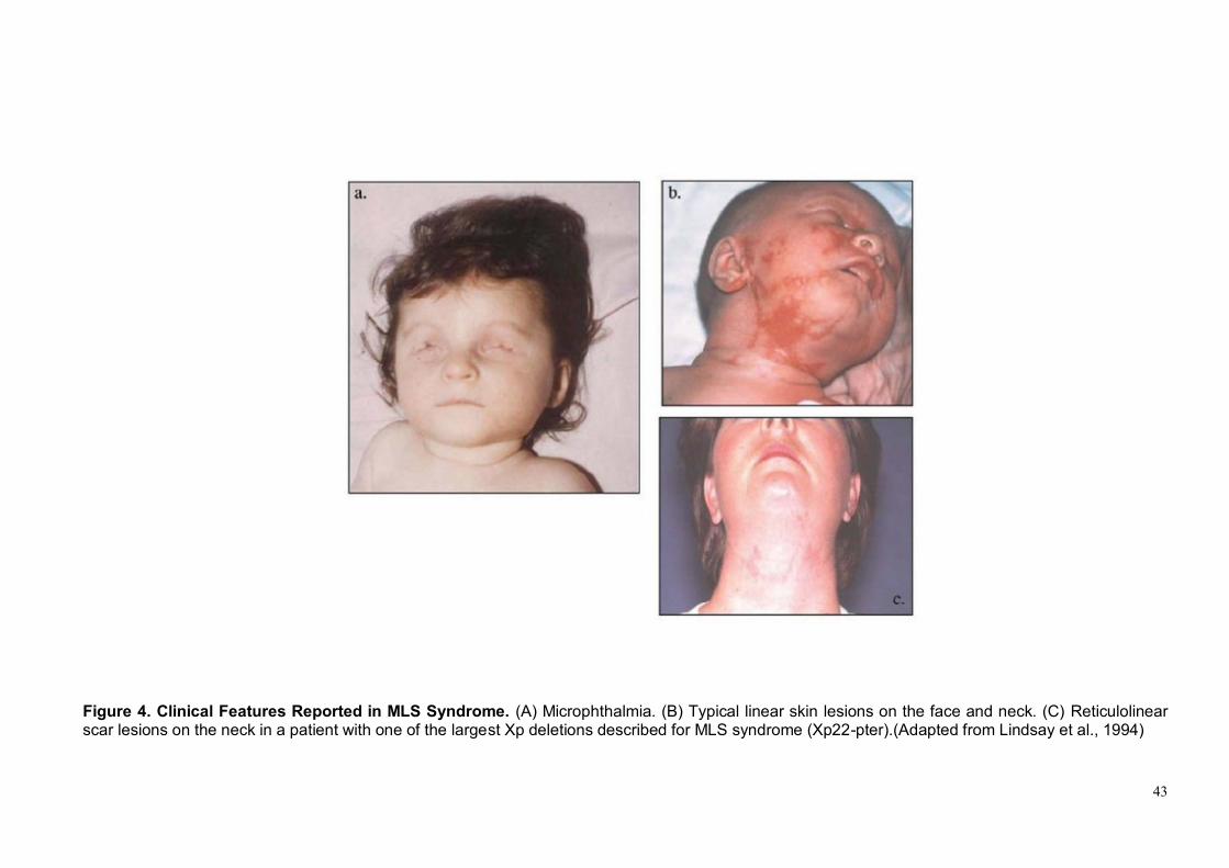
Figure 8. The extrinsic (death receptor-mediated) and intrinsic (mitochondria
mediated) central apoptotic pathways………………………………........47 Figure 9. The mechanisms of apoptosome formation and caspase activation initiated by cytochrome c release………………………………………….48 Figure 10. Schematic presentation of the mitochondrial biochemical alterations
in the course of Apaf 1-independent caspase 9 activation. ………….49 Figure 11. Selected stages of Medaka development…………………………….…50 Figure 12. ClustalW multiple alignment of the human (hHCCS) and the two medaka (olhccsa and olhccsb) HCCS amino acid sequences……….71 Figure 13. Structure and expression of the olhccs transcripts in medaka……….72 Figure 14. Effects of the morpholinos injections in medaka embryos…………….73 Figure 15. Morpholinos against olhccsa efficiently interfere with its translation...74
Figure 16. Analysis of cell proliferation in hccs-deficient embryos………………..75
Figure 17. Increase of apoptosis in the retina of olhccs-deficient embryos……...76
Figure 18. TUNEL assay on medaka heart………………………………………….77
Figure 19. Coinjection of caspase inhibitors to rescue the olhccsa knockdown
microphthalmic phenotype………………………………………………..78 Figure 20. Involvement of Mitochondrial-Dependent cell death pathway in olhccsa knockdown microphthalmic phenotype………………………...79

7
Figure 21. Apaf1-independent cell death in the retina of hccs-deficient
embryos……………………………………………………………………..80 Figure 22. Impairment of mitochondrial respiratory chain in yeast………………..81 Figure 23. TEM analysis of mitochondrial morphology in hccs-deficient embryos……………………………………………………………………..82 Figure 24. Detection of ROS levels in olhccsa MO-injected fish………………….83
Figure 25. Analysis of retinal cells type specific markers…………………………..84 Figure 26. Analysis of dorso-ventral pattern on olhccsa MO-injected fish………85

8
ABSTRACT
The Microphthalmia with linear skin defects (MLS) syndrome is an X- linked
dominant male-lethal neuro-developmental disorder associated to mutations in the
holocytochrome c-type synthetase (HCCS) transcript. Female patients display
unilateral or bilateral microphthalmia and linear skin defects, additional features
include central nervous system (CNS) malformation and mental retardation. HCCS
codifies a mitochondrial protein that catalyzes the attachment of heme to both
apocytochrome c and c1, necessary for proper functioning of the mitochondrial
respiratory chain. Although mutation analysis clearly indicates a role for HCCS in
the pathogenesis of this genetic condition, the molecular mechanisms underlying
the developmental anomalies in the presence of HCCS dysfunction are still
unknown. Previous studies demonstrated the early lethality of mouse embryonic
Hccs knock-out stem cells. To overcome the problem of the possible embryonic
lethality, we decided to generate an animal model for MLS syndrome in the
medaka fish (Oryzia latipes) using a morpholino-based technology. Fish models
(zebrafish and medaka) are considered good models to study developmental
biology processes and in particular eye developmental defects.
Three specific morpholinos directed against different portions of the olhccs
transcript have been designed and injected and our data indicated that all
morpholinos effectively downregulate the expression of the olhccs gene. The
injection of the three different morpholinos resulted in a pathological phenotype,
which resembles the human condition. Morphants displayed microphthalmia,
coloboma, and microcephaly associated to a severe cardiac pathology. To date,
this is the only animal model that recapitulates the phenotype observed in MLS
syndrome. Analysis with markers for specific retinal cell types showed defects in

9
differentiation of the ventral neural retina. Characterization of morphants revealed
that hccs down-regulation results in impairment of mitochondrial functions,
overproduction of reactive oxygen species (ROS) and a strong increase of
apoptosis mediated by activation of the mitochondrial-dependent cell death
pathway in the CNS and in the eyes. Our results clearly indicate that HCCS plays
a critical role in mitochondria and imply that MLS should be considered a
mitochondrial disease.
It is well established that the intrinsic mitochondrial dependent apoptotic pathway
rely on the formation of apoptosomes, which require the presence and/or the
activity of cytochrome c, Apaf1, and caspase 9. Detailed studies of the
mechanisms that underlie intrinsic apoptosis have shown that the heme group of
cytochrome c is necessary for Apaf1 activation, apoptosome formation and
activation of caspase 9. Interestingly, our data indicate that, in our model, the
mitochondrial dependent apoptosis is triggered by caspase 9 activation and occur
in a Bcl-dependent but apoptosome-independent manner suggesting that at least
in some tissues the apoptosis can occur in a non-canonical way. Our data support
the evidence of an apoptosome-indipendent activation of caspase 9 and suggest
the possibility that this event might be tissue specific. Our study shed new light into
the functional role of HCCS in the mitochondria. In addition, we provide strong
evidences that mitochondrial mediated apoptotic events underlie microphtalmia
providing new insights into the mechanisms of this developmental defect.

10
1. INTRODUCTION

11
1.1 The vertebrate eye development.
The eye is a bilateral organ that originates from a single field positioned in the
anterior portion of the neural plate. This undifferentiated primordium reaches its
final complexity through a series of inductive and morphogenetic events that are
coordinated by specific genetic programs, which, by enlarge, are conserved
among different vertebrate species. A schematic representation of the main
events underlying a correct development of the eye is depicted in Figure 1.
The basic components of the complex optic system are derived from four
embryonic sources: forebrain neuroectoderm, intercalating mesoderm, surface
ectoderm, and neural crest. The neuroectoderm differentiates into the retina, iris,
and optic nerve; the surface ectoderm gives rise to lens and corneal epithelium;
the mesoderm differentiates into the extraocular muscles and the fibrous and
vascular coats of the eye; and neural crest cells become the corneal stroma sclera
and corneal endothelium.. During neurulation, eye progenitor cells converge
medially and are surrounded rostrally and laterally by telencephalic precursors and
caudally and medially by cells that will form the diencephalon. The first
morphological sign of eye development in vertebrates is the bilateral evagination
of anterior diencephalon in the early neurula after the formation and differentiation
of the neural tube. In mammals, this is marked by the appearance of the optic pit,
whereas in fish and amphibians a bulging of the optic primordia is observed (Chow
and Lang, 2001). Continued evagination of the optic primordial leads to the
formation of the optic vesicles connected to the diencephalon by a small canal, the
optic stalk (Figure 1 A). These extend towards the overlying, non-neural surface
ectoderm that will ultimately give rise to the lens and cornea. Mesenchyme
between the optic vesicle and the surface ectoderm (apparent in mammals and
chick) is displaced as the two tissues come into close physical contact (Figure 1

12
B). This is a critical period in eye development during which inductive signals
between the optic vesicle and the surface ectoderm are thought to exchange. At
this stage, the presumptive lens also shows the first morphological signs of
development. This is characterized by formation of the lens placode, a thickening
of the surface ectoderm that comes into contact with the optic vesicle (Chow and
Lang, 2001). Molecularly, lens placode formation coincides with the onset of
crystalline expression. These protein families are expressed at high levels in a
lens preferential manner and are required for generating and maintaining lens
transparency (Cvekl and Piatigorsky, 1996; Graw, 1996; Wistow and Piatigorsky,
1988; Wride, 1996).
Coordinated invagination of the lens placode and the optic vesicle results in the
formation of the lens vesicle and a double-layered optic cup and provides the first
indication of the final shape of the eye (Figure 1 C). The inner layer of the optic
cup (facing the lens) forms the neural retina (NR), while the outer layer of the optic
cup gives rise to the retinal pigment epithelium (RPE) which will be formed of a
single layer of cells containing melanin (Figure 1 D).
At the ventral extremity of the optic vesicle, the process of invagination forms a
groove that runs continuously from the ventral-most region of the NR and along
the ventral aspect of the optic stalk to the junction with the neural tube.
The point at which the laterally growing edges of the optic cup fuse is known as
the choroidal (or optic) fissure. This structure provides a channel for blood vessels
within the eye and an exit route for projecting axons. Normally, the optic fissure
closes during development: its failure to close leads to a pathological condition
called coloboma.
Development of the cornea from the surface ectoderm overlying the lens and from
migrating neural crest–derived mesenchyme, is a highly coordinated multistep
process. Briefly, it first involves the secretion of a collagen-rich extracellular matrix

13
by the corneal epithelium (ectoderm overlying the lens). This primary stroma
attracts a wave of neural crest–derived mesenchymal cells from the region
surrounding the eye that coincides with its hydration and the migration of a second
wave of neural crest–derived mesenchymal cells. Eventually, an increased level of
thyroxine triggers the dehydration and compaction of the posterior stroma that
ultimately leads to the formation of the mature, transparent cornea (Chow and
Lang, 2001).
The remaining parts of the adult eye, such as the ciliary body and iris are derived
from the distal tip of the optic cup at the point where the inner and outer optic cup
layers meet (Beebe, 1986) (Figure 1 C).
In vertebrates, the cells that compose the optic vesicle neuroepithelium are initially
morphologically and molecularly indistinguishable and therefore are all potentially
competent to originate the different retinal cells type, the optic stalk or the RPE.
They reach their final complexity through a series of inductive and morphogenetic
events. Information, in the form of signaling molecules, derived from the
surrounding tissues and the neuroepithelium itself, modulate and restrict the
expression of different transcription factors and can drive the differentiation
towards a specific cell type (Figure 2). For example extracellular signals, derived
from the surface ectoderm in contact with the prospective NR or from the
periocular mesenchyme surrounding the presumptive RPE, pattern the distal optic
vesicle. The surface ectoderm secretes high levels of two members of Fibroblast
Growth Factor (FGF) signaling molecules family, FGF1 and FGF2, (Nguyen and
Arnheiter, 2000) while members of the Transforming Growth Factor-β (TGF-β)
signaling molecules superfamily, such as activins or the related Bone
Morphogenetic Proteins (Bmp) - Bmp4 and Bmp7 - are expressed in the
surrounding mesenchyme and/or the presumptive RPE itself (Fuhrmann et al.,
2000). FGF and TGF-β/BMP signaling act antagonistically on the specification of

14
RPE and NR precursors. The first activates NR specification but inhibits RPE
formation by activation of the mitogen-activated protein kinase (MAPK) cascade
which, in turn, promote the expression of Ceh10 homeodomain-contain homolog
(Chx10), the best candidate to impose a NR character to the native optic vesicle
cells, and reduce the expression of Orthodenticle homeobox 2 (Otx2) and
Microphtalmia transcriptor-associated factor (MITF), transcription factors crucial for
the RPE identity. In contrast the induction of TGFβ/BMP signaling by extraocular
mesenchyme is essential for the activation of the RPE molecular markers such as
MITF, and has an inhibitory effect on the Chx10.
At the end of the differentiation process, in the adult vertebrate NR there are six
types of neurons and one type of glial cells (Müller glial cells), which constitute
three cellular layers: rod and cone photoreceptors in the outer nuclear layer (ONL),
horizontal, bipolar, and amacrine interneurons and Müller glial cells in the inner
nuclear layer (INL), and ganglion and displaced amacrine cells in the ganglion cell
layer (GCL) (Figure 3 A). These seven types of cells are differentiated from
common progenitors in a temporal order widely conserved during evolution from
fish to mammals: ganglion cells first, followed by horizontal cells, cones and
amacrine cells, and rods and bipolar cells and Müller glial cells last (Belecky-
Adams et al., 1996; Carter-Dawson and LaVail, 1979; Cepko et al., 1996; Hu and
Easter, 1999; La Vail et al., 1991; Stiemke and Hollyfield, 1995; Young, 1985)
(Figure 3 B). Although there is an exact temporal order, the differentiation stage of
each cell type is in part contemporaneous with the differentiation stage of the
preceding cell type (Figure 3 B). Thus, retinal development consists of three
successive processes: (i) proliferation of progenitors, (ii) neurogenesis, and (iii)
gliogenesis. According to the model of "competence", the different retinal cell
types derived from a single common progenitor which change competency over
time under the control of extrinsic (such as neurotrophic factors) and intrinsic

15
regulators (such as transcription factors) (Austin et al., 1995; Belliveau and Cepko,
1999; Belliveau et al., 2000; Harris, 1997; Holt et al., 1988; Livesey and Cepko,
2001; Marquardt and Gruss, 2002; Turner and Cepko, 1987; Turner et al., 1990).
Thus, in this model progenitors pass through intrinsically determined competence
states, during which they are capable of giving rise to a limited subset of cell types
under the influence of extrinsic signals (Altshuler et al., 1993; Ezzeddine et al.,
1997; Furukawa et al., 2000; Guillemot and Cepko, 1992; Kelley et al., 1994;
Zhang and Yang, 2001). TGF, EGF (epidermal growth factor) and ILF (leukemia
inhibitor factor) are just some examples of extrinsic factors that can stimulate the
production of specific retinal cells types, while leading to suppression of other
(Lillien and Wancio, 1998).
Indeed genes coding for transcription factors of the family b-helix-loop-helix, as
Ath5, mash1, NeuroD, or genes containing a homeodomain such as Otx2, Chx10,
Pax6, Six3 and Crx work as intrinsic regulators (Bramblett et al., 2004; Brown et
al., 2001; Burmeister et al., 1996; Dyer et al., 2003; Inoue et al., 2002; Li et al.,
2004; Marquardt et al., 2001; Mathers et al., 1997; Morrow et al., 1999; Satow et
al., 2001; Tomita et al., 2000). These genes are important both for differentiation
and maintenance of retinal cell types; in fact many of them are expressed at high
levels in specific cellular regions also when the retina is completely differentiated.
In contrast, genes involved in proliferation as c-myc and cyclin D1 are expressed
in a small number of retinal progenitor cells that remain proliferating in the ciliary
margin zone (CMZ). Thus the development of the eye is an highly complex
process, and the sequential and coordinated expression of other numerous genes
encoding for transcription factors, cofactors, signal transduction molecules,
membrane receptors and others, more or less well characterized, play a key role
in different stages of eye development and may be responsible for different eye
malformations.

16
1.2 Microphthalmia and anophtalmia: an overview
Eye diseases represent one of the most common groups of genetic disorders in
the human population. Over 200 different forms of ocular heritable disorders have
been described and it has been estimated that about 27% of the phenotypes
described in OMIM (On-line Mendelian Inheritance in Man) affect the eye.
Congenital anophthalmia and microphthalmia are rare defects of the globe
resulting from abnormalities in the development of the primary optic vesicle.
Anophthalmia and microphthalmia describe, respectively, the absence of an eye
and the presence of a small eye within the orbit. The term anophthalmia is used
where there is no visible ocular remnant. However, ultrasound often reveals a
buried microphthalmic remnant or cyst. Microphthalmia refers to an eye with
reduced volume and is usually defined in terms of corneal diameter and axial
length. It may be associated with other eye developmental anomalies including
lens and optic nerve abnormalities, orbital cyst and coloboma, which is a more
regional eye defect that is caused by defective closure of the embryonic fissure of
the optic cup.
The birth prevalence of anophthalmia and microphthalmia has been generally
estimated to be 3 and 14 per 100,000 population respectively, although other
evidence puts the combined birth prevalence of these malformations at up to 30
per 100,000 population with microphthalmia reported in up to 11% of blind children
(Adapted from Morrison et al., 2002; Shaw et al., 2005). High-resolution cranial
imaging, post-mortem examination and genetic studies suggest that these
conditions represent a phenotypic continuum.
Both anophthalmia and microphthalmia may be unilateral or bilateral and may
occur as an isolated clinical sign or as part of a syndrome (in one-third of cases).
17
Epidemiological studies have predicted both environmental and heritable factors in
causing these defects, in fact anophthalmia/microphthalmia have complex
aetiology with chromosomal, monogenic and environmental causes identified.
Environmental factors playing a contributory role include gestational-acquired
infections, maternal vitamin A deficiency, exposure to X-rays, solvent misuse and
thalidomide exposure (Verma and Fitzpatrick, 2007). However, evidences for the
role of environmental causes are both more circumstantial and accounts for a
smaller proportion of cases compared to heritable factors.
Genetic eye disease can be broadly divided into two main categories,
degenerative disorders and developmental anomalies. While for the degenerative
diseases, molecular genetics studies have been able, in recent years, to shed light
on the molecular basis of a significant number of cases (Farrar et al., 2002), the
pathogenetic causes of eye developmental anomalies remain very elusive in the
majority of conditions. This is particularly true for the
anophthalmia/microphthalmia, with the exception of a few instances where
chromosomal duplications, deletions and translocations and monogenic mutations
have been implicated and are typically associated with characteristic syndromes.
In the majority of known cases these defects are likely to be caused by
disturbances of the morphogenetic pathway that controls eye development.
Genetic studies demonstrated that mutations in several genes principally involved
in ocular development often cause eye malformations such as anophthalmia and
microphthalmia and mutations in these transcripts are associated to specific
syndromes including eye developmental defects (Table 1).
For example SOX2, regulator of retinal neural progenitor competence (Taranova
et al., 2006), has to date been identified as a major causative gene for
anophthalmia/microphthalmia. Cytogenetic studies placed the locus at 3q26.3, and
de novo heterozygous loss-of-function point mutations in this transcript have been

18
Name of Gene (Inheritance)
Name of syndrome Ocular manifestations Systemic manifestations
SOX2 3q26.3-q27 (AD)
SOX2 anophthalmia syndrome, some cases of AEG
Anophthalmia, microphthalmia, dystrophy
Hypothalamic – pituitary abnormalities, growth failure, genital tract malformation, developmental delay, seizures, oesophageal atresia
OTX2 14q21-14q22 (AD)
Anophthalmia, microphthalmia, coloboma, microcornea, cataract, retinal dystrophy, optic nerve hypoplasia
Agenesis of the corpus callosum, developmental delay
PAX2 10q24.3-q25 (AD)
Renal-coloboma or Papillorenal syndrome
Coloboma, microphthalmia Renal hypoplasia
PAX6 11p13 (AD)
Aniridia, Peters' anomaly, foveal hypoplasia, keratopathy
Abnormalities of pituitary, pancreatic, and brain development
CHD7 8q12.1 (AD)
CHARGE syndrome Microphthalmia, coloboma Heart defects, choanal atresia, retarded growth and development, genital hypoplasia, ear anomalies, and deafness
PTCH 9q22.3 (AD)
Basal cell naevus /Gorlin's syndrome
Microphthalmia, coloboma, cyst Palmer pits, medulloblastoma basal cell carcinoma
SHH 7q36 (AD)
Holoprosencephaly-3 (HPE3)
Cyclopia, coloboma, hypotelorism Preaxial polydactyly, cleft lip and palate
CHX10 14q24.3 (AR)
Anophthalmia, microphthalmia, coloboma, cataract, iris abnormalities
FOXC1 6p25 (AD)
Axenfeld–Rieger syndrome
Iris hypoplasia, iridogoniodysgenesis, glaucoma
Dental abnormalities, midface abnormalities
HCCS Xp22 (X-linked)
Microphthalmia with linear skin defects
Microphthalmia, sclerocornea Linear skin defects, agenesis of corpus callosum
BRIP1 17q22 (AD)
Fanconi anaemia Microphthalmia Bone marrow failure, breast cancer, growth retardation, hearing loss, thumb and kidney abnormalities
DPD 1p22 (AR)
Microphthalmia, coloboma, nystagmus Epilepsy, mental retardation, motor retardation
SIX3 2p21 (AD)
Holoprosencephaly 2 Cyclopia, Microphthalmia, coloboma hypotelorism, microcephaly, craniofacial anormalities
SIX6 14q23 (AD)
Microphthalmia, cataract, nystagmus Pituitary abnormalities
PITX2 4p25 (AD)
Rieger syndrome Iris hypoplasia, iridogoniodysgenesi, glaucoma
Maxillary hypoplasia, dental abnormalities, excess periumbilical skin
POMT1 9q34.1 (AR)
Walker-Warburg syndrome
Microphthalmia, cataract, retinal dysplasia and detachment, coloboma, optic nerve hypoplasia
Developmental delay, muscular dystrophy, hydrocephalus, agyria, epilepsy
BCOR Xq27-q28 (X-linked)
Oculofaciocardiodental syndrome
Microphthalmia, congenital cataract Mental retardation, heart defects, dental and facial abnormalities
RX 18q21.3 (AD)
Anophthalmia, microphthalmia, sclerocornea
FRAS1 4q21 (AR)
Fraser Syndrome Microphthalmia, cryptophthalmos Genital and kidney abnormalities, finger webbing
FREM2 13q13.3 (AR)
Fraser Syndrome Microphthalmia, cryptophthalmos Genital and kidney abnormalities, finger webbing
HESX1 3p21.2-p21.1 (AD)
Septo-optic dysplasia Optic nerve hypoplasia Agenesis of the corpus callosum, panhypopituitarism, and absent septum pellucidum
MAF 16q22-q23 (AD)
Cataract, anterior segment dysgenesis, coloboma
Nephritic syndrome
RAB3GAP 2q21.3 (AR)
Warburg Micro Syndrome
Microphthalmia, microcornea, optic atrophy, cataract
Microcephaly, mental retardation, hypoplasia of corpus callosum,
Table 1. Selected genes and syndromes associated with eye malformations. (Adapted from Ragge et al., 2007)

19
shown to account for 10–20% of severe bilateral anophthalmia/microphthalmia
(Morrison et al., 2002; Shaw et al., 2005), the most common phenotype being
bilateral anophthalmia.
PAX6, on chromosome 11p13, has been studied more extensively than most other
eye genes. In humans, heterozygous loss-of-function mutations typically produce
aniridia (OMIM 106210), a congenital pan-ocular malformation associated with
severe visual impairment; however PAX6 was also the first gene implicated in
human anophthalmia (Glaser et al., 1994). Although PAX6 mutations are an
extremely rare cause of anophthalmia, there has recently been interest in a
possible co-operative role between PAX6 and SOX2. It has been shown that
PAX6 and SOX2 co-bind to a regulatory element driving lens induction in the chick
(Kondoh et al., 2004), which suggests that lens induction failure could be
responsible for microphthalmia in patients with mutations in these genes
(Fitzpatrick and van Heyningen, 2005). As expected with genes expressed in the
developing brain, patients with inherited PAX6 and SOX2 mutations exhibit CNS
malformations in addition to dominantly inherited anophthalmia/microphthalmia
(Fitzpatrick and van Heyningen, 2005; Sisodiya et al., 2006).
Mutations in three genes with retinal expression are associated with
anophthalmia/microphthalmia, possibly through failure of retinal differentiation.
Heterozygous loss-of-function mutations of OTX2 (on chromosome 14q22,
autosomal dominant inheritance) have been shown to be associated with a wide
range of ocular disorders from anophthalmia and microphthalmia to retinal defects.
CNS malformations and mental retardation are common in patients with OTX2
mutations (Ragge et al., 2005). RAX, located on chromosome 18q21.32, is linked
to about 2% of inherited anophthalmia/microphthalmia (Voronina et al., 2004).
Similarly, CHX10 mutations (chromosome 14q24.3) account for about 2% of
isolated microphthalmia (Ferda Percin et al., 2000).

20
The SIX3 gene is responsible for several cases of holoprosencephaly but in one
patient it has been associated to microphtalmia and coloboma without other
classical signs of holoprosencephaly (Wallis et al., 1999). Mutations in the PAX2
gene, playing an important role in the morphogenesis of the ventral developing
eye (Torres et al., 1996), are responsible for the renal-coloboma syndrome, which
is also characterized by optic nerve coloboma (Sanyanusin et al., 1995).
Unlike the previous, which are all transcription factors principally involved in ocular
development, mutations in gene that are not directly correlated to eye
development, such as RAB3GAP and BCOR, it has been also described. The first,
involved in the Warburg Micro Syndrome, encoding for a protein involved in the
GTPase signal transduction and regulates neurotransmitter release and synaptic
plasticity (Sakane et al., 2006). BCOR (BCL6 corepressor) encoding for a protein
ubiquitously expressed in human tissues and mutation in its transcript are
responsible for oculofaciocardiodental syndrome (OFCD) and Lenz
microphthalmia (Ng et al., 2004). The protein encoded by this gene was identified
as an interacting corepressor of BCL6, a POZ/zinc finger transcription repressor
that is required for germinal center formation and may influence apoptosis
(Gearhart et al., 2006).
However, it must be emphasized that eye developmental anomalies, and
anophthalmia/microphthalmia in particular, seem to be a very heterogeneous
group of disorders with many different genes involved, as suggested by the limited
number of mutations so far identified in the patients analyzed.
Numerous genes encoding for transcription factors, cofactors, signal transduction
molecules, membrane receptors and others, more or less well characterized, play
a key role in different stages of eye development and may be responsible for
different eye malformations.
21
Genetic counselling can be challenging due to the extensive range of genes
responsible and wide variation in phenotypic expression. Appropriate counselling
is indicated if the mode of inheritance can be identified. Differential diagnoses
include cryptophthalmos, cyclopia and synophthalmia, and congenital cystic eye.
Patients are often managed within multidisciplinary teams consisting of
ophthalmologists, pediatricians and/or clinical geneticists, especially for syndromic
cases. Treatment is directed towards maximizing existing vision and improving
cosmesis through simultaneous stimulation of both soft tissue and bony orbital
growth. Mild to moderate microphthalmia is managed conservatively with
conformers. Severe microphthalmia and anophthalmia rely upon additional
remodeling strategies of endo-orbital volume replacement (with implants,
expanders and dermis-fat grafts) and soft tissue reconstruction. The potential for
visual development in microphthalmic patients is dependent upon retinal
development and other ocular characteristics.
The aetiology of anophthalmia/microphthalmia underlies the entire developmental
biology of ocular formation and remains a field where our knowledge is increasing
exponentially. Despite the progresses made, much work is still needed to
understand the processes underlying these complex diseases, which are a
significant cause of childhood blindness. Even if these processes are elucidated in
the future, novel therapeutic approaches to prevent these conditions from
occurring could still be precluded by very early ocular development in the fetus.
1.3 The molecular basis of microphtalmia with linear skin lesion
(MLS) syndrome
Among the genetic forms of anophthalmia/microphthalmia, Microphthalmia with
linear skin lesions syndrome (MLS, OMIM 309801), first described in the 1990 (al-

22
Gazali et al., 1990), represents one of the most puzzling genetic disorder. The
MLS syndrome, also known as MIDAS (microphthalmia, dermal aplasia and
sclerocornea), is a rare X-linked dominant condition characterized by unilateral or
bilateral microphthalmia and linear skin defects, which are limited to the face and
neck, consisting of areas of aplastic skin which heal with age to form
hyperpigmented areas, in affected females and in utero lethality for males. Other
ocular abnormalities are variable and can include: sclerocornea; orbital cysts;
microcornea; eyelid fissures; corneal leukoma; iridocorneal adhesion (Peters
anomaly); congenital glaucoma with total/peripheral anterior synechiae; aniridia;
cataracts; a remnant of the anterior hyaloid artery; vitreous opacity; and
hypopigmented areas of the retinal pigment epithelium (Cape et al., 2004;
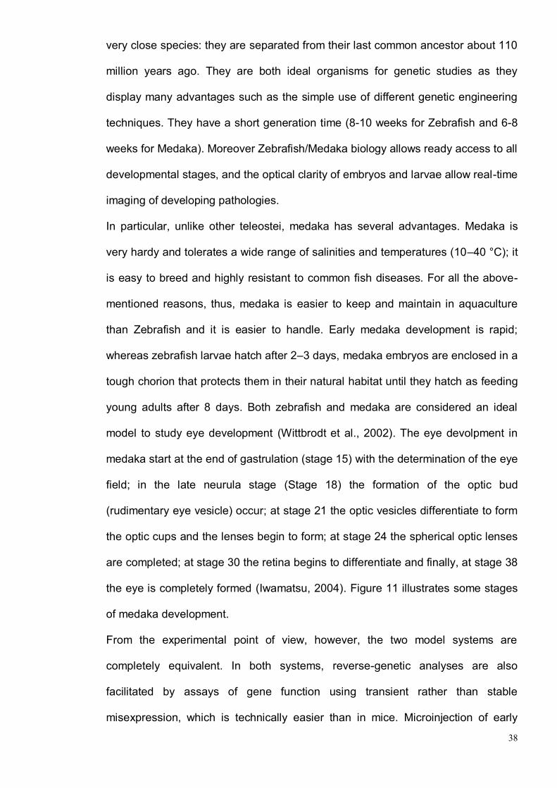
Kobayashi et al., 1998; Wimplinger et al., 2006). In Figure 4 and Table 2 are
depicted and described some of the most typical clinical signs observed in this
genetic condition.
Moreover additional features in female patients include central nervous system
anomalies (such as agenesis of corpus callosum, ventriculomegaly, microcephaly
reported in about 40% of affected individuals), mental retardation (in about 25% of
affected individuals) and congenital heart defects (arrhythmias, septum defects,
cardiomyopathy) (al-Gazali et al., 1990; Happle et al., 1993; Lindsay et al., 1994;
Sharma et al., 2008; Temple et al., 1990; Van den Veyver, 2002; Zvulunov et al.,
1998).
In the majority of cases, patients carry deletions or unbalanced translocations
involving the Xp22.3 region resulting in segmental monosomy of this chromosome
(Morleo et al., 2005). With the exception of eight males that show a 46 XX
karyotype and an translocation Xp,Yp (that causes Xp monosomy in one of two X
chromosomes) (Kapur et al., 2008; Morleo et al., 2005), MLS patients are all
females.

23
Clinical feature Occurrence (%)
Cutaneous
Linear skin defects 92
Nail dystrophy 13
Ear pit 5
Ocular
Microphthalmia 89
Corneal clouding/opacities 35
Sclerocornea 33
Cataracts 8
Hypopigmentation of the RPE 5
Developmental
Short stature 74
Developmental delay 37
CNS
Agenesis of the corpus callosum, Ventriculomegaly, Microcephaly 40
Mental retardation 25
Seizures 3
Cardiac
Supraventricular tachycardia 13
Atrial/ventricular septal defects 13
Hypertrophic cardiomyopathy 5
Bradycardia 5
A–V block 5
Table 2. Clinical Features Reported in MLS Syndrome (Adapted from Sharma et al., 2008)
An high degree of intra- as well as inter-familial clinical variability has been
observed in this condition possibly related to the role of X-inactivation (Franco and
Ballabio, 2006; Morleo and Franco, 2008; Van den Veyver, 2001). In fact the
manifestations vary among affected individuals and, although most of them display
the classic phenotype of MLS syndrome, many have only a subset of
characteristic features: some show the characteristic skin defects without ocular
abnormalities, whereas others have eye abnormalities without skin defects (Morleo
and Franco, 2008). No genotype-phenotype correlations have been observed
since the high phenotypic variability is not correlated to the extent of Xp-terminal
deletion. An example is a female with a normal phenotype except for typical MLS

24
syndrome skin defects (Figure 4 C) who had an affected female fetus with
anencephaly. Cytogenetic analysis revealed that both mother and fetus had the
same Xp22 deletion that was one of the largest Xp deletions described for MLS
syndrome (Lindsay et al., 1994).
Skewed X inactivation has been detected in 16 out of the 17 MLS patients
analyzed to date (Cain et al., 2007; Ogata et al., 1998; Schluth et al., 2007;
Wimplinger et al., 2006; Wimplinger et al., 2007). It has been proposed that the
most severe MLS syndrome clinical manifestations are observed in females whose
normal X chromosome is inactivated in the affected tissue or at a specific time of
embryonic development; conversely, a milder phenotype or the total absence of
MLS syndrome clinical manifestations may result from totally skewed
X-chromosome inactivation that forces preferential activation of the unaffected X,
not only in blood cells, but also in tissues such as the eye and skin (Morleo and
Franco, 2008).
The “MLS minimal critical region”, spanning approximately 610 Kb in Xp22.2
region, has been first defined through a combination of cytogenetic analysis and
breakpoint mapping on somatic cell hybrids from ten MLS patients with deletions
and translocations involving the Xp22 region (Wapenaar et al., 1993; Wapenaar et
al., 1994). Three genes are located in the critical interval, including MID1, HCCS,
and ARHGAP67. MID1 is mutated in Opitz G/BBB syndrome (Quaderi et al., 1997;
Schaefer et al., 1997); ARHGAP6 gene codes for a Rho GTPase–activating
protein (Rho GAP) that functions as a GAP for the small GTPase RhoA, as well as
a protein implicated in reorganization of the actin cytoskeleton (Prakash et al.,
2000; Schaefer et al., 1997); HCCS encodes a mitochondrial holocytochrome
c–type synthase, also known as “heme lyase”, that catalyzes the covalent
attachment of heme to both apocytochrome c and c1 (Bernard et al., 2003;
Schwarz and Cox, 2002). In the 2002 Prakash and colleagues showed that in vivo
25
generated deletions involving the equivalent critical MLS region in the mouse
(MLSΔ), encompassing Hccs as well as parts of Mid1 and Arhgap6, lead to
lethality of hemizygous, homozygous, and heterozygous embryos early in
development. This lethality can be rescued by overexpression of the human HCCS
gene from a BAC clone, providing the proof that lethality is indeed due to loss of
HCCS (Prakash et al., 2002) and defining HCCS as the most convincing candidate
gene for these disease.
After more than 10 years of effort, in 2006, the conclusive evidence that HCCS is
the gene responsible for MLS syndrome have been found: the group of Prof.
Brunella Franco, at the TIGEM of Naples, in collaboration with the group of Prof.
Kerstin Kutsche (Hamburg University) identified loss-of-function mutations
associated with the MLS phenotype. More in detail, de novo heterozygous point
mutations, a missense (p.R217C) and a nonsense mutation (p.R197X),
respectively, were identified in HCCS in two patients with MLS and a normal
karyotype. Functional analysis demonstrate that both mutant proteins (R217C and
Δ197-268) were not able to complement a Saccharomyces cerevisiae mutant
deficient for the HCCS orthologue Cyc3p, in contrast to HCCS wild type (Figure 5).
Moreover, ectopically expressed HCCS wild type and the R217C mutant protein
are targeted to mitochondria in CHO-K1 cells, while the C-terminal truncated
Δ197-268 mutant failed to be sorted to mitochondria (Figure 6). In addition,
characterization of a familial case revealed the presence of an 8.6-kb deletion
comprising HCCS exons 1 and 2, the first 83 bp of exon 3, the 5‟ untranslated
exons 1a and 1b of MID1, as well as the respective intronic sequences. These
results provided strong evidence for the involvement of the holocytochrome c-type
synthase in the pathogenesis of this disorder (Wimplinger et al., 2006). In the 2007
a novel missense mutation (p.E159K) has been identified; it leads to loss-of-
function of the encoded holocytochrome c-type synthase, in a sporadic female
26
patient with microphthalmia of both eyes and bilateral sclerocornea without skin
lesions, confirming that the phenotypic variability described in MLS cases is not
correlated to the extent of Xp-terminal deletion or to the presence or nature of the
HCCS mutations.
1.4 HCCS and its role in mitochondrial functioning
HCCS encodes a mitochondrial holocytochrome c–type synthase, also known as
“heme lyase,” composed of 268 aa (Bernard et al., 2003; Schwarz and Cox, 2002)
and located on the outer surface of the inner mitochondrial membrane (Schaefer
et al., 1996). It is ubiquitously expressed with the strongest expression observed in
heart and skeletal muscle (Schaefer et al., 1996; Van den Veyver et al., 1998).
HCCS catalyzes the covalent attachment of heme to both apocytochrome c and
c1, the precursor forms, thereby leading to the mature forms, holocytochrome c
and c1, which are necessary for proper functioning of the mitochondrial respiratory
chain (Bernard et al., 2003; Moraes et al., 2004). In addition to the well-known role
of cytochrome c in oxidative phosphorylation (OXPHOS), it is released from
mitochondria in response to a variety of intrinsic death-promoting stimuli, which in
turn result in caspase-dependent cell death (Jiang X et al., 2004).
In S. cerevisiae, two heme lyases exist, the cytochrome c- (Cyc3p) and the
cytochrome c1- specific heme lyase (Cyt2p), whereas only a single heme lyase is
required for maturation of both cytochrome c and c1 in higher eukaryotes (Bernard
et al., 2003; Schaefer et al., 1996). They are located on the outer surface of the
inner mitochondrial membrane and defects in either of the yeast heme lyases
result in loss of respiratory growth of the respective strain (Dumont et al., 1987).
Cyt2p and Cyc3p proteins have been extensively studied and well characterized,
especially together with cytochrome c and its secondary involvement in

27
programmed cell death (Li et al., 2000; Liu et al., 1996).
HCCS is the first mammalian holocytochrome c-type synthetases to be described
in the literature and it is homologous to the class of heme lyase identified first in
organisms such S. cerevisiae, N. crassa and C. elegans (Drygas et al., 1989;
Dumont et al., 1987; Zollner et al., 1992).
The protein encoded by the human gene identified by Schaefer and colleagues
(Schaefer et al., 1996) showed a 35% of identity with the Cyt2p and Cyc3p protein
sequences and seems to be the only example in higher eukaryotes. The amino
acid sequences of human and murine proteins shows 85% of identity and identical
length. Through complementation studies with both CYC3- and CYT2-deficient
yeast strains, Cox T. and colleagues demonstrated specific and complete rescue
of CYC3− growth-deficient phenotype supporting the conclusion that the human
protein possesses HCCS activity (Schwarz and Cox, 2002).
The HCCS gene covers a genomic segment of 11 kb and is ubiquitously
expressed (brain, placenta, kidney, lung, pancreas) with higher levels in heart and
skeletal muscle (Schaefer et al., 1996; Van den Veyver et al., 1998). Moreover by
RT-PCR analysis, Schwarz and colleagues also detected an abundant Hccs
expression in adult and fetal mouse eye (Schwarz and Cox, 2002). This data were
confirmed by in situ hybridization analysis (ISH) on mouse embryos performed in
the laboratory of Prof. Brunella Franco (unpublished data) (Figure 7). At E11.5,
E13.5 and E18.5 a specific signal in the eyes and in the encephalon is detected.
The absence or insufficiency of the human HCCS enzyme activity could give rise
to a nuclear-encoded respiratory chain defect with complications reminiscent of
the mitochondrial myopathies (pathologies in those tissues requiring the highest
amounts of energy). Consistent with this prediction is the observation that the
highest levels of mouse Hccs mRNA are found in the adult heart and with the
observation of strong Hccs expression in ocular tissue, which also has

28
considerable energy requirements. This expression profile is reminiscent of that
seen for other genes whose protein products function in the mitochondrial
respiratory chain (Wang et al., 1999). The HCCS haploinsufficiency could explain
some of the MLS clinical features such as the cardiomyopathy present in 18% of
cases, the dysgenesis of the corpus callosum present in 40% of cases and the
neurological problems commonly observed in MLS patients. These phenotypes
are in fact consistent with the involvement of a mitochondrial respiratory chain
enzyme. Nevertheless unique skin lesions restricted to the head and neck and the
microphthalmia are also diagnostic features and perhaps difficult to explain by
deficiency of a mitochondrial enzyme. The typical features of mitochondrial
diseases, that are a clinically heterogeneous group of disorders arising as a result
of dysfunction of the mitochondrial respiratory chain, include neuromuscular
hypotonia, ataxia, encephalomyopathy and various myopathies (DiMauro and
Moraes, 1993) (Table 3). Interestingly, MLS patients do not display
encephalomyopathies or signs of muscle involvement. However, mutations in
genes involved in mitochondrial oxidative phosphorylation often cause
neurological disorders. An example is represented by SCO2 and SURF,
responsible of infantile cardioencephalomyopathy and of Leigh syndrome,
respectively. Both these diseases are associated with deficiency of cytochrome c
oxidase (COX), that catalyzes the transfer of reducing equivalents from
cytochrome c to molecular oxygen (Papadopoulou et al., 1999; Zhu et al., 1998).
Is therefore not difficult to link HCCS to neurological disorders that characterize
the MLS syndrome as well as to cardiac defects found in 18% MLS cases.
Moreover it is interesting to underlie that some mitochondrial disorders only affect
a single organ. In particular the Leber hereditary optic neuropathy (LHON) is an
example of mitochondrial diseases with a typical ocular phenotype. LHON disease
has been associated with many missense mutations in the mtDNA and presents in

29
Disorder Primary Features Additional Features
Alpers-Huttenlocher syndrome •Hypotonia •Seizures • Liver failure
• Renal tubulopathy
Chronic progressive external ophthalmoplegia (CPEO)
• External ophthalmoplegia • Bilateral ptosis
• Mild proximal myopathy
Kearns-Sayre syndrome (KSS)
• PEO onset at age <20 years • Pigmentary retinopathy •CSF protein >1g/L, cerebellar ataxia, heart block
• Bilateral deafness • Myopathy • Dysphagia • Diabetes mellitus • Dementia
Pearson syndrome
• Sideroblastic anemia of childhood • Pancytopenia • Exocrine pancreatic failure
• Renal tubular defects
Infantile myopathy and lactic acidosis (fatal and non-fatal forms)
• Hypotonia in 1st year of life • Feeding and respiratory difficulties
• Fatal form may be associated with a cardiomyopathy and/or the Toni-Fanconi-Debre syndrome
Leigh syndrome (LS)
• Subacute relapsing encephalopathy • Cerebellar and brain stem signs • Infantile onset
• Basal ganglia lucencies • Maternal history of neurologic disease or Leigh Sy
Neurogenic weakness with ataxia and retinitis pigmentosa (NARP)
• Late-childhood or adult-onset peripheral neuropathy • Ataxia • Pigmentary retinopathy
• Basal ganglia lucencies • Abnormal electroretinogram • Sensorimotor neuropathy
Mitochondrial encephalomyopathy with lactic acidosis and stroke-like episodes (MELAS)
• Stroke-like episodes at age <40 years • Seizures and/or dementia • Ragged-red fibers and/or lactic acidosis
• Diabetes mellitus • Cardiomyopathy hypertrophic • Bilateral deafness • Pigmentary retinopathy • Cerebellar ataxia
Myoclonic epilepsy myopathy sensory ataxia (MEMSA)
• Myopathy • Seizures • Cerebellar ataxia
• Dementia • Peripheral neuropathy • Spasticity
Myoclonic epilepsy with ragged-red fibers (MERRF)
• Myoclonus • Seizures • Cerebellar ataxia • Myopathy
• Dementia • Optic atrophy • Bilateral deafness • Peripheral neuropathy • Spasticity
Leber hereditary optic neuropathy (LHON)
• Subacute painless bilateral visual failure • Median age of onset 24 years
Table 3. Clinical Syndromes of Mitochondrial Diseases (Adapted from Chinnery PF.
2010)

30
mid-life as acute or subacute central vision loss leading to central scotoma and
blindness (Yu-Wai-Man et al., 2009).
In addition, although the main function of the mitochondrion is the production of
energy in the form of ATP, it is well known its central role in many other metabolic
tasks. One of these is the regulation of intrinsic pathway of cell death a key
process required for proper development of the CNS (Valenciano et al., 2009).
Thus, even if HCCS is one of the few genes involved in eye disease that is not
directly involved in eye development, it could be play also an important role in
apoptosis since it is necessary for the maturation cytochrome c a key regulator of
intrinsic cell death pathway (Ow et al., 2008).
1.5 Mitochondrial-mediated apoptosis
Apoptosis is a process of particular importance for the proper development of the
CNS and the eye. The regulatory mechanism of survival/death during neuronal
development was not yet fully characterize but it is clear that apoptosis plays a key
role in the balance between proliferation and differentiation. Apoptosis (derived
from a Greek word meaning “falling off”, as leaves from a tree in Autumn) is the
main morphological feature of the process of programmed cell death or “cell
suicide”. It is a widespread, physiological phenomenon which occurs during the
embryonic development of multicellular organisms (Glucksmann, 1965;
Oppenheim, 1991) and represents the most common mechanism to regulate the
size of cell populations during development, as well as in adult life. In the
developing vertebrate nervous system, for example, around half or more of the
nerve cells normally die soon after they are born. Deregulated apoptosis has been
implicated in diverse pathologies, including cancer and neurodegenerative
disease, and it has been considered the final common pathway resulting from a
31
variety of primary defects (Tait and Green, 2010; Vecino et al., 2004). In vertebrate
cells, apoptosis typically proceeds through one of two signalling cascades termed
the intrinsic (or mitochondria-dependent) and extrinsic pathways (also known as
“death receptor pathway”). Figure 8 illustrates a schematic representation of the
extrinsic (death receptor-mediated) and intrinsic (mitochondria-mediated) central
apoptotic pathways.
Both the extrinsic and the intrinsic routes to apoptosis ultimately lead to cell
shrinkage, chromatin condensation, nuclear fragmentation (which is frequently
accompanied by internucleosomal DNA fragmentation), blebbing, and
phosphatidylserine exposure on the surface of the plasma membrane (Zamzami et
al., 1996)
In the extrinsic pathway, apoptosis is triggered by the ligand-induced activation of
death receptors at the cell surface. Death receptors include the tumor necrosis
factor (TNF) receptor-1, CD95/Fas (the receptor of CD95L/FasL), as well as the
TNF-related apoptosis inducing ligand (TRAIL) receptors-1 and -2.
Death receptor ligation causes the recruitment of adaptor molecules, such as FAS-
associated death domain protein (FADD), that bind, dimerize and activate an
“initiator” caspase, caspase 8. Active caspase 8 directly cleaves and activates the
“executioner” caspases, caspase 3 and caspase 7. The “executioner” caspases
through the cleavage of numerous proteins ultimately lead to the phagocytic
recognition and engulfment of the dying cell (Debatin and Krammer, 2004).
In the intrinsic pathway, mitochondrial outer membrane permeabilization (MOMP),
which leads to the release of proapoptotic proteins from the mitochondrial
intermembrane space (IMS), is the crucial event driving initiator caspase activation
and apoptosis. Following its release from mitochondria, cytochrome c binds
apoptotic protease activating factor 1 (APAF1), inducing its conformational change
and oligomerization and leading to the formation of a caspase activation platform
32
termed the apoptosome. APAF 1 preexists in the cytosol as a monomer, and its
activation depends on the presence of cytochrome c (Cyt c) and ATP/dATP (Cain
et al., 2002). The apoptosome recruits, dimerizes and activates an initiator
caspase, caspase 9, which, in turn, cleaves and activates caspase 3 and
caspase 7.
The mitochondrion also releases other pro-apoptotic proteins such as SMAC (also
known as DIABLO) and OMI (also known as HTRA2) blocking the X-linked
inhibitor of apoptosis protein (XIAP)-mediated inhibition of caspase activity.
MOMP can even commit a cell to die when caspases are not activated. This
“caspase-independent death” (Chipuk and Green, 2005) can occur by release of
caspase independent death effectors including apoptosis-inducing factor (AIF)
(Susin et al., 1999b) and endonuclease G (Li et al., 2001).
The MOMP is a highly regulated process, primarily controlled through interactions
between pro- and anti-apoptotic members of the B cell lymphoma 2 (BCL-2) family
(Tait and Green, 2010).
Crosstalk between the extrinsic and intrinsic pathways occurs through caspase 8-
mediated cleavage of BCL-2 homology 3 (BH3)-interacting domain death agonist
(BID; a BH3 domain-only protein), leading to BID activation and MOMP.
While the extrinsic pathway mediates apoptosis through the specialized subset of
death signals detected at the plasma membrane, the intrinsic pathway on the other
hand transduces a wide variety of extracellular and intracellular stimuli including
loss of survival/trophic factors, toxins, radiation, hypoxia, oxidative stress,
ischaemia-reperfusion and DNA damage (Foo et al., 2005). Reactive oxygen
species (ROS) excessively produced by the respiratory chain can also cause the
progressive mitochondrial damage leading to apoptosis (Skulachev, 1997).
During the past decade, it has become obvious that the mitochondria play a critical
role in the regulation of cell death, and that MOMP and release of intermembrane

33
space proteins are important features for this process. Moreover mitochondria are
known to modulate and synchronize Calcium signaling which has long been
recognized as a participant in apoptotic pathways (Giacomello et al., 2007). Also
the fragmentation of the mitochondrial network and remodelling of the
mitochondrial cristae are both required processes for the progression of apoptosis
(Scorrano et al., 2002); mitochondrial fusion and fission machinery controls
mitochondrial shape and physiology including mitochondrial remodeling during
apoptosis. Thus mitochondria exert both vital and lethal functions in physiological
and pathological scenario: the mitochondrion is not only the cell‟s powerhouse, it is
also its arsenal.
The mitochondrion sequesters a potent cocktail of pro-apoptotic proteins and the
most prominent among these is cytochrome c. After release from mitochondria, the
biochemistry of how cytochrome c triggers caspase activation is very complex. It
was found that cytochrome c can interact with the C-terminal WD40 repeats of
Apaf 1 and that this interaction is required for activation of the pathway (Lassus et
al., 2002). An in vitro de novo reconstitution of apoptosome was achieved by using
purified recombinant Apaf 1, procaspase 9, procaspase 3, and highly purified
horse cytochrome c. When all the proteins are incubated together in the presence
of nucleotide dATP/ATP, caspase 3 is activated. A striking phenomenon observed
in this in vitro system is that Apaf 1 and cytochrome c are induced into a huge
complex in a dATP/ATP-dependent manner to form the apoptosome (Zou et al.,
1999) (Figure 9). It has been demonstrated that apocytochrome c (the cytochrome
c without the heme group) binds Apaf 1 but that this interaction is insufficient for
caspase activation (Martin and Fearnhead, 2002).
Although the biochemistry of apoptosome formation has been reconstituted by
using purified components in vitro, an understanding of the composition of the
native apoptosome in apoptotic cells has emerged only recently. The successful
34
immunoprecipitation of catalytically active apoptosomes from Jurkat cells has
revealed that the native apoptosome (at least in this cell type) contains caspase 3
and XIAP in addition to Apaf 1 and caspase 9 (Hill et al., 2004). This study also
intimates that the interaction of XIAP with caspase 9 is necessary for caspase 3
association with the apoptosome. Interestingly, it has been suggested that Smac
or Omi/HtrA2, which are coordinately released from the mitochondrial
intermembrane space with cytochrome c, may displace XIAP from the
apoptosome and thereby increase apoptosomal activity (Twiddy et al., 2004).
These findings raise the intriguing possibility that despite its ability to inhibit
caspases, XIAP may initially recruit caspase 3 to the apoptosome, with
subsequent displacement of XIAP by Smac or Omi leading to full apoptosomal
activation.
Despite recent progress in understanding the role of the apoptosome during
development, adult tissue homeostasis, and pathogenesis (Schafer and Kornbluth,
2006), a number of questions remain concerning its precise mechanism of
activation/formation. For example, we do not yet know the precise binding site of
cytochrome c on Apaf 1, nor do we know how dADP is exchanged for dATP after
the initial hydrolysis of dATP.
Moreover in the original model, the formation of the apoptosome is assumed to be
the only mechanism to convert procaspase 9 to the active form in the cytosol.
However, recent data that uncouple cytochrome c, Apaf 1 and caspase 9
activation in numerous cell death models have been reported (Hao et al., 2005; Ho
et al., 2004; Ho et al., 2007; Katoh et al., 2008; Mills et al., 2006).
Interesting a model of cytochrome c knock-in mice, engineered to express a
mutant allele with a point mutation rendering it unable to activate Apaf 1, but
competent for cellular respiration, yielded some data that challenge our current
understanding of apoptosome function (Hao et al., 2005). In contrast to fibroblasts,

35
thymocytes from these animals retain sensitivity to apoptotic stimuli despite the
inactivation of cytochrome c. After γ-irradiation, caspase activation occurs in the
absence of detectable Apaf 1 oligomerization, but in an Apaf 1-dependent manner.
This suggests the presence (at least in certain cell types) of a cytochrome c
independent, Apaf 1-dependent mechanism of caspase 9 activation.
Moreover Apaf 1-deficient primary myoblasts but not fibroblast could activate
caspase 9, which suggested that coupling of caspase 9 with Apaf 1 is cell type-
specific (Ho et al., 2004). A similar observation was made with Drosophila
melanogaster lacking ARK, the fly homologue of Apaf 1(Mills et al., 2006).
Interestingly, a population of procaspase 9/caspase 9 and other caspases pre-
exist in the intermembrane space of mitochondria and participate in apoptosis
(Costantini et al., 2002; Johnson and Jarvis, 2004; Samali et al., 1999; Susin et al.,
1999a). Cytosolic translocation of these molecules can be prevented by Bcl-2
(Costantini et al., 2002; Katoh et al., 2004).
In addiction it has been shown that procaspase 9 is able to homo-dimerize to gain
its enzyme activity in the absence of Apaf 1 as evidenced by bacterial expression
systems, in vitro translation and biochemical analyses (Boatright et al., 2003; Pop
et al., 2006; Renatus et al., 2001; Sadhukhan et al., 2006; Srinivasula et al., 1998).
In particular, procaspase 9 is dimerized by higher concentrations of kosmotropes,
salts able to stabilize proteins, such as 1 M citrate (Boatright et al., 2003; Pop et
al., 2006). Citric acid is the first product in the Krebs cycle whose reactions are
controlled by the electron transfer system maintaining inner membrane potential
(Δψm) for ATP synthesis. It has been showed that an hypoactive Δψm, caused by
oxidative stress, leads to an accumulation of citrate, probably due to a feedback
control of the Krebs cycle by the electron transfer system. The accumulation of
citrate could be a cause of intra-mitochondrial caspase 9 activation (Katoh et al.,
2008). Thus in this model, depicted in Figure 10, Katoh and colleagues propose

36
that an Apaf 1-independent mitochondrial in situ caspase 9 activation may be
caused by the major metabolic reactions (Krebs cycle) in response to physiological
stresses, like the ROS overproduction (Katoh et al., 2008).
It is clear that apoptosis-inducing mechanisms distinct from the apoptosome model
deserve more extensive investigation by various approaches, and the continued
study of apoptosomal formation, function, and regulation may soon make the
apoptosome a viable therapeutic target not only for cancer treatment, but also for
degenerative and developmental disorders.
1.6 Oryzias latipes as a model system to study developmental
defects and genetic diseases.
As stated above MLS syndrome is an X-linked dominant male lethal disorders and
previous studies demonstrated the early lethality of Hccs knock-out mouse
embryos (Prakash et al., 2002). Recently a heart-specific conditional Hccs
knockout mouse was generated. It has been reported that hemizygous males as
well as homozygous females die in utero between 10.5 and 12.5 dpc. In contrast
heterozygous females appeared normal after birth. Analyses of heterozygous
embryos revealed the expected 50:50 ratio of Hccs deficient to normal cardiac
cells at mid-gestation, as expected for random X-inactivation; however, diseased
tissue contributed progressively less over time and by birth represented only 10%
of cardiac tissue volume. This change was accounted for by increased proliferation
of remaining healthy cardiac cells resulting in a fully functional heart (Drenckhahn
et al., 2008).
Although this model can explain the phenotypically variability of cardiac defects in
MLS patients, the molecular mechanisms underlying the eye and brain
developmental anomalies in the presence of HCCS dysfunction are still unknown.

37
Towards this aim we thus decided to generate a model for this disease in a
simpler vertebrate species, Oryzias latipes (Medakafish), where we planned to
perform gain and loss of function studies to better define the function of this gene
and its role in the pathogenesis of MLS syndrome.
Medakafish is a particularly amenable model system for this kind of analysis since
its use is less time and resource consuming, as compared, for instance, with mice
(Ishikawa, 2000). In addition, over-expression of mutated and wild-type mRNA or
injections of morpholinos allows to test the function of wild type and dominant
negative forms of specific gene or to study the functional loss of the same
transcript. Moreover this strategy would allow us to overcome the problem of early
embryonic lethality since both over-expression of mutated or wild-type mRNAs and
injections of morpholinos don‟t abolish completely the gene function.
Physiology, embryology and genetics of medaka have been widely studied in the
past 100 years. Already in 1913, the medaka was used to show Mendelian
inheritance in vertebrates (Ishikawa, 1913; Toyama, 1916). Then, genetic studies
on medaka, have been focused on the molecular basis of pigmentation and sex
determination (Baroiler et al. 1999; Wada et al. 1998; Matsuda et al. 1998,
Matsuda et al.1999, Yamamoto T. 1958). In the past few years this model was a
very useful tool to identify some important genes involved in the eye development
(Fukada et al. 1995: Simeon A. 1998, Zhou et al. 2000; Chaing et al.1996;
Macdonald et al.1995; Ekker et al.1995; Mathers et al.2000).
In addition, the complete sequencing of the human genome and other vertebrate
species has greatly contributed to the use of this model to study various biological
processes underlying the embryonic development. Different comparative studies
among vertebrates have demonstrated an highly conservation in terms of genomic
sequences and molecular processes, also in model systems such as teleostei
(Danio rerio / Zebrafish and Oryzias latipes / Medaka). Zebrafish and medaka are
38
very close species: they are separated from their last common ancestor about 110
million years ago. They are both ideal organisms for genetic studies as they
display many advantages such as the simple use of different genetic engineering
techniques. They have a short generation time (8-10 weeks for Zebrafish and 6-8
weeks for Medaka). Moreover Zebrafish/Medaka biology allows ready access to all
developmental stages, and the optical clarity of embryos and larvae allow real-time
imaging of developing pathologies.
In particular, unlike other teleostei, medaka has several advantages. Medaka is
very hardy and tolerates a wide range of salinities and temperatures (10–40 °C); it
is easy to breed and highly resistant to common fish diseases. For all the above-
mentioned reasons, thus, medaka is easier to keep and maintain in aquaculture
than Zebrafish and it is easier to handle. Early medaka development is rapid;
whereas zebrafish larvae hatch after 2–3 days, medaka embryos are enclosed in a
tough chorion that protects them in their natural habitat until they hatch as feeding
young adults after 8 days. Both zebrafish and medaka are considered an ideal
model to study eye development (Wittbrodt et al., 2002). The eye devolpment in
medaka start at the end of gastrulation (stage 15) with the determination of the eye
field; in the late neurula stage (Stage 18) the formation of the optic bud
(rudimentary eye vesicle) occur; at stage 21 the optic vesicles differentiate to form
the optic cups and the lenses begin to form; at stage 24 the spherical optic lenses
are completed; at stage 30 the retina begins to differentiate and finally, at stage 38
the eye is completely formed (Iwamatsu, 2004). Figure 11 illustrates some stages
of medaka development.
From the experimental point of view, however, the two model systems are
completely equivalent. In both systems, reverse-genetic analyses are also
facilitated by assays of gene function using transient rather than stable
misexpression, which is technically easier than in mice. Microinjection of early

39
embryos with either mRNA or antisense morpholino oligonucleotides results in
transient gene overexpression or knockdown, respectively (Wittbrodt et al., 2002).
These can be a great advantage in terms of speed and allow studying a highly
specific gene function, without any laborious, time and resource consuming
techniques.
The identification of thousands of early developmental fish mutants through
genetic screens that were carried out in the 1990s, established the fish as a
mainstream model in developmental biology. Recently, the same attributes that
have propelled the rise of fish in developmental biology research have also
prompted the increased use of this organism as a model for several human
diseases. Many fish models of monogenic human genetic diseases have already
been generated through forward and reverse genetic approaches, allowing an
enhanced understanding of the basic cell-biological processes that underlie the
disease phenotype of the specific genetic diseases under study beyond that
gained from existing models (Lieschke and Currie, 2007; Wittbrodt et al., 2002).
For all the above mentioned considerations, we believe that the Medakafish could
be a powerful tool to study the function of the HCCS gene and to understand the
molecular basis of the Microphthalmia with linear skin lesions syndrome.

40
Figure 1. Schematic overview of vertebrate eye development.
In panels A–D, presumptive or differentiated eye tissues are color-coded in the following manner: blue, lens/cornea; green, neural retina; yellow, retinal pigmented epithelium (RPE); purple, optic stalk; red, ventral forebrain/prechordal mesenchyme; grey, mesenchyme. (A) Formation of the optic vesicle is initiated by an evagination (indicated by arrow) of the presumptive forebrain region resulting in the formation of the optic pit (OP). The optic vesicle region is divided into dorso-distal region (green), which contains the presumpitve neural retina (PNR) and RPE (not shown), and the proximo-ventral region, which gives rise to the presumptive ventral optic stalk (POS); PLE, presumptive lens ectoderm; M, mesenchyme; VF, ventral forebrain; PCM, prechordal mesoderm. (B) Continued growth of the optic vesicle culminates with a period of close contact between the lens placode (LP) and the presumptive neural retina (NR) during which important inductive signal likely exchange: RPE, presumptive retinal pigmented epithelium; VOS, ventral optic stalk; DOS, dorsal optic stalk. (C) Invagination of the optic vesicle results in formation of the lens vesicle (LV) and neural retina (NR) and establishes the overall structure of the eye. The point at which the neural retina and RPE meet gives rise to components of the ciliary body and iris (C/I). (D) Mature eye: C, cornea; LE, lens epithelium; LF, lens fiber cells; I, iris; CB, ciliary body; GCL,ganglion cell layer; INL, inner nuclear layer; ONL, outer nuclear layer; ON, optic nerve. (Adapted from Chaw and Lang 2001)

41
Figure 2. Schematic representation of the optic vesicle patterning. (A) Unpatterned optic vesicle: all the neuroepithelial cells are indistinguishable
(mixed colour-code) and express a common set of transcription factors. TGFb-likesignals from the extraocular mesenchyme favour cells of the optic vesicle to become RPE (red arrow), whereas FGF signals from the lens placode repress RPE (red line) and activate neural retina (green arrow) identity. (B) Patterned optic vesicle: different transcriptional regulators become restricted to the presunptive RPE(red) and neural retina (green). (C) Differentiated optic cup. (Adapted from Martinez-Morales et al., 2004)

42
Figure 3. Schematic structure of the neural retina and its differentiation. (A) Vertebrate neural retina composed of seven types of retinal cells which
constitute three cellular layers. GCL, ganglion cell layer; INL, inner nuclear layer; ONL, outer nuclear layer. (B) Retinal cells are differentiated in an order conserved among many species: ganglion cells first and Müller glial cells last. (Adapted from Hatakeyama and Kageyama, 2004).
A B
43
Figure 4. Clinical Features Reported in MLS Syndrome. (A) Microphthalmia. (B) Typical linear skin lesions on the face and neck. (C) Reticulolinear scar lesions on the neck in a patient with one of the largest Xp deletions described for MLS syndrome (Xp22-pter).(Adapted from Lindsay et al., 1994)

44
Figure 5. HCCS mutant proteins are not able to complement S. cerevisiae CYC3 deficiency. Functional complementation of the S. cerevisiae strain
B-8025 (Cyc3−). B-8025 was transformed with human wild-type HCCS (HCCS WT), the mutants Δ197–268 and R217C, or yeast CYC3 (Cyc3p) expression constructs and was grown on minimal medium. Saturated and diluted cultures were spotted on glycerol medium and incubated at 30°C. The top row shows spots of saturated cultures, and the middle and bottom rows show spots of dilutions. Note partial restoration of growth by Cyc3p and wild-type HCCS, whereas no growth was observed for the untransformed strain or that expressing HCCS Δ197–268 or HCCS R217C. Strain B-7553 served as wild-type growth control. (Adapted from Wimplinger et al., 2006)

45
Figure 6. Targeting of ectopically expressed HCCS wild-type and mutant proteins to mitochondria. Subcellular localization of different N-terminally HA-tagged HCCS proteins
ectopically expressed in CHO-K1 cells (A, D, and G) and staining of endogenous mitochondria by MitoTracker (B, E, and H) are shown. HA-tagged HCCS wild-type protein (A [green]) is targeted to mitochondria (B [red]), as shown by colocalization with the MitoTracker (C [yellow]). Similarly, HA-tagged HCCS R217C mutant protein (D [green]) shows a mitochondrial (E [red]) distribution (F [yellow]). In contrast, the truncated HCCS Δ197–268 protein is diffusively dispersed in the cell (G), and the two fluorescence patterns (G and H) show no overlap (I). (Adapted from Wimplinger et al., 2006).

46
Figure 7. HCCS expression analysis in mouse. In situ hybridisation analysis (ISH) on
wild type mice at E11.5, E13.5 and E18.5. A specific signal in the eyes and in the encephalon is detected. The first row show images of whole mouse ISH while the other rows illustrates the results of ISH on sagittal and frontal sections. (unpublished data)
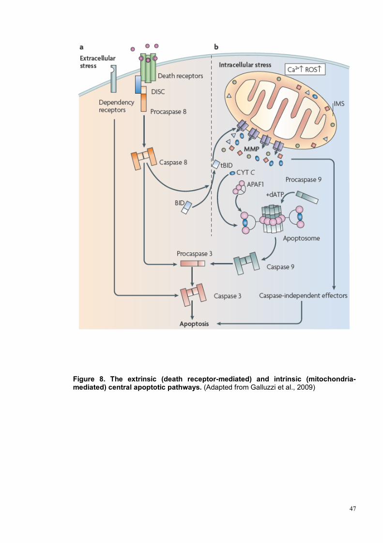
47
Figure 8. The extrinsic (death receptor-mediated) and intrinsic (mitochondria-mediated) central apoptotic pathways. (Adapted from Galluzzi et al., 2009)

48
Figure 9. The mechanisms of apoptosome formation and caspase activation initiated by cytochrome c release. (From Jiang and Wang, 2004)
49
Figure 10 Schematic presentation of the mitochondrial biochemical alterations in the course of Apaf 1-independent caspase 9 activation.
The mitochondrial inner membrane is illustrated with the electron transfer system (complexes I-IV), ATP synthase and citrate transporter (oval). A part of the Krebs cycle in the matrix is depicted. The outer membrane with pores permeable to the metabolites is also shown. Biochemical changes revealed by this study are highlighted in magenta. Bold arrows indicate an increase (upward) or decrease (downward) in each physiological parameter. Bar-headed lines indicate inhibition. The hypo-Δψm condition and the results with thioredoxin (TRX) predict free radical ([O2]–) production by which aconitase is inactivated. Dimerization of procaspase 9 by a topical increase in citrate is hypothesized. Cytosolic translocation of caspase 9 is inhibited by Bcl-2, although the mechanism has not been identified (blue box) (Adapted from Katoh et al., 2008).
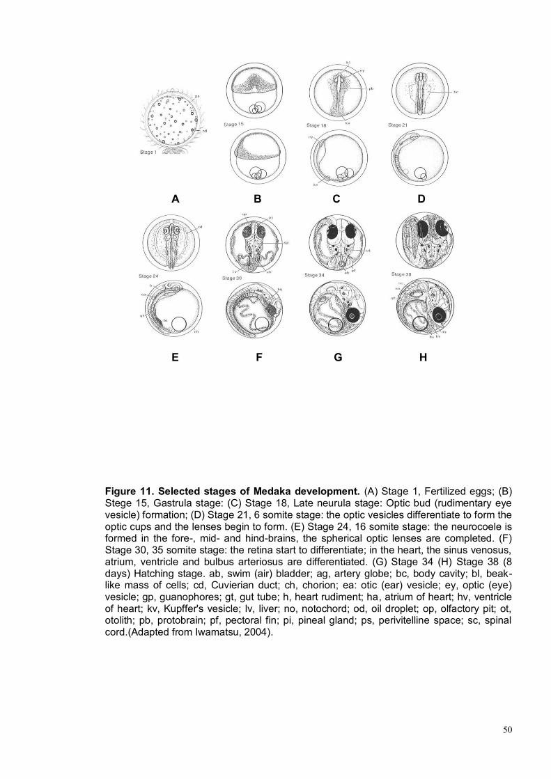
50
Figure 11. Selected stages of Medaka development. (A) Stage 1, Fertilized eggs; (B) Stege 15, Gastrula stage: (C) Stage 18, Late neurula stage: Optic bud (rudimentary eye vesicle) formation; (D) Stage 21, 6 somite stage: the optic vesicles differentiate to form the optic cups and the lenses begin to form. (E) Stage 24, 16 somite stage: the neurocoele is formed in the fore-, mid- and hind-brains, the spherical optic lenses are completed. (F) Stage 30, 35 somite stage: the retina start to differentiate; in the heart, the sinus venosus, atrium, ventricle and bulbus arteriosus are differentiated. (G) Stage 34 (H) Stage 38 (8 days) Hatching stage. ab, swim (air) bladder; ag, artery globe; bc, body cavity; bl, beak-like mass of cells; cd, Cuvierian duct; ch, chorion; ea: otic (ear) vesicle; ey, optic (eye) vesicle; gp, guanophores; gt, gut tube; h, heart rudiment; ha, atrium of heart; hv, ventricle of heart; kv, Kupffer's vesicle; lv, liver; no, notochord; od, oil droplet; op, olfactory pit; ot, otolith; pb, protobrain; pf, pectoral fin; pi, pineal gland; ps, perivitelline space; sc, spinal cord.(Adapted from Iwamatsu, 2004).
A B C D
E F G H

51
2. MATERIALS AND METHODS

52
2.1 Medaka Stocks
Wild type Oryzias latipes of the cab strain were maintained in an in-house facility
in a constant re-circulating system at 28°C on a 14 hours light/10 hours dark cycle.
Embryos were staged according to Iwamatsu 2004 (Iwamatsu, 2004).
2.2 Isolation and characterization of olhccs
To identify the olhccs gene, were performed a BLAST searches with the human
HCCS protein sequence (NP_005324 [GenBank]) in the Ensembl madaka
database (www.ensembl.org) and identified putative olhccs transcripts. The entire
olhccs coding sequence including part of the 5‟untranslated region were isolated
by RT-PCR amplification from a cDNA derived from a pool of medaka embryos at
different stages. Total RNAs were isolated from medaka embryos homogenized in
TRIzol reagent (Invitrogen Carlsbad, CA) using a sterile pestle. RNA was isolated
by chloroform extraction, isopropanol precipitation, and washed in 75% ethanol.
Contaminating genomicDNA was removed with Dnase I (Roche, Basel,
Switzerland). RNA was reverse transcribed to cDNA using the Superscript III First
Strand Synthesis Kit (Invitrogen). PCR was performed using 2 μl of the reverse
transcription reaction as a template with the High Fidelity PCR system (Roche,
Basel, Switzerland) and using the following olhccs specific primers:
olhccsaFw 5‟- TGCCGGTCGGTGGGTCCTTTG-3‟
olhccsaRv 5‟- CCAGTCTGCTGAAGCGCTGCAC-3‟
olhccsbFw 5‟- GATGTCAGTCACCCATCACG-3‟

53
2.3 Morpholinos (MO) and mRNAs injections
olhccs specific morpholinos (MO) (Gene Tools) were designed on the two different
5‟UTR region of the transcripts (olhccsa-MO and olhccsb-MO) and on the first
common exon-intron splice site (olhccs-MO) as follow:
olhccsa-MO 5‟- AGGTGTAGACGCAGAAGCGCCCATC-3‟
olhccsb-MO 5‟- GACTGACATCCCGATGGAGAGACCA-3‟
olhccs-MO 5‟-TGAAGTCAGGAACGTACCATGTTAG-3‟
The following MO containing five mismatches (olhccsa-mmMO) with respect to the
olhccsa-MO sequence was used as control (mismatches are in red):
olhccsa-mmMO 5‟-ATGTGTAAACGCATAAGCTCCCATT
olApaf1 specific MO were also designed on the 5‟UTR region of olApaf1:
olApaf1-MO 5‟- CTTCAGGCAAGTCACCTCCGACCAT-3‟
MOs were injected in a range of concentrations (0,03 - 0,4 mM). Their efficiency
was measured as the ability of interfering with eGFP expression using a reporter
construct. The pCS2/olhccsa-GFP, pCS2/olhccsb-GFP and pCS2/olhccs-GFP
reporter plasmids were constructed cloning the complementary region of the MOs
in frame with the eGFP coding sequence. MOs were individually co-injected with
the different eGFP mRNAs (25 ng/µL) and RFP mRNA (25 ng/µL). The inhibitory
efficiency of each MO was measured by quantification of the green/red
fluorescence ratio (eGFP/RFP) intensity using Photoshop CS3 software (Adobe)
54
to measure average pixel intensity of RFP and eGFP, as previously reported
(Conte et al., 2010b; Esteve et al., 2004). Selected MO working concentrations
was 0,3 mM for olhccsa-MO, 0,1 mM for olhccsb-MO, 0,06 mM for olhccs-MO, 0,1
mM for olApaf1-MO. MOs were injected into one blastomere of the embryos at the
one-two cell stage. Control embryos were always injected with either olhccsa-
mmMO or eGFP mRNA to follow the efficiency of the injections as well as for
testing possible defects associated with the injection procedures. At least 3
independent experiments were conducted for each marker and condition.
Activation of p53 is an occasional off-targeting effect of MO injections (Robu et al.,
2007), and can be counteracted by injection of a p53 Mo (Eisen and Smith, 2008).
Thus possible non-specific effects of olhccsa-MO were ruled out by coinjecting it
with a Mo designed against medaka p53 (p53MO) (Conte et al., 2010b).
olhccsa mRNA carrying silent mutations at the MO binding site, hBcl-xL mRNA,
eGFP mRNA and RFP mRNA were cloned and transcribed out of pCS2+ vector
using the SP6 mMessage mMachine kit (Ambion) according to manufacturer
instructions. The synthesized mRNAs were quantified and re-suspended in 1x
Yamamoto Ringer (Yamamoto and Yamagami, 1975). Rescue experiments were
performed by co-injecting olhccsa or hBcl-xL mRNA with olhccsa-MO. olhccs
mRNA (200 ng/µL) or hBcl-xL mRNA (200 ng/µL) were co-injected with olhccsa-
MO into one blastomere of the embryos at the one-two cell stage. All the injection
solutions included 25 ng/µL of eGFP mRNA as a lineage tracer.
2.4 Caspase inhibitors
Caspase inhibitors (CalBiochem) were caspase 1 inhibitor I (Ac-YVAD-CHO),
caspase inhibitor I (Z-VAD-FMK), and caspase 9 inhibitor III (Ac-LEHDCMK).
Stocks were diluted in DMSO or distilled water. Caspase inhibitors at final

55
concentration of 5 µM were co-injected with olhccsa-MO as previously reported
(Walker and Harland, 2009).
2.5 Whole-Mount In Situ Hybridization
Whole-mount in situ hybridization was carried out as using digoxygenin-labeled
antisense RNA riboprobes as previously described (Loosli et al., 1998). Probes for
olOtx2, olPax6, olBmp4, olTbx5, olVax2, olCyclinD1 and olAth5, have been
previously described (Conte et al., 2010a; Del Bene et al., 2007; Ruiz et al., 2009)
The cDNA of the olhccs, olIslet2 genes were isolated by RT-PCR amplification
with the following specific primers:
olhccsaFw 5‟- TGCCGGTCGGTGGGTCCTTTG-3‟
olhccsaRv 5‟- CCAGTCTGCTGAAGCGCTGCAC-3‟
islet2Fw 5‟-TCTGTTCTCCCAAGACCCGCTA-3‟
islet2Rv 5‟-AGGCTGGACAACATCAGACGA-3‟
Selected embryos were fixed in 10 ml of 4% paraformaldehyde prepared in 2X in
2X PTW (PBS containing 0.1% Tween) for 1 hour at room temperature and then
12 hours at 4°C. The embryos were dechorionated and washed 4 times with 1X
PTW. Finally, embryos were dehydrated in methanol 100% and stored at -20°C
(embryos in methanol survive to several months of storage without degeneration).
Embryos were gradually rehydrated washing with 75% methanol/PTW, 50%
methanol/PTW and 25%methanol/PTW. Than samples, were treated with 10mg/ml
proteinase K in PTW for a different amount of time (from 5 to 60 minutes)
depending on the specific embryonic stage and washed twice with freshly
prepared 2mg/ml glycine in PTW. After the embryos were refixed in 4%

56
paraformaldehyde/PTW at room temperature for 20 minutes and washed through
five changes of PTW. The embryos were pre-hybridized for at least 1 hour at 65°C
with hybridization buffer prepared as follow: Formamide 100% 25ml, SSC 20X
(pH7.0) 12.5 ml, Heparin (50mg/m) 150 μl, Torula-RNA 250 mg, Tween20 10%.
The probes were added and the samples were hybridized overnight at 65°C. After
the hybridization step, embryos were washed at 65°C with 50%
formammide/2xSSCT, 2xSSCT and 0.2xSSCT. Then the embryos were incubated
at room temperature with a blocking solution (5% serum/PTW) for two hours in
agitation. The samples were then incubated for 12 hours at 4° with 200 µl of anti-
DIG antibody (1:4000 dilution). Then the samples were washed 3 times for 10
minutes with the SB solution (0.1M Tris pH 9.5, 0.1M sodium chloride, 50 mM,
Magnesium chloride, 0.1% Tween). Subsequently the embryos were placed in the
appropriate colour solution with specific reagents NBT / BCIP (Boehringer).
The reaction was blocked with TE/Tween 0.1% solution, the embryos were again
fixed in 4% paraformaldehyde/PTW for 20 minutes, washed with PTW1X and
stored in glycerol. The embryos were embedded in a mix of BSA/Gelatine and
sectioned with vibratome. Bright-field images were obtained on a Leica DM-6000
microscope. Adobe Photoshop was used to adjust image brightness and contrast.
A minimum of 20 embryos, obtained from independent injections, were hybridized
for each marker and condition.
2.6 Immunohistochemistry
Embryos at different developmental stages were collected and fixed. The embryos
were dechorionated, dehydrated and stored at -20°C in methanol 100% as in 2.5.
Embryos were gradually rehydrated washing with 75% methanol/PTW, 50%
methanol/PTW and 25%methanol/PTW. The samples, were treated with 10mg/ml
57
proteinase K in PTW for a different amount of time (from 5 to 60 minutes)
depending on the specific embryonic stage and washed twice with freshly
prepared 2mg/ml glycine in PTW. The embryos were then refixed 4%
paraformaldehyde/PTW at room temperature for 20 minutes and washed through
five changes of PBS/ 0,05% TritonX-100. Embryos were placed in the blocking
solution (0,1 M phosphate buffer pH 7.2; 1% DMSO; 5% Fetal Bovine Serum FBS;
0,05% TritonX-100). Embryos were stained in toto with anti-Phospho-Histone3
antibodies (1:100) (Cell Signaling), followed by biotinylated-secondary antibodies
and peroxidase-coupled streptavidin (ABC-Elite kit, Vector Laboratories). Antigen
localization was detected by incubation in PBS containing 30 µg/mL DAB to which
0.003% hydrogen peroxide was added. The embryos were embedded in a mix of
BSA/Gelatine and sectioned with vibratome. At least ten retinas were analyzed for
each stage. The number of positive cells was normalized to the total area of retina
determined with the Image-J analysis software.
For immunofluorescence OCT embedded fixed embryos were cryostat-sectioned.
The sections were permeabilized boiling in citrate buffer 1 minute in the
microwave. Permeabilized sections were washed with PTW, and blocked in FBS
5% / PTW for 1 hour. Sections were incubated overnight at 4°C with the following
primary antibodies: α-active caspase 3 (1:500) (BD Pharmingen™), α-rhodopsin
(1:10000; Sigma), α-syntaxin (1:1000; Sigma) and α-GS6 antibodies (1:500;
Chemicon). Alexa-488–conjugated goat anti-rabbit or antimouse (1:1,000;
Invitrogen) IgGs were used as secondary antibodies. Propidium iodide (PI)
(1:1000) was added for nuclear staining. Stained sections were mounted with
Vectashield (Vector Laboratories). Microscopy analysis was performed with a
Leica DM6000 microscope and with Leica TCS SP2 AOBS confocal microscope.
Adobe Photoshop was used to adjust image brightness and contrast. The results
were obtained from four independent experiments.

58
2.7 TUNEL Staining
Embryos at different developmental stages were collected and fixed. The embryos
were dechorionated, dehydrated and stored at -20°C in methanol 100% as in 2.5.
Embryos were gradually rehydrated washing with 75% methanol/PTW, 50%
methanol/PTW and 25%methanol/PTW. The samples, were treated with 10mg/ml
proteinase K in PTW for a different amount of time (from 5 to 60 minutes) and
washed twice with freshly prepared 2mg/ml glycine in PTW. After the embryos
have been refixed 4% paraformaldehyde/PTW and washed through five changes
of PBS/0,1% TritonX-100. Then embryos were permeabilized in a 0,1 %
NaCitrate/0,1% TritonX-100 in ice for 15 minutes.
Embryos were processed with terminal deoxynucleotidyl transferase-mediated
dUTP nick-end labeling (TUNEL) kit (Roche Diagnostics) according to
manufacturer‟s instructions. The embryos were embedded in a mix of
BSA/Gelatine and sectioned with vibratome. At least ten embryos were analyzed
for each stage.
2.8 Transmission electron microscopy (TEM)
The embryos were fixed overnight at 4°C in 2.5% glutaraldehyde / 2%
paraformaldehyde in PTW. The material was then rinsed in buffer and post-fixed
in 1% osmium tetroxide in PBS for 1 hour at 4°C. After dehydration in a graded
series of ethanol, the material was embedded in an Epon-Araldite mixture.
Ultrathin sections, obtained with a Leica Ultracut UCT, were routinely stained (5%
uranyl acetate following by 1% lead citrate) and observed with a JEOL-JEM-1011
transmission electron microscope operating at 100 kV.

59
2.9 Detection of ROS levels
Reactive oxygen species (ROS) accumulation, assessed by the levels of the
oxidized form of the cell-permeant ROS indicator acetyl ester of 5- (and 6-)
chloromethyl-2‟,7‟-dichlorodihydrofluorescein diacetate (CM-H2DCFDA)
(Invitrogen), was detected in live fish as follows. Injected embryos at stage 24
were in vivo dechorionated, several groups of three embryos were put in the wells
of a 96-well plate, and excess medium was removed using a fire-polished glass
Pasteur pipette. Embryos were incubated in 1 µg/ml CM-H2DCFDA for up to 2 h,
and the fluorescence was measured with the Fluoroscan Ascent FL plate reader
(Thermo) (excitation 485 nm, emission 520 nm) after every 10 min. The
spontaneous oxidation of CM-H2DCFDA was monitored in the wells without fish,
and the levels were low and did not change with time. H2O2 solution served as a
positive control.

60
3. RESULTS

61
3.1 Identification and characterization of olhccs
To identify the olhccs gene, we performed BLAST searches with the human HCCS
protein sequence (NP_005324 [GenBank]) in the Ensembl madaka database and
identified putative olhccs transcripts, which were confirmed by PCR cloning and
sequence analysis. olhccs encodes for two transcript variants (olhccsa and
olhccsb) derived from an alternative splicing; the predicted proteins of 281 aa and
258 aa differ in the N-terminal portion and shown respectively 64% and 69% of
identity with the human HCCS protein (Figure 12 and Figure 13 A.). In order to
analyzes the expression patter of the gene in medaka we performed an in situ
hybridization (ISH) analysis using a probe that is able to recognize both
transcripts: according with the human and mouse hccs expression data (Ramskold
et al., 2009; Schaefer et al., 1996; Schwarz and Cox, 2002; Franco, unpublished
data), olhccs seems to be ubiquitously expressed but a very strong expression is
detectable in muscles, heart, CNS (Figure 13 B-D) and in the eye where the
expression is evident from early stages (stage 24) (Figure 13 E). The expression
of olhccs continued throughout the development and at stage 38, when the retina
is fully differentiated, becomes more specific for the ganglion and amacrine cell
layers, and for the ciliary marginal zone (CMZ) , although lower level of transcripts
are also detectable in other layers (Figure 13 F-H).
3.2 Knockdown of olhccs results in a specific phenotype recapitulating
the human MLS condition
To determine and analyze the olhccs function during development we employed
antisense morpholinos (MO) to achieve functional gene knockdown. We used
three different MOs designed against the 5‟UTR of the two transcript variants
62
olhccsa and olhccsb (olhccsa-MO and olhccsb-MO) and against the first common
splice donor site (olhccs-MO). The oIhccs-MO is predicted to downregulate both
transcripts. These MOs were independently injected at 1–2 cell stage and then the
embryos were phenotypically analyzed.
Injection of olhccs-MO resulted in a very severe phenotype and about the 80% of
fish die before the gastrulation stage. Injection of both olhccsa-MO and olhccsb-
MO induce in the fish defects that resemble the human MLS condition: the
embryos show a severe ocular phenotype with microphtalmia, alteration of RPE,
and coloboma in 50% of cases (Figure 14 A-C, I). In particular reduction in eye
size was evident starting from stage 24 and the eyes treated with olhccsa-MO
showed a reduction in size of about 50% compared with the size of the eyes of the
uninjected controls at stage 38 (Figure 14 G). These defects are associated also
with microcephaly and a cardiovascular pathology consisted of significant
pericardial edema, bradycardia (reduced heart rate), ventricular asystole (failure to
contract), and failure to develop into a loop (red arrows in Figure 14 H-I). These
defects were progressive, eventually leading to death after stage 38.
Of note, injection of olhccsa-MO and olhccsb-MO yielded indistinguishable
phenotypes. The olhccsa-MO was thus used for analysis and quantization in all
subsequent experiments, with all phenotypic observations additionally verified
using the olhccsb-MO. To ensure the specificity of the olhccsa-MO, we performed
all the appropriate control experiments (Eisen and Smith, 2008). We were able to
rescue the phenotype by coinjecting the MO with the wt olhccs mRNA and this
experiment demonstrate that all the defects observed in the injected embryos are
due to specific downregulation of olhccs (Figure 14 D). In addition, a mismatch MO
with five base substitutions in its sequence (olhccsa-mmMO) did not show any
phenotype at an equivalent dose (Figure 14 E).

63
Finally, it has been proposed that an activation of p53 could represent a non
specific off-target effect of some MOs (Robu et al., 2007). In this case the
phenotype can be rescued by co-injection of the morpholino against the sequence
under study and that of p53 (Eisen and Smith, 2008). We thus co-injected olhccsa-
MO with p53-MO (Conte et al., 2010b) and no amelioration of the phenotype was
observed (Figure 14 F) indicating that in this case the activation of p53 is not
involved in the observed phenotype.
To further ensure that olhccsa-MO was blocking the translation of olhccs, we
constructed and coinjected with the olhccsa-MO a GFP reporter, where the coding
region of the GFP was fused to the 5‟UTR of olhccsa (olhccsa-GFP) (Figure 15 A).
If the olhccsa-MO binds the target sequence and represses translation of the GFP,
the embryo would lack green fluorescence. To obtain a quantitative measurement
of the repression of GFP by the MO, RNA coding for RFP was also coinjected and
the green/red fluorescence ratio was quantified as previously described (Conte et
al., 2010b; Esteve et al., 2004). As illustrated in figure 15 C a reporter GFP fused
to the 5‟UTR of olhccsa, was translationally repressed when coinjected with
olhccsa-MO. However, when the same GFP construct was coinjected with
olhccsa-mmMO, it was not able to repress the translation of the GFP (Figure 15 D)
further supporting the specificity and quality of our controls.
Together, these results demonstrate that olhccsa-MO is specifically able to inhibit
translation and therefore abolish the function of olhccsa in vivo, and that this leads
to the induction of a specific phenotype resembling the human MLS condition.
3.3 Knockdown of olhccs leads to an increase of apoptosis.
The microphthamia is a one of most common defects associated with HCCS
mutations (Sharma et al., 2008; Wimplinger et al., 2006; Wimplinger et al., 2007).
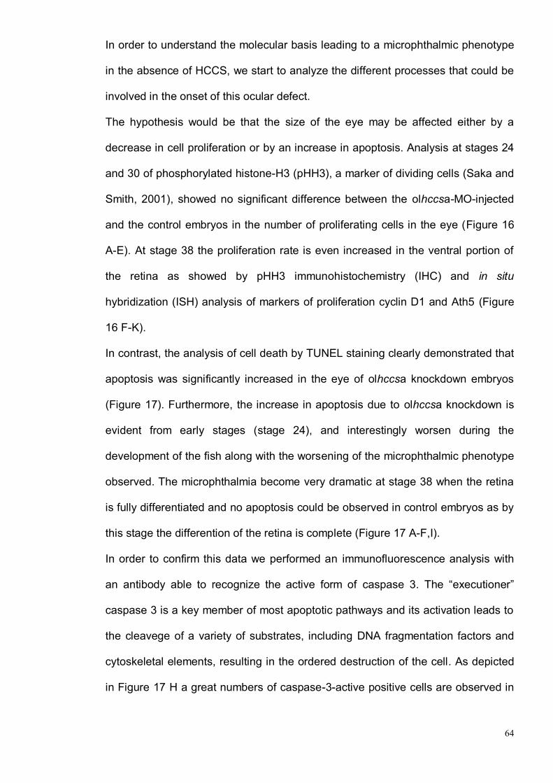
64
In order to understand the molecular basis leading to a microphthalmic phenotype
in the absence of HCCS, we start to analyze the different processes that could be
involved in the onset of this ocular defect.
The hypothesis would be that the size of the eye may be affected either by a
decrease in cell proliferation or by an increase in apoptosis. Analysis at stages 24
and 30 of phosphorylated histone-H3 (pHH3), a marker of dividing cells (Saka and
Smith, 2001), showed no significant difference between the olhccsa-MO-injected
and the control embryos in the number of proliferating cells in the eye (Figure 16
A-E). At stage 38 the proliferation rate is even increased in the ventral portion of
the retina as showed by pHH3 immunohistochemistry (IHC) and in situ
hybridization (ISH) analysis of markers of proliferation cyclin D1 and Ath5 (Figure
16 F-K).
In contrast, the analysis of cell death by TUNEL staining clearly demonstrated that
apoptosis was significantly increased in the eye of olhccsa knockdown embryos
(Figure 17). Furthermore, the increase in apoptosis due to olhccsa knockdown is
evident from early stages (stage 24), and interestingly worsen during the
development of the fish along with the worsening of the microphthalmic phenotype
observed. The microphthalmia become very dramatic at stage 38 when the retina
is fully differentiated and no apoptosis could be observed in control embryos as by
this stage the differention of the retina is complete (Figure 17 A-F,I).
In order to confirm this data we performed an immunofluorescence analysis with
an antibody able to recognize the active form of caspase 3. The “executioner”
caspase 3 is a key member of most apoptotic pathways and its activation leads to
the cleavege of a variety of substrates, including DNA fragmentation factors and
cytoskeletal elements, resulting in the ordered destruction of the cell. As depicted
in Figure 17 H a great numbers of caspase-3-active positive cells are observed in
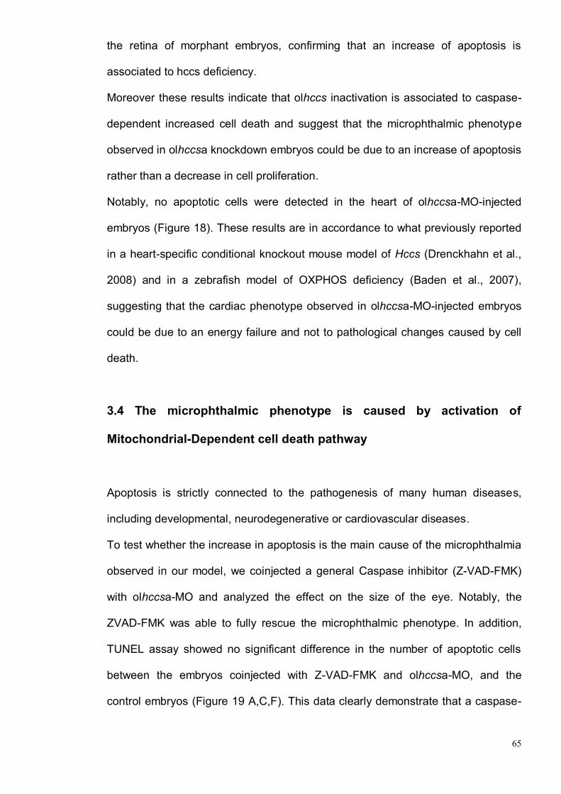
65
the retina of morphant embryos, confirming that an increase of apoptosis is
associated to hccs deficiency.
Moreover these results indicate that olhccs inactivation is associated to caspase-
dependent increased cell death and suggest that the microphthalmic phenotype
observed in olhccsa knockdown embryos could be due to an increase of apoptosis
rather than a decrease in cell proliferation.
Notably, no apoptotic cells were detected in the heart of olhccsa-MO-injected
embryos (Figure 18). These results are in accordance to what previously reported
in a heart-specific conditional knockout mouse model of Hccs (Drenckhahn et al.,
2008) and in a zebrafish model of OXPHOS deficiency (Baden et al., 2007),
suggesting that the cardiac phenotype observed in olhccsa-MO-injected embryos
could be due to an energy failure and not to pathological changes caused by cell
death.
3.4 The microphthalmic phenotype is caused by activation of
Mitochondrial-Dependent cell death pathway
Apoptosis is strictly connected to the pathogenesis of many human diseases,
including developmental, neurodegenerative or cardiovascular diseases.
To test whether the increase in apoptosis is the main cause of the microphthalmia
observed in our model, we coinjected a general Caspase inhibitor (Z-VAD-FMK)
with olhccsa-MO and analyzed the effect on the size of the eye. Notably, the
ZVAD-FMK was able to fully rescue the microphthalmic phenotype. In addition,
TUNEL assay showed no significant difference in the number of apoptotic cells
between the embryos coinjected with Z-VAD-FMK and olhccsa-MO, and the
control embryos (Figure 19 A,C,F). This data clearly demonstrate that a caspase-

66
dependent cell death pathway is responsible for the microphthalmic phenotype
observed in our model.
In order to elucidate which pathway was involved in the activation of the caspase
cascade, we tested two different specific Caspase inhibitors (Caspase 1 and
Caspase 9) for their ability to rescue the apoptotic phenotype in morphant
embryos. While the inhibitor of caspase 1 did not display any effect, the Caspase
9-specific inhibitor, Ac-LEHD-CMK, was able to fully rescue the microphthalmia
(Figure 19).
The initiator caspase 9 is one of the most important members of the mitochondria-
mediated cell death pathway and its activation in response to hccs knock-down
strongly suggested the involvement of the “intrinsic pathway” in the onset of
microphtalmia. To test this hypothesis we tried to rescue the phenotype by the
over-expression of the anti-apoptotic gene Bcl-xL. The protein encoded by this
transcript belongs to the BCL-2 protein family and is located at the outer
mitochondrial membrane where regulates the opening of the outer mitochondrial
membrane channel (VDAC). As expected, the over-expression of Bcl-xL was also
able to rescue the microphthalmia confirming an activation of the intrinsic cell-
death pathway (Figure 20).
Together these results clearly indicate the involvement of Mitochondrial-
Dependent cell death pathway in the onset of the microphthalmic phenotype
associated to olhccs down-regulation.
3.5 olhccs down-regulation leads to activation of caspase 9 in an
apopotosoma-independent manner.
Our data demonstrated that the downregulation of olhccs leads to an increase of
Mitochondrial mediated apoptosis which is responsible for the onset of the

67
microphtalmic phenotype. In particular we showed that an activation of “initiator”
caspase 9 underlie this process.
Detailed studies of the mechanisms underlying intrinsic apoptosis have shown that
the heme group of cytochrome c is necessary for Apaf1 activation, apoptosome
formation and caspase 9 activation (Martin and Fearnhead, 2002). However, as
described in the introduction, recent models propose that the activation of caspase
9 can occur in a apoptosome-indipendent manner. We thus decided to test if in our
model an apoptosoma-independent caspase 9 activation can occur by looking at
the effects of Apaf1 down-regulation in olhccsa-MO injected embryos.
To achieve a functional gene knockdown we designed morpholinos against the
olApaf1 5‟UTR (olApaf1-MO). olApaf1-MO and olhccsa-MO were coinjected at 1–2
cell stage and then embryos were analyzed by TUNEL assay.
Interestingly, our results showed that Apaf1 down-regulation was not able to block
the apoptotic cascade and to rescue the microphtalmic phenotype (Figure 21),
thus indicating that in our model the activation of cell death is mediated by an
apoptosome-independent caspase 9 activation.
3.6 Impairment of mitochondrial function and overproduction of
reactive oxygen species (ROS) in olhccs knockdown embryos.
Mitochondria have long been known to be critical for cell survival due to their role
in energy metabolism and for their pivotal role in progression through apoptotic
pathways. HCCS acts binding heme to cytochrome c and c1 (Bernard et al., 2003;
Schwarz and Cox, 2002) and the holo-form of proteins are necessary for their
activity. We thus, analyzed the effect of HCCS deficiency on mitochondrial
functions.

68
For this purpose in collaboration with Dr. I. Ferrero and Dr. P. Goffrini (University
of Parma) we performed HCCS complementation studies in yeast.
We used a strain, of Saccharomyces cerevisiae mutant deficient for the HCCS
orthologue Cyc3 which is not able to grow on non-fermentable carbon sources like
glycine. In this condition, the cell can‟t use glycolysis but needs mitochondrial
oxidative phosphorylation to produce ATP. The respiratory growth can then be
restored transforming the yeast with Cyc3 or HCCS (Bernard et al., 2003; Schwarz
and Cox, 2002; Wimplinger et al., 2006) (Figure 22 A).
The analysis of the cytochromes absorption spectra shows that both cytochromes
c and b, and a+a3 (component respectively of Complex III or Complex IV of
mitochondrial respiratory chain) were reduced, indicating a block at the distal end
of the respiratory chain (Figure 22 B).
Analysis of OXPHOS complexes activity in hccs-deficient medaka embryos are
still in progress in collaboration with Prof. M. Zeviani at Carlo Besta Institute of
Neurology, Milan. Nevertheless, as expected from an OXPHOS-deficient tissue,
Transmission Electron Microscopy (TEM) analysis on morphants retina shows the
presence of abnormal mitochondria, with internal disorganization of the cristae
(Figure 23). This result further supports the key role of hccs in the physiology of
fish mitochondria.
It has been demonstrated that when the respiratory chain is inhibited downstream
of complex III, electrons coming from succinate oxidation could also lead to
superoxide anion generation by reverse electron transport from Complex II to
Complex I (Lambert and Brand, 2004; St-Pierre et al., 2002). Moreover ROS
production increases when electron transport is reduced, which occurs at low
respiratory rates (Korshunov et al., 1997) or in pathological situations (Wallace,
2005). We thus checked the production of ROS in hccs-deficient embryos and, as
expected, olhccs morphants showed increased levels of ROS, as detected by
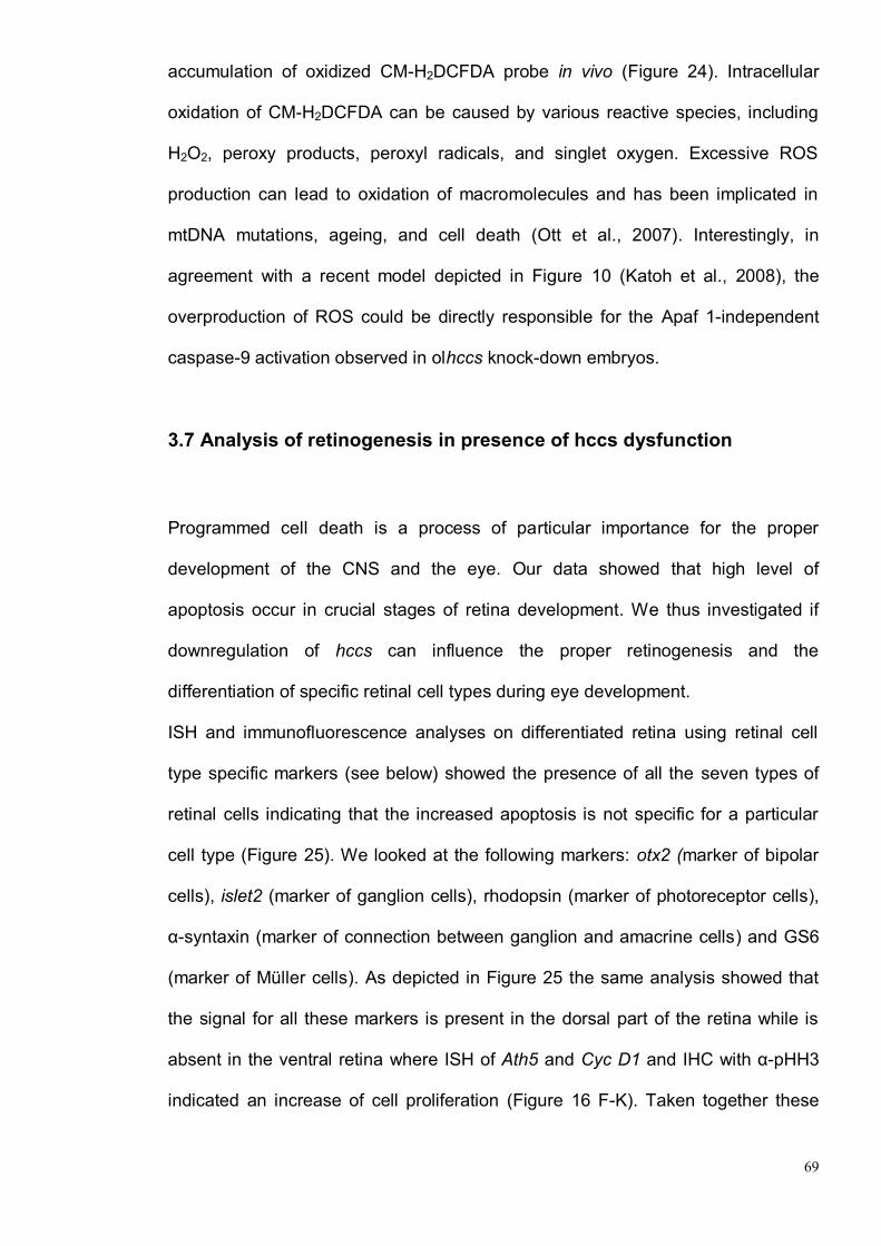
69
accumulation of oxidized CM-H2DCFDA probe in vivo (Figure 24). Intracellular
oxidation of CM-H2DCFDA can be caused by various reactive species, including
H2O2, peroxy products, peroxyl radicals, and singlet oxygen. Excessive ROS
production can lead to oxidation of macromolecules and has been implicated in
mtDNA mutations, ageing, and cell death (Ott et al., 2007). Interestingly, in
agreement with a recent model depicted in Figure 10 (Katoh et al., 2008), the
overproduction of ROS could be directly responsible for the Apaf 1-independent
caspase-9 activation observed in olhccs knock-down embryos.
3.7 Analysis of retinogenesis in presence of hccs dysfunction
Programmed cell death is a process of particular importance for the proper
development of the CNS and the eye. Our data showed that high level of
apoptosis occur in crucial stages of retina development. We thus investigated if
downregulation of hccs can influence the proper retinogenesis and the
differentiation of specific retinal cell types during eye development.
ISH and immunofluorescence analyses on differentiated retina using retinal cell
type specific markers (see below) showed the presence of all the seven types of
retinal cells indicating that the increased apoptosis is not specific for a particular
cell type (Figure 25). We looked at the following markers: otx2 (marker of bipolar
cells), islet2 (marker of ganglion cells), rhodopsin (marker of photoreceptor cells),
α-syntaxin (marker of connection between ganglion and amacrine cells) and GS6
(marker of Müller cells). As depicted in Figure 25 the same analysis showed that
the signal for all these markers is present in the dorsal part of the retina while is
absent in the ventral retina where ISH of Ath5 and Cyc D1 and IHC with α-pHH3
indicated an increase of cell proliferation (Figure 16 F-K). Taken together these

70
data demontrate that morphants display a differentiation defect of the ventral
retina, although all retinal cell types seem to be present.
Our data indicated that the increased apoptosis due to hccs down-regulation is not
specific for the ventral part of the retina. Thus, in order to understand the
differentiation problem of the ventral retina, we also analyzed the expression of
genes involved in dorsal-ventral pattern specification. However, ISH analysis on
stage 24 embryos using specific probes for Pax6, Vax2, Tbx5, and Bmp4, gene
involved in specification of dorsal-ventral pattern, do not show changes in the their
expression (Figure 26). These data suggest that in olhccs-deficient embryos the
dorsal-ventral pattern is not affected and further experiments will be necessary to
explain the phenotype observed in the ventral retina.
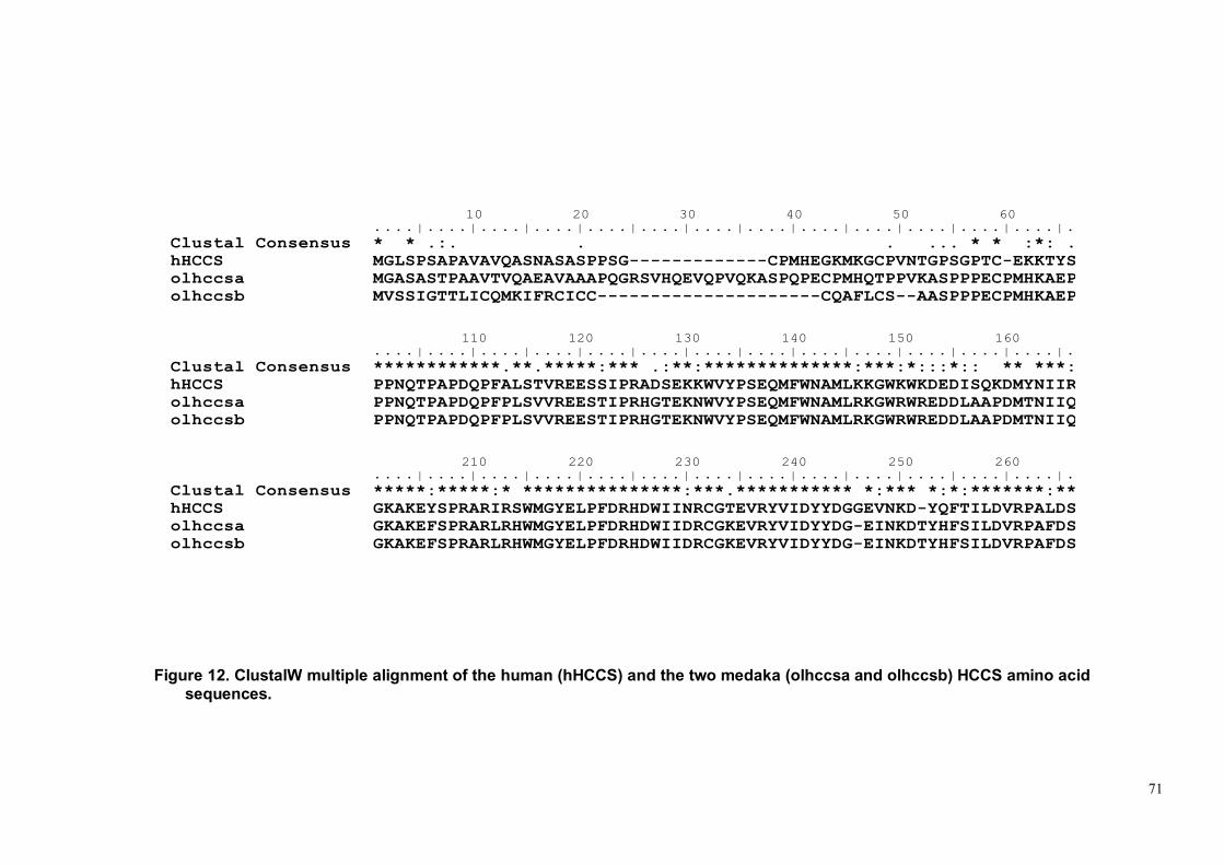
71
Figure 12. ClustalW multiple alignment of the human (hHCCS) and the two medaka (olhccsa and olhccsb) HCCS amino acid
sequences.
10 20 30 40 50 60 70 80 90 100....|....|....|....|....|....|....|....|....|....|....|....|....|....|....|....|....|....|....|....|
Clustal Consensus * * .:. . . ... * * :*: . ****::**::****:* :** :.::**:*:**
hHCCS MGLSPSAPAVAVQASNASASPPSG-------------CPMHEGKMKGCPVNTGPSGPTC-EKKTYSVPAHQERAYEYVECPIRGTAAENKENLDPSNLMP
olhccsa MGASASTPAAVTVQAEAVAAAPQGRSVHQEVQPVQKASPQPECPMHQTPPVKASPPPECPMHKAEPGPAHQNQAYQFVECPMR-AAAGVRSDIDPANMMP
olhccsb MVSSIGTTLICQMKIFRCICC---------------------CQAFLCS--AASPPPECPMHKAEPGPAHQNQAYQFVECPMR-AAAGVRSDIDPANMMP
110 120 130 140 150 160 170 180 190 200....|....|....|....|....|....|....|....|....|....|....|....|....|....|....|....|....|....|....|....|
Clustal Consensus ************.**.*****:*** .:**:**************:***:*:::*:: ** ***:***:******:**********.******:* ***
hHCCS PPNQTPAPDQPFALSTVREESSIPRADSEKKWVYPSEQMFWNAMLKKGWKWKDEDISQKDMYNIIRIHNQNNEQAWKEILKWEALHAAECPCGPSLIRFG
olhccsa PPNQTPAPDQPFPLSVVREESTIPRHGTEKNWVYPSEQMFWNAMLRKGWRWREDDLAAPDMTNIIQIHNKNNEQAWQEILKWEALHAGECPCGPTLKRFG
olhccsb PPNQTPAPDQPFPLSVVREESTIPRHGTEKNWVYPSEQMFWNAMLRKGWRWREDDLAAPDMTNIIQIHNKNNEQAWQEILKWEALHAGECPCGPTLKRFG
210 220 230 240 250 260 270 280....|....|....|....|....|....|....|....|....|....|....|....|....|....|....|....|...
Clustal Consensus *****:*****:* ***************:***.*********** *:*** *:*:*******:***.***************
hHCCS GKAKEYSPRARIRSWMGYELPFDRHDWIINRCGTEVRYVIDYYDGGEVNKD-YQFTILDVRPALDSLSAVWDRMKVAWWRWTS
olhccsa GKAKEFSPRARLRHWMGYELPFDRHDWIIDRCGKEVRYVIDYYDG-EINKDTYHFSILDVRPAFDSLGAVWDRMKVAWWRWTS
olhccsb GKAKEFSPRARLRHWMGYELPFDRHDWIIDRCGKEVRYVIDYYDG-EINKDTYHFSILDVRPAFDSLGAVWDRMKVAWWRWTS
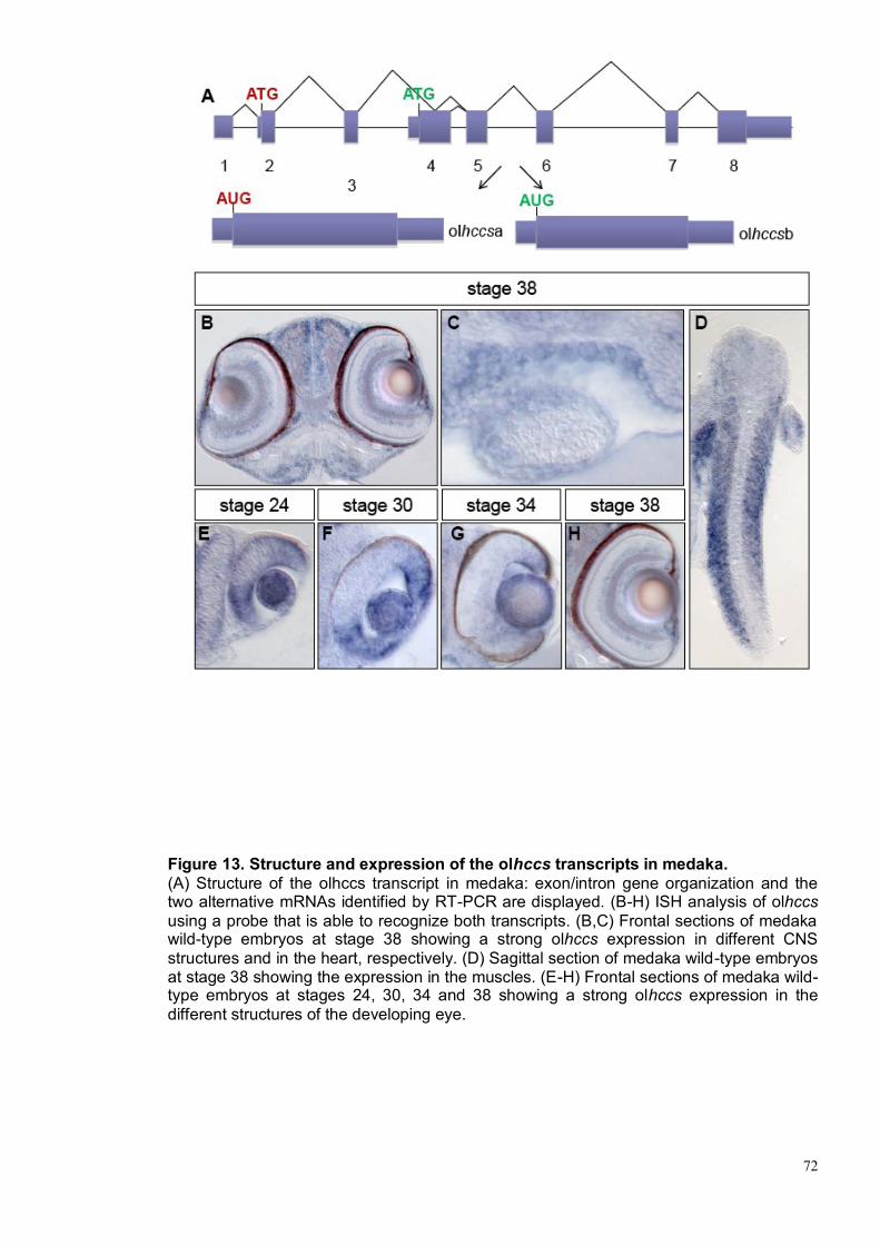
72
Figure 13. Structure and expression of the olhccs transcripts in medaka.
(A) Structure of the olhccs transcript in medaka: exon/intron gene organization and the two alternative mRNAs identified by RT-PCR are displayed. (B-H) ISH analysis of olhccs
using a probe that is able to recognize both transcripts. (B,C) Frontal sections of medaka wild-type embryos at stage 38 showing a strong olhccs expression in different CNS
structures and in the heart, respectively. (D) Sagittal section of medaka wild-type embryos at stage 38 showing the expression in the muscles. (E-H) Frontal sections of medaka wild-type embryos at stages 24, 30, 34 and 38 showing a strong olhccs expression in the
different structures of the developing eye.

73
Figure 14. Effects of the morpholinos injections in medaka embryos. Bright-field stereomicroscopy images of wild-type embryos (A,H) and of olhccsa-MO (B,I), olhccsb-MO (C), olhccsa-MO + olhccsa mRNA (D), olhccsa-mmMO (E), olhccsa-MO + p53-MO (F) -injected medaka embryos at
stage 38. A-F dorsal view. H-I lateral view. The embryos injected with morpholinos display a phenotype resembling the human MLS condition. The ocular phenotype includes microphthalmia (B,C,I), coloboma (black arrow) (I). (G) Retinal dimension analysis at stages 24 and 38. The ocular defects are associated with microcephalia (B,C,I) and cardiac defects (red arrows) (I). In wild-type embryos, the ventricle is looped behind the atrium (H). In hccs-deficient embryos the heart is not looped and is surrounded by pericardial edema (I). a, atrium; v, ventricle.

74
Figure 15. Morpholinos against olhccsa efficiently interfere with its translation. (A) Schematic representation of the construct (olhccsa-GFP) used to test the efficiency of olhccsa-MO: the construct contained the 5′ portion of olhccsa gene fused in frame with the GFP coding region. (B–D) Dorsal views of stage 19 embryos injected with RFP and olhccsa-GFP mRNA alone (B) or in association with olhccsa-MO (C), or with a control morpholino containing five mismatches with respect to the original olhccsa-MO sequence (olhccsa-mmMO) (D). Note how olhccsa-MO but not olhccsa-mmMO,
significantly inhibits GFP expression. (E) Percentage of inhibition, quantified by GFP/RFP intensity using Adobe-Photoshop software.
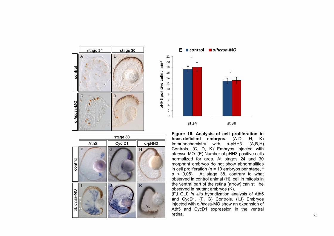
75
Figure 16. Analysis of cell proliferation in hccs-deficient embryos. (A-D, H, K)
Immunochemistry with α-pHH3. (A,B,H) Controls. (C, D, K) Embryos injected with olhccsa-MO. (E) Number of pHH3-positive cells
normalized for area. At stages 24 and 30 morphant embryos do not show abnormalities in cell proliferation (n = 10 embryos per stage, * p < 0,05). At stage 38, contrary to what observed in control animal (H), cell in mitosis in the ventral part of the retina (arrow) can still be observed in mutant embryos (K). (F,I G,J) In situ hybridization analysis of Ath5
and CycD1. (F, G) Controls. (I,J) Embryos injected with olhccsa-MO show an expansion of
Ath5 and CycD1 expression in the ventral retina.

76
Figure 17. Increase of apoptosis in the retina of olhccs-deficient embryos. (A-F) TUNEL assay. (A, B, C) Controls. (D, E, F) Embryos injected with olhccsa-MO show a dramatic increase of cell death at all stages analysed. (I) Number of TUNEL-positive cells for eye (n = 10 embryos per stage, * p < 0,05). (G,H) Immunofluorescence analysis with α-active-Caspase 3 on control (G) and olhccsa-MO-injected (H) embryos at stage 38.
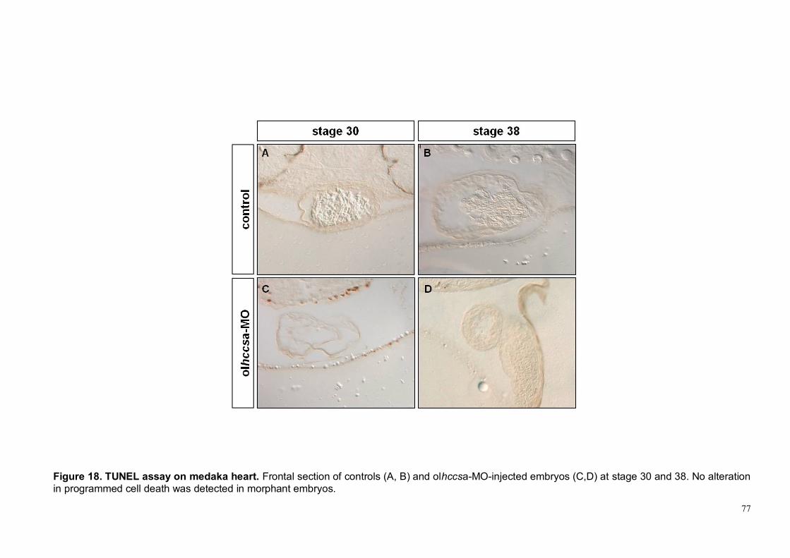
77
Figure 18. TUNEL assay on medaka heart. Frontal section of controls (A, B) and olhccsa-MO-injected embryos (C,D) at stage 30 and 38. No alteration
in programmed cell death was detected in morphant embryos.
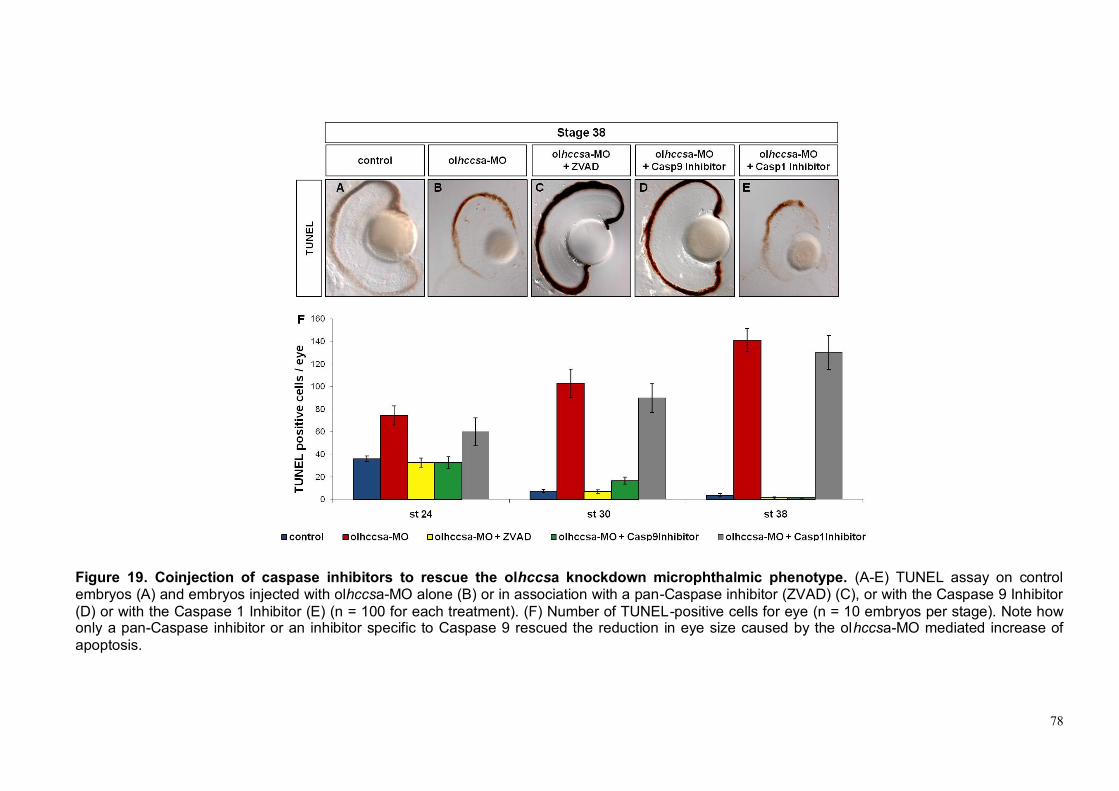
78
Figure 19. Coinjection of caspase inhibitors to rescue the olhccsa knockdown microphthalmic phenotype. (A-E) TUNEL assay on control embryos (A) and embryos injected with olhccsa-MO alone (B) or in association with a pan-Caspase inhibitor (ZVAD) (C), or with the Caspase 9 Inhibitor
(D) or with the Caspase 1 Inhibitor (E) (n = 100 for each treatment). (F) Number of TUNEL-positive cells for eye (n = 10 embryos per stage). Note how only a pan-Caspase inhibitor or an inhibitor specific to Caspase 9 rescued the reduction in eye size caused by the olhccsa-MO mediated increase of
apoptosis.

79
Figure 20. Involvement of Mitochondrial-Dependent cell death pathway in olhccsa knockdown microphthalmic phenotype. (A-E) TUNEL assay on control embryos (A) and embryos injected with olhccsa-MO alone (B) or in association with the Caspase 9 Inhibitor (C) or with the Bcl-xL mRNA (D) (n = 100 for each treatment).
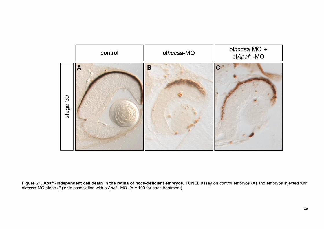
80
Figure 21. Apaf1-independent cell death in the retina of hccs-deficient embryos. TUNEL assay on control embryos (A) and embryos injected with olhccsa-MO alone (B) or in association with olApaf1-MO. (n = 100 for each treatment).

81
Figure 22. Impairment of mitochondrial respiratory chain in yeast. (A) The S. cerevisiae strain B-8025 (Cyc3Δ) was transformed with yeast CYC3,
human HCCS or empty expression construct pYEX4T, and grown on fermentable (glucose) or respiratory (glycine) substrates. Note the CYC3 mutant strain is not able to grow on non-fermentable carbon sources and needs complementation to show respiratory growth. (B) Cytochrome spectra of the heme lyase mutant strain B-8025 transformed with yeast CYC3, human HCCS or empty vector. The arrows indicate the absorption maxima of the bands of cytochromes c and c1 (550 nm), b (560 nm), and a+a3 (603 nm). The spectra were reduced in CYC3 mutant, indicating a block at the distal end of the respiratory chain. In collaboration Dr. I. Ferrero and Dr. P. Goffrini (University of Parma)

82
Figure 23. TEM analysis of mitochondrial morphology in hccs-deficient embryos. Retinal sections of controls (A) and olhccsa-MO-injected embryos (B) at stage 38. (C,D) Magnifications showing several mitochondria in photoreceptor cells. Note the presence of abnormal mitochondria, with internal disorganization of the cristae (arrows in D) in the morphants compared to controls (C).

83
Figure 24. Detection of ROS levels in olhccsa MO-injected fish. Accumulation of the CMH2DCFDA dye, used as indicator of ROS levels, in olhccsa MO-injected and control embryos at stage 24. ROS levels are increased in olhccsa MO-injected fish. Values represent means of ten samples. Each
sample is represented by a group of 3 fish.

84
Figure 25. Analysis of retinal cells type specific markers. Retinal sections of controls (A-E) and olhccsa-MO-injected embryos (F-J) at stage 38. (A,F B,G) ISH analysis to look at the expression pattern of otx2 and islet2. (C,H D,I E,J) Immunofluorescence with α-rhodopsin, α-syntaxin and α-GS6
antibodies. Note the absence of all signals in the morphant ventral retina (asterisks in F-J).

85
Figure 26. Analysis of dorso-ventral pattern on olhccsa MO-injected fish. Retinal sections of controls (A-D) and olhccsa-MO-injected embryos (E-H) at stage 24. (A,E B,F C,G D,H) ISH analysis to detect the pattern of expression of pax6, vax2, tbx5 and bmp4. Injected embryos display the same
pattern of expression of control embryos indicating that the dorso-ventral patterning of the retina does not seem to be perturbed.

86
4. DISCUSSION
87
Eye diseases represent one of the most common groups of genetic disorders in
the human population. Over 200 different forms of ocular heritable disorders have
been described and it has been estimated that about 27% of the phenotypes
described in OMIM affect the eye. In the majority of conditions the pathogenetic
causes of eye developmental anomalies remain very elusive. This is particularly
true for Microphthalmia and Anophthalmia both characterized by major structural
eye malformations. Among the genetic forms of Microphthalmia/Anophthalmia,
MLS syndrome, first described in the 1990, represents one of the most puzzling
genetic disorder. As fully described in chapter 1.3 MLS is a rare X-linked dominant
male lethal neurodevelopmental disorder characterized by a syndromic form of
microphthalmia associated to skin abnormalities (linear skin lesions), central
nervous system anomalies and congenital heart defects. An high degree of intra-
as well as interfamilial clinical variability has been observed in this condition
possibly related to the role of X-inactivation (Franco B et al.,2006; Van den Veyver
IB, 2001). In the past fifteen years, several research groups have been actively
searching for the gene responsible for this disorder. The MLS critical region it has
been defined by deletion mapping in the 1993 (Wapenaar et al., 1993; Wapenaar
et al., 1994). However, the conclusive evidence that HCCS is the gene responsible
for MLS syndrome have been found only in 2006 (Wimplinger et al., 2006).
HCCS encodes a mitochondrial holocytochrome c–type synthase and catalyzes
the covalent attachment of heme to both apocytochrome c and c1. Functional
studies demonstrated that this protein plays a critical role in mithocondrial function
(Bernard et al., 2003; Schwarz and Cox, 2002)
Although mutation analysis clearly indicates a role for HCCS in the pathogenesis
of this genetic condition, the molecular mechanisms underlying the developmental
defects observed in the presence of HCCS dysfunction are still unknown. Previous

88
studies demonstrated the early lethality of Hccs knock-out mouse embryos
(Prakash et al., 2002).
Medakafish is considered an ideal model to study developmental biology
processes and in particularly the eye development (Wittbrodt et al., 2002)..
HCCS is a protein highly conserved in evolution and in particular the two proteins
codified by the two transcript variants (olhccsa and olhccsb) identified in medaka
showed, respectively, 64% and 69% of identity with the human HCCS protein.
Moreover our data showed also that the hccs expression pattern in medaka is
consistent with what has been reported concerning the expression of the HCSS
transcript in human and mouse (Ramskold et al., 2009; Schaefer et al., 1996;
Schwarz and Cox, 2002; Franco, unpublished data). Moreover our data, in
addition to what previously reported, showed that a strong expression in the eye
was evident from early stages (stage 24) and continued throughout the
development. The expression become more specific, for the ganglion and
amacrine cell layers, and for the ciliary marginal zone (CMZ) at later stages (stage
38), although lower level of transcripts are also detectable also in other layers. The
differences in the expression of hccs between the different retinal cell types could
be explain by a specific request in mitochondrial energy of some types of retinal
cells, such as the ganglion cells in which rich accumulations of mitochondria are
observed in the soma, neurofiber layer, and pre-laminar region (unmyelinated
axons) (Barron et al., 2004).
Taken together this finding indicated medaka like a good model to study the hccs
function and its role in the pathogenesis of MLS syndrome. We thus decided to
generate a model for this disease in medakafish, where we planned to perform
loss of function studies to better define the function of this gene and its role in the
pathogenesis of MLS syndrome.

89
Moreover medaka is also a particularly amenable system model for reverse-
genetic analyses. Microinjection of early embryos with either mRNA or antisense
morpholino oligonucleotides results in transient gene overexpression or
knockdown, respectively and allow studying a highly specific gene function,
without any laborious, time and resource consuming techniques. In addition, the
use of graded concentrations of specific morpholinos, directed against the
alternative forms olhccsa and olhccsb allowed us to obtain a partial gene
inactivation. Different attempts using different dose of morpholinos allowed us to
establish the dose that reduces the level of hccs activity to a degree that impairs
normal development but is not immediately lethal. This is a great advantage to
study diseases, such as MLS, where the total inactivation of the gene is lethal.
Capitalizing on the characteristics of medaka, we have investigated the effects of
hccs deficiency in different organ‟s development and function. Our results
demonstrated that medaka indeed represents an informative and powerful model
to study the MLS syndrome.
To determine and analyze the function of olhccs during development we used
three different MOs designed against the 5‟UTR of the two-transcript variants
olhccsa and olhccsb (olhccsa-MO and olhccsb-MO) and against the first common
splice donor site (olhccs-MO). Our experiments showed that the injection of the
three different morpholinos resulted in a pathological phenotype, which resembles
the human MLS condition: the embryos show a severe ocular phenotype with
microphthalmia, alteration of retinal pigmented epithelium (RPE), and coloboma
associated to microcephaly and a severe cardiovascular pathology. Not
surprisingly the injection of olhccs-MO, which is able to downregulate both
transcripts, resulted in a very severe phenotype and about the 70% of fish die
before the gastrulation stage. All suggested controls (Eisen and Smith, 2008) were
employed to demonstrate that these phenotype were not secondary to injection

90
trauma or MO toxicity but specific to hccs deficiency. These strategy allowed as to
generate an animal model which, to date, is the only animal model recapitulating
the phenotype observed in MLS syndrome.
We demonstrated that HCCS, in yeast as well as in fish, is necessary for proper
mitochondrial functioning and in particular that hccs-deficiency leads to a block at
the distal end of the respiratory chain and to an overproduction of reactive
oxidative species (ROS) which are known to be involved in oxidation of
macromolecules, mtDNA mutations, aging, and cell death (Ott et al., 2007;
Skulachev, 1997). These results further supprt the critical role of HCCS and
expand our knowledge on the mithocondrial function of this protein. On the basis
of our results we propose that MLS syndrome could be considered a mitochondrial
disease. As showed in Table 3 mitochondrial diseases are a clinically
heterogeneous group of disorders that display involvement of multiple organs,
often with the prominent presence of clinical signs affecting the CNS and the
muscles (DiMauro and Moraes, 1993). However, there are examples in which
mithochondrial dysfunction affect a single organ (e.g., the eye in Leber hereditary
optic neuropathy). Although neurological disorders and cardiac defects are
common features of mitochondrial disorders, HCCS seems to be the first human
gene codifying for a mitochondrial respiratory chain protein which when mutated
causes microphthalmia.
We hypothesized that the eye size may be affected either by a decrease in cell
proliferation or by an increase in apoptosis. Our data showed that the cell
proliferation was not affected in early stages and seemed even increased in the
ventral portion of the retina at later stages. In contrast our results revealed a
dramatic increase in caspase-dependent apoptosis in the eye of olhccsa
knockdown embryos. Furthermore, the increase in apoptosis due to olhccsa
knockdown is evident from early stages (stage 24), and interestingly worsen

91
during the development of the fish along with the worsening of the microphthalmic
phenotype, suggesting that deregulation of this process can underlie the onset of
microphthalmia. Notably, no apoptotic cells were detected in the heart of olhccsa-
MO-injected embryos. These results are in accordance to what previously reported
in a heart-specific conditional knockout mouse model of Hccs (Drenckhahn et al.,
2008) and in a zebrafish model of OXPHOS deficiency (Baden et al., 2007),
suggesting that the cardiac phenotype observed in olhccsa-MO-injected embryos
could be due to an energy failure and not to pathological changes caused by cell
death.
Apoptosis is a critical process for the proper development of different organs and
in particular of the CNS and the eye and is strictly connected to the pathogenesis
of many human diseases, including developmental or neurodegenerative diseases
(Tait and Green, 2010; Valenciano et al., 2009). Interesting, our data clearly
showed that the downregulation of olhccs leads to an increase of Mitochondrial
mediated apoptosis. We clearly demonstrated that an activation of this pathway is
the main cause of the microphtalmia in our model. Both specific inhibition of
caspase 9 and overexpression of Bcl-xL are indeed able to block cell death in hccs
deficient embryos and to completely rescue the microphthalmic phenotype
observed in olhccs-MO injected fish.
Activation of the initiator caspase-9 determines the induction of the mitochondrion-
linked intrinsic pathway of apoptosis in response to diverse cellular stresses (Foo
et al., 2005). In the original model, the formation of the „apoptosome‟ comprising of
the apoptosis protease activating factor 1 (Apaf 1), cytochrome c and procaspase-
9 is assumed to be the only mechanism to convert procaspase-9 to the active form
caspase-9 in the cytosol (Cain et al., 2002). Detailed studies of the mechanisms
underlying intrinsic apoptosis have shown that the heme group of cytochrome c is
necessary for Apaf1 activation, apoptosome formation and caspase 9 activation

92
(Martin and Fearnhead, 2002). However, recent data that uncouple cytochrome c,
Apaf 1 and caspase-9 activation in numerous cell death models have challenged
this view (Hao et al., 2005; Ho et al., 2004; Ho et al., 2007; Katoh et al., 2008; Mills
et al., 2006) and suggested us that, in our model, a caspase 9 activation can
occurs in a apoptosome-indipendent way.
Although the majority of studies to date have described localization of the
caspase-9 enzyme within the cytosol, pro- and processed-caspase9 has also
reportedly been found sequestered in the mitochondrial intermembrane space in
some cell types. These molecules can then be released upon mitochondrial outer
membrane permeabilization (MOMP) into the cytosol where they participate to the
induction of apoptosis (Costantini et al., 2002; Ho et al., 2007; Susin et al., 1999a).
To support this observation, among other data, Krajewski et al. showed the
release of caspase-9 from mitochondria during neuronal apoptosis and ischemia in
rat and canine models (Krajewski et al., 1999). In this conditions the caspase-9 is
secluded behind the outer mitochondrial membrane and thus separated from
Apaf 1, a cytosolic protein that has never been detected in the mitochondrial
fraction; thus an apoptosome-independent caspase 9 activation it has been
proposed. The fact that the caspase 9 may be secluded behind the outer
membrane of mitochondria underlines the importance of mitochondrial membrane
permeabilization as a rate limiting step of the apoptotic cascade. Anti-apoptotic
members of the Bcl-2 family fully prevent the translocation of pre-processed
caspase-9 from mitochondria (Costantini et al., 2002).
Interestingly, our results showed that Apaf 1 down-regulation was not able to block
the apoptotic cascade and to rescue the microphtalmic phenotype, indicating that
indeed, as we hypothesized, in our model the activation of cell death is mediated
by an apoptosome-independent but Bcl-xL-dependent caspase 9 activation.

93
Moreover, it has been reported that requirement and coupling of caspase-9 with
Apaf1 and cytochrome c are both context dependent and cell type specific (Hao et
al., 2005; Ho et al., 2004; Katoh et al., 2008). This could explain the finding that
the increased apoptosis is not detectable in the heart of olhccsa-MO-injected
embryos, but is specific for the CNS and the eyes.
Although apoptosis-inducing mechanisms distinct from the apoptosome model
deserve more extensive investigation by various approaches, several possible
mechanisms for caspase-9 activation without the involvement of cytochrome c and
Apaf1 have been postulated: (1) the activation of a Lysosomal Cell Death Pathway
(Gyrd-Hansen et al., 2006); (2) the activation of caspase-9 by endoplasmic
reticulum (ER)-specific caspase-12 (Morishima et al., 2002); (3) ROS-dependent
caspase-9 activation (Katoh et al., 2008; Katoh et al., 2004; Kim and Park, 2003).
The finding that overexpression of Bcl-xL is able to rescue the apoptotic
phenotype in our model excludes the first hypothesis because activation of this
pathway is bcl-independent (Gyrd-Hansen et al., 2006).
We can‟t completely exclude the second hypothesis because a bcl-dependent
mechanism (Morishima et al., 2004) in ER-dependent apoptosis has been
described (Morishima et al., 2004). Our data, however, suggested that an
implication of ROS in triggering caspase 9 activation is more consistent. Indeed, in
p53(-/-) mouse embryonic fibroblasts (MEFs), ER stress-induced apoptosis is
almost suppressed (Li et al., 2006) while down-regulation of p53 in hccs deficient
embryos does not ameliorate the microphthalmic phenotype. In addition, we
observed that hccs deficient embryos displayed strongly increased levels of ROS
at early stages (stage 24) and this could be directed linked to an OXPHOS
impairment. It has been demonstrated that when the respiratory chain is inhibited
downstream of complex III, electrons coming from succinate oxidation could also

94
lead to superoxide anion generation by reverse electron transport from Complex II
to Complex I (Lambert and Brand, 2004; St-Pierre et al., 2002).
Moreover, it has been shown that procaspase-9 is able to homo-dimerize to gain
its enzyme activity in the absence of Apaf 1 as evidenced by bacterial expression
systems, in vitro translation and biochemical analyses (Boatright et al., 2003; Pop
et al., 2006; Renatus et al., 2001; Sadhukhan et al., 2006; Srinivasula et al., 1998).
In particular, procaspase-9 is dimerized by higher concentrations of kosmotropes,
which are salts able to stabilize proteins, such as 1 M citrate (Boatright et al.,
2003; Pop et al., 2006). Citric acid is the first product in the Krebs cycle whose
reactions are controlled by the electron transfer system maintaining inner
membrane potential (Δψm) for ATP synthesis. It has been showed that an
hypoactive Δψm, caused by oxidative stress, leads to an accumulation of citrate,
probably due to a feedback control of the Krebs cycle by the electron transfer
system. The accumulation of citrate could be a cause of intra-mitochondrial
caspase 9 activation (Katoh et al., 2008). Thus in this model, depicted in Figure
10, Katoh and colleagues propose that an Apaf 1-independent mitochondrial in
situ caspase 9 activation may be caused by the major metabolic reactions (Krebs
cycle) in response to physiological stresses, like the ROS overproduction. More
specifically, they showed that an increased level of ROS produced from the
respiratory chain can inactivate aconitase, an iron-sulfur (Fe-S) protein that
catalyzes hydration of citrate at the beginning of the Krebs cycle, and thereby
causes citrate accumulation in the matrix (Katoh et al., 2008).
This model could well describe what occur in hccs deficient model in which there is
a constitutive block of mitochondrial respiratory chain.
Furthemore, many mitochondrial disease have been linked to ROS overproduction
and this is particularly true for diseases with severe involving of CNS and eyes
(Biousse et al., 2002; Liu et al., 2009; Quinzii et al., 2010; Wallace and Fan, 2009).

95
To clearly demonstrate that on overproduction of ROS underlie the activation of
apoptosis we are currently testing the effect of same known antioxidant
compounds (such as N-acetylcysteine, tyoredoxin) on the microphthalmic
phenotype. Moreover tacking advantage of the fish model it should be possible to
screen chemical libraries to search for new compounds with antioxidant activity.
We investigated also if the downregulation of hccs can influence the proper
retinogenesis and the differentiation of specific retinal cell types during eye
development since our data showed that high level of apoptosis occur in crucial
stages of retina development. Our results indicate that in the morphants, although
all retinal cell types are present, there is a differentiation defect of cells of the
ventral retina. Since the increased apoptosis due to hccs down-regulation is not
specific for the ventral part of the retina we investigated if hccs down-regulation
could affects the dorsal-ventral pattern duing eye development. However our
analysis indicate that in olhccs-deficient embryos the dorsal-ventral pattern is not
affected and further experiments will be necessary to explain the developmental
defect observed in the ventral retina.
Conclusions
We have generated an animal model for Microphthalmia with linear skin lesions
syndrome by taking advantage of the chracteristics of the medaka model. Our
results revealed that the fish with downregulation of the hccs gene reproduce the
phenotype observed in the human disease and that this model represent an
informative and powerful tool to study the MLS syndrome and the effects of hccs
deficiency on organ development and function. To date, this is the only animal
model recapitulating the phenotype observed in MLS condition. Characterization of
morphants revealed that hccs down-regulation results in impairment of
96
mitochondrial functions, overproduction of reactive oxygen species (ROS) and a
strong increase of apoptosis mediated by activation of Mitochondrial-Dependent
cell death pathway in the CNS and in the eyes, indicating that HCCS plays a
critical role in mitochondria and that MLS could be considered a mitochondrial
disease. Interestingly, our data showed that, in our model, the mitochondrial
dependent apoptosis is triggered by caspase 9 activation and occur in a Bcl-
dependent but apoptosome-independent manner suggesting that at least in some
tissues the apoptosis can happen in a non-canonical way. Our data support the
evidence of an apoptosome-independent activation of caspase 9 and suggest the
possibility that this event might be tissue specific. Finally our model provides
strong evidences that mitochondrial mediated apoptotic events underlie
microphtalmia providing new insights into the mechanisms of this developmental
defect.

97
5. ACKNOWLEDGEMENTS
I wish to thank Dr. Ivan Conte for scientific and technical support, Dr. Marinella
Pirozzi for confocal microscopy, Dr. Rosarita Tatè for TEM analysis, the Institute
of Genetics and Biophysics CNR, Naples and the Telethon Institute of Genetics
and Medicine (TIGEM), Naples. I wish to thank the Company of Biologists for the
Development travelling fellowship and the European Molecular Biology
Organization for EMBO short term fellowship spent in the laboratory of Dr. Paola
Bovolenta, Institute Cajal-CSIC Madrid, Spain. I wish to thank in particular my tutor
Prof. Brunella Franco, my internal supervisor Prof. Sandro Banfi and my external
supervisor Prof. Paola Bovolenta.
98
6. REFERENCES
al-Gazali, L.I., Mueller, R.F., Caine, A., Antoniou, A., McCartney, A., Fitchett, M.,
and Dennis, N.R. (1990). Two 46,XX,t(X;Y) females with linear skin defects and
congenital microphthalmia: a new syndrome at Xp22.3. J Med Genet 27, 59-63.
Altshuler, D., Lo Turco, J.J., Rush, J., and Cepko, C. (1993). Taurine promotes the
differentiation of a vertebrate retinal cell type in vitro. Development 119, 1317-
1328.
Austin, C.P., Feldman, D.E., Ida, J.A., Jr., and Cepko, C.L. (1995). Vertebrate
retinal ganglion cells are selected from competent progenitors by the action of
Notch. Development 121, 3637-3650.
Baden, K.N., Murray, J., Capaldi, R.A., and Guillemin, K. (2007). Early
developmental pathology due to cytochrome c oxidase deficiency is revealed by a
new zebrafish model. J Biol Chem 282, 34839-34849.
Barron, M.J., Griffiths, P., Turnbull, D.M., Bates, D., and Nichols, P. (2004). The
distributions of mitochondria and sodium channels reflect the specific energy
requirements and conduction properties of the human optic nerve head. Br J
Ophthalmol 88, 286-290.
Beebe, D.C. (1986). Development of the ciliary body: a brief review. Trans
Ophthalmol Soc U K 105 ( Pt 2), 123-130.
Belecky-Adams, T., Cook, B., and Adler, R. (1996). Correlations between terminal
mitosis and differentiated fate of retinal precursor cells in vivo and in vitro: analysis
with the "window-labeling" technique. Dev Biol 178, 304-315.
Belliveau, M.J., and Cepko, C.L. (1999). Extrinsic and intrinsic factors control the
genesis of amacrine and cone cells in the rat retina. Development 126, 555-566.
Belliveau, M.J., Young, T.L., and Cepko, C.L. (2000). Late retinal progenitor cells
show intrinsic limitations in the production of cell types and the kinetics of opsin
synthesis. J Neurosci 20, 2247-2254.

99
Bernard, D.G., Gabilly, S.T., Dujardin, G., Merchant, S., and Hamel, P.P. (2003).
Overlapping specificities of the mitochondrial cytochrome c and c1 heme lyases. J
Biol Chem 278, 49732-49742.
Biousse, V., Pardue, M.T., Wallace, D.C., and Newman, N.J. (2002). The eyes of
mito-mouse: mouse models of mitochondrial disease. J Neuroophthalmol 22, 279-
285.
Boatright, K.M., Renatus, M., Scott, F.L., Sperandio, S., Shin, H., Pedersen, I.M.,
Ricci, J.E., Edris, W.A., Sutherlin, D.P., Green, D.R., et al. (2003). A unified model
for apical caspase activation. Mol Cell 11, 529-541.
Bramblett, D.E., Pennesi, M.E., Wu, S.M., and Tsai, M.J. (2004). The transcription
factor Bhlhb4 is required for rod bipolar cell maturation. Neuron 43, 779-793.
Brown, N.L., Patel, S., Brzezinski, J., and Glaser, T. (2001). Math5 is required for
retinal ganglion cell and optic nerve formation. Development 128, 2497-2508.
Burmeister, M., Novak, J., Liang, M.Y., Basu, S., Ploder, L., Hawes, N.L., Vidgen,
D., Hoover, F., Goldman, D., Kalnins, V.I., et al. (1996). Ocular retardation mouse
caused by Chx10 homeobox null allele: impaired retinal progenitor proliferation
and bipolar cell differentiation. Nat Genet 12, 376-384.
Cain, C.C., Saul, D., Attanasio, L., Oehler, E., Hamosh, A., Blakemore, K., and
Stetten, G. (2007). Microphthalmia with linear skin defects (MLS) syndrome
evaluated by prenatal karyotyping, FISH and array comparative genomic
hybridization. Prenat Diagn 27, 373-379.
Cain, K., Bratton, S.B., and Cohen, G.M. (2002). The Apaf-1 apoptosome: a large
caspase-activating complex. Biochimie 84, 203-214.
Cape, C.J., Zaidman, G.W., Beck, A.D., and Kaufman, A.H. (2004). Phenotypic
variation in ophthalmic manifestations of MIDAS syndrome (microphthalmia,
dermal aplasia, and sclerocornea). Arch Ophthalmol 122, 1070-1074.
Carter-Dawson, L.D., and LaVail, M.M. (1979). Rods and cones in the mouse
retina. II. Autoradiographic analysis of cell generation using tritiated thymidine. J
Comp Neurol 188, 263-272.

100
Cepko, C.L., Austin, C.P., Yang, X., Alexiades, M., and Ezzeddine, D. (1996). Cell
fate determination in the vertebrate retina. Proc Natl Acad Sci U S A 93, 589-595.
Chipuk, J.E., and Green, D.R. (2005). Do inducers of apoptosis trigger caspase-
independent cell death? Nat Rev Mol Cell Biol 6, 268-275.
Chow, R.L., and Lang, R.A. (2001). Early eye development in vertebrates. Annu
Rev Cell Dev Biol 17, 255-296.
Conte, I., Carrella, S., Avellino, R., Karali, M., Marco-Ferreres, R., Bovolenta, P.,
and Banfi, S. (2010a). miR-204 is required for lens and retinal development via
Meis2 targeting. Proc Natl Acad Sci U S A 107, 15491-15496.
Conte, I., Marco-Ferreres, R., Beccari, L., Cisneros, E., Ruiz, J.M., Tabanera, N.,
and Bovolenta, P. (2010b). Proper differentiation of photoreceptors and amacrine
cells depends on a regulatory loop between NeuroD and Six6. Development 137,
2307-2317.
Costantini, P., Bruey, J.M., Castedo, M., Metivier, D., Loeffler, M., Susin, S.A.,
Ravagnan, L., Zamzami, N., Garrido, C., and Kroemer, G. (2002). Pre-processed
caspase-9 contained in mitochondria participates in apoptosis. Cell Death Differ 9,
82-88.
Cvekl, A., and Piatigorsky, J. (1996). Lens development and crystallin gene
expression: many roles for Pax-6. Bioessays 18, 621-630.
Debatin, K.M., and Krammer, P.H. (2004). Death receptors in chemotherapy and
cancer. Oncogene 23, 2950-2966.
Del Bene, F., Ettwiller, L., Skowronska-Krawczyk, D., Baier, H., Matter, J.M.,
Birney, E., and Wittbrodt, J. (2007). In vivo validation of a computationally
predicted conserved Ath5 target gene set. PLoS Genet 3, 1661-1671.
DiMauro, S., and Moraes, C.T. (1993). Mitochondrial encephalomyopathies. Arch
Neurol 50, 1197-1208.
Drenckhahn, J.D., Schwarz, Q.P., Gray, S., Laskowski, A., Kiriazis, H., Ming, Z.,
Harvey, R.P., Du, X.J., Thorburn, D.R., and Cox, T.C. (2008). Compensatory
growth of healthy cardiac cells in the presence of diseased cells restores tissue
homeostasis during heart development. Dev Cell 15, 521-533.

101
Drygas, M.E., Lambowitz, A.M., and Nargang, F.E. (1989). Cloning and analysis of
the Neurospora crassa gene for cytochrome c heme lyase. J Biol Chem 264,
17897-17906.
Dumont, M.E., Ernst, J.F., Hampsey, D.M., and Sherman, F. (1987). Identification
and sequence of the gene encoding cytochrome c heme lyase in the yeast
Saccharomyces cerevisiae. EMBO J 6, 235-241.
Dyer, M.A., Livesey, F.J., Cepko, C.L., and Oliver, G. (2003). Prox1 function
controls progenitor cell proliferation and horizontal cell genesis in the mammalian
retina. Nat Genet 34, 53-58.
Eisen, J.S., and Smith, J.C. (2008). Controlling morpholino experiments: don't stop
making antisense. Development 135, 1735-1743.
Esteve, P., Lopez-Rios, J., and Bovolenta, P. (2004). SFRP1 is required for the
proper establishment of the eye field in the medaka fish. Mech Dev 121, 687-701.
Ezzeddine, Z.D., Yang, X., DeChiara, T., Yancopoulos, G., and Cepko, C.L.
(1997). Postmitotic cells fated to become rod photoreceptors can be respecified by
CNTF treatment of the retina. Development 124, 1055-1067.
Farrar, G.J., Kenna, P.F., and Humphries, P. (2002). On the genetics of retinitis
pigmentosa and on mutation-independent approaches to therapeutic intervention.
EMBO J 21, 857-864.
Ferda Percin, E., Ploder, L.A., Yu, J.J., Arici, K., Horsford, D.J., Rutherford, A.,
Bapat, B., Cox, D.W., Duncan, A.M., Kalnins, V.I., et al. (2000). Human
microphthalmia associated with mutations in the retinal homeobox gene CHX10.
Nat Genet 25, 397-401.
Fitzpatrick, D.R., and van Heyningen, V. (2005). Developmental eye disorders.
Curr Opin Genet Dev 15, 348-353.
Foo, R.S., Mani, K., and Kitsis, R.N. (2005). Death begets failure in the heart. J
Clin Invest 115, 565-571.
Franco, B., and Ballabio, A. (2006). X-inactivation and human disease: X-linked
dominant male-lethal disorders. Curr Opin Genet Dev 16, 254-259.

102
Fuhrmann, S., Levine, E.M., and Reh, T.A. (2000). Extraocular mesenchyme
patterns the optic vesicle during early eye development in the embryonic chick.
Development 127, 4599-4609.
Furukawa, T., Mukherjee, S., Bao, Z.Z., Morrow, E.M., and Cepko, C.L. (2000).
rax, Hes1, and notch1 promote the formation of Muller glia by postnatal retinal
progenitor cells. Neuron 26, 383-394.
Galluzzi, L., Blomgren, K., and Kroemer, G. (2009). Mitochondrial membrane
permeabilization in neuronal injury. Nat Rev Neurosci 10, 481-494.
Gearhart, M.D., Corcoran, C.M., Wamstad, J.A., and Bardwell, V.J. (2006).
Polycomb group and SCF ubiquitin ligases are found in a novel BCOR complex
that is recruited to BCL6 targets. Mol Cell Biol 26, 6880-6889.
Giacomello, M., Drago, I., Pizzo, P., and Pozzan, T. (2007). Mitochondrial Ca2+ as
a key regulator of cell life and death. Cell Death Differ 14, 1267-1274.
Glaser, T., Jepeal, L., Edwards, J.G., Young, S.R., Favor, J., and Maas, R.L.
(1994). PAX6 gene dosage effect in a family with congenital cataracts, aniridia,
anophthalmia and central nervous system defects. Nat Genet 7, 463-471.
Glucksmann, A. (1965). Cell death in normal development. Arch Biol (Liege) 76,
419-437.
Graw, J. (1996). Genetic aspects of embryonic eye development in vertebrates.
Dev Genet 18, 181-197.
Guillemot, F., and Cepko, C.L. (1992). Retinal fate and ganglion cell differentiation
are potentiated by acidic FGF in an in vitro assay of early retinal development.
Development 114, 743-754.
Gyrd-Hansen, M., Farkas, T., Fehrenbacher, N., Bastholm, L., Hoyer-Hansen, M.,
Elling, F., Wallach, D., Flavell, R., Kroemer, G., Nylandsted, J., et al. (2006).
Apoptosome-independent activation of the lysosomal cell death pathway by
caspase-9. Mol Cell Biol 26, 7880-7891.
Hao, Z., Duncan, G.S., Chang, C.C., Elia, A., Fang, M., Wakeham, A., Okada, H.,
Calzascia, T., Jang, Y., You-Ten, A., et al. (2005). Specific ablation of the

103
apoptotic functions of cytochrome C reveals a differential requirement for
cytochrome C and Apaf-1 in apoptosis. Cell 121, 579-591.
Happle, R., Daniels, O., and Koopman, R.J. (1993). MIDAS syndrome
(microphthalmia, dermal aplasia, and sclerocornea): an X-linked phenotype
distinct from Goltz syndrome. Am J Med Genet 47, 710-713.
Harris, W.A. (1997). Cellular diversification in the vertebrate retina. Curr Opin
Genet Dev 7, 651-658.
Hatakeyama, J., and Kageyama, R. (2004). Retinal cell fate determination and
bHLH factors. Semin Cell Dev Biol 15, 83-89.
Hill, M.M., Adrain, C., Duriez, P.J., Creagh, E.M., and Martin, S.J. (2004). Analysis
of the composition, assembly kinetics and activity of native Apaf-1 apoptosomes.
EMBO J 23, 2134-2145.
Ho, A.T., Li, Q.H., Hakem, R., Mak, T.W., and Zacksenhaus, E. (2004). Coupling
of caspase-9 to Apaf1 in response to loss of pRb or cytotoxic drugs is cell-type-
specific. EMBO J 23, 460-472.
Ho, A.T., Li, Q.H., Okada, H., Mak, T.W., and Zacksenhaus, E. (2007). XIAP
activity dictates Apaf-1 dependency for caspase 9 activation. Mol Cell Biol 27,
5673-5685.
Holt, C.E., Bertsch, T.W., Ellis, H.M., and Harris, W.A. (1988). Cellular
determination in the Xenopus retina is independent of lineage and birth date.
Neuron 1, 15-26.
Hu, M., and Easter, S.S. (1999). Retinal neurogenesis: the formation of the initial
central patch of postmitotic cells. Dev Biol 207, 309-321.
Inoue, T., Hojo, M., Bessho, Y., Tano, Y., Lee, J.E., and Kageyama, R. (2002).
Math3 and NeuroD regulate amacrine cell fate specification in the retina.
Development 129, 831-842.
Ishikawa, Y. (2000). Medakafish as a model system for vertebrate developmental
genetics. Bioessays 22, 487-495.

104
Iwamatsu, T. (2004). Stages of normal development in the medaka Oryzias
latipes. Mech Dev 121, 605-618.
Jiang, X., and Wang, X. (2004). Cytochrome C-mediated apoptosis. Annu Rev
Biochem 73, 87-106.
Johnson, C.R., and Jarvis, W.D. (2004). Caspase-9 regulation: an update.
Apoptosis 9, 423-427.
Kapur, R., Tu, E.Y., Toyran, S., Shah, P., Vangveeravong, S., Lloyd, W.C., 3rd,
and Edward, D.P. (2008). Corneal pathology in microphthalmia with linear skin
defects syndrome. Cornea 27, 734-738.
Katoh, I., Sato, S., Fukunishi, N., Yoshida, H., Imai, T., and Kurata, S. (2008).
Apaf-1-deficient fog mouse cell apoptosis involves hypo-polarization of the
mitochondrial inner membrane, ATP depletion and citrate accumulation. Cell Res
18, 1210-1219.
Katoh, I., Tomimori, Y., Ikawa, Y., and Kurata, S. (2004). Dimerization and
processing of procaspase-9 by redox stress in mitochondria. J Biol Chem 279,
15515-15523.
Kelley, M.W., Turner, J.K., and Reh, T.A. (1994). Retinoic acid promotes
differentiation of photoreceptors in vitro. Development 120, 2091-2102.
Kim, J.Y., and Park, J.H. (2003). ROS-dependent caspase-9 activation in hypoxic
cell death. FEBS Lett 549, 94-98.
Kobayashi, M., Kiyosawa, M., Toyoura, T., and Tokoro, T. (1998). An XX male
with microphthalmos and sclerocornea. J Pediatr Ophthalmol Strabismus 35, 122-
124.
Kondoh, H., Uchikawa, M., and Kamachi, Y. (2004). Interplay of Pax6 and SOX2
in lens development as a paradigm of genetic switch mechanisms for cell
differentiation. Int J Dev Biol 48, 819-827.
Korshunov, S.S., Skulachev, V.P., and Starkov, A.A. (1997). High protonic
potential actuates a mechanism of production of reactive oxygen species in
mitochondria. FEBS Lett 416, 15-18.

105
Krajewski, S., Krajewska, M., Ellerby, L.M., Welsh, K., Xie, Z., Deveraux, Q.L.,
Salvesen, G.S., Bredesen, D.E., Rosenthal, R.E., Fiskum, G., et al. (1999).
Release of caspase-9 from mitochondria during neuronal apoptosis and cerebral
ischemia. Proc Natl Acad Sci U S A 96, 5752-5757.
La Vail, M.M., Rapaport, D.H., and Rakic, P. (1991). Cytogenesis in the monkey
retina. J Comp Neurol 309, 86-114.
Lambert, A.J., and Brand, M.D. (2004). Inhibitors of the quinone-binding site allow
rapid superoxide production from mitochondrial NADH:ubiquinone oxidoreductase
(complex I). J Biol Chem 279, 39414-39420.
Lassus, P., Opitz-Araya, X., and Lazebnik, Y. (2002). Requirement for caspase-2
in stress-induced apoptosis before mitochondrial permeabilization. Science 297,
1352-1354.
Li, J., Lee, B., and Lee, A.S. (2006). Endoplasmic reticulum stress-induced
apoptosis: multiple pathways and activation of p53-up-regulated modulator of
apoptosis (PUMA) and NOXA by p53. J Biol Chem 281, 7260-7270.
Li, K., Li, Y., Shelton, J.M., Richardson, J.A., Spencer, E., Chen, Z.J., Wang, X.,
and Williams, R.S. (2000). Cytochrome c deficiency causes embryonic lethality
and attenuates stress-induced apoptosis. Cell 101, 389-399.
Li, L.Y., Luo, X., and Wang, X. (2001). Endonuclease G is an apoptotic DNase
when released from mitochondria. Nature 412, 95-99.
Li, S., Mo, Z., Yang, X., Price, S.M., Shen, M.M., and Xiang, M. (2004). Foxn4
controls the genesis of amacrine and horizontal cells by retinal progenitors.
Neuron 43, 795-807.
Lieschke, G.J., and Currie, P.D. (2007). Animal models of human disease:
zebrafish swim into view. Nat Rev Genet 8, 353-367.
Lillien, L., and Wancio, D. (1998). Changes in Epidermal Growth Factor Receptor
Expression and Competence to Generate Glia Regulate Timing and Choice of
Differentiation in the Retina. Mol Cell Neurosci 10, 296-308.
Lindsay, E.A., Grillo, A., Ferrero, G.B., Roth, E.J., Magenis, E., Grompe, M.,
Hulten, M., Gould, C., Baldini, A., Zoghbi, H.Y., et al. (1994). Microphthalmia with

106
linear skin defects (MLS) syndrome: clinical, cytogenetic, and molecular
characterization. Am J Med Genet 49, 229-234.
Liu, C.Y., Lee, C.F., and Wei, Y.H. (2009). Role of reactive oxygen species-elicited
apoptosis in the pathophysiology of mitochondrial and neurodegenerative
diseases associated with mitochondrial DNA mutations. J Formos Med Assoc 108,
599-611.
Liu, X., Kim, C.N., Yang, J., Jemmerson, R., and Wang, X. (1996). Induction of
apoptotic program in cell-free extracts: requirement for dATP and cytochrome c.
Cell 86, 147-157.
Livesey, F.J., and Cepko, C.L. (2001). Vertebrate neural cell-fate determination:
lessons from the retina. Nat Rev Neurosci 2, 109-118.
Loosli, F., Koster, R.W., Carl, M., Krone, A., and Wittbrodt, J. (1998). Six3, a
medaka homologue of the Drosophila homeobox gene sine oculis is expressed in
the anterior embryonic shield and the developing eye. Mech Dev 74, 159-164.
Marquardt, T., Ashery-Padan, R., Andrejewski, N., Scardigli, R., Guillemot, F., and
Gruss, P. (2001). Pax6 is required for the multipotent state of retinal progenitor
cells. Cell 105, 43-55.
Marquardt, T., and Gruss, P. (2002). Generating neuronal diversity in the retina:
one for nearly all. Trends Neurosci 25, 32-38.
Martin, A.G., and Fearnhead, H.O. (2002). Apocytochrome c blocks caspase-9
activation and Bax-induced apoptosis. J Biol Chem 277, 50834-50841.
Martinez-Morales, J.R., Rodrigo, I., and Bovolenta, P. (2004). Eye development: a
view from the retina pigmented epithelium. Bioessays 26, 766-777.
Mathers, P.H., Grinberg, A., Mahon, K.A., and Jamrich, M. (1997). The Rx
homeobox gene is essential for vertebrate eye development. Nature 387, 603-607.
Mills, K., Daish, T., Harvey, K.F., Pfleger, C.M., Hariharan, I.K., and Kumar, S.
(2006). The Drosophila melanogaster Apaf-1 homologue ARK is required for most,
but not all, programmed cell death. J Cell Biol 172, 809-815.
107
Moraes, C.T., Diaz, F., and Barrientos, A. (2004). Defects in the biosynthesis of
mitochondrial heme c and heme a in yeast and mammals. Biochim Biophys Acta
1659, 153-159.
Morishima, N., Nakanishi, K., Takenouchi, H., Shibata, T., and Yasuhiko, Y.
(2002). An endoplasmic reticulum stress-specific caspase cascade in apoptosis.
Cytochrome c-independent activation of caspase-9 by caspase-12. J Biol Chem
277, 34287-34294.
Morishima, N., Nakanishi, K., Tsuchiya, K., Shibata, T., and Seiwa, E. (2004).
Translocation of Bim to the endoplasmic reticulum (ER) mediates ER stress
signaling for activation of caspase-12 during ER stress-induced apoptosis. J Biol
Chem 279, 50375-50381.
Morleo, M., and Franco, B. (2008). Dosage compensation of the mammalian X
chromosome influences the phenotypic variability of X-linked dominant male-lethal
disorders. J Med Genet 45, 401-408.
Morleo, M., Pramparo, T., Perone, L., Gregato, G., Le Caignec, C., Mueller, R.F.,
Ogata, T., Raas-Rothschild, A., de Blois, M.C., Wilson, L.C., et al. (2005).
Microphthalmia with linear skin defects (MLS) syndrome: clinical, cytogenetic, and
molecular characterization of 11 cases. Am J Med Genet A 137, 190-198.
Morrison, D., FitzPatrick, D., Hanson, I., Williamson, K., van Heyningen, V., Fleck,
B., Jones, I., Chalmers, J., and Campbell, H. (2002). National study of
microphthalmia, anophthalmia, and coloboma (MAC) in Scotland: investigation of
genetic aetiology. J Med Genet 39, 16-22.
Morrow, E.M., Furukawa, T., Lee, J.E., and Cepko, C.L. (1999). NeuroD regulates
multiple functions in the developing neural retina in rodent. Development 126, 23-
36.
Ng, D., Thakker, N., Corcoran, C.M., Donnai, D., Perveen, R., Schneider, A.,
Hadley, D.W., Tifft, C., Zhang, L., Wilkie, A.O., et al. (2004).
Oculofaciocardiodental and Lenz microphthalmia syndromes result from distinct
classes of mutations in BCOR. Nat Genet 36, 411-416.

108
Nguyen, M., and Arnheiter, H. (2000). Signaling and transcriptional regulation in
early mammalian eye development: a link between FGF and MITF. Development
127, 3581-3591.
Ogata, T., Wakui, K., Muroya, K., Ohashi, H., Matsuo, N., Brown, D.M., Ishii, T.,
and Fukushima, Y. (1998). Microphthalmia with linear skin defects syndrome in a
mosaic female infant with monosomy for the Xp22 region: molecular analysis of
the Xp22 breakpoint and the X-inactivation pattern. Hum Genet 103, 51-56.
Oppenheim, R.W. (1991). Cell death during development of the nervous system.
Annu Rev Neurosci 14, 453-501.
Ott, M., Gogvadze, V., Orrenius, S., and Zhivotovsky, B. (2007). Mitochondria,
oxidative stress and cell death. Apoptosis 12, 913-922.
Ow, Y.P., Green, D.R., Hao, Z., and Mak, T.W. (2008). Cytochrome c: functions
beyond respiration. Nat Rev Mol Cell Biol 9, 532-542.
Papadopoulou, L.C., Sue, C.M., Davidson, M.M., Tanji, K., Nishino, I., Sadlock,
J.E., Krishna, S., Walker, W., Selby, J., Glerum, D.M., et al. (1999). Fatal infantile
cardioencephalomyopathy with COX deficiency and mutations in SCO2, a COX
assembly gene. Nat Genet 23, 333-337.
Pop, C., Timmer, J., Sperandio, S., and Salvesen, G.S. (2006). The apoptosome
activates caspase-9 by dimerization. Mol Cell 22, 269-275.
Prakash, S.K., Cormier, T.A., McCall, A.E., Garcia, J.J., Sierra, R., Haupt, B.,
Zoghbi, H.Y., and Van Den Veyver, I.B. (2002). Loss of holocytochrome c-type
synthetase causes the male lethality of X-linked dominant microphthalmia with
linear skin defects (MLS) syndrome. Hum Mol Genet 11, 3237-3248.
Prakash, S.K., Paylor, R., Jenna, S., Lamarche-Vane, N., Armstrong, D.L., Xu, B.,
Mancini, M.A., and Zoghbi, H.Y. (2000). Functional analysis of ARHGAP6, a novel
GTPase-activating protein for RhoA. Hum Mol Genet 9, 477-488.
Quaderi, N.A., Schweiger, S., Gaudenz, K., Franco, B., Rugarli, E.I., Berger, W.,
Feldman, G.J., Volta, M., Andolfi, G., Gilgenkrantz, S., et al. (1997). Opitz G/BBB
syndrome, a defect of midline development, is due to mutations in a new RING
finger gene on Xp22. Nat Genet 17, 285-291.

109
Quinzii, C.M., Lopez, L.C., Gilkerson, R.W., Dorado, B., Coku, J., Naini, A.B.,
Lagier-Tourenne, C., Schuelke, M., Salviati, L., Carrozzo, R., et al. (2010).
Reactive oxygen species, oxidative stress, and cell death correlate with level of
CoQ10 deficiency. FASEB J 24, 3733-3743.
Ragge, N.K., Brown, A.G., Poloschek, C.M., Lorenz, B., Henderson, R.A., Clarke,
M.P., Russell-Eggitt, I., Fielder, A., Gerrelli, D., Martinez-Barbera, J.P., et al.
(2005). Heterozygous mutations of OTX2 cause severe ocular malformations. Am
J Hum Genet 76, 1008-1022.
Ragge, N.K., Subak-Sharpe, I.D., and Collin, J.R. (2007). A practical guide to the
management of anophthalmia and microphthalmia. Eye (Lond) 21, 1290-1300.
Ramskold, D., Wang, E.T., Burge, C.B., and Sandberg, R. (2009). An abundance
of ubiquitously expressed genes revealed by tissue transcriptome sequence data.
PLoS Comput Biol 5, e1000598.
Renatus, M., Stennicke, H.R., Scott, F.L., Liddington, R.C., and Salvesen, G.S.
(2001). Dimer formation drives the activation of the cell death protease caspase 9.
Proc Natl Acad Sci U S A 98, 14250-14255.
Robu, M.E., Larson, J.D., Nasevicius, A., Beiraghi, S., Brenner, C., Farber, S.A.,
and Ekker, S.C. (2007). p53 activation by knockdown technologies. PLoS Genet 3,
e78.
Ruiz, J.M., Rodriguez, J., and Bovolenta, P. (2009). Growth and differentiation of
the retina and the optic tectum in the medaka fish requires olSfrp5. Dev Neurobiol
69, 617-632.
Sadhukhan, R., Leone, J.W., Lull, J., Wang, Z., Kletzien, R.F., Heinrikson, R.L.,
and Tomasselli, A.G. (2006). An efficient method to express and refold a truncated
human procaspase-9: a caspase with activity toward Glu-X bonds. Protein Expr
Purif 46, 299-308.
Saka, Y., and Smith, J.C. (2001). Spatial and temporal patterns of cell division
during early Xenopus embryogenesis. Dev Biol 229, 307-318.
Sakane, A., Manabe, S., Ishizaki, H., Tanaka-Okamoto, M., Kiyokage, E., Toida,
K., Yoshida, T., Miyoshi, J., Kamiya, H., Takai, Y., et al. (2006). Rab3 GTPase-

110
activating protein regulates synaptic transmission and plasticity through the
inactivation of Rab3. Proc Natl Acad Sci U S A 103, 10029-10034.
Samali, A., Cai, J., Zhivotovsky, B., Jones, D.P., and Orrenius, S. (1999).
Presence of a pre-apoptotic complex of pro-caspase-3, Hsp60 and Hsp10 in the
mitochondrial fraction of jurkat cells. EMBO J 18, 2040-2048.
Sanyanusin, P., Schimmenti, L.A., McNoe, L.A., Ward, T.A., Pierpont, M.E.,
Sullivan, M.J., Dobyns, W.B., and Eccles, M.R. (1995). Mutation of the PAX2 gene
in a family with optic nerve colobomas, renal anomalies and vesicoureteral reflux.
Nat Genet 9, 358-364.
Satow, T., Bae, S.K., Inoue, T., Inoue, C., Miyoshi, G., Tomita, K., Bessho, Y.,
Hashimoto, N., and Kageyama, R. (2001). The basic helix-loop-helix gene hesr2
promotes gliogenesis in mouse retina. J Neurosci 21, 1265-1273.
Schaefer, L., Ballabio, A., and Zoghbi, H.Y. (1996). Cloning and characterization of
a putative human holocytochrome c-type synthetase gene (HCCS) isolated from
the critical region for microphthalmia with linear skin defects (MLS). Genomics 34,
166-172.
Schaefer, L., Prakash, S., and Zoghbi, H.Y. (1997). Cloning and characterization
of a novel rho-type GTPase-activating protein gene (ARHGAP6) from the critical
region for microphthalmia with linear skin defects. Genomics 46, 268-277.
Schafer, Z.T., and Kornbluth, S. (2006). The apoptosome: physiological,
developmental, and pathological modes of regulation. Dev Cell 10, 549-561.
Schluth, C., Cossee, M., Girard-Lemaire, F., Carelle, N., Dollfus, H., Jeandidier,
E., and Flori, E. (2007). Phenotype in X chromosome rearrangements: pitfalls of X
inactivation study. Pathol Biol (Paris) 55, 29-36.
Schwarz, Q.P., and Cox, T.C. (2002). Complementation of a yeast CYC3
deficiency identifies an X-linked mammalian activator of apocytochrome c.
Genomics 79, 51-57.
Scorrano, L., Ashiya, M., Buttle, K., Weiler, S., Oakes, S.A., Mannella, C.A., and
Korsmeyer, S.J. (2002). A distinct pathway remodels mitochondrial cristae and
mobilizes cytochrome c during apoptosis. Dev Cell 2, 55-67.

111
Sharma, V.M., Ruiz de Luzuriaga, A.M., Waggoner, D., Greenwald, M., and Stein,
S.L. (2008). Microphthalmia with linear skin defects: a case report and review.
Pediatr Dermatol 25, 548-552.
Shaw, G.M., Carmichael, S.L., Yang, W., Harris, J.A., Finnell, R.H., and Lammer,
E.J. (2005). Epidemiologic characteristics of anophthalmia and bilateral
microphthalmia among 2.5 million births in California, 1989-1997. Am J Med Genet
A 137, 36-40.
Sisodiya, S.M., Ragge, N.K., Cavalleri, G.L., Hever, A., Lorenz, B., Schneider, A.,
Williamson, K.A., Stevens, J.M., Free, S.L., Thompson, P.J., et al. (2006). Role of
SOX2 mutations in human hippocampal malformations and epilepsy. Epilepsia 47,
534-542.
Skulachev, V.P. (1997). Membrane-linked systems preventing superoxide
formation. Biosci Rep 17, 347-366.
Srinivasula, S.M., Ahmad, M., Fernandes-Alnemri, T., and Alnemri, E.S. (1998).
Autoactivation of procaspase-9 by Apaf-1-mediated oligomerization. Mol Cell 1,
949-957.
St-Pierre, J., Buckingham, J.A., Roebuck, S.J., and Brand, M.D. (2002). Topology
of superoxide production from different sites in the mitochondrial electron transport
chain. J Biol Chem 277, 44784-44790.
Stiemke, M.M., and Hollyfield, J.G. (1995). Cell birthdays in Xenopus laevis retina.
Differentiation 58, 189-193.
Susin, S.A., Lorenzo, H.K., Zamzami, N., Marzo, I., Brenner, C., Larochette, N.,
Prevost, M.C., Alzari, P.M., and Kroemer, G. (1999a). Mitochondrial release of
caspase-2 and -9 during the apoptotic process. J Exp Med 189, 381-394.
Susin, S.A., Lorenzo, H.K., Zamzami, N., Marzo, I., Snow, B.E., Brothers, G.M.,
Mangion, J., Jacotot, E., Costantini, P., Loeffler, M., et al. (1999b). Molecular
characterization of mitochondrial apoptosis-inducing factor. Nature 397, 441-446.
Tait, S.W., and Green, D.R. (2010). Mitochondria and cell death: outer membrane
permeabilization and beyond. Nat Rev Mol Cell Biol 11, 621-632.

112
Taranova, O.V., Magness, S.T., Fagan, B.M., Wu, Y., Surzenko, N., Hutton, S.R.,
and Pevny, L.H. (2006). SOX2 is a dose-dependent regulator of retinal neural
progenitor competence. Genes Dev 20, 1187-1202.
Temple, I.K., Hurst, J.A., Hing, S., Butler, L., and Baraitser, M. (1990). De novo
deletion of Xp22.2-pter in a female with linear skin lesions of the face and neck,
microphthalmia, and anterior chamber eye anomalies. J Med Genet 27, 56-58.
Tomita, K., Moriyoshi, K., Nakanishi, S., Guillemot, F., and Kageyama, R. (2000).
Mammalian achaete-scute and atonal homologs regulate neuronal versus glial fate
determination in the central nervous system. EMBO J 19, 5460-5472.
Torres, M., Gomez-Pardo, E., and Gruss, P. (1996). Pax2 contributes to inner ear
patterning and optic nerve trajectory. Development 122, 3381-3391.
Turner, D.L., and Cepko, C.L. (1987). A common progenitor for neurons and glia
persists in rat retina late in development. Nature 328, 131-136.
Turner, D.L., Snyder, E.Y., and Cepko, C.L. (1990). Lineage-independent
determination of cell type in the embryonic mouse retina. Neuron 4, 833-845.
Twiddy, D., Brown, D.G., Adrain, C., Jukes, R., Martin, S.J., Cohen, G.M.,
MacFarlane, M., and Cain, K. (2004). Pro-apoptotic proteins released from the
mitochondria regulate the protein composition and caspase-processing activity of
the native Apaf-1/caspase-9 apoptosome complex. J Biol Chem 279, 19665-
19682.
Valenciano, A.I., Boya, P., and de la Rosa, E.J. (2009). Early neural cell death:
numbers and cues from the developing neuroretina. Int J Dev Biol 53, 1515-1528.
Van den Veyver, I.B. (2001). Skewed X inactivation in X-linked disorders. Semin
Reprod Med 19, 183-191.
Van den Veyver, I.B. (2002). Microphthalmia with linear skin defects (MLS),
Aicardi, and Goltz syndromes: are they related X-linked dominant male-lethal
disorders? Cytogenet Genome Res 99, 289-296.
Van den Veyver, I.B., Subramanian, S., and Zoghbi, H.Y. (1998). Genomic
structure of a human holocytochrome c-type synthetase gene in Xp22.3 and
mutation analysis in patients with Rett syndrome. Am J Med Genet 78, 179-181.
113
Vecino, E., Hernandez, M., and Garcia, M. (2004). Cell death in the developing
vertebrate retina. Int J Dev Biol 48, 965-974.
Verma, A.S., and Fitzpatrick, D.R. (2007). Anophthalmia and microphthalmia.
Orphanet J Rare Dis 2, 47.
Voronina, V.A., Kozhemyakina, E.A., O'Kernick, C.M., Kahn, N.D., Wenger, S.L.,
Linberg, J.V., Schneider, A.S., and Mathers, P.H. (2004). Mutations in the human
RAX homeobox gene in a patient with anophthalmia and sclerocornea. Hum Mol
Genet 13, 315-322.
Walker, J.C., and Harland, R.M. (2009). microRNA-24a is required to repress
apoptosis in the developing neural retina. Genes Dev 23, 1046-1051.
Wallace, D.C. (2005). A mitochondrial paradigm of metabolic and degenerative
diseases, aging, and cancer: a dawn for evolutionary medicine. Annu Rev Genet
39, 359-407.
Wallace, D.C., and Fan, W. (2009). The pathophysiology of mitochondrial disease
as modeled in the mouse. Genes Dev 23, 1714-1736.
Wallis, D.E., Roessler, E., Hehr, U., Nanni, L., Wiltshire, T., Richieri-Costa, A.,
Gillessen-Kaesbach, G., Zackai, E.H., Rommens, J., and Muenke, M. (1999).
Mutations in the homeodomain of the human SIX3 gene cause
holoprosencephaly. Nat Genet 22, 196-198.
Wang, J., Wilhelmsson, H., Graff, C., Li, H., Oldfors, A., Rustin, P., Bruning, J.C.,
Kahn, C.R., Clayton, D.A., Barsh, G.S., et al. (1999). Dilated cardiomyopathy and
atrioventricular conduction blocks induced by heart-specific inactivation of
mitochondrial DNA gene expression. Nat Genet 21, 133-137.
Wapenaar, M.C., Bassi, M.T., Schaefer, L., Grillo, A., Ferrero, G.B., Chinault, A.C.,
Ballabio, A., and Zoghbi, H.Y. (1993). The genes for X-linked ocular albinism
(OA1) and microphthalmia with linear skin defects (MLS): cloning and
characterization of the critical regions. Hum Mol Genet 2, 947-952.
Wapenaar, M.C., Schiaffino, M.V., Bassi, M.T., Schaefer, L., Chinault, A.C.,
Zoghbi, H.Y., and Ballabio, A. (1994). A YAC-based binning strategy facilitating

114
the rapid assembly of cosmid contigs: 1.6 Mb of overlapping cosmids in Xp22.
Hum Mol Genet 3, 1155-1161.
Wimplinger, I., Morleo, M., Rosenberger, G., Iaconis, D., Orth, U., Meinecke, P.,
Lerer, I., Ballabio, A., Gal, A., Franco, B., et al. (2006). Mutations of the
mitochondrial holocytochrome c-type synthase in X-linked dominant
microphthalmia with linear skin defects syndrome. Am J Hum Genet 79, 878-889.
Wimplinger, I., Shaw, G.M., and Kutsche, K. (2007). HCCS loss-of-function
missense mutation in a female with bilateral microphthalmia and sclerocornea: a
novel gene for severe ocular malformations? Mol Vis 13, 1475-1482.
Wistow, G.J., and Piatigorsky, J. (1988). Lens crystallins: the evolution and
expression of proteins for a highly specialized tissue. Annu Rev Biochem 57, 479-
504.
Wittbrodt, J., Shima, A., and Schartl, M. (2002). Medaka--a model organism from
the far East. Nat Rev Genet 3, 53-64.
Wride, M.A. (1996). Cellular and molecular features of lens differentiation: a review
of recent advances. Differentiation 61, 77-93.
Young, R.W. (1985). Cell differentiation in the retina of the mouse. Anat Rec 212,
199-205.
Yu-Wai-Man, P., Griffiths, P.G., Hudson, G., and Chinnery, P.F. (2009). Inherited
mitochondrial optic neuropathies. J Med Genet 46, 145-158.
Zamzami, N., Susin, S.A., Marchetti, P., Hirsch, T., Gomez-Monterrey, I., Castedo,
M., and Kroemer, G. (1996). Mitochondrial control of nuclear apoptosis. J Exp Med
183, 1533-1544.
Zhang, X.M., and Yang, X.J. (2001). Regulation of retinal ganglion cell production
by Sonic hedgehog. Development 128, 943-957.
Zhu, Z., Yao, J., Johns, T., Fu, K., De Bie, I., Macmillan, C., Cuthbert, A.P.,
Newbold, R.F., Wang, J., Chevrette, M., et al. (1998). SURF1, encoding a factor
involved in the biogenesis of cytochrome c oxidase, is mutated in Leigh syndrome.
Nat Genet 20, 337-343.

115
Zollner, A., Rodel, G., and Haid, A. (1992). Molecular cloning and characterization
of the Saccharomyces cerevisiae CYT2 gene encoding cytochrome-c1-heme
lyase. Eur J Biochem 207, 1093-1100.
Zou, H., Li, Y., Liu, X., and Wang, X. (1999). An APAF-1.cytochrome c multimeric
complex is a functional apoptosome that activates procaspase-9. J Biol Chem 274,
11549-11556.
Zvulunov, A., Kachko, L., Manor, E., Shinwell, E., and Carmi, R. (1998).
Reticulolinear aplasia cutis congenita of the face and neck: a distinctive cutaneous
manifestation in several syndromes linked to Xp22. Br J Dermatol 138, 1046-1052.
Related Documents


