A Locus for Posterior Polymorphous Corneal Dystrophy (PPCD3) Maps to Chromosome 10 Satoko Shimizu 1,2 , Charles Krafchak 1,3 , Nobuo Fuse 1,4 , Michael P. Epstein 5,6 , Miriam T. Schteingart 7 , Alan Sugar 1 , Maya Eibschitz-Tsimhoni 1 , Catherine A. Downs 1 , Frank Rozsa 1 , Edward H. Trager 1 , David M. Reed 1 , Michael Boehnke 5 , Sayoko E. Moroi 1 , and Julia E. Richards 1,3,* 1Department of Ophthalmology & Visual Sciences, W.K. Kellogg Eye Center, University of Michigan, Ann Arbor, Michigan 2Currently at Department of Ophthalmology, Teikyo University, Tokyo, Japan 3Department of Epidemiology, University of Michigan, Ann Arbor, Michigan 4Currently at Department of Ophthalmology, Tohoku University, Sendai, Japan 5Department of Biostatistics, University of Michigan, Ann Arbor, Michigan 6Department of Human Genetics, Emory University, Atlanta, Georgia 7Andersen Eye Center, Saginaw, Michigan Abstract Posterior polymorphous corneal dystrophy (PPCD) is an autosomal dominant disorder characterized by corneal endothelial abnormalities, which can lead to blindness due to loss of corneal transparency and sometimes glaucoma. We mapped a new locus responsible for PPCD in a family in which we excluded the previously reported PPCD locus on 20q11, and the region containing COL8A2 on chromosome 1. Results of a 317-marker genome scan provided significant evidence of linkage of PPCD to markers on chromosome 10, with single-point LOD scores of 2.63, 1.63, and 3.19 for markers D10S208 (at θ ^ = 0.03), D10S1780 (at θ ^ = 0.00), and D10S578 (at θ ^ = 0.06). A maximum multi-point LOD score of 4.35 was found at marker D10S1780. Affected family members shared a haplotype in an 8.55 cM critical interval that was bounded by markers D10S213 and D10S578. Our finding of another PPCD locus, PPCD3, on chromosome 10 indicates that PPCD is genetically heterogeneous. Guttae, a common corneal finding sometimes observed along with PPCD, were found among both affected and unaffected members of the proband’s sib ship, but were absent in the younger generations of the family. Evaluation of phenotypic differences between family members sharing the same affected haplotype raises questions about whether differences in disease severity, including differences in response to surgical interventions, could be due to genetic background or other factors independent of the PPCD3 locus. INTRODUCTION Posterior polymorphous corneal dystrophy (PPCD [MIM122000]), also sometimes referred to as PPMD, is a corneal dystrophy characterized by thickening of Descemet’s membrane and transformation of corneal endothelial cells into cells with an epithelial-like appearance [Krachamer, 1985]. The clinical phenotype of PPCD can vary from relatively benign Descemet’s thickening to severe progression towards vision loss from corneal edema [Cibis *Correspondence to: Julia E. Richards, PhD, Department of Ophthalmology & Visual Sciences, University of Michigan, 1000 Wall Street, Ann Arbor, MI 48105. E-mail: [email protected]. NIH Public Access Author Manuscript Am J Med Genet A. Author manuscript; available in PMC 2005 October 7. Published in final edited form as: Am J Med Genet A. 2004 November 1; 130(4): 372–377. NIH-PA Author Manuscript NIH-PA Author Manuscript NIH-PA Author Manuscript

Welcome message from author
This document is posted to help you gain knowledge. Please leave a comment to let me know what you think about it! Share it to your friends and learn new things together.
Transcript
A Locus for Posterior Polymorphous Corneal Dystrophy (PPCD3)Maps to Chromosome 10
Satoko Shimizu1,2, Charles Krafchak1,3, Nobuo Fuse1,4, Michael P. Epstein5,6, Miriam T.Schteingart7, Alan Sugar1, Maya Eibschitz-Tsimhoni1, Catherine A. Downs1, Frank Rozsa1,Edward H. Trager1, David M. Reed1, Michael Boehnke5, Sayoko E. Moroi1, and Julia E.Richards1,3,*
1Department of Ophthalmology & Visual Sciences, W.K. Kellogg Eye Center, University of Michigan, AnnArbor, Michigan
2Currently at Department of Ophthalmology, Teikyo University, Tokyo, Japan
3Department of Epidemiology, University of Michigan, Ann Arbor, Michigan
4Currently at Department of Ophthalmology, Tohoku University, Sendai, Japan
5Department of Biostatistics, University of Michigan, Ann Arbor, Michigan
6Department of Human Genetics, Emory University, Atlanta, Georgia
7Andersen Eye Center, Saginaw, Michigan
AbstractPosterior polymorphous corneal dystrophy (PPCD) is an autosomal dominant disorder characterizedby corneal endothelial abnormalities, which can lead to blindness due to loss of corneal transparencyand sometimes glaucoma. We mapped a new locus responsible for PPCD in a family in which weexcluded the previously reported PPCD locus on 20q11, and the region containing COL8A2 onchromosome 1. Results of a 317-marker genome scan provided significant evidence of linkage ofPPCD to markers on chromosome 10, with single-point LOD scores of 2.63, 1.63, and 3.19 formarkers D10S208 (at θ = 0.03), D10S1780 (at θ = 0.00), and D10S578 (at θ = 0.06). A maximummulti-point LOD score of 4.35 was found at marker D10S1780. Affected family members shared ahaplotype in an 8.55 cM critical interval that was bounded by markers D10S213 and D10S578. Ourfinding of another PPCD locus, PPCD3, on chromosome 10 indicates that PPCD is geneticallyheterogeneous. Guttae, a common corneal finding sometimes observed along with PPCD, were foundamong both affected and unaffected members of the proband’s sib ship, but were absent in theyounger generations of the family. Evaluation of phenotypic differences between family memberssharing the same affected haplotype raises questions about whether differences in disease severity,including differences in response to surgical interventions, could be due to genetic background orother factors independent of the PPCD3 locus.
INTRODUCTIONPosterior polymorphous corneal dystrophy (PPCD [MIM122000]), also sometimes referred toas PPMD, is a corneal dystrophy characterized by thickening of Descemet’s membrane andtransformation of corneal endothelial cells into cells with an epithelial-like appearance[Krachamer, 1985]. The clinical phenotype of PPCD can vary from relatively benignDescemet’s thickening to severe progression towards vision loss from corneal edema [Cibis
*Correspondence to: Julia E. Richards, PhD, Department of Ophthalmology & Visual Sciences, University of Michigan, 1000 WallStreet, Ann Arbor, MI 48105. E-mail: [email protected].
NIH Public AccessAuthor ManuscriptAm J Med Genet A. Author manuscript; available in PMC 2005 October 7.
Published in final edited form as:Am J Med Genet A. 2004 November 1; 130(4): 372–377.
NIH
-PA Author Manuscript
NIH
-PA Author Manuscript
NIH
-PA Author Manuscript
et al., 1977; Threlkeld et al., 1994; Weisenthal and Streeten, 1997]. In about 40% of the cases,PPCD includes glaucoma [Bourgeois et al., 1984]. In the subject of this report, family UM:139, Moroi et al. [2003] previously found that PPCD is excluded from the 30 cM geneticinclusion interval for autosomal dominant PPCD [Heon et al., 2002] on chromosome 20q11.This locus, which was referred to as a PPMD locus when reported by Heon et al. [2002], hasbeen designated PPCD1 by the Human Genome Nomenclature Committee. Moroi et al.[2003] also indicated that PPCD in family UM:139 is unlikely to be the result of a mutation atthe autosomal dominant congenital hereditary endothelial dystrophy locus (CHED1[MIM121700]) located within and potentially allelic to the chromosome 20q PPCD locus[Toma et al., 1995], the autosomal recessive CHED2 locus (MIM217700) on chromosome 20p[Chan et al., 1982; Hand et al., 1999]. This paper also found that PPCD in this family is unlikelyto be the result of mutations in the COL8A2 gene (MIM120252) on chromosome 1p [Biswaset al., 2001], which causes Fuchs endothelial corneal dystrophy (FECD) and has been giventhe alias PPCD2 by the Human Genome Nomenclature Committee because of the observationof a COL8A2 FECD mutation in two members of one PPCD family [Biswas et al., 2001]. Inthis study we provide significant evidence of a new PPCD locus, PPCD3, and discussphenotypic variability within a single large family with PPCD.
METHODSSubjects
Twenty-six members of family UM:139 provided informed consent and blood samplesaccording to a protocol approved by the Institutional Review Board for Human SubjectResearch of the University of Michigan Medical School. One or more of the authors (MTS,AS, ME, and SEM) performed complete ocular examinations on 23 of the family members,which included slit lamp bio-microscopy, gonioscopy, measurement of intraocular pressure,optic disc examination, and corneal pachymetry. The remaining three individuals were assessedbased on records obtained from their ophthalmologists. The presence or absence of guttae inthe central portion of the cornea was evaluated by using a thin slit beam on the highestmagnification on a slit lamp biomicroscope. Specular microscopy was not performed on thisfamily.
Based on slit-lamp biomicroscopic evaluation of the cornea [Cibis et al., 1977; Miller andKrachmer, 1997], participants were classified as having PPCD if they showed any of thefollowing: vesicular, geographic, or band-like lesions at the level of Descemet’s membrane,or posterior vesicles in at least one eye. If outside ophthalmologic records did not specify thepresence or absence of bands and vesicles, then the affected status was based on the clinicaldiagnosis of PPCD. In addition, all subjects examined by the authors were evaluated for thepresence of guttae based on the finding of excrescences visible on specular reflection of theposterior corneal surface. The physicians who carried out the exams and assigned affectionstatus had no knowledge of the subjects’ genotypes. Based on a lack of reported hearingproblems or a history of kidney disease among family members, we presumed that we werenot dealing with Alport syndrome (MIM 301050, MIM104200, MIM 203780), which cansometimes include PPCD as a feature of the disease [Alport, 1927; Colville and Savige,1997].
Molecular Genetic AnalysisGenomic DNA was isolated from 26 members of UM:139 using Puregene (Gentra Systems,Minneapolis, MN) according to the manufacturer’s instructions. An initial genome scan using317 fluorescence-labeled microsatellite autosomal markers from the ABI Prism LinkageMapping Set MD-10 Version 1 was performed (Applied Biosystems MD-10 v. 1, Foster City,CA). Markers were PCR-amplified according to the manufacturer’s recommended protocols
Shimizu et al. Page 2
Am J Med Genet A. Author manuscript; available in PMC 2005 October 7.
NIH
-PA Author Manuscript
NIH
-PA Author Manuscript
NIH
-PA Author Manuscript
and size fractionated on 4% acrylamide gel using an ABI377 automated sequencer (AppliedBiosystems). Based on data suggesting possible linkage, we tested eight additional markers onchromosome 10, and six additional markers on chromosome 13 (ABI Prism Linkage MappingSet MD-10 Version 2 (Applied Biosystems; Research Genetics, Foster City). Previously,another 15 markers had been run in the vicinity of the PPCD-related loci on chromosomes 1and 20 [Moroi et al., 2003].
Data were transferred electronically from the ABI sequencer into the Cicada database (E.Trager, University of Michigan, Eyegene Server http://eyegene.ophthy.med.umich.edu) andformatted with the use of an analysis and formatting program called Madeline (E. Trager,University of Michigan, Eyegene Server http://eyegene.ophthy.med.umich.edu). Clinical datawere managed with the use of an Access database (Microsoft Corp., Redman, WA) and weredisplayed with Cyrillic (Cherwell Scientific Publishing Ltd., Palo Alto, CA) and Madeline.
Marker allele frequencies were estimated by a maximum likelihood method using all familymembers and taking their relationships into account [Boehnke, 1991]. Possible genotypingincompatibilities were evaluated by the method of O’Connell and Weeks [1998] with the useof the program Pedcheck and by the method of Sobel and Lange [1996] as implemented inSIMWALK2. Single-point linkage analysis was performed by the method of LOD scores[Morton, 1955] using the program MENDEL [Lange et al., 1988]. Multi-point linkage analysiswas performed with the use of the Markov chain Monte Carlo method of Sobel and Lange[1996] through use of the program SIMWALK2. Haplotype construction was conductedindependently by manual haplotype construction and by using SIMWALK2 [Sobel and Lange,1996]. The test for linkage was performed under an autosomal dominant model, assuming adisease allele frequency of 0.001, 1% sporadic rate, and 90% penetrance based on one apparentcase of non-penetrance in this family. For purposes of analysis, individuals were designatedaffected if they were diagnosed with PPCD or unaffected if they had a normal ophthalmologicexam. Individuals with other ophthalmologic findings were treated as having unknownphenotype for purposes of analysis. Tests for significant differences in the pachymetry dataused a two-sample t-test and the data appeared approximately normally distributed.
RESULTSWe have previously reported that the transmission of PPCD in family UM:139 appears to beautosomal dominant with incomplete penetrance (Fig. 1) [Moroi et al., 2003]. The 26 sampledindividuals include 13 individuals affected with PPCD and one case of apparent non-penetrancethat may represent age-related penetrance (IV-2). The posterior corneal findings, pachymetry,iris features, angle features on gonioscopy, and ocular diagnosis for examined family membersare summarized in Table I.
The proband (IV-10) has been described in detail previously [Moroi et al., 2003]. Her clinicalcourse showed an aggressive form of PPCD that required 17 procedures on her left eye andeight procedures on her right eye. An unusual manifestation of her disease was the documentedgrowth of the retrocorneal membrane onto the crystalline lens and intraocular lens. Ourprevious paper [Moroi et al., 2003] presented data supporting the diagnosis of PPCD in theproband, including images from light and transmission electron microscopy of the cornealbutton as well as use of anticytokeratin antibodies to evaluate presence or absence ofcytokeratins in the epithelial and endothelial layers of the corneal button. Only one other familymember, IV-11, has needed penetrating keratoplasty; however, his clinical course has, so far,not been complicated by the aggressive growth of a retrocorneal membrane and developmentof secondary glaucoma.
Shimizu et al. Page 3
Am J Med Genet A. Author manuscript; available in PMC 2005 October 7.
NIH
-PA Author Manuscript
NIH
-PA Author Manuscript
NIH
-PA Author Manuscript
The presence of guttae in seven of the eight siblings for whom information is available, and inno one in the two younger generations, is consistent with prior reports of guttae as being anage-related finding [Kaufman et al., 1966; Lorenzetti et al., 1967; Jackson et al., 1999]. Guttaewere present in both affected and unaffected individuals, and not present in one of theindividuals with PPCD, which we interpret to mean that guttae in this family may beindependent of the PPCD phenotype.
The mean corneal thickness, obtained by pachymetry, was larger for the PPCD family members(mean 600 microns, standard deviation (SD) 41 microns) than for the normal family members(mean 577 microns, SD 29 microns). For all individuals carrying the affected haplotype,including individual V-18 who carries the affected haplotype for part of the identified geneticinclusion interval, the mean was 592 microns (SD 47). In individuals with guttae, the meanthickness was 578 microns (SD 47). There was substantial overlap of the ranges and no strongevidence for a difference in mean values (P > 0.13 for all pairwise comparisons).
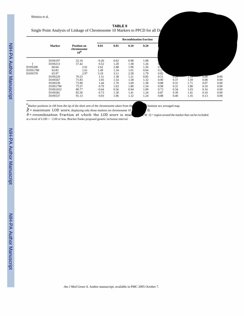
Single-point analyses of family UM:139 suggested linkage of PPCD to markers onchromosome 10. Utilizing data from the whole family, we obtained maximum single-pointLOD scores of 2.63, 1.63, and 3.19 for markers D10S208 (at θ = 0.03), D10S1780 (atθ = 0.00), and D10S578 (at θ = 0.06), respectively (Table II). An affecteds-only analysisproduced maximum single-point LOD scores of 2.76, 1.32, and 2.05 at markers D10S208 (atθ = 0.00), D10S1780 (at θ = 0.00), and D10S578 (at θ = 0.06), respectively.
Subsequent multi-point analyses of family UM:139 suggested significant evidence of linkageon the short arm of chromosome 10 according to the criteria of Morton [1955]. A maximummulti-point LOD score of 4.3 was found at marker D10S1780 when data from the whole familywere used (Fig. 2), with a 5.1 cM one-LOD support interval whose boundaries are roughlyapproximated by D10S208 and D10S578. An analogous affecteds-only multipoint analysisalso suggested significant evidence of linkage of PPCD with a maximum multi-point LODscore of 3.53 at marker D10S208 (Fig. 2), although it appears that the LOD score value remainsalmost constant between D10S280 and D10S1780. An affected haplotype was identified andrecombination events were identified that flank the 8.55 cM critical interval between markersD10S213 and D10S578.
In addition to the region with the highest observed LOD score, we also evaluated other regionsshowing some evidence of linkage. We were initially interested in the possibility of analternative location on chromosome 13 because of the maximum single-point LOD score of2.60 at D13S263 in the affecteds-only analysis. To follow up on this result, we evaluated thisregion at the 5 cM level by screening six additional markers, including three markers in an 18cM region surrounding D13S263. The result was a broad peak with maximum multipoint LODscore of 1.76 at D13S263 for the affecteds-only analysis and a narrower peak with maximummultipoint LOD score of 1.51 between D13S217 and D13S1287 when all data from the familywere used. Haplotype analysis failed to identify any single haplotype shared by all affectedfamily members, and all haplotypes present in affected family members were also present inunaffected individuals.
As further support for the argument that the gene responsible for PPCD in this family is locatedon chromosome 10p, it is important that data from other points in the genome fail to identifyalternative locations and that such data support exclusion of other PPCD-associated regionspreviously reported. Our previous report excluded the region on chromosome 20 containingPPCD1 and the VSX1 gene (MIM605020) and the region on chromosome 1 containingCOL8A2 (MIM 120252) [Hand et al., 1999; Biswas et al., 2001; Heon et al., 2002; Moroi etal., 2003]. We conclude that we have excluded the region on chromosome 2q that contains twoof the Alport syndrome genes COL4A3 (MIM120070) and COL4A4 (MIM120131) [Kashtan,
Shimizu et al. Page 4
Am J Med Genet A. Author manuscript; available in PMC 2005 October 7.
NIH
-PA Author Manuscript
NIH
-PA Author Manuscript
NIH
-PA Author Manuscript
1999; Kent et al., 2002] based on our finding of LOD scores below −3.0 across that wholeregion of chromosome 2. In addition, we can dismiss the X-linked Alport syndrome geneCOL4A5 (MIM 303630) [Kashtan, 1999] based on the autosomal dominant mode ofinheritance in this family.
Once we had confirmed that our data suggest a locus on chromosome 10 and do not supportlocations elsewhere, we considered possible candidate genes within the PPCD3 geneticinclusion interval. The 8.55 cM region between D10S213 and D10S578 contains 26 knowngenes and hundreds of predicted genes and expressed sequence tags (EST’s) [Kent et al.,2002]. Since the homeodomain transcription factor VSX1 has been implicated in PPCD [Heonet al., 2002], it is of interest to note that the PPCD3 critical interval contains the transcriptionfactor, TCF8 (MIM189909), which has both homeodomain and zinc finger motifs [Williamset al., 1992; Funahashi et al., 1993; Franklin et al., 1994; Kent et al., 2002]. Although severalcollagen genes have been implicated in PPCD [Biswas et al., 2001] and in Alport syndrome,which can include PPCD [Colville and Savige, 1997; Kashtan, 1999], none of the knowncollagen genes are located in the interval between D10S213 and D10S578. Prioritization ofcandidate genes within this interval will call for additional bioinformatic analysis andevaluation of gene expression in relevant ocular tissues.
DISCUSSIONAnalysis of the genome scan data for family UM:139 has provided significant evidence for anew PPCD locus (PPCD3) in a 8.55 cM region on chromosome 10p near D10S1780. Whilewe initially identified substantial single-point linkage evidence for a marker on chromosome13, additional marker genotyping and multipoint analysis resulted in a substantial decrease inthe linkage evidence. Thus, while chromosome 13 has not been formally excluded as containinga locus responsible for PPCD in this family, the evidence for this region is not strong. Wecannot rule out the possibility that a locus in this region might be playing a role in the phenotypein some members of family UM:139, but there is no evidence of correlation of a specifichaplotype with disease severity or presence of specific characteristics such as guttae.
Our data allow us to exclude the region on chromosome 2 that contains two collagen genesimplicated in Alport syndrome [Kashtan, 1999]. Previously, we excluded both the mappedPPCD locus on chromosome 20 [Heon et al., 2002] and the reported candidate PPCD gene onchromosome 1 [Biswas et al., 2001] as being responsible for PPCD in this family. Thus, thevalidity of this new PPCD locus is supported by: (1) significant evidence of linkage to markersin the vicinity of D10S1780, (2) a lack of evidence that the locus is elsewhere in the genome,and (3) evidence of exclusion of the other regions of the genome reportedly involved in PPCD.
In family UM:139, the expressivity of the PPCD phenotype varied widely, a characteristicpreviously described in PPCD families [Cibis et al., 1977]. Typically, the clinical course ofPPCD is slowly progressive, and occasionally the clinical course may be severe with cornealdecompensation and secondary glaucoma as found for proband, IV-10 [Moroi et al., 2003].The striking feature of the proband, IV-10, was the documented accelerated growth of theretrocorneal membrane on the crystalline lens and intraocular lens after surgical interventions.A comparably aggressive pattern to the disease has not been seen in the other 12 affectedindividuals, but if surgical intervention or penetrating trauma is what induces the membranegrowth, then we would not know whether other family members have the potential for asimilarly serious reaction since only one other family member has had a penetrating surgicalprocedure or trauma.
Another clinical feature worth noting is the presence of guttae in most members of generationIV, both in those with and those without PPCD. The underlying genetic components of guttae
Shimizu et al. Page 5
Am J Med Genet A. Author manuscript; available in PMC 2005 October 7.
NIH
-PA Author Manuscript
NIH
-PA Author Manuscript
NIH
-PA Author Manuscript

remain unknown. Guttae were absent in reports of some families with PPCD [McGee and Falls,1953] and present in four of five PPCD individuals in one generation of a three-generationfamily [Threlkeld et al., 1994]. At present, it appears that the guttae are segregatingindependently of PPCD in the one generation affected with guttae, and our data are consistentwith previous observations of guttae as an age-related finding [Goar, 1934; Kaufman et al.,1966; Lorenzetti et al., 1967; Nagaki et al., 1996]. If the one member of generation IV, who isknown to currently lack guttae, were to develop guttae, then possible mitochondrial inheritancewould become a consideration in this family with PPCD and guttae. This concept is reinforcedby the fact that multiple genetic and biochemical scenarios have been reported for Fuchsendothelial corneal dystrophy (MIM136800), a disorder that also includes guttae [Fuchs,1910; Wilson and Bourne, 1988; Albin, 1988; Gottsch et al., 2003].
In summary, we have identified a new PPCD3 locus on chromosome 10p in family UM:139.Given the previously reported locus on chromosome 20q and the report of COL8A2involvement in PPCD, it is clear that this disease is a genetically heterogeneous disorder.Determination of the underlying genetic components of PPCD will provide fundamentalinsights into the pathologic processes affecting the corneal endothelium and Descemet’smembrane. Localization of a new PPCD locus will also provide new tools with which to studythe relationship between PPCD and glaucoma.
ELECTRONIC-DATABASE INFORMATIONSee Online Mendelian Inheritance in Man (OMIM), http://www.ncbi.nlm.nih.gov/Omim/ forPPCD or PPMD (MIM122000), CHED1 (MIM121700), CHED2 (MIM217700), AlportSyndrome (MIM 301050, MIM104200, MIM 203780), COL4A3 (MIM120070, COL4A4(MIM 120131), COL4A5 (MIM 303630), COL8A2 (MIM120252), TCF8 (MIM189909), andVSX1 (MIM605020).
See the Eyegene Server http://eyegene.ophthy.med.umich.edu to download data formattingprogram Madeline and its documentation without charge.
ReferencesAlbin RL. Fuch’s corneal dystrophy in a patient with mitochondrial DNA mutations. J Med Genet
1988;35:258–259. [PubMed: 9541117]Alport AC. Hereditary familial congenital haemorrhagic nephritis. Brit Med J 1927;1:504–506.Biswas S, Munier FL, Yardley J, Hart-Holden N, Perveen R, Cousin P, Sutphin JE, Noble B, Batterbury
M, Kielty C, Hackett A, Bonshek R, Ridgway A, McLeod D, Sheffield VC, Stone EM, SchorderetDF, Black GCM. Missense mutations in COL8A2, the gene encoding the alpha-2 chain of type VIIIcollagen, cause two forms of corneal endothelial dystrophy. Hum Mol Genet 2001;10:2415–2423.[PubMed: 11689488]
Boehnke M. Allele frequency estimation from data on relatives. Am J Hum Genet 1991;48:22–25.[PubMed: 1985459]
Bourgeois J, Shields MB, Thresher R. Open-angle glaucoma associated with posterior polymorphousdystrophy. A clinicopathologic study. Ophthalmol 1984;91:420–423.
Chan CC, Green WR, Barraquer J, Barraquer-Somers E, de la Cruz ZC. Similarities between posteriorpolymorphous and congenital hereditary endothelial dystrophies: A study of 14 buttons of 11 cases.Cornea 1982;1:155–172.
Cibis GW, Krachmer JA, Phelps CD, Weingeist TA. The clinical spectrum of posterior polymorphousdystrophy. Arch Ophthalmol 1977;95:1529–1537. [PubMed: 302697]
Colville D, Savige J. Alport syndrome: A review of ocular manifestations. Ophthal Genet 1997;18:161–173.
Franklin AJ, Jetton TL, Shelton KD, Magnuson MA. BZP, a novel serum-responsive zinc finger proteinthat inhibits gene transcription. Mol Cell Biol 1994;14:6773–6788. [PubMed: 7935395]
Shimizu et al. Page 6
Am J Med Genet A. Author manuscript; available in PMC 2005 October 7.
NIH
-PA Author Manuscript
NIH
-PA Author Manuscript
NIH
-PA Author Manuscript
Fuchs E. Dystrophia epithelialis corneae. Albrecht Von Graefes Arch Ophthalmol 1910;76:478–508.Funahashi J, Sekido R, Murai K, Kamachi Y, Kondoh H. Delta-crystallin enhancer binding protein delta
EF1 is a zinc finger-homeodomain protein implicated in postgrastrulation embyrogenesis.Development 1993;119:433–446. [PubMed: 7904558]
Goar EL. Dystrophy of the corneal endothelium (cornea guttata), with a report of a histologicalexamination. Am J Ophthalmol 1934;17:215.
Gottsch JD, Bowers AL, Margulies EH, Seitzman GD, Kim SW, Saha S, Jun AS, Stark WJ, Liu SH.Serial analysis of gene expression in the corneal endothelium of Fuchs’ dystrophy. Invest OphthalmolVis Sci 2003;44:594–599. [PubMed: 12556388]
Hand CK, Harmon DL, Kennedy SM, FitzSimon JS, Collum LMT, Parfrey NA. Localization of the genefor autosomal recessive congenital hereditary endothelial dystrophy (CHED2) to chromosome 20 byhomozygosity mapping. Genomics 1999;61:1–4. [PubMed: 10512674]
Heon E, Greenberg A, Koop KK, Rootman D, Vincent AL, Billingsley G, Priston M, Dorval KM, ChowRL, McInnes RR, Heathcote G, Westall C, Sutphin JE, Semina E, Bremner R, Stone EM. VSX1: Agene for posterior polymorphous dystrophy and keratoconus. Hum Mol Genet 2002;11:1029–1036.[PubMed: 11978762]
Jackson AJ, Robinson FO, Frazer DG, Archer DB. Corneal guttata: A comparative clinical and specularmicrographic study. Eye 1999;13:737–743. [PubMed: 10707136]
Kashtan CE. Alport syndrome. An inherited disorder of renal, ocular, and cochlear basement membranes.Medicine 1999;78:338–360. [PubMed: 10499074]
Kaufman HE, Capella JA, Robbins JE. The human corneal endothelium. Am J Ophthalmol 1966;61:835–841. [PubMed: 16874980]
Kent WJ, Sugnet CW, Furey TS, Roskin KM, Pringle TH, Zahler AM, Haussler D. The human genomebrowser at UCSC. Genome Research 2002;12:996–1006. [PubMed: 12045153]Accessed April 2003.
Krachamer JH. Posterior polymorphous corneal dystrophy: A disease characterized by epithelial-likeendothelial cells which influence management and prognosis. Tr Am Ophth Soc 1985;83:413–475.[PubMed: 3914130]
Lange K, Weeks D, Boehnke M. Programs for pedigree analysis: MENDEL, FISHER, and dGENE.Genet Epidemiol 1988;5:471–472. [PubMed: 3061869]
Lorenzetti DWC, Uotila MH, Parikh N, Kaufman HE. Central cornea guttata: Incidence in the generalpopulation. Am J Ophthalmol 1967;64:1155. [PubMed: 6072991]
McGee HB, Falls HF. Hereditary polymorphous deep degeneration of the cornea. Arch Ophthalmol1953;50:462–467.
Miller CA, Krachmer JH. 1997. Endothelial dystrophies. In: Kaufman HE, Barron BA, McDonald MB,editors. The cornea. 2nd edn. Boston, MA: Butterworth-Heinemann. p 453–475.
Moroi SE, Gokhale PA, Schteingart MT, Sugar A, Downs CA, Shimizu S, Krafchak C, Fuse N, ElnerSG, Elner VM, Flint A, Epstein MP, Boehnke M, Richards JE. Clinicopathologic correlation andgenetic analysis in a case of posterior polymorphous corneal dystrophy. Am J Ophthalmol2003;135:461–470. [PubMed: 12654361]
Morton NE. Sequential tests for the detection of linkage. Am J Hum Genet 1955;7:277–318. [PubMed:13258560]
Nagaki Y, Hayasaka S, Kitagawa K, Yamamoto S. Primary cornea guttata in Japanese patients withcataract: Specular microscopic observations. Jpn J Ophthalmol 1996;40:520–525. [PubMed:9130056]
O’Connell JR, Weeks DE. PedCheck: A program for identification of genotype incompatibilities inlinkage analysis. Am J Hum Genet 1998;63:259–266. [PubMed: 9634505]
Sobel E, Lange K. Descent graphs in pedigree analysis: Applications to haplotyping, location scores, andmarker-sharing statistics. Am J Hum Genet 1996;58:1323–1337. [PubMed: 8651310]
Threlkeld AB, Green WR, Quigley HA, de la Cruz Z, Stark WJ. A clinicopathologic study of posteriorpolymorphous dystrophy: Implications for pathogenetic mechanism of the associated glaucoma. TrAm Ophth Soc 1994;92:133–165. [PubMed: 7886861]
Toma NM, Ebenezer ND, Inglehearn CF, Plant C, Ficker LA, Bhattacharya SS. Linkage of congenitalhereditary endothelial dystrophy to chromosome 20. Hum Mol Genet 1995;4:2395–2398. [PubMed:8634716]
Shimizu et al. Page 7
Am J Med Genet A. Author manuscript; available in PMC 2005 October 7.
NIH
-PA Author Manuscript
NIH
-PA Author Manuscript
NIH
-PA Author Manuscript

Weisenthal RW, Streeten BW. 1997. Posterior membrane dystrophies. In: Krachmer JH, Mannis MJ,Holland EJ, editors. Cornea Vol. II. Cornea and external disease: Clinical diagnosis and management.St. Louis, MO: Mosby-Year Book, Inc. p 1063–1090.
Williams TM, Montoya G, Wu Y, Eddy RL, Byers MG, Shows TB. The TCF8 gene encoding a zincfinger protein (Nil-2-a) resides on human chromosome 10p11.2. Genomics 1992;14:194–196.[PubMed: 1427828]
Wilson SE, Bourne WM. Fuchs’ dystrophy. Cornea 1988;7:2–18. [PubMed: 3280235]
Shimizu et al. Page 8
Am J Med Genet A. Author manuscript; available in PMC 2005 October 7.
NIH
-PA Author Manuscript
NIH
-PA Author Manuscript
NIH
-PA Author Manuscript

Fig. 1.Family UM:139 with PPCD. Among those individuals for whom molecular data weregenerated for use in the analysis (marked with +), filled symbols are individuals with PPCD,open symbols are individuals deemed unaffected with PPCD, and a cross within symbolindicates individuals who were scored as having an indeterminate PPCD status for purposesof analysis. Open symbols not marked with + are individuals for whom molecular data werenot generated; they are free of PPCD by family report, but were not examined by us. Arrowindicates proband. Squares represent males; circles represent females. Diagonal line throughsymbol indicates individual is deceased. Box to the left of the + indicates presence (black dot)or absence (gray box) of guttae. Data on guttae are not available for individuals not markedwith a box. Other significant eye disease in the family includes: angle-closure glaucoma in theproband (IV-10); primary open-angle glaucoma (POAG) in III-11; blindness from an accidentin I-1; blindness from unspecified glaucoma in II-5; and blindness from cataract surgery inIII-1. Haplotypes are displayed for each individual genotyped, with a genetic inclusion intervalbetween D10S213 and D10S578 indicated by recombination events apparent in data forindividuals V-2 and VI-1. A recombination event in individual V-18 is not considered to furtherreduce the interval since this individual is unaffected in a family showing evidence of reducedpenetrance.
Shimizu et al. Page 9
Am J Med Genet A. Author manuscript; available in PMC 2005 October 7.
NIH
-PA Author Manuscript
NIH
-PA Author Manuscript
NIH
-PA Author Manuscript
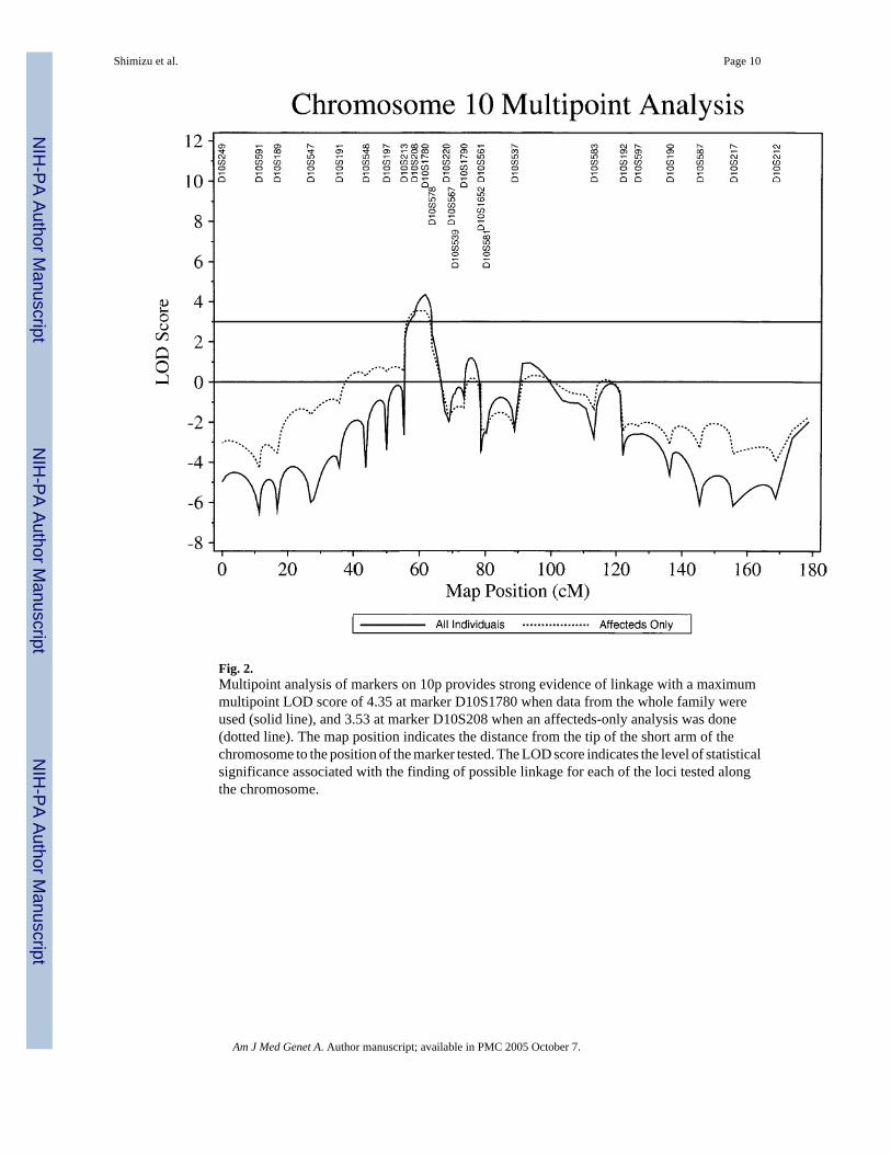
Fig. 2.Multipoint analysis of markers on 10p provides strong evidence of linkage with a maximummultipoint LOD score of 4.35 at marker D10S1780 when data from the whole family wereused (solid line), and 3.53 at marker D10S208 when an affecteds-only analysis was done(dotted line). The map position indicates the distance from the tip of the short arm of thechromosome to the position of the marker tested. The LOD score indicates the level of statisticalsignificance associated with the finding of possible linkage for each of the loci tested alongthe chromosome.
Shimizu et al. Page 10
Am J Med Genet A. Author manuscript; available in PMC 2005 October 7.
NIH
-PA Author Manuscript
NIH
-PA Author Manuscript
NIH
-PA Author Manuscript

NIH
-PA Author Manuscript
NIH
-PA Author Manuscript
NIH
-PA Author Manuscript
Shimizu et al. Page 11TA
BLE
ISu
mm
ary
of O
cula
r Fin
ding
s and
Ocu
lar D
iagn
oses
in E
xam
ined
Mem
bers
of F
amily
UM
:139
Cor
neal
find
ings
(OD
/OS)
Iris
find
ings
(OD
/OS)
Gon
iosc
opy
(OD
/OS)
Indi
vidu
alA
ffect
ed st
atus
Ocu
lar
diag
nosi
sPo
ster
ior
vesi
cles
Post
erio
r ba
ndG
utta
ePa
chym
etry
OD
/O
S (μ
m)
Ect
ropi
onuv
eae
“gla
ss m
embr
ance
”Fi
ne S
ynec
hiae
Bro
ad sy
nech
iae
III-
11I
POA
G−/−
−/−
−/−
548/
550
−/−
−/−
−/−
−/−
III-
19U
Nor
mal
−/−
−/−
−/−
618/
601
−/−
−/−
N/A
IV-2
IG
utta
e−/−
−/−
+/+
521/
522
−/−
−/−
−/−
−/−
IV-3
IG
utta
e−/−
−/−
+/+
589/
579
−/−
−/−
−/−
−/−
IV-5
APP
CD
gut
tae
−/−
+/−
+/−
563/
589
−/−
−/−
−/−
−/−
IV-7
APP
CD
+/+
−/−
−/−
582/
567
−/−
−/−
−/−
−/−
IV-1
0 (p
roba
nd)
APP
CD
gut
tae
angl
e-cl
osur
egl
auco
ma
+/+
−/−
+/+
580/
680
+/+
+/+
−/−
+/+
IV-1
1A
PPC
D−/
++/−
N/A
720/
610
−/−
−/−
−/−
−/−
IV-1
2A
PPC
D G
utta
e+/−
+/−
+/+
567/
603
−/−
−/−
−/−
−/−
IV-1
5I
Gut
tae
−/−
−/−
+/+
N/A
−/−
−/−
−/−
−/−
IV-1
7I
Gut
tae
−/−
−/−
+/+
N/A
−/−
−/−
−/−
−/−
V-2
APP
CD
a+/
+N
/AN
/AN
/AV
-5U
Nor
mal
−/−
−/−
−/−
520/
523
−/−
−/−
−/−
−/−
V-1
3A
PPC
D+/
++/
+−/−
637/
656
−/−
−/−
−/−
−/−
V-1
8U
Nor
mal
−/−
−/−
−/−
571/
582
−/−
−/−
−/−
−/−
V-2
1A
PPC
D+/−
−/−
−/−
582/
575
−/−
−/−
N/A
V-2
2A
PPC
D+/
+−/−
−/−
611/
586
−/−
−/−
−/−
−/−
V-2
3U
Nor
mal
−/−
−/−
−/−
599/
602
−/−
−/−
−/−
−/−
V-2
4U
Nor
mal
−/−
−/−
−/−
593/
592
−/−
−/−
−/−
−/−
V-2
5A
PPC
D+/
+−/−
−/−
640/
623
−/−
−/−
−/+
−/+
V-2
6U
Nor
mal
−/−
−/−
−/−
581/
565
−/−
−/−
−/−
−/−
V-2
7U
Nor
mal
−/−
−/−
−/−
607/
590
−/−
−/−
−/−
−/−
V-2
8U
Nor
mal
−/−
−/−
−/−
N/A
N/A
N/A
VI-
1A
PPC
Da
+/+
−/−
−/−
N/A
N/A
N/A
VI-
3A
PPC
Da
+/+
−/−
−/−
540/
550
N/A
N/A
VI-
12A
PPC
D+/
+−/−
−/−
573/
574
−/−
−/−
N/A
a Dia
gnos
is b
ased
on
reco
rds f
rom
out
side
oph
thal
mol
ogis
t, O
D, r
ight
eye
; OS,
left
eye;
I, in
dete
rmin
ate
stat
us fo
r pur
pose
s of a
naly
sis;
A, a
ffec
ted
stat
us fo
r pur
pose
s of a
naly
sis;
U, u
naff
ecte
dst
atus
for p
urpo
ses o
f ana
lysi
s, N
/A in
dica
tes t
hat t
he in
form
atio
n is
not
ava
ilabl
e.
Am J Med Genet A. Author manuscript; available in PMC 2005 October 7.
NIH
-PA Author Manuscript
NIH
-PA Author Manuscript
NIH
-PA Author Manuscript
Shimizu et al. Page 12
TABLE IISingle Point Analysis of Linkage of Chromosome 10 Markers to PPCD for all Data in Family UM:139
Recombination fraction
Marker Position onchromosome
10a
0.01 0.05 0.10 0.20 0.30 0.40 Z θ θ(−2)
D10S197 52.10 −0.26 0.62 0.98 1.08 0.81 0.36 1.10 0.17 0.00[ D10S213 57.42 0.52 1.20 1.38 1.26 0.88 0.37 1.39 0.12 0.00
D10S208 60.64 2.61 2.62 2.48 1.96 1.26 0.49 2.63 0.03 0.00D10S1780 63.83 1.61 1.49 1.34 1.01 0.64 0.27 1.63 0.00 0.00D10S578 65.97 2.97 3.19 3.11 2.58 1.79 0.82 3.19 0.06 0.00
D10S220 70.23 1.51 1.38 1.21 0.85 0.51 0.20 1.55 0.00 0.00D10S567 71.83 1.05 1.54 1.58 1.32 0.90 0.37 1.59 0.08 0.00D10S539 72.90 1.44 1.70 1.69 1.38 0.88 0.31 1.71 0.07 0.00D10S1790 75.57 0.79 1.63 1.80 1.54 0.98 0.31 1.80 0.10 0.00D10S1652 80.77 −0.64 0.56 0.94 1.00 0.73 0.34 1.03 0.16 0.00D10S581 82.50 0.73 1.30 1.41 1.24 0.87 0.39 1.41 0.10 0.00D10S537 91.13 0.03 1.06 1.32 1.24 0.88 0.40 1.35 0.13 0.00
aMarker positions in cM from the tip of the short arm of the chromosome taken from the Marshfield Institute sex averaged map.Z = maximum LOD score, displaying only those markers on chromosome 10 with Z ≥ 1.0,θ = recombination fraction at which the LOD score is maximized, θ(−2) = region around the marker that can be excludedat a level of LOD = −2.00 or less. Bracket flanks proposed genetic inclusion interval.
Am J Med Genet A. Author manuscript; available in PMC 2005 October 7.
Related Documents