This article was downloaded by: [University of Udine] On: 25 June 2012, At: 06:14 Publisher: Psychology Press Informa Ltd Registered in England and Wales Registered Number: 1072954 Registered office: Mortimer House, 37-41 Mortimer Street, London W1T 3JH, UK Neurocase: The Neural Basis of Cognition Publication details, including instructions for authors and subscription information: http://www.tandfonline.com/loi/nncs20 A left basal ganglia case of dynamic aphasia or impairment of extra-language cognitive processes? Cristiano Crescentini a , Alberta Lunardelli a c , Alessandro Mussoni a , Antonietta Zadini c & Tim Shallice a b a International School for Advanced Studies SISSA-ISAS, Trieste, Italy b Institute of Cognitive Neuroscience, University College, London, UK c Ospedale Maggiore, Trieste, Italy Available online: 19 Jun 2008 To cite this article: Cristiano Crescentini, Alberta Lunardelli, Alessandro Mussoni, Antonietta Zadini & Tim Shallice (2008): A left basal ganglia case of dynamic aphasia or impairment of extra-language cognitive processes?, Neurocase: The Neural Basis of Cognition, 14:2, 184-203 To link to this article: http://dx.doi.org/10.1080/13554790802108380 PLEASE SCROLL DOWN FOR ARTICLE Full terms and conditions of use: http://www.tandfonline.com/page/terms-and-conditions This article may be used for research, teaching, and private study purposes. Any substantial or systematic reproduction, redistribution, reselling, loan, sub-licensing, systematic supply, or distribution in any form to anyone is expressly forbidden. The publisher does not give any warranty express or implied or make any representation that the contents will be complete or accurate or up to date. The accuracy of any instructions, formulae, and drug doses should be independently verified with primary sources. The publisher shall not be liable for any loss, actions, claims, proceedings, demand, or costs or damages whatsoever or howsoever caused arising directly or indirectly in connection with or arising out of the use of this material.

Welcome message from author
This document is posted to help you gain knowledge. Please leave a comment to let me know what you think about it! Share it to your friends and learn new things together.
Transcript
This article was downloaded by: [University of Udine]On: 25 June 2012, At: 06:14Publisher: Psychology PressInforma Ltd Registered in England and Wales Registered Number: 1072954 Registered office: MortimerHouse, 37-41 Mortimer Street, London W1T 3JH, UK
Neurocase: The Neural Basis of CognitionPublication details, including instructions for authors and subscription information:http://www.tandfonline.com/loi/nncs20
A left basal ganglia case of dynamic aphasiaor impairment of extra-language cognitiveprocesses?Cristiano Crescentini a , Alberta Lunardelli a c , Alessandro Mussoni a , AntoniettaZadini c & Tim Shallice a ba International School for Advanced Studies SISSA-ISAS, Trieste, Italyb Institute of Cognitive Neuroscience, University College, London, UKc Ospedale Maggiore, Trieste, Italy
Available online: 19 Jun 2008
To cite this article: Cristiano Crescentini, Alberta Lunardelli, Alessandro Mussoni, Antonietta Zadini & Tim Shallice(2008): A left basal ganglia case of dynamic aphasia or impairment of extra-language cognitive processes?,Neurocase: The Neural Basis of Cognition, 14:2, 184-203
To link to this article: http://dx.doi.org/10.1080/13554790802108380
PLEASE SCROLL DOWN FOR ARTICLE
Full terms and conditions of use: http://www.tandfonline.com/page/terms-and-conditions
This article may be used for research, teaching, and private study purposes. Any substantial orsystematic reproduction, redistribution, reselling, loan, sub-licensing, systematic supply, or distributionin any form to anyone is expressly forbidden.
The publisher does not give any warranty express or implied or make any representation that thecontents will be complete or accurate or up to date. The accuracy of any instructions, formulae, anddrug doses should be independently verified with primary sources. The publisher shall not be liable forany loss, actions, claims, proceedings, demand, or costs or damages whatsoever or howsoever causedarising directly or indirectly in connection with or arising out of the use of this material.
© 2008 Psychology Press, an imprint of the Taylor & Francis Group, an Informa business
http://www.psypress.com/neurocase DOI: 10.1080/13554790802108380
NEUROCASE2008, 14 (2), 184–203
NNCS A left basal ganglia case of dynamic aphasia or impairment of extra-language cognitive processes?
Dynamic Aphasia in Left Basal Ganglia Lesion Cristiano Crescentini,1 Alberta Lunardelli,1,3 Alessandro Mussoni,1 Antonietta Zadini,3 and Tim Shallice1,2
1International School for Advanced Studies SISSA-ISAS, Trieste, Italy2Institute of Cognitive Neuroscience, University College, London, UK3Ospedale Maggiore, Trieste, Italy
We report the case of OTM who presented with dynamic aphasia following a stroke that occurred in the leftbasal ganglia. He showed drastically reduced spontaneous speech in the context of well preserved naming, rep-etition and comprehension skills. OTM was particularly impaired in generating words, sentences and phraseswhen cued by a stimulus allowing many response options. By contrast, when a single response was strongly sug-gested by a stimulus, he could generate verbal responses adequately. OTM’s non-verbal response generationabilities varied across tasks. He performed in the normal range in a motor movement generation test and heproduced as many figures as controls when tested on a figural fluency task. He showed, however, many perse-verations on this test. Moreover in a random number generation task he produced more responses that werepart of ascending and descending series of numbers. The patient’s impairments are interpreted as a conse-quence of two deficits. The first of these consists of an inability to generate verbal responses particularly in sit-uations of high competition and involves the function of left frontal regions. The second deficit is one ofimpaired novel thought generation as evidenced by perseverations. This second deficit has been proposed to bea function of basal ganglia damage.
Keywords: Dynamic aphasia; Failure of inhibition; Verbal alternatives; Basal ganglia; Cortical-subcortical loops.
INTRODUCTION
Dynamic aphasia consists of an impairment in propo-sitional language production characterized by anexceptionally reduced spontaneous speech in the con-text of well-preserved naming, articulation, prosodyand repetition skills. Patients with dynamic aphasiahave great difficulty in the initiation and elaborationof self-generated utterances. Nevertheless they may beable to describe pictures and to answer certain typesof direct questions (Luria, 1970). Dynamic aphasiagenerally occurs after lesions confined to the left cere-bral hemisphere specifically involving frontal regions
(see Robinson, Shallice, & Cipolotti, 2005, for areview). Both neurodegenerative disorders and focallesions have been reported to cause this disorder.
Recently several theoretical accounts fordynamic aphasia have been proposed; most ofthese accounts interpret such a syndrome withinthe domain of language while other explanationsextend beyond this domain. Among the accountsthat interpret dynamic aphasia within the domainof language, some consider the disorder as due toa specific impairment in creating preverbal mes-sages, following the theoretical framework pro-posed by Levelt (1989, 1999). Preverbal messages
This research was partially supported by a grant from PRIN to Tim Shallice and Raffaella Rumiati.Address correspondence to Cristiano Crescentini, Cognitive Neuroscience Sector, International School for Advanced Studies
(SISSA), Via Beirut 2–4, 34014 Trieste, Italy (E-mail: [email protected] and [email protected]).
Dow
nloa
ded
by [
Uni
vers
ity o
f U
dine
] at
06:
14 2
5 Ju
ne 2
012
DYNAMIC APHASIA IN LEFT BASAL GANGLIA LESION 185
consist of conceptual structures expressed in termof lexical concepts, which are in turn associatedwith the corresponding words in the language.For example, Warren, Warren, Fox, and War-rington (2003) described a patient (ADY) with afrontal lobe dementia who was impaired whenasked to combine various lexical concepts inorder to create a new sentence or a phrase, andparticularly when required to retrieve lexical con-cepts from among others or when asked to linkthem in new ways.
A similar interpretation has also been advocatedby Robinson et al. (2005) to account for thelanguage disturbances shown by patient CH whopresented with focal atrophy to the left frontal lobe.CH showed a defective conceptual preparation oflanguage, which was particularly evident when lexicalconcepts had to be generated under conditions ofhigh competition, that is, by choosing them fromamong others equally plausible such as when asubject has to complete a sentence that can be com-pleted in many different ways. According toRobinson et al. (2005) when stimuli activate manyverbal response options, selection between themcannot be accomplished by a damaged system;nevertheless, an appropriate response can beproduced when lexical concepts are stronglysuggested by the context, namely in conditions oflow competition. CH’s impairment was shown tobe specific to the language domain since anaccurate examination of his non-verbal generationabilities showed that these were intact. Robinson,Blair, and Cipolotti (1998) also reported the case ofthe patient ANG who was seen to be impaired inthe selection of a verbal response when there weremany competing options.
As stated before, dynamic aphasia has alsobeen held to occur due to impairments extendingbeyond the domain of language. For instanceRaymer, Rowland, Haley, and Crosson (2002)suggested that the deficits observed in case ofdynamic aphasia may also involve the ability toproduce non-verbal responses. In a similar fash-ion Robinson, Shallice, and Cipolotti (2006)reported the case of a patient with progressivesupranuclear palsy (PSP) who, despite his propo-sitional language impairment, performed well onword and sentence level generation tasks thatrequired a single response, but did not do so innon-verbal generation tasks. Following a dis-course level generation analysis, Robinson et al.(2006) claimed that their patient (KAS) wasdefective in the generation of a “fluent sequence
of novel thought” (Robinson et al., 2006, p.1344). These authors drew a distinction betweentwo subtypes of dynamic aphasia. They arguedthat patients that present with word and sentencelevel generation deficits have language specificimpairment and generally left inferior frontalgyrus lesions. By contrast, the second type ofdynamic aphasia could be associated with bilat-eral frontal and subcortical damage; the patientsaffected by this second subtype should presentwith verbal–non-verbal generation deficits as wellas with problems with discourse level generationtests. The authors also claimed that such patientsare supposed to be unimpaired in the word andsentence level generation tests.
In explaining the dynamic aphasia suffered by apatient (CO) who had a bilateral striatocapsular inf-arction, Gold et al. (1997) stressed the importance ofthe circuit from the dorsolateral prefrontal cortex tothe dorsolateral caudate (Alexander & Crutcher,1990). The authors claimed that a damage to thiscircuit caused decreased spontaneous speech in theirpatient, difficulties in executive functions and, mostimportantly, also an inability to adopt effectivestrategies for the retrieval of information from thesemantic network. CO was a rare case of dynamicaphasia following a subcortical lesion.
The fact that impairments in executive function-ing can be found even in non-demented patientssuffering from subcortical lesions has also recentlybeen explained as due to disturbance of subcortical-frontal circuits (Kramer, Reed, Mungas, Weiner, &Chui, 2002). According to this view, subcorticalischemic vascular lesions would cause the disruptionof dorsolateral prefrontal-subcortical circuits andproduce signs of dysexecutive syndrome.
However, the functional role of subcorticalstructures in language and aphasia remainsunclear. Some authors (e.g., Alexander, Naeser, &Palumbo, 1987) have suggested that the basal gan-glia may not be involved in language at all, thoughthey do recognize a role for some pathways withinthe white matter. In a related fashion, Nadeau andCrosson (1997) discussed four possible accounts ofsubcortical aphasia involving structures other thanthe thalamus, namely those of (a) diaschisis, (b)direct involvement of basal ganglia structures inlanguage processes, (c) disconnection of corticalstructures involved in language, and (d) deregula-tion of release of cortically formulated languagesegments. They argued that these mechanisms donot easily account for the high degree of heteroge-neity in language impairment following subcortical
Dow
nloa
ded
by [
Uni
vers
ity o
f U
dine
] at
06:
14 2
5 Ju
ne 2
012
186 CRESCENTINI ET AL.
lesions to structures other than the thalamus. Theyproposed instead that cortical hypoperfusion,which often follows striato-capsular infarction,may be the crucial factor. Nadeau and Crosson(1997) also argue that neither the caudate nor theputamen play a critical role in language.
More recently, Hillis et al. (2002) have stressedthe importance of cortical hypoperfusion in theexplanation of the language deficits of patientswith subcortical stroke. These authors tested apopulation of 115 patients within 24 h of onset orprogression of stroke symptoms for aphasia orhemispatial neglect. Many of these patients (44)only had subcortical infarcts. The authors usedsome imaging techniques that allow the assessmentof the structural lesion and also an evaluation of thefunctional lesion in early stroke. Such techniqueswere diffusion-weighted imaging (DWI) and perfusion-weighted imaging (PWI). Hillis et al. (2002) alsostudied whether the restoration of perfusion to thecortex was reflected in the resolution of aphasia.The authors found that most of the patients whohad only left hemisphere subcortical lesionssuffered from aphasia and that all patients also hadcortical hypoperfusion; according to Hillis andcolleagues, this suggested that the language deficitssuffered by the patients with subcortical lesions couldbe explained by cortical hypoperfusion. Finally, theseauthors also showed that the small group of patientswhich benefited from reversal of cortical hypoper-fusion also showed resolution of aphasia.
The evidence reported above suggests that corti-cal hypoperfusion plays an important role in theaphasic deficits of patients with subcortical lesions;however, this evidence does not completely excludethe possibility that subcortical lesions can moredirectly cause language deficits. In fact, a more dir-ect role of basal ganglia in language has been pro-posed by others. Grossman, Stern, Gollomp,Vernon, and Hurtig (1994) reported a verb-learningimpairment in Parkinson’s disease (PD) patients.In this study, some PD patients were administereda verb-learning task in the context of a forced-choice sentence picture-matching task. Thesepatients were seen to have a deficit in “appreciatinggrammatical information represented in the newverb” (Grosmann et al., 1994, p. 413). Ullmanet al. (1997) reported evidence for a role of basalganglia structures in grammatical processing. Theyfound a correlation between the right-sided hypok-inesia of PD patients and their difficulties in theproduction of regular English past tense. Ullmanet al. (1997) claimed that “one kind of basal gan-
glia lesion, which leads to the suppression of motoractivity (Parkinson’s disease), also led to the sup-pression of rule use” (Ullman et al., 1997, p. 274).A specific role for the striatum in the application oflinguistic rules has recently been proposed byTeichmann, Dupoux, Kouider, and Bachoud-Lévi(2006) in a population of Huntington’s disease (HD)patients. These patients were administered a task ofacceptability judgments of conjugated verbs andnonword forms and a task of lexical decision, andwere found to be impaired in rule application whilepresenting preserved lexical abilities.
An involvement of basal ganglia structures inlexical-semantic processing has also been proposedfollowing studies on patients with subcorticallesions. Copland, Chenery, and Murdoch (2000a;see also Copland, Chenery, & Murdoch, 2000b)tested a group of 14 patients with chronic nontha-lamic subcortical (NS) lesions using both standardaphasia batteries (The Western Aphasia Battery,WAB; The Boston Naming Test, BNT) and teststhat tap more complex language functions (Test ofLanguage Competence-Expanded Edition, TLC-E;Test of Word Knowledge, TOWK). These authorsfound that their patients performed as well as nor-mal controls on standard aphasia tests. The excep-tion was given by word fluency in which most ofthe patients were impaired. Eight patients also hadproblems in the BNT test. However, when assessedon more complex language functions (TLC-E andTOWK) the NS patients showed impaired per-formance on all the subtests (the only exceptionwas the Making inferences subtest of the TLC-Ewhere only two patients were impaired). Accordingto Copland and colleagues, the tests on which theNS patients had the greatest difficulties requiredlexical-semantic manipulation, use of languagestrategies, cognitive-linguistic flexibility as well asthe ability to select between alternative solutions forthe same linguistic input. The authors proposed thatNS lesions impair the operations of the lexical-semantic system, in particular generative languageand the interpretation of meaning at sentence level(Copland et al., 2000a, p. 6). Importantly, Coplandand colleagues argued that their findings were con-sistent with the proposal (see Wallesch & Papagno,1988) according to which basal ganglia, and moregenerally the fronto-striatal loops, have a role inthe process of selection of relevant lexical itemsand in the inhibition of irrelevant ones.
Copland (2003) provided further evidence for arelated position, through a semantic priming study.Patients with nonthalamic subcortical vascular
Dow
nloa
ded
by [
Uni
vers
ity o
f U
dine
] at
06:
14 2
5 Ju
ne 2
012

DYNAMIC APHASIA IN LEFT BASAL GANGLIA LESION 187
lesions (NS), Parkinson’s disease patients (PD),patients with cortical lesions, and controls wererequired to perform lexical decisions to targets,which were either nonwords or words that wererelated or not to the prime. They were adminis-tered auditory prime-target pairs of four differenttypes (subordinate unrelated, bat-river; dominantunrelated, foot-money; subordinate related, bank-river and dominant related, bank-money). Therewere two conditions of prime-target interstimulusinterval (ISI; short, 200 ms, and long, 1250 ms). Inneither NS nor PD patients was there a selectivesemantic facilitation of the dominant meaning atlong ISI. The authors suggested that damage tofrontal-subcortical systems led to impairedcontrolled lexical processing in these patients. Inparticular, they claimed that dysfunction in thebasal ganglia produced impairments in theselective engagement of the semantic network dueto damaged inhibitory mechanisms.
Further evidence for the involvement of basalganglia in lexical processes comes also fromneuroimaging studies. In an fMRI experiment,Crosson et al. (2003) administered 4 tasks to 21subjects: generation of nonsense syllables, genera-tion of words given a rhyming word, generation ofwords given a semantic category in two conditions:at a slow rate and at a fast rate. They found aninvolvement of the left pre-supplementary motorarea (pre-SMA), dorsal caudate nucleus and ven-tral anterior thalamic circuit in lexical retrieval,since some of these brain areas were active whilesubjects performed word generation from rhymingwords and from category but, on the other hand,were silent during nonsense syllable generation.According to Crosson et al. (2003), subcorticalstructures have the function of maintaining a biastowards a lexical alternative chosen from amongothers in competition during controlled word selec-tion. The role of basal ganglia structures in theattention-controlled access to lexical-semanticinformation has been proposed by Longworth,Keenan, Barker, Marslen-Wilson, and Tyler (2005)not to be specific to language. These authorshave argued that patients with lesions to thebasal ganglia might have separate but paralleldeficits in semantic and syntactic processes.However, they proposed that such deficits mayreflect an impairment of a function involved inlanguage comprehension and production whichis not specific to language, namely that of inhibi-tion of competing alternatives during later con-trolled processes.
In this study we report the case of patient OTMwho was classified as a dynamic aphasic followinga vascular accident to his left basal ganglia (seeFigure 1). Together with his language output disor-der OTM presented with many signs of executivedysfunction. In this respect our case shows similar-ities with that reported by Gold et al. (1997). How-ever, Gold et al.’s patient was not given tasks toinvestigate the level of competition in the responseset of the type used by Robinson et al. (1998, 2005,2006). In the present study, we present a patientwith subcortical damage defined with a series oftasks commonly used with other dynamic aphasicpatients. In addition, we aim to account forsubcortical language functions in terms of con-temporary language production models (forinstance Levelt, 1999). The case of OTM is alsoparticularly relevant to the status of dynamic apha-sia as a language specific disorder and on whetherfunctionally distinct sub-varieties of this syndromeexist, as has been suggested by Robinson et al.(2005, 2006).
CASE REPORT
OTM is a 67-year-old man, right-handed, with8 years of education, who had retired from workas a solderer. In May 2005 he suffered from anischemic stroke. A CT scan revealed a lesioninvolving the left basal ganglia, the corona radi-ata, and the surrounding white matter. Within thebasal ganglia the lesion involved the putamen andextended to the capsule close to the dorsal cau-date (see Figure 1). A few days after his admit-tance to the Department of Neurology of theOspedali Riuniti in Trieste he had a secondstroke, this time concerning the right parietallobe. When he was discharged he was referred fora neuropsychological evaluation. The testing wascarried out at the Neuropsychology Laboratoryof the Ospedali Riuniti in Trieste over a series ofsessions that took place between September andOctober 2005.
Figure 1. OTM’s CT scan. Four images are presented in radi-ological convention (left hemisphere on the right; see text fordetails on the lesion).
Dow
nloa
ded
by [
Uni
vers
ity o
f U
dine
] at
06:
14 2
5 Ju
ne 2
012
188 CRESCENTINI ET AL.
Neuropsychological assessment
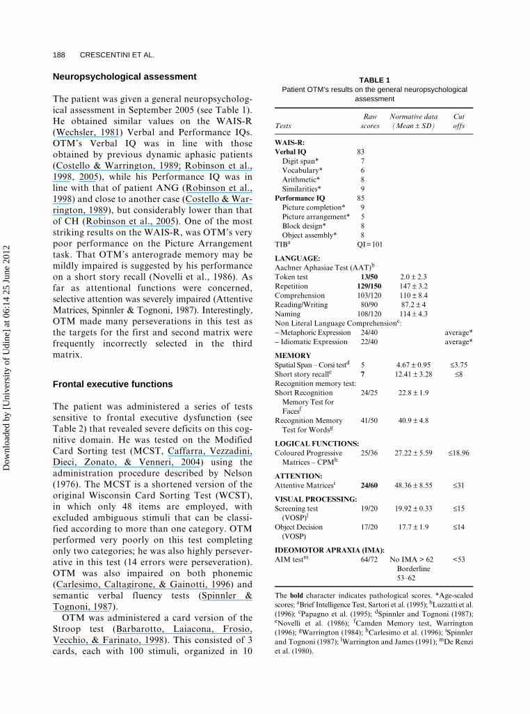
The patient was given a general neuropsycholog-ical assessment in September 2005 (see Table 1).He obtained similar values on the WAIS-R(Wechsler, 1981) Verbal and Performance IQs.OTM’s Verbal IQ was in line with thoseobtained by previous dynamic aphasic patients(Costello & Warrington, 1989; Robinson et al.,1998, 2005), while his Performance IQ was inline with that of patient ANG (Robinson et al.,1998) and close to another case (Costello & War-rington, 1989), but considerably lower than thatof CH (Robinson et al., 2005). One of the moststriking results on the WAIS-R, was OTM’s verypoor performance on the Picture Arrangementtask. That OTM’s anterograde memory may bemildly impaired is suggested by his performanceon a short story recall (Novelli et al., 1986). Asfar as attentional functions were concerned,selective attention was severely impaired (AttentiveMatrices, Spinnler & Tognoni, 1987). Interestingly,OTM made many perseverations in this test asthe targets for the first and second matrix werefrequently incorrectly selected in the thirdmatrix.
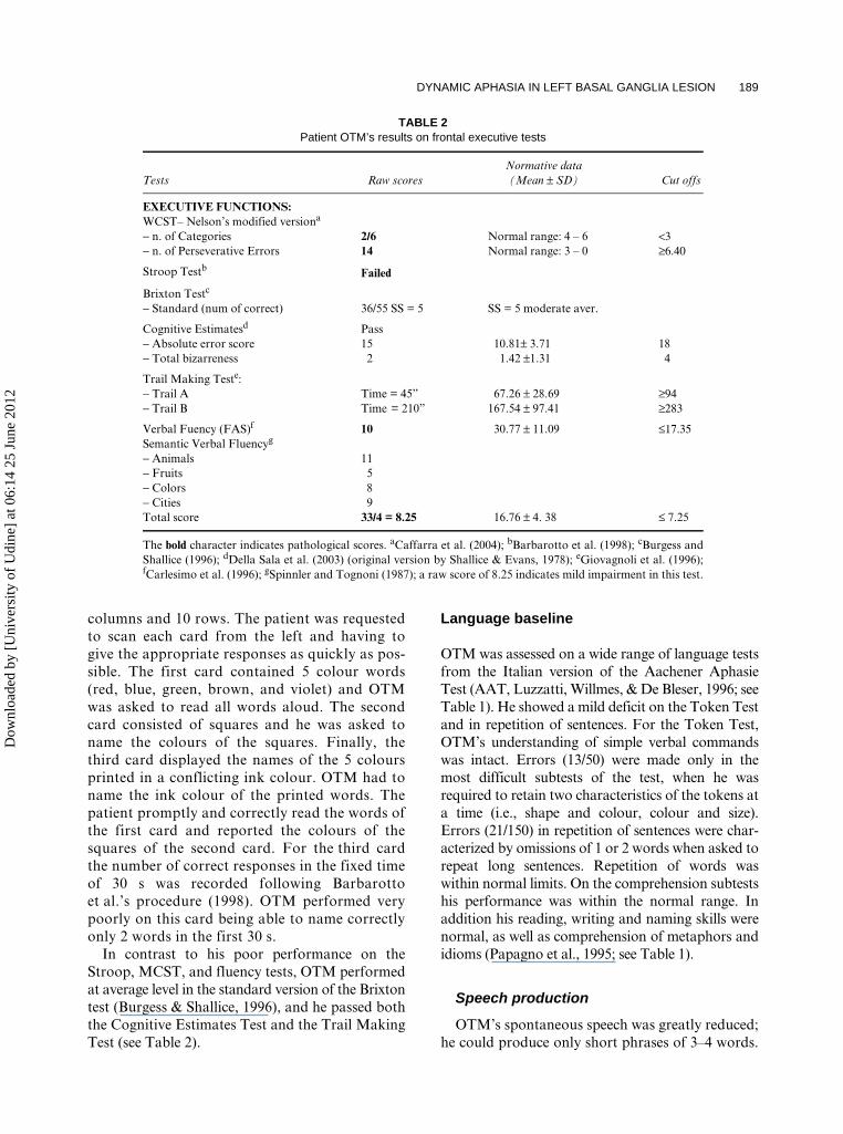
Frontal executive functions
The patient was administered a series of testssensitive to frontal executive dysfunction (seeTable 2) that revealed severe deficits on this cog-nitive domain. He was tested on the ModifiedCard Sorting test (MCST, Caffarra, Vezzadini,Dieci, Zonato, & Venneri, 2004) using theadministration procedure described by Nelson(1976). The MCST is a shortened version of theoriginal Wisconsin Card Sorting Test (WCST),in which only 48 items are employed, withexcluded ambiguous stimuli that can be classi-fied according to more than one category. OTMperformed very poorly on this test completingonly two categories; he was also highly persever-ative in this test (14 errors were perseveration).OTM was also impaired on both phonemic(Carlesimo, Caltagirone, & Gainotti, 1996) andsemantic verbal fluency tests (Spinnler &Tognoni, 1987).
OTM was administered a card version of theStroop test (Barbarotto, Laiacona, Frosio,Vecchio, & Farinato, 1998). This consisted of 3cards, each with 100 stimuli, organized in 10
TABLE 1 Patient OTM’s results on the general neuropsychological
assessment
TestsRaw
scoresNormative data(Mean ± SD)
Cut offs
WAIS-R:Verbal IQ 83
Digit span* 7Vocabulary* 6Arithmetic* 8Similarities* 9
Performance IQ 85Picture completion* 9Picture arrangement* 5Block design* 8Object assembly* 8
TIBa QI=101
LANGUAGE:Aachner Aphasiae Test (AAT)b
Token test 13/50 2.0 ± 2.3Repetition 129/150 147 ± 3.2Comprehension 103/120 110 ± 8.4Reading/Writing 80/90 87.2 ± 4Naming 108/120 114 ± 4.3Non Literal Language Comprehensionc:− Metaphoric Expression 24/40 average*− Idiomatic Expression 22/40 average*
MEMORYSpatial Span – Corsi testd 5 4.67 ± 0.95 ≤3.75Short story recalle 7 12.41 ± 3.28 ≤8Recognition memory test:Short Recognition
Memory Test for Facesf
24/25 22.8 ± 1.9
Recognition Memory Test for Wordsg
41/50 40.9 ± 4.8
LOGICAL FUNCTIONS:Coloured Progressive
Matrices – CPMh25/36 27.22 ± 5.59 ≤18.96
ATTENTION:Attentive Matricesi 24/60 48.36 ± 8.55 ≤31
VISUAL PROCESSING:Screening test
(VOSP)l19/20 19.92 ± 0.33 ≤15
Object Decision (VOSP)
17/20 17.7 ± 1.9 ≤14
IDEOMOTOR APRAXIA (IMA):AIM testm 64/72 No IMA > 62
Borderline 53–62
<53
The bold character indicates pathological scores. *Age-scaledscores; aBrief Intelligence Test, Sartori et al. (1995); bLuzzatti et al.(1996); cPapagno et al. (1995); dSpinnler and Tognoni (1987);eNovelli et al. (1986); fCamden Memory test, Warrington(1996); gWarrington (1984); hCarlesimo et al. (1996); iSpinnlerand Tognoni (1987); lWarrington and James (1991); mDe Renziet al. (1980).
Dow
nloa
ded
by [
Uni
vers
ity o
f U
dine
] at
06:
14 2
5 Ju
ne 2
012
DYNAMIC APHASIA IN LEFT BASAL GANGLIA LESION 189
columns and 10 rows. The patient was requestedto scan each card from the left and having togive the appropriate responses as quickly as pos-sible. The first card contained 5 colour words(red, blue, green, brown, and violet) and OTMwas asked to read all words aloud. The secondcard consisted of squares and he was asked toname the colours of the squares. Finally, thethird card displayed the names of the 5 coloursprinted in a conflicting ink colour. OTM had toname the ink colour of the printed words. Thepatient promptly and correctly read the words ofthe first card and reported the colours of thesquares of the second card. For the third cardthe number of correct responses in the fixed timeof 30 s was recorded following Barbarottoet al.’s procedure (1998). OTM performed verypoorly on this card being able to name correctlyonly 2 words in the first 30 s.
In contrast to his poor performance on theStroop, MCST, and fluency tests, OTM performedat average level in the standard version of the Brixtontest (Burgess & Shallice, 1996), and he passed boththe Cognitive Estimates Test and the Trail MakingTest (see Table 2).
Language baseline
OTM was assessed on a wide range of language testsfrom the Italian version of the Aachener AphasieTest (AAT, Luzzatti, Willmes, & De Bleser, 1996; seeTable 1). He showed a mild deficit on the Token Testand in repetition of sentences. For the Token Test,OTM’s understanding of simple verbal commandswas intact. Errors (13/50) were made only in themost difficult subtests of the test, when he wasrequired to retain two characteristics of the tokens ata time (i.e., shape and colour, colour and size).Errors (21/150) in repetition of sentences were char-acterized by omissions of 1 or 2 words when asked torepeat long sentences. Repetition of words waswithin normal limits. On the comprehension subtestshis performance was within the normal range. Inaddition his reading, writing and naming skills werenormal, as well as comprehension of metaphors andidioms (Papagno et al., 1995; see Table 1).
Speech production
OTM’s spontaneous speech was greatly reduced;he could produce only short phrases of 3–4 words.
TABLE 2 Patient OTM’s results on frontal executive tests
Tests Raw scoresNormative data (Mean ± SD) Cut offs
EXECUTIVE FUNCTIONS:WCST– Nelson’s modified versiona
− n. of Categories 2/6 Normal range: 4 – 6 <3− n. of Perseverative Errors 14 Normal range: 3 – 0 ≥6.40
Stroop Testb Failed
Brixton Testc
− Standard (num of correct) 36/55 SS = 5 SS = 5 moderate aver.
Cognitive Estimatesd Pass− Absolute error score 15 10.81± 3.71 18− Total bizarreness 2 1.42 ±1.31 4
Trail Making Teste:− Trail A Time = 45” 67.26 ± 28.69 ≥94− Trail B Time = 210” 167.54 ± 97.41 ≥283
Verbal Fuency (FAS)f 10 30.77 ± 11.09 ≤17.35Semantic Verbal Fluencyg
− Animals 11− Fruits 5− Colors 8− Cities 9Total score 33/4 = 8.25 16.76 ± 4. 38 ≤ 7.25
The bold character indicates pathological scores. aCaffarra et al. (2004); bBarbarotto et al. (1998); cBurgess andShallice (1996); dDella Sala et al. (2003) (original version by Shallice & Evans, 1978); eGiovagnoli et al. (1996);fCarlesimo et al. (1996); gSpinnler and Tognoni (1987); a raw score of 8.25 indicates mild impairment in this test.
Dow
nloa
ded
by [
Uni
vers
ity o
f U
dine
] at
06:
14 2
5 Ju
ne 2
012
190 CRESCENTINI ET AL.
Family members claimed he tended not to speak athome, while before the stroke he had been veryloquacious. He showed mild disprosodia and mildarticulatory problems. When asked to speak, he wasvery often unable to communicate what he appearedto want to say, and he usually stopped in the middleof a sentence. When asked to describe something(i.e., what he remembered of his first stroke) hisanswers were characterized by many repetitions,interruptions and circumlocutions. For example,when asked to describe how he felt after his strokehe produced the following: meglio . . . ma più ditanto non so come parlar . . . non so come parlar . . .parlo un pò con mia moglie . . . ma più di tanto . . .(50 s) (I feel better . . . but I do not know how tospeak . . . I do not know how to speak . . . I speak alittle with my wife . . . but not so much . . .).
OTM’s speech was also analysed using tasksthat elicited speech from pictorial stimuli. In orderto obtain a spontaneous speech sample we askedOTM to generate a story from three simplepictures taken from the BADA (Battery forthe Assessment of Aphasic Deficits, Miceli,Laudanna, Burani, & Capasso, 1994). He wasgiven 1 min to produce a story for each picture.OTM was also asked to talk about both his jobwhen he worked and the city where he lives. Oneminute was again given for each topic. Five Italiancontrol subjects matched to OTM for age, educa-tion, and gender carried out the same tasks (seeTable 3 for their performance and for comparisonwith OTM). The modified t-test of Crawford andGarthwaite (2002) was used to contrast the per-formance of OTM with that of control subjects.OTM produced significantly less words than con-trols when he was required to speak about his joband his city, whereas there was only a trendtowards a difference between OTM and controlson the story generation task. Globally OTM pro-duced less words (word/minute ratio; see Table 3)than normal subjects but he was close to the rate
of word production of other dynamic aphasicpatients. OTM always began the story generationtask providing good descriptions of the pictures hesaw. Only rarely did he attempt to construct astory as required. By contrast, controls providedless detailed descriptions but tried to produce astory for each single picture. These behaviourscould be the reason why OTM did not differ fromcontrols in the story generation task while doingso when asked to describe, for example, his city. Inthis latter task, stimuli (i.e., describe your city)provide less constrains for verbal production rela-tive to pictures which act as cues for production inthe story generation task.
Summary
In sum, OTM showed impairments in intellectual,language, and executive functions as well as havingproblems in verbal memory and selective attention.Within the language domain OTM had a dissocia-tion between dramatically reduced spontaneouspropositional speech and either preserved or onlymild impaired use of language in comprehensionand naming skills. The diagnosis derived from theAAT (Luzzatti et al., 1996) was that OTM was notaffected by aphasic disturbances. However, his dys-phasic impairment falls within the classification ofpure dynamic aphasia (Luria, 1970, 1973; Robinsonet al., 2005, 2006).
The next experimental series of tests was designedto investigate OTM’s language output disorder.
WORD AND SENTENCE LEVEL GENERATION TASKS
The aim of this experimental series of tests was topinpoint the nature of OTM’s word and sentencelevel generation impairment. Most of the tests
TABLE 3 Speech rate of patient OTM, five matched controls, and four other dynamic aphasic patients
OTM Controls T(4) ANGa MPb CHc KASd
Story generation (3 × 1min)# 50 130.2 ± 45.15 −1.62Event description (2 × 1 min)# 39 169 ± 47.5 -2.5Speech rate (words per min) 17.8 59.8 ± 17.63 −2.17 29.2 <20 12.0 23.0
The total number of words produced are reported in each cell. The standard deviation for controls is also reported.#See text for details on these tests. The bold character indicates significant differences between controls and OTM.aRobinson et al. (1998); bRaymer et al. (2002); cRobinson et al. (2005); dRobinson et al. (2006).
Dow
nloa
ded
by [
Uni
vers
ity o
f U
dine
] at
06:
14 2
5 Ju
ne 2
012
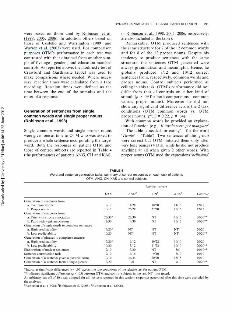
DYNAMIC APHASIA IN LEFT BASAL GANGLIA LESION 191
were based on those used by Robinson et al.(1998, 2005, 2006). In addition others based onthose of Costello and Warrington (1989) andWarren et al. (2003) were used. For comparisonpurposes OTM’s performance in each test wascontrasted with that obtained from another sam-ple of five age-, gender-, and education-matchedcontrols. As reported above, the modified t-test ofCrawford and Garthwaite (2002) was used tomake comparisons where needed. Where neces-sary, reaction times were calculated from a taperecording. Reaction times were defined as thetime between the end of the stimulus and theonset of a response.
Generation of sentences from single common words and single proper nouns (Robinson et al., 1998)
Single common words and single proper nounswere given one at time to OTM who was asked toproduce a whole sentence incorporating the targetword. Both the responses of patient OTM andthose of control subjects are reported in Table 4(the performances of patients ANG, CH and KAS,
of Robinson et al., 1998, 2005, 2006, respectively,are also included in the table).
Remarkably, OTM produced sentences withthe same structure for 7 of the 12 common wordsand for 8 of the 12 proper nouns. Despite histendency to produce sentences with the samestructure, the sentences OTM generated werealways grammatical and meaningful. Hence, heglobally produced 8/12 and 10/12 correctsentences from, respectively, common words andproper nouns. Control subjects performed atceiling in this task. OTM’s performance did notdiffer from that of controls on either kind ofstimuli (p > .09 for both comparisons – commonwords, proper nouns). Moreover he did notshow any significant difference across the 2 taskconditions (OTM common words vs. OTMproper nouns, χ2(1) = 0.22, p = .64).
With common words he provided an explana-tion of function (e.g., ‘Il tavolo serve per mangiare’– ‘The table is needed for eating’ – for the word‘Tavolo’ – ‘Table’). Two sentences of this groupwere correct but OTM initiated them only aftervery long pauses (>13 s), while he did not produceanything at all when given 2 other words. Withproper nouns OTM used the expressions ‘bellissimo’
TABLE 4 Word and sentence generation tasks: summary of correct responses on each task of patients
OTM, ANG, CH, KAS and control subjects
Number correct
OTM ANGa CHb KASc Controls
Generation of sentences froma. Common words 8/12 11/28 10/30 14/15 12/12b. Proper nouns 10/12 26/28 22/30 15/15 12/12
Generation of sentences froma. Pairs with strong association 23/30* 22/30 NT 15/15 30/30**b. Pairs with weak association 13/30 4/30 NT 15/15 30/30**
Generation of single words to complete sentencesa. High predictability 19/20* NT NT NT 20/20b. Low predictability 10/20 NT NT NT 20/20**
Generation of phrases to complete sentencesa. High predictability 17/20* 9/12 19/22 10/10 20/20b. Low predictability 10/20 3/12 11/22 10/10 20/20**
Elaboration of nuclear sentences 2/10 3/20 NT 5/5 10/10**Sentence construction task 9/10 14/15 9/10 8/10 10/10Generation of a sentence given a pictorial scene 10/10 34/34 20/20 15/15 10/10Generation of a sentence from a single picture 2/20 0/6 NT 9/10 20/20**
*Indicates significant differences (p < .05) across the two conditions of the relative test for patient OTM.**Indicates significant differences (p < .05) between OTM and control subjects in the test. NT=not tested.An arbitrary cut-off of 10 s was adopted for all the tests reported in this section; responses generated after this time were excluded bythe analyses.aRobinson et al. (1998); bRobinson et al. (2005); cRobinson et al. (2006).
Dow
nloa
ded
by [
Uni
vers
ity o
f U
dine
] at
06:
14 2
5 Ju
ne 2
012
192 CRESCENTINI ET AL.
and ‘bravissimo’ (‘very beautiful and very good’)(e.g., ‘Pippo Baudo’ prompted ‘Pippo Baudo e’ unbravissimo conduttore’ –‘Pippo Baudo is a verygood showman’). For two of these stimuli he didnot make any attempt to produce a sentence.
Clearly, OTM did not have problems in generat-ing a response in this task; however he appeared touse a compensatory strategy in coping with taskdemands (explain function for common words andreport a description for proper nouns). Thus, whenhe was asked to vary the structure of the sentenceshe was producing, he complained that the taskwas too difficult and that he did not have anyother word in mind. Moreover, OTM may haveperseverated on this test, as have other patients withsubcortical lesions when tested on similar tasks(Esmonde, Giles, Xuereb, & Hodges, 1996; see alsopatient MP, Raymer et al., 2002, for strategy use).OTM performed this task differently by ANG andCH who had more difficulties in generating sen-tences from common words than from propernouns. As stated also in Robinson et al. (2006),proper nouns are associated with a dominantresponse or with few verbal response options, whilecommon nouns tend to elicit many response options.
Generation of a sentence from word pairs with strong and weak associations (Robinson et al., 1998)
The stimuli used in this task were the same of those ofRobinson et al. (1998). They consisted of stronglyassociated word pairs (e.g., ‘giraffa-collo’, ‘giraffe-neck’) and weakly associated word pairs (e.g.,‘bambino-dolce’, ‘baby-sweet’). OTM was asked toproduce a sentence incorporating both words of thepair. As for patient ANG, OTM had more difficultiesin producing sentences from weakly associated wordpairs than from strongly associated ones (χ2(1) =5.62, p < .02) (see Table 4). Control subjects had nodifficulty with this task.
Generation of single words to complete sentences with high and low response predictability (Robinson et al., 1998)
This task consisted of 20 sentence frames to be com-pleted with a single word where there were fewresponse options (e.g., ‘The boy sent the letterwithout the . . .’ with ‘stamp’ being the selectedresponse for 98% of the subjects; (Bloom & Fischler,
1980; ‘Il ragazzo spedì la lettera senza’. . . ‘fran-cobollo’, is the Italian version) and also 20 sentenceswhich had many response options (e.g., ‘The police-man had never seen a man so . . .’ with ‘drunk’ beingthe most often selected response, 18% of thesubjects; ‘La polizia non aveva mai visto un uomocosì . . .’, is the Italian version). The sentences givento the patient were an Italian translation of eitherthe corresponding sentences used in a previousstudy with a dynamic aphasic patient (Robinsonet al., 1998) or of the sentences of Bloom and Fischler(1980).
OTM produced appropriate responses (in lessthan 2 s on average) for almost all the sentenceswith high response predictability (with lowcompetition in the response set). By contrast hisperformance was significantly impaired on the lowresponse predictability sentences (with highcompetition in the response set) (χ2(1) = 8.02, p < .006,see Table 4). Control subjects performed at ceilingin this task.
Generation of phrases to complete sentences with high and low response predictability (Robinson et al., 1998)
This task consisted of sentence frames, based onthose of Robinson et al. (1998), to be completed bythe addition of a whole phrase. Twenty had fewplausible verbal response options for their comple-tion (e.g., ‘The hairdresser moved next to the ladywith her scissors and . . .’; ‘La parruchiera ando’verso la donna con le forbici e . . .’, is the Italian ver-sion) while 20 sentences had many plausible verbalresponse options for their completion (e.g., ‘Theman sat in his chair and . . .’; ‘L’uomo si sedettesulla sua poltrona e . . .’ is the Italian version). Eachsentence was read to the patient. As for patientsANG and CH, OTM produced significantly morephrases in completing sentences with a highlypredictable response (the mean voice onset time forthe correct responses was 2.8 s) than in case of sen-tences with low predictability of the response (meanvoice onset time 3.2 s; χ2(1) = 4.10, p < .05) (seeTable 4). Controls perform at ceiling on this task.
Elaboration of nuclear sentences (Warren et al., 2003)
OTM was presented with 10 complete sentencesand asked to produce a second sentence developing
Dow
nloa
ded
by [
Uni
vers
ity o
f U
dine
] at
06:
14 2
5 Ju
ne 2
012
DYNAMIC APHASIA IN LEFT BASAL GANGLIA LESION 193
the theme of the first. OTM did not provide anyanswer for 3 out of 10 sentences. For five sentencesOTM provided only a completion without generatinga separate sentence (e.g., for the sentence: ‘La raga-zza ascoltava la storia . . .’, ‘The lady was listeningto the story . . .’, after more than 15 s he said ‘delsuo ragazzo’, ‘of her boyfriend’). For the remainingtwo sentences OTM produced a correct response.Controls had no difficulty with this task.
Sentence construction task (Costello & Warrington, 1989)
Dynamic aphasia can also be present in the con-text of concomitant syntactical or verbal plan-ning impairments. OTM was provided with 10sentences of 4 to 8 words in length which had tobe rearranged. Single words were printed on sep-arate pieces of paper and had to be reorganizedto construct a meaningful sentence. OTM per-formed well in this task (9/10). His only hesita-tion concerned one of the two 7-word sentences.He rearranged the target sentence: ‘Ieri Luca harotto la sua macchina nuova’ (‘Yesterday Lucabroke his new car’) as ‘Ieri Luca ha rotto sua lamacchina nuova’. Insofar as this task is one ofverbal planning (Costello & Warrington, 1989)this ability was shown to be spared in OTM.Controls also had no problem in this task (seeTable 4).
Generation of a sentence given a pictorial scene (Robinson et al., 1998)
As shown in Table 4 previous studies with dynamicaphasics had found that patients were able to pro-vide simple descriptions of pictorial scenes. In thistest OTM was given a series of pictures taken fromthe BADA (Miceli et al., 1994) and was told to cre-ate a sentence that described the content of the pic-ture. The patient was able to do this for all thepictures he saw (10/10). Controls also performederrorless in this task.
Generation of a sentence from a single picture (Robinson et al., 1998)
Patient ANG (Robinson et al., 1998) was impairedwhen asked to produce whole sentences (i.e., morethan a simple description) that incorporated themeaning of single pictures with which he was
presented. OTM was presented with 20 single pic-tures selected from the BADA battery (Miceli etal., 1994) and required to produce a whole sentenceincorporating the meaning of the picture, but notto simply describe it. Pictures were divided in twogroups: 10 were of objects whereas 10 were of eitherpersons or animals doing an action. The pictures fromthe two groups were presented at two differenttimes. Like patient ANG, OTM’s performance wasseriously impaired in this test (only 2 out of 20 sen-tences were correct). In most cases, OTM merelyproduced a description of the picture rather thangenerating a whole sentence as required. Controlsperformed at ceiling (see Table 4).
Summary and conclusions from the word and sentence generation tasks
The word and sentence level generation testsshowed that OTM has difficulties in generatingwords, sentences and phrases. However, in cer-tain cases he was able to produce plausiblewords and phrases. OTM was particularlyimpaired when stimuli activated many responseoptions (phrases and words low in response pre-dictability, word pairs weak in association). Bycontrast he generally performed much betterwith stimuli that strongly constrained a response(phrases and words high in response predictabil-ity, word pairs strong in association). In theserespects OTM’s performance resembled those ofthe dynamic aphasic patients ANG and CH(Robinson et al., 1998, 2005). The results fromthis series of tests showed that OTM had prob-lems in generating verbal messages in conditionof high competition between multiple responseoptions. However, OTM behaved differentlyfrom ANG or CH in some ways. Qualitatively heperformed just in the normal range in the genera-tion of sentences from proper nouns and com-mon words. Nevertheless, as reported above,he appeared to use a compensatory strategy inthis task.
Gold et al. (1997) reported the case of patientCO who also became dynamic aphasic following abasal ganglia lesion, namely a bilateral striatocap-sular infarction. CO also presented with signs ofexecutive dysfunction. CO performed poorly on asemantic categorization task and on a proceduraldiscourse test. The authors argued that he suf-fered from an impairment of concept formation aswell as from defective semantic strategy formation.
Dow
nloa
ded
by [
Uni
vers
ity o
f U
dine
] at
06:
14 2
5 Ju
ne 2
012
194 CRESCENTINI ET AL.
They held that the latter problem was confirmedby the difficulty the patient had in sorting itemsfrom very closely related categories by contrastwith his intact performance in sorting items frommore distantly related categories. Gold et al.(1997) suggested that CO’s deficits might reflect amore general impairment in the ability to organizeknowledge in a hierarchical fashion. The followingsection aims to assess whether OTM’s problems inproducing verbal responses in situations of highcompetition may be due to deficits in conceptgeneration and in semantic strategy formation.
ROUTINE ACTIVITIES DESCRIPTION AND SEMANTIC STRATEGY FORMATION
Procedural discourse task (Gold et al., 1997)
In Gold et al.’s (1997) version of the task the patienthad to describe the steps involved in carrying out aseries of common actions such as preparing a cup ofcoffee. The patient performed the task in two condi-tions. First he described the steps involved in theprocedures without any prompting, whereas in thesecond the experimenter provided the patient withcues such as ‘the first step is . . .’ etc.
We administered a similar task to OTM butchanging some of the required actions to fit withthose used by Rumiati, Zanini, Vorano, andShallice (2001). These authors provided a list ofthe verbal descriptions of the discrete stepsinvolved in a series of multi-objects-actions(MOT) that they administered to their apraxicpatients. They distinguished a total of 47 stepsfor 10 common tasks with 7 tasks being divisibleinto 5 discrete steps and 3 tasks into 4 steps. Theprocedures were: Preparing orange juice, Makingcoffee using a coffee pot, Lighting a cigarette,Preparing a letter for posting, Pouring water froma bottle, Lighting an electric torch, Hanging asmall picture, Lighting a candle, Sharpen a pencil,Peeling a potato.
OTM was asked to describe the steps he usu-ally goes through in carrying out these actions.He performed as well as normal subjects (n = 5)in this task. OTM gave an average of 3.9 stepsfor each action while controls gave descriptionsformed of 4.3 steps on average with the differ-ence being not significant (t(4) = –0.97; modifiedt-test, Crawford & Garthwaite, 2002). GivenOTM’s ability to perform the task he was not
administered the sequence cued condition usedby Gold et al. (1997).
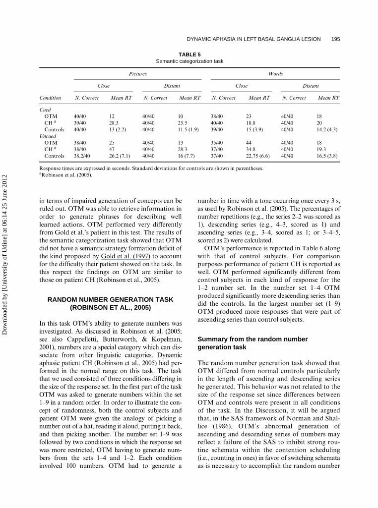
Semantic categorization task (Gold et al., 1997)
Before assessing OTM’s abilities in semantic strat-egy formation and semantic categorization, we eval-uated his ability to access semantic representations.OTM was tested using only the picture section ofThe Pyramids and Palm Trees Test (Howard &Patterson, 1992), a test specifically aimed to assessthe integrity of the semantic network. His perform-ance was well within the normal range and almost atceiling (50/52) indicating that he could recognize theitems and retrieve conceptual and semantic informa-tion about them.
Given that OTM’s ability to access meaning frompictures was intact we gave him a semantic categori-zation task. We used the procedure developed byRobinson et al. (2005) and the categories theseauthors employed. They had found that the dynamicaphasic patient CH was unimpaired in this task.OTM was required to sort items into two categories.A set of 80 words and a set of 80 pictures were cho-sen. Each of the two groups of stimuli formed 16 cat-egories, each consisting of five highly associateditems. Pairs of categories were used with their degreeof association being either distant or close. OTM wasgiven 10 cards at the same time and was required tosort the pictures or the words into two groups of fiveeach. In one condition the names of the two catego-ries were given in advance to the patient (cued condi-tion) and in another the names of the categories werenot given (uncued condition).
OTM’s performance on this task is reported inTable 5 together with CH’s performance (Robinsonet al., 2005) and that of four control subjects whowere administered the task. OTM’s performanceon the task was good and similar to that of CH(Robinson et al., 2005) and of normal controls.OTM was only somewhat slower than controls inthe uncued condition of the closely related wordpairs category (t(3) = 2.88, p < .05).
Summary and conclusions from the procedural discourse and the semantic categorization tasks
Given OTM’s intact performance on the proce-dural discourse task, an explanation of his deficits
Dow
nloa
ded
by [
Uni
vers
ity o
f U
dine
] at
06:
14 2
5 Ju
ne 2
012
DYNAMIC APHASIA IN LEFT BASAL GANGLIA LESION 195
in terms of impaired generation of concepts can beruled out. OTM was able to retrieve information inorder to generate phrases for describing welllearned actions. OTM performed very differentlyfrom Gold et al.’s patient in this test. The results ofthe semantic categorization task showed that OTMdid not have a semantic strategy formation deficit ofthe kind proposed by Gold et al. (1997) to accountfor the difficulty their patient showed on the task. Inthis respect the findings on OTM are similar tothose on patient CH (Robinson et al., 2005).
RANDOM NUMBER GENERATION TASK (ROBINSON ET AL., 2005)
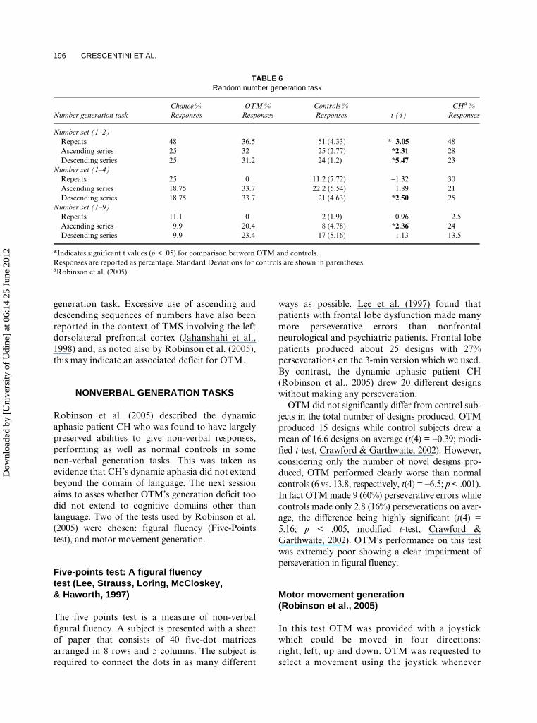
In this task OTM’s ability to generate numbers wasinvestigated. As discussed in Robinson et al. (2005;see also Cappelletti, Butterworth, & Kopelman,2001), numbers are a special category which can dis-sociate from other linguistic categories. Dynamicaphasic patient CH (Robinson et al., 2005) had per-formed in the normal range on this task. The taskthat we used consisted of three conditions differing inthe size of the response set. In the first part of the taskOTM was asked to generate numbers within the set1–9 in a random order. In order to illustrate the con-cept of randomness, both the control subjects andpatient OTM were given the analogy of picking anumber out of a hat, reading it aloud, putting it back,and then picking another. The number set 1–9 wasfollowed by two conditions in which the response setwas more restricted, OTM having to generate num-bers from the sets 1–4 and 1–2. Each conditioninvolved 100 numbers. OTM had to generate a
number in time with a tone occurring once every 3 s,as used by Robinson et al. (2005). The percentages ofnumber repetitions (e.g., the series 2–2 was scored as1), descending series (e.g., 4–3, scored as 1) andascending series (e.g., 3–4, scored as 1; or 3–4–5,scored as 2) were calculated.
OTM’s performance is reported in Table 6 alongwith that of control subjects. For comparisonpurposes performance of patient CH is reported aswell. OTM performed significantly different fromcontrol subjects in each kind of response for the1–2 number set. In the number set 1–4 OTMproduced significantly more descending series thandid the controls. In the largest number set (1–9)OTM produced more responses that were part ofascending series than control subjects.
Summary from the random number generation task
The random number generation task showed thatOTM differed from normal controls particularlyin the length of ascending and descending serieshe generated. This behavior was not related to thesize of the response set since differences betweenOTM and controls were present in all conditionsof the task. In the Discussion, it will be arguedthat, in the SAS framework of Norman and Shal-lice (1986), OTM’s abnormal generation ofascending and descending series of numbers mayreflect a failure of the SAS to inhibit strong rou-tine schemata within the contention scheduling(i.e., counting in ones) in favor of switching schemataas is necessary to accomplish the random number
TABLE 5 Semantic categorization task
Condition
Pictures Words
Close Distant Close Distant
N. Correct Mean RT N. Correct Mean RT N. Correct Mean RT N. Correct Mean RT
CuedOTM 40/40 12 40/40 10 38/40 23 40/40 18CH a 39/40 28.3 40/40 25.5 40/40 18.8 40/40 20Controls 40/40 13 (2.2) 40/40 11.5 (1.9) 39/40 15 (3.9) 40/40 14.2 (4.3)
UncuedOTM 38/40 25 40/40 13 35/40 44 40/40 18CH a 38/40 47 40/40 28.3 37/40 34.8 40/40 19.3Controls 38.2/40 26.2 (7.1) 40/40 16 (7.7) 37/40 22.75 (6.6) 40/40 16.5 (3.8)
Response times are expressed in seconds. Standard deviations for controls are shown in parentheses.aRobinson et al. (2005).
Dow
nloa
ded
by [
Uni
vers
ity o
f U
dine
] at
06:
14 2
5 Ju
ne 2
012
196 CRESCENTINI ET AL.
generation task. Excessive use of ascending anddescending sequences of numbers have also beenreported in the context of TMS involving the leftdorsolateral prefrontal cortex (Jahanshahi et al.,1998) and, as noted also by Robinson et al. (2005),this may indicate an associated deficit for OTM.
NONVERBAL GENERATION TASKS
Robinson et al. (2005) described the dynamicaphasic patient CH who was found to have largelypreserved abilities to give non-verbal responses,performing as well as normal controls in somenon-verbal generation tasks. This was taken asevidence that CH’s dynamic aphasia did not extendbeyond the domain of language. The next sessionaims to asses whether OTM’s generation deficit toodid not extend to cognitive domains other thanlanguage. Two of the tests used by Robinson et al.(2005) were chosen: figural fluency (Five-Pointstest), and motor movement generation.
Five-points test: A figural fluency test (Lee, Strauss, Loring, McCloskey, & Haworth, 1997)
The five points test is a measure of non-verbalfigural fluency. A subject is presented with a sheetof paper that consists of 40 five-dot matricesarranged in 8 rows and 5 columns. The subject isrequired to connect the dots in as many different
ways as possible. Lee et al. (1997) found thatpatients with frontal lobe dysfunction made manymore perseverative errors than nonfrontalneurological and psychiatric patients. Frontal lobepatients produced about 25 designs with 27%perseverations on the 3-min version which we used.By contrast, the dynamic aphasic patient CH(Robinson et al., 2005) drew 20 different designswithout making any perseveration.
OTM did not significantly differ from control sub-jects in the total number of designs produced. OTMproduced 15 designs while control subjects drew amean of 16.6 designs on average (t(4) = −0.39; modi-fied t-test, Crawford & Garthwaite, 2002). However,considering only the number of novel designs pro-duced, OTM performed clearly worse than normalcontrols (6 vs. 13.8, respectively, t(4) = −6.5; p < .001).In fact OTM made 9 (60%) perseverative errors whilecontrols made only 2.8 (16%) perseverations on aver-age, the difference being highly significant (t(4) =5.16; p < .005, modified t-test, Crawford &Garthwaite, 2002). OTM’s performance on this testwas extremely poor showing a clear impairment ofperseveration in figural fluency.
Motor movement generation (Robinson et al., 2005)
In this test OTM was provided with a joystickwhich could be moved in four directions:right, left, up and down. OTM was requested toselect a movement using the joystick whenever
TABLE 6 Random number generation task
Number generation taskChance % Responses
OTM % Responses
Controls % Responses t (4)
CHa % Responses
Number set (1–2)Repeats 48 36.5 51 (4.33) *-3.05 48Ascending series 25 32 25 (2.77) *2.31 28Descending series 25 31.2 24 (1.2) *5.47 23
Number set (1–4)Repeats 25 0 11.2 (7.72) −1.32 30Ascending series 18.75 33.7 22.2 (5.54) 1.89 21Descending series 18.75 33.7 21 (4.63) *2.50 25
Number set (1–9)Repeats 11.1 0 2 (1.9) −0.96 2.5Ascending series 9.9 20.4 8 (4.78) *2.36 24Descending series 9.9 23.4 17 (5.16) 1.13 13.5
*Indicates significant t values (p < .05) for comparison between OTM and controls.Responses are reported as percentage. Standard Deviations for controls are shown in parentheses.aRobinson et al. (2005).
Dow
nloa
ded
by [
Uni
vers
ity o
f U
dine
] at
06:
14 2
5 Ju
ne 2
012
DYNAMIC APHASIA IN LEFT BASAL GANGLIA LESION 197
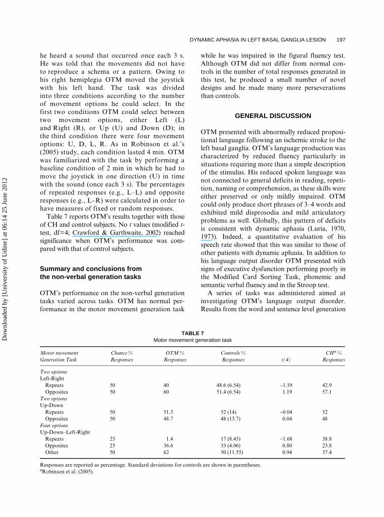
he heard a sound that occurred once each 3 s.He was told that the movements did not haveto reproduce a schema or a pattern. Owing tohis right hemiplegia OTM moved the joystickwith his left hand. The task was dividedinto three conditions according to the numberof movement options he could select. In thefirst two conditions OTM could select betweentwo movement options, either Left (L)and Right (R), or Up (U) and Down (D); inthe third condition there were four movementoptions: U, D, L, R. As in Robinson et al.’s(2005) study, each condition lasted 4 min. OTMwas familiarized with the task by performing abaseline condition of 2 min in which he had tomove the joystick in one direction (U) in timewith the sound (once each 3 s). The percentagesof repeated responses (e.g., L–L) and oppositeresponses (e.g., L–R) were calculated in order tohave measures of fixed or random responses.
Table 7 reports OTM’s results together with thoseof CH and control subjects. No t values (modified t-test, df=4; Crawford & Garthwaite, 2002) reachedsignificance when OTM’s performance was com-pared with that of control subjects.
Summary and conclusions fromthe non-verbal generation tasks
OTM’s performance on the non-verbal generationtasks varied across tasks. OTM has normal per-formance in the motor movement generation task
while he was impaired in the figural fluency test.Although OTM did not differ from normal con-trols in the number of total responses generated inthis test, he produced a small number of noveldesigns and he made many more perseverationsthan controls.
GENERAL DISCUSSION
OTM presented with abnormally reduced proposi-tional language following an ischemic stroke to theleft basal ganglia. OTM’s language production wascharacterized by reduced fluency particularly insituations requiring more than a simple descriptionof the stimulus. His reduced spoken language wasnot connected to general deficits in reading, repeti-tion, naming or comprehension, as these skills wereeither preserved or only mildly impaired. OTMcould only produce short phrases of 3–4 words andexhibited mild disprosodia and mild articulatoryproblems as well. Globally, this pattern of deficitsis consistent with dynamic aphasia (Luria, 1970,1973). Indeed, a quantitative evaluation of hisspeech rate showed that this was similar to those ofother patients with dynamic aphasia. In addition tohis language output disorder OTM presented withsigns of executive dysfunction performing poorly inthe Modified Card Sorting Task, phonemic andsemantic verbal fluency and in the Stroop test.
A series of tasks was administered aimed atinvestigating OTM’s language output disorder.Results from the word and sentence level generation
TABLE 7 Motor movement generation task
Motor movement Generation Task
Chance % Responses
OTM % Responses
Controls % Responses t(4)
CHa % Responses
Two optionsLeft-Right
Repeats 50 40 48.6 (6.54) −1.19 42.9Opposites 50 60 51.4 (6.54) 1.19 57.1
Two optionsUp-Down
Repeats 50 51.3 52 (14) −0.04 52Opposites 50 48.7 48 (13.7) 0.04 48
Four optionsUp-Down–Left-Right
Repeats 25 1.4 17 (8.45) −1.68 38.8Opposites 25 36.6 33 (4.06) 0.80 23.8Other 50 62 50 (11.55) 0.94 37.4
Responses are reported as percentage. Standard deviations for controls are shown in parentheses.aRobinson et al. (2005).
Dow
nloa
ded
by [
Uni
vers
ity o
f U
dine
] at
06:
14 2
5 Ju
ne 2
012
198 CRESCENTINI ET AL.
tests showed that OTM had great difficulties whenrequired to produce phrases and words; his prob-lems became evident when stimuli did not suggest adominant response. In a similar fashion to patientANG (Robinson et al., 1998), OTM had more dif-ficulties in producing sentences from word pairswith weak association between them than from astrongly associated word pairs and had also manymore problems in completing low predictabilitysentences, either with phrases or words, than highpredictability ones. In a similar fashion to ANG(Robinson et al., 1998) and CH (Robinson et al.,2005) but differently from ROH (Costello & War-rington, 1989), OTM did not show any problem inthe sentence construction task. Tests of speech elic-itation from pictures (generation of a sentencegiven a pictorial scene and from a single picture)produced similar results to those obtained in otherdynamic aphasic patients (Robinson et al., 1998,2005); indeed OTM was severely impaired in pro-ducing sentences from single pictures whereas hecould give a simple description of them.
We also assessed whether OTM suffered fromthe same kind of deficits described by Gold et al.(1997) in their dynamic aphasic patient CO, whoalso had a basal ganglia infarction. These authorsproposed that their patient suffered from deficits insemantic strategy formation and concept genera-tion, being impaired in both tasks of semantic cate-gorization and procedural discourse. OTM’sperformance in the latter task showed that hecould correctly describe routine activities, he couldgenerate concepts and combine them in sequences.OTM also performed almost errorless in thesemantic categorization task as done by patientCH (Robinson et al., 2005). As discussed byRobinson et al. (2005), the fact that CH and OTMperformed normally in the semantic categorizationtest indicates that they do not have a primary defi-cit in semantic strategy formation.
OTM’s ability to generate numbers was also investi-gated. In a random number generation task heperformed differently from control subjects producingmore ascending and descending series of numbers. Weinterpret these findings as due to a failure of suppres-sion of strong routine responses (i.e., counting in ones).
Non-verbal response generation skills werefound to be preserved in 1 task; OTM was withinthe normal range in the motor movement gen-eration task. His performance on figural fluencywas however impaired; the number of responseswas in the normal range but 9/15 of these wereperseverations.
Possible explanations of OTM’s dynamic aphasia
As previously reported in the text, OTM also sufferedfrom a stroke involving the right parietal lobe.Interestingly, there has been reported evidenceshowing that language performance may be influ-enced by the side of space to which subjects arepaying attention. Thus spatial information mayinfluence language functions. For instance, Coslett(1999) investigated the performance of severalgroups of patients on both motor and languagetasks. He found that subjects with right parietallesions performed better when they directed atten-tion to stimuli in the right hemispace. The aim ofour study was not to further assess whether spatialfactors influence OTM’s performance on languagetasks. Nonetheless, OTM did not show any sign ofneglect when, for instance, the tests which involvevisual material in space (e.g., Progressive matrices)are considered. Moreover he did not have greaterdifficulties on the left than the right in tests whichinvolve space such as Attentive Matrices. Accord-ingly, OTM’s problems in generating responseswhen alternative options are available, and hishigh number of perseverative responses are morelikely due to damage to fronto-striatal regions.
As briefly reported in the Introduction, anumber of explanations have been proposed forforms of dynamic aphasia extending both withinthe domain of language and beyond the latter. Asfar as accounts of dynamic aphasia that considerthe disorder within the domain of language areconcerned, for reasons similar to those presentedby Robinson et al. (2005) we can rule out the pos-sibility that OTM’s deficits lay in damage to amechanism forming the linear scheme of a sentence(Luria, 1970, 1973). Similarly, the possibility of animpairment in verbal planning which Costello andWarrington (1989) considered for their patient(ROH) can be rejected for OTM. Our patientperformed almost errorless in the sentence con-struction test on which ROH had great difficulties.
In addition to showing problems in both seman-tic categorization and procedural discourse tasks,patient CO (Gold et al., 1997) had been impairedin design fluency, category fluency tasks and alsoin some tests of frontal system functions, such asthe Trail making B and Wisconsin Card Sort. Dueto this empirical evidence, Gold et al. concludedthat CO’s deficit in propositional language couldbe attributed to associated deficits in executivefunctions. As already described, OTM was
Dow
nloa
ded
by [
Uni
vers
ity o
f U
dine
] at
06:
14 2
5 Ju
ne 2
012
DYNAMIC APHASIA IN LEFT BASAL GANGLIA LESION 199
impaired in some tests of executive functions aswell. OTM also showed difficulties in the short storyrecall (see Table 1), a task that might be strategydependent, and also in the picture arrangementsubtest of the WAIS (see Table 1).
As far as the word and sentence generation tasksare concerned, we have shown that the number ofresponse options associated with a task (verbalresponse alternative options) was an importantfactor helping to give rise to OTM’s performancein many of the tasks we used. In a similar fashionto CH (Robinson et al, 2005), OTM was notimpaired when a stimulus strongly suggested aresponse. By contrast, OTM had great difficultieswhen stimuli activated many response alternatives.As stated in Robinson et al. (2005), a deficit in theselection of verbal responses in propositionalspeech is likely to be due to defective mechanismsof conceptual preparation (Levelt, 1989, 1999).Lexical concepts have sometimes to be generatedin conditions of high competition, namely whenmany of them are simultaneously activated. Theseconditions are those that pose the greatest require-ments to the conceptual preparation mechanisms,particularly when the latter are defective. Theempirical evidence collected in this study suggeststhat OTM, similarly to patients ANG and CH(Robinson et al., 1998, 2005, respectively), suffersfrom an impairment in verbal response generationin situations of high competition.
However, differently from CH, OTM also hadproblems in figural fluency and in the randomnumber generation task. OTM made many persevera-tions in the figural fluency test while he gave increasedascending and descending series of numbers in therandom number generation task. As brieflyreported previously in the text, we suggest that adeficit of inhibition can explain OTM’s problemswith numbers. Among other processes assumed tobe involved in reasonable performance of therandom number generation task, one consists ofinhibiting the most familiar strategy of countingupwards or downwards in ones which, in the termi-nology of the SAS model, is the strongest schema inthe contention scheduling. Thus it is possible thatOTM’s pattern of performance in this task reflects afailure of the SAS to inhibit strong routine schemata(i.e., counting in ones) in favor of less commonactions (i.e., counting in twos). An account based onfailure of suppression may also explain OTM’sperseverative responses in the figural fluency task.
Nevertheless, an alternative possibility existsfor explaining perseverations on figural fluency.
Perseverations may reflect a deficit to generatenovel content. We have shown that OTM was ableto produce only 6 different figures relative to con-trols which generated almost 14 new designs in thefigural fluency test. OTM’s inability to generatenovel content may also be seen to fit with his diffi-culty in some language tasks. For instance, OTMdid not have any problem in producing a responsewhen given common words or proper nouns; how-ever, he appeared to perseverate or to use a com-pensatory strategy in this task (see also hisproblems in sentence generation from single pic-tures or elaboration of nuclear sentences in whichnovel material has to been generated).
In summary, we propose that two deficits con-tribute to the overall pattern of performance ofpatient OTM. An impairment of verbal responsegeneration in conditions of high competition ispresent along with one of novel thought generationevidenced by perseveration and due to eitherfailure of inhibition or inability to generate novelcontent. These two deficits may be related to dam-age of different regions within the fronto-striatalcircuits, namely, left frontal regions and basalganglia, as is argued in the next section.
As reported in the Introduction, Robinson et al.(2006) proposed a distinction between two subtypesof dynamic aphasia. Since discourse generation hasnot been specifically tested in OTM, it is difficult tosay to which subtype his dynamic aphasia relates.We suggest that OTM has a similar impairment tothe first subtype of dynamic aphasia in which a ver-bal generation impairment is present along withassociated left frontal lesions. However, a profilerelated to the second subtype of Robinson et al.(2006) cannot be ruled out given that problems inresponse generation may be also present in domainsother than language (figural fluency) in OTM.
Anatomical implications
The case of OTM, in a similar fashion to those ofthe dynamic aphasic patients CO and KAS (Goldet al., 1997; Robinson et al., 2006, respectively; seealso Raymer et al., 2002), suggests that subcorticallesions which give rise to dynamic aphasia alsolead to deficits in non-verbal domains. OTM’s pat-tern of performance on both verbal and non-verbaltasks suggests that he suffered from two differentdeficits: impaired verbal response generation insituations of high competition and impaired novelthought generation as evidenced by perseveration.
Dow
nloa
ded
by [
Uni
vers
ity o
f U
dine
] at
06:
14 2
5 Ju
ne 2
012
200 CRESCENTINI ET AL.
With regard to the first deficit, there is a consider-able body of neuropsychological and neuroimagingevidence supporting the role of the left inferiorfrontal gyrus (LIFG) in the process of conflict res-olution, which is made by selecting informationfrom among competing alternatives (Barch, Braver,Sabb, & Noll, 2000; Persson et al., 2004; Thompson-Schill & Botvinick, 2006; Thompson-Schill,D’Esposito, Aguirre, & Farah, 1997; Thompson-Schill et al., 1998; Zhang, Feng, Fox, Gao, & Tan,2004). Recently, Badre, Poldrack, Paré-Blagoev,Insler, and Wagner (2005) have provided fMRIevidence supporting the fact that selection could besubserved by the left mid-ventro lateral prefrontalcortex (VLPFC) while controlled retrieval processescould be subserved by the left anterior VLPFC.
Referring back to dynamic aphasia, the litera-ture suggests the involvement of the LIFG in thissyndrome. As reported in the Introduction, bothneurodegenerative and focal lesions have been seento lead to dynamic aphasia. A review of these casessupports the role of the LIFG in the emergence ofthe reduced verbal output, which is generallyobserved in dynamic aphasic patients (Costello &Warrington, 1989; Esmonde et al., 1996; Raymeret al., 2002; Robinson et al., 1998, 2005; Snowden,Griffiths, & Neary, 1996; Warren et al., 2003).According to this evidence, the findings on OTMsuggest that his verbal response generation impair-ment may be due to a frontostriatal damagedisrupting the left frontal region function of selectionin situations of high competition. Admittedly, aword of caution is necessary with regard to the CTscan: in fact this is not 100% sensitive for ischemicdamage, so that the precise extent of the frontallesion in OTM cannot be exactly defined.
As reported in the Introduction, there is evidencesuggesting the involvement of basal ganglia incontrolled cognitive processes (Copland, 2003;Copland et al., 2000a, 2000b; Crosson et al., 2003;Redgrave, Prescott, & Gurney, 1999). In particular,recent studies assign the basal ganglia a non-languagespecific role in the inhibition of competingalternatives (Castner et al., 2007, in press; Crescentini,Mondolo, Biasutti, & Shallice, 2008; Longworthet al., 2005). Different studies have proposed thatdeficits in the SAS may also reflect in poorperformance on tasks requiring inhibition ofprepotent or irrelevant responses. Lesions or mal-function of basal ganglia seem to be particularlyimportant in causing failure of inhibition. As anexample, Dujardin, Degreef, Rogelet, Defebvre,and Destee (1999) (see also Bouquet, Bonnaud, &
Gil, 2003; Castner et al., 2007, in press; Crescentiniet al., 2008 for similar findings) have shown thatPD patients had problems in inhibiting task irrele-vant information in a modified version of theStroop test and of the Brown–Peterson paradigm.Moreover, in an fMRI study, Rossel, Bullmore,Williams, and David (2001) studied the braincorrelates of automatic and controlled processingin a semantic priming environment. Automatic andcontrolled processing were investigated using,respectively, short and long prime-target delays.The authors found that the putamen was preferen-tially activated at long intervals and this led themto suggest a role for this structure in controlledsemantic processes. The authors suggested that theputamen may be involved in the processes ofresponse selection and inhibition. Even morerecently, McNab and Klingberg (2008) have pro-posed that the basal ganglia operate to allow onlytask-relevant information into the working memory(see also Frank, Loughry, & O’Reilly, 2001). Thiswould be possible through the selective filtering ofthis information.
The role of basal ganglia in the suppression ofcompeting alternatives in the motor realm is wellknown (see Mink, 1996). Nevertheless, the studiesbriefly reported above suggest that Mink’s frameworkcan also be applied to domains other than motorbehavior (i.e., semantic priming, word selection,cognitive inhibition, see also Crosson, Benjamin, &Levy, 2007).
Accordingly, we suggest that a failure of inhibitioncould explain the problems that OTM had in the ran-dom number generation task. Moreover, it is also apossible explanation for his perseverative responses.
From another perspective, it is now usuallyassumed that different areas of the frontal lobesproject to separate areas of the striatum so that paral-lel frontal-subcortical circuits are formed (Alexander& Crutcher, 1990; Zgaljardic et al., 2006). Amongthese, the dorsolateral prefrontal circuit involves:Brodmann’s areas 9- and 10-dorsolateral caudate-pallidum, the substantia nigra-ventral anterior, thedorsomedial nuclei of the thalamus and the dorsola-teral prefrontal cortex. The relevance of this circuithas been stressed by Gold et al. (1997) in order toaccount for several of the signs of frontal dysfunctionshown by their patient. OTM also showed impair-ments which correspond to malfunctions of the dor-solateral prefrontal cortex. Moreover, thecomponents of this circuit have been seen to be criti-cally implicated in response suppression during therandom number generation task (Brown, Soliveri, &
Dow
nloa
ded
by [
Uni
vers
ity o
f U
dine
] at
06:
14 2
5 Ju
ne 2
012
DYNAMIC APHASIA IN LEFT BASAL GANGLIA LESION 201
Jahanshahi, 1998, Jahanshahi et al., 1998) and alsoduring other tasks requiring challenging responseinhibition such as the Stroop task (Alexander, Stuss,Picton, Shallice, & Gillingham, 2007). Furthermore,recent evidence shows that the basal ganglia may beimplicated in each part of the two-process model(selection and controlled retrieval) of control ofsemantic memory proposed by Badre et al. (2005; seeLongworth et al., 2005). Finally, that this could bethe case has been confirmed more recently by furtherevidence collected on Parkinson’s disease patients(Crescentini et al., 2008).
In sum we propose that an impairment to thebasal ganglia, and the consequent abnormal func-tioning of subcortical-frontal circuits, are import-ant factors in helping to give rise to the deficit ofnovel thought generation found in OTM and,more generally, to his pure dynamic aphasia.
Conclusions
We report the case of OTM who presented withdynamic aphasia following a lesion to the left basalganglia. We interpreted the pattern of performanceof OTM as due to two deficits. The first concernsthe ability to generate verbal responses and is par-ticularly present when stimuli activate many com-peting response options. The second deficitconsists of defective generation of novel thoughtsas evidenced by perseveration. We hold the firstdeficit to arise from a frontostriatal damage dis-rupting the left frontal region functions, and thesecond from a damage to the basal ganglia.
Original manuscript received 16 October 2007Revised manuscript accepted 27 March 2008
First published online 4 June 2008
REFERENCES
Alexander, G. E., & Crutcher, M. D. (1990). Functionalarchitecture of basal ganglia circuits: Neural substratesof parallel processing. Trends in the Neurosciences, 13,266–271.
Alexander, M. P., Naeser, M. A., & Palumbo, C. (1987).Correlations of subcortical CT lesion sites and aphasiaprofiles. Brain, 110, 961–991.
Alexander, M. P., Stuss, D. T., Picton, T., Shallice, T., &Gillingham, S. (2007). Regional frontal injuries causedistinct impairments in cognitive control. Neurology,68, 1515–1523.
Badre, D., Poldrack, R. A., Paré-Blagoev, E. J., Insler,R. Z., & Wagner, A. D. (2005). Dissociable controlled
retrieval and generalized selection mechanisms in ven-trolateral prefrontal cortex. Neuron, 47, 907–918.
Barbarotto, R., Laiacona, M., Frosio, R., Vecchio, M.,& Farinato, A. (1998). A normative study on visualreaction times and two Stroop color-word tests. Ital-ian Journal of Neurological Science, 19, 161–170.
Barch, D. M., Braver, T. S., Sabb, F. W., & Noll, D. C.(2000). Anterior cingulate and the monitoring ofresponse conflict: Evidence from an fMRI study ofovert verb generation. Journal of Cognitive Neuro-science, 12, 298–309.
Bloom, P. A., & Fischler, I. (1980). Completion norms for329 sentence contexts. Memory and Cognition, 8, 631–642.
Bouquet, C. A., Bonnaud, V., & Gil, R. (2003). Investi-gation of supervisory attentional system functions inpatients with Parkinson’s disease using the Haylingtask. Journal of Clinical and Experimental Neuropsy-chology, 25, 751–760.
Brown, R. G., Soliveri, P., & Jahanshahi, M. (1998).Executive processes in Parkinson’s disease-randomnumber generation and response suppression.Neuropsychologia, 36, 1355–1362.
Burgess, P. W., & Shallice, T. (1996). Response suppres-sion, initiation and strategy use following frontal lobelesions. Neuropsychologia, 34, 263–273.
Caffarra, P., Vezzadini, G., Dieci, F., Zonato, F., &Venneri, A. (2004). Modified Card Sorting Test:Normative data. Journal of Clinical and ExperimentalNeuropsychology, 26, 246–250.
Cappelletti, M., Butterworth, B., & Kopelman, M.(2001). Spared numerical abilities in a case of semanticdementia. Neuropsychologia, 39, 1224–1239.
Carlesimo, G. A., Caltagirone, C., & Gainotti, G.(1996). The Mental Deterioration Battery: Norma-tive data, diagnostic reliability and qualitativeanalyses of cognitive impairment. European Neu-rology, 36, 378–384.
Castner, J. E., Chenery, H. J., Silburn, P. A., Smith,E. R., Coyne, T. J., Sinclair, F., & Copland, D. A.(in press). The effects of subthalamic deep brainstimulation on noun/verb generation and selectionfrom competing alternatives in Parkinson’sdisease. Journal of Neurology, Neurosurgery, andPsychiatry.
Castner, J. E., Copland, D. A., Silburn, P. A., Coyne, T.J., Sinclair, F., & Chenery, H. J. (2007).Lexical-semantic inhibitory mechanisms in Parkinson’sdisease as a function of subthalamic stimulation.Neuropsychologia, 14, 3167–3177.
Copland, D. (2003). The basal ganglia and semanticengagement: Potential insights from semantic priming inindividuals with subcortical vascular lesions, Parkinson’sdisease, and cortical lesions. Journal of the Interna-tional Neuropsychological Society, 9, 1041–1052.
Copland, D. A., Chenery, H. J., & Murdoch, B. E. (2000a).Persistent deficits in complex language function follow-ing dominant nonthalamic subcortical lesions. Journalof Medical Speech-Language Pathology, 8, 1–14.
Copland, D. A., Chenery, H. J., & Murdoch, B. E.(2000b). Processing lexical ambiguities in word tri-plets: Evidence of lexicalsemantic deficits followingdominant nonthalamic subcortical lesions. Neuropsy-chology, 14, 379–390.
Dow
nloa
ded
by [
Uni
vers
ity o
f U
dine
] at
06:
14 2
5 Ju
ne 2
012
202 CRESCENTINI ET AL.
Coslett, H. B. (1999). Spatial influences on motor andlanguage function. Neuropsychologia, 37, 695–706.
Costello de Lacy, A., & Warrington, E. K. (1989).Dynamic aphasia. The selective impairment of verbalplanning. Cortex, 25, 103–114.
Crawford, J. R., & Garthwaite, P. H. (2002). Investiga-tion of the single case in neuropsychology: Confidencelimits on the abnormality of test scores and test scoredifferences. Neuropsychologia, 40, 1196–1208.
Crescentini, C., Mondolo, F., Biasutti, E., & Shallice, T.(2008). Supervisory and routine processes in nounand verb generation in nondemented patients withParkinson’s disease. Neuropsychologia, 46, 434–447.
Crosson, B., Benefield, H., Cato, M. A., Sadek, J. R.,Moore, A. B., Wierenga, C. E., Gopinath, K.,Soltysik, D., Bauer, R. M., Auerbach, E. J., Gokcay,D., Leonard, C. M., & Briggs, R. W. (2003). Left andright basal ganglia and frontal activity during lan-guage generation: Contributions to lexical, semantic,and phonological processes. Journal of the Interna-tional Neuropsychological Society, 9, 1061–1077.
Crosson, B., Benjamin, M., & Levy, I. (2007). Role of thebasal ganglia in language and semantics: Supportingcast. In J. Hart, & M. Kraut (Eds), Neural basis ofsemantic memory. London: Cambridge University Press.
De Renzi, E., Motti, F., & Nichelli, P. (1980). Imitatinggestures. A quantitative approach to ideomotorapraxia. Archives of Neurology, 37, 6–10.
Della Sala, S., MacPherson, S. E., Phillips, L. H., Sacco,L., & Spinnler, H. (2003). How many camels are therein Italy? Cognitive estimates standardised on the Italianpopulation. Neurological Sciences, 24, 10–15.
Dujardin, K., Degreef, J. F., Rogelet, P., Defebvre, L., &Destee, A. (1999). Impairment of the supervisoryattentional system in early untreated patients with Par-kinson’s disease. Journal of Neurology, 246, 783–788.
Esmonde, T., Giles, E., Xuereb, J., & Hodges, J. (1996).Progressive supranuclear palsy presenting withdynamic aphasia. Journal of Neurology, Neurosur-gery, and Psychiatry, 60, 403–410.
Frank, M. J., Loughry, B., & O’Reilly, R. (2001).Interactions between frontal cortex and basal gan-glia in working memory: A computational model.Cognitive, Affective, and Behavioral Neuroscience,1, 137–160.
Giovagnoli, A. R., Pesce, M., Del Mascheroni, S.,Simoncelli, M., Laiacona, M., & Capitani, E. (1996).Trail making test: Normative values from 287 normaladult controls. The Italian Journal of NeurologicalSciences, 17, 305–309.
Gold, M., Nadeau, S. E., Jacobs, D. H., Adair, J. C.,Rothi, L. J. G., & Heilman, K. M. (1997). Adynamicaphasia: A transcortical motor aphasia with defectivesemantic strategy formation. Brain and Language, 57,374–393.
Grossman, M., Stern, M. B., Gollomp, S., Vernon, G., &Hurtig, H. I. (1994). Verb learning in Parkinson’s dis-ease. Neuropsychology, 8, 413–423.
Hillis, A. E., Wityk, R. J., Barker, P. B., Beauchamp, N.J., Gailloud, P., Murphy, K., Cooper, O., & Metter,E. J. (2002). Subcortical aphasia and neglet in acutestroke: The role of cortical hypoperfusion. Brain, 125,1094–1104.
Howard, D., & Patterson, K. (1992). Pyramids and PalmTrees: A test of semantic access from pictures andwords. Bury St Edmunds: Thames Valley.
Jahanshahi, M., Profice, P., Brown, R. G., Ridding, M.C., Dirnberger, G., & Rothwell, J. C. (1998). Theeffects of transcranial magnetic stimulation over thedorsolateral prefrontal cortex on suppression ofhabitual counting during random number generation.Brain, 121, 1533–1544.
Kramer, J. H., Reed, B. R., Mungas, D., Weiner, M. W.,& Chui, H. C. (2002). Executive dysfunction in sub-cortical ischaemic vascular disease. Journal of Neurol-ogy and Neurosurgery Psychiatry, 72, 217–220.
Lee, G. P., Strauss, E., Loring, D. W., McCloskey, L., &Haworth, J. M. (1997). Sensitivity of figural fluencyon the Five-Point Test to focal neurological disease.The Clinical Neuropsychologist, 11, 59–68.
Levelt, W. J. M. (1989). Speaking: From intention toarticulation. Cambridge, MA: MIT Press.
Levelt, W. J. M. (1999). Producing spoken language:A blueprint of the speaker. In C. M. Brown, &P. Hagoort (Eds), The neurocognition of language (pp.83–122). Oxford: Oxford University Press.
Longworth, C. E., Keenan, S. E., Barker, R. A.,Marslen-Wilson, W. D., & Tyler, L. K. (2005).The basal ganglia and rule-governed language use:Evidence from vascular and degenerative conditions.Brain, 128, 584–596.
Luria, A. R. (1970). Traumatic aphasia. The Hague:Mouton.
Luria, A. R. (1973). The working brain: An introduction toneuropsychology. Harmondsworth, UK: Penguin Books.
Luzzatti, C., Willmes, K., & De Bleser, R. (1996).Aachener Aphasie Test. Organizzazioni Speciali,Firenze.
McNab, F., & Klingberg, T. (2008). Prefrontal cortexand basal ganglia control access to working memory.Nature Neuroscience, 11, 103–107.
Miceli, G., Laudanna, A., Burani, C., & Capasso, R.(1994). Batteria per l’analisi dei deficit afasici. BADA.Cepsag Università Cattolica del Sacro Cuore Poli-clinico Gemelli.
Mink, J. W. (1996). The basal ganglia: Focused selectionand inhibition of competing motor programs.Progresses in Neurobiology, 50, 381–425.
Nadeau, S. E., & Crosson, B. (1997). Subcortical apha-sia. Brain and Language, 58, 355–402.
Norman, D. A., & Shallice, T. (1986). Attention toaction: Willed and automatic control of behaviour.Reprinted in revised form in R. J. Davidson, G.E.Schwartz, & D. Shapiro (Eds), Consciousness andself-regulation (Vol. 4, pp. 1–18). New York: PlenumPress.
Novelli, G., Papagno, C., Capitani, E., Laiacona, M.,Vallar, G., & Cappa, S. F. (1986). Tre test clinici dimemoria verbale a lungo termine. Archivio di Psicolo-gia Neurologia e Psichiatria, 47, 278–296.
Papagno, C., Cappa, S. F., Capitani, E., Forelli, A.,Garavaglia, G., Laiacona, M., et al. (1995). La comp-rensione non letterale del linguaggio: Taratura di untest di comprensione di metafore e di espressioniidiomatiche. Archivio di Psicologia Neurologia ePsichiatria, 56, 402–420.
Dow
nloa
ded
by [
Uni
vers
ity o
f U
dine
] at
06:
14 2
5 Ju
ne 2
012
DYNAMIC APHASIA IN LEFT BASAL GANGLIA LESION 203
Persson, J., Sylvester, C. C., Nelson, J. K., Welsh, K. M.,Jonides, J., & Reuter-Lorenz, P. A. (2004). Selectionrequirements during verb generation: Differentialrecruitment in older and younger adults. Neuroimage,23, 1382–1390.
Raymer, A. M., Rowland, L., Haley, M., & Crosson, B.(2002). Nonsymbolic movement training to improvesentence generation in transcortical motor aphasia:A case study. Aphasiology, 16, 493–506.
Redgrave, P., Prescott, T. J., & Gurney, K. (1999). Thebasal ganglia: A vertebrate solution to the selectionproblem? Neuroscience, 89, 1009–1023.
Robinson, G., Blair, J., & Cipolotti, L. (1998). Dynamicaphasia: An inability to select between competingverbal responses? Brain, 121, 77–89.
Robinson, G., Shallice, T., & Cipolotti, L. (2005). A failureof high level verbal response selection in progressivedynamic aphasia. Cognitive Neuropsychology, 22,661–694.
Robinson, G., Shallice, T., & Cipolotti, L. (2006).Dynamic aphasia in progressive supranuclear palsy:A deficit in generating a fluent sequence of novelthought. Neuropsychologia, 44, 1344–1360.
Rossel, S. L., Bullmore, E. T., Williams, S. C. R., &David, A. S. (2001). Brain activation during automaticand controlled processing of semantic relations: Apriming experiment using lexical-decision. Neuropsy-chologia, 39, 1167–1176.
Rumiati, R. I., Zanini, S., Vorano, L., & Shallice, T.(2001). A form of ideational apraxia as a selectivedeficit of contention scheduling. Cognitive Neuropsy-chology, 18, 617–642.
Sartori, G., Colombo, L., Vallar, G., Rusconi, M. L., &Pinarello, A. (1995). TIB-Test di Intelligenza Breveper la valutazione del quoziente intellettivo attuale epre-morboso. [TIB-Brief Test of Intelligence forassessing present and pre-morbid intelligence].Professione di Psicologo. Giornale dell’Ordine degliPsicologi, 4, 1–24.
Shallice, T., & Evans, M. E. (1978). The involvement ofthe frontal lobes in cognitive estimation. Cortex, 14,294–303.
Snowden, J. S., Griffiths, H. L., & Neary, D. (1996).Progressive language disorder associated with frontallobe degeneration. Neurocase, 2, 429–440.
Spinnler, H., & Tognoni, G. (1987). Standardizzazione etaratura italiana di test neuropsicologici. The ItalianJournal of Neurological Sciences, 6, 1–120.
Teichmann, M., Dupoux, E., Kouider, S., & Bachoud-Lévi, A. C. (2006). The role of the striatum in
processing language rules: Evidence from wordperception in Huntington’s disease. Journal of CognitiveNeuroscience, 18, 1555–1569.
Thompson-Schill, S. L., & Botvinick, M. M. (2006).Resolving conflict: A response to Martin and Cheng.Psychonomic Bulletin & Review, 13, 402–408.
Thompson-Schill, S. L., D’Esposito, M., Aguirre, G. K., &Farah, M. J. (1997). Role of left inferior prefrontalcortex in retrieval of semantic knowledge: A reevalu-ation. Proceedings of the National Academy of SciencesUSA, 94, 14792–14797.
Thompson-Schill, S. L., Swick, D., Farah, M. J., D’Esposito,M., Kan, I. P., & Knight, R. T. (1998). Verb generation inpatients with focal frontal lesions: A neuropsychologicaltest of neuroimaging findings. Proceedings of the NationalAcademy of Sciences USA, 95, 15855–15860.
Ullman, M. T., Corkin, S., Coppola, M., Hickok, G.,Growdon, J. H., Koroshetz, W. J., et al. (1997). Aneural dissociation within language: Evidence thatthe mental dictionary is part of declarative memory,and that grammatical rules are processed by the pro-cedural system. Journal of Cognitive Neuroscience, 9,266–276.
Wallesch, C. W., & Papagno, C. (1988). Subcorticalaphasia. In F. C. Rose, R. Whurr, M. A. Wyke(Eds.), Aphasia (pp. 256–287). London: Whurr Pub-lishers.
Warren, J. D., Warren, J. E., Fox, N. C., & Warrington, E.K. (2003). Nothing to say, something to sing: Primaryprogressive dynamic aphasia. Neurocase, 9, 140–155.
Warrington, E. K. (1984). Recognition Memory Test.Windsor, UK: NFER-Nelson.
Warrington, E. K. (1996). The Camden Memory Tests.Hove, UK: Psychology Press.
Warrington, E. K., & James, M. (1991). The VisualObject and Space Perception Battery. Bury StEdmunds, UK: Thames Valley Test Company.
Wechsler, D. (1981). Wechsler Adult Intelligence Scale-Revised. San Antonio, TX: The PsychologicalCorporation.
Zgaljardic, D. J., Borod, J. C., Foldi, N. S., Mattis, P. J.,Gordon, M. F., Feigin, A., & Eidelberg, D. (2006).An examination of executive dysfunction associatedwith frontostriatal circuitry in Parkinson’s disease.Journal of Clinical and Experimental Neuropsychology,28, 1127–1144.
Zhang, J. X., Feng, C. M., Fox, P. T., Gao, J. H., &Tan, L. H. (2004). Is left inferior frontal gyrus ageneral mechanism for selection? Neuroimage, 23,596–603.
Dow
nloa
ded
by [
Uni
vers
ity o
f U
dine
] at
06:
14 2
5 Ju
ne 2
012
Related Documents