RESEARCH ARTICLE A Hypothetical Model of Crossing Bombyx mori Nucleopolyhedrovirus through Its Host Midgut Physical Barrier Yang Cheng 1¤ , Xue-Yang Wang 1 , Hao Hu 2 , Nabil Killiny 2 , Jia-Ping Xu 1 * 1. School of Life Sciences, Anhui Agricultural University, Hefei, People’s Republic of China, 2. Department of Entomology and Nematology, Citrus Research and Education Center, IFAS, University of Florida, Lake Alfred, Florida, United States of America * [email protected] ¤ Current address: Institute of Zoology, Chinese Academy of Sciences, Chaoyang District, Beijing, People’s Republic of China Abstract Bombyx mori nucleopolyhedrovirus (BmNPV) is a primary pathogen of silkworm (B. mori) that causes severe economic losses each year. However, the molecular mechanisms of silkworm-BmNPV interactions, especially the silkworm proteins that can interact with the virus, are still largely unknown. In this study, the total and membrane proteins of silkworm midguts were displayed using one- and two- dimensional electrophoresis. A virus overlay assay was used to detect B. mori proteins that specifically bind to BmNPV particles. Twelve proteins were located and identified using mass spectrometry, and the different expression of the corresponding genes in BmNPV susceptible and resistant silkworm strains also indicated their involvement in BmNPV infection. The 12 proteins are grouped based on their potential roles in viral infection, for example, endocytosis, intracellular transportation, and host responses. Based on these results, we hypothesize the following: I) vacuolar ATP synthase catalytic subunit A and subunit B may be implicated in the process of the membrane fusion of virus and the release of the nucleocapsid into cytoplasm; II) actin, enolase and phosphoglycerate kinase are cytoskeleton associated proteins and may play an important role in BmNPV intracellular transportation; III) mitochondrial prohibitin complex protein 2, ganglioside-induced differentiation-associated protein, calreticulin, regucalcin-like isoform X1 and 60 kDa heat shock protein are involved in cell apoptosis regulation during BmNPV infection in larvae midguts; IV) ribosomal P0 may be associated with BmNPV infection by regulating gene expression of BmNPV; V) arginine kinase has a role in the antiviral activities against BmNPV. Our work should prove OPEN ACCESS Citation: Cheng Y, Wang X-Y, Hu H, Killiny N, Xu J-P (2014) A Hypothetical Model of Crossing Bombyx mori Nucleopolyhedrovirus through Its Host Midgut Physical Barrier. PLoS ONE 9(12): e115032. doi:10.1371/journal.pone.0115032 Editor: Yi Li, Wuhan Bioengineering Institute, China Received: October 1, 2014 Accepted: November 17, 2014 Published: December 12, 2014 Copyright: ß 2014 Cheng et al. This is an open- access article distributed under the terms of the Creative Commons Attribution License, which permits unrestricted use, distribution, and repro- duction in any medium, provided the original author and source are credited. Data Availability: The authors confirm that all data underlying the findings are fully available without restriction. All relevant data are within the paper and its Supporting Information files. Funding: This work was supported by the Natural Science Foundation of China (31472148), the National 863 plans projects of China (2011AA100306) and International cooperation project of Anhui province (1403062018). The funders had no role in study design, data collection and analysis, decision to publish, or preparation of the manuscript. Competing Interests: The authors have declared that no competing interests exist. PLOS ONE | DOI:10.1371/journal.pone.0115032 December 12, 2014 1 / 17

Welcome message from author
This document is posted to help you gain knowledge. Please leave a comment to let me know what you think about it! Share it to your friends and learn new things together.
Transcript

RESEARCH ARTICLE
A Hypothetical Model of Crossing Bombyxmori Nucleopolyhedrovirus through ItsHost Midgut Physical BarrierYang Cheng1¤, Xue-Yang Wang1, Hao Hu2, Nabil Killiny2, Jia-Ping Xu1*
1. School of Life Sciences, Anhui Agricultural University, Hefei, People’s Republic of China, 2. Department ofEntomology and Nematology, Citrus Research and Education Center, IFAS, University of Florida, Lake Alfred,Florida, United States of America
¤ Current address: Institute of Zoology, Chinese Academy of Sciences, Chaoyang District, Beijing, People’sRepublic of China
Abstract
Bombyx mori nucleopolyhedrovirus (BmNPV) is a primary pathogen of silkworm (B.
mori) that causes severe economic losses each year. However, the molecular
mechanisms of silkworm-BmNPV interactions, especially the silkworm proteins that
can interact with the virus, are still largely unknown. In this study, the total and
membrane proteins of silkworm midguts were displayed using one- and two-
dimensional electrophoresis. A virus overlay assay was used to detect B. mori
proteins that specifically bind to BmNPV particles. Twelve proteins were located
and identified using mass spectrometry, and the different expression of the
corresponding genes in BmNPV susceptible and resistant silkworm strains also
indicated their involvement in BmNPV infection. The 12 proteins are grouped based
on their potential roles in viral infection, for example, endocytosis, intracellular
transportation, and host responses. Based on these results, we hypothesize the
following: I) vacuolar ATP synthase catalytic subunit A and subunit B may be
implicated in the process of the membrane fusion of virus and the release of the
nucleocapsid into cytoplasm; II) actin, enolase and phosphoglycerate kinase are
cytoskeleton associated proteins and may play an important role in BmNPV
intracellular transportation; III) mitochondrial prohibitin complex protein 2,
ganglioside-induced differentiation-associated protein, calreticulin, regucalcin-like
isoform X1 and 60 kDa heat shock protein are involved in cell apoptosis regulation
during BmNPV infection in larvae midguts; IV) ribosomal P0 may be associated
with BmNPV infection by regulating gene expression of BmNPV; V) arginine kinase
has a role in the antiviral activities against BmNPV. Our work should prove
OPEN ACCESS
Citation: Cheng Y, Wang X-Y, Hu H, Killiny N, XuJ-P (2014) A Hypothetical Model of CrossingBombyx mori Nucleopolyhedrovirus through ItsHost Midgut Physical Barrier. PLoS ONE 9(12):e115032. doi:10.1371/journal.pone.0115032
Editor: Yi Li, Wuhan Bioengineering Institute,China
Received: October 1, 2014
Accepted: November 17, 2014
Published: December 12, 2014
Copyright: � 2014 Cheng et al. This is an open-access article distributed under the terms of theCreative Commons Attribution License, whichpermits unrestricted use, distribution, and repro-duction in any medium, provided the original authorand source are credited.
Data Availability: The authors confirm that all dataunderlying the findings are fully available withoutrestriction. All relevant data are within the paperand its Supporting Information files.
Funding: This work was supported by the NaturalScience Foundation of China (31472148), theNational 863 plans projects of China(2011AA100306) and International cooperationproject of Anhui province (1403062018). Thefunders had no role in study design, data collectionand analysis, decision to publish, or preparation ofthe manuscript.
Competing Interests: The authors have declaredthat no competing interests exist.
PLOS ONE | DOI:10.1371/journal.pone.0115032 December 12, 2014 1 / 17
informative by providing multiple protein targets and a novel direction to investigate
the molecular mechanisms of the interactions between silkworms and BmNPV.
Introduction
The silkworm, Bombyx mori L. (Lepidoptera: Bombycidae), is an economically
important insect for production of silk and recombinant proteins, and also a good
model of the Lepidoptera [1]. Bombyx mori nucleopolyhedrovirus (BmNPV) is a
primary pathogen of the domestic silkworm, and always causes severe economic
loss [2].
In the NPV replication cycle, there are two different virion phenotypes, which
are the occlusion-derived virus (ODV) and the budded virus (BV) [3]. BVs infect
a broad range of cell types and transmit virus among insect tissues within an
infected larva, whereas ODVs are contained in polyhedrons and form occlusion
bodies (OBs) which infect only columnar epithelial cells of the insect midguts and
are required for the oral transmission of virus between insect hosts [4, 5].
At present, the molecular interaction mechanisms between BmNPV and B. mori
remain unclear. Studies on the functions of anti-viral proteins isolated from B.
mori were reported frequently in the last ten years. It has been reported that B.
mori serine protease-2, lipase-1 and alkaline trypsin protein purified from the
digestive juice of B. mori larvae showed strong antiviral activity to BmNPV in
vitro [6–8]. Using the fluorescent differential display (FDD) technique, Bms3a
was found related to BmNPV resistance in silkworm [9]. But the roles of these
proteins in the process of BmNPV infection or B. mori anti-infection are not
stated, meanwhile, studies on the interactions between BmNPV and B. mori at the
system-wide level of larva using the methodology of far-western blot and mass
spectrometry have not been reported yet. Far-western blot, also known as virus
overlay assay, has been used successfully to detect proteins that are potential virus
receptors in the body of insect vectors [10]. Initially, several proteins within
Myzuspersicae were found to bind to potato leaf roll virus (PLRV) in vitro [11].
Since then, many virus-binding proteins were determined in host insects. Kikkert
et al. found a 94-kDa thrips protein that exhibited specific binding to tomato
spotted wilt virus (TSWV) particles in Frankliniella occidentalis and Thrips tabaci
using virus overlay assays [12]. Bandla et al also found out that a 50-kDa protein
in larval midguts of F. occidentalis exhibited interaction with TSWV using similar
method almost at the same time [13]. But the virus-binding proteins were not
identified exactly. The development of mass spectrometry in recent years makes
the identification of proteins feasible. For example, 5 proteins of Laodelphax
striatellus exhibited interactions with Rice stripe virus (RSV) using virus overlay
assays and they were identified using Nano LC-ESI-CID-MS/MS analysis [10].
In our study, virus overlay assays were performed in the screening for BmNPV
binding proteins from larval midguts of B. mori. The results showed that twelve
BmNPV Binding Proteins
PLOS ONE | DOI:10.1371/journal.pone.0115032 December 12, 2014 2 / 17
proteins of B. mori bound specifically to purified BmNPV particles in vitro, and
these potential proteins were identified using MALDI-TOF/TOF MS analysis. The
potential functions of these binding proteins were further investigated and
speculated reasonably.
Results
Interaction between BmNPV particles and silkworm midgut
proteins
Virus overlay assays were applied to ascertain whether specific binding of BmNPV
particles to B. mori midgut total proteins or lipid-associated midgut membrane
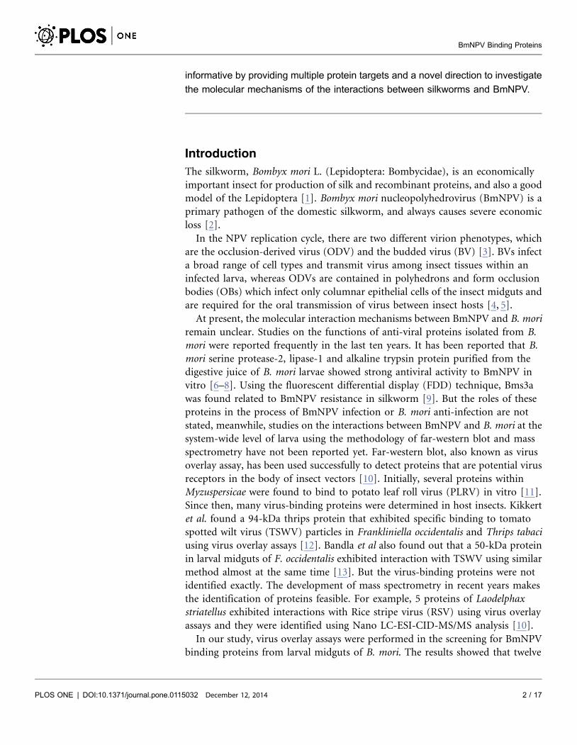
proteins would occur. Total proteins were characterized by 12% SDS-PAGE
followed by far-Western blot experiment, and three clear bands indicated by
arrows were detected (Fig. 1A). In order to determine the three protein bands, a
parallel SDS-PAGE assay was performed (Fig. 1B), and three protein bands
named a, b and c, respectively, were cut out for MALDI-TOF/TOF MS analysis.
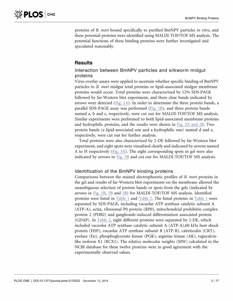
Similar experiments were performed to both lipid-associated membrane proteins
and hydrophilic proteins, and the results were shown in Fig. 2A and 2B. Two
protein bands (a lipid-associated one and a hydrophilic one) named d and e,
respectively, were cut out for further analysis.
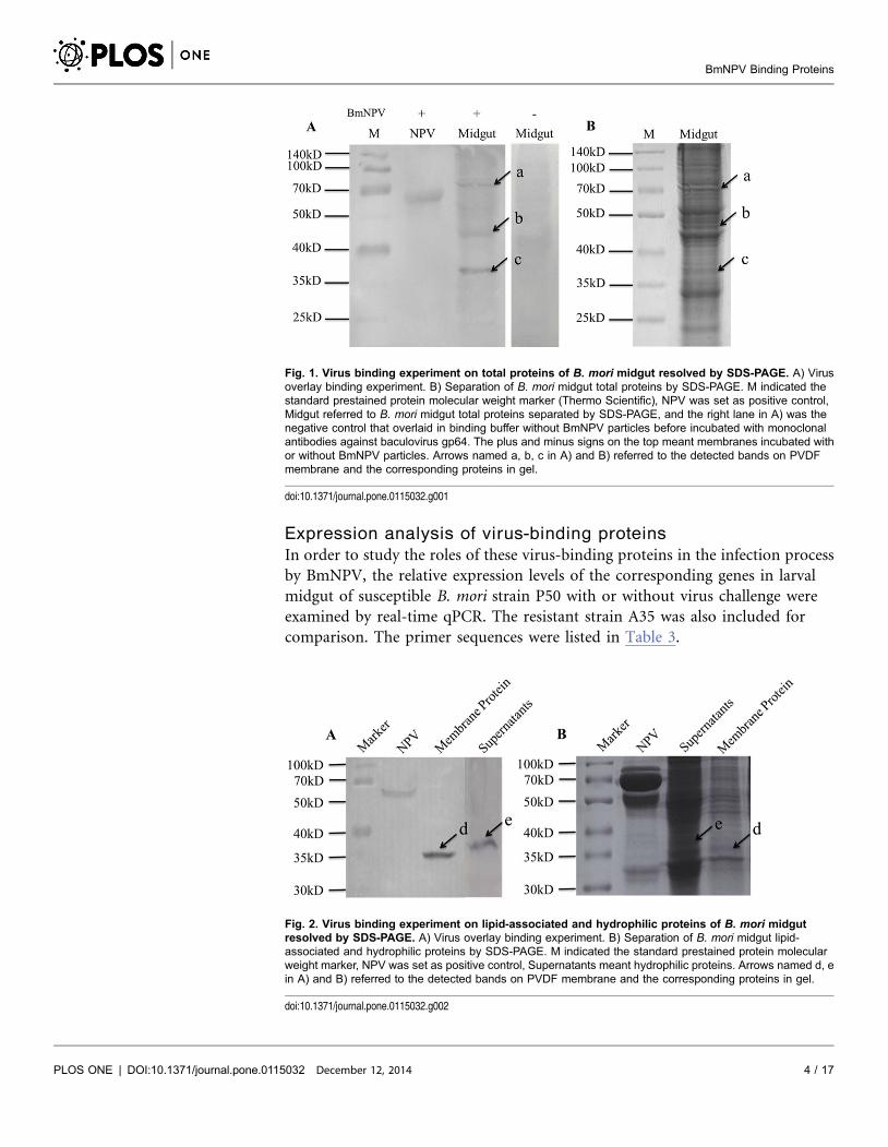
Total proteins were also characterized by 2-DE followed by far-Western blot
experiment, and eight spots were visualized clearly and indicated by arrows named
A to H respectively (Fig. 3A). The eight corresponding spots in gel were also
indicated by arrows in Fig. 3B and cut out for MALDI-TOF/TOF MS analysis.
Identification of the BmNPV binding proteins
Comparisons between the stained electrophoretic profiles of B. mori proteins in
the gel and results of far-Western blot experiments on the membrane allowed the
unambiguous selection of protein bands or spots from the gels (indicated by
arrows in Fig. 1B, 2B and 3B) for MALDI-TOF/TOF MS analysis. Identified
proteins were listed in Table 1 and Table 2. The listed proteins in Table 1 were
separated by SDS-PAGE, including vacuolar ATP synthase catalytic subunit A
(ATP-A), actin, ribosomal P0 protein (RP0), mitochondrial prohibitin complex
protein 2 (PHB2) and ganglioside-induced differentiation associated protein
(GDAP). In Table 2, eight different proteins were separated by 2-DE, which
included vacuolar ATP synthase catalytic subunit A (ATP-A),60 kDa heat shock
protein (HSP), vacuolar ATP synthase subunit B (ATP-B), calreticulin (CRT),
enolase (En), phosphoglycerate kinase (PGK), arginine kinase (AK), regucalcin-
like isoform X1 (RCX1). The relative molecular weights (MW) calculated in the
NCBI database for these twelve proteins were in good agreement with the
experimentally observed values.
BmNPV Binding Proteins
PLOS ONE | DOI:10.1371/journal.pone.0115032 December 12, 2014 3 / 17
Expression analysis of virus-binding proteins
In order to study the roles of these virus-binding proteins in the infection process
by BmNPV, the relative expression levels of the corresponding genes in larval
midgut of susceptible B. mori strain P50 with or without virus challenge were
examined by real-time qPCR. The resistant strain A35 was also included for
comparison. The primer sequences were listed in Table 3.
Fig. 1. Virus binding experiment on total proteins of B. mori midgut resolved by SDS-PAGE. A) Virusoverlay binding experiment. B) Separation of B. mori midgut total proteins by SDS-PAGE. M indicated thestandard prestained protein molecular weight marker (Thermo Scientific), NPV was set as positive control,Midgut referred to B. mori midgut total proteins separated by SDS-PAGE, and the right lane in A) was thenegative control that overlaid in binding buffer without BmNPV particles before incubated with monoclonalantibodies against baculovirus gp64. The plus and minus signs on the top meant membranes incubated withor without BmNPV particles. Arrows named a, b, c in A) and B) referred to the detected bands on PVDFmembrane and the corresponding proteins in gel.
doi:10.1371/journal.pone.0115032.g001
Fig. 2. Virus binding experiment on lipid-associated and hydrophilic proteins of B. mori midgutresolved by SDS-PAGE. A) Virus overlay binding experiment. B) Separation of B. mori midgut lipid-associated and hydrophilic proteins by SDS-PAGE. M indicated the standard prestained protein molecularweight marker, NPV was set as positive control, Supernatants meant hydrophilic proteins. Arrows named d, ein A) and B) referred to the detected bands on PVDF membrane and the corresponding proteins in gel.
doi:10.1371/journal.pone.0115032.g002
BmNPV Binding Proteins
PLOS ONE | DOI:10.1371/journal.pone.0115032 December 12, 2014 4 / 17
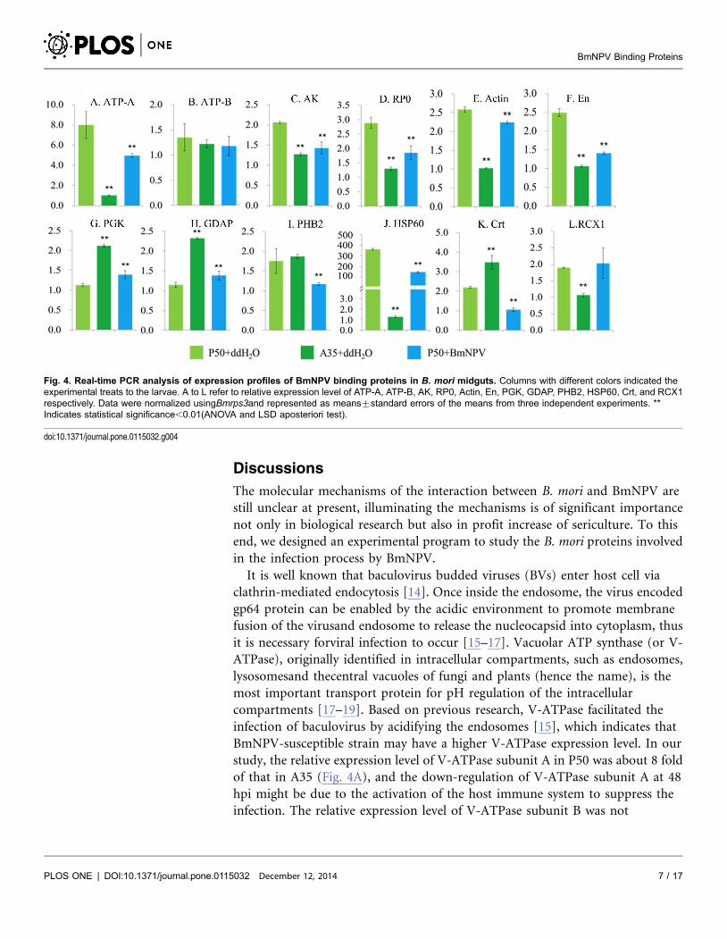
Real-time qPCR analysis was performed between BmNPV infected P50 larvae
and the control group treated with ddH2O. Fifth instar molt larvae of P50 were
starved overnight and fed with 500 OB of BmNPV T3 strain or ddH2O per larva
orally. The relative expression levels of PGK (Fig. 4G) and GDAP (Fig. 4H) in P50
midguts were up-regulated significantly (P,0.01) at 48hpi, while the levels of
eight genes, ATP-A (Fig. 4A), AK (Fig. 4C), RP0 (Fig. 4D), actin (Fig. 4E), En
(Fig. 4F), PHB2 (Fig. 4I), HSP (Fig. 4J), Crt (Fig. 4K), respectively, were down-
regulated significantly (P,0.01) at 48hpi. The relative expression levels of ATP-B
(Fig. 4B) and RCX1 (Fig. 4L) were not significantly different between the infected
and the control.
The relative expression levels of most examined genes, except ATP-B (Fig. 4B)
and PHB2 (Fig. 4I), were significantly different between P50 and A35 treated with
ddH2O. The relative expression levels of seven examined genes in P50, ATP-A
(Fig. 4A), AK (Fig. 4C), RP0 (Fig. 4D), Actin (Fig. 4E), En (Fig. 4F), HSP
(Fig. 4J), RCX1 (Fig. 4L) respectively, were significantly higher than those in A35
(P,0.01), while the levels of three examined genes in P50, PGK (Fig. 4G), GDAP
(F. 4H), Crt (Fig. 4K), respectively, were significantly lower than those in A35
(P,0.01).
Fig. 3. Virus binding experiment on total proteins of B. mori midgut resolved by 2-DE. A) Virus overlay binding experiment. B) Separation of B. morimidgut total proteins by 2-DE. Molecular mass was indicated on the left and isoelectric point (PI) range on the top. Arrows named A to H in A) and B) referredto the detected spots on PVDF membrane and the corresponding proteins in gel, respectively.
doi:10.1371/journal.pone.0115032.g003
Table 1. Identification of B. mori proteins separated by SDS-PAGE.
Bands Identified proteins Score Coverage NCBI code Mw (kDa)
a Vacuolar ATP synthase catalytic subunit A, Bombyx mori (ATP-A) 1715 43% gi|148298878 68.558
b Actin, Anopheles gambiae 952 39% gi|158293921 42.703
c Ribosomal P0 protein, Bombyx mori (RP0) 222 18% gi|37359627 34.197
d Mitochondrial prohibitin complex protein 2, Bombyx mori (PHB2) 66 8% gi|114051710 33.212
e Ganglioside-induced differentiation-associated-protein, Bombyx mori (GDAP) 125 13% gi|315633209 38.479
doi:10.1371/journal.pone.0115032.t001
BmNPV Binding Proteins
PLOS ONE | DOI:10.1371/journal.pone.0115032 December 12, 2014 5 / 17
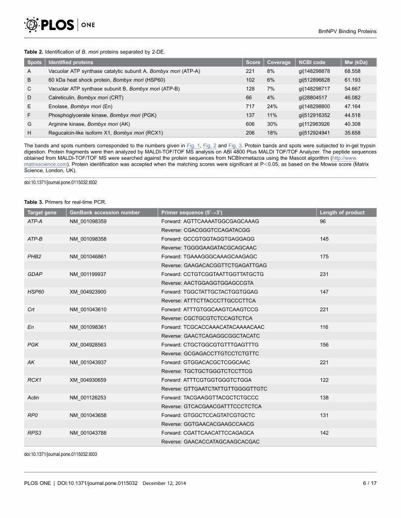
Table 2. Identification of B. mori proteins separated by 2-DE.
Spots Identified proteins Score Coverage NCBI code Mw (kDa)
A Vacuolar ATP synthase catalytic subunit A, Bombyx mori (ATP-A) 221 8% gi|148298878 68.558
B 60 kDa heat shock protein, Bombyx mori (HSP60) 102 6% gi|512896628 61.193
C Vacuolar ATP synthase subunit B, Bombyx mori (ATP-B) 128 7% gi|148298717 54.667
D Calreticulin, Bombyx mori (CRT) 66 4% gi|28804517 46.082
E Enolase, Bombyx mori (En) 717 24% gi|148298800 47.164
F Phosphoglycerate kinase, Bombyx mori (PGK) 137 11% gi|512916352 44.518
G Arginine kinase, Bombyx mori (AK) 606 30% gi|112983926 40.308
H Regucalcin-like isoform X1, Bombyx mori (RCX1) 206 18% gi|512924941 35.658
The bands and spots numbers corresponded to the numbers given in Fig. 1, Fig. 2 and Fig. 3. Protein bands and spots were subjected to in-gel trypsindigestion. Protein fragments were then analyzed by MALDI-TOF/TOF MS analysis on ABI 4800 Plus MALDI TOF/TOF Analyzer. The peptide sequencesobtained from MALDI-TOF/TOF MS were searched against the protein sequences from NCBInrmetazoa using the Mascot algorithm (http://www.matrixscience.com). Protein identification was accepted when the matching scores were significant at P,0.05, as based on the Mowse score (MatrixScience, London, UK).
doi:10.1371/journal.pone.0115032.t002
Table 3. Primers for real-time PCR.
Target gene GenBank accession number Primer sequence (5’R3’) Length of product
ATP-A NM_001098359 Forward: AGTTCAAAATGGCGAGCAAAG 96
Reverse: CGACGGGTCCAGATACGG
ATP-B NM_001098358 Forward: GCCGTGGTAGGTGAGGAGG 145
Reverse: TGGGGAAGATACGCAGCAAC
PHB2 NM_001046861 Forward: TGAAAGGGCAAAGCAAGAGC 175
Reverse: GAAGACACGGTTCTGAGATTGAG
GDAP NM_001199937 Forward: CCTGTCGGTAATTGGTTATGCTG 231
Reverse: AACTGGAGGTGGAGCCGTA
HSP60 XM_004923900 Forward: TGGCTATTGCTACTGGTGGAG 147
Reverse: ATTTCTTACCCTTGCCCTTCA
Crt NM_001043610 Forward: ATTTGTGGCAAGTCAAGTCCG 221
Reverse: CGCTGCGTCTCCAGTCTCA
En NM_001098361 Forward: TCGCACCAAACATACAAAACAAC 116
Reverse: GAACTCAGAGGCGGCTACATC
PGK XM_004928563 Forward: CTGCTGGCGTGTTTGAGTTTG 156
Reverse: GCGAGACCTTGTCCTCTGTTC
AK NM_001043937 Forward: GTGGACACGCTCGGCAAC 221
Reverse: TGCTGCTGGGTCTCCTTCG
RCX1 XM_004930659 Forward: ATTTCGTGGTGGGTCTGGA 122
Reverse: GTTGAATCTATTGTTGGGGTTGTC
Actin NM_001126253 Forward: TACGAAGGTTACGCTCTGCCC 138
Reverse: GTCACGAACGATTTCCCTCTCA
RP0 NM_001043658 Forward: GTGGCTCCAGTATCGTGCTC 131
Reverse: GGTGAACACGAAGCCAACG
RPS3 NM_001043788 Forward: CGATTCAACATTCCAGAGCA 142
Reverse: GAACACCATAGCAAGCACGAC
doi:10.1371/journal.pone.0115032.t003
BmNPV Binding Proteins
PLOS ONE | DOI:10.1371/journal.pone.0115032 December 12, 2014 6 / 17
Discussions
The molecular mechanisms of the interaction between B. mori and BmNPV are
still unclear at present, illuminating the mechanisms is of significant importance
not only in biological research but also in profit increase of sericulture. To this
end, we designed an experimental program to study the B. mori proteins involved
in the infection process by BmNPV.
It is well known that baculovirus budded viruses (BVs) enter host cell via
clathrin-mediated endocytosis [14]. Once inside the endosome, the virus encoded
gp64 protein can be enabled by the acidic environment to promote membrane
fusion of the virusand endosome to release the nucleocapsid into cytoplasm, thus
it is necessary forviral infection to occur [15–17]. Vacuolar ATP synthase (or V-
ATPase), originally identified in intracellular compartments, such as endosomes,
lysosomesand thecentral vacuoles of fungi and plants (hence the name), is the
most important transport protein for pH regulation of the intracellular
compartments [17–19]. Based on previous research, V-ATPase facilitated the
infection of baculovirus by acidifying the endosomes [15], which indicates that
BmNPV-susceptible strain may have a higher V-ATPase expression level. In our
study, the relative expression level of V-ATPase subunit A in P50 was about 8 fold
of that in A35 (Fig. 4A), and the down-regulation of V-ATPase subunit A at 48
hpi might be due to the activation of the host immune system to suppress the
infection. The relative expression level of V-ATPase subunit B was not
Fig. 4. Real-time PCR analysis of expression profiles of BmNPV binding proteins in B. mori midguts. Columns with different colors indicated theexperimental treats to the larvae. A to L refer to relative expression level of ATP-A, ATP-B, AK, RP0, Actin, En, PGK, GDAP, PHB2, HSP60, Crt, and RCX1respectively. Data were normalized usingBmrps3and represented as means¡standard errors of the means from three independent experiments. **Indicates statistical significance,0.01(ANOVA and LSD aposteriori test).
doi:10.1371/journal.pone.0115032.g004
BmNPV Binding Proteins
PLOS ONE | DOI:10.1371/journal.pone.0115032 December 12, 2014 7 / 17
significantly different in the three groups (Fig. 4B), indicating that subunit A was
more important in BmNPV infection.
Cytoskeleton is important for maintenance of cell shape, cell motility and
intracellular transport [21], and it is generally thought that viruses need
cytoskeleton during infection [20]. Here the interaction of several cytoskeleton-
associated proteins with BmNPV was determined, and they were actin, enolase
(En) and phosphoglycerate kinase (PGK). It is known that actin is the major
component of microfilaments [22], and En and PGK are enzymes involved in
glycolysis initially [23, 24]. Recent studies demonstrated that these two enzymes
had their roles in diseases and immune responses [25, 26]. Boone et al. reported
that PGK could bind to actin and plasminogen [27]. Plasminogen is the zymogen
of plasmin, a key component of the fibrinolytic system, in which plasminogen
plays two main functions of blood clot dissolution and extracellular matrix
disintegration [28]. En has been found on the tegument surface of
Schistosomabovis, where it acts as a plasminogen receptor to avoid blood clot
formation and facilitates the infection of the host [28, 29]. In our study, a
significant lower relative expression level of En in A35 (Fig. 4F) indicates its role
in virus infection.
In lieu of an adaptive immune system, apoptosis plays a central role in
regulating cellular or environmental stimuli in Lepidopteran insect cells during
virus infection [30], where larvae resist baculovirus infection by selective
apoptosis of the infected cells from midguts epithelium and by sloughing off the
infected cells [31]. Using virus overlay assay, five proteins including mitochon-
drial prohibitin complex protein 2 (PHB2), ganglioside induced differentiation
associated protein (GDAP), Calreticulin (CRT), Regucalcin-like isoform X1
(RCX1) and 60 kDa heat shock protein (HSP60) of B. mori were identified
interacting with BmNPV in our study. Previous researches have revealed their
functions in apoptosis. It was reported that PHB2 inhibited apoptosis and
regulated the mitochondrial morphology by interacting with HAX-1 [32, 33];
CRT could be induced to form a complex with gC1qR and prevented apoptosis
[34]; overexpression of regucalcin had a suppressive effect on cell death and
apoptosis induced by various factors in cloned normal rat kidney proximal
tubular epithelial NRK52E cells [35]. Based on their role of apoptosis suppression,
hosts needed to decrease the expression of these proteins to promote apoptosis
and prevent virus infection when exposed to virus, and this explained the notable
down-regulation of PHB2 and CRT at 48 hpi (Fig. 4I and K). Gao et al. reported
that higher expression level of BmGDAP in B. mori midguts during BmCPV
infection could activate the apoptosis and death programs of infected cells by
accelerating the mitochondrial division [36]. We determined the binding of
BmGDAP and BmNPV, and found its similar expression level in B. mori midguts
during BmNPV infection (Fig. 4H). Furthermore, we also observed BmNPV-
resistant strain had a much higher expression level of GDAP in midguts (Fig. 4H),
and this might explain the higher resistance against BmNPV than susceptible
strain. HSP60 is well known as a chaperon in that facilitates protein folding
directly [37]. Vabulas et al. reported that endocytosed HSP60 could use Toll-like
BmNPV Binding Proteins
PLOS ONE | DOI:10.1371/journal.pone.0115032 December 12, 2014 8 / 17
receptor 2 (TLR2) and TLR4 to activate the Toll/interleukin-1 receptor signaling
pathway in innate immune cells [38]. Furthermore, HSP60 was determined to
have the ability of binding to the 39-UTR of the Murine hepatitis virus genome
[39], and the contribution of HSP60 in anti-apoptotic program was confirmed in
tumors in vivo [40]. The notable higher expression level (approximate 300 fold) of
HSP60 in midguts of BmNPV-susceptible strain than in resistant strain (Fig. 4J)
indicates its important role in BmNPV infection. The regulation of the five
apoptosis-related genes (GDAP, PHB2, HSP60, CRT and RCX1) observed in this
study is in congruent with their potential roles in response to virus infection, for
example, up-regulation of the pro-apoptosis GDAP (Fig. 4H), while down-
regulation of the other three anti-apoptosis genes (Fig. 4I, J and K), which in turn
supported our hypothesized model below.
Ribosomes are important components of protein translation, and they contain
a structure called ribosomal stalk [41], which forms a lateral protrusion from the
ribosome and is composed of acidic proteins P0, P1, and P2 in eukaryotic cells
[42]. Ribosomal P0 protein (RP0) functions as a scaffold for the stalk structure by
interacting with 28S rRNA, and promotes Potato Virus A (PVA) infection of
Nicotiana benthamiana by regulating PVA RNA expression [43]. We demon-
strated the binding of BmNPV to RP0 of B. mori in vitro and determined its
relative expression level in BmNPV susceptible and resistant strains of B. mori
here, which (Fig. 4D) indicated potential role of RP0 in BmNPV infection
process.
In recent years, arginine kinase (AK) has been studied not only in insects [44]
but also in shrimps [45]. AK is a phosphagen kinase catalyzing the reversible
transfer of the phosphoryl group of ATP to arginine yielding ADP and
phosphoarginine, and has allergenic potential contributing to allergies against
silkworm [46]. Injection of AK into Litopenaeusvannamei increased the mortality
of shrimp infected with white spot syndrome virus (WSSV) indicating the
involvement of AK in WSSV infection [47]. Kang et al. made a conclusion that AK
was involved in the antiviral process of B. mori larvae against BmNPV infection by
determining the relative expression level in BmNPV susceptible and resistant
strains of B. mori [48]. Based on our results, the interaction between BmAK and
BmNPV in vitro was confirmed by virus overlay assay, and the significantly higher
expression of BmAK in susceptible strain (Fig. 4C) suggested its role in the
infection process of BmNPV.
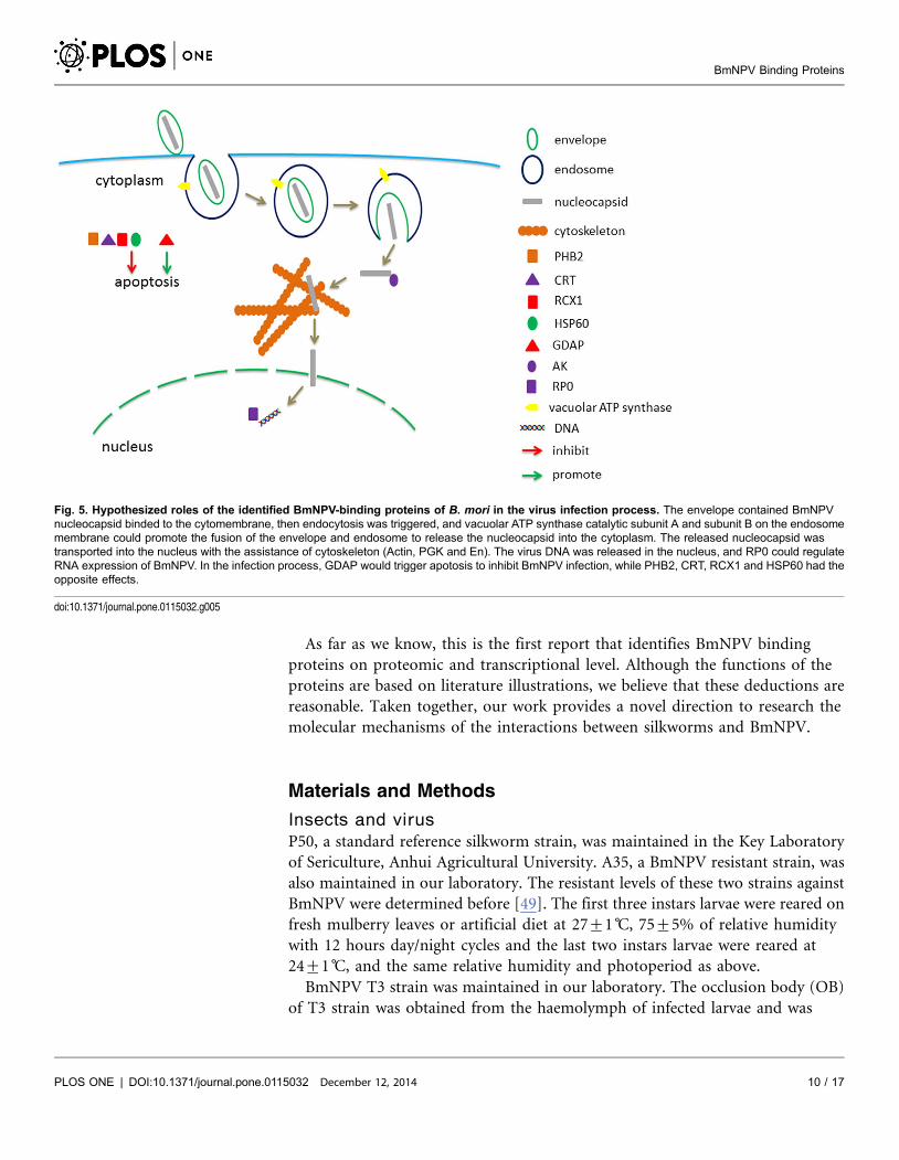
Based on the results and analysis above, we hypothesize the roles of these
binding proteins in the process of crossing BmNPV through silkworm midgut
cells. The BmNPV nucleocapsid contained envelope binds to the cytomembrane,
and endocytosis is triggered. Then ATP-A and ATP-B on the endosome
membrane promote the fusion of the envelope and endosome to release the
nucleocapsid into cytoplasm. The released nucleocapsid is transported into the
nucleus by the assistance of cytoskeleton (Actin, PGK and En). The virus DNA is
released in the nucleus, and RP0 could regulate RNA expression of BmNPV. In
the infection process, GDAP would trigger apoptosis to inhibit BmNPV infection,
while PHB2, CRT, RCX1 and HSP60 had the opposite effects (Fig. 5).
BmNPV Binding Proteins
PLOS ONE | DOI:10.1371/journal.pone.0115032 December 12, 2014 9 / 17
As far as we know, this is the first report that identifies BmNPV binding
proteins on proteomic and transcriptional level. Although the functions of the
proteins are based on literature illustrations, we believe that these deductions are
reasonable. Taken together, our work provides a novel direction to research the
molecular mechanisms of the interactions between silkworms and BmNPV.
Materials and Methods
Insects and virus
P50, a standard reference silkworm strain, was maintained in the Key Laboratory
of Sericulture, Anhui Agricultural University. A35, a BmNPV resistant strain, was
also maintained in our laboratory. The resistant levels of these two strains against
BmNPV were determined before [49]. The first three instars larvae were reared on
fresh mulberry leaves or artificial diet at 27¡1 C, 75¡5% of relative humidity
with 12 hours day/night cycles and the last two instars larvae were reared at
24¡1 C, and the same relative humidity and photoperiod as above.
BmNPV T3 strain was maintained in our laboratory. The occlusion body (OB)
of T3 strain was obtained from the haemolymph of infected larvae and was
Fig. 5. Hypothesized roles of the identified BmNPV-binding proteins of B. mori in the virus infection process. The envelope contained BmNPVnucleocapsid binded to the cytomembrane, then endocytosis was triggered, and vacuolar ATP synthase catalytic subunit A and subunit B on the endosomemembrane could promote the fusion of the envelope and endosome to release the nucleocapsid into the cytoplasm. The released nucleocapsid wastransported into the nucleus with the assistance of cytoskeleton (Actin, PGK and En). The virus DNA was released in the nucleus, and RP0 could regulateRNA expression of BmNPV. In the infection process, GDAP would trigger apotosis to inhibit BmNPV infection, while PHB2, CRT, RCX1 and HSP60 had theopposite effects.
doi:10.1371/journal.pone.0115032.g005
BmNPV Binding Proteins
PLOS ONE | DOI:10.1371/journal.pone.0115032 December 12, 2014 10 / 17
purified by repeated and differential centrifugation following the previously
published protocol [50], and the concentration of the virus (OB/ml) was
determined using haemocytometer. The budded virus (BV) was supplied by
Jiangsu University.
Extraction of total proteins
Fifth instar larvae were dissected at the third day after molting, and the midguts
were frozen with liquid nitrogen and then pulverized. Total protein extraction was
performed as described by the Instruction Manual of ReadyPrep Protein
Extraction Kit (Bio-Rad). Briefly, 100 mg pulverized midgut was added into a
2 mL microcentrifuge tube containing 1 mL of 2-D Rehydration/Sample Buffer 1
with 10 mL of 200 mM ReadyPrep tributylphosphine reducing agent, 10 mL of
40% (w/v) ampholyte (Bio-Rad, Bio-Lyte 3/10) and 10 mL of 100 mM
phenylmethanesulfonyl fluoride (PMSF), and the sample was sonicated until lysis
was complete on ice. After a centrifugation(Hettichi MIKRO 220R, 1195A) at
16,0006g for 30 min at 4 C the supernatant was transferred to a clean tube. The
protein sample was characterized by 12% SDS-PAGE, and the concentration was
determined with Bradford using BSA as standard.
Extraction of lipid-associated membrane proteins
Lipid-associated membrane proteins from larvae midgut were isolated as
described by Dickerson et al. [51] with some modifications. Briefly, 100 mg of
pulverized midgut was added into a 2 mL microcentrifuge tube containing 1 mL
of ice-cold Triton X-114 extraction buffer[2.0%(v/v) triton X-114, 300 mM NaCl,
20 mM Tris-HCl (pH7.5), 1 mM PMSF], and the sample was sonicated until lysis
was complete on ice. After a centrifugation at 16,0006g for 10 min at 4 C, the
supernatant was transferred into a clean tube on ice. The supernatant was
incubated in a 30 C water-bath for 5 min and was layered onto a sucrose cushion
of equal volume[6.0% (w/v) sucrose, 150 mM NaCl, 0.06%(v/v) Triton X-114,
10 mM Tris-HCl (pH 7.5)] in a clean tube, and warmed in 30 C water-bath for
5 min followed by a centrifugation at 3006g for 3 min at room temperature
(RT). The aqueous phase was transferred to another tube and treated again as
described above, and the remaining detergent phase was saved for later use. Both
the lipoproteins and hydrophilic proteins were precipitated by adding nine times
volume of ice-cold acetone. The precipitated proteins were suspended in 10 mM
Tris-HCl (pH7.5) for concentration determinations with Bradford.
Sodium dodecyl sulfatepolyacrylamide gel electrophoresis(SDS-
PAGE) and two-dimensional electrophoresis (2-DE)
For SDS-PAGE, 56protein loading buffer (50 mM Tris-HCl pH 8.0,
250 mMDTT, 5% SDS, 50% Glycerol, 0.04% Bromophenol Blue) was added to
the total protein sample and the purified BmNPV particles. Samples were boiled
BmNPV Binding Proteins
PLOS ONE | DOI:10.1371/journal.pone.0115032 December 12, 2014 11 / 17
for 10 min and loaded onto a sodium dodecyl sulfate (SDS) polyacrylamide gel.
Electrophoresis was performed in the Bio-Rad Mini-protean Tetra system. In the
procedure of 2-DE, the first step, isoelectric focusing (IEF), was performed in Bio-
Rad protean IEF cell. Briefly, samples (125 mL/strip) were loaded on ReadyStrip
IPG strips (7 cm) with linear pH 3–10 gradients. After active rehydration (50V)
for 14 h, IEF was performed following a voltage step-gradient (250V linear for
1 h, 500V rapid for 1 h, 4000V linear for 3 h, and 4000V rapid for 20,000 V?h) at
20 C, with a maximum current of 50 mA/strip. Before the second step (SDS-
PAGE), the IPG strips were first equilibrated for 15 min in a solution containing
6 M urea, 2% (w/v) SDS, 0.375 M Tris-HCl pH 8.8, 20% (v/v) glycerol, and 2%
(w/v) DTT, and then for 15 min in the same solution, substituting DTT with
2.5% (w/v) iodoacetamide. SDS-PAGE was carried out in 1 mm-thick 12% SDS
polyacrylamide gels. Gels were run under a constant current of 20 mA until the
bromphenol blue dye front migrated to the bottom.
Virus overlay assay (far-western blot)
After electrophoresis, proteins in a gel were transferred onto a polyvinylidenedi-
fluoride (PVDF) membrane (Millipore) following a Biometre wet blotting
procedure (150 mA for 120 min). To locate potential virus-binding proteins from
SDS polyacrylamide gels or 2-DE gels for identification, the parallel gel was
stained with Coomassie brilliant blue G-250.
The far-Western blot was performed as described by Wu et al. [52], with some
modifications. Proteins on the membrane were denatured and renatured in AC
buffer (100 mM NaCl, 20 mMTris-HCl pH 7.6, 0.5 mM EDTA, 10% glycerol,
0.1% Tween-20, 2% no-fat milk, 1 mM DTT) by gradually reducing the
guanidine-HCl concentration. Briefly, the membrane was incubated in the AC
buffer containing 6 M guanidine-HCl for 30 min at RT, and then washed with the
AC buffer containing 3 M guanidine-HCl for 30 min at RT. This is followed by
washing with the AC buffer containing 0.1 M and no guanidine-HCl AC buffer at
4 C, for 30 min and 1 h, respectively, and then blocked for 30 min at RT in
blocking buffer [PBST (137 mM NaCl, 2.7 mM KCl, 10 mM Na2HPO4, 2 mM
KH2PO4, pH7.5, 0.05% (v/v) Tween-20), 5% (w/v) no-fat milk]. The membrane
was subsequently incubated overnight in binding buffer (100 mM NaCl, 20 mM
Tris-HCl pH 7.5, 0.5 mM EDTA, 10% (v/v) glycerol, 2% (w/v) no-fat milk, and
1 mM DTT) containing purified BmNPV particles (5 mg/mL). After washed three
times for 10 min each in PBST, the membrane was incubated with monoclonal
antibodies (MAbs) against baculovirus gp64 (Santa Cruz) with a dilution of 1:500
in blocking buffer for 3 hours at RT. After washing as above, antigen-antibody
complexes were detected with a horseradish peroxidase (HRP)-conjugated goat
anti-mouse secondary antibody (1:5000 dilution) in blocking buffer for 1.5 hours
at RT. After another series of washes, immobilized conjugates on the membrane
were visualized in HRP substrate solution (Tiangen). For the negative control, the
membrane was incubated overnight in binding buffer without BmNPV particles,
followed by antibody incubation.
BmNPV Binding Proteins
PLOS ONE | DOI:10.1371/journal.pone.0115032 December 12, 2014 12 / 17
Protein identification
Comparisons between the stained gels and the results of far-western blot
experiments on the membrane allowed the unambiguous selection of protein
bands from SDS polyacrylamide gels or spots from 2-DE gels for MALDI-TOF/
TOF MS analysis on ABI 4800 Plus MALDI TOF/TOF Analyzer. Selected protein
bands or spots were cut out from gels, and the pieces were destained with 100 mM
NH4HCO3 in 30% (v/v) acetonitrile for 15 min. The liquid phase was removed,
and the gel pieces were completely lyophilized, followed by swelling in a digestion
buffer containing 10ng/ml trypsin (Promega) overnight at 37 C. The digested
peptides were lyophilized and dissolved in 20% (v/v) acetonitrile. One microliter
of the sample was added to the target site and taken into the analyzer after dried
naturally. The peptide sequences obtained from MALDI-TOF/TOF MS were
searched against the protein sequences from NCBInrmetazoa using the Mascot
algorithm (http://www.matrixscience.com). The search parameters were set as
follows: Type of search, MS/MS Ion Search; enzyme, trypsin; fixed modifications,
carbamidomethyl (C); variable modifications, Acetyl (Protein N-term),
Deamidated (NQ), Dioxidation (W), Oxidation (M); Mass values, Monoisotopic;
Protein Mass, Unrestricted; mass tolerances for MS/MS were 100 ppm and 0.5 Da;
max missed cleavages, one. Protein identification was accepted when the matching
scores were significant at P,0.05, as based on the Mowse score (Matrix Science,
London, UK).
RNA isolation and cDNA synthesis
Based on the reported time course of viral proliferation and expression of NPV
responsive genes in fat body and haemocytes of susceptible and resistant strains
[53], silkworm from the infected and control groups were dissected at 48 hour
post inoculation (h.p.i), and the midguts were frozen with liquid nitrogen and
then pulverized. One hundred milligram of midgut was added into a RNAase free
microcentrifuge tube containing 1000 mL TRIZOL Reagent (Life Technologies),
then homogenized with a pellet pestle motor. Total RNA was extracted from the
midguts using TRIZOL Reagent according to the manufacturer’s instructions of
Invitrogen. The ratios of A260/280 and the concentrations for the RNA samples
were determined by NanoDrop 2000 spectrophotometer (Thermo Scientific).
Total RNA samples were treated with RT reagent kit with gDNA Eraser
(TaKaRa) to remove genomic DNA and the first strand cDNA was synthesized
according to the manufacturer’s instructions. Briefly, 2.0 mL of 56gDNA Eraser
buffer, 1.0 mL of gDNA Eraser, and 1.0 mg of total RNA were mixed in a 200 mL
PCR tube and added up RNase Free dH2O to 10 mL, and then incubated at room
temperature for 5 minutes. Four micro liter of 56PrimeScript buffer, 1.0 mL of
PrimeScript RT Enzyme Mix I, and 1.0 mL of RT Primer Mix were added to the
previous tube, then added up to 20 mL with RNase Free dH2O. The mix was
incubated at 37 C for 15 minutes followed by 85 C for 5 seconds and stored at
220 C for later use.
BmNPV Binding Proteins
PLOS ONE | DOI:10.1371/journal.pone.0115032 December 12, 2014 13 / 17
qPCR analysis
qPCR was carried out in a 25 mL reaction mix containing 12.5 mL of SYBR Premix
Ex Taq (TaKaRa), 1 mL of 1:5 diluted cDNA template, 1 mL of each of the primers
(10 mM) and 9.5 mL ddH2O. The thermal cycling profile consisted of initial
denaturation at 95 C for 30 s and 40 cycles at 95 C for 5 s, 60 C for 30 s, and 72 C
for 20 s. Relative expression levels were calculated using the 22DDCt method
where DDCt5DCt sample2DCt reference following the previously published
protocol [54]. PCR reactions were performed on Bio-Rad CFX96TM Real-Time
System using SYBR Green to detect dsDNA synthesis. PCR amplification was
performed in triplicate wells. The B. mori ribosomal protein s3 gene (Bmrps3) was
set as an internal control. Data were normalized using Bmrps3, and the statistical
analysis was conducted using ANOVA and LSD aposteriori test (P,0.01).
Supporting Information
S1 Figure. Details of ATP-A (band) identified by MALDI-TOF/TOF MS.
doi:10.1371/journal.pone.0115032.s001 (TIF)
S2 Figure. Details of Actin identified by MALDI-TOF/TOF MS.
doi:10.1371/journal.pone.0115032.s002 (TIF)
S3 Figure. Details of RP0 identified by MALDI-TOF/TOF MS.
doi:10.1371/journal.pone.0115032.s003 (TIF)
S4 Figure. Details of PHB2 identified by MALDI-TOF/TOF MS.
doi:10.1371/journal.pone.0115032.s004 (TIF)
S5 Figure. Details of GDAP identified by MALDI-TOF/TOF MS.
doi:10.1371/journal.pone.0115032.s005 (TIF)
S6 Figure. Details of ATP-A (spot) identified by MALDI-TOF/TOF MS.
doi:10.1371/journal.pone.0115032.s006 (TIF)
S7 Figure. Details of HSP60 identified by MALDI-TOF/TOF MS.
doi:10.1371/journal.pone.0115032.s007 (TIF)
S8 Figure. Details of ATP-B identified by MALDI-TOF/TOF MS.
doi:10.1371/journal.pone.0115032.s008 (TIF)
S9 Figure. Details of Crt identified by MALDI-TOF/TOF MS.
doi:10.1371/journal.pone.0115032.s009 (TIF)
S10 Figure. Details of En identified by MALDI-TOF/TOF MS.
doi:10.1371/journal.pone.0115032.s010 (TIF)
S11 Figure. Details of PGK identified by MALDI-TOF/TOF MS.
doi:10.1371/journal.pone.0115032.s011 (TIF)
S12 Figure. Details of AK identified by MALDI-TOF/TOF MS.
doi:10.1371/journal.pone.0115032.s012 (TIF)
S13 Figure. Details of RCX1 identified by MALDI-TOF/TOF MS.
doi:10.1371/journal.pone.0115032.s013 (TIF)
BmNPV Binding Proteins
PLOS ONE | DOI:10.1371/journal.pone.0115032 December 12, 2014 14 / 17
Author Contributions
Conceived and designed the experiments: JPX. Performed the experiments: YC
XYW. Analyzed the data: YC XYW HH JPX. Contributed reagents/materials/
analysis tools: JPX. Wrote the paper: YC HH NK JPX.
References
1. Goldsmith MR, Shimada T, Abe H (2005) The genetics and genomics of the silkworm, Bombyx mori.Annu Rev Entomol 50: 71–100.
2. Miao XX, Xu SJ, Li MH, Li MW, Huang JH, et al. (2005) Simple sequence repeat-based consensuslinkage map of Bombyx mori. Proc Natl Acad Sci U S A 102: 16303–16308.
3. Maeda S (1989) Expression of foreign genes in insects using baculovirus vectors. Annu Rev Entomol34: 351–372.
4. Rohrmann GF (1992) Baculovirus structural proteins. J Gen Virol 73 (4): 749–761.
5. Song JJ, Wang RR, Deng F, Wang HL, Hu ZH (2008) Functional studies of per os infectivity factors ofHelicoverpa armigera single nucleocapsid nucleopolyhedrovirus. J Gen Virol 89: 2331–2338.
6. Nakazawa H, Tsuneishi E, Ponnuvel KM, Furukawa S, Asaoka A, et al. (2004) Antiviral activity of aserine protease from the digestive juice of Bombyx mori larvae against nucleopolyhedrovirus. Virology321: 154–162.
7. Ponnuvel KM, Nakazawa H, Furukawa S, Asaoka A, Ishibashi J, et al. (2003) A Lipase Isolated fromthe Silkworm Bombyx mori Shows Antiviral Activity against Nucleopolyhedrovirus. J Virol 77: 10725–10729.
8. Ponnuvel KM, Nithya K, Sirigineedi S, Awasthi AK (2012) In vitro antiviral activity of an alkalinetrypsin from the digestive juice of Bombyx mori larvae against nucleopolyhedrovirus. Arch InsectBiochem 81: 90–104.
9. Xu JP, Chen KP, Liu MH, Yao Q, Gao GT, et al. (2008) Identification and characterization of Bms3a inBombyx mori L. Afr J Biotechnol 7: 3424–3430.
10. Li S, Xiong RY, Wang XF, Zhou YJ (2011) Five Proteins of Laodelphax striatellus Are PotentiallyInvolved in the Interactions between Rice Stripe Virus and Vector. PLoS ONE 6: e26585.
11. van den Heuvel JF, Verbeek M, van der Wilk F (1994) Endosymbiotic bacteria associated withcirculative transmission of potato leafroll virus by Myzus persicae. J Gen Virol 75 (10): 2559–2565.
12. Kikkert M, Meurs C, van de Wetering F, Dorfmuller S, Peters D, et al. (1998) Binding of TomatoSpotted Wilt Virus to a 94-kDa Thrips Protein. Phytopathology 88: 63–69.
13. Bandla MD, Campbell LR, Ullman DE, Sherwood JL. (1998) Interaction of Tomato Spotted WiltTospovirus (TSWV) Glycoproteins with a Thrips Midgut Protein, a Potential Cellular Receptor for TSWV.Phytopathology 88: 98–104.
14. Long G, Pan XY, Kormelink R, Vlak JM (2006) Functional entry of baculovirus into insect andmammalian cells is dependent on clathrin-mediated endocytosis. J Virol 80: 8830–8833.
15. Blissard GW, Wenz JR (1992) Baculovirus gp64 envelope glycoprotein is sufficient to mediate pH-dependent membrane fusion. J Virol 66: 6829–6835.
16. Kingsley DH, Behbahani A, Rashtian A, Blissard GW, Zimmerberg J (1999) A discrete stage ofbaculovirus GP64-mediated membrane fusion. Mol Biol Cell 10: 4191–4200.
17. Hinton A, Bond S, Forgac M (2009) V-ATPase functions in normal and disease processes. PflugersArch 457: 589–598.
18. Beyenbach KW, Wieczorek H (2006) The V-type H+ ATPase: molecular structure and function,physiological roles and regulation. J Exp Biol 209: 577–589.
19. Forgac M (2007) Vacuolar ATPases: rotary proton pumps in physiology and pathophysiology. Nat RevMol Cell Biol 8: 917–929.
BmNPV Binding Proteins
PLOS ONE | DOI:10.1371/journal.pone.0115032 December 12, 2014 15 / 17
20. Radtke K, Dohner K, Sodeik B (2006) Viral interactions with the cytoskeleton: a hitchhiker’s guide tothe cell. Cell Microbiol 8: 387–400.
21. Matthews JD, Morgan R, Sleigher C, Frey TK (2013) Do viruses require the cytoskeleton? Virol J 10:121.
22. Pollard TD, Cooper JA (2009) Actin, a central player in cell shape and movement. Science 326: 1208–1212.
23. Stern H, Mirsky AE (1952) The isolation of wheat germ nuclei and some aspects of their glycolyticmetabolism. Journal Gen Physiol 36: 181–200.
24. Blanchaer MC, Williams HR, Weiss P (1955) Presence of the glycolytic enzymes D-glyceraldehyde-3-phosphate dehydrogenase, phosphoglycerate kinase and triosephosphate isomerase in hemolysates ofpreserved blood. Am J Physiol 181: 602–608.
25. Pancholi V (2001) Multifunctional alpha-enolase: its role in diseases. Cell Mol Life Sci 58: 902–920.
26. Wesolowska A, Jaros S, Norbury LJ, Jaros D, Zygner W, et al. (2013) Microarray analysis of ratimmune responses to liver fluke infection following vaccination with Fasciola hepatica phosphoglyceratekinase. Exp Parasitol 134: 33–38.
27. Boone TJ, Burnham CAD, Tyrrell GJ (2011) Binding of group B streptococcal phosphoglycerate kinaseto plasminogen and actin. Microb Pathogenesis 51: 255–261.
28. de la Torre-Escudero E, Manzano-Roman R, Perez-Sanchez R, Siles-Lucas M, Oleaga A (2010)Cloning and characterization of a plasminogen-binding surface-associated enolase from Schistosomabovis. Vet Parasitol 173: 76–84.
29. Vanegas G, Quinones W, Carrasco-Lopez C, Concepcion JL, Albericio F, et al. (2007) Enolase as aplasminogen binding protein in Leishmania mexicana. Parasitol Res 101: 1511–1516.
30. Wu Y, Wu YJ, Hui T, Wu HL, Wu Y, et al. (2013) Reaper homologue IBM1 in silkworm Bombyx moriinduces apoptosis upon baculovirus infection. Febs Lett 587: 600–606.
31. Narayanan K (2004) Insect defence: its impact on microbial control of insect pests. Curr Sci India 86:800–814.
32. Suzuki Y, Demoliere C, Kitamura D, Takeshita H, Deuschle U, et al. (1997) HAX-1, a novelintracellular protein, localized on mitochondria, directly associates with HS1, a substrate of Src familytyrosine kinases. J Immunol 158: 2736–2744.
33. Cilenti L, Soundarapandian MM, Kyriazis GA, Stratico V, Singh S, et al. (2004) Regulation of HAX-1anti-apoptotic protein by Omi/HtrA2 protease during cell death. J Biol Chem 279: 50295–50301.
34. Watthanasurorot A, Jiravanichpaisal P, Soderhall K, Soderhall I (2013) A calreticulin/gC1qRcomplex prevents cells from dying: a conserved mechanism from arthropods to humans. J Mol Cell Biol5: 120–131.
35. Nakagawa T, Yamaguchi M (2005) Overexpression of regucalcin suppresses apoptotic cell death incloned normal rat kidney proximal tubular epithelial NRK52E cells: change in apoptosis-related geneexpression. J Cell Biochem 96: 1274–1285.
36. Gao K, Deng X, Qian H, Wu P, Qin G, et al. (2012) Cloning, characterization, and expression analysis ofa novel BmGDAP1 gene from silkworm, Bombyx mori, involved in cytoplasmic polyhedrosis virusinfection. Gene 497: 208–213.
37. Hartl FU, Hayer-Hartl M (2002) Protein folding - Molecular chaperones in the cytosol: from nascentchain to folded protein. Science 295: 1852–1858.
38. Vabulas RM, Ahmad-Nejad P, da Costa C, Miethke T, Kirschning CJ, et al. (2001) EndocytosedHSP60s use toll-like receptor 2 (TLR2) and TLR4 to activate the Toll/interleukin-1 receptor signalingpathway in innate immune cells. J Biol Chem 276: 31332–31339.
39. Nanda SK, Johnson RF, Liu Q, Leibowitz JL (2004) Mitochondrial HSP70, HSP40, and HSP60 bind tothe 3’ untranslated region of the Murine hepatitis virus genome. Arch Virol 149: 93–111.
40. Ghosh JC, Dohi T, Kang BH, Altieri DC (2008) Hsp60 regulation of tumor cell apoptosis. J Biol Chem283: 5188–5194.
41. Gonzalo P, Reboud JP (2003) The puzzling lateral flexible stalk of the ribosome. Biol Cell 95: 179–193.
BmNPV Binding Proteins
PLOS ONE | DOI:10.1371/journal.pone.0115032 December 12, 2014 16 / 17
42. Wool IG, Chan YL, Gluck A, Suzuki K (1991) The primary structure of rat ribosomal proteins P0, P1,and P2 and a proposal for a uniform nomenclature for mammalian and yeast ribosomal proteins.Biochimie 73: 861–870.
43. Hafren A, Eskelin K, Makinen K (2013) Ribosomal protein P0 promotes Potato virus A infection andfunctions in viral translation together with VPg and eIF(iso)4E. J Virol 87: 4302–4312.
44. Wang HC, Zhang L, Zhang L, Lin Q, Liu NN (2009) Arginine kinase: Differentiation of gene expressionand protein activity in the red imported fire ant, Solenopsis invicta. Gene 430: 38–43.
45. Rattanarojpong T, Wang HC, Lo CF, Flegel TW (2007) Analysis of differently expressed proteins andtranscripts in gills of Penaeus vannamei after yellow head virus infection. Proteomics 7: 3809–3814.
46. Liu ZG, Xia LX, Wu YL, Xia QY, Chen JJ, et al. (2009) Identification and Characterization of an ArginineKinase as a Major Allergen from Silkworm (Bombyx mori) Larvae. Int Arch Allergy Imm 150: 8–14.
47. Ma FF, Liu QH, Guan GK, Li C, Huang J (2014) Arginine kinase of Litopenaeus vannamei involved inwhite spot syndrome virus infection. Gene 539: 99–106.
48. Kang LQ, Shi HS, Liu XY, Zhang CY, Yao Q, et al. (2011) Arginine kinase is highly expressed in aresistant strain of silkworm (Bombyx mori, Lepidoptera): Implication of its role in resistance to Bombyxmori nucleopolyhedrovirus. Comp Biochem Phys 158: 230–234.
49. Cheng Y, Wang XY, Du C, Gao J, Xu JP (2014) Expression analysis of several antiviral related genes toBmNPV in different resistant strains of silkworm, Bombyx mori. J Insect Sci 14(76):1–9.
50. Rahman MM, Gopinathan KP (2004) Systemic and in vitro infection process of Bombyx morinucleopolyhedrovirus. Virus Res 101: 109–118.
51. Dickerson HW, Clark TG, Findly RC (1989) Icthyophthirius multifiliis has membrane-associatedimmobilization antigens. J Protozool 36: 159–164.
52. Wu Y, Li Q, Chen XZ (2007) Detecting protein-protein interactions by Far western blotting. Nat Protoc 2:3278–3284.
53. Bao YY, Lv ZY, Liu ZB, Xue J, Xu YP, et al. (2010) Comparative analysis of Bombyx morinucleopolyhedrovirus responsive genes in fat body and haemocyte of B. mori resistant and susceptiblestrains. Insect Mol Biol 19: 347–358.
54. Livak KJ, Schmittgen TD (2001) Analysis of relative gene expression data using real-time quantitativePCR and the 2-DDCT method. Methods 25: 402–408.
BmNPV Binding Proteins
PLOS ONE | DOI:10.1371/journal.pone.0115032 December 12, 2014 17 / 17
Related Documents