A HUPO test sample study reveals common problems in mass spectrometry–based proteomics Alexander W Bell 1 , Eric W Deutsch 2 , Catherine E Au 1 , Robert E Kearney 3 , Ron Beavis 4 , Salvatore Sechi 5 , Tommy Nilsson 6 , John J M Bergeron 1 & HUPO Test Sample Working Group 7 We performed a test sample study to try to identify errors leading to irreproducibility, including incompleteness of peptide sampling, in liquid chromatography–mass spectrometry–based proteomics. We distributed an equimolar test sample, comprising 20 highly purified recombinant human proteins, to 27 laboratories. Each protein contained one or more unique tryptic peptides of 1,250 Da to test for ion selection and sampling in the mass spectrometer. Of the 27 labs, members of only 7 labs initially reported all 20 proteins correctly, and members of only 1 lab reported all tryptic peptides of 1,250 Da. Centralized analysis of the raw data, however, revealed that all 20 proteins and most of the 1,250 Da peptides had been detected in all 27 labs. Our centralized analysis determined missed identifications (false negatives), environmental contamination, database matching and curation of protein identifications as sources of problems. Improved search engines and databases are needed for mass spectrometry–based proteomics. Liquid chromatography–mass spectrometry (LC-MS) has become the most popular technique for proteomics analysis. In this strategy, proteins of a sample are typically separated by PAGE and then digested with trypsin. After extraction from the gel, peptides are separated by liquid chromatography and upon elution are ionized via electrospray into the mass spectrometer for char- acterization by mass analysis. The mass spectrometer subsequently selects peptides for fragmentation to yield mass values that are then used to identify the peptide and the corresponding protein by searching sequence databases. This technique, termed tandem mass spectrometry (MS), is repeated to continuously select ionized peptides from the liquid chromatography column. Depending on protein abundance and complexity, the mass spectrometer type and its setup, up to about 15,000 peptides and up to about 4,000 proteins can be identified in a single experiment 1 . Despite the high mass accuracy of modern mass spectrometers, the general perception of the reliability of MS-based proteomics is that it is low. Previous test sample studies have demonstrated that there is both a lack of reproducibility between different laboratories as well as a general inability to identify purified proteins in samples of low complexity 2 (http://www.abrf.org/Research Groups/ProteomicsStandardsResearchGroup/EPosters/ABRFsPRG Study2006poster.pdf). This is in part due to the stochastic nature of peptide sampling by the mass spectrometer and the inherent bias toward peptides of higher concentrations, which also confounds the statistical challenges and pitfalls associated with MS-based analyses, particularly when samples are rich in protein complexity. Protein solubilization, protein separation, protease digestion, peptide separa- tion and peptide selection, all involve steps and protocols that vary greatly among labs, and different commercially available tandem mass spectrometers have different mass accuracies and different rates of peptide selection for fragmentation. The use of different search engines to decode tandem mass spectra and match them to databases of theoretical tryptic peptides is also a source of variability 3 , because of differences in the search engines themselves as well as different false discovery rates 4,5 . Furthermore, the matching of high-quality tandem mass spectra to different databases may lead to irreproducibility as protein databases vary greatly in terms of their curation, completeness and comprehensiveness 6–8 . Despite variability in instruments, search engines and databases, the high mass accuracy of modern mass spectrometers 9 should assure a 100% success rate of protein identi- fication for those tryptic peptides that readily ionize and for which high-quality tandem mass spectra can be obtained. Prior work in analytical chemistry and genomics 10–14 has demonstrated the benefits of standardized test sample efforts for testing the reproducibility of technology platforms. To address the question of reproducibility in LC-MS–based proteomics 15 , the Human Proteome Organization (HUPO) created a test sample working group to carry out a controlled study involving 27 different labs. We produced a test sample made up of 20 human proteins of high purity and at equimolar ratios. To test for any potential stochastic bottleneck as a consequence of current data-dependent acquisition methods 16 , all 20 proteins were selected to contain at least one unique tryptic peptide of 1,250 ± 5 Da each with a different amino acid sequence. The primary task given to members © 2009 Nature America, Inc. All rights reserved. RECEIVED 18 DECEMBER 2008; ACCEPTED 3 APRIL 2009; PUBLISHED ONLINE 17 MAY 2009; CORRECTED AFTER PRINT 29 JUNE 2009; DOI:10.1038/NMETH.1333 1 Department of Anatomy and Cell Biology, McGill University, Montreal, Canada. 2 The Institute for Systems Biology, Seattle, Washington, USA. 3 Department of Biomedical Engineering, McGill University, Montreal, Canada. 4 Biomedical Research Centre, University of British Columbia, Vancouver, Canada. 5 Division Diabetes, Endocrinology and Metabolic Diseases, National Institute of Diabetes and Digestive and Kidney Diseases, National Institutes of Health, Bethesda, Maryland, USA. 6 The Research Institute of the McGill University Health Centre and the Department of Medicine, McGill University, Montreal, Canada. 7 A full list of authors appears at the end of this paper. Correspondence should be addressed to J.J.M.B. ([email protected]). NATURE METHODS | VOL.6 NO.6 | JUNE 2009 | 423 ANALYSIS

Welcome message from author
This document is posted to help you gain knowledge. Please leave a comment to let me know what you think about it! Share it to your friends and learn new things together.
Transcript
A HUPO test sample study reveals common problemsin mass spectrometry–based proteomicsAlexander W Bell1, Eric W Deutsch2, Catherine E Au1, Robert E Kearney3, Ron Beavis4, Salvatore Sechi5,Tommy Nilsson6, John J M Bergeron1 & HUPO Test Sample Working Group7
We performed a test sample study to try to identify errors
leading to irreproducibility, including incompleteness of peptide
sampling, in liquid chromatography–mass spectrometry–based
proteomics. We distributed an equimolar test sample, comprising
20 highly purified recombinant human proteins, to 27
laboratories. Each protein contained one or more unique tryptic
peptides of 1,250 Da to test for ion selection and sampling in
the mass spectrometer. Of the 27 labs, members of only 7 labs
initially reported all 20 proteins correctly, and members of
only 1 lab reported all tryptic peptides of 1,250 Da. Centralized
analysis of the raw data, however, revealed that all 20 proteins
and most of the 1,250 Da peptides had been detected in all 27
labs. Our centralized analysis determined missed identifications
(false negatives), environmental contamination, database
matching and curation of protein identifications as sources of
problems. Improved search engines and databases are needed for
mass spectrometry–based proteomics.
Liquid chromatography–mass spectrometry (LC-MS) has becomethe most popular technique for proteomics analysis. In thisstrategy, proteins of a sample are typically separated by PAGEand then digested with trypsin. After extraction from the gel,peptides are separated by liquid chromatography and upon elutionare ionized via electrospray into the mass spectrometer for char-acterization by mass analysis. The mass spectrometer subsequentlyselects peptides for fragmentation to yield mass values that are thenused to identify the peptide and the corresponding protein bysearching sequence databases. This technique, termed tandem massspectrometry (MS), is repeated to continuously select ionizedpeptides from the liquid chromatography column. Depending onprotein abundance and complexity, the mass spectrometer type andits setup, up to about 15,000 peptides and up to about 4,000proteins can be identified in a single experiment1.
Despite the high mass accuracy of modern mass spectrometers,the general perception of the reliability of MS-based proteomics isthat it is low. Previous test sample studies have demonstrated
that there is both a lack of reproducibility between differentlaboratories as well as a general inability to identify purifiedproteins in samples of low complexity2 (http://www.abrf.org/ResearchGroups/ProteomicsStandardsResearchGroup/EPosters/ABRFsPRGStudy2006poster.pdf). This is in part due to the stochastic nature ofpeptide sampling by the mass spectrometer and the inherent biastoward peptides of higher concentrations, which also confounds thestatistical challenges and pitfalls associated with MS-based analyses,particularly when samples are rich in protein complexity. Proteinsolubilization, protein separation, protease digestion, peptide separa-tion and peptide selection, all involve steps and protocols that varygreatly among labs, and different commercially available tandem massspectrometers have different mass accuracies and different rates ofpeptide selection for fragmentation. The use of different searchengines to decode tandem mass spectra and match them to databasesof theoretical tryptic peptides is also a source of variability3, because ofdifferences in the search engines themselves as well as different falsediscovery rates4,5. Furthermore, the matching of high-quality tandemmass spectra to different databases may lead to irreproducibility asprotein databases vary greatly in terms of their curation, completenessand comprehensiveness6–8. Despite variability in instruments, searchengines and databases, the high mass accuracy of modern massspectrometers9 should assure a 100% success rate of protein identi-fication for those tryptic peptides that readily ionize and for whichhigh-quality tandem mass spectra can be obtained.
Prior work in analytical chemistry and genomics10–14 hasdemonstrated the benefits of standardized test sample efforts fortesting the reproducibility of technology platforms. To address thequestion of reproducibility in LC-MS–based proteomics15, theHuman Proteome Organization (HUPO) created a test sampleworking group to carry out a controlled study involving 27 differentlabs. We produced a test sample made up of 20 human proteins ofhigh purity and at equimolar ratios. To test for any potentialstochastic bottleneck as a consequence of current data-dependentacquisition methods16, all 20 proteins were selected to contain at leastone unique tryptic peptide of 1,250 ± 5 Da each with a differentamino acid sequence. The primary task given to members
©20
09 N
atu
re A
mer
ica,
Inc.
All
rig
hts
res
erve
d.
RECEIVED 18 DECEMBER 2008; ACCEPTED 3 APRIL 2009; PUBLISHED ONLINE 17 MAY 2009; CORRECTED AFTER PRINT 29 JUNE 2009; DOI:10.1038/NMETH.1333
1Department of Anatomy and Cell Biology, McGill University, Montreal, Canada. 2The Institute for Systems Biology, Seattle, Washington, USA. 3Department ofBiomedical Engineering, McGill University, Montreal, Canada. 4Biomedical Research Centre, University of British Columbia, Vancouver, Canada. 5Division Diabetes,Endocrinology and Metabolic Diseases, National Institute of Diabetes and Digestive and Kidney Diseases, National Institutes of Health, Bethesda, Maryland, USA.6The Research Institute of the McGill University Health Centre and the Department of Medicine, McGill University, Montreal, Canada. 7A full list of authors appears at theend of this paper. Correspondence should be addressed to J.J.M.B. ([email protected]).
NATURE METHODS | VOL.6 NO.6 | JUNE 2009 | 423
ANALYSIS
of the 27 labs was to identify all 20 human proteins and allunique peptides (22) of mass 1,250 ± 5 Da and to report these tothe lead investigator, A.W.B. We encouraged members of the labs touse whatever optimized procedures and instrumentation they rou-tinely used, without constraints, which would allow us to assess anytrends in those procedures or instruments that were the most effective.We had the labs use the same version of the National Centerfor Biotechnology Information (NCBI) nr human protein database(27 November 2006) so as to minimize variability in data matchingand reporting.
For the first time, to our knowledge, in a proteomics testsample study, each of the participating laboratories is publiclyidentified here, though all data have been rendered anonymousto prevent tracking to any individual lab. This test sample
experiment goes beyond previous efforts as after the findingsfrom these the 27 labs were initially reported to us, we commu-nicated back to them the potential sources of misidentificationsuch that most errors could be corrected. Furthermore, werequested that members of each lab deposit all raw data,methodology, peak lists, peptide statistics and protein identifi-cations into Tranche17 for subsequent submission to the Pro-teomics Identifications Database (PRIDE)18. The availability ofthe raw data allowed us to centrally analyze all data. This analysisshowed that even though members of most participating labsinitially did not report all 20 proteins and the 22 1,250-Dapeptides correctly, their raw data clearly indicated that mostparticipants should have been able to identify all 20 proteins aswell as most of the 22 1,250-Da peptides.
©20
09 N
atu
re A
mer
ica,
Inc.
All
rig
hts
res
erve
d.
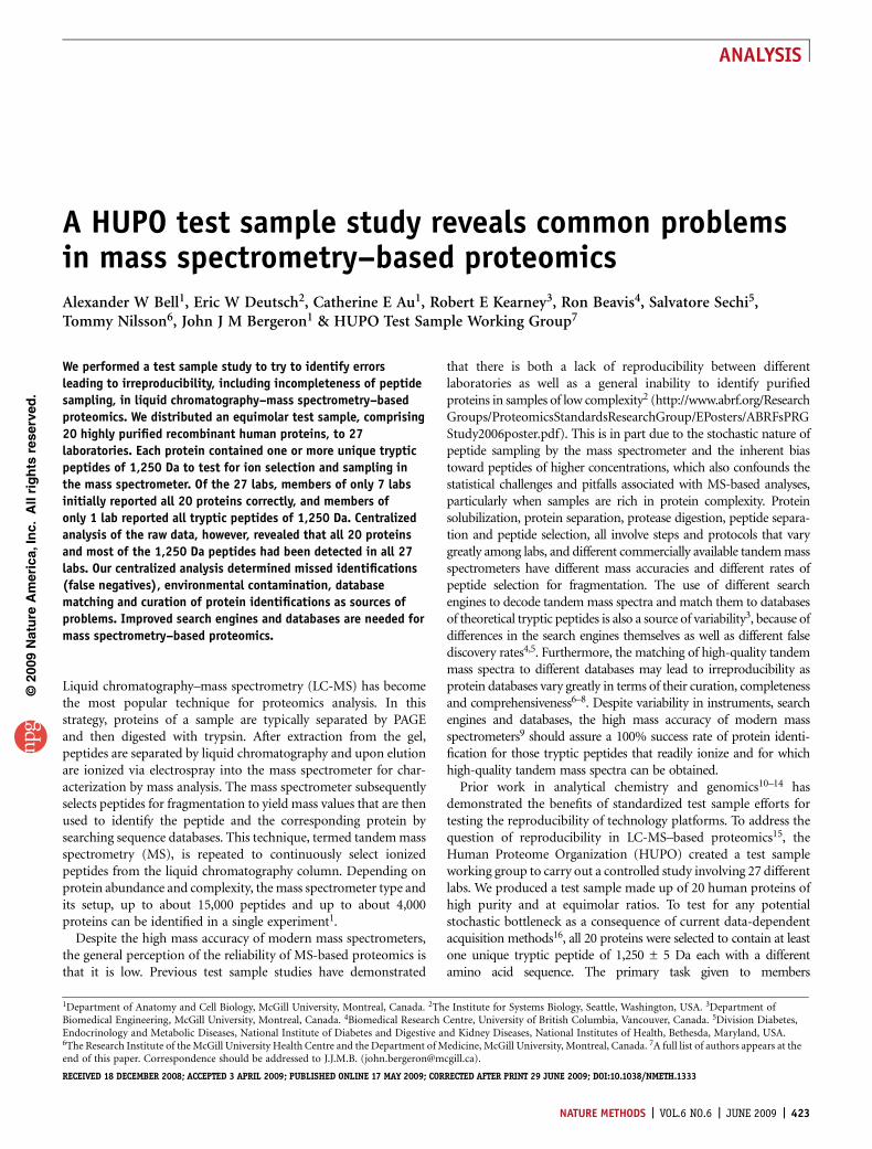
Table 1 | Results of test sample reporting of 24 academic labs (1–24) and 3 vendors (A–C)
Lab or vendor
A 1 2 3 4 5 6 7 8 B 9 10 11 12 13 14 15 16 17 18 19 20 21 22 C 23 24
Abbreviationa Group I Group II Group III Group IV
KHK + + + + + + + + + + + + + + + + + + + + + DB + + + + +
ATPAF2 + + + + + + + + + + + + + + + + + + + + + + ST N TR + TR
SETD3 + + + + + + + + + + + + + + + + + + + + + DB + + + + +
SPRY2 + + + + + + + + + + + + + + + + + + + + + + + + + + TR
GLB1L3 + + + + + + + + + + + + + + + + + + + + N + N + + TR +
FYTTD1 + + + + + + + + + + N N N N + + N N N N + + + + TR + TR
IHPK1 + + + + + + + + + N + + + N + + + + N N + + + + + ST TR
IFRD1 + + + + + + + + + + + + + + + + + + + + N + + N + + N
GCNT3 + + + + + + + + + + + + + + + + + + + + + + + + + TR +
EIF2S3 + + + + + + + + + + + + + + + + + + + + + + + A + + +
F2 + + + + + + + + + + + + + + + + + + N + AC + + A TR ST TR
FARP2 + + + + + + + + + + + + + N + + + + + N + + + + + + +
ENOX1 + + + + + + + N + N + N N + + + N + N N + N + + + + N
KLHL13 + + + + + + + + + + + + + + + + + + + N + + + N + ST +
NIBP + + + + + + + + N + N + + + + N + N + + + + + + + + +
MARS + + + + + + + + + + + + + + + + + + + + + + + + + TR +
NUP210 + + + + + + + + + + + + + + + + + + + + + + + + + + +
THBS4 + + + + + + + + + + + + + + + + + + + + + + ST A TR + +
KIAA0746 + + + + + + + + + + + + + + + + + + + + N N ST + TR TR +
HIRA + + + + + + + + + + + + + + + + + + + + + + + + + + N
Reportedb 20 20 20 20 20 20 20 20 20 20 20 20 20 20 24 22 21 22 30 25 22 22 21 21 20 18 18
Correctb 20 20 20 20 20 20 20 19 19 18 18 18 18 17 20 19 18 18 16 15 16 16 16 14 15 13 12
False positivesb 1 1 1 1
Contaminantsb 3 2 1 2 1 1 1 3 1 3 5 5 3
Redundantb 1 8 3 2 1 2
Score (%) 100 90 90 81 81 81 81 72 83 82 77 74 42 45 58 58 61 47 56 47 40
SDS-PAGEc + + + + + + + + +
Mass spectrometerd QT IT IT QT IT H TT QT QT QT IT IT H QT H H QT H IT H IT IT IT TT QT H IT
Peaklist sofwaree P D E M X E Ex Di Di PP E B DTA PP Xc Ex Di Xc B E Sp B Xc Ex Sp Sp Sp
Search enginef O M S Sp M M M M M O M S M O S M M O S S Sp S O M Sp Sp S
Turnaroundg 11 16 61 70 90 114 51 41 103 22 53 42 70 44 89 93 85 92 63 108 46 48 69 57 14 48 63
Groups I–IV identify labs scoring 100% (group I), those with naming (N) errors (group II) and those with naming errors as well as false positive, contaminant and redundant identifications (groupIII). Group IV includes labs with these errors as well as errors attributed to acrylamide alkylation (AC), database searching (DB), excessive stringency (ST), undersampling (A) or trypsinization(TR)-related errors. ‘+’ indicates a correct identification.aNotation used throughout the paper. bThe number of proteins in each category are indicated. c+ here indicates which analyses used gel separation.dMass spectrometers used were: ion trap (IT); QToF (QT); hybrid (H) including LTQ-FT or LTQ-Orbitrap; and ToFToF (TT). ePeaklists were generated by using the following software: Bioworks Browser (Thermo Electron)(B); Data Analysis mzXML (D); Distiller (Matrix Science) (Di); DTA Supercharge (DTA); Extract_msn (Thermo Electron) (E); Explorer (Applied Biosystems) (Ex); Masslynx (Waters) (M); ProteinLynx Global Server(Waters) (P); Protein Pilot (Applied Biosystems) (PP); Spectrum Mill (Agilent) (Sp); X! Tandem (X); and Xcaliber (Thermo Electron) (Xc). fDatabase search engines used were: Mascot (Matrix Science) (M); Sequest(Thermo Electron) (S); Spectrum Mill (Sp); and other (O) that include IdentityE (PLGS, http://www.waters.com), ProteinPilot (Applied Biosystems) or X! Tandem. gTurnaround time is given in days.
424 | VOL.6 NO.6 | JUNE 2009 | NATURE METHODS
ANALYSIS
RESULTSTest sample proteinsTo create the test sample, we selected 20 proteins in the molecularweight range of 32–110 kDa from the open reading frame (ORF)19
collection and the mammalian gene collection20 (SupplementaryMethods online). The criteria (Supplementary Fig. 1a online) forselection included a purity of about 95%, unique tryptic peptidesequences and the presence of at least one tryptic peptide of 1,250 ±5 Da (Supplementary Fig. 1b,c). We expressed the candidateproteins in Escherichia coli and purified them following a produc-tion strategy by using ion exchange and reverse phase chromato-graphy or by preparative electrophoresis purification frominclusion bodies (Supplementary Methods). One-dimensionalSDS-PAGE revealed the purity of the 20 purified proteins (Supple-mentary Fig. 1d) at 95% or greater (Supplementary Table 1
online) as evaluated by densitometry (Supplementary Fig. 2 andSupplementary Table 2 online). MS analysis of the 20 purifiedproteins revealed a vector-derived N-terminal extension of 7 aminoacids present on each of the proteins (Supplementary Fig. 3online). MS analysis of the test sample confirmed quality (Supple-mentary Fig. 4 and Supplementary Tables 2,3 online) and stability(Supplementary Fig. 5 and Supplementary Table 4 online) beforedistribution to the 27 labs.
Protein identificationWe instructed members of the 27 selected labs to use the NCBI nrhuman protein database of November 27, 2006 with exact matchesfor all 20 test sample proteins (Supplementary Fig. 6 and Supple-mentary Table 5 online) for protein identification. The individualresults from the labs are reported in Supplementary Table 6 online
©20
09 N
atu
re A
mer
ica,
Inc.
All
rig
hts
res
erve
d.
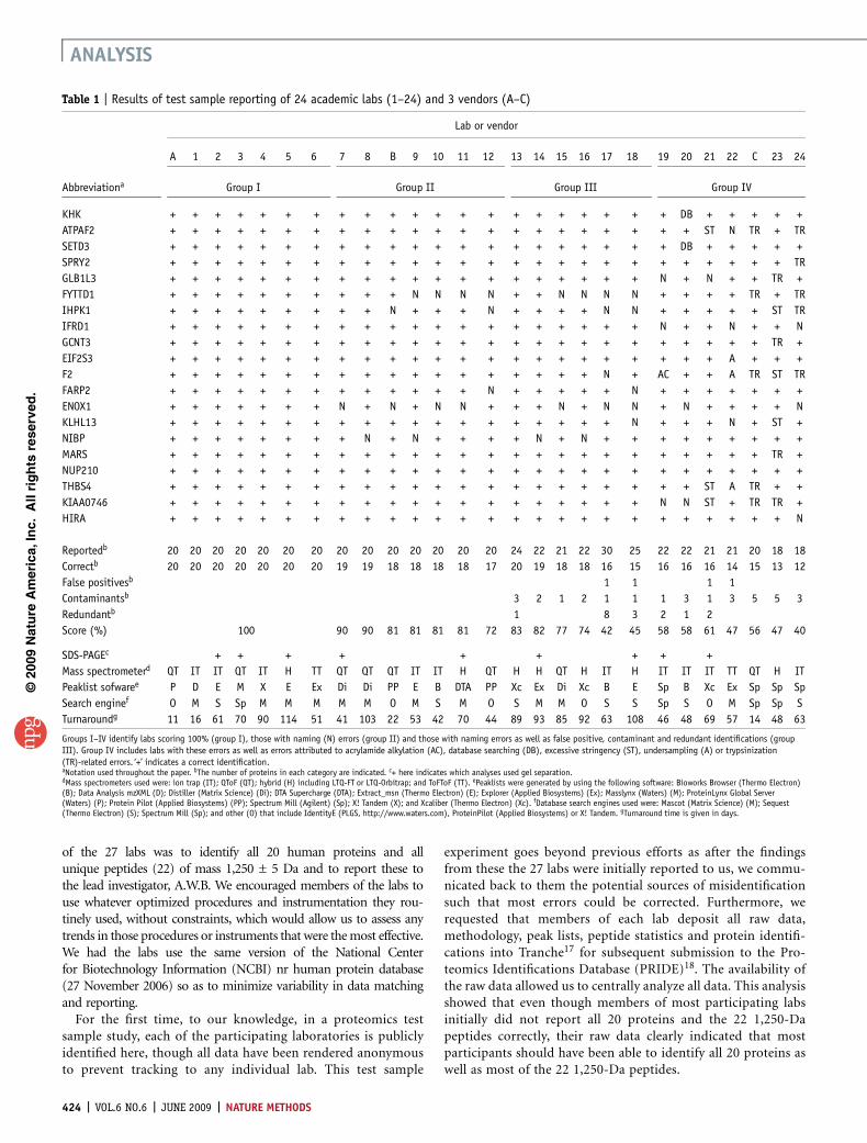
Table 2 | Designed peptide mass complexity reporting of 24 academic labs and 3 vendors
Lab or vendor
Abbreviation A 1 2 3 4 5 6 7 8 B 9 10 11 12 13 14 15 16 17 18 19 20 21 22 C 23 24 22R CR 23R 24R
KHK + + + + + + + + + + + + + + + + +
ATPAF + + + + + + + + + +
SETD3 + + + + + + + + +
SPRY2 + + + + + + + + + + + +
GLB1L3 + + + + + + + + + +
FYTTD1 + + + + + + + + + + + + + + + + +
IHPK1 + + + + + + + + + +
IFRD1 + + + + + + + + +
GCNT3 + + + + + + + + + + + + + + +
EIF2S3 + + + + + + + + + + + + + + + + + +
F2 + + + + + + + + + + + + + + +
FARP2 + + + + + + + + + + + + + + + + + + +
ENOX1 + + + + + + + + + + + + + + + + + + + +
KLHL13 + + + + + + + + + + + + + + + + + + +
NIBP + + + + +
MARS + + + + + + + + + + + + + + + + + + +
NUP210 + + + + + + + + + + + + + + + + + + + + + + + + +
NUP210 + + + + + +
THBS4 + + + + + + + + + + + + + + + + +
KIAA0746 + + + + + + + + + + + + + + + +
HIRA + + + + + + + + + + + + + + + + + + + +
HIRA + + + + + +
Total (initial) 15 9 3 16 12 15 9 14 3 11 12 10 15 4 10 22 14 8 11 15 1 8 8 4 0 7 12 0 13
Total (final) 18 10 22 16 19 11 18 11 22 11 2 11 16
True positive 3 1 3 1 3
False positive
Contaminants 1 1 6 1 3 1 3
Analysis scoring:
Initial score (%) 68 41 14 68 55 64 41 59 14 50 55 45 49 18 41 100 64 36 50 57 2 26 36 18 0 NR 32 55 0 59
Final score (%) 82 45 14 100 73 64 41 86 14 50 55 45 82 18 50 100 64 36 50 57 2 26 36 18 9 NR 50 73 0 59
Report scoring:
Centralized (count) 10 15 20 19 18 21 10 19 5 21 19 22 22 6 17 22 18 13 14 22 6 16 16 3 NRD 2 14 15 11 18
Score (%) DRD 67 15 DRD 89 71 90 100 60 52 63 45 82 67 65 100 78 62 85 68 17 50 50 DRD NRD NR 79 DRD 0 72
Initial and final reporting of the number of peptides of mass 1,250 ± 5 Da with increases in totals related to tabulation of mass-shifted cysteine-containing peptides. + indicates a correctidentification. Analysis scoring was calculated from the fraction of correct peptide identifications and the accuracy of reporting peptides of mass 1,250 Da, whereas the report scoring was based onthe fraction of correct peptide identifications reported divided by the number identified by the centralized analysis. Results not reported, NR; no raw data, NRD; submitted and data reprocessingdifference, DRD. DRDs are indicated by fewer peptides identified by the centralized analysis as compared to the number reported. Positive identifications included cysteine-containing peptides thathave been alkylated, peptides including missed trypsin cleavage and oxidized methionine residues. Labs A, 4–9, 11 and 13 reported peptides assigned at o95% confidence. Lab 6 used iTRAQ.
NATURE METHODS | VOL.6 NO.6 | JUNE 2009 | 425
ANALYSIS
and are summarized in Table 1. Analysis of the reports revealedclear differences in the number of tandem MS spectra assignedbased on the instrument used (Supplementary Fig. 7 online) butincorrect reporting of false positive and contaminating proteinswere not specifically linked to any MS platform or search engine.
Initially, members of only 7 labs (classified as group I) correctlyidentified all 20 proteins (Table 1). The labs classified as group IIencountered naming errors. Labs classified as group III encoun-tered naming errors, false positive and redundant identifications(Supplementary Fig. 8 and Supplementary Table 7 online). Noredundant identifications were reported by members of any lab thatused the Mascot (Matrix Science) search engine (n ¼ 11) whereaslabs using Sequest and SpectrumMill did report redundant identi-fications. Labs classified as group IV encountered several problems.We distributed another aliquot of the sample to labs that indicatedtrypsinization problems (labs C, 23 and 24; Supplementary Table 8online). Members of lab 22, who had a problem with under-sampling, (Supplementary Table 9 online) performed an addi-tional analysis with their remaining sample. Other errorsencountered by group IV included incomplete matching of tandem
MS spectra resulting from acrylamidealkylation (Supplementary Fig. 9 online),database search errors (SupplementaryTable 10 online) and the use of overlystringent identification criteria (Supplemen-tary Table 11 online), all of which resulted inmissed identifications. We devised a scoringsystem to take incorrect reporting intoaccount. After we discussed the problemswith members of each laboratory (Supple-mentary Table 12 online) and in some caseshad them perform repeat analyses, all iden-tified all 20 proteins, achieving a uniformscore of 100% (data not shown).
Peptide samplingWe also assessed the completeness of pep-tide sampling and selection in the massspectrometer by assessing the ability of the27 labs to detect the 22 designed trypticpeptides of mass 1,250 ± 5 Da (Supple-mentary Table 13 online), six of whichcontained cysteine residues whose massincreased as a consequence of reductionand alkylation as routinely used beforeprotein trypsinization. Initially, membersof only one lab (lab 14) reported detectionof all 22 peptides (Table 2) and only anadditional three groups (labs 17, B and CR;R indicates repeat analysis) reported detect-ing any peptides that contained cysteines.Several groups incorrectly reported pep-tides of 1,250 ± 5 Da derived from contam-inating proteins. Several groups alsoreported peptides of 1,250 ± 5 Da as a resultof a single missed trypsin cleavage (denotedas a true positive). We requested that theselabs perform a reassessment as describedabove for protein reporting.
We used our scoring system to assess both the analysis and thereporting of the 1,250 ± 5 Da tryptic peptides. Initially, onlymembers of lab 14 achieved a 100% score. After guidance, membersof lab 3 achieved 100% success by correcting for cysteine-contain-ing peptides and excluding peptides derived from contaminants.All other groups reported insufficient data. To distinguish betweenincomplete reporting and incomplete sampling, we compared the1,250-Da peptides that were reported to those that were identifiedby the centralized analysis (see below). Results from labs 10, 11, 14and 18 (but not lab 3) had data for all 22 1,250-Da peptides.However, members of labs 10, 11 and 18 could not report thepeptides and our centralized analysis failed to identify the 22peptides in the data from lab 3 (Table 2). Besides lab 14, only lab7 achieved 100% reporting of all 1,250-Da peptides in their dataset(a total of 19 peptides, as assessed by our centralized analysis of thedata) (Table 2 and Supplementary Table 13).
Data deposition to Tranche and PRIDEWe asked members of the 27 labs to transfer their raw MS data, themethodologies used, peak lists, peptide statistics and protein
©20
09 N
atu
re A
mer
ica,
Inc.
All
rig
hts
res
erve
d.
KHK
a
b
79% 94%
ATPAF2
SETD3
SPRY2
GLB1L3
FYTTD1
IHPK1
IFRD1
GCNT3
EIF2S3
FARP2
ENOX1
KLHL13
NIBP
MARS
NUP210
THBS4KIAA0746
HIRA
0 100 200 300 400 500 600 700 800 900 1,000
500
20 test sample proteins
20 test sample proteins
Trypsin
Keratins
E. coli
E. coliContaminants
250
250
500
0
F2
Position of peptide in the protein sequence
Num
ber of redundanttryptic peptides excluding
1,250 Da peptides
Num
ber of redundant1,250 D
a tryptic peptides
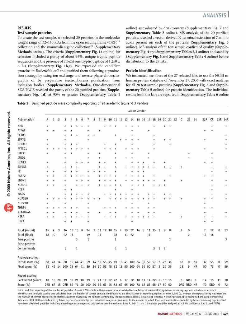
Figure 1 | Number of tandem mass spectra assigned to tryptic peptides. (a) Comparison of protein
abundance (percent of total redundant peptides) from the centralized analysis of the raw data collected in
the 27 labs (left) and after removal of individual lab contaminants including keratins as well as trypsin
(right). (b) Peptide heatmap representation for each of the 20 proteins from the centralized analysis of
the raw data from all 27 labs, revealing the frequency of observation of a given peptide as well as its
position in the protein sequence. Raw data from lab 24 were excluded (Online Methods).
426 | VOL.6 NO.6 | JUNE 2009 | NATURE METHODS
ANALYSIS

identifications to Tranche, a repository for raw data. Initial pro-blems related to the transfer of data to Tranche were all overcome.Tranche hash and passphrase codes are available in SupplementaryTable 14 online. PRIDE personnel transferred a copy of all datafrom Tranche to PRIDE, a centralized public data repository for thestandardized reporting of proteomics results. As evaluated byPRIDE personnel, the initially deposited data had several problemsincluding incomplete files, proprietary software formats and screen-shots of data displays in software rather than actual data files. Thewide variety of data formats encountered faithfully represents theheterogeneity in the field concerning proteomics bioinformatics. Italso appears that the implementation of community standards fordata reporting and exchange is not yet at a level that accommodatedthe minimal requirements for these 20 test proteins.
Centralized analysis of the raw dataTo independently assess the individual analyses of the 27 labs, wedownloaded all raw data from Tranche. We reanalyzed the collectiveraw data centrally using a uniform protocol of database searchingusing X! Tandem21 and post-processing with the Trans ProteomicPipeline22 to assign probabilities to all identifications and globalfalse discovery rates as well as to determine the total numberof tandem MS spectra assigned, number of distinct peptidesand amino acid sequence coverage (Supplementary Tables 13and 15 online).
We found that members of the majority of the labs had in factgenerated raw data of sufficient quality to identify all 20 proteinsand most of the 22 1,250-Da peptides. We identified discrepanciesbetween the submitted results (Supplementary Table 12) and the
centrally reprocessed results (Supplementary Table 15) for labs 2,4, 5, 8, 10, 11, 16, 19, 20, 21, 22R, 24 and CR, largely owing to thedifferent data analysis strategies used in these labs. The centralizedanalysis included checks for experimental artifacts including pyro-Glu formation, deamidations and nontryptic cleavages.
For all 27 labs, the majority of tandem mass spectra (79%) wereassigned to the 20 recombinant human proteins, but 21% of thespectra were assigned to contaminants that included E. coli pro-teins, trypsin, keratins and other proteins (Fig. 1a and Supple-mentary Table 15). The centralized analysis also revealed that all 22predicted tryptic peptides of 1,250 Da were observed in only 4 labs,three of which used a Fourier transform ion cyclotron resonance(FTICR) instrument (Tables 1 and 2). These instruments reportedthe highest number of assigned tandem mass spectra, therebyincreasing the likelihood of identifying all of the 1,250-Da peptides(Supplementary Fig. 7). Tandem mass spectra matching the1,250-Da peptides were variable for each of the 20 proteins(Fig. 1b) and were variably detected in our centralized analysis(Supplementary Fig. 10 online).
The centralized analysis also revealed (i) that the majority oftandem MS spectra assigned to keratins (human keratins KRT1,KRT2, KRT9 and KRT10 are commonly found in mature epidermaltissue and are also present in laboratory dust and fingerprints, ratherthan hair- or wool-derived keratins) were largely attributed tostrategies that used one-dimensional PAGE (SupplementaryFig. 11 and Supplementary Table 15 online); (ii) that E. coliproteins were found by members of all but 2 labs (SupplementaryFig. 11 and Supplementary Table 15) and most likely were presentin the provided sample; (iii) that other protein contaminants (forexample, albumin and casein) were found in datasets from a specificsubset of labs (5 labs found albumin, 5 casein and 3 both proteins;albumin was incorrectly reported as human when in fact it wasbovine, and both bovine serum albumin and casein are likelyabundant proteins used in these labs for standardization); and(iv) that autolytic trypsin peptides resulted from added trypsin.Excluding the contaminants introduced in the labs, 94% of thetandem mass spectra were accounted for by the 20 recombinantproteins, and the remaining tandem MS spectra were assigned to theE. coli proteins (Fig. 1a). False negatives (one or more of the 20recombinant proteins not detected) were likely a consequence ofvariability in trypsin digestion and the stochastic sampling of themass spectrometry analysis.
Labs that used exclusively liquid phase separations in general hadfewer spectra that could be assigned to epidermal keratins than labsthat used a combination of protein separation by gel electrophor-esis followed by in-gel digestion, peptide extraction and high-performance liquid chromatography peptide separation beforetandem MS analysis (Supplementary Fig. 11). This trend isprobably caused by the fact that each gel slice was exposed to theenvironment individually, effectively increasing the load of envir-onmental contaminants. The number of spectra that could beassigned to keratins was also broadly correlated with the identifica-tion of low-concentration sample source contaminants (E. coliproteins) and reagent proteins (trypsin), suggesting that in mostcases these proteins were present at substantially lower concentra-tions than the 20 test sample proteins (Supplementary Table 15).
Our centralized analysis confirmed that raw data initiallyreported by members of 4 labs were incomplete (SupplementaryTable 15). Repeat analysis in these labs generated sufficient data to
©20
09 N
atu
re A
mer
ica,
Inc.
All
rig
hts
res
erve
d.
0 50
Total
a
b
c
Lab 19Lab 20
TotalLab 20
TotalLab 19Lab 24
Lab 24RLab C
Lab CR
Lab 21Lab 24
Lab 24RLab C
Lab CR
100 150
ATPAF2
SETD3
F2
200 250 300
0 50 100 150 200
0 100 200 300
600 300 200 4000
400 500 600
250 300
Position of the peptide in the protein sequence
Number of redundanttryptic peptides excluding1,250 Da peptides
Number of redundant1,250 Da tryptic peptides
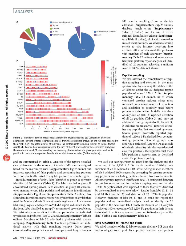
Figure 2 | Discrepancies between reported data and centralized analysis
identify erroneous reporting. (a–c) Peptide heatmap comparisons of the
centralized analysis compiled for all 27 labs (total), with the data from
selected indicated individual labs for the proteins ATPAF2 (a), SETD3 (b) and
F2 (c). Blue, the 1,250 Da peptides; red, all other tryptic peptides. Scale bar
represents the number of redundant peptides. Missed cleavages account for
the different degree of shading for peptides of 1,250 Da.
NATURE METHODS | VOL.6 NO.6 | JUNE 2009 | 427
ANALYSIS

identify the 20 proteins. No tandem mass spectra were initiallyobserved for the ATAF2 protein in labs 24 and C (Fig. 2), but in arepeat analysis, they generated sufficient tandem mass spectra(marked as 24R and CR) to characterize the protein as well asthe 1,250-Da peptide. However, members of labs 19, 20 and 21generated sufficient tandem mass spectra for protein ATPAF2,members of lab 20 generated sufficient tandem mass spectra forprotein SETD3, and members of labs 19 and C generated sufficienttandem mass spectra for protein F2 but still did not initially reportthe identification of these proteins. We determined that membersof lab 20 had a database-matching problem for protein SETD3 andmembers of lab 19 had an acrylamide modification problem forprotein F2. Lab 24 had a trypsinization problem for protein F2,which was fixed upon repeat analysis (24R). Although lab C initiallyreported a trypsinization problem for the F2 protein, the raw dataproved otherwise. Lab C’s repeat analysis (CR) revealed moretandem mass spectra assigned to protein F2 but insufficient datafor the peptide of mass 1,250 Da. Detailed central analysis of eachlab’s data submitted to Tranche justified the removal of results fromlab 24 (but not of this lab’s repeat analysis, 24R) from the heat mapshown in Figure 1b. Inspection of the results from lab 24 (Supple-mentary Table 13) revealed that B95% of the tandem mass spectrawere assigned to peptides with cyclized N-terminal glutamineamino acid (pyroGln), which is not typical for analysis of trypticpeptides. Additional in-depth analysis of the raw data did notidentify tandem mass spectra; aberrant chemically induced mod-ifications may have been introduced.
DISCUSSIONOur results demonstrate that, of 27 labs, members of only 7 labsinitially characterized an equimolar sample of 20 human proteins.However, our centralized analysis of the raw data demonstratedthat members of each of the labs, with a few exceptions, had in factgenerated mass spectrometry data of very high quality, more thansufficient to identify all 20 proteins and most of the 22 1,250-Dapeptides. This demonstrates the important need for education andtraining to properly apply such a complex technology.
Most notably, we found generic problems in databases to be themajor hurdle for the correct characterization of proteins in the testsample. The search engines used in this study at present cannotdistinguish among different identifiers for the same protein, deriv-ing from the way the databases are constructed. Indeed, the searchengines used either for the centralized data analysis or by theindividual labs suggest an erroneous confidence to the assignmentsof peptides and proteins. This erroneous confidence necessitates theuse of manual verification of both the peptide assignments andprotein assignments for low-confidence identifications.
An extended standardized FASTA format (http://psidev.info/index.php?q¼node/317) has been proposed by HUPO ProteomicsStandards Initiative (PSI) that would resolve the problem ofstandardized annotation. Presently, manual curation of tandemMS data search results is needed for correct reporting. This includesthe nonredundant assignments of tandem MS spectra to overcomethe common errors in the apparent characterization of differentproteins that are one and the same. We have observed thatalgorithms used by different search engines to calculate molecularweight are variable (data not shown). It is therefore reasonable tosuggest that a common method for calculating molecular weight be
chosen and used throughout the community. Additionally, theautomatic matching of tandem mass spectra of high quality to aprotein-coding genome with a single representative protein foreach gene could overcome several of the current errors in proteinnaming and redundancies.
A test sample containing 20 proteins at 5 pmol equimolarabundance is not representative of a proteomics study with com-plex mixtures. However, a routine 100% success rate of protein and1,250-Da peptide identification of such a test sample could beimplemented as a standard, as well as the routine deposition of rawdata into Tranche. This would enable a greater degree of trust in theconclusions deduced for proteomics studies in general. A limitednumber of the 20 test sample protein mixtures have been preparedand are available by contacting the lead author (A.W.B.). Thesesamples, however, are stored in 7.5 M urea, which leads to variablecarbamylation, and this may affect trypsinization as well as dataanalysis. Such test samples should be helpful as a benchmarkingtool for researchers embarking on a proteomics study with complexmixtures. At the least, their abilities to collect sufficient data forunambiguous identification of 20 human proteins and 22 1,250-Dapeptides can be assessed. A peptide-by-peptide comparison ofresults from any individual lab with those from a centralizedanalysis of the data should be informative to the inability ofany lab’s members to detect proteins or specific peptides. For anylarge-scale, multilab proteomics effort, we recommend the useof a centralized analysis, especially if data are generated on morethan one platform, generated in more than one location orcollected over time.
Our study allowed us to deduce several guidelines for performingany proteomics experiment. Sources of lab-derived contaminationneed to be identified and monitored closely, with the two majorsources being environmental contamination carried over from priorexperiments and keratins (largely from gel-based analysis). The useof target-decoy search strategies should be made mandatory, andfalse discovery rates should be reported. The monitoring of uniquepeptides and unique tandem mass spectra is needed to ensure thatthe minimum list of protein identifications is reported, to address theissue of redundant identifications (sequence variants of the sameprotein). A gene-centric database could ensure that only a singledescriptive name would be assigned to each protein sequence,eliminating aliases. The creation of tools for transforming data(raw data, peak lists, peptide lists and protein lists) into standardizedformats would aid the ease of submission to repositories such asTranche. The distribution of all data deposited in Tranche to thecommunity, via PRIDE, Human ProteinPedia, PeptideAtlas andGPM, would facilitate centralized data analysis which may helplead to new insights in proteomics experiments.
In summary, our analysis showed that even with a sampleconsisting of highly purified human proteins, members of manyparticipating labs had difficulties in reporting data correctly. How-ever, the majority of the participants deposited raw data, each withmore than sufficient coverage of the 20 proteins. Thus a majorcontributing factor to erroneous reporting resides at the level ofdatabase and search engines used and once corrected for,provided an almost perfect score for most participants. Therefore,we expect that once databases and search engines have beenimproved and made compatible with MS-based proteomics, theaccuracy of data reporting will increase and along with it, thefidelity of proteomics.
©20
09 N
atu
re A
mer
ica,
Inc.
All
rig
hts
res
erve
d.
428 | VOL.6 NO.6 | JUNE 2009 | NATURE METHODS
ANALYSIS
METHODSMethods and any associated references are available in the onlineversion of the paper at http://www.nature.com/naturemethods/.
Note: Supplementary information is available on the Nature Methods website.
ACKNOWLEDGMENTSSupported in part by Canadian Institutes of Health Research to the HUPO HeadQuarters (S. Ouellette) for coordination of this HUPO test sample initiative. A.W.B.and C.E.A. were supported by Genome Quebec and McGill University. We thank D.Juncker, G. Temple, J. van Oostrum, G. Omenn, K. Colwill, J. Langridge and M.Hallett for their comments on the manuscript, and D.M. Desiderio for helpfulcomments on the manuscript. This test sample effort builds on pioneering effortsfrom several other groups and especially Association of Biomolecular ResourceFacilities. This study is a HUPO test sample initiative and HUPO welcomescollaborative efforts to benefit proteomics. We acknowledge the following sources ofgrant support: E.W.D. is supported by the National Heart, Lung and Blood Institute,National Institutes of Health (NIH), under contract N01-HV-28179; the University ofCalifornia, Los Angeles Burnham Institute for Medical Research NIH grant numberRR020843; University of California, Los Angeles (National Heart, Lung and BloodInstitute P01-008111); University of Michigan, NIH P41RR018627; Beijing ProteomeResearch Center, affiliated with The Beijing Institute of Radiation Medicine forNational Key Programs for Basic Research grant 2006CB910801 and Hi-Tech Researchgrant 2006AA02A308. We acknowledge access and use of The University CollegeDublin Conway Mass Spectrometry Resource instrumentation, supported by ScienceFoundation, Ireland grant 04/RPI/B499. PRIDE, J.A.V. is a postdoctoral fellow of the‘‘Especializacion en Organismos Internacionales’’ program from the Spanish Ministryof Education and Science. L.M. is supported by the ‘‘ProDaC’’ grant LSHG-CT-2006-036814 of the EU. Samuel Lunenfeld Research Institute, Mount Sinai, Toronto issupported by Genome Canada through Ontario Genomics Institute. J.A.V. and L.M.thank H. Hermjakob and R. Apweiler for their support. A.W.B. thanks L. Roy andZ. Bencsath-Makkai for help in data submission and analysis.
AUTHOR CONTRIBUTIONSA.W.B. coordinated all steps of the study. C.E.A., T.N. and J.J.M.B. coordinateddata analysis and the final manuscript. E.W.D., R.B. and R.K. did the centralizedanalysis of the collective data retrieved from the raw data supplied from each labto Tranche. S.A.C., P.P., L.M., E.K., C.D., S.S., X.Q., K.W., T.P.C., K.P. and T.A.B. providedcomments. Invitrogen prepared, designed and distributed the test sample proteins.
COMPETING INTERESTS STATEMENTThe authors declare competing financial interests: details accompany the full-textHTML version of the paper at http://www.nature.com/naturemethods/
Published online at http://www.nature.com/naturemethods/Reprints and permissions information is available online athttp://npg.nature.com/reprintsandpermissions/
1. de Godoy, L.M. et al. Comprehensive mass-spectrometry-based proteomequantification of haploid versus diploid yeast. Nature 455, 1251–1254 (2008).
2. Turck, C.W. et al. The Association of Biomolecular Resource Facilities ProteomicsResearch Group 2006 study: relative protein quantitation. Mol. Cell. Proteomics 6,1291–1298 (2007).
3. Boutilier, K. et al. Comparison of different search engines using validated MS/MStest datasets. Anal. Chim. Acta 534, 11–20 (2005).
4. Elias, J.E., Haas, W., Faherty, B.K. & Gygi, S.P. Comparative evaluation of massspectrometry platforms used in large-scale proteomics investigations. Nat.Methods 2, 667–675 (2005).
5. Kapp, E.A. et al. An evaluation, comparison, and accurate benchmarking ofseveral publicly available MS/MS search algorithms: sensitivity and specificityanalysis. Proteomics 5, 3475–3490 (2005).
6. Bell, A.W., Nilsson, T., Kearney, R.E. & Bergeron, J.J. The protein microscope:incorporating mass spectrometry into cell biology. Nat. Methods 4, 783–784(2007).
7. Gilchrist, A. et al. Quantitative proteomics analysis of the secretory pathway.Cell 127, 1265–1281 (2006).
8. Klie, S. et al. Analyzing large-scale proteomics projects with latent semanticindexing. J. Proteome Res. 7, 182–191 (2008).
9. Zubarev, R. & Mann, M. On the proper use of mass accuracy in proteomics.Mol. Cell. Proteomics 6, 377–381 (2007).
10. Cortez, L. The implementation of accreditation in a chemical laboratory.Trends Analyt. Chem. 18, 638–643 (1999).
11. Lander, E.S. et al. Initial sequencing and analysis of the human genome.Nature 409, 860–921 (2001).
12. Yates, J.R. III., Gilchrist, A., Howell, K.E. & Bergeron, J.J. Proteomics oforganelles and large cellular structures. Nat. Rev. Mol. Cell Biol. 6, 702–714(2005).
13. Shi, L., Perkins, R.G., Fang, H. & Tong, W. Reproducible and reliable microarrayresults through quality control: good laboratory proficiency and appropriatedata analysis practices are essential. Curr. Opin. Biotechnol. 19, 10–18(2008).
14. Anonymous. Making the most of microarrays. Nat. Biotechnol. 24, 1039(2006).
15. Anonymous. Proteomics’ new order. Nature. 437, 169 (2005).16. Domon, B. & Aebersold, R. Challenges and opportunities in proteomics data
analysis. Mol. Cell. Proteomics 5, 1921–1926 (2006).17. Falkner, J.A., Hill, J.A. & Andrews, P.C. Proteomics FASTA archive and reference
resource. Proteomics 8, 1756–1757 (2008).18. Martens, L. et al. PRIDE: the proteomics identifications database. Proteomics 5,
3537–3545 (2005).19. Liang, F. et al. ORFDB: an information resource linking scientific content to a
high-quality Open Reading Frame (ORF) collection. Nucleic Acids Res. 32,D595–D599 (2004).
20. Strausberg, R.L., Feingold, E.A., Klausner, R.D. & Collins, F.S. The mammalian genecollection. Science 286, 455–457 (1999).
21. Craig, R. & Beavis, R.C. TANDEM: matching proteins with tandem mass spectra.Bioinformatics 20, 1466–1467 (2004).
22. Keller, A., Eng, J., Zhang, N., Li, X.J. & Aebersold, R. A uniform proteomics MS/MSanalysis platform utilizing open XML file formats. Mol. Syst. Biol. 1, 2005.0017(2005).
©20
09 N
atu
re A
mer
ica,
Inc.
All
rig
hts
res
erve
d.
The full list of authors and affiliations is as follows:Thomas A Beardslee8, Thomas Chappell9, Gavin Meredith10, Peter Sheffield11, Phillip Gray12, Mahbod Hajivandi10, Marshall Pope10, Paul Predki10,
Majlinda Kullolli13, Marina Hincapie13, William S Hancock13, Wei Jia14, Lina Song14, Lei Li14, Junying Wei14, Bing Yang14, Jinglan Wang14, Wantao Ying14,
Yangjun Zhang14, Yun Cai14, Xiaohong Qian14, Fuchu He14, Helmut E Meyer15, Christian Stephan15, Martin Eisenacher15, Katrin Marcus15, Elmar Langenfeld15,
Caroline May15, Steven A Carr16, Rushdy Ahmad16, Wenhong Zhu17, Jeffrey W Smith17, Samir M Hanash18, Jason J Struthers18, Hong Wang18, Qing Zhang18,
Yanming An19, Radoslav Goldman19, Elisabet Carlsohn20, Sjoerd van der Post20, Kenneth E Hung21, David A Sarracino22, Kenneth Parker21, Bryan Krastins22,
Raju Kucherlapati21, Sylvie Bourassa23, Guy G Poirier24, Eugene Kapp25, Heather Patsiouras25, Robert Moritz25, Richard Simpson25, Benoit Houle26, Sylvie LaBoissiere27,
Pavel Metalnikov28, Vivian Nguyen29, Tony Pawson29, Catherine C L Wong30, Daniel Cociorva30, John R Yates III30, Michael J Ellison31, Ana Lopez-Campistrous31,
Paul Semchuk31, Yueju Wang32, Peipei Ping32, Giuliano Elia33, Michael J Dunn33, Kieran Wynne33, Angela K Walker34, John R Strahler34, Philip C Andrews34,
Brian L Hood35,36, William L Bigbee35,37, Thomas P Conrads35,36, Derek Smith38, Christoph H Borchers38, Gilles A Lajoie39, Sean C Bendall39, Kaye D Speicher40,
David W Speicher40, Masanori Fujimoto41, Kazuyuki Nakamura41, Young-Ki Paik42, Sang Yun Cho42, Min-Seok Kwon42, Hyoung-Joo Lee42, Seul-Ki Jeong42,
An Sung Chung42, Christine A Miller43, Rudolf Grimm43, Katy Williams44, Craig Dorschel45, Jayson A Falkner34, Lennart Martens46 & Juan Antonio Vizcaıno46
8Verdezyne, Inc., Carlsbad, California, USA. 9BioGrammatics Incorporated, Carlsbad, California, USA. 10Invitrogen Corporation, Carlsbad, California, USA.11Allergan, Irvine, California, USA. 12Ambry Genetics, Aliso Viejo, California, USA. 13Barnett Institute and Department of Chemistry and Chemical Biology,
NATURE METHODS | VOL.6 NO.6 | JUNE 2009 | 429
ANALYSIS
©20
09 N
atu
re A
mer
ica,
Inc.
All
rig
hts
res
erve
d.
Northeastern University, Boston, Massachusetts, USA. 14State Key Laboratory of Proteomics, Beijing Proteome Research Center, Changping District, Beijing,
China. 15Bochum University, Ruhr-Universitaet Bochum, Bochum, Germany. 16Proteomics, Broad Institute of Massachusetts Institute of Technology and Harvard,
Cambridge, Massachusetts, USA. 17Burnham Institute for Medical Research, La Jolla, California, USA. 18Fred Hutchinson Cancer Research Center, Seattle,
Washington, USA. 19Georgetown University, Department of Oncology, Washington, DC, USA. 20Goteborg Proteomics Centre: The Proteomics Core Facility,
Sahlgrenska Academy, University of Goteborg, Goteborg, Sweden. 21Harvard Partners Center for Genetics and Genomics, Cambridge, Massachusetts, USA.22Thermo-Fisher BRIMS Center, Cambridge, Massachusetts, USA. 23Proteomics Platform, Quebec Genomic Center, Laval University Medical Research Center,
Quebec, Canada. 24Health and Environment Unit, Laval University Medical Research Center, Quebec, Canada. 25Joint Proteomics Laboratory, Ludwig Institute
for Cancer Research and The Walter & Eliza Hall Institute for Medical Research, Parkville, Australia. 26Genizon BioSciences Incorporated, Saint Laurent,
Canada. 27McGill University and Genome Quebec Innovation Centre, Montreal, Canada. 28Ontario Cancer Biomarker Network, MaRS Centre, Toronto, Canada.29Samuel Lunenfeld Research Institute, Mount Sinai Hospital, Toronto, Canada. 30The Scripps Research Institute, Department of Chemical Physiology, La Jolla,
California, USA. 31Department of Biochemistry, University of Alberta, Edmonton, Canada. 32Departments of Physiology, Medicine and Division of Cardiology,
David Geffen School of Medicine, University of California, Los Angeles, California, USA. 33Proteome Research Centre, Conway Institute of Biomolecular and
Biomedical Research, University College Dublin, Dublin, Ireland. 34Department of Biological Chemistry, University of Michigan, Ann Arbor, Michigan, USA.35Clinical Proteomics Facility, University of Pittsburgh Cancer Institute, Pittsburgh, Pennsylvania, USA. 36Department of Pharmacology and Chemical Biology,
University of Pittsburgh School of Medicine, Pittsburgh, Pennsylvania, USA. 37Department of Pathology, University of Pittsburgh School of Medicine,
Magee-Womens Research Institute, Pittsburgh, Pennsylvania, USA. 38University of Victoria, Victoria, Canada. 39Department of Biochemistry, University of
Western Ontario, London, Ontario, Canada. 40The Wistar Institute, Philadelphia, Pennsylvania, USA. 41Department of Biochemistry and Functional Proteomics,
Yamaguchi University Graduate School of Medicine, Ube, Yamaguchi, Japan. 42Yonsei Proteome Research Center, Yonsei University, Sudaemoon-ku, Seoul,
Korea. 43Agilent Technologies Incorporated, Santa Clara, California, USA. 44Applied Biosystems, Foster City, California, USA. 45Waters Corporation, Milford,
Massachusetts, USA. 46EMBL Outstation, European Bioinformatics Institute, Wellcome Trust Genome Campus, Hinxton, Cambridge, UK.
430 | VOL.6 NO.6 | JUNE 2009 | NATURE METHODS
ANALYSIS

ONLINE METHODSTest sample generation and distribution. As more completelydescribed in the Supplementary Methods, all test sampleproteins were cloned23 and expressed24 in E. coli, purified frominclusion bodies under denaturing conditions and mixed inequimolar (5 pmol) amounts. A committee made up of fundingagency representatives (NIH and Canadian Institutes of HealthResearch), journal editors and the HUPO Executive Committeeproposed a list of 55 labs. Invitations to participate wereextended to 41 labs and 24 accepted. Also, 6 mass spectrometervendors were selected by the HUPO Industrial Advisory Board(IAB) and all agreed to participate but only 3 provided results.The 27 labs that participated are indicated here as co-authors.Dried samples containing 5 picomoles of each protein wereshipped on dry ice, along with detailed examples of LC-MSproteomics analyses (http://www.invitrogen.com/etc/medialib/en/filelibrary/pdf.Par.72904.File.tmp/HumanProteinStandardsforMassSpectrometry.pdf). Samples were shipped from Invitrogenand deliveries were overnight (by DHL in the USA and DHLInternational or FedEx International express overseas; 1 to 3business day delivery). Delivery to Australia was delayed ontwo occasions owing to incomplete customs-related documen-tation that resulted in the samples attaining ambient tempera-tures and hence their replacement. Another two samples werereceived at the recipient institutes but did not arrive at the hostlab. One vial was reported to be empty as negligible signal wasobserved by Coomassie blue staining of a two-dimensional gel.In all cases, more material was supplied. Participants wereinstructed to use a specified NCBI nr database (http://portal.proteomics.mcgill.ca:8080/hupo-standards/nr_human_20061127_v2.fasta), to report details of methodologies used andproteins identified and to deposit raw data and reports toTranche (http://tranche.proteomecommons.org/) (SupplementaryNote online).
Instructions to laboratories and vendors. Test samples weredistributed to participating laboratories, who were instructed to(i) identify the 20 human proteins, (ii) report the details of theidentifications (protein name, NCBI gi number, sequence coverage,number of peptides and number of tandem MS spectra) followingthe criteria of ref. 25 and (iii) report the details of methodology.The following description of the sample was supplied: ‘‘The sampleis an equimolar mixture (5 pmol) of 20 human proteins that wereexpressed in E. coli under conditions to maximize inclusion bodyformation. The expression system results in an N-terminal exten-sion of 7 amino acids (sequence MYKKAGT) followed by theencoded initiator methionine. The 20 proteins were purified bypreparative SDS PAGE or 2D-LC (anion exchange and reversedphase) to 4 95% purity. Trypsin digestion of the purifiedconstructs results in the generation of a tripeptide (MYK) plusfree K or a tetrapeptide (MYKK) resulting from 1 missed cleavageand an N-terminal extension of 3 (AGT) or 4 (KAGT, 1 missedcleavage) amino acids. Contaminants do not exceed 1% in the finalmixture.’’ Details regarding the proteomics MS analysis as well asthe selection and purification of the test sample proteins byInvitrogen were also supplied (poster presentation (http://www.invitrogen.com/etc/medialib/en/filelibrary/pdf.Par.72904.File.tmp/HumanProteinStandardsforMassSpectrometry.pdf) that was pre-sented at the HUPO 5th Annual World Congress).
Protein identification reports were scored based on acceptablenames as found in the specified database. For reassessment, eachlab was instructed to make corrections based on naming; redun-dant, false positive and contaminant identifications; and acrylamidealkylation of cysteines. Labs that did not achieve 100% afterreassessment were requested to repeat the analysis of anotheraliquot of the sample.
Reporting of peptides of mass 1,250 ± 5 Da was requested, withreassessment as above, and reports were scored twofold, foranalysis and reporting completeness.
Database selection. To limit variation in data evaluation, a singledatabase, the NCBInr human protein database of 27 November,2006, was selected. The NCBInr database contained all 20 testproteins with their exact matches represented.
Previous efforts to benchmark proteomics through test sampleshave usually allowed participating labs to choose whatever databasethey felt might be the most appropriate to match their tandem massspectra. As we have argued elsewhere6,26, most databases are still in aconstant flux changing from one release to another. These changeslead to increased variation in data evaluation. Here we comparedthe predicted amino acid sequence of the 20 test proteins selected asidentified above with the NCBI nonredundant database, the Uni-versal Protein Resource (UniProt) and the International ProteinIndex (IPI) databases (Supplementary Table). Comparisons weremade by using blastp (http://www.ncbi.nlm.nih.gov/BLAST/). Thereciprocal matching (database to ORF and ORF to database)process revealed differences in protein length as well as aminoacid substitutions, most of which occurred in the IPI database andare likely to be related to the specific assembly process of the IPI27.Longer or shorter sequences in the database indicate extensions ortruncations and/or differences in editing (removal of potentialintrons) the predicted DNA sequences. Amino acid substitutionsare indicated by orange and green shading. An exact match isindicated by 100% identity in both directions. From this databaseassessment only the NCBInr database had all recombinant pro-teins with their exact matches represented.
Data reporting. The number of proteins reported and numbercorrect are indicated as are the number of false positive (proteinsidentified by shared peptides) and contaminant (proteins not inthe sample) identifications and those proteins identified morethan once but reported as separate proteins (redundant). After theinitial reporting by members of the 27 labs (numbers and lettersare used to identify academic labs and vendors, respectively), oneof us (A.W.B.) discussed with members of each lab problemsassociated with providing nondescriptive names (for example,hypothetical protein, ORF), and also the reporting of redundantidentifications, and false positive and contaminating proteins.Problems associated with spurious alkylation of cysteine residuesby acrylamide during preparative electrophoresis were also dis-cussed. Participants were requested to reassess search results and tosubmit updated final reports. A scoring system was devised to takeinto account incomplete reporting as well as erroneous identifica-tions. The score (Table 1) was calculated as follows: score ¼fraction identified (number correct / 20) � accuracy (numbercorrect / number reported) � 100. For Table 1, details for theproteomics analyses on a lab-by-lab basis including proteinseparation, mass spectrometer, peaklist software and database
©20
09 N
atu
re A
mer
ica,
Inc.
All
rig
hts
res
erve
d.
doi:10.1038/nmeth.1333 NATURE METHODS
search engine as well as turn-around time (time from the labreceiving the sample until results were submitted by email (average67 days)) are indicated. All labs used trypsin. Mass spectrometersused included: ion trap (IT); QToF (QT); hybrid (H) includingLTQ-FT or LTQ-Orbitrap; and ToFToF (TT). Peaklists weregenerated by using the following software: Bioworks Browser(Thermo Electron) (B), Data Analysis mzXML (D), Distiller(Matrix Science) (Di), DTA Supercharge (DTA), Extract_msn(Thermo Electron) (E), Explorer (Applied Biosystems) (Ex),Masslynx (Waters) (M), ProteinLynx Global Server (Waters) (P),Protein Pilot (Applied Biosystems) (PP), Spectrum Mill (Agilent)(Sp), X! Tandem (X), and Xcaliber (Thermo Electron) (Xc), andall labs used default parameter with lab 5 including total ioncurrent (TIC) threshold of 100 and a minimum of 10 peaks, andlab 7 including correlation threshold (CT) of 0.7, signal-to-noiseratio (SNR) of 20, reject width outliers and baseline correction.Database search engines included: Mascot (Matrix Science) (M),Sequest (Thermo Electron) (S), Spectrum Mill (Sp), and others(O) that include IdentityE (PLGS, http://www.waters.com), Pro-teinPilot (Applied Biosystems) or X! Tandem. All procedures usedare reported in Tranche (Supplementary Table 14).
The methodology, the peak lists, the peptide statistics andprotein identification data were transferred to Tranche, a reposi-tory for raw data. Detailed instructions (Supplementary Note)were provided to each participating lab with regards to thepreparation and transferring of supporting data and informationto Tranche (http://www.proteomecommons.org/dev/dfs/examples/hupo-2007/Tranche-HUPO.jsp). All problems in the transfer ofdata from host labs to Tranche (for example, compact disk andcourier transmission, firewall problems, unresponsive servers)were overcome. The transfer of data culminated with the genera-tion of a Tranche hash and passphrase codes that were returned bye-mail to the submitter and to one of us (A.W.B.). The final set ofcodes is listed in Supplementary Table 14).
Transfer of peaklists, search results, peptide statistics and proteinidentification data from Tranche to PRIDE by the PRIDE person-nel led to the successful transfer of 29 datasets (accession numbers:8130–8158). The data can be accessed by these accession numbersor by project name (HUPO test samples) from the ‘Browseexperiments’ portal at PRIDE. The information in PRIDE com-prises protein identifications and spectra from all the groupsinvolved, and all the associated metadata.
Centralized analysis of the collective data. To provide an inde-pendent assessment of all individual analyses, we reanalyzed alldata collectively by using a uniform protocol of searching withX! Tandem21 and post-processing with the Trans Proteomic Pipe-line22 to assign probabilities to all identifications and global falsediscovery rates.
Raw data and supporting documentation as deposited by eachlab to Tranche were downloaded by using Tranche hash andpassphrase codes (Supplementary Table 14 online). For labs1–5, 7, 9–14, 15_1, 16–21, 23R, 24, 24R and A, raw massspectrometer output files were deposited in the native instrumentvendor format. These files were transformed into the open XMLformat mzXML28. Labs 6, 8, 15_2, 22R and B did not provide massspectrometer output files, and in these cases, the text-format peaklist files were used in the centralized analysis. For labs C and CR,mzData files were submitted and used for the analysis. Lab A data
were acquired in MSe (ref. 29) mode that include low energy (MSscans) and high energy (fragmentation scans) scans withoutpeptide ion selection. Standard processing techniques cannot beapplied to the output MSe spectra because co-eluting peptide ionsare fragmented simultaneously. For the centalized analysis, lab Aprovided PKL files with time-deconvolved peaklists. These PKLfiles were converted to mzXML and processed in the same manneras the others. For lab 7, the conversion from vendor format tomzXML did not sum consecutive scans, which would haveresulted in approximately twice as many identified spectra. Forthis reason, the MGF files provided by the lab that alreadycontained summed scans were used for the analysis.
All of the datasets were subjected to a uniform processing andvalidation to provide a homogeneous analysis environment in anattempt to minimize data processing differences among the groups.The tandem mass spectra were searched against a reference databaseconstructed from a) the human IPI 3.50 protein list (http://www.ebi.ac.uk/IPI/), b) the non-redundant E. coli database distributed byNCI ABCC dated 2008-02-06 (ftp://ftp.ncifcrf.gov/pub/nonredun/),c) the cRAP set of common contaminant proteins from the GlobalProteome Machine database (GPMDB) dated 2008-10-01 (http://www.thegpm.org/cRAP/index.html), d) the 20 recombinant proteinspresent in the test samples with the vector-derived N-terminalextension of 7 amino acids and e) finally an appended set of decoyproteins derived by scrambling all tryptic peptides in the targetsequences described above. A copy of this constructed database isavailable at http://www.peptideatlas.org/tmp/HsIPI3.50_Ec_cRAP_20_TargetDecoy.fasta. The spectra were searched using the X!Tandem search engine21 with the K-score plugin30.
The search parameter files used for each experiment are avail-able in the centalized reanalysis Tranche project file (Supplemen-tary Table 14 online). In general, the search parameters were: 2allowed missed cleavages, precursor m/z tolerance from �2.1 to+4.1, fragment m/z tolerance 0.4. Searches were performed withvariable methionine oxidation, pyro-glutamic acid formation(from N-terminal glutamic acid and glutamine) and variableiodoacetamide and acrylamide modifications on cysteine oriTRAQ modifications, if appropriate. If the native data containedcharge state information, it was used; when charge state informa-tion was not available, either +1 or both +2, +3 were searched.Consideration for potential ion pairs that might degrade MSanalysis (that is, glutamic acid and aspartic acid residues incarboxylate form and ion-paired with Na+ or K+) revealed anegligible contribution, and these ion pairs were not included.
Validation of the search results was performed using the TransProteomic Pipeline (TPP) software suite22. The TPP tool Peptide-Prophet31 modeled the correct and incorrect spectrum assign-ments, calculating a probability of being correct to each matchbased on the models. The ProteinProphet tool32 was then used toadjust the identification probabilities based on corroboratingevidence of other identifications that include tandem MS ofsimilar matching characteristics but of lower quality within eachdataset and, notably, perform a protein-inference step that coa-lesces the identifications that map to multiple proteins into singleconsensus identifications. This processing and validation produceda high-quality set of identifications for each lab. A final centralizedprocessing of all PeptideProphet results through a single Protein-Prophet run yields a global picture of all proteins detected by the27 labs in the mass spectrometry analyses.
©20
09 N
atu
re A
mer
ica,
Inc.
All
rig
hts
res
erve
d.
NATURE METHODS doi:10.1038/nmeth.1333

23. Khan, S. et al. Identification of the dominant translation start site in the attB1sequence of the pET-DEST42 Gateway vector. Protein Expr. Purif. 49, 102–107(2006).
24. Fahnert, B., Lilie, H. & Neubauer, P. Inclusion bodies: formation and utilisation.Adv. Biochem. Eng. Biotechnol. 89, 93–142 (2004).
25. Carr, S. et al. The need for guidelines in publication of peptide and proteinidentification data: Working Group on Publication Guidelines for Peptideand Protein Identification Data. Mol. Cell. Proteomics 3, 531–533 (2004).
26. Au, C.E. et al. Organellar proteomics to create the cell map. Curr. Opin. Cell Biol.19, 376–385 (2007).
27. Kersey, P.J. et al. The International Protein Index: an integrated database forproteomics experiments. Proteomics 4, 1985–1988 (2004).
28. Pedrioli, P.G. et al. A common open representation of mass spectrometry data andits application to proteomics research. Nat. Biotechnol. 22, 1459–1466 (2004).
29. Silva, J.C. et al. Quantitative proteomic analysis by accurate mass retention timepairs. Anal. Chem. 77, 2187–2200 (2005).
30. MacLean, B., Eng, J.K., Beavis, R.C. & McIntosh, M. General framework fordeveloping and evaluating database scoring algorithms using the TANDEM searchengine. Bioinformatics 22, 2830–2832 (2006).
31. Keller, A., Nesvizhskii, A.I., Kolker, E. & Aebersold, R. Empirical statistical modelto estimate the accuracy of peptide identifications made by MS/MS and databasesearch. Anal. Chem. 74, 5383–5392 (2002).
32. Nesvizhskii, A.I., Keller, A., Kolker, E. & Aebersold, R. A statistical model for identifyingproteins by tandem mass spectrometry. Anal. Chem. 75, 4646–4658 (2003).
©20
09 N
atu
re A
mer
ica,
Inc.
All
rig
hts
res
erve
d.
doi:10.1038/nmeth.1333 NATURE METHODS
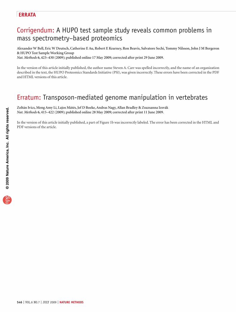
Corrigendum: A HUPO test sample study reveals common problems in mass spectrometry–based proteomicsAlexander W Bell, Eric W Deutsch, Catherine E Au, Robert E Kearney, Ron Beavis, Salvatore Sechi, Tommy Nilsson, John J M Bergeron & HUPO Test Sample Working GroupNat. Methods 6, 423–430 (2009); published online 17 May 2009; corrected after print 29 June 2009.
In the version of this article initially published, the author name Steven A. Carr was spelled incorrectly, and the name of an organization described in the text, the HUPO Proteomics Standards Initiative (PSI), was given incorrectly. These errors have been corrected in the PDF and HTML versions of this article.
Erratum: Transposon-mediated genome manipulation in vertebratesZoltán Ivics, Meng Amy Li, Lajos Mátés, Jef D Boeke, Andras Nagy, Allan Bradley & Zsuzsanna IzsvákNat. Methods 6, 415–422 (2009); published online 28 May 2009; corrected after print 11 June 2009.
In the version of this article initially published, a part of Figure 1b was incorrectly labeled. The error has been corrected in the HTML and PDF versions of the article.
546 | VOL.6 NO.7 | JULY 2009 | nature methods
errata
©20
09 N
atu
re A
mer
ica,
Inc.
All
rig
hts
res
erve
d.
Related Documents