A homozygous p.Glu150Lys mutation in the opsin gene of two Pakistani families with autosomal recessive retinitis pigmentosa Maleeha Azam, 1,2 Muhammad Imran Khan, 1 Andreas Gal, 3 Alamdar Hussain, 1 Syed Tahir Abbas Shah, 1 Muhammad Shakil Khan, 1,4 Ahmed Sadeque, 1 Habib Bokhari, 1 Rob W.J. Collin, 2,5 Ulrike Orth, 3 Maria M. van Genderen, 6 A.I. den Hollander, 2,5 Frans P. M. Cremers, 1,2,5 Raheel Qamar 1,4 1 Department of Biosciences, COMSATS Institute of Information Technology, Islamabad, Pakistan; 2 Department of Human Genetics, Radboud University Nijmegen Medical Centre, Nijmegen, The Netherlands; 3 Department of Human Genetics, University Medical Centre Hamburg Eppendorf, Germany; 4 Shifa College of Medicine, Islamabad, Pakistan; 5 Nijmegen Centre for Molecular Life Sciences, Radboud University Nijmegen Medical Centre, Nijmegen, The Netherlands; 6 Bartiméus Institute for the Visually Impaired, Zeist, The Netherlands Purpose: To identify the gene mutations responsible for autosomal recessive retinitis pigmentosa (arRP) in Pakistani families. Methods: A cohort of consanguineous families with typical RP phenotype in patients was screened by homozygosity mapping using microsatellite markers that mapped close to 21 known arRP genes and five arRP loci. Mutation analysis was performed by direct sequencing of the candidate gene. Results: In two families, RP21 and RP53, homozygosity mapping suggested RHO, the gene encoding rhodopsin, as a candidate disease gene on chromosome 3q21. In six out of seven affected members from the two families, direct sequencing of RHO identified a homozygous c.448G>A mutation resulting in the p.Glu150Lys amino acid change. This variant was first reported in PMK197, an Indian arRP family. Single nucleotide polymorphism analysis in RP21, RP53, and PMK197 showed a common disease-associated haplotype in the three families. Conclusions: In two consanguineous Pakistani families with typical arRP phenotype in the patients, we identified a disease-causing mutation (p.Glu150Lys) in the RHO gene. Single nucleotide polymorphism analysis suggests that the previously reported Indian family (PMK197) and the two Pakistani families studied here share the RHO p.Glu150Lys mutation due to a common ancestry. Retinitis pigmentosa (RP) is a clinically and genetically heterogeneous group of ophthalmic diseases that are characterized by night blindness and gradual loss of peripheral vision due to a progressive degeneration of the photoreceptor cells in the retina. RP is a major cause of inherited blindness in adulthood, with a worldwide prevalence of 1 in 3,500– 4,000 individuals [1-4]. The diagnosis of individuals affected by RP is typically based on fundus examination and the electroretinogram (ERG). In affected individuals, the fundus shows a waxy pallor of the optic discs, retinal vessel attenuation, and peripheral pigmentary alterations with bone spicule deposits. The ERG typically shows a photoreceptor dysfunction with the rod-derived responses being more affected than the cone-derived ones [5]. In addition to clinical heterogeneity of RP, there is a significant genetic heterogeneity of the disease, with different modes of inheritance [6-8]. To date more than 40 different RP- associated genes have been identified (RetNet). These genes encode proteins that are involved either in the Correspondence to: Raheel Qamar, Department of Biosciences, COMSATS Institute of Information Technology, Islamabad, Pakistan; Phone: +92-51-9235033; FAX: +92-51-4442805; email: [email protected] phototransduction cascade, retinoid metabolism, cell–cell interaction, or photoreceptor development. Some of these proteins are transcription or splicing factors, whereas others are intracellular transport molecules [9,10]. Rhodopsin is an abundant rod photoreceptor-specific transmembrane protein, which upon photoexcitation initiates the phototransduction cascade. Heterozygous mutations of the corresponding gene (RHO; MIM 180380) account for approximately 30% of all autosomal dominant RP (adRP) cases in the USA and the UK, whereas the relative frequency and prevalence of RHO mutations is uncertain in the Asian population [11,12]. Up to now there have been more than 100 adRP-causing mutations, but only two autosomal recessive RP-associated mutations have been reported in the coding sequence of RHO (RetNet) [13-15]. The first arRP case, a French-Canadian female patient, carried an apparently homozygous null mutation in exon 4 [14], whereas in the second case, in a consanguineous Indian family, a homozygous c.448G>A (p.Glu150Lys) mutation in exon 2 was reported [15]. Here we report the identification of the p.Glu150Lys RHO mutation in affected individuals from two Pakistani families with arRP. Molecular Vision 2009; 15:2526-2534 <http://www.molvis.org/molvis/v15/a271> Received 9 September 2009 | Accepted 25 November 2009 | Published 3 December 2009 © 2009 Molecular Vision 2526

Welcome message from author
This document is posted to help you gain knowledge. Please leave a comment to let me know what you think about it! Share it to your friends and learn new things together.
Transcript

A homozygous p.Glu150Lys mutation in the opsin gene of twoPakistani families with autosomal recessive retinitis pigmentosa
Maleeha Azam,1,2 Muhammad Imran Khan,1 Andreas Gal,3 Alamdar Hussain,1 Syed Tahir Abbas Shah,1
Muhammad Shakil Khan,1,4 Ahmed Sadeque,1 Habib Bokhari,1 Rob W.J. Collin,2,5 Ulrike Orth,3Maria M. van Genderen,6 A.I. den Hollander,2,5 Frans P. M. Cremers,1,2,5 Raheel Qamar1,4
1Department of Biosciences, COMSATS Institute of Information Technology, Islamabad, Pakistan; 2Department of Human Genetics,Radboud University Nijmegen Medical Centre, Nijmegen, The Netherlands; 3Department of Human Genetics, University MedicalCentre Hamburg Eppendorf, Germany; 4Shifa College of Medicine, Islamabad, Pakistan; 5Nijmegen Centre for Molecular LifeSciences, Radboud University Nijmegen Medical Centre, Nijmegen, The Netherlands; 6Bartiméus Institute for the Visually Impaired,Zeist, The Netherlands
Purpose: To identify the gene mutations responsible for autosomal recessive retinitis pigmentosa (arRP) in Pakistanifamilies.Methods: A cohort of consanguineous families with typical RP phenotype in patients was screened by homozygositymapping using microsatellite markers that mapped close to 21 known arRP genes and five arRP loci. Mutation analysiswas performed by direct sequencing of the candidate gene.Results: In two families, RP21 and RP53, homozygosity mapping suggested RHO, the gene encoding rhodopsin, as acandidate disease gene on chromosome 3q21. In six out of seven affected members from the two families, direct sequencingof RHO identified a homozygous c.448G>A mutation resulting in the p.Glu150Lys amino acid change. This variant wasfirst reported in PMK197, an Indian arRP family. Single nucleotide polymorphism analysis in RP21, RP53, and PMK197showed a common disease-associated haplotype in the three families.Conclusions: In two consanguineous Pakistani families with typical arRP phenotype in the patients, we identified adisease-causing mutation (p.Glu150Lys) in the RHO gene. Single nucleotide polymorphism analysis suggests that thepreviously reported Indian family (PMK197) and the two Pakistani families studied here share the RHO p.Glu150Lysmutation due to a common ancestry.
Retinitis pigmentosa (RP) is a clinically and geneticallyheterogeneous group of ophthalmic diseases that arecharacterized by night blindness and gradual loss of peripheralvision due to a progressive degeneration of the photoreceptorcells in the retina. RP is a major cause of inherited blindnessin adulthood, with a worldwide prevalence of 1 in 3,500–4,000 individuals [1-4]. The diagnosis of individuals affectedby RP is typically based on fundus examination and theelectroretinogram (ERG). In affected individuals, the fundusshows a waxy pallor of the optic discs, retinal vesselattenuation, and peripheral pigmentary alterations with bonespicule deposits. The ERG typically shows a photoreceptordysfunction with the rod-derived responses being moreaffected than the cone-derived ones [5]. In addition to clinicalheterogeneity of RP, there is a significant geneticheterogeneity of the disease, with different modes ofinheritance [6-8]. To date more than 40 different RP-associated genes have been identified (RetNet). These genesencode proteins that are involved either in the
Correspondence to: Raheel Qamar, Department of Biosciences,COMSATS Institute of Information Technology, Islamabad,Pakistan; Phone: +92-51-9235033; FAX: +92-51-4442805; email:[email protected]
phototransduction cascade, retinoid metabolism, cell–cellinteraction, or photoreceptor development. Some of theseproteins are transcription or splicing factors, whereas othersare intracellular transport molecules [9,10].
Rhodopsin is an abundant rod photoreceptor-specifictransmembrane protein, which upon photoexcitation initiatesthe phototransduction cascade. Heterozygous mutations of thecorresponding gene (RHO; MIM 180380) account forapproximately 30% of all autosomal dominant RP (adRP)cases in the USA and the UK, whereas the relative frequencyand prevalence of RHO mutations is uncertain in the Asianpopulation [11,12]. Up to now there have been more than 100adRP-causing mutations, but only two autosomal recessiveRP-associated mutations have been reported in the codingsequence of RHO (RetNet) [13-15]. The first arRP case, aFrench-Canadian female patient, carried an apparentlyhomozygous null mutation in exon 4 [14], whereas in thesecond case, in a consanguineous Indian family, ahomozygous c.448G>A (p.Glu150Lys) mutation in exon 2was reported [15]. Here we report the identification of thep.Glu150Lys RHO mutation in affected individuals from twoPakistani families with arRP.
Molecular Vision 2009; 15:2526-2534 <http://www.molvis.org/molvis/v15/a271>Received 9 September 2009 | Accepted 25 November 2009 | Published 3 December 2009
© 2009 Molecular Vision
2526

Molecular Vision 2009; 15:2526-2534 <http://www.molvis.org/molvis/v15/a271> © 2009 Molecular Vision
2527
METHODSFamily collection: A total of 29 arRP families representativeof the autosomal recessive form of the disease were selectedfrom different areas of northern and central Pakistan.Pedigree analysis and clinical ophthalmic examination: Afteran initial linkage analysis, described below, twoconsanguineous Pakistani families were ascertained; RP21was from Dera Ghazi Khan (central Pakistan) and familyRP53, which was identified later during control panelscreening for the RHO mutation, was from Abbottabad(northern Pakistan). Affected individuals had typical RPsymptoms with early onset of night blindness. Medical andfamily histories of the participants were then obtained withthe help of a questionnaire, and the pedigree structure wasdrawn (Figure 1) [16]. Clinical examination andcharacterization of the affected and unaffected individualswas done by an experienced ophthalmologist. For thispurpose, individuals IV-1 and IV-7 of RP21 and V-1 and V-3of RP53 were then selected for further detailed investigations,including funduscopy of the retina and ERG studies.Ethics committee approval: This study adhered to the tenetsof the Declaration of Helsinki. The consent of both familieswas obtained for this study, which was approved by the ShifaCollege of Medicine, Shifa International Hospital EthicsCommittee Institutional Review Board after an extensivereview of the protocol.Sample collection and DNA isolation: Peripheral bloodsamples of each individual, for which haplotypes are depictedin Figure 1, except RP21-III-6 were drawn by 10 ml sterilesyringes (Terumo Co. BL, Philipines) and were collectedusing vacutainer tubes containing acid citrate dextrose(Becton Dickinson, Franklin Lakes, NJ). All the bloodsamples were stored at 4 °C till processed further. Bloodsamples of 100 random, unrelated, healthy control individualswere also collected and stored as above for determination ofallele frequency in the general Pakistani population. GenomicDNA was isolated using an organic phenol-chloroformextraction method [17,18], briefly the protocol consisted ofcell lysis and protein digestion followed by phenol-chloroform extraction of the peptides, and precipitation ofDNA with ethanol. The precipitated DNA was re-suspendedin Tris-EDTA buffer and stored at 4 °C before use.Linkage mapping and haplotype analysis of arRP genes: Tofind the causative gene in the arRP families, homozygositymapping using polymorphic microsatellite markers for theknown arRP loci was performed. Microsatellite markersmapped by the Cooperative Human Linkage Center wereobtained from Invitrogen, Inc. (Carlsbad, CA). Cytogeneticlocations of these markers as well as the length of theamplified products were obtained from the Human GenomeDatabase and the Marshfield Medical Center database. Table1 lists the arRP genes and loci with the flanking markers usedfor homozygosity mapping. Each microsatellite marker was
amplified in a 25-μl volume consisting of the following:0.5 mM deoxynucleotide triphosphates (dNTPs), 1X TaqBuffer (10 mM Tris-HCl, pH 9.0, 50 mM KCl, 0.1% TritonX-100, 0.01% [w/v] gelatin), 1.5 mM MgCl2, 0.3 μM of eachforward and reverse primer, 2.5 U Taq polymerase, and 50 nggenomic DNA. The thermal cycling conditions were asfollows: initial denaturation of 90 °C for 2 min, followed by40 cycles of 90 °C for 1 min, 55 °C for 1 min, 72 °C for 1 min,and a final extension at 72 °C for 7 min. Genotypes weredefined following PCR and 8% PAGE. Images of the gelswere visualized by UV transillumination after staining withethidium bromide, captured with a digital camera, anddocumented using the software BioCap MW Version 11.01software from Vilber Lourmat (Marne-La-Vallee Cedex1,France). Haplotypes were constructed based upon the allelicpatterns of microsatellites.
After suggestive linkage was observed, additionalflanking markers were selected from the above mentioneddatabases, and haplotypes were constructed for twogenerations in both the pedigrees. The Lod score analysis ofall the genotyped individuals for family RP21 (excludingRP21-III-6 for whom the haplotype was logically deduced)for the markers was performed with the EasyLinkage softwarepackage version 5.02 (Friedrich Alexander UniversityErlangen, Nuremberg, Germany) [19].Sequence analysis of RHO: The sequencing primers forRHO were designed for the upstream promoter sequence andthe five coding exons with Primer3 software (WhiteheadInstitute for Biomedical Research, Cambridge, MA; Table 2).All exons of the RHO gene were bidirectionally sequencedwith an automated DNA sequencer.Statistical analysis: Genotype frequencies of cases andcontrols were compared to determine association/correlation,and chi-square (χ2) tests were performed at the 95%confidence interval to calculate the difference between theobserved and the expected frequencies of the variant.Founder effect detection: To determine the status of anyfounder effect in the Pakistani families RP21 and RP53 andthe Indian family PMK197, microsatellite and singlenucleotide polymorphism (SNP) analysis were performed inall three families. Informative SNPs surrounding the RHOgene were selected, using the UCSC genome browser(University of California Santa Cruz, Santa Clara, CA) andhaplotypes were constructed for all three families (Figure 2).
RESULTSIn family RP21, there are four generations with five affectedindividuals in the fourth generation (Figure 1A), suggestingthat RP has an autosomal recessive inheritance pattern (Figure1). The onset of RP in this family was in the first decade, withtypical symptoms of the disease including night blindness andprogressive visual field loss. Fundus examination of theaffected members of the family revealed typical features of

Figure 1. Pedigrees and 3q21 marker haplotypes spanning the RHO gene in autosomal recessive retinitis pigmentosa families RP21 (A) andRP53 (B). A: Family RP21 consists of four generations. White circles represent unaffected females, filled circles affected females, whitesquares unaffected males, and filled squares affected males. Deceased individuals are shown with a slanting line across the symbol. Alsoshown are the marker loci and their positions in an 18.5-Mb interval, which were used for fine mapping of the disease locus. Marker allelesizes were not accurately determined and therefore we only give two-allele designations. Arrowheads designate crossovers that define the6.8-Mb minimal critical region between D3S1589 and D3S2322. B: Family RP53 consists of five generations. Patient V-3 in one branch ofthe family is homozygous for marker alleles flanking RHO. The disease in affected individual III-12 could be RHO unrelated, suggestinglocus heterogeneity in the different branches of the family. Haplotypes were established for all individuals for which marker alleles are depicted,except for RP21-III-6, the haplotypes of whom were deduced (indicated with brackets around the haplotypes).
Molecular Vision 2009; 15:2526-2534 <http://www.molvis.org/molvis/v15/a271> © 2009 Molecular Vision
2528
RP, including optic disc pallor, attenuation of the retinalvessels, atrophy of the peripheral retinal pigment epithelium(RPE), and formation of bone spicule pigmentation. Inaddition, affected individuals had macular edema in theposterior pole and small white dots in the mid-periphery at thelevel of the RPE (Figure 3). The ERG showed unrecordablerod and cone responses with undetectable 30-Hz flicker ERG(data not shown). In contrast, in unaffected individuals thefundus was completely normal and the rod response and the
mixed rod–cone response had normal amplitudes and implicittimes, with normal oscillatory potentials (data not shown).Single flash photopic ERG and photopic 30-Hz flickerresponses were also normal, thus the ERG pattern wasunremarkable in these individuals.
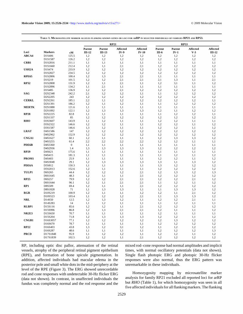
Homozygosity mapping by microsatellite markeranalysis for family RP21 excluded all reported loci for arRPbut RHO (Table 1), for which homozygosity was seen in allfive affected individuals for all flanking markers. The flanking
TABLE 1. MICROSATELLITE MARKER ALLELES FLANKING KNOWN GENES OR LOCI FOR ARRP IN SELECTED INDIVIDUALS OF FAMILIES RP21 AND RP53.
RP21 RP53
Loci MarkersParentIII-12
ParentIII-13
AffectedIV-9
AffectedIV-10
ParentIII-6
ParentIV-1
AffectedV-3
AffectedIII-12
ABCA4 D1S406 125.5 1,2 1,2 1,2 1,2 1,2 1,2 1,2 1,2 D1S1587 126.2 1,2 1,2 1,2 1,2 1,2 1,2 1,2 1,2CRB1 D1S2816 211.1 1,1 1,1 1,1 1,1 1,1 1,1 1,1 1,1 D1S1660 212.4 1,2 1,1 2,1 2,1 1,2 1,2 1,2 1,2USH2A D1S474 233.9 1,2 1,2 1,2 1,2 1,2 1,2 1,2 1,2 D1S2827 234.5 1,2 1,2 1,2 1,2 1,2 1,2 1,2 1,2RPE65 D1S2806 100.4 1,2 1,3 2,1 2,1 1,1 1,1 1,1 1,1 D1S219 101.5 1,2 1,2 2,1 2,1 2,1 1,3 1,1 2,1RP32 D1S2808 131.9 1,2 1,1 2,1 1,1 1,2 3,1 1,1 1,2 D1S2896 134.2 1,1 2,1 1,1 1,1 1,1 1,1 1,1 1,1 D1S485 136.9 1,2 1,2 2,1 1,2 1,2 1,2 1,2 1,2SAG D2S2348 242.2 2,1 1,2 1,2 1,1 1,1 1,2 1,2 1,2 D2S2205 243 1,2 1,2 1,2 1,2 1,2 1,2 1,2 1,2CERKL D2S2261 185.1 2,2 1,1 1,2 1,2 1,2 1,2 1,2 1,1 D2S1391 186.2 1,2 1,1 1,2 1,1 1,1 1,2 1,2 1,2MERTK D2S1888 121.6 1,1 1,1 1,1 1,1 1,1 1,1 1,1 1,1 D2S1892 122.1 1,2 1,3 1,3 1,3 1,2 1,1 1,2 1,2RP28 D2S2225 82.8 1,1 1,1 1,1 1,1 1,1 1,2 1,2 1,1 D2S1337 85 1,2 1,2 1,2 1,2 1,2 1,2 1,2 1,2RHO D3S3607 143.9 1,1 1,2 1,1 1,1 1,2 1,2 1,1 1,1 D3S2322 146.6 1,2 1,2 1,1 1,1 1,2 1,2 1,1 1,2 D3S1587 146.6 1,3 1,2 1,1 1,1 1,2 1,2 1,1 1,2LRAT D4S1586 147 1,2 1,2 1,2 1,2 1,2 1,2 1,2 1,2 D4S2962 152.9 1,2 1,2 1,2 1,2 1,2 1,2 1,2 1,2CNGA1 D4S1627 60.1 1,2 1,1 1,1 1,1 1,2 1,3 1,3 1,2 D4S3255 61.4 2,2 2,2 2,2 2,2 1,2 1,2 1,1 1,1PDE6B D4S3360 0 1,1 1,1 1,1 1,1 1,1 1,1 1,1 1,1 D4S2936 1.4 1,3 1,3 1,3 1,3 1,2 1,2 1,2 1,2RP29 D4S621 175.6 1,1 1,1 1,1 1,1 1,1 1,1 1,1 1,1 D4S415 181.3 1,1 1,1 1,1 1,1 1,1 1,2 1,1 1,2PROM1 D4S403 25.9 1,1 1,1 1,1 1,1 1,2 1,2 1,1 1,2 D4S3048 29.1 1,2 1,3 1,3 1,3 1,3 1,1 1,3 1,2PDE6A D5S812 150.3 1,1 1,1 1,1 1,1 1,3 1,2 1,2 1,2 D5S2013 152.6 1,2 1,1 1,1 1,1 1,2 1,2 1,2 1,2TULP1 D6S265 44.4 1,2 1,2 1,2 2,1 1,2 1,3 1,2 1,3 D6S1645 48.3 1,2 1,1 1,1 2,1 1,2 1,2 1,2 1,2RP25 D6S257 79.9 1,2 1,2 2,1 2,1 1,2 1,2 2,1 1,2 D6S1644 96.1 1,2 1,1 2,1 2,1 1,1 1,2 1,1 1,2RP1 D8S509 69.4 1,2 1,1 1,1 2,1 1,2 1,2 1,2 1,2 D8S1828 71 1,1 1,3 1,3 1,1 1,1 1,3 1,3 1,2RGR D10S219 100.9 1,2 1,1 1,1 1,2 1,2 1,1 2,1 1,2 D10S523 103.4 1,1 1,2 1,2 2,1 1,1 1,3 1,3 1,2NRL D14S50 12.5 1,2 1,3 1,2 1,1 1,2 1,2 2,1 1,1 D14S283 14 1,1 1,2 1,1 1,1 1,1 1,2 1,1 1,1RLBP1 D15S116 85.6 1,2 1,2 1,1 2,1 1,2 1,2 1,2 1,2 D15S996 86.8 1,2 1,3 2,1 2,1 1,2 1,1 1,1 1,2NR2E3 D15S650 70.7 1,1 1,1 1,1 1,1 1,2 1,1 1,1 1,1 D15S204 71.8 1,2 1,3 1,3 1,3 1,2 1,2 1,1 1,2CNGB1 D16S3057 77.1 1,2 1,2 1,2 1,2 1,2 1,3 1,2 1,2 D16S670 78.7 1,1 1,1 1,1 1,1 1,2 1,2 1,2 1,2RP22 D16S403 43.8 1,3 1,2 3,1 1,2 1,1 1,2 1,2 1,2 D16S287 48.6 1,1 1,1 1,1 1,1 1,2 1,2 1,2 1,3PRCD D17S1848 95.9 1,1 1,2 1,2 1,1 1,2 1,2 1,1 1,2 D17S1839 102.5 1,2 1,2 1,1 2,1 1,2 1,2 1,2 1,2
Molecular Vision 2009; 15:2526-2534 <http://www.molvis.org/molvis/v15/a271> © 2009 Molecular Vision
2529
cM
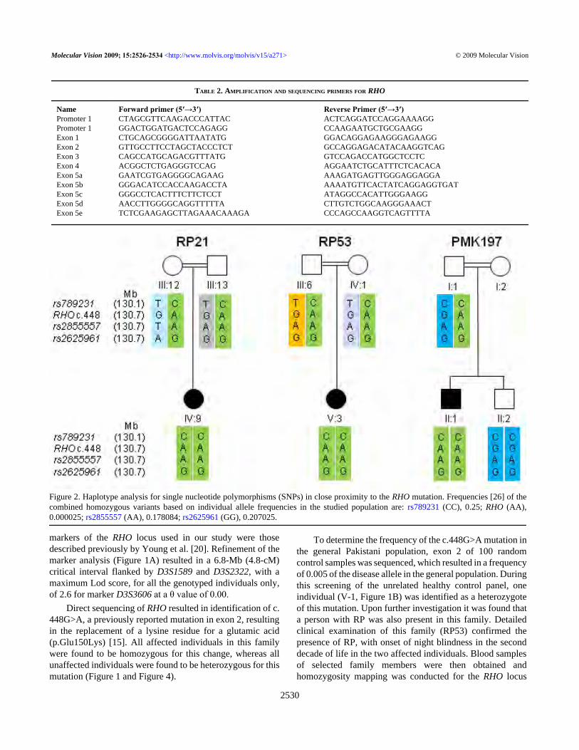
TABLE 2. AMPLIFICATION AND SEQUENCING PRIMERS FOR RHO
Name Forward primer (5′→3′) Reverse Primer (5′→3′)Promoter 1 CTAGCGTTCAAGACCCATTAC ACTCAGGATCCAGGAAAAGGPromoter 1 GGACTGGATGACTCCAGAGG CCAAGAATGCTGCGAAGGExon 1 CTGCAGCGGGGATTAATATG GGACAGGAGAAGGGAGAAGGExon 2 GTTGCCTTCCTAGCTACCCTCT GCCAGGAGACATACAAGGTCAGExon 3 CAGCCATGCAGACGTTTATG GTCCAGACCATGGCTCCTCExon 4 ACGGCTCTGAGGGTCCAG AGGAATCTGCATTTCTCACACAExon 5a GAATCGTGAGGGGCAGAAG AAAGATGAGTTGGGAGGAGGAExon 5b GGGACATCCACCAAGACCTA AAAATGTTCACTATCAGGAGGTGATExon 5c GGGCCTCACTTTCTTCTCCT ATAGGCCACATTGGGAAGGExon 5d AACCTTGGGGCAGGTTTTTA CTTGTCTGGCAAGGGAAACTExon 5e TCTCGAAGAGCTTAGAAACAAAGA CCCAGCCAAGGTCAGTTTTA
Molecular Vision 2009; 15:2526-2534 <http://www.molvis.org/molvis/v15/a271> © 2009 Molecular Vision
2530
markers of the RHO locus used in our study were thosedescribed previously by Young et al. [20]. Refinement of themarker analysis (Figure 1A) resulted in a 6.8-Mb (4.8-cM)critical interval flanked by D3S1589 and D3S2322, with amaximum Lod score, for all the genotyped individuals only,of 2.6 for marker D3S3606 at a θ value of 0.00.
Direct sequencing of RHO resulted in identification of c.448G>A, a previously reported mutation in exon 2, resultingin the replacement of a lysine residue for a glutamic acid(p.Glu150Lys) [15]. All affected individuals in this familywere found to be homozygous for this change, whereas allunaffected individuals were found to be heterozygous for thismutation (Figure 1 and Figure 4).
To determine the frequency of the c.448G>A mutation inthe general Pakistani population, exon 2 of 100 randomcontrol samples was sequenced, which resulted in a frequencyof 0.005 of the disease allele in the general population. Duringthis screening of the unrelated healthy control panel, oneindividual (V-1, Figure 1B) was identified as a heterozygoteof this mutation. Upon further investigation it was found thata person with RP was also present in this family. Detailedclinical examination of this family (RP53) confirmed thepresence of RP, with onset of night blindness in the seconddecade of life in the two affected individuals. Blood samplesof selected family members were then obtained andhomozygosity mapping was conducted for the RHO locus
Figure 2. Haplotype analysis for single nucleotide polymorphisms (SNPs) in close proximity to the RHO mutation. Frequencies [26] of thecombined homozygous variants based on individual allele frequencies in the studied population are: rs789231 (CC), 0.25; RHO (AA),0.000025; rs2855557 (AA), 0.178084; rs2625961 (GG), 0.207025.

Molecular Vision 2009; 15:2526-2534 <http://www.molvis.org/molvis/v15/a271> © 2009 Molecular Vision
2531
(Figure 1B). Homozygosity mapping and sequencing resultedin identification of 11 heterozygotes, including the affectedindividual III-12, while only one female, V-3, washomozygous for the c.448G>A mutation. Sequencing of theremaining exons and upstream regulatory regions of RHO inIII-12 did not reveal any additional RHO mutations.
In addition to the 100 control samples, exon 2 of theRHO gene was also sequenced using the DNA samples of 28probands from a Pakistani RP cohort to determine theprevalence of the c.448G>A mutation. None of the affectedindividuals were found to carry this mutation. The χ2 analysisresulted in a significant association of the homozygous mutantallele A (p<0.05) with the disease when calculated for all theindividuals of both the families and also when compared withthe control samples (Table 3).
Founder effect analysis of the c.448G>A mutation in thetwo Pakistani families (RP21, RP53) and the Indian family(PMK197) [15] revealed that all affected members share thesame homozygous haplotype consisting of three highlyinformative SNPs surrounding the RHO gene mutation(Figure 2), therefore suggesting a common ancestry betweenthe three families.
DISCUSSIONTwo apparently unrelated, ethnically diverse, Pakistani arRPfamilies from different geographical regions of Pakistan,
RP21 from the Punjab province and RP53 from the NorthWest Frontier province, were identified and analyzed byhomozygosity mapping using highly informativemicrosatellite markers flanking the known arRP genes andloci. Initially, one of the families (RP21) showedhomozygosity for the RHO (RP4) locus at 3q22.1 in allaffected individuals of the family.
Mutations in the rhodopsin gene have previously beenshown to result in RP. However, most of the RHO mutationshave been found in families with adRP, Human GenomeMutation Database (HGMD, rhodopsin mutation), whereasonly two mutations have been shown to cause RP with anautosomal recessive mode of inheritance [14,15].
In this study, we identified the c.448G>A mutation in theRHO gene in two different consanguineous arRP familiesfrom Pakistan and also determined the frequency of thepathogenic allele in the normal population. While determiningthe population frequency, we found the c.448G>A mutationin the heterozygous state in one out of 100 control samples,but we did not find it in any of the other 28 families from ourRP panel that were screened for the c.448G>A mutation.Further investigations of the family of the heterozygousindividual from the control samples resulted in theidentification of an additional arRP family (RP53). DNAsequencing revealed the same causative mutation in onebranch of the family. There seems to be genetic heterogeneity
Figure 3. Fundus photograph of anindividual affected with retinitispigmentosa. The fundus appearance ofindividual RP21-IV-1 is characteristicof advanced RP, with optic disc pallor,attenuation of the retinal vessels,peripheral pigment epithelial atrophy,and bone spicule pigmentation. Theposterior pole shows macular edema,and small white dots are present in themid-periphery at the level of the retinalpigment epithelium

Molecular Vision 2009; 15:2526-2534 <http://www.molvis.org/molvis/v15/a271> © 2009 Molecular Vision
2532
of the RP in this family as individual III-12 may either carrya second, yet unidentified, variant in the RHO intronicsequences or a mutation in a different gene. The statisticalanalysis for the association/correlation showed that the mutantallele A is significantly associated (p<0.05) with the diseasephenotype because the AA genotype is always observed inknown affected individuals and is absent from the normalpopulation (Table 3, Figure 2).
This c.448G>A mutation has previously been reported tocause arRP in an Indian family from southern India [15],which is ethnically different and geographically isolated fromthe present families. We therefore compared, in the twoPakistani families and the Indian family, some of the SNPsfrom the surrounding region of RHO to determine if they sharea haplotype, suggesting a founder mutation in these families.Indeed, our analysis revealed a shared haplotype in thepatients of the three families, suggesting a common ancestry.This is remarkable as the three families belong to diverse
ethnic groups and have lived at their present location forseveral generations with no relative having moved to any otherpart of the subcontinent, at least for the last 10 generations;thus any commonality in ancestry must be more than severalhundred years old.
Genetic screening of RP families is not only helpful inunderstanding the molecular mechanism of the disease; it isalso important for genetic counseling. Traditionalconsanguineous marriages are major risk factors forautosomal recessive diseases, including retinopathies.Families have little knowledge of the inheritance of thediseases, and through such studies as the present one, manyfamilies could be counseled appropriately, as was done formembers of families RP21 and RP53.
The rod visual pigment rhodopsin consists of theapoprotein opsin and the covalently bound chromophore 11-cis-retinal [21]. This protein is a heptahelical G protein-coupled receptor synthesized at a high level in the rod inner
Figure 4. Sequence analysis of RHO in family RP21. A: Sequence trace of part of exon 2 of an affected individual (RP21-IV-1) showing thehomozygous mutant sequence c.448G>A. B: Sequencing results of RP21-IV-7 showing the heterozygous c.448G>A sequence. C: Sequencetrace of an unaffected individual with the homozygous wild-type c.448G. d
TABLE 3. CHI SQUARE ANALYSIS.
A Unaffected family members Patients p (χ2)GG/GA 20 1 <0.05 (22.04)
AA 0 6B Controls+Unaffected family members Patients p (χ2)
GG/GA 119 1 <0.05 (107.10)AA 0 6
A: χ2 analysis of families RP21 and RP53 showing association of alleles AA with the disease. B: χ2 analysis of the families andthe controls.

Molecular Vision 2009; 15:2526-2534 <http://www.molvis.org/molvis/v15/a271> © 2009 Molecular Vision
2533
segment and transported subsequently to the rod outersegment [22]. The seven transmembrane segments (H1–H7)are sequentially linked by extracellular (E1–E3) andcytoplasmic (C1–C3) loops [23]. The first reported nullmutation (p.Glu249Ter) associated with arRP was shown toabolish the function of the helices H6 and H7, which containsthe retinal binding site. Remarkably, this mutation caused anabnormal ERG pattern even in heterozygotes [14].
Residue Glu150, which is embedded at the edge of thephospholipid bilayer, is located near the C-terminal region ofthe C2 loop, which is important for proper folding of opsinand its binding to transducin and thus for the initiation of thephototransduction cascade [24]. However, Zhu et al. [25] haveshown that the homozygous p.Glu150Lys change does notaffect binding of opsin to transducin but instead affects theexport of the protein opsin from the Golgi apparatus. This isdue to improper glycosylation of the protein, which results inretention of the protein inside the Golgi, leading to aninsufficient supply of opsin to the rod outer segments. Theprocessing of rhodopsin protein has been shown to be normalin the p.Glu150Lys heterozygotes [25].
We conclude that linkage and mutation analysis of knownretinal disease genes in Pakistani patients with RP resulted inthe identification of a RHO mutation in two out of 30 familiesthat very likely have a common ancestor and are also distantlyrelated with the Indian family previously found to harbor thismutation. We also observed genetic heterogeneity in one ofthese two families. These findings have facilitated accurategenetic counseling of patients and unaffected familymembers.
ACKNOWLEDGMENTSThe authors dedicate this paper to the memory of ProfessorDr. Aslam Malik who, until the day he died, was constantlygoing into the field with us to conduct clinical investigationsof the individuals. Without his efforts and constantencouragement, we could not have completed this work. Wealso thank all the families for donating their blood and fullycooperating with us. This work was supported by grant no.530 awarded (to R.Q.) by the Higher Education Commissionof Pakistan. Part of the funding for this work was provided byCOMSATS Institute of Information Technology (to A.H.).This work was also financially supported by the FoundationFighting Blindness, USA (to A.I.d.H.). We also acknowledgethe Higher Education Commission of Pakistan for awardinga IRSIP Scholarship to M.A. to perform part of the researchat the Radboud University, Nijmegen Medical Center,Nijmegen, the Netherlands.
REFERENCES1. Berson EL. Retinitis pigmentosa. The Friedenwald Lecture.
Invest Ophthalmol Vis Sci 1993; 34:1659-76. [PMID:8473105]
2. Jay M. On the heredity of retinitis pigmentosa. Br J Ophthalmol1982; 66:405-16. [PMID: 7093178]
3. Evans K, Bhattacharya S. Retinal dystrophies: a moleculargenetic approach. In: Pawlowitzki IH, Edwards JH,Thompson EA, editors. Genetic Mapping of Disease Genes.San Diego: Academic Press; 1997. p. 247–54.
4. Bunker CH, Berson EL, Bromley WC, Hayes RP, Roderick TH.Prevalence of retinitis pigmentosa in Maine. Am JOphthalmol 1984; 97:357-65. [PMID: 6702974]
5. Hood DC, Birch DG. Light adaptation of human rod receptors:the leading edge of the human a-wave and models of rodreceptor activity. Vision Res 1993; 33:1605-18. [PMID:8236849]
6. Boughman JA, Caldwell RJ. Genetic and clinicalcharacterization of a survey population with retinitispigmentosa. Clinical, Structural and Biochemical Advancesin Hereditary Eye Disorders. New York: Alan R. Liss Inc.;1982; P. 147–66.
7. Sullivan LS, Daiger SP. Inherited retinal degeneration:exceptional genetic and clinical heterogeneity. Mol MedToday 1996; 2:380-6. [PMID: 8885257]
8. Rivolta C, Sharon D, DeAngelis MM, Dryja TP. Retinitispigmentosa and allied diseases: numerous diseases, genes andinheritance patterns. Hum Mol Genet 2002; 11:1219-27.[PMID: 12015282]
9. Maubaret C, Hamel C. Genetics of retinitis pigmentosa:metabolic classification and phenotype/genotypecorrelations. J Fr Ophtalmol 2005; 28:71-92. [PMID:15767903]
10. Hims MM, Diager SP, Inglehearn CF. Retinitis pigmentosa:genes, proteins and prospects. Dev Ophthalmol 2003;37:109-25. [PMID: 12876833]
11. Gregory-Evans K, Bhattacharya SS. Genetic blindness: currentconcepts in the pathogenesis of human outer retinaldystrophies. Trends Genet 1998; 14:103-8. [PMID: 9540407]
12. Inglehearn CF, Tarttelin EE, Plant C, Peacock RE, Al-Maghtheh M, Vithana E. A linkage survey of 20 dominantretinitis pigmentosa families: frequencies of the nine knownloci and evidence for further heterogeneity. J Med Genet1998; 35:1-5. [PMID: 9475085]
13. Sung CH, Davenport CM, Hennessey JC, Maumenee IH,Jacobson SG, Heckenlively JR, Nowakowski R, Fishman G,Gouras P, Nathans J. Rhodopsin mutations in autosomaldominant retinitis pigmentosa. Proc Natl Acad Sci USA 1991;88:6481-4. [PMID: 1862076]
14. Rosenfeld PJ, Cowley GS, McGee TL, Sandberg MA, BersonEL, Dryja TP. A null mutation in the rhodopsin gene causesrod photoreceptor dysfunction and autosomal recessiveretinitis pigmentosa. Nat Genet 1992; 1:209-13. [PMID:1303237]
15. Kumaramanickavel G, Maw M, Denton MJ, John S, SrikumariCR, Orth U, Oehlmann R, Gal A. Missense rhodopsinmutation in a family with recessive RP. Nat Genet 1994;8:10-1. [PMID: 7987385]Letter
16. Bennett RL, Steinhaus KA, Uhrich SB, O'Sullivan CK, RestaRG, Lochner DD, Markei DS, Vincent V, Hamanishi J.Recommendations for standardized human pedigreenomenclature. Am J Hum Genet 1995; 56:745-52. [PMID:7887430]
17. Ausubel FM, Brent R, Kingston RE, More DD, Seidman JG,Smith JA, Struhl K, editors. Current protocols in molecular
http://www.ncbi.nlm.nih.gov/entrez/query.fcgi?cmd=Retrieve&db=PubMed&dopt=abstract&list_uids=8473105
http://www.ncbi.nlm.nih.gov/entrez/query.fcgi?cmd=Retrieve&db=PubMed&dopt=abstract&list_uids=8473105
http://www.ncbi.nlm.nih.gov/entrez/query.fcgi?cmd=Retrieve&db=PubMed&dopt=abstract&list_uids=7093178
http://www.ncbi.nlm.nih.gov/entrez/query.fcgi?cmd=Retrieve&db=PubMed&dopt=abstract&list_uids=6702974
http://www.ncbi.nlm.nih.gov/entrez/query.fcgi?cmd=Retrieve&db=PubMed&dopt=abstract&list_uids=8236849
http://www.ncbi.nlm.nih.gov/entrez/query.fcgi?cmd=Retrieve&db=PubMed&dopt=abstract&list_uids=8236849
http://www.ncbi.nlm.nih.gov/entrez/query.fcgi?cmd=Retrieve&db=PubMed&dopt=abstract&list_uids=8885257
http://www.ncbi.nlm.nih.gov/entrez/query.fcgi?cmd=Retrieve&db=PubMed&dopt=abstract&list_uids=9540407
http://www.ncbi.nlm.nih.gov/entrez/query.fcgi?cmd=Retrieve&db=PubMed&dopt=abstract&list_uids=9475085
http://www.ncbi.nlm.nih.gov/entrez/query.fcgi?cmd=Retrieve&db=PubMed&dopt=abstract&list_uids=1862076
http://www.ncbi.nlm.nih.gov/entrez/query.fcgi?cmd=Retrieve&db=PubMed&dopt=abstract&list_uids=1303237
http://www.ncbi.nlm.nih.gov/entrez/query.fcgi?cmd=Retrieve&db=PubMed&dopt=abstract&list_uids=1303237

Molecular Vision 2009; 15:2526-2534 <http://www.molvis.org/molvis/v15/a271> © 2009 Molecular Vision
2534
biology, supplement 27. New York: J. Wiley and Sons; 1987.p. 2.4.1.
18. Sambrook J, Fritsch EF, Maniatis T. Molecular Cloning: ALaboratory Manual, 2nd ed. Cold Spring Harbor, New York:Cold Spring Harbor Laboratory Press; 1989.
19. Lindner TH, Hoffmann K. easyLINKAGE: A PERL script foreasy and automated two-/multi-point linkage analyses.Bioinformatics 2005; 21:405-7. [PMID: 15347576]
20. Yong RYY, Chee CKL, Yap EPH. A two-stage approachidentifies a Q344X mutation in the rhodopsin gene of aChinese Singaporean family with autosomal dominantretinitis pigmentosa. Ann Acad Med Singapore 2005;34:94-9. [PMID: 15726226]
21. Okada T, Sugihara M, Bondar AN, Elstner M, Entel P, Buss V.The retinal conformation and its environment in rhodopsin inlight of a new 2.2 A crystal structure. J Mol Biol 2004;342:571-83. [PMID: 15327956]
22. Tam BM, Moritz OL. Dark rearing rescues P23H rhodopsin-induced retinal degeneration in a transgenic Xenopus laevis
model of Retinitis Pigmentosa: A chromophore-dependentmechanism characterized by production of N-terminallytruncated mutant Rhodopsin. J Neurosci 2007; 27:9043-53.[PMID: 17715341]
23. Sakmar TP. Structure of rhodopsin and the superfamily ofseven-helical receptors: the same and not the same. Curr OpinCell Biol 2002; 14:189-95. [PMID: 11891118]
24. Ridge KD, Palczewski K. Visual rhodopsin sees the light:structure and mechanism of G protein signaling. J Biol Chem2007; 282:9297-301. [PMID: 17289671]
25. Zhu L, Imanishi Y, Filipek S, Alekseev A, Jastrzebska B, SunW, Saperstein DA, Palczewski K. Autosomal recessiveretinitis pigmentosa and E150K mutation in the opsin gene. JBiol Chem 2006; 281:22289-98. [PMID: 16737970]
26. The International HapMap Consortium. A second generationhuman haplotype map of over 3.1 million SNPs. Nature 2007;449:851-61. [PMID: 17943122]
The print version of this article was created on 1 December 2009. This reflects all typographical corrections and errata to thearticle through that date. Details of any changes may be found in the online version of the article.
http://www.ncbi.nlm.nih.gov/entrez/query.fcgi?cmd=Retrieve&db=PubMed&dopt=abstract&list_uids=7887430
Related Documents