Citation: Biselli, R.; Nisini, R.; Lista, F.; Autore, A.; Lastilla, M.; De Lorenzo, G.; Peragallo, M.S.; Stroffolini, T.; D’Amelio, R. A Historical Review of Military Medical Strategies for Fighting Infectious Diseases: From Battlefields to Global Health. Biomedicines 2022, 10, 2050. https://doi.org/10.3390/ biomedicines10082050 Academic Editor: Willibald Wonisch Received: 27 July 2022 Accepted: 13 August 2022 Published: 22 August 2022 Publisher’s Note: MDPI stays neutral with regard to jurisdictional claims in published maps and institutional affil- iations. Copyright: © 2022 by the authors. Licensee MDPI, Basel, Switzerland. This article is an open access article distributed under the terms and conditions of the Creative Commons Attribution (CC BY) license (https:// creativecommons.org/licenses/by/ 4.0/). biomedicines Review A Historical Review of Military Medical Strategies for Fighting Infectious Diseases: From Battlefields to Global Health Roberto Biselli 1,†,‡ , Roberto Nisini 2, * ,‡ , Florigio Lista 3 , Alberto Autore 4 , Marco Lastilla 5 , Giuseppe De Lorenzo 6 , Mario Stefano Peragallo 7,† , Tommaso Stroffolini 8,† and Raffaele D’Amelio 9,† 1 Ispettorato Generale della Sanità Militare, Stato Maggiore della Difesa, Via S. Stefano Rotondo 4, 00184 Roma, Italy 2 Dipartimento di Malattie Infettive, Istituto Superiore di Sanità, Viale Regina Elena 299, 00161 Roma, Italy 3 Dipartimento Scientifico, Policlinico Militare, Comando Logistico dell’Esercito, Via S. Stefano Rotondo 4, 00184 Roma, Italy 4 Osservatorio Epidemiologico della Difesa, Ispettorato Generale della Sanità Militare, Stato Maggiore della Difesa, Via S. Stefano Rotondo 4, 00184 Roma, Italy 5 Istituto di Medicina Aerospaziale, Comando Logistico dell’Aeronautica Militare, Viale Piero Gobetti 2, 00185 Roma, Italy 6 Comando Generale dell’Arma dei Carabinieri, Dipartimento per l’Organizzazione Sanitaria e Veterinaria, Viale Romania 45, 00197 Roma, Italy 7 Centro Studi e Ricerche di Sanità e Veterinaria, Comando Logistico dell’Esercito, Via S. Stefano Rotondo 4, 00184 Roma, Italy 8 Dipartimento di Malattie Infettive e Tropicali, Policlinico Umberto I, 00161 Roma, Italy 9 Dipartimento di Medicina Clinica e Molecolare, Sapienza Università di Roma, Via di Grottarossa 1035-1039, 00189 Roma, Italy * Correspondence: [email protected]; Tel.: +39-06-49902659 † Retired. ‡ These Authors contributed equally to this work. Abstract: The environmental conditions generated by war and characterized by poverty, undernu- trition, stress, difficult access to safe water and food as well as lack of environmental and personal hygiene favor the spread of many infectious diseases. Epidemic typhus, plague, malaria, cholera, typhoid fever, hepatitis, tetanus, and smallpox have nearly constantly accompanied wars, frequently deeply conditioning the outcome of battles/wars more than weapons and military strategy. At the end of the nineteenth century, with the birth of bacteriology, military medical researchers in Germany, the United Kingdom, and France were active in discovering the etiological agents of some diseases and in developing preventive vaccines. Emil von Behring, Ronald Ross and Charles Laveran, who were or served as military physicians, won the first, the second, and the seventh Nobel Prize for Physiology or Medicine for discovering passive anti-diphtheria/tetanus immunotherapy and for identifying mosquito Anopheline as a malaria vector and plasmodium as its etiological agent, respectively. Meanwhile, Major Walter Reed in the United States of America discovered themosquito vector of yellow fever, thus paving the way for its prevention by vector control. In this work, the military relevance of some vaccine-preventable and non-vaccine-preventable infectious diseases, as well as of biological weapons, and the military contributions to their control will be described. Currently, the civil–military medical collaboration is getting closer and becoming interdependent, from research and development for the prevention of infectious diseases to disasters and emergencies management, as recently demonstrated in Ebola and Zika outbreaks and the COVID-19 pandemic, even with the high biocontainment aeromedical evacuation, in a sort of global health diplomacy. Keywords: the military; infectious diseases; passive immunization; vaccines; antibodies; active immunization; biological agents; war Biomedicines 2022, 10, 2050. https://doi.org/10.3390/biomedicines10082050 https://www.mdpi.com/journal/biomedicines

Welcome message from author
This document is posted to help you gain knowledge. Please leave a comment to let me know what you think about it! Share it to your friends and learn new things together.
Transcript
Citation: Biselli, R.; Nisini, R.; Lista,
F.; Autore, A.; Lastilla, M.; De
Lorenzo, G.; Peragallo, M.S.;
Stroffolini, T.; D’Amelio, R. A
Historical Review of Military Medical
Strategies for Fighting Infectious
Diseases: From Battlefields to Global
Health. Biomedicines 2022, 10, 2050.
https://doi.org/10.3390/
biomedicines10082050
Academic Editor: Willibald Wonisch
Received: 27 July 2022
Accepted: 13 August 2022
Published: 22 August 2022
Publisher’s Note: MDPI stays neutral
with regard to jurisdictional claims in
published maps and institutional affil-
iations.
Copyright: © 2022 by the authors.
Licensee MDPI, Basel, Switzerland.
This article is an open access article
distributed under the terms and
conditions of the Creative Commons
Attribution (CC BY) license (https://
creativecommons.org/licenses/by/
4.0/).
biomedicines
Review
A Historical Review of Military Medical Strategies for FightingInfectious Diseases: From Battlefields to Global HealthRoberto Biselli 1,†,‡ , Roberto Nisini 2,*,‡, Florigio Lista 3, Alberto Autore 4, Marco Lastilla 5,Giuseppe De Lorenzo 6 , Mario Stefano Peragallo 7,† , Tommaso Stroffolini 8,† and Raffaele D’Amelio 9,†
1 Ispettorato Generale della Sanità Militare, Stato Maggiore della Difesa, Via S. Stefano Rotondo 4,00184 Roma, Italy
2 Dipartimento di Malattie Infettive, Istituto Superiore di Sanità, Viale Regina Elena 299, 00161 Roma, Italy3 Dipartimento Scientifico, Policlinico Militare, Comando Logistico dell’Esercito, Via S. Stefano Rotondo 4,
00184 Roma, Italy4 Osservatorio Epidemiologico della Difesa, Ispettorato Generale della Sanità Militare, Stato Maggiore della
Difesa, Via S. Stefano Rotondo 4, 00184 Roma, Italy5 Istituto di Medicina Aerospaziale, Comando Logistico dell’Aeronautica Militare, Viale Piero Gobetti 2,
00185 Roma, Italy6 Comando Generale dell’Arma dei Carabinieri, Dipartimento per l’Organizzazione Sanitaria e Veterinaria,
Viale Romania 45, 00197 Roma, Italy7 Centro Studi e Ricerche di Sanità e Veterinaria, Comando Logistico dell’Esercito, Via S. Stefano Rotondo 4,
00184 Roma, Italy8 Dipartimento di Malattie Infettive e Tropicali, Policlinico Umberto I, 00161 Roma, Italy9 Dipartimento di Medicina Clinica e Molecolare, Sapienza Università di Roma, Via di Grottarossa 1035-1039,
00189 Roma, Italy* Correspondence: [email protected]; Tel.: +39-06-49902659† Retired.‡ These Authors contributed equally to this work.
Abstract: The environmental conditions generated by war and characterized by poverty, undernu-trition, stress, difficult access to safe water and food as well as lack of environmental and personalhygiene favor the spread of many infectious diseases. Epidemic typhus, plague, malaria, cholera,typhoid fever, hepatitis, tetanus, and smallpox have nearly constantly accompanied wars, frequentlydeeply conditioning the outcome of battles/wars more than weapons and military strategy. Atthe end of the nineteenth century, with the birth of bacteriology, military medical researchers inGermany, the United Kingdom, and France were active in discovering the etiological agents of somediseases and in developing preventive vaccines. Emil von Behring, Ronald Ross and Charles Laveran,who were or served as military physicians, won the first, the second, and the seventh Nobel Prizefor Physiology or Medicine for discovering passive anti-diphtheria/tetanus immunotherapy andfor identifying mosquito Anopheline as a malaria vector and plasmodium as its etiological agent,respectively. Meanwhile, Major Walter Reed in the United States of America discovered the mosquitovector of yellow fever, thus paving the way for its prevention by vector control. In this work, themilitary relevance of some vaccine-preventable and non-vaccine-preventable infectious diseases,as well as of biological weapons, and the military contributions to their control will be described.Currently, the civil–military medical collaboration is getting closer and becoming interdependent,from research and development for the prevention of infectious diseases to disasters and emergenciesmanagement, as recently demonstrated in Ebola and Zika outbreaks and the COVID-19 pandemic,even with the high biocontainment aeromedical evacuation, in a sort of global health diplomacy.
Keywords: the military; infectious diseases; passive immunization; vaccines; antibodies; activeimmunization; biological agents; war
Biomedicines 2022, 10, 2050. https://doi.org/10.3390/biomedicines10082050 https://www.mdpi.com/journal/biomedicines
Biomedicines 2022, 10, 2050 2 of 96
1. Introduction
The military worldwide have always been challenged with the issue of infectiousdiseases, which may deeply influence the outcome of battles/wars. The military are par-ticularly exposed to the risk of infectious diseases for a series of reasons, including thecommunity life, often in precarious environmental conditions regarding the hygiene ofwater and food supply, sanitation, the traumatism with contaminated wounds, and thepossibility to be exposed to extreme temperatures and to diseases unknown in their countryof origin, for which no natural immunization has, therefore, been developed [1,2]. In 431BCE, the outcome of the Peloponnesian war between the Athens of Pericles and Sparta wasdetermined by the so-called “plague of Athens”, a terrible epidemic responsible for thedeath of approximately one-third of the Athens’ population, of Pericles and two of his sons,which seems to have been due to an outbreak of Salmonella typhi, as recently reported [3,4].More recently, Napoleon lost 90% of his army deployed to Haiti, 27,000/30,000 soldiersincluding the commander, who was Napoleon’s brother-in-law, as a consequence of yellowfever, which was endemic in Haiti, but unknown to the French troops, which were, there-fore, highly vulnerable. This situation induced Napoleon to retire from the New Worldand leave Louisiana for the then-nascent United States of America (USA) to concentratehis efforts in Europe [3]. It has been estimated that among the 600,000 French soldierswho lost their lives in war during the eighteenth century, over 50% were due to disease.During the war campaign in Madagascar, 1895–1896, 30% of French soldiers lost their lives,approximately one hundred to combat wounds and 4500 to infectious diseases (malaria,typhoid, dysentery) [5]. In the USA troops, the ratio of death rate for disease/death rate forcombat wounds was 7:1 during the Mexican war (1846–1848) and 5:1 during the Spanishwar in 1898. Conversely, among the Germans during the Franco-Prussian War of 1870 andamong the Japanese and the Russians in the Russo-Japanese War of 1904, the number ofwounded was higher than the number of sick soldiers [6].
Consequently, the issue of infectious diseases has been faced by the military healthservices often earlier than the civilian counterpart, and the contribution provided by themilitary scientists to the birth of passive immunization and the development of activeimmunization was relevant starting from the end of the nineteenth century. Moreover,many vaccines have been developed and often tested in the military, considering thatpre-enrollment screening, easy follow-up and a standardized way of life make the militaryan ideal population for studying the safety and efficacy of a drug/vaccine. A surveycarried out by the World Health Organization (WHO) in 1998 showed that 47 out of52 participating countries (90%) had a compulsory vaccination program for the military [7].The lethal and/or incapacitating power of certain infectious diseases has also been exploitedto fight enemies, and armies have developed strategies to use pathogens or toxins asbiological weapons.
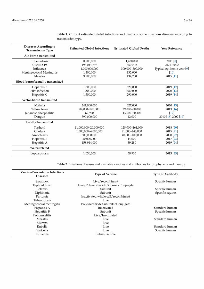
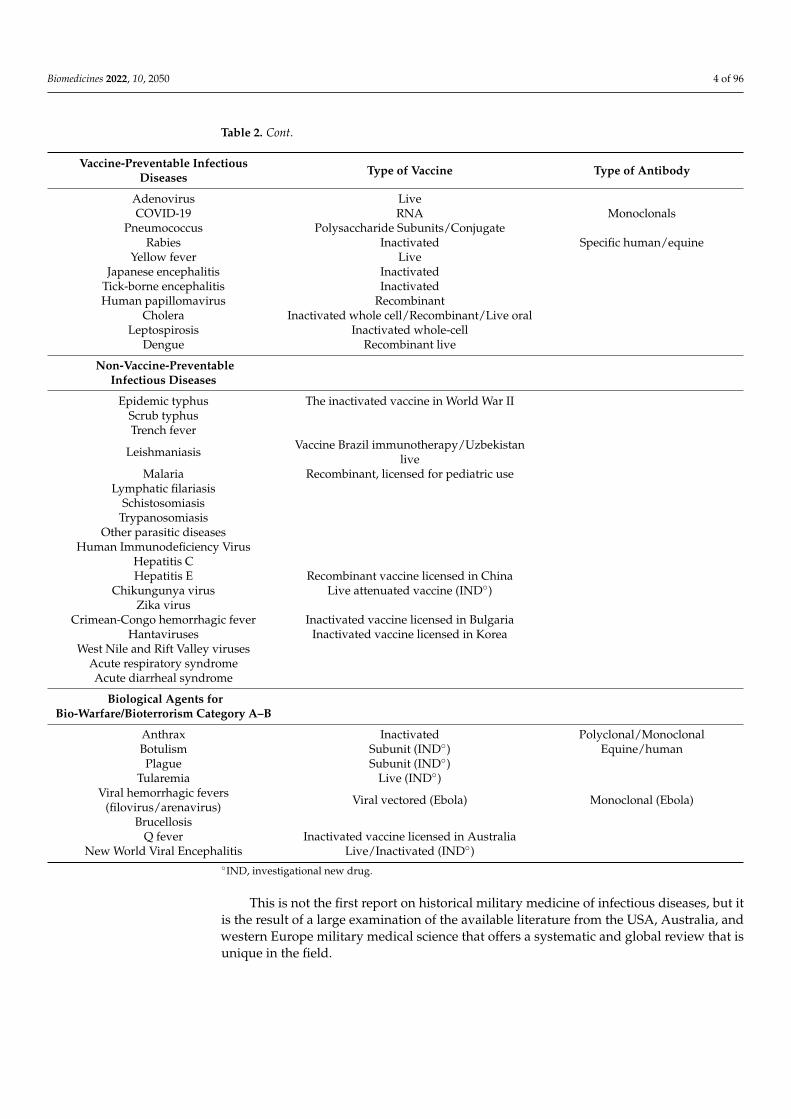
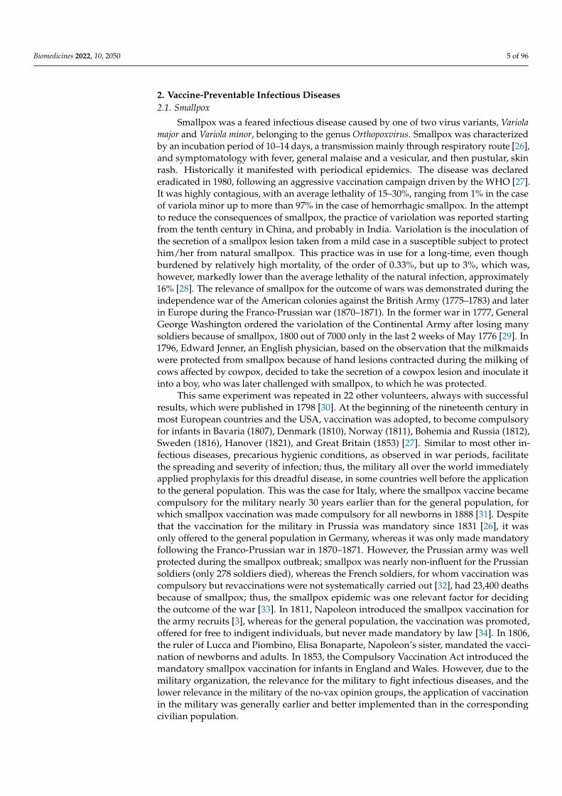
In this paper, a historical approach to the military fight against infectious diseasesis reviewed by describing the military involvement in (i) vaccine- and (ii) non-vaccine-preventable diseases; (iii) acute respiratory and (iv) diarrheal syndromes, (v) the study ofmajor agents developed for biological warfare and (vi) the high biocontainment aeromedicalevacuation. Although the military are particularly exposed to some specific infectiousdiseases, which are widespread and often burdened by high mortality (Table 1), they maybe also at higher risk for other infectious diseases, whose spreading is favored by thespecific environmental conditions that characterize the military life. Thus, we extended ourreview to cover all the main infectious threats to the military and the role of military healthservices and scientists in their containment (Table 2).
Biomedicines 2022, 10, 2050 3 of 96
Table 1. Current estimated global infections and deaths of some infectious diseases according totransmission type.
Diseases According toTransmission Type Estimated Global Infections Estimated Global Deaths Year Reference
Air-borne transmitted
Tuberculosis 8,700,000 1,400,000 2011 [8]COVID-19 195,044,798 650,702 2021–2022Influenza 1,000,000,000 300,000–500,000 Typical epidemic year [9]
Meningococcal Meningitis 1,200,000 135,000 [10]Measles 9,700,000 134,200 2015 [11]
Blood-borne/sexually transmitted
Hepatitis B 1,500,000 820,000 2019 [12]HIV infection 1,500,000 680,000 2020 [13]Hepatitis C 1,500,000 290,000 2019 [14]
Vector-borne transmitted
Malaria 241,000,000 627,000 2020 [15]Yellow fever 84,000–170,000 29,000–60,000 2013 [16]
Japanese encephalitis 67,900 13,600–20,400 [17]Dengue 390,000,000 12,000 2010 [18] 2002 [19]
Fecally transmitted
Typhoid 11,000,000–20,000,000 128,000–161,000 2018 [20]Cholera 1,300,000–4,000,000 21,000–143,000 2015 [21]
Amoebiasis 500,000,000 40,000–100,000 2000 [22]Hepatitis E 20,000,000 44,000 2017 [23]Hepatitis A 158,944,000 39,280 2019 [24]
Water-related
Leptospirosis 1,030,000 58,900 2015 [25]
Table 2. Infectious diseases and available vaccines and antibodies for prophylaxis and therapy.
Vaccine-Preventable InfectiousDiseases Type of Vaccine Type of Antibody
Smallpox Live/recombinant Specific humanTyphoid fever Live/Polysaccharide Subunit/Conjugate
Tetanus Subunit Specific humanDiphtheria Subunit Specific equinePertussis Inactivated whole cell/recombinant
Tuberculosis LiveMeningococcal meningitis Polysaccharide Subunits/Conjugate
Hepatitis A Inactivated Standard humanHepatitis B Subunit Specific human
Poliomyelitis Live/InactivatedMeasles Live Standard humanMumps LiveRubella Live Standard humanVaricella Live Specific humanInfluenza Subunits/Live
Biomedicines 2022, 10, 2050 4 of 96
Table 2. Cont.
Vaccine-Preventable InfectiousDiseases Type of Vaccine Type of Antibody
Adenovirus LiveCOVID-19 RNA Monoclonals
Pneumococcus Polysaccharide Subunits/ConjugateRabies Inactivated Specific human/equine
Yellow fever LiveJapanese encephalitis Inactivated
Tick-borne encephalitis InactivatedHuman papillomavirus Recombinant
Cholera Inactivated whole cell/Recombinant/Live oralLeptospirosis Inactivated whole-cell
Dengue Recombinant live
Non-Vaccine-PreventableInfectious Diseases
Epidemic typhus The inactivated vaccine in World War IIScrub typhusTrench fever
Leishmaniasis Vaccine Brazil immunotherapy/Uzbekistanlive
Malaria Recombinant, licensed for pediatric useLymphatic filariasis
SchistosomiasisTrypanosomiasis
Other parasitic diseasesHuman Immunodeficiency Virus
Hepatitis CHepatitis E Recombinant vaccine licensed in China
Chikungunya virus Live attenuated vaccine (IND◦)Zika virus
Crimean-Congo hemorrhagic fever Inactivated vaccine licensed in BulgariaHantaviruses Inactivated vaccine licensed in Korea
West Nile and Rift Valley virusesAcute respiratory syndromeAcute diarrheal syndrome
Biological Agents forBio-Warfare/Bioterrorism Category A–B
Anthrax Inactivated Polyclonal/MonoclonalBotulism Subunit (IND◦) Equine/humanPlague Subunit (IND◦)
Tularemia Live (IND◦)Viral hemorrhagic fevers
(filovirus/arenavirus) Viral vectored (Ebola) Monoclonal (Ebola)
BrucellosisQ fever Inactivated vaccine licensed in Australia
New World Viral Encephalitis Live/Inactivated (IND◦)◦IND, investigational new drug.
This is not the first report on historical military medicine of infectious diseases, but itis the result of a large examination of the available literature from the USA, Australia, andwestern Europe military medical science that offers a systematic and global review that isunique in the field.
Biomedicines 2022, 10, 2050 5 of 96
2. Vaccine-Preventable Infectious Diseases2.1. Smallpox
Smallpox was a feared infectious disease caused by one of two virus variants, Variolamajor and Variola minor, belonging to the genus Orthopoxvirus. Smallpox was characterizedby an incubation period of 10–14 days, a transmission mainly through respiratory route [26],and symptomatology with fever, general malaise and a vesicular, and then pustular, skinrash. Historically it manifested with periodical epidemics. The disease was declarederadicated in 1980, following an aggressive vaccination campaign driven by the WHO [27].It was highly contagious, with an average lethality of 15–30%, ranging from 1% in the caseof variola minor up to more than 97% in the case of hemorrhagic smallpox. In the attemptto reduce the consequences of smallpox, the practice of variolation was reported startingfrom the tenth century in China, and probably in India. Variolation is the inoculation ofthe secretion of a smallpox lesion taken from a mild case in a susceptible subject to protecthim/her from natural smallpox. This practice was in use for a long-time, even thoughburdened by relatively high mortality, of the order of 0.33%, but up to 3%, which was,however, markedly lower than the average lethality of the natural infection, approximately16% [28]. The relevance of smallpox for the outcome of wars was demonstrated during theindependence war of the American colonies against the British Army (1775–1783) and laterin Europe during the Franco-Prussian war (1870–1871). In the former war in 1777, GeneralGeorge Washington ordered the variolation of the Continental Army after losing manysoldiers because of smallpox, 1800 out of 7000 only in the last 2 weeks of May 1776 [29]. In1796, Edward Jenner, an English physician, based on the observation that the milkmaidswere protected from smallpox because of hand lesions contracted during the milking ofcows affected by cowpox, decided to take the secretion of a cowpox lesion and inoculate itinto a boy, who was later challenged with smallpox, to which he was protected.
This same experiment was repeated in 22 other volunteers, always with successfulresults, which were published in 1798 [30]. At the beginning of the nineteenth century inmost European countries and the USA, vaccination was adopted, to become compulsoryfor infants in Bavaria (1807), Denmark (1810), Norway (1811), Bohemia and Russia (1812),Sweden (1816), Hanover (1821), and Great Britain (1853) [27]. Similar to most other in-fectious diseases, precarious hygienic conditions, as observed in war periods, facilitatethe spreading and severity of infection; thus, the military all over the world immediatelyapplied prophylaxis for this dreadful disease, in some countries well before the applicationto the general population. This was the case for Italy, where the smallpox vaccine becamecompulsory for the military nearly 30 years earlier than for the general population, forwhich smallpox vaccination was made compulsory for all newborns in 1888 [31]. Despitethat the vaccination for the military in Prussia was mandatory since 1831 [26], it wasonly offered to the general population in Germany, whereas it was only made mandatoryfollowing the Franco-Prussian war in 1870–1871. However, the Prussian army was wellprotected during the smallpox outbreak; smallpox was nearly non-influent for the Prussiansoldiers (only 278 soldiers died), whereas the French soldiers, for whom vaccination wascompulsory but revaccinations were not systematically carried out [32], had 23,400 deathsbecause of smallpox; thus, the smallpox epidemic was one relevant factor for decidingthe outcome of the war [33]. In 1811, Napoleon introduced the smallpox vaccination forthe army recruits [3], whereas for the general population, the vaccination was promoted,offered for free to indigent individuals, but never made mandatory by law [34]. In 1806,the ruler of Lucca and Piombino, Elisa Bonaparte, Napoleon’s sister, mandated the vacci-nation of newborns and adults. In 1853, the Compulsory Vaccination Act introduced themandatory smallpox vaccination for infants in England and Wales. However, due to themilitary organization, the relevance for the military to fight infectious diseases, and thelower relevance in the military of the no-vax opinion groups, the application of vaccinationin the military was generally earlier and better implemented than in the correspondingcivilian population.
Biomedicines 2022, 10, 2050 6 of 96
In 1980, the WHO declared smallpox eradicated, after the last case of natural smallpoxoccurring in Somalia in 1977, and recommended vaccination interruption, considering thatthe risk of adverse events was higher than the risk of smallpox infection. However, in somecountries, the military continued to be immunized, as a prevention for the possible useof smallpox as a biological agent on a population that was not protected anymore. Thefear of the possible use of smallpox as a biological weapon became more pressing after theepisode of anthrax sent by mail; thus, USA President Bush ordered that the health workersand the military were compulsorily immunized. However, vaccination was interruptedafter having vaccinated approximately 500,000 military subjects and 40,000 health work-ers, for the relatively high frequency of adverse events [35]. Nonetheless, the last NorthAtlantic Treaty Organization (NATO) document on the vaccinations for the military inthe 30 NATO countries reports that 3/25 countries that have reported their military vacci-nation schedule, still maintain smallpox vaccine for selected categories of personnel [36].New, less reactogenic, tissue-culture-based live attenuated, and subunit smallpox vaccineformulations are studied for the risk that smallpox may be used as a biological weapon [37],or for protection against naturally occurring monkeypox. Moreover, by collecting the bloodof the immunized people, it was possible to produce specific polyclonal Ig, which wereprotective and could be used in emergencies, with significantly lower adverse events thanthe vaccine [35]. Thus, despite smallpox having been eradicated since 1980, the interestfor the military is still great, in light of its possible use as a biological weapon, of categoryA. The military contribution is the worldwide early military vaccination, which may havecontributed to its eradication.
2.2. Typhoid Fever
Typhoid fever is a serious infection caused by Salmonella typhi, a Gram-negative bacte-rial microorganism, which may infect through ingestion of contaminated water or food.The disease is characterized by high fever, headache, gastralgia, diarrhea or constipation,hepato-splenomegaly and possible complications, such as intestinal perforation. In thepre-antibiotic era, mortality was as high as 20%. Salmonella has three antigens, the O and Hantigens, thermostable and thermolabile, respectively, and a third antigen Vi, for virulence.The diagnosis may be carried out by stool culture, blood culture and serologically, by thesearch for specific anti-O and anti-H antibodies. Some people may become chronic carriersof S. typhi, continuing to release bacteria in their stools, thus spreading the disease. Typhoidfever is a classic example of an infectious disease spreading in unfavorable hygienic condi-tions, with lack of access to safe water and food, as may be observed during the war. This,joined with the severe clinical picture and the relatively high lethality, makes the disease ofgreat interest to the military. In addition to the environmental prophylaxis, the search foran effective vaccine has registered the activity of military researchers from Germany, GreatBritain, France, Italy, and the USA. The development of the typhoid vaccine has been tradi-tionally attributed to Almroth Wright, Professor of Pathology at the British Army MedicalSchool at Netley, even though documents prove that Wright, appointed by the Director ofthe Army Medical Service to develop a typhoid vaccine and worried to be unable to comply,was reassured after knowing the results obtained by Prof. Richard Pfeiffer in Germanyabout the development of a typhoid vaccine [38,39]. Pfeiffer, a military doctor of the Ger-man Army, was seconded to the Laboratory of Robert Koch at the University of Berlin, andapplied with success to bacteriology and immunology, by observing that a heated S. typhiculture, inoculated subcutaneously in man, could induce antibody-mediated agglutination.These data were described by Pfeiffer and Kolle in 1897 [40]. In 1896, Wright publisheda paper that was not focused on typhoid vaccination [41], while his paper on typhoidvaccination was contemporaneous to the paper of Pfeiffer and Kolle in 1897 [42]. However,independently of who was the first, this activity witnesses the interest of the military inpreventing this dreadful disease. The first chance to test the vaccine’s effectiveness was theAnglo-Boer War in southern Africa in 1899, during which the British Army used early formsof the typhoid vaccine. Among 14,626 immunized British soldiers, 1417 contracted typhoid
Biomedicines 2022, 10, 2050 7 of 96
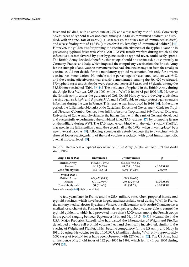
fever and 163 died, with an attack rate of 9.7% and a case fatality rate of 11.5%. Conversely,48,754 cases of typhoid fever occurred among 313,618 unimmunized soldiers, and 6991died, with an attack rate of 15.5% (p < 0.0000001 vs. the attack rate of immunized soldiers),and a case fatality rate of 14.34% (p = 0.002965 vs. lethality of immunized soldiers) [43].However, the golden test for proving the vaccine effectiveness of the typhoid vaccine inpreventing typhoid fever was World War I (WWI) trench warfare during which all theinfectious diseases favored by poor hygiene, such as typhoid fever, could easily spread.The British Army decided, therefore, that troops should be vaccinated, but, contrarily toGermany, France, and Italy, which imposed the compulsory vaccination, the British Army,for the strength of anti-vaccine movements that had obtained exemption from the smallpoxvaccine, could not decide for the mandatory typhoid vaccination, but only for a warmvaccine recommendation. Nonetheless, the percentage of vaccinated soldiers was 94%,and the vaccine effectiveness was clearly demonstrated; among the 604,420 vaccinated,570 typhoid cases and 34 deaths were observed versus 295 cases and 89 deaths among the38,580 non-vaccinated (Table 3) [44]. The incidence of typhoid in the British Army duringthe Anglo-Boer War was 285 per 1000, while in WWI, it fell to <1 per 1000 [45]. Moreover,the British Army, under the guidance of Col. David Harvey, could develop a trivalentvaccine against S. typhi and S. paratyphi A and B (TAB), able to better protect against entericinfections during the war in France. This vaccine was introduced in 1916 [46]. In the sameperiod, the Italian microbiologist Aldo Castellani, Director of Government Clinic for Tropi-cal Diseases, Colombo, Ceylon, later full Professor of Tropical and Sub-Tropical Diseases,University of Rome, and physician in the Italian Navy with the rank of General, developedand successfully experimented the combined killed TAB vaccine [47], by promoting its useon the military during WWI. The TAB vaccine, combined with the tetanus toxoid (TABTe),was used in the Italian military until the second half of the 1980s, when it was replaced by anew live oral vaccine [48], following a comparative study between the two vaccines, whichshowed lower reactogenicity of the oral vaccine associated with good immunogenicity,even at mucosal level [49].
Table 3. Effectiveness of typhoid vaccine in the British Army (Anglo-Boer War, 1899 and WorldWar I, 1915).
Anglo-Boer War Immunized Unimmunized p
British Army 14,626 (4.46%) 313,618 (95.54%)Disease 1417 (9.7%) 48,754 (15.5%) <0.0000001
Case-fatality rate 163 (11.5%) 6991 (14.34%) 0.002965
World War IBritish Army 604,420 (94%) 38,580 (6%)
Disease 570 (0.094%) 295 (0.764%) <0.0000001Case-fatality rate 34 (5.96%) 89 (30.2%) <0.0000001
From references [43,44] slightly modified.
A few years later, in France and the USA, military researchers prepared inactivatedtyphoid vaccines, which have been largely and successfully used during WWI. In France,the military medical doctor Hyacinthe Vincent, in collaboration with André Chantemesse, amedical researcher of the Pasteur Institute, developed a typhoid vaccine, able to control thetyphoid epidemic, which had provoked more than 65,000 cases among the French troopsin the period ranging between September 1914 and May 1915 [50,51]. Meanwhile in theUSA, Major Frederick Russell, who had visited the laboratories of Wright and Pfeiffer,developed a whole cell typhoid vaccine, heat and chemically inactivated, similar to thevaccine of Wright and Pfeiffer, which became compulsory for the US Army and Navy in1911. By using this vaccine for the 4,100,000 USA military during WWI, only approximately2000 cases of typhoid fever have been observed with 227 deaths [52]. The US Army hadan incidence of typhoid fever of 142 per 1000 in 1898, which fell to <1 per 1000 duringWWI [53].
Biomedicines 2022, 10, 2050 8 of 96
In conclusion, the first typhoid vaccines, all developed by military researchers, eventhough reactogenic and incompletely protective, showed a satisfying protection in theunfavorable hygienic conditions of the trench warfare such as of the one of WWI. Duringthe 1970s, a new live attenuated oral typhoid vaccine was developed from a wild-typeS. typhi strain Ty2 made defective from the galactose-epimerase gene and Vi antigen bychemical mutagenesis [54]; it was approved in Europe in 1983 and in the USA in 1989. A Vipolysaccharide injectable typhoid vaccine was developed in the 1970s and is used in manyworld countries. Moreover, in the second half of the 1980s, the Vi polysaccharide–proteinconjugate was also developed [55]. The conjugate vaccine, in which the Vi polysaccharideis linked to a protein matrix, which may be represented by tetanus toxoid, or diphtheriatoxoid, or CRM197 (a recombinant, avirulent analogous of diphtheria toxin) or recombi-nant exotoxin A of Pseudomonas aeruginosa, compared to the plain polysaccharide vaccine,allows a T-independent antigen to be transformed into a T-dependent one, thus elicitingmemory cells. However, despite that it represents a more effective vaccine than the plainpolysaccharide, the conjugation process is complex and expensive; thus, it has currentlyonly been approved in endemic countries, such as India and Nepal [56].
Currently, with the improvement of hygienic conditions, typhoid fever has virtuallydisappeared in developed countries; thus, in the military of many developed countries, suchas Italy, typhoid vaccination is only compulsory for troops deployed abroad, in developingcountries with unfavorable epidemiological situations. The WHO estimates an annualincidence of 11–20 million typhoid cases and annual deaths of 128,000–161,000, mainlyoccurring in developing areas of Africa, the Americas, Southeast Asia and Western Pacificregions [20]. Typhoid vaccination is present in all the 25 NATO countries out of 30, whichhave reported the vaccination schedule for the military. In 18 countries, the used vaccine isthe inactivated one, whereas in the remaining seven, it is the live attenuated one. In none ofthese 25 countries vaccination is it addressed to the whole military personnel, but analogouswith Italy, it is addressed to selected categories only [36]. The first vaccine developmentwas uniquely carried out by the military, and it was crucial in disease containment. S. typhihas been included among the biological agents, category B [57].
2.3. Tetanus
Tetanus is a potentially lethal disease caused by the anaerobic microorganism Clostrid-ium tetani, which produces a neurotoxin toxin (tetanospasmin). The severe symptomatologyof the disease is characterized by spastic palsy, due to the inhibition of the inhibitory neuro-transmitters of nerve terminals of lower motor neurons, the nerves activating voluntarymuscles [58]. The spores of C. tetani are resistant in the soil; thus, the wounds with necroticparts contaminated by topsoil are at particular risk of developing the infection. In absenceof therapy, the disease is virtually always lethal. Emil Adolf von Behring, a German militaryphysician expert in disinfection, joined the Robert Koch’s Institute of Hygiene in 1890, afterleaving the Army. In that period, in France with Louis Pasteur and in Germany with RobertKoch, microbiology and immunology were emerging. In particular, the Koch’s Laboratorycollected many scientists around, including Behring, Richard Pfeiffer who with Kolle andWright in Great Britain, will share credit for developing the typhoid vaccine, Paul Ehrlich,bacteriologist, and immunologist, Shibasaburo Kitasato, who isolated the C. tetani. Behringand Kitasato, in December 1890, published one paper describing that the inoculation ofsterilized cultures of tetanus in rabbits induced the appearance of antitoxins in the blood,as proven by the inoculation of this immune blood in mice that resulted protected by achallenge with tetanus [59]. A week later, Behring published another paper to extend thisobservation to diphtheria as well [60]. Based on these premises, Behring inoculated theserum of a previously immunized animal to diphtheria toxin in an eight-year-old boy withsevere diphtheria, who later had a full recovery. The lethality rate of diphtheria in thefollowing 10 years decreased from 50% to 13% [61]. This represented the birth of passiveimmunization, which has later been applied to different clinical contexts, including therecently set up of monoclonal antibodies to severe acute respiratory syndrome coronavirus
Biomedicines 2022, 10, 2050 9 of 96
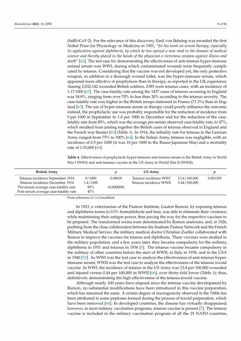
(SaRS-CoV-2). For the relevance of this discovery, Emil von Behring was awarded the firstNobel Prize for Physiology or Medicine in 1901, “for his work on serum therapy, especiallyits application against diphtheria, by which he has opened a new road in the domain of medicalscience and thereby placed in the hands of the physician a victorious weapon against illness anddeath” [62]. The test case for demonstrating the effectiveness of anti-tetanus hyper-immuneanimal serum was WWI, during which contaminated wounds were frequently compli-cated by tetanus. Considering that the vaccine was not developed yet, the only protectiveweapon, in addition to a thorough wound toilet, was the hyper-immune serum, whichappeared more effective in prophylaxis than in therapy, as reported in the UK experience.Among 2,032,142 wounded British soldiers, 2385 were tetanus cases, with an incidence of1.17:1000 [63]. The case-fatality rate among the 1437 cases of tetanus occurring in Englandwas 34.8%, ranging from over 70% to less than 20% according to the tetanus severity. Thecase-fatality rate was higher in the British troops stationed in France (71.3%) than in Eng-land [63]. The use of hyper-immune serum as therapy could poorly influence the outcome;instead, the prophylactic use was probably responsible for the reduction of incidence from9 per 1000 in September to 1.4 per 1000 in December and for the reduction of the case-fatality rate from 85%, which was the average pre-serum observed case-fatality rate, to 47%,which resulted from joining together the British cases of tetanus observed in England andthe French war theater [63] (Table 4). In 1914, the lethality rate for tetanus in the GermanArmy ranged from 75% to 100% [64]. In the Italian Army, tetanus was negligible, with anincidence of 0.5 per 1000 (it was 10 per 1000 in the Russo-Japanese War) and a mortalityrate of 1:33,000 [65].
Table 4. Effectiveness of prophylactic hyper-immune anti-tetanus serum in the British Army in WorldWar I (WWI) and anti-tetanus vaccine in the US Army in World War II (WWII).
British Army p US Army p
Tetanus incidence September 1914 9/1000 0.04018 Tetanus incidence WWI 13.4/100,000 0.001305Tetanus incidence December 1914 1.4/1000 Tetanus incidence WWII 0.44/100,000
Pre-serum average case-fatality rate 85% <0.0000001Post-serum average case-fatality rate 47%
From references [63,66] modified.
In 1923, a veterinarian of the Pasteur Institute, Gaston Ramon, by exposing tetanusand diphtheria toxins to 0.5% formaldehyde and heat, was able to eliminate their virulence,while maintaining their antigen power, thus paving the way for the respective vaccines tobe prepared. The transformed toxins were denominated by Ramon anatoxins, and, in 1926,profiting from the close collaboration between the Institute Pasteur Network and the FrenchMilitary Medical Service, the military medical doctor Christian Zoeller collaborated withRamon to improve the vaccines for tetanus and diphtheria. These vaccines were studied inthe military population, and a few years later, they became compulsory for the military,diphtheria in 1931 and tetanus in 1936 [5]. The tetanus vaccine became compulsory inthe military of other countries before the start of WWII, in Italy in 1938, and in the USAin 1940 [52]. As WWI was the test case to analyze the effectiveness of anti-tetanus hyper-immune serum, WWII was the test case to analyze the effectiveness of the tetanus toxoidvaccine. In WWI, the incidence of tetanus in the US Army was 13.4 per 100,000 woundedand injured versus 0.44 per 100,000 in WWII [66], over thirty-fold lower (Table 4); thus,definitively demonstrating the high effectiveness of the tetanus toxoid vaccine.
Although nearly 100 years have elapsed since the tetanus vaccine development byRamon, no substantial modifications have been introduced in this vaccine preparation,which has remained the same. A certain degree of reactogenicity observed in the 1940s hasbeen attributed to some peptones formed during the process of toxoid preparation, whichhave been removed [66]. In developed countries, the disease has virtually disappeared;however, in most military vaccination programs, tetanus vaccine is present [7]. The tetanusvaccine is included in the military vaccination program of all the 25 NATO countries,
Biomedicines 2022, 10, 2050 10 of 96
which have reported the respective vaccination schedule out of the 30 ones, 23 of which forthe whole personnel and in another two for selected categories [36]. In Italy, the tetanustoxoid vaccine was included in the vaccination program for infants only in 1968, thirtyyears later than for the military. The relevance of military vaccination as a public healthmeasure for tetanus prevention was witnessed in Italy and France, until the conscriptionwas present in both countries, by the unbalanced epidemiological situation of the few casesof tetanus annually reported, which were characterized by a marked preponderance of oldfemales, who were not covered by vaccination because it was not administered during themilitary service, which was only compulsory for males, nor in infancy, because it was notintroduced in the infant vaccination schedule yet [5]. Currently, an open issue is the dura-bility of vaccine-induced antibodies and thus the right timing for booster administration tomaintain the protective antibody levels without risking hyper-immunization [67–69]. Themilitary contributed to the discovery of passive immunotherapy and to the collaboration tovaccine development.
2.4. Diphtheria
Diphtheria is an infectious disease caused by the toxigenic strains of the Gram-positiveCorynebacterium diphtheriae, of which three main biotypes exist: gravis, intermedius, andmitis. The infection is localized in the high airways, where the toxin causes rhinitis,pharyngitis, and laryngitis. The toxin may induce myocarditis and polyneuropathy; thedisease is generally observed in <15-year-old boys and the case-fatality rate is 5–17% [70].Diphtheria has been described in the sixteenth and seventeenth centuries in Spain, withrecurrent epidemics in the eighteenth century in the USA, in the nineteenth and twentiethin Europe and more recently even in Asia and Africa. The etiologic agent was identified byEdwin Klebs in 1883 and was cultured by Friedrich Löffler, who demonstrated the toxinas well, whereas the progress in passive and active immunization is parallel to the one oftetanus, and it has been reported above in the paragraph of tetanus.
Considering that in non-vaccinated subjects, the disease is generally observed in<15-year-old boys, diphtheria is not apparently of military interest. However, the militarymust travel to different world countries, and if they are exposed to the etiologic agent inconditions of insufficient immune protection, they may be infected and become carriers,thus spreading the infection. This appears to have been the case for the start of a diphtheriaepidemic occurring in the period 1990–1995 in the newly independent states of the formerSoviet Union, where 47,808 cumulative cases of diphtheria occurred, 1746 of which werefatal [71]. A cluster of diphtheria infection was described in the members of a militaryconstruction battalion in Moscow in 1990. It must be considered that the Soviet troopshad been present, in the period 1980–1989, in Afghanistan, which reported to the WHO13,628 cases of diphtheria in the same period. Considering that the notification systemfor infectious diseases in the former Soviet Union was completely separated betweenmilitary and civilian populations, civilian health authorities were not immediately aware ofthese diphtheria cases occurring in the military; thus, the actions for limiting the infectionspreading were late and largely ineffective [71]. However, the causes for the spreading ofthe infection were largely unknown, but a high rate of unimmunized children and waningimmunity in adults was certainly present; thus, even in the armies of developed countries,where diphtheria has been eradicated, particular attention to maintaining the antibodylevels above the threshold for protection has become mandatory. In Italy, diphtheria boosterwas added to the compulsory vaccination schedule for the military after demonstrationof the relatively low percentage of recruits with protective antibody levels [72]. However,even though in the military much attention has been paid to the need to maintain protectiveantibody levels for diphtheria, a survey made up among the military medical servicesof 52 world countries showed that the tetanus vaccine was present in the compulsoryvaccine program for the military in 45/52 (87%), whereas diphtheria was only present in30/52 (58%) [7]. Currently, the diphtheria vaccine is included in the military vaccinationprogram of all 25 NATO countries, which have reported the respective vaccination schedule
Biomedicines 2022, 10, 2050 11 of 96
out of the 30, 22 of which are for the whole personnel and the other three for selectedcategories [36]. The outbreak of diphtheria in the newly independent states of the formerSoviet Union in the 1990s is a clear example of how the military may become involuntarycarriers of disease; thus, the military health authorities should not only combat infectiousdiseases for assuring the operational readiness but even closely collaborate with civilianhealth authorities in order to prevent possible military-mediated outbreaks. The completeseparation of civilian and military notification systems for infectious diseases in the formerSoviet Union was instead an example of a flawed organization, which has allowed thehappening of such a dramatic event.
2.5. Pertussis
Pertussis is a highly contagious infectious disease, known for many centuries, causedby the Gram-negative coccobacillus Bordetella pertussis, which was isolated and cultivatedby Jules Bordet and Octave Gengou in 1906 [73]. The most relevant symptom is whoopingcough, which may be accompanied by inflammation of the high airways and may becomplicated by apnea, pneumonia, rib fractures, insomnia, hospitalization, and rarelydeath [74]. The disease was generally observed in infancy, but in the last 20–30 years, it haseven been observed in adults [74], thus acquiring an interest for the military, consideringthat in many countries, limited outbreaks in the military have been described [75–79]. In theItalian military, a study carried out in the 1990s showed that more than 90% of subjects hadspecific cell-mediated and antibody immunity to B. pertussis and that symptoms suggestiveof pertussis were absent in the military [80].
Two types of vaccines are available, the first one is whole-cell, older, and inactivated,whereas the second vaccine, developed in the 1970s, but practically available since the1990s, is acellular, recombinant and may only contain one, two, or three of the mainvirulence factors of the microorganism, represented by the pertussis toxin, the pertactin,and the filamentous hemagglutinin. The whole-cell vaccine is more reactogenic, however,it seems quite more effective and able to provide more durable protection. Both vaccinesare combined with tetanus and diphtheria, in a trivalent diphtheria/tetanus/pertussis(DTP) or diphtheria/tetanus/acellular pertussis (DtaP). Pertussis is now, in both the USAand Europe, particularly present in adults, who represent the major reservoir for theinfection [81]. Currently, 21/25 countries reporting the vaccination military program amongthe 30 countries considered in the document of the NATO standardization agreement forvaccination of 2021 declare having pertussis included in the program, in 18 countries for allthe military personnel, in two out of the remaining three countries for selected categories(deployable, alert, risk personnel) and in one country for recommendation only [36]. Theuse of the trivalent DTP/DtaP vaccine in the military is a relevant measure of publichealth, particularly in the countries with conscription because maintaining a high level ofimmunity reduces the microorganisms’ circulation.
2.6. Tuberculosis (TB)
TB is a severe disease, whose infectious nature was demonstrated by the Frenchmilitary physician Jean-Antoine Villemin in 1865, and which was published in 1868 [82],through inoculation of material from infected humans to laboratory rabbits. TB is caused byMycobacterium tuberculosis, discovered in 1882 by Robert Koch, who was awarded the NobelPrize for Physiology or Medicine in 1905 [83]. The microorganism is transmitted throughairways and may induce, after an average period of 3–9 months up to two years [84],either a latent or active disease, generally at lung level, but, more rarely, everywhere in thebody. It is estimated that one-third of the world population is infected, the large majoritywith a latent infection and a minority, which in 2011 was represented by 8.7 million cases,with active infection, and 1.4 million deaths [8]. In 1895, a French military physician,Albert Calmette, who founded the Pasteur Institute in Saigon and later directed the PasteurInstitute in Lille, started his studies on tuberculosis and, together with the veterinarianCamille Guérin, developed a live attenuated vaccine for TB, which was successfully tested
Biomedicines 2022, 10, 2050 12 of 96
for the first time in 1921 [85]. This vaccine uses attenuated Mycobacterium bovis and isknown as Bacillus Calmette–Guérin (BCG), after the names of its discoverers.
Similar to many other infectious diseases, TB spread increases in unfavorable en-vironmental conditions, such as insane housing, overcrowding or hypo-nutrition, thatcharacterize poverty and occur during wars, but community life may also favor TB spread-ing [86]. In the US military, the epidemiology of TB has been analyzed since the CivilWar (1861–1865) up to the last wars in Iraq and Afghanistan. TB was more frequent in themilitary up to WWI and lower than in the civilian population in the following years [87].During the American Civil War, the morbidity rate for TB was 924/100,000 and the mortal-ity rate 261/100,000, whereas during the Spanish–American War (1898) the mortality ratewas slightly reduced to 197/100,000, and during WWI, the morbidity rate increased up to1168/100,000, with a higher prevalence of cases among soldiers who had remained in theUSA compared with those who were deployed to Europe [86]. With WWII, lung X-ray wasextensively employed to improve the screening before enrollment, thus preventing newcases coming from the contagion with infected comrades. From WWII, the influence of waron the epidemiology of TB seems inapparent, even when the wars occurred in countriesendemic to TB, such as Korea, Iraq, and Afghanistan; the epidemiological curve of incidencecontinued to descend, until 0.4/100,000 in 2012, which represents a value eight-fold lowerthan in the USA civilian population [87]. However, a crucial point for reducing the casesof active TB in the military is to identify with the highest possible precision the subjectswith latent TB among the applicants for military service during the pre-enrollment medicalscreening, for these cases to be adequately treated before enrollment, thus preventing thepossible development of active TB as a consequence of the stress of the military life [88]. Inthe UK, the situation is quite different, considering that TB still presents a morbidity rate of12.3/100,000 in the general population, mainly due to immigrants from high-endemicitycountries (70/100,000 among immigrants versus 4/100,000 of UK-born people), but evendependent on risk factors, such as smoking, alcohol consumption, immunosuppression,and concomitant diseases, such as HIV infection and diabetes. The situation is similar in theUK military, in which historically at the end of the nineteenth century, TB represented thefirst cause of medical discharge from active service (300/100,000 in 1891). In the first half ofthe twentieth century, the situation improved by showing a reduction of approximately50% (an average of 150/100,000), a behavior that was observed during WWII and evenafterward, up to half a century. In the second half of the century, a series of initiativeswere taken, including pre-enrollment screening, the diagnosis and treatment of latent TBinfections and the offer of BCG to skin-negative subjects who had not received BCG ininfancy. Based on a careful study, it emerged that the risk of TB was higher in older veteranswho entered the Army before the implementation of preventive measures [89]. In Italy, astudy carried out on over 2000 soldiers in the 1990s found a prevalence of latent infections(tuberculin-positive, asymptomatic subjects) of over 6% [90], a percentage not dissimilarfrom the percentage of the US Army in the same period [91]. Based on this result, in 2001,the norm of article 10 of Act 1088/1970 requiring that all skin-negative soldiers wouldhave been vaccinated with BCG at enrollment was cancelled. The reactogenicity and theuncertain protection induced by BCG in adults [92] did not justify its administration in thepresence of a relatively reduced prevalence. Moreover, in 2005, the conscription in Italy wasabolished, thus deeply modifying the socio-epidemiology in the military. In addition to anumerical reduction of the military personnel, even the community life was reduced andonly maintained during the training and operational periods, thus reducing the occasionsfor infections spreading. The lung TB in the period 1986–1997 in the Italian military had anannual incidence ranging from 8 to 13.5/100,000, higher than that observed in the age- andsex-matched civilian population, with an average annual incidence of 10.4/100,000 [93],whereas in the period 2008–2018, the annual incidence was always lower than 1/100,000,except for in 2013 and 2017, when it was 1.68/100,000 and 2.1/100,000, respectively, withan annual average incidence of 0.675/100,000 and a reduction of 15.4-fold (Table 5). Onlysix NATO countries maintain the BCG for the military; however, in only two countries, it
Biomedicines 2022, 10, 2050 13 of 96
is administered to the whole military personnel; in one, it is only recommended, and inthe last three, it is administered to selected categories of personnel, who are exposed as aconsequence of occupational risk or deployed in high-risk areas [36]. A rising problem isthe multi-drug-resistant TB (MDR-TB), caused by isoniazid- and rifampin-resistant My-cobacterium tuberculosis; this issue has been considered of awareness for the military, notonly because of the difficulties in the treatment of patients with MDR-TB but also becausedrug-resistant Mycobacterium tuberculosis is a pathogen included among the biologicalagents, category C in the Centers for Disease Control and Prevention (CDC) classification,and studies aimed at counteracting its infection are of strategic interest. In conclusion, evenfor TB, the role of military physicians in the demonstration of the infectious nature of thedisease and the prophylactic vaccine, as well as in its epidemiology, especially in wartime,witnesses the interest of the military and the contribution the military provided.
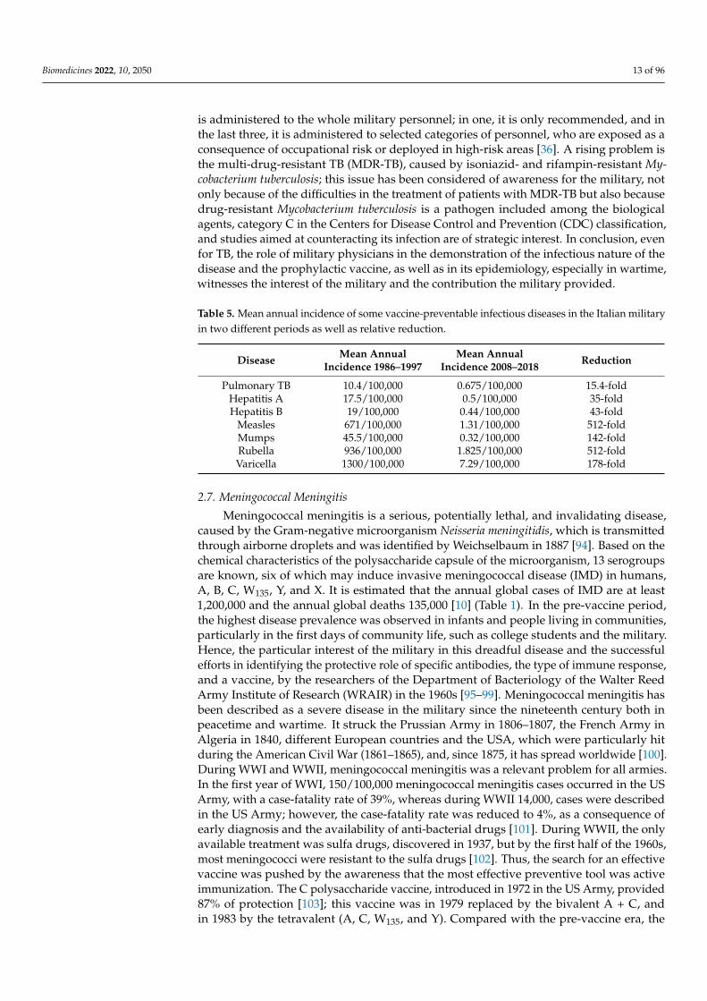
Table 5. Mean annual incidence of some vaccine-preventable infectious diseases in the Italian militaryin two different periods as well as relative reduction.
Disease Mean AnnualIncidence 1986–1997
Mean AnnualIncidence 2008–2018 Reduction
Pulmonary TB 10.4/100,000 0.675/100,000 15.4-foldHepatitis A 17.5/100,000 0.5/100,000 35-foldHepatitis B 19/100,000 0.44/100,000 43-fold
Measles 671/100,000 1.31/100,000 512-foldMumps 45.5/100,000 0.32/100,000 142-foldRubella 936/100,000 1.825/100,000 512-foldVaricella 1300/100,000 7.29/100,000 178-fold
2.7. Meningococcal Meningitis
Meningococcal meningitis is a serious, potentially lethal, and invalidating disease,caused by the Gram-negative microorganism Neisseria meningitidis, which is transmittedthrough airborne droplets and was identified by Weichselbaum in 1887 [94]. Based on thechemical characteristics of the polysaccharide capsule of the microorganism, 13 serogroupsare known, six of which may induce invasive meningococcal disease (IMD) in humans,A, B, C, W135, Y, and X. It is estimated that the annual global cases of IMD are at least1,200,000 and the annual global deaths 135,000 [10] (Table 1). In the pre-vaccine period,the highest disease prevalence was observed in infants and people living in communities,particularly in the first days of community life, such as college students and the military.Hence, the particular interest of the military in this dreadful disease and the successfulefforts in identifying the protective role of specific antibodies, the type of immune response,and a vaccine, by the researchers of the Department of Bacteriology of the Walter ReedArmy Institute of Research (WRAIR) in the 1960s [95–99]. Meningococcal meningitis hasbeen described as a severe disease in the military since the nineteenth century both inpeacetime and wartime. It struck the Prussian Army in 1806–1807, the French Army inAlgeria in 1840, different European countries and the USA, which were particularly hitduring the American Civil War (1861–1865), and, since 1875, it has spread worldwide [100].During WWI and WWII, meningococcal meningitis was a relevant problem for all armies.In the first year of WWI, 150/100,000 meningococcal meningitis cases occurred in the USArmy, with a case-fatality rate of 39%, whereas during WWII 14,000, cases were describedin the US Army; however, the case-fatality rate was reduced to 4%, as a consequence ofearly diagnosis and the availability of anti-bacterial drugs [101]. During WWII, the onlyavailable treatment was sulfa drugs, discovered in 1937, but by the first half of the 1960s,most meningococci were resistant to the sulfa drugs [102]. Thus, the search for an effectivevaccine was pushed by the awareness that the most effective preventive tool was activeimmunization. The C polysaccharide vaccine, introduced in 1972 in the US Army, provided87% of protection [103]; this vaccine was in 1979 replaced by the bivalent A + C, andin 1983 by the tetravalent (A, C, W135, and Y). Compared with the pre-vaccine era, the
Biomedicines 2022, 10, 2050 14 of 96
vaccine introduction reduced morbidity by over 90%, whereas the case-fatality rate didnot result to be significantly modified, always remaining around 7% [101]. In Italy, theburden of meningococcal meningitis in the military became particularly relevant duringthe 1980s (in 1985, an incidence of 17/100,000 cases, 92% serogroup C, and in 1986, anincidence of 7/100,000, 95% serogroup C, were observed, compared with an incidenceof 0.8/100,000 in the general population [104,105]); thus, the bivalent A + C vaccinationwas introduced since 1 January 1987. Vaccination was effective in reducing the burdenof meningococcal meningitis A and C, showing an effectiveness of 91.2% [104,106], animmunogenicity of 84% and 91% of protective seroconversion for polysaccharides A and C,respectively, with the appearance of mainly oligoclonal specific antibodies, and safety [107].In 1991, the tetravalent polysaccharide ACW135Y vaccine was introduced, recently largelyreplaced by the protein–conjugate formulation. However, the tetravalent polysaccharidevaccine maintains its validity because of its good immunogenicity and the long durabilityof induced antibody response, which were recently examined at 9 months [108] and 5years [109]. In the French military, the vaccine was introduced in 1996, and 2 years later, itsprotective effectiveness was calculated to be 100% [110]. Currently, the tetravalent vaccineACW135Y is included in the vaccination program of 24/25 NATO countries which havereplied out of the 30, in 10 countries for the whole military personnel and in the otherfourteen for selected categories [36].
A vaccine based on polysaccharide antigen could not be pursued only for meningo-coccal polysaccharide B, considering that polysaccharide B has a chemical structure closeto human brain polysaccharide, thus resulting in being poorly immunogenic or, evenworse, auto immunogenic [111]. Therefore, the approach for obtaining an effective anti-Bvaccine was long, laborious, and not based on the use of polysaccharides as antigens; rather,through the innovative approach of reverse vaccinology, a recombinant protein vaccinewas achieved only in 2005 [112]. This vaccine proved to be mildly moderately reactogenicin infants, particularly when administered in association with other vaccines; however, itwas proven that the concurrent administration of paracetamol significantly reduced reac-togenicity without interfering with the immune response [113]. Due to the relative rarityof IMD, not many significative studies on efficacy in the pre-registration phase have beencarried out, however, the vaccine has been approved based on its immunogenicity [113].The effectiveness in preventing IMD has been demonstrated in the real-world [114]. Vac-cination with meningococcal B vaccine has been included in the national immunizationprogram (NIP) of the UK, Ireland, and Italy; 12 European Member States have made anassessment to include the vaccine in the NIP, three are recommending the vaccine with-out reimbursement, whereas five are not recommending, as of March 2015 [115]. Only5/25 NATO countries, which had reported the respective military vaccination program,declare having meningococcal B vaccine included in their vaccination program for themilitary; in two cases the vaccine is only recommended, whereas in the remaining three,it is compulsory for selected categories of military personnel [36]. Even though sporadiccases are still observed, the vaccine introduction induced a substantial reduction of IMD inboth civilians and militaries [116]. In Italy, the anti-meningococcal polysaccharide vaccinehas been introduced in the compulsory vaccination program for the military thirty yearsbefore its availability for free in infants; however, the meningococcal B vaccine has beenfreely offered to infants since 2017, and it has not been included in the vaccination schedulefor the military yet.
2.8. Hepatitis A
Hepatitis A is a disease caused by an RNA virus (HAV), transmitted via the fecal–oralroute, by contaminated water and food, that easily spreads in poor hygienic conditionsand overcrowding. It was so largely widespread in the military, both in peacetime andmainly in wartime, that it was even known as “camp jaundice” [117]. In 2019, the globalannual infections were estimated to be 158,944,000, an increase of nearly 14% comparedwith 1990, and the annual deaths were 39,280 [24] (Table 1). Poor hygienic conditions
Biomedicines 2022, 10, 2050 15 of 96
and overcrowding as risk factors for the military were present in the literature up to 1990,whereas in more recent times, the major risk factor for the military has been the deploymentto countries of high endemicity [118]. One epidemiological study in Italy in the decade1987–1997 revealed a similar incidence in the military and the age- and sex-matched civilianpopulation [93]. Moreover, a study carried out in the Italian military in 2003 documentedthat Italy passed from a prevalence of 66.3% positive subjects for anti-HAV antibodies in1981 to 5.3% in 2003, thus from high to low HAV endemicity in 20 years, with the militaryreflecting the epidemiology of the general population [119]. A similar behavior of anti-HAVseroprevalence was even observed in the French military [120]. In the Italian military, theannual incidence in the period 1986–1997 ranged from 5 to 60/100,000, with an averageannual incidence of 17.5/100,000 [93], whereas in the period 2008–2018, it ranged from0.35 to 0.66/100,000, an average annual incidence of 0.5/100,000, a reduction of 35-fold(Table 5). In 1953, for the first time, the definition of hepatitis A and hepatitis B, to identifythe infectious (shorter incubation time, fecal-oral transmission, better prognosis) versus theserum-transmitted (higher incubation time, serum transmission, worse prognosis) hepatitis,respectively, was reported by an expert committee of the WHO [121]. However, until 1942,when an outbreak of acute viral hepatitis involving nearly 50,000 US Army personnelfollowing yellow fever vaccination [122], no clear idea that at least two types of hepatitiscould occur was still present: only the study of this outbreak, and the clarification thatthe outbreak was not due to a side effect of yellow fever vaccine, but to the preparationof the vaccine with human serum contaminated with virus hepatitis, has allowed a bettercomprehension of acute hepatitis to be achieved.
A disease with the characteristics of epidemic or infectious jaundice was describedduring the British–American War of 1812, but especially during the American Civil War,when 87,326 cases of jaundice were recorded by the Medical Corps of the Union Army [123].In WWI, epidemic jaundice represented a relevant problem for the French, British, andGerman armies, whereas this was not the case for the US Army, and in WWII, the US Armyregistered over 180,000 cases of infectious jaundice, with a case-fatality rate of 0.3% [124].However, following the occupation of Italy and Germany, where infectious jaundice wasendemic, the US military registered an increase in cases, which in Italy reached the incidenceof 37/1000 and in Germany continued even after the end of the war [116]. This observationallowed the first epidemiological studies to be carried out by US researchers in a newlyestablished hepatitis center in Bavaria [115]. During the Korean War in 1950–1951, in acountry of high endemicity, the cases of jaundice in the troops hospitalized or isolated wereover 4000 [124].
During WWII, US military researchers demonstrated the protective role of the pooledgamma-globulin plasma fraction against epidemic jaundice [125]. During the Korean War,a randomized double-blind study driven by US military researchers on intramuscular IgGadministration to soldiers could establish that the passively immunized subjects resultedprotected from hepatitis A, B, and non-A non-B for 6 months [126]. Even though passiveimmunization has been used for a long time for the protection of travelers and militarypersonnel, more recently an inactivated vaccine was developed by US researchers ofWRAIR in collaboration first with Robert Purcell at the National Institute of Health [NIH]and later with SmithKline Beecham (SKB), now GlaxoSmithKline. This vaccine provedto be safe, immunogenic, and highly protective (94% after two doses) in a large phaseIII study in Thailand on approximately 20,000 individuals and 20,000 controls who hadreceived hepatitis B vaccine [127]; based on these results, the vaccine was approved bythe Food and Drug Administration (FDA) in 1995 [116]. The vaccine, administered intwo doses 6 months apart not only demonstrated to be highly immunogenic but eveneffective, by inducing a long, probably a life-long, protection. The persistence of anti-hepatitis A antibodies following vaccination is so long that in a recent study, the durabilityof vaccine-induced antibodies could not even be calculated because the curve representingmean antibody titers was slightly ascending in joining the levels found at 9 months and5 years post-vaccination [109]. HAV vaccine has been introduced in the military vaccination
Biomedicines 2022, 10, 2050 16 of 96
program of all 25/30 NATO countries, which have reported the vaccination program forthe military: in 12/25 countries, the vaccination is indicated for all the military personneland in the remaining 13 countries for selected categories [36]. Currently, HAV infection,which has historically represented a real obstacle to the operational readiness of the military,does not represent a problem for the military anymore, not even when deployed to highendemicity countries. The military contribution in the fight against hepatitis A has beencrucial for epidemiology, the demonstration of protection by passive immunization withhuman immunoglobulins, and vaccine development.
2.9. Hepatitis B
Hepatitis B is a disease caused by a DNA virus (HBV), which may cause either acuteor chronic disease. Chronic disease may eventually induce liver cirrhosis and/or hepato-carcinoma. The disease is highly contagious and may be transmitted by contaminatedblood and blood derivatives, sexual route and perinatally. The diagnosis may be madeby identifying: the surface antigen (HBsAg) of the virus released in biological fluids; theantibody response to viral antigens (serum antibodies to the viral core (HBcAb), surface(HBsAb), and/or envelope (HBeAb) antigens); or by amplification of viral genes by poly-merase chain reaction (PCR) at serum and hepatic levels. It is estimated that, worldwide,approximately 2 billion people have come in contact with HBV [128] and the WHO esti-mates that in 2019, 296 million people were living with chronic HBV infection; each year1,500,000 new infections and 820,000 deaths occur, the majority from severe sequelae ofhepatitis B, such as cirrhosis and hepatocarcinoma [12] (Table 1). This blood-borne diseaseis of interest for the military, considering that wounds may be a source of contagion andwhole blood transfusions are used as resuscitation tools, consequently, the need that thesoldiers are “walking blood banks”, thus free of blood-borne viruses, such as HBV, hepatitisC virus (HCV), human immunodeficiency virus (HIV) types I and II, and human T-celllymphotropic virus (HTLV) types I and II, is imperative [129].
A pre-enrollment screening for blood-borne viral infections to prevent admission tothe military seems, therefore, the best preventive measure. However, in a survey carriedout by the WHO in 1998 in over half of the countries reporting to the WHO (107/193),only 76 replied; of these, 53 declared having a central military laboratory to perform thescreening of the recruits, 27/53 (51%) for HIV, 17/53 (32%) for HBV and 7/53 (13%) forHCV [7]. Currently, the situation is probably improved, even in consideration that in 1991in different world countries, the compulsory HBV vaccination for infants was introduced;thus, in the last decade, the applicants for military service had generally been vaccinatedin infancy. The vaccine, which was made available as plasma-derived in the first halfof the 1980s, and, since 1986 as recombinant, is effective and, after having completedthe whole vaccination cycle (three administrations), provides a long, probably life-long,protection [130]. Moreover, in 24/25 NATO countries hepatitis B vaccine is present, in 15for the whole personnel and in 9 for selected categories [36]. However, in some NATOcountries, in which the access to HBV vaccination in infant age has been delayed, theprevalence of serum HBV infection markers was still quite high in the first decade of thiscentury [131,132].
The combined influence of entry screening, awareness of the risk of infection due tosexual activity as a consequence of the HIV infection prevention programs, and vaccinationhas determined a rate of infection slightly lower in the US military (0.23%) than in thecorresponding civilian population (0.3–0.5%) [133]. The influence of vaccines may beinferred by the significant difference between the rate observed in the older cohort, bornbefore 1979, generally not vaccinated, and the rate observed in the younger cohort, born inor after 1979, generally vaccinated, 0.39% vs. 0.13%, respectively (p = 0.016, Yates corrected,two tails, χ2). Conversely, the influence of social factors and fear of HIV infection may beobserved in the dramatic decline, in less than a decade, of HBV markers in two Italianmilitary populations of approximately 5000 individuals each, the first from the Italian Navyanalyzed in 1981 and the second from the Italian Air Force analyzed in 1990. HbsAg and
Biomedicines 2022, 10, 2050 17 of 96
HbcAb were 3.4% and 16.8%, respectively, in 1981, whereas they declined to 1.6% and 5.8%,respectively, in 1990 [134]. Even a study of incidence in the same period on approximately1300 Italian students at a military school located in the Italian region with the highestprevalence of HBV infection, followed-up for eight months, showed seroconversion toHBV markers of only two subjects (0.24/100 person-years of exposure), thus witnessing alow spreading of HBV markers among the Italian recruits [135]. In the Italian military inthe period 1986–1997, the annual incidence of HB ranged from 7 to 33/100,000, with anaverage annual incidence of 19/100,000 [92], whereas in the period 2008–2018, only fourcases have been reported, an annual incidence ranging from 0.32 to 0.65/100,000 cases, anaverage of 0.44/100,000, a 43-fold reduction (Table 5). This epidemiological situation andthe consideration that currently the cohorts of recruits have been previously vaccinatedwhen entering the military life are at the basis of the decision of the Italian military healthauthorities to eliminate the HBV vaccine from the military vaccination schedule, thusavoiding an expensive, useless, and unjustified booster.
2.10. Poliomyelitis
Poliomyelitis is a severe disease caused by an enterovirus, of which three types, 1, 2,and 3, are known. The disease may be transmitted through the nasopharynx, through anoral–oral way, by feces, or through a fecal–oral way, and after infection, the virus entersthe bloodstream. This virus is highly contagious, and up to 100% of households maybe infected, but in 95%, the infection runs asymptomatically or pauci-symptomatically,whereas in the remaining 5%, the symptoms are characterized by fever, headache, fatigue,nausea, vomiting, and neck stiffness, for meningitis. In some subjects, the virus, which hasa marked neurotropism, localizes at the spinal level, most frequently in the anterior horncells of the cord, thus eventually determining an asymmetric flaccid paralysis, particularlyin the arms. More rarely, the virus may localize at the bulbar level, thus compromising vitalfunctions, such as circulation and respiration, with consequent high mortality [136]. Thecase-fatality rate of paralytic cases was 2–5% for children and 15–30% for adults [137]. Thedisease in the pre-vaccine era was largely widespread worldwide; in 1956, the inactivatedtrivalent vaccine developed by Jonas Salk was introduced, whereas in 1962, it was largelyreplaced by the oral, living, vaccine developed by Albert Sabin [136]. The use of vaccineshas allowed the disease spread to be dramatically reduced; however, in 1988, the WHOdecided to start an eradication campaign with the objective to eliminate the disease bythe year 2000. Despite that the eradication campaign could not achieve eradication by2000, the 350,000 estimated cases in 1988 were reduced to 3000 in the year 2000 [138].Currently, the viral types 2 and 3 have been declared eradicated; thus, the wild virus isonly type 1, which is still present in Afghanistan and Pakistan, where in the last years, ithas even increased [139], and sporadic cases are reemerging in other politically unstablecountries and sometimes sites of conflicts, such as Syria, Iraq, Cameroon, Equatorial Guinea,Ethiopia, Kenia, Nigeria, and Somalia [140]. In the process of eradication, in addition to thedifficulties created by war and political instability, a further complication derives from thefecal elimination of a vaccine virus in countries where the oral, living vaccine is, or was,used. The live attenuated vaccine virus may revert to virulence; thus, being able of induceparalytic polio in vaccine recipients, particularly in those with immunodepression [139].All these difficulties may delay the date of eradication; consequently, vaccination should bemaintained at least until eradication.
Although the disease has been known for a long time, with the first evidence identifiedapproximately 1500 years BCE, poliomyelitis did not induce outbreaks until the end ofthe nineteenth century, when outbreaks of infantile paralysis occurred in Scandinaviaand the USA [141]. The disease was not considered relevant for the military, because itscarcely occurred in adults, and even during WWI, no outbreaks were described, despitethe poor hygienic conditions and sanitation. However, in the interwar period, the casesof poliomyelitis in adults increased, and in the course of WWII, the US military registered1023 cases with over 20% of deaths [142]. Out of the 1023 cases, 446 occurred in the troops
Biomedicines 2022, 10, 2050 18 of 96
who remained in the USA, whereas 577 occurred in the troops deployed overseas, inparticular in Egypt, Italy, and the Philippines. Although these figures do not appear sohigh if compared with another severe, “military”, infectious disease such as meningococcalmeningitis, for which 14,000 cases were described during WWII with a case-fatality rate of4%. Polio had over 20% of mortality, 42% of discharge for disability and was the infectiousdisease with the highest number of lost days, with only 34% of infected military returningto duty, figures not comparable with other infectious diseases [142]. Nonetheless, poliohas never been considered a “military” infectious disease, and vaccination is maintainedonly to make the military ready to be deployed everywhere, even in countries such asAfghanistan, where wild poliovirus is still circulating, and yearly cases due to poliovirustype 1 are notified. Out of the 25/30 NATO countries reporting the vaccination programfor the military, all maintain an inactivated polio vaccine, 16 for all the military personneland nine for selected categories [36]. The vaccine-induced antibodies are well stimulatedby inactivated vaccine even though the priming is carried out with oral vaccine [139], andtheir durability above the threshold for protection has been calculated in 10–20 years foranti-type 1 and 3 antibodies [109], data in line with the literature [143]. Maintaining theanti-polio inactivated booster for the military creates ulterior protection to prevent thepossibility that soldiers returning from a mission to endemic areas become involuntarycarriers of wild poliovirus; moreover, it is a relevant measure of public health, because itreduces the viral circulation, thus contributing to the eradication campaign of the GlobalPolio Eradication Initiative.
2.11. Measles
Measles is a disease caused by a virus derived from the agent of the cattle rinderpest,which adapted to humans 5000–10,000 years ago [144]. It is air-borne transmitted and ishighly contagious (one infected person may infect on average 9–18 susceptible individuals,more than the smallpox virus, which may infect 5–7 susceptible individuals, and influenza,which may infect 2–3 susceptible subjects). The disease is characterized by fever, cough,coryza, maculopapular rash, and conjunctivitis; however, the virus is carried by lympho-cytes and may localize in the lymphoid tissue and everywhere in the body, with possiblesevere complications, such as pneumonitis, keratoconjunctivitis, and encephalitis. Theinfection of lymphocytes causes a transitory immunodepression, and the measles viruswas the first infectious agent for which induced immunodepression was demonstrated.The Nobel Laureate John Franklin Enders developed the first live attenuated vaccine in1960 [92]. Measles was responsible for over 2 million deaths annually in the pre-vaccineera, but even now, it is still responsible for over 100,000 deaths per year. In 2015, the globalannual cases were estimated to be over 9,700,000 (only 245,928 cases reported), and theglobal annual deaths were 134,200 [11] (Table 1). Despite the RNA genome being generallycharacterized by a high rate of mutations, both the wild virus and vaccine strains are stable,making it not necessary to update the vaccine to a newly circulating mutated virus, asrequired for the influenza vaccine. The inclusion of this live attenuated vaccine in theExpanded Program of Immunization (EPI) in 1980 contributed to the reduction of measlesmorbidity and mortality, particularly in areas such as Sub-Saharan Africa, with the high-est morbidity and case-fatality rate [145]. Measles eradication by a global immunizationprogram is in theory possible, as the vaccine is effective and no animal reservoir is known.However, the deadline of 2010 for its eradication set by WHO in the European region wasnot respected, and to date, measles prevalence is still quite high (11%) in this area, whereasin some countries, an increase was reported after 2010 [145,146]. Various causes can behypothesized for this failure, including the vaccine hesitancy caused by the publicationand diffusion on mass media of the false association of measles/mumps/rubella (MMR)vaccination with autism, which led many parents to not vaccinate their children [92].
Measles has represented a relevant problem, even for the military, particularly up tothe twentieth century. For example, in the American Revolutionary War and the AmericanCivil War, measles was one of the main causes of death among the soldiers [52]. During

Biomedicines 2022, 10, 2050 19 of 96
the whole Civil War, measles caused 67,763 cases and 4246 deaths (case-fatality rate of6.27%) in the Union Army [147]. The case-fatality rate was 6% and 11% for white andblack soldiers, respectively [148]. A reduction in the impact of measles on the US militaryin the war was observed in the following years. The morbidity in the Union Army inthe first 2 years of war (1861–1862) was 56/1000, with a case-fatality rate of 2/1000 [149].Morbidity (42/1000) and mortality (0.45/1000) caused by measles decreased in the first2 years (1898–1899) of the Spanish–American War. In 2 years of WWI (1917–1918), thereported morbidity was 28/1000 and the mortality 0.7/1000. The progressive reductionof morbidity and mortality was confirmed in WWII when over 300,000 admissions tohospitals were registered for measles, mumps, rubella, and varicella [52], but a limitednumber of US soldiers died of measles. Finally, during the Vietnam War, no death tomeasles was registered among US soldiers. The progressive reduction of cases and deathsfor measles cannot be explained by medical progress, considering that no immunoglobulinsor antibiotics for the possible bacterial super-infections, nor vaccines, were still available inthe first phase of observed reduction. A possible explanation that has been proposed for thisphenomenon is the epidemiological isolation of recruits. In the nineteenth and first yearsof the twentieth century, the majority of soldiers were enrolled from rural, isolated areas,where the possibility to acquire measles and natural immunization in infancy was scarce.Overcrowded barracks, tents and battle camps forced young men coming from differentareas of the country to live in close contact, creating the best conditions for viral spreadamong susceptible individuals upon the emergence of new cases [148]. Measles, in the firstpart of the last century, was mainly complicated by bacterial pneumonia, more often causedby Streptococcus haemolyticus, currently known as Streptococcus pyogenes, largely presentin apparently healthy carriers, and able to induce pneumonia, and sometimes empyema,in a respiratory tree already damaged by measles virus [150]. In 1915, the HighlandDivision of the British Army suffered a measles outbreak associated with scarlet fever; outof 529 soldiers with measles, 65 died, a case-fatality rate of 12%, greater than that observedduring the American Civil War [151]. In 1917, measles and pneumonia were responsible for30% of all USA deaths in the troops [52]. This same paradigm of a bacterial super-infectionon a viral disease was repeated in 1918 with the Spanish influenza pandemic, whose highmortality was largely dependent on the bacterial super-infection, with severe cases ofpneumonia, which were frequently lethal in the pre-antibiotic era. Conversely, prior to thetwentieth century, measles-associated deaths were mainly due to lethal gastrointestinalcomplications and a hemorrhagic illness known as black measles [152].
Even in the post-vaccine era, the military, due to the high contagiousness of the diseaseand the community life, which is characteristic of the military population, seem to bemore exposed to measles than the general population, as observed in Italy in the period1986–1997 [93], and France in 2011 [153]. This observation pushed the military medicalauthorities in Italy and in France to introduce the compulsory measles vaccination in thetrivalent formulation MMR, which was developed by Dr. Maurice Hilleman in Merck, afterleaving WRAIR (in Italy, it was introduced in the military vaccination program in 1998 [48],whereas only in 1999 was vaccination offered for free to infants, to become compulsory onlyat the end of 2017, following a large measles outbreak in January of the same year [154]). InItaly, the effectiveness was found to be 95% [154], and even the immunogenicity was good,considering that 96% of vaccinees showed post-vaccine protective antibody levels [140].However, the high prevalence of pre-vaccine antibody positivity, probably due to naturalimmunization [140], induces to believe that pre-vaccine screening may be the best policyto adopt, such as in the USA [52]. In Italy, in the period 1986–1997, measles annualincidence ranged from 70 to 1300 cases per 100,000, with an average annual incidence of671/100,000 versus an annual incidence ranging from 0.33 to 4.2/100,000 in the period2008–2018, an average annual incidence of 1.31/100,000, and a 512-fold reduction (Table 5).This epidemiological situation probably reflects not only the effectiveness of MMR, whichwas introduced in 1998, but even the socio-environmental transformation due to thepassage, in 2005, from the mandatory conscription to the professional army. This resulted

Biomedicines 2022, 10, 2050 20 of 96
in a reduction of occasions of disease transmission, consequent to the reduction of thenumber of military personnel, but mainly to the marked reduction of the requirement forthe soldiers to live in barracks, a rule that has remained limited to training or operationalmilitary personnel. Currently, 23/25 NATO countries reporting the military vaccinationprogram maintain the measles vaccination, in 18 countries for all the military personneland in five for selected categories of personnel [36]; in all these countries, the administeredvaccine is the trivalent MMR. Despite that measles responds well to the vaccine, such that itdoes not represent a severe risk for public health in most countries anymore, the eradicationprocess is quite hard to reach, even in some European countries [155]; thus, guard mustremain high, even because there is the awareness that the disease-induced protection islifelong, whereas the vaccination-induced protection is not, and currently, there is an opendiscussion on how many boosters are needed, in addition to the two already acceptedvaccine administrations [156], for maintaining protection in the different environmentalconditions [140]. The military are particularly exposed because of being a close communityand because of operational activity, which may put them in contact with under-vaccinatedpopulations where the virus is still highly circulating; thus, it should be desirable that themilitary is always updated with this vaccination, even by periodical checks, which mayverify the state of immunization [157]. However, a measles outbreak has been recentlyreported even in a highly vaccinated population [158].
2.12. Mumps
Mumps is a disease caused by the Mumps virus, a member of the Paramyxoviridaefamily in the genus Rubulavirus that naturally infects only humans. Mumps generallyhas mild clinical course, characterized by swelling of salivary glands, especially parotid,accompanied by fever, headache, and malaise, but complications such as aseptic meningitisin up to 10%, orchitis in approximately 25% of post-pubertal male subjects, pancreatitis,deafness in approximately 4% of subjects, and rarely encephalitis, which may inducepermanent disabilities or even death, may occur [159]. The infection is transmitted withmoderate-high effectiveness by respiratory route, is only observed in humans, and has anincubation time of 2–4 weeks with a clinical course of 1–2 weeks [160]. A live attenuatedvaccine has been developed in the 1960s [161]; it may contain different viral strains, withmajor or minor reactogenicity/efficacy, and it is generally administered in a combinedformulation, similar to MMR. One mostly used strain, because of its safety and efficacy, isnamed Jeryl Lynn, after the daughter of Dr. Maurice Hilleman, who isolated the virus fromher throat and prepared the attenuated vaccine strain.
In the eighteenth century, mumps was known and occurred worldwide, particularlyin crowded environments such as in schools, colleges, prisons, and military barracks [162],with an annual incidence of >100/100,000 [159]. However, in the military, an even higherincidence of 6000/100,000 was observed [163]. In the first year of WWI in the USA, mumpsspread explosively when recruits coming from rural areas or cities of the USA were as-signed together in military barracks [164,165]. Recruits from rural areas perhaps had fewerprobabilities compared to recruits from cities to come in contact with infectious agents andacquire natural immunization at infancy; thus, they were more susceptible to this and otherviral infections. The epidemics followed a periodical trend, with a period of approximately3 years and a higher peak during WWII [166]. In the USA, mumps vaccine was initiallymade available in 1967 to specific categories; then, from 1968 to 1977, it was graduallyextended to all children of 12 months of age. The annual mumps incidence from 88/100,000in 1968 decreased to 2.5/100,000 in 1982 with a net reduction of 97% [166]. Despite twoperiods of mumps resurgence in the decade 1983–1992 and in the 15-year 1993–2008 period,generally occurring in schools and colleges of rural USA populations, no resurgence wasobserved in the military, probably for the vaccination policy of the military with MMRsince 1991. A crucial point is the choice of the vaccine, considering that some vaccinestrains are effective but poorly attenuated, such as Urabe Am9, which was responsible inthe Italian military for a post-vaccine outbreak due to the vaccine strain, as molecularly

Biomedicines 2022, 10, 2050 21 of 96
demonstrated [167]. The vaccine-induced mumps for scarce vaccine strain attenuationmay possibly be one of the reasons for finding 70% of mumps vaccine efficacy, comparedwith 95% of vaccine efficacy for measles and rubella [168]. However, the mumps vaccineeffectiveness is quoted ranging from 69% to 88% [169], and a mumps outbreak has beenreported in a French military Parachuting Unit in 2013, in the majority vaccinated withtwo MMR doses, characterized by a high attack rate, ranging from 21.6% to 25% [170].The mumps occurrence in highly vaccinated populations is a well-known phenomenoneven in other countries [171,172], and different hypotheses have been proposed for itsinterpretation, including early waning of immunity or antigenic variance that may reducethe efficacy of the vaccine against new circulating strains, as frequently observed withinfluenza vaccine [172]. Another crucial point is the number of boosters that should beadministered for maintaining antibody levels above the threshold for protection. Currently,it has been established that in countries where two vaccine doses at approximately fiveyears of distance are administered in infancy, immunization is protective with an effec-tiveness of over 99% of disease reduction, a percentage higher than that observed in thecountries where vaccination schedule is based on only one vaccine administration [159].However, the need for further booster(s) is still a matter of discussion and has not been es-tablished yet. Only one mumps case has been reported in the Italian military in the decade2008–2018, an incidence of 0.32/100,000; thus, it has virtually disappeared, whereas, in theperiod 1986–1997, when the MMR was lacking in the compulsory vaccination schedulefor the Italian military, it ranged from 25 to 65/100,000 cases, an average annual incidenceof 45.5/100,000 [93]. The ratio of reduction is over 142-fold (Table 5); however, for thisdramatic reduction, the same considerations spent for measles on the passage in Italyfrom mandatory conscription to professional army in 2005 are valid. Considering thatthe administered vaccine is MMR, among the 25 NATO countries reporting the respectivemilitary vaccination schedule, the mumps vaccine, similar to measles, is administered in23 countries, in 18 of them for the whole military and in five for selected categories [36].
2.13. Rubella
Rubella is a viral disease caused by Rubivirus rubellae, a member of the genus Rubivirus,with a generally mild clinical course, rash and lymphadenopathy, mainly at nuchal level.The major complication of rubella is fetus infection, which may provoke miscarriage orcongenital rubella syndrome (CRS), a severe condition characterized by congenital ocular,hearing, heart, brain, or endocrine disabilities [173]. Despite that an effective live attenuatedvaccine has been developed by Dr. Maurice Hilleman in the 1960s [174], CRS is still presentwith approximately 100,000 cases per year [173]. The disease has no animal reservoir,has an effective vaccine and has been eradicated in the Americas since 2009 and in GreatBritain; thus, it is an optimal candidate for global eradication, even in consideration of itslower transmissibility compared with measles, provided that a suitable percentage of herdimmunity, which may range from <70% to >90% according to the different world areas, isachieved and maintained [173].
The interest of rubella for the military is not only witnessed by the outbreaks observedin wartime and peacetime, but even and especially by the fact that the virus was firstidentified and isolated by military researchers of the WRAIR in the US Army recruits in1961 [175]. The rubella vaccine was adopted in the US Army in 1972 [52]. The effect ofvaccine introduction in reducing rubella cases was dramatic. In the three years beforevaccine introduction, the number of rubella cases notified in the USA was 47,745, whereasin 2005, the CDC announced that endemic rubella was eradicated in the USA [124]. In theItalian military, rubella showed an over four-fold incidence increase in the period 1991–1995compared with the period 1976–1980 (1150/100,000 vs. 280/100,000, respectively), with anannual incidence ranging from 50/100,000 to 2300/100,000 in the period 1986–1997 andan average annual incidence of 936/100,000 [93], whereas in the period 2008–2018, only11 cases have been registered, 10 of which were in 2008, an incidence of 3.32/100,000, andone in 2013, an incidence of 0.33/100,000, and an average annual incidence of 1.825/100,000,
Biomedicines 2022, 10, 2050 22 of 96
a reduction of 512-fold (Table 5). However, for this dramatic reduction, the considerationsspent for measles and mumps on the passage in Italy from mandatory conscription toprofessional army in 2005 should be taken into account. Even for rubella, the situation inthe NATO countries is identical to the situation reported for measles and mumps, with18 countries using MMR for the whole personnel and five for selected categories of militarypersonnel [36]. MMR vaccination in the military is a relevant measure of public health evenin countries where MMR is provided in infancy, where, acting as a booster, it contributes toreducing viral circulation.
2.14. Varicella
Varicella or chickenpox is a disease caused by a DNA herpesvirus that generallyinduces a mild disease, characterized by fever, malaise, and vesicular erythema. Thedisease has high transmissibility, with an R0 estimated at around 10–12 [176], by airborneroute of the virus coming from skin vesicles [177]. Varicella only occurs in humans andis present at global level, with an average annual incidence of 13–16/1000, but greaterthan 100/1000 in the <9-year-old children [177]. However, this epidemiological patternis generally observed in temperate areas, because in tropical areas, the adult age is morefrequently represented [177]. The clinical course tends to be self-limiting in children,whereas it may be complicated in adults in pregnancy, in which varicella may cause fetalmalformations (congenital varicella syndrome) in approximately 1% if infection occurs inthe first two trimesters, and in immunosuppressed people, in whom it may be responsiblefor death in up to 15–18% [177,178]. In 1974, Takahashi developed a live attenuatedvaccine [179], which has been shown to be safe and effective. Its systematic use with twodoses has deeply modified the disease epidemiology, with a reduction of over 95% ofincidence, hospitalizations, and deaths in children in the USA [177].
Varicella is highly contagious; thus, it has represented a problem for the military inthe pre-vaccine era, in analogy to measles, rubella, mumps, and pertussis. Even thoughmost recruits are protected when they join the military, nonetheless, some dozen casesoccur each year, as in Israel [180], considering that vaccine-induced seroprotection seemsto be lower than disease-induced protection [181]. In some countries, a marked increase invaricella infection has been observed in the military between the 1970s and the 1980s [182]or between the 1970s and 1990s [93]. In the Italian military in the period 1986–1997, theannual incidence ranged from 800 to 1900/100,000 cases with an average annual incidenceof 1300/100,000 [93], whereas it ranged from 2.4 to 12.6/100,000 in the period 2008–2018with an average annual incidence of 7.29/100,000, a reduction of 178-fold (Table 5). Thisseems more a probable expression of the transformation of the military service in Italythan of the effect of vaccination, considering that in 2005, compulsory conscription wassubstituted with the professional army. In Italy, similar to many other countries, eventhough vaccination is mandatory, in practice, it is only administered to those who donot refer having suffered the disease or carried out vaccination in infancy, a methodthat does not appear as reliable, particularly in the presence of negative history [183].Moreover, vaccination is applied in only 10/25 NATO countries reporting the militaryvaccination schedule, in half of which is either compulsory for all the military personnelor compulsory/recommended for selected categories of personnel [36]. Finally, in somecountries, the percentage of susceptible recruits to varicella is quite high, of the orderof 50% in the current period [178]. In conclusion, despite the availability of a safe andeffective tool for varicella prevention, it appears that the vaccine is not as largely used inthe military and, even when it is used, the policy to limit vaccine administration to thoselacking documentation of infant vaccination or disease may reduce its impact on diseaseprevention. This may probably explain why the reduction rate of varicella is lower than thereduction rate of measles and rubella in the Italian military.

Biomedicines 2022, 10, 2050 23 of 96
2.15. Influenza
Influenza is an acute respiratory disease that is transmitted by respiratory route, char-acterized by fever, cough, myalgias, and a generally benign clinical course of approximately2–8 days. However, sometimes, particularly in children less than 5 years of age, older adults,subjects with underlying diseases, and in pregnant women, influenza may be complicated,mainly by pneumonia and even by multi-organ failure, with possible hospitalization anddeath [184]. The WHO estimates that annually approximately 1 billion people becomeinfected with seasonal influenza, with approximately 3–5 million severe influenza and300,000–500,000 deaths [9] (Table 1). The etiological agent is a highly mutant RNA virus,of which four types are known, A, B, C, and D, the first being responsible for epidemicsand pandemics, and the most severe clinical forms [185]. All four types may be found inhumans and other animal species, such as swine, horses, dogs, seals, bats, and the largestreservoir, represented by wild aquatic birds [186]. The virus A expresses in its surfacetwo proteins, hemagglutinin (H), responsible for the infection, through the attachmentto the corresponding receptors on respiratory cells, of which 18 subtypes are currentlyknown, and neuraminidase (N), responsible for detachment from cell to infect other cells,of which 11 subtypes are known [186]. Type B, which may be responsible for epidemics,C, which has been associated with mild symptoms, and D, which has not been associatedwith pathology in humans, may be found in animals and humans. Currently, two A strainsare circulating, H1N1 and H3N2, and two B subtypes, B/Yamagata and B/Victoria [185].Hemagglutinin and neuraminidase, as first observed by Dr. Maurice Hilleman at WRAIRin 1957 [29], undergo annual slight antigenic modifications, defined “drifts”, and periodicmarked antigenic transformations, defined “shifts”, which are responsible for pandemicssince the immune system does not recognize the brand-new antigen. In 1918, a terribleinfluenza pandemic, called “Spanish flu” started inside the USA military, at the trainingcamps of recruits of the American Expeditionary Force (AEF), due to the strain H1N1,which was responsible for an estimated infection of one-third of humankind and death ofapproximately 50 million subjects, with a case-fatality rate of over 2.5% vs. 0.1% observedin other pandemics [187]. In 1957, a new pandemic, due to the strain H2N2, defined as“Asian flu”, was responsible for approximately 1.5 million deaths, followed in 1968 by anew pandemic, due to the strain H3N2, defined as “Hong Kong flu”, which was responsiblefor approximately 1 million deaths. Finally, in 2009, a new pandemic, due to a swine strainH1N1, was responsible for an estimated 300,000 deaths [188].
The “Spanish” influenza pandemic was the worst. It deeply hit the military, at thebeginning the US military, and afterward the military and the civilian populations of othercountries, including different European countries, Africa, India and Asia, Australia andNew Zealand [189]. However, the rate of infection was always higher in the military thanin the corresponding civilian population [190]. This pandemic developed along three suc-cessive waves, starting in spring 1918 with a relatively mild disease and then proceeding tofall and winter–spring 1919 with two highly lethal waves. The high lethality was observednot only in the extreme life’s ages, as in other influenza epidemics or pandemics, but alsoin young adults. This wide distribution of lethality had a dramatic demographic and eco-nomic impact on the working and productive sectors of the interested population, higherthan the war itself [187]. The virulence of the influenza virus was unique, unprecedented,and never observed afterward [191], but many other causes may have contributed to the ex-traordinary severity of the pandemics in wartime, including overcrowding, undernutrition,and stress due to the war, thus making the disease spread and the bacterial super-infectionwith consequent pneumonia easier. The high case-fatality rate, in general, and for youngadults in particular, remains without an answer, despite several, careful studies [191]. In1918, two months before the armistice of November, a peculiar event occurred that willnever be repeated: the simultaneous outbreak of influenza and malaria in the EgyptianExpeditionary Force in Palestine, in which out of 315,000 soldiers, 773 died from malariaand 934 from influenza–pneumonia. Disease victims outnumbered those due to combatby over 37 to 1. Moreover, out of 40,000 men of the Desert Mounted Corps, 19,652 sick

Biomedicines 2022, 10, 2050 24 of 96
soldiers were evacuated due to malaria from Plasmodium falciparum, a condition that causedthe interruption of combat operations [192]. However, the US military tolerated a highinfluenza pandemic burden in 1918–1919, such that their engagement in studying andpreventing influenza was witnessed by establishing, in 1941, the Board for the Investigationand Control of Influenza and Other Epidemic Diseases in the Army, which evolved into theArmy Epidemiological Board in 1944 and the Armed Forces Epidemiological Board (AFEB)in 1949 [125]. This structure supported the studies for the development of the influenzavaccine [193,194], which was tested on the military. Starting in 1943, army personnel wereimmunized against virus A, prior to the licensure to Parke Davis, in order to preventpossible influenza outbreaks during troop mobilization [125]. Moreover, AFEB supportedreal-world studies of vaccine effectiveness in the military [195–200]. Influenza virus ishighly mutant, and the immunization success is closely dependent on the matching be-tween the circulating and the vaccine viral strains; thus, the WHO has organized a networkof collaborating laboratories, in order to early identify the circulating strain and give preciseindications to the industry for the seasonal vaccine preparation [201]. The US militaryparticipates in such a network with the Armed Forces Health Surveillance Center, Divisionof Global Emerging Infections Surveillance and Response System (AFHSC-GEIS), whichsupports at least 52 national influenza centers and other country-specific influenza, regionaland US-based, emerging infectious disease reference laboratories (44 civilian, 8 military)in 46 countries worldwide for surveillance and response [202]. Even the French militaryhas implemented a surveillance system for influenza, called the military influenza surveil-lance system (MISS), as further evidence of the relevance of influenza to the military [203].Finally, even the Italian Armed Forces have organized an Influenza Surveillance Systemin coordination with the civilian Influenza Surveillance Network (Influnet), driven by theItalian National Institute of Health. All these activities aim to contrast a fearsome infectiousdisease, which, even though did not recur with the high virulence of the Spanish flu pan-demic, has shown an easy capability of spreading in favorable environmental conditions,such as those encountered in the military [201]. However, although influenza is considereda threat to the military, flu vaccination was only compulsory in the US military, on thebasis of a WHO survey [7,201]. More recently, influenza vaccination has become presentin the military vaccination program of 24/25 NATO countries that report the vaccinationprogram for the military; however, in only nine countries for the whole military person-nel, two of these nine countries uniquely recommend [36]. The relatively scarce use ofimmunization for influenza in the military is probably a consequence of the relatively pooreffectiveness of the influenza vaccine in young adults [204], which is parallel to vaccineimmunogenicity [140].
2.16. Adenovirus
Adenoviruses are a group of over 50 serotypes of a DNA virus, which may be trans-mitted by respiratory route, conjunctiva (in case of contact with contaminated hands), andfecal–oral route. They may induce acute respiratory disease, conjunctivitis, and gastroin-testinal infections. Premises for epidemics are environmental conditions characterized bycommunity life with overcrowding, a situation often encountered in the military, partic-ularly the recruits, who are exposed especially in the first 3–5 weeks of training [205]. Anew virus, later denominated adenovirus [206], was identified in the first half of the 1950sby Dr. Hilleman and Dr. Werner at WRAIR [207]. It was later recognized that adenovirusincludes different serotypes and that types 4 and 7 were particularly implicated in acuterespiratory disease in the military [124]. Adenoviruses were later recognized as the mainetiological agent of acute respiratory disease in the military, with up to 80% of infected and20% of hospitalized subjects [208]. Dr Hilleman developed a formalin-inactivated bivalentvaccine including serotypes 4 and 7, which was successfully tested for safety and efficacy,showing to be safe and over 90% effective, and was licensed in 1958. However, due tothe risk of contamination by the oncogenic virus SV40, the license was retired in 1963 [29].New live oral vaccines for serotypes 4 and 7 were developed in the 1960s by a group of
Biomedicines 2022, 10, 2050 25 of 96
military researchers led by Col. Edward Buescher and were tested in the military [29].These vaccines proved to be safe, highly immunogenic, and protective [209,210] and wereregularly administered to the US recruits on the first day of their arrival at the trainingcamps starting in 1971 [29]. However, in 1996, this vaccination was interrupted, as thevaccines were not produced anymore by the unique manufacturer; thus, the US Departmentof Defense made a contract with another manufacturer [125], and in 2011, vaccination ofthe military was resumed [211], with a dramatic decline of febrile respiratory illness and ofadenovirus respiratory infections, which decreased 100-fold [212]. This vaccine is licensedby the FDA for US military personnel, ages 17 through 50, who may be at higher risk for in-fection from these two adenovirus types [36]. Although the issue of adenovirus respiratoryinfection has been deeply studied by the US military, it has been reported in the militaryof other countries since the 1970s until now [213–218]. However, among the 25 NATOcountries reporting vaccination schedules for the military, only one country reports thatadenovirus vaccination is recommended for all recruits [36]. This is probably due to theadenovirus epidemiology in these countries, frequently involving serotypes for whichvaccine is not available. Moreover, the relevance itself of the problem may be overlooked bythe lack of pathognomonic symptomatology and the difficult access to molecular and/orserological diagnosis.
2.17. Coronavirus Disease 2019 (COVID-19)
Coronavirus disease 2019 (COVID-19) is a potentially lethal respiratory disease, firstdescribed in China at the end of 2019 and still ongoing, caused by an RNA coronavirus(SARS-CoV-2, because similar to the SARS-CoV described in China in 2003), with highcontagiousness, so that in a few weeks from the first description, it was declared a pandemicby the WHO [219,220]. As of 23 May 2022, it has caused 525,618,514 total cases and 6,277,339total deaths, thus showing an average global attack rate of 6.78% and case-fatality rate of1.19% (https://coronavirus.jhu.edu/map.html (accessed on 21 July 2022)). From the samedata bank, the post-vaccine average annual new cases and deaths have been calculated.The average values of new cases and deaths referred to a 28-day period occurring in thelast year (2021–2022) and were multiplied by 13 to refer to the length of one year; the resultswere 195,044,798 annual new cases and 650,702 annual deaths (Table 1). Compared withthe dreadful Spanish flu of more than a century ago, the attack rate and the case-fatalityrate of COVID-19 are markedly lower, considering that in the Spanish flu, the estimatedattack rate was as high as approximately 30% [221], and the estimated lethality 50 milliondeaths [222]. Nonetheless, the current pandemic is representing a great challenge forall the countries and the respective health services, which are overwhelmed by the highnumber of patients who are hospitalized, particularly in intensive care units, for the moresevere cases, during the acute phases of the pandemic. The response to the pandemic byresearch was unprecedented and could develop and make available in less than one-yeareffective vaccines [223], monoclonal antibodies, and anti-viral agents, even though thegreat variability of the RNA virus has generated viral variants of concern, more aggressiveand/or more transmissible, which may make the disease control uncertain. In the researchfor an effective vaccine, the Chinese military had an early and relevant role [224,225].Although the pandemic is still ongoing and has not been eradicated nor transformed intoan endemic disease, the vaccine’s effectiveness, especially against severe disease and itscomplications, including hospitalizations and death, is definitively demonstrated [226].
The military are exposed to the infection not only for their community life but evenfor the direct management of the pandemic for its control, which offers a variety of oppor-tunities for exposure to the virus [227]. However, even though the military are particularlyexposed to the virus and their rate of infection may significantly differ or not from thecivilian population, they are expected to overcome the disease without complications,considering that they are generally young and in good health [228]. A comparison betweenthe study of the COVID-19 outbreak in the aircraft carrier Theodore Roosevelt and thecruise ship Diamond Princess is a clear demonstration of the statement above (Table 6).
Biomedicines 2022, 10, 2050 26 of 96
Theodore Roosevelt is a ship with a crew of 4779 members, 1271 of whom have beenfound to be serologically confirmed COVID-19 infected (26.6%) and 60 had suspectedCOVID-19 for suggestive symptomatology, in the absence of positive serology. Out ofthese 1331 (27.85%) confirmed and suspected COVID-19 infected subjects, 23 (1.73%) havebeen hospitalized, 4 (0.3%) needed intensive care, and one died [229]. Diamond Princessis a cruise ship that started a cruise on 20 January 2020 with approximately 3700 passen-gers and crew members, during which an outbreak of 712 COVID-19 infected subjectsoccurred (19.24%, p < 0.0000001 vs. Theodore Roosevelt), with 36 (5%, p = 0.00003448 vs.Theodore Roosevelt) hospitalized, and 13 (1.83%, p = 0.00001793 vs. Theodore Roosevelt)deaths [230]. The significant difference in the attack rates, higher in the military ship, isprobably related to the tighter available spaces for sleeping in the military ship, comparedto the more comfortable cabins of the Diamond Princess, where social distancing andisolation are easier to reach, whereas the higher rates of hospitalization and death in theDiamond Princess is probably related to the military being young and in good health. Lastyear, another outbreak occurred on another US Navy ship, with a crew of approximately350 members. The infected crew members were 22 (attack rate 6.3%), all were fully vacci-nated and, although symptomatic, no severe cases were observed, none were hospitalizedand no death occurred [231]. This observation is a testimony of the effectiveness of thevaccine on hospitalizations and deaths and of the limited protection against infection, inthe presence of the aggressive viral Delta variant. A similar observation has been madeon vaccinated British military personnel deployed to Western Africa. A total of 15 outof 26 soldiers had symptomatic, but not severe, COVID-19 infection, despite being fully(11) or partially (4) vaccinated [232]. Even the infection-induced protection is not absolute,as demonstrated in Marine Corps recruits, who are admitted to the basic training after aquarantine period and a baseline negative quantitative polymerase chain reaction (qPCR)and a serological test for specific antibodies. The risk of infection in the seropositive recruitswas five-fold lower than that of the seronegative recruits, thus underlining marked, butnot absolute, infection-induced protection [233]. The relevance of the community life tothe infection spread has even been clearly demonstrated in non-embarked personnel, suchas Marine Corps recruits before being admitted to basic training. They had to follow a2-week quarantine period at home followed by 2 weeks on a college campus, during whichthe recruits were asked to wear masks and to adopt social distancing. At the end of thissecond 2-week period, approximately 2% of recruits were SARS-CoV-2 positive by qPCR,thus underlining the relevance of community life for the infection rate, despite the rightand checked behavioral control measures [234]. Even in the Bolivian military, the rate ofinfection is higher than in the civilian population (2.5% vs. 1.26%, p < 0.0000001), whereasthe rate of mortality is significantly lower (1.9% vs. 6.19%, p < 0.0000001) [235]. The rateof infection even in the Brazilian military is higher than in the civilian population [236],whereas the opposite is observed in the Korean military [237], thus confirming that therate of infection may depend on many variables, including the coverage of the vaccinationin the military compared with the general population. Moreover, despite the vaccine’seffectiveness against severe disease, the protection against infection seems to be quite lim-ited, in particular for some types of viral variants of concern; thus, the research is activelyengaged in developing more effective vaccines, possibly a “universal” vaccine [238], suchas the one that is desirable to obtain even for influenza [239]. However, despite that nodocuments are yet available on the vaccination coverage of the military in all the countriesof the world, it may be hypothesized that in all countries, the military have been considereda category to be primarily vaccinated, such as health care workers and vulnerable patients.The COVID-19 pandemic has the characteristic of profoundly interfering with societalfunctioning and stability, even for the relevant sequelae of the acute disease (so-called longCOVID-19) that may be observed in over one-third of the subjects [240] and may markedlyreduce fitness to work [241], thus fully justifying the marked interest of the military forCOVID-19 and their involvement in the management of the pandemic, in the picture ofclose civil–military collaboration in several world countries [242].
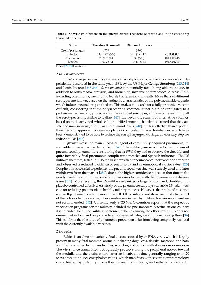
Biomedicines 2022, 10, 2050 27 of 96
Table 6. COVID-19 infections in the aircraft carrier Theodore Roosevelt and in the cruise shipDiamond Princess.
Ships Theodore Roosevelt Diamond Princess p
Crew/passengers 4779 3700Infected 1331 (27.85%) 712 (19.24%) <0.0000001
Hospitalized 23 (1.73%) 36 (5%) 0.00003448Deaths 1 (0.075%) 13 (1.83%) 0.00001793
From [229,230] modified.
2.18. Pneumococcus
Streptococcus pneumoniae is a Gram-positive diplococcus, whose discovery was inde-pendently described in the same year, 1881, by the US Major George Sternberg [243,244]and Louis Pasteur [245,246]. S. pneumoniae is potentially fatal, being able to induce, inaddition to otitis media, sinusitis, and bronchitis, invasive pneumococcal disease (IPD),including pneumonia, meningitis, febrile bacteremia, and death. More than 90 differentserotypes are known, based on the antigenic characteristics of the polysaccharide capsule,which induces neutralizing antibodies. This makes the search for a fully protective vaccinedifficult, considering that the polysaccharide vaccines, either plain or conjugated to aprotein matrix, are only protective for the included serotypes, and a vaccine including allthe serotypes is impossible to realize [247]. However, the search for alternative vaccines,based on the inactivated whole cell or purified proteins, has demonstrated that they aresafe and immunogenic, at cellular and humoral levels [248], but less effective than expected;thus, the only approved vaccines are plain or conjugated polysaccharide ones, which havebeen demonstrated to be able to reduce the nasopharyngeal carriage, a necessary step forreducing IDP [247].
S. pneumoniae is the main etiological agent of community-acquired pneumonia, re-sponsible for nearly a quarter of them [249]. The military are sensitive to the problem ofpneumococcal pneumonia, considering that in WWI they had to observe the dreadful andquite invariably fatal pneumonia complicating measles and Spanish influenza. The USmilitary, therefore, tested in 1945 the first hexavalent pneumococcal polysaccharide vaccineand observed a reduced incidence of pneumonia and pneumococcal carrier rates [125].Despite this successful experience, the pneumococcal vaccine was scarcely used and laterwithdrawn from the market [250], due to the higher confidence placed at that time in thenewly available antibiotics compared to vaccines to deal with the pneumococcal diseaseissue [251]. More recently, the US military organized a large randomized, double-blind,placebo-controlled effectiveness study of the pneumococcal polysaccharide 23-valent vac-cine for reducing pneumonia in healthy military trainees. However, the results of this largeand well-performed study on more than 150,000 recruits did not show any protective effectof the polysaccharide vaccine, whose routine use in healthy military trainees was, therefore,not recommended [252]. Currently, only 8/25 NATO countries report that the respectivevaccination programs for the military included the pneumococcal vaccine; in one country,it is intended for all the military personnel, whereas among the other seven, it is only rec-ommended in four, and only considered for selected categories in the remaining three [36].This confirms that the issue of pneumonia prevention is far from being completely resolvedwith the currently available vaccines.
2.19. Rabies
Rabies is an almost invariably fatal disease, caused by an RNA virus, which is largelypresent in many feral mammal animals, including dogs, cats, skunks, raccoons, and bats,and it is transmitted to humans by bites, scratches, and contact with skin lesions or mucosae.The virus, once transmitted, retrogradely proceeds along the peripheral nerves towardthe medulla and the brain, where, after an incubation time generally ranging from 20to 90 days, it induces encephalomyelitis, which manifests with severe symptomatology,characterized by difficulty in swallowing and hydrophobia, and either an encephalitic

Biomedicines 2022, 10, 2050 28 of 96
(furious) or paralytic (dumb) form, in 80% and 20% of cases, respectively [253]. The WHOestimates that globally there are at least 55,000 deaths each year from rabies, especially inAsia and Africa [254]. No effective therapy exists, but an effective inactivated vaccine andpassive immunotherapy with human rabies immunoglobulins (RIGs) are available. In caseof bite and suspected infection, post-exposure prophylaxis (PEP) may be administered assoon as possible, by cleansing the wound and inoculating human RIGs at 20 IU/kg aroundthe wound [255], and by administering four vaccine doses intramuscularly in two weeks(0, 3, 7, and 14 days) for minor contacts [256].
Despite that the military are not actively engaged in rabies research, rabies is a diseaseof military interest, in particular for deployed active service members [257]. In the USArmed Forces in the period 2011–2018, 22,709 animal bites were reported, which is anaverage of eight animal bites per day [258]. Animal bites with consequent rabies have beenobserved during the Vietnam war [259]. After the Vietnam war, rabies was still a problemin the Philippines, where in 1984, 315 potential rabies exposures were managed and 79 ofthem received PEP [260]. The British Army had to manage 62 animal bites when deployedto Bosnia–Herzegovina in 1995–1996 [257]. The possible shortage of RIGs may heavilyinfluence the outcome of an at-risk animal bite in deployed personnel; thus, pre-exposureprophylaxis by active immunization has been considered to avoid the need of administeringRIGs in the PEP [256]. All 25 NATO countries reporting the military vaccinating programinclude rabies vaccine for selected military categories; however, in three countries, rabiesvaccine is only recommended [36].
2.20. Yellow Fever
Yellow fever is a potentially lethal disease caused by an RNA flavivirus, which istransmitted by the bite of infected mosquitoes of the species Aedes aegypti and Hemagogus, en-demic in Sub-Saharan Africa and tropical Central and South America [91]. The case-fatalityrate of the disease is estimated at approximately 35%; modeling studies have estimatedin 2013 the burden of yellow fever in 84,000–170,000 cases with 29,000–60,000 deaths [16](Table 1). The incubation period is 3–6 days, and the disease may run asymptomatic orwith a mild, not specific, symptomatology, with fever, myalgia, backache, headache, loss ofappetite, nausea or vomiting, for 3–4 days. Most patients heal from the infection, whereasa few patients may enter a toxic phase one day after the end of symptomatology, withmulti-organ failure, icterus and bleeding from the nose, mouth, eyes or stomach; half ofthese patients die within 7–10 days [16].
Yellow fever was endemic in Africa, and in the sixteenth century, it traveled to theAmericas, following the slave trade, thus becoming endemic in the coastal areas of Centraland South America and even in the southern and eastern coast of North America to Boston.From 1668 to 1893, over 135 epidemics of yellow fever occurred in the USA. In 1793, anepidemic of yellow fever killed 10% of the Philadelphia population and in 1878, anotherepidemic killed 20,000 people in the Mississippi valley [261]. At that time, nothing wasknown about the biology of the disease and the way it is transmitted. At the start of theSpanish–American war in 1898, the US troops were decimated by yellow fever in Cuba.Thus, the Surgeon General of the US Army, Gen. George Miller Sternberg, organized aYellow Fever Commission, coordinated by Major Walter Reed, and composed of MajorsJames Carroll, Aristides Agramonte, and Jesse Lazear, with the duty of clarifying theway of transmission of the disease in order to prevent infection spreading [261]. TheCommission went to Cuba to begin its activity in June 1900. It started by verifying theetiological hypothesis proposed by the Italian microbiologist Giuseppe Sanarelli, who in1897, announced to have found the etiological agent of yellow fever, which was namedBacillus icteroides. The commission ruled out this hypothesis and then focused its activityon taking into account the work of Carlos Finlay, a Cuban physician who had suggested atransmission through the mosquito Aedes aegypti by performing specific experiments onhuman volunteers, which were unsuccessful. Finlay tried to expose healthy volunteersto the bite of mosquitoes 2–6 days after the mosquitoes had bitten a patient with yellow
Biomedicines 2022, 10, 2050 29 of 96
fever; however, he never succeeded in observing a clear case of infection transmission. Thereason was clarified over 10 years later by the observations of the US physician Henry RoseCarter in 1898, relative to the “extrinsic incubation” of yellow fever in the mosquito, whichwas calculated in approximately 2 weeks. The Yellow Fever Commission thus repeatedFinlay’s experiments, by taking into account the “extrinsic incubation” time of Carter andsucceeded in demonstrating the transmissibility of the etiological agent by mosquitoes, thusproviding scientific evidence to Finlay’s hypothesis. Considering that there is not an animalmodel for yellow fever, the commission used healthy human volunteers, including thesame members of the commission and one of its members, Jesse Lazear, who died in 1900, atthe age of 34 years [262]. The observations of the Yellow Fever Commission were publishedin 1901 (JAMA 1901;36: 431–40), and the Major physician US Army William C Gorgas,responsible for health in Cuba, received the disposition to free Havana of mosquitoes. Hiswork was excellent because in 90 days, he transformed the epidemiological situation ofHavana, in which one case of yellow fever per day was described on average from 1762 to1901, whereas after mosquito disinfestation, it was free of the disease [261]. Thus, the fightagainst yellow fever was won in this phase by the US military.
The viral etiological agent was only isolated in 1927 from a sick man in Ghana. A livevaccine, attenuated by 200 subcultures of this virus, designated 17D strain, was developedin the 1930s by Theiler and Smith [263]; Theiler was awarded the Nobel Prize for Physiologyor Medicine in 1951 [92]. The vaccine is generally safe, highly immunogenic and protectivefor long periods, considering that the presence of neutralizing antibodies has been foundafter 30–35 years from vaccination [264]. Currently, all 25 NATO countries reporting therespective military vaccination program include yellow fever vaccine, in 24 for selectedcategories, whereas in one country for all the military personnel. With the use of the vaccine,yellow fever does not represent a problem for the military at the global level anymore [36].Yellow fever virus has been included among the possible biological agents, category C [57].
2.21. Japanese Encephalitis (JE)
Japanese encephalitis (JE) is a potentially lethal disease caused by an RNA flavivirustransmitted by the bite of infected Culex mosquitoes, in particular Culex tritaeniorhynchus;however, even other mosquito species may be vectors. The virus is endemic in large partsof South, South-East Asia and the Western Pacific, including an estimated population ofover 3 billion people, particularly in rural areas, where the risk factor is living in proximityof rice fields and pig rearing [265]. It is estimated that the annual JE cases are 67,900, with13,600–20,400 deaths (Table 1). The infection may run asymptomatic in most patients; inone case out of 250 infections, the disease is severe. After an incubation period of 4–14 days,symptomatology starts with high fever, chills, myalgias, headache, and mental confusion;however, opisthotonos and even acute flaccid paralysis may occur. The disease occurspreferentially in children <10 years, in whom it is generally more severe. The case-fatalityrate of the severe disease is 20–30%, and approximately 30% of the survivors presentpermanent neurologic or psychologic disabilities [17].
The interest for the US military started during WWII; in 1942, a research team wasestablished at WRAIR, with the duty of developing a vaccine for JE [257]. Even MajorAlbert Sabin received by the Commission on Neurotropic Virus Diseases of the ArmyEpidemiological Board the task to develop a JE vaccine [125]. The vaccine was a formalin-inactivated JE virus cultured in the brains of mice; it was used on 250,000 US soldiersduring the war, starting in 1945, after an outbreak of JE in the US military stationed inOkinawa [125]. Albert Sabin had the opportunity to study and describe this outbreakand the use of the vaccine [266]. Even after the war, Albert Sabin and the US militarycollaborated with Japanese researchers for studying together the JE vaccine [267]. TheUS military suffered a relevant outbreak of 300 cases of JE in 1950 among the US troopsstationed in Korea during the Korean War, although the US military were all vaccinated,and 16 lethal cases were observed [257]. Even during the Vietnam War, cases of JE in theUS Air Force personnel were described [257], however, no reduction of US force fighting
Biomedicines 2022, 10, 2050 30 of 96
strength was observed [268]. Following the Korea outbreak, which had demonstrated thepoor protection provided by the first used vaccine, the army interrupted the vaccinationof the US military assigned to the Far East Command [125]. At the end of the 1950s,researchers of WRAIR working in Japan contributed to providing new knowledge on JEecology [269]. The studies for the development of a new vaccine resumed in the 1980s, ledby the CDC; however, the conclusive phase III studies were carried out in Thailand, underthe leadership of Col. Charles Hoke, of the US Army Medical Component, Armed ForcesResearch Institute of Medical Sciences (AFRIMS) in Bangkok, Thailand, during which amonovalent (Nakayama strain) and bivalent (Nakayama and Beijing-1 strains) vaccinewere studied in comparison with placebo. The results showed 91% of efficacy for bothmonovalent and bivalent vaccines [270,271]. This study could be carried out due to theprevious research at AFRIMS of the military researcher Donald Scott Burke, who had setup a diagnostic test for anti-JE IgM in serum and liquor [272,273]. Another study wascarried out by the WRAIR researchers on 538 US soldiers with monovalent JE vaccine,which confirmed the safety and the high immunogenicity of the vaccine and ruled out thepossible interference with a previous yellow fever vaccination, another flavivirus [274]. In2005, the production of the mouse brain-derived JE inactivated vaccine was discontinuedby the manufacturing company because it was considered too reactogenic and poorlyimmunogenic [275]. Currently, in the USA, the only approved vaccine is IXIARO (JE-VC),which is a Vero cell-culture-derived inactivated vaccine [276]. However, even live andrecombinant live vaccines are available [17,274]. Twenty-one out of the twenty-five NATOcountries report that the military vaccination program includes the JE vaccine for selectedcategories of personnel [36]. The Italian military soldiers participating in the INTERFET(International Force to East Timor) mission in 1999 were vaccinated with the monovalent(Nakayama strain) mouse brain-derived JE vaccine, without side effects. However, some ofthem were infected by the dengue virus, and the previous vaccination with the JE vaccinehas been considered partly cross-protective even for dengue [277].
2.22. Tick-Borne Encephalitis (TBE)
Tick-borne encephalitis (TBE) is a disease caused by an RNA flavivirus transmittedby the bite of ticks. The virus is present in many animals, such as wild rodents, deer,boar, dog, fox, sheep, cattle, and bat, and is transmitted to humans by ticks of the familyIxodidae, in particular Ixodes Ricinus and Ixodes persulcatus. Humans are a dead-end hostand may even be infected by alimentary route, by eating raw contaminated dairy. Threesubtypes of the virus are responsible for the respective diseases that are endemic in central,eastern and northern Europe (western subtype), eastern Europe, Russia and northern Asia(Siberian subtype), and eastern Russia as well as some parts of China and Japan (far easternsubtype) [278]. Although two-thirds of the cases are asymptomatic, the disease caused bythe western subtype has a biphasic pattern, with a first phase characterized by nonspecificsymptomatology (fever, myalgia, headache, fatigue, nausea) and a second phase, followinga free interval, by meningoencephalitis, myelitis or paralysis, whereas the far easternsubtype is associated with a monophasic disease. Moreover, the European disease ismilder (mortality 0.5–2%, and neurological sequelae up to 10%) than the disease causedby the far eastern subtype, which has a mortality of up to 20% and a higher prevalenceof permanent neurological sequelae [279]. The diagnosis may be made by molecular orserological approaches. However, virus identification by molecular methods is poorly usedbecause the viremia is present for a short time, and the reliability of serological tests isreduced for the possible cross-reaction among different flaviviruses. There is no availabletreatment, whereas an inactivated vaccine from a cell-cultured virus has been shown tobe safe and protective in over 95% of recipients after three-dose administration [278]. Theannual world cases of TBE are approximately 10,000–12,000 [280], whereas in Europe, over3000 annual TBE cases are hospitalized [281]. The disease tends to be more frequent inmales than in females in Europe and more severe in >50–60-year-old subjects, who are lessresponsive to the vaccine [278,282,283].
Biomedicines 2022, 10, 2050 31 of 96
The interest for the military of TBE is linked to the country where the military liveor are deployed to, whether TBE is endemic or not. The US military, which started tobe interested in TBE in the mid-1980s [284], vaccinated the troops deployed to Bosnia in1996 with an accelerated schedule (0, 7, and 28 days instead of 0, 1–3, and 9–12 months)of TBE vaccine in order to be readily protected; 80% of seroconversion rate was observedafter the third vaccine dose, and the vaccine proved to be safe, with only 7/3981 (0.18%)vaccinees reporting self-limited symptoms. However, the infection risk was relatively low,considering that only 4/959 (0.42%) unvaccinated soldiers seroconverted [285]. Amongthe 25 NATO countries reporting the respective vaccination schedule for the military,twenty-two include the TBE vaccine, six of these countries (all European where TBE isendemic) provided to the whole military personnel, and in the other sixteen, only to selectedcategories [36]. The TBE virus has been included among biological agents, category C [57].
2.23. Human Papillomavirus (HPV)
Human papillomavirus (HPV) is a DNA virus, which infects epithelial basal cells, atcutaneous and mucosal levels, and may induce different cutaneous and mucosal lesionsand even cancers. There are more than 100 serotypes, some of which may cause cervix,anal, penile, and oropharynx cancers, with serotypes 16 and 18 being the most frequentlyimplicated in cancers. However, even serotypes 31, 33, 45, 52, and 58 may be consideredhigh risk for cancer induction, whereas serotypes 6 and 11 are generally associated withanogenital warts, such as condyloma acuminatum, and are considered low-risk HPV [286].The main way of HPV transmission is through sexual intercourse; thus, the militaryworldwide are at special risk [287], hence their interest in HPV, even considering that asafe and effective HPV vaccine is currently available. Three recombinant vaccines areavailable, the bivalent (16 and 18 serotypes), the tetravalent (6, 11, 16, and 18 serotypes),and the nine-valent (6, 11, 16, 18, 31, 33, 45, 52, and 58 serotypes). It may be calculatedthat with bivalent and tetravalent vaccines, the protection against cancer is approximately70%, whereas it increases to approximately 90% with the nine-valent one [92]. Only oneNATO country reports HPV mandatory vaccination in the military schedule, whereas inthe other six countries, the vaccination is only recommended [36]. A longitudinal study inthe US military showed that 14.6% of male recruits were HPV positive for serotypes 6, 11,16 or 18 at entry and 34.2% of those originally negative for these serotypes seroconvertedto one or more of them after 10 years [288]. However, more recently, an epidemiologicalsurvey on genital HPV infections developed during a 9-year long follow-up, between1 January 2012 and 31 December 2020, has shown a significant reduction of infection forboth genders, female service members from 261.2/10,000 to 163.1/10,000 person-years(37.5% of reduction) and male service members from 40.6 to 16.9/10,000 person-years (66.1%of reduction), a decrease that has been attributed, at least in part, to the introduction of avaccine for females in 2006 and for males in 2010, which, even though it is not mandatory, isencouraged and offered to service members [289]. In a relatively low number of countries(76/195, 39%), the HPV vaccine has been introduced as mandatory and in most cases foryoung females, with a gradient of application ranging from 10% for low-income countries to69% for high-income countries, thus clearly indicating the negative influence of poverty onthe possibility of introducing this relatively expensive vaccine. The global HPV vaccinationcoverage is estimated to be as low as 12.2% [290]. Australia was the first country to organize,in 2007, an eradication program of the cancer of the cervix [291]. Despite that the fightagainst cervical cancer is a priority considering the high number of annual cases and deaths,especially in low-income countries [290], and for this reason, the vaccination campaign hasmainly been addressed to young girls before starting sexual activity, the vaccination of themales should also be considered to prevent male cancers [292].
HPV vaccination in the military could contribute to the reduction of HPV-relatedcases of cancers if mandatory, considering that the military are at a higher risk of infectionthan the matched civilian population, and the simple recommendation of vaccinationcannot reach critical coverage, considering the stigma linked to the sexually transmitted

Biomedicines 2022, 10, 2050 32 of 96
diseases [293,294]. Moreover, a cost-effectiveness estimate allows one to compare the carecost per case of anal cancer of USD 52,700 or 146,100 per case of oropharyngeal cancerversus USD 450 for HPV vaccination [295]. Thus, this hesitancy in making HPV vaccinationmandatory for the military is quite surprising, and it diverges from the historical behaviorof the military, that for many infectious diseases has generally anticipated the generalpopulation in vaccine research and application. Probably, this was the expression of adifferent time, in which infectious diseases could heavily influence the outcome of battlesand war more than the combat capacity. Moreover, HPV is not acutely incapacitating,considering that it may induce deferred neoplastic disease. However, a larger vaccine use,especially in countries with compulsory conscription, may represent a relevant measure ofpublic health.
2.24. Cholera
Cholera is a bacterial disease that can be transmitted through water or food contam-inated with Vibrio cholerae, 01 and 0139 serogroups, endemic in 50 countries and able toinduce epidemics. It is estimated that annually 1.3–4 million people become infected, re-sulting in 21,000–143,000 annual deaths [296] (Table 1). Seven pandemics since 1817 spreadfrom Asia to all over the world. Right rehydration may lower the mortality from over 50%to 0.2% [21]. The prevention consists of water sterilization and sanitation. Cholera wasfirst reported by the British military in 1770 [297]. Similar to all the infectious diarrhealsyndromes linked to poor hygienic conditions, it has always been considered a threatby the military. In 1855, during the Crimean War, the Piedmont–Sardinia expeditionaryforce was deeply hit by cholera; 2728/18,000 military personnel fell ill with cholera, anattack rate of 15%, and 1230 died, a case-fatality rate of 45% [298]. A live vaccine againstcholera was first developed by Jaime Ferran in Spain [299], but it was ultimately the vaccinedeveloped by the German scientist Wilhelm Kolle in 1896, using heat-inactivated cholerabacilli, that came into general use and that served as a model for cholera vaccines for thenext century [300]. As a military physician and hygienist during WWI, Kolle was highlysuccessful in vaccination against cholera. This vaccine was widely used during WWI in themilitary, such as by the Italian Army when, in August 1915, cholera broke out in the Italiantroops deployed along the Isonzo river. The anti-cholera mass vaccination of the militarywas then ordered and subsequently extended to civilians residing in closely affected areas.This approach allowed the containment of the epidemic, which remained almost confinedto the military community and only marginally affected the civilian population. In 1915,the observed cases were 14,000, whereas they were reduced to only 170 in 1916 [301]. Thesedata demonstrate that vaccination campaigns can be carried out safely even during theepidemic phases, helping to provide useful information to the scientific world to betterunderstand the effectiveness of this vaccine. However, this vaccine was painful and didnot give long-lasting immunity.
Furthermore, it is worth noting the contributions of US military investigators on thefront lines of cholera research. The US Navy’s involvement with cholera began in Cairo,Egypt, during the 1947 cholera epidemic, when the commander of the Naval MedicalResearch Unit (NAMRU) 3, Robert A. Phillips, made some interesting observations. Heestablished that the stools of patients with cholera were isotonic with their blood [302]and did not contain proteins; thus, allowing him to argue that no mucosal damage waspresent [297]. This observation allowed the rehydration of patients by infusion of isotonicelectrolyte solutions to be possible, as even confirmed in a cholera outbreak in BangkokThailand, where Phillips applied his method [303]. This rehydration method allowed themortality of cholera to be reduced from 20–30% to less than 1%; thus, saving a large numberof lives. Later, in 1961 in Manila, Phillips discovered that isotonic electrolyte solutionscontaining glucose could be orally administered to rehydrate patients with cholera andother diarrheal diseases. This further observation made the rehydration method accessibleeven to developing countries for its lower cost, thus allowing millions of lives to be saved in

Biomedicines 2022, 10, 2050 33 of 96
the past several decades [297]. Richard Finkelstein, a civilian working at WRAIR, isolatedthe cholera exotoxin, which he called cholerogen, in 1963 [304].
Finally, the US military contributed to developing and testing improved choleravaccines. Col. Jose Sanchez and colleagues, from WRAIR [305], and even in collaborationwith the US Navy Medical Research Institute Detachment—Lima, Peru [306], studied akilled, whole-cell, vaccine plus recombinant cholera toxin B subunit (WC/rBS), and Col.David Taylor and other colleagues from WRAIR contributed to basic science research intoa live attenuated O139 Vibrio cholerae vaccine prototype [307,308].
In addition, the US Department of Defense contributed to basic science research into alive attenuated cholera vaccine at the Armed Forces Research Institute of Medical Sciencesin Bangkok [309] and at the Indonesian US NAMRU in Jakarta [310], respectively.
While considering the advances in the development of vaccines, also due to the contri-bution of the military, cholera is still a major global health problem in unsanitary conditions.Current cholera vaccines, represented by a two-dose killed whole cell monovalent (01) plusrecombinant cholera B subunit of cholera toxin (WC-rBS), a two-dose killed whole cellbivalent (01 and 0139) (WC), and a single-dose live oral attenuated vaccine (CVD-103 HgR),are safe, feasible to use and represent a public health tool in the prevention of the disease,along with hygiene measures [311]. Currently, such as for typhoid fever, in the military ofmost NATO countries (21/25), including Italy, cholera vaccination is present in the vaccina-tion schedule, but only for the troops deployed to at-risk epidemiological countries [36].V. cholerae has been included among the possible biological agents, category B [57].
2.25. Leptospirosis
Leptospirosis is a potentially fatal bacterial disease caused by Leptospira, an aerobicbacterium containing in its structure a lipopolysaccharide similar to the one found inGram-negative bacteria [312]. Leptospira is present in different wild and domestic animals;however, the main reservoir for human infections is Rattus norvegicus [313]. Leptospira isexcreted in rat urine; thus, contaminating soil and water. Humans are accidental hosts,who may be infected through the trans-cutaneous or trans-mucosal passage, profiting fromcuts or abrasions of the skin or conjunctival and/or oral mucosae [314]. Leptospirosisis therefore an occupational zoonosis; the most exposed worker categories are sewageworkers, farmers in rainy areas and the military, particularly during exercises in marshysoils. The disease may be mild and self-limited; however, in some subjects and with someserovars, the disease may be severe, as in the case of Weil’s disease, caused by serovarsof the icterohaemorrhagiae serogroup, in which the mortality is over 10%, or the severepulmonary hemorrhage syndrome, which may have a case-fatality rate of over 50% [314].Annually leptospirosis is estimated to be responsible for 1.03 million clinical cases with58,900 deaths [25] (Table 1).
The etiological agent was discovered in 1915 by Japanese [315] and German [316,317]physicians, whereas the severe form of the disease had been described by Weil in 1886 [318].However, the disease was present before and in the seventeenth century in New England,and in the eighteenth and nineteenth centuries in Europe, illnesses with the characteristicsof leptospirosis had been described, particularly by military doctors [319]. Leptospirosis isa disease of interest for the military, because it is frequently associated with the military,both in wartime, considering the precarious hygienic conditions, particularly in humidtrench warfare, and in peacetime, for training in standing water [320]. It was describedduring the second independence war in Italy in 1859 [319], during WWI and WWII, andthe Vietnam War [308]. In the summer of 1942, there was an outbreak of febrile exanthemat Fort Bragg, which involved 40 US soldiers and recurred in the summer of 1943 and1944, whose etiology was only clarified in 1952 by Major US Army William Gochenourand colleagues following isolation of Leptospira autumnalis [321]. In the same year, MajorGochenour and colleagues were able to diagnose as leptospiral meningitis an outbreakof “aseptic meningitis” occurring in 1949 in US soldiers serving in Okinawa [322]. In the1980s, Dr. Ernest T. Takafuji from WRAIR and colleagues were able to successfully test
Biomedicines 2022, 10, 2050 34 of 96
the efficacy of chemoprophylaxis with doxycycline against leptospirosis on US soldierstraining in field exercises in the Panama Canal [323]. Although an inactivated whole-cellvaccine has been available for more than a century, it is largely used in animals, whereas itis rarely used in humans, despite its effectiveness, due to its specific protection only againstsingle serovars (Spirolept®, produced by Sanofi-Pasteur, only protects against Leptospiraicterohaemorrhagiae), the quite heavy schedule, characterized by three subcutaneously ad-ministered doses followed by biannual boosters, and its reactogenicity [313,324]. Althoughinformation about the number of world countries adopting the leptospirosis vaccine for themilitary is lacking, among NATO countries, only two consider leptospirosis vaccination inoccupationally exposed military personnel [36].
2.26. Dengue
Dengue is the most prevalent arthropod-borne viral disease [325], responsible for anestimated 390,000,000 annual infections, a quarter of which are symptomatic [18] (Table 1).The virus is an RNA flavivirus, of which four serotypes (1, 2, 3, and 4) are known, andis transmitted by the same vector of yellow fever virus, Aedes aegypti; however, in somegeographical regions, other vectors, such as Aedes albopictus and Aedes polynesiensis, mayeven transmit the virus [326]. The infection may run completely asymptomatic, whereasin an estimated 25% of cases, it may induce non-specific fever, dengue fever, denguehemorrhagic fever, and dengue shock syndrome. Dengue hemorrhagic fever is frequentlyobserved in children and dengue shock syndrome, if severe, may be responsible for deathin 9.3%, but up to 47%, of cases with profound shock [327]. Dengue is of interest to themilitary because it is highly prevalent at the global level and may heavily reduce theoperational readiness of the soldiers, even though the annual mortality is estimated to bequite low, 12,000, mainly occurring among children [19] (Table 1). In a recent quantitativealgorithm to quantify the burden of infectious diseases for the US military, dengue ranksthird, after malaria and bacterial diarrhea [328]. Moreover, the prevention of dengueconsists of the defense from the vector, considering that a satisfying vaccine registeredin many world countries is still lacking. Although different types of vaccines are understudy, including the live recombinant ones, inactivated, subunits with the envelope (E)protein alone or together with the precursor of the membrane (prM), only one tetravalentrecombinant live on a YF17D backbone has been licensed in Mexico in December 2015, withthe name Dengvaxia® [329]. Afterward, other endemic countries registered this productwith their respective regulatory authorities. This vaccine is administered according toa 0/6/12-month schedule and has the highest efficacy of 76.9% against serotype 4 andthe lowest of 43% against serotype 2. However, the cumulative efficacy was substantiallyhigher, 78.2%, in people already exposed to dengue compared to naïve [329]. In differentprojects for vaccine and monoclonal antibodies development, the US military, at WRAIRand Naval Medical Research Center (NMRC), are involved as further evidence of theinterest in dengue for the military [329–331]. In addition to the diagnostic problem for thecross-reaction with other flaviviruses and the possible cross-protection between differentflaviviruses [332,333], for dengue virus only the issue of antibody-dependent enhancement(ADE) has been described, which is the facilitated antibody-mediated viral entry intothe cells through the FcγR [326]. ADE has been considered as the main reason for thewaning, after approximately 2 years from an infection with a dengue serotype, of the cross-protection against the other three serotypes (heterotypic protection), whereas the homotypicprotection is lifelong [329], in line with the long persistence of protective antibodies [334].After the waning of heterotypic protection, people are more exposed to severe forms ofdengue by heterologous serotypes [335]. This peculiar behavior of humoral anti-dengueimmunity has to be taken into account when developing a dengue vaccine.
The US military have reviewed the burden of dengue from the American–Spanish War,through the Philippines, where they could observe that the disease more easily occurred inurban than in rural areas and that reinfection was not rare. During WWII, dengue occurredin many war theaters, particularly in the South Pacific, New Guinea and the Philippines; in
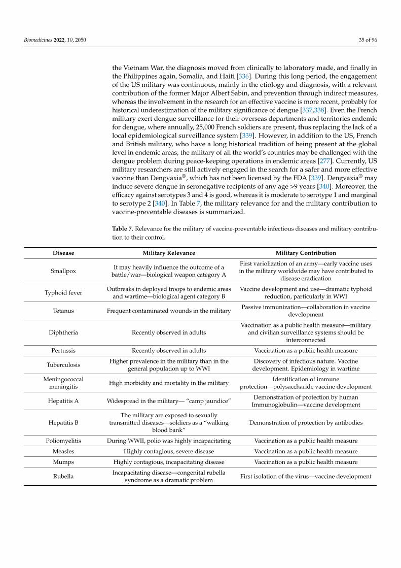
Biomedicines 2022, 10, 2050 35 of 96
the Vietnam War, the diagnosis moved from clinically to laboratory made, and finally inthe Philippines again, Somalia, and Haiti [336]. During this long period, the engagementof the US military was continuous, mainly in the etiology and diagnosis, with a relevantcontribution of the former Major Albert Sabin, and prevention through indirect measures,whereas the involvement in the research for an effective vaccine is more recent, probably forhistorical underestimation of the military significance of dengue [337,338]. Even the Frenchmilitary exert dengue surveillance for their overseas departments and territories endemicfor dengue, where annually, 25,000 French soldiers are present, thus replacing the lack of alocal epidemiological surveillance system [339]. However, in addition to the US, Frenchand British military, who have a long historical tradition of being present at the globallevel in endemic areas, the military of all the world’s countries may be challenged with thedengue problem during peace-keeping operations in endemic areas [277]. Currently, USmilitary researchers are still actively engaged in the search for a safer and more effectivevaccine than Dengvaxia®, which has not been licensed by the FDA [339]. Dengvaxia® mayinduce severe dengue in seronegative recipients of any age >9 years [340]. Moreover, theefficacy against serotypes 3 and 4 is good, whereas it is moderate to serotype 1 and marginalto serotype 2 [340]. In Table 7, the military relevance for and the military contribution tovaccine-preventable diseases is summarized.
Table 7. Relevance for the military of vaccine-preventable infectious diseases and military contribu-tion to their control.
Disease Military Relevance Military Contribution
Smallpox It may heavily influence the outcome of abattle/war—biological weapon category A
First variolization of an army—early vaccine usesin the military worldwide may have contributed to
disease eradication
Typhoid fever Outbreaks in deployed troops to endemic areasand wartime—biological agent category B
Vaccine development and use—dramatic typhoidreduction, particularly in WWI
Tetanus Frequent contaminated wounds in the military Passive immunization—collaboration in vaccinedevelopment
Diphtheria Recently observed in adultsVaccination as a public health measure—military
and civilian surveillance systems should beinterconnected
Pertussis Recently observed in adults Vaccination as a public health measure
Tuberculosis Higher prevalence in the military than in thegeneral population up to WWI
Discovery of infectious nature. Vaccinedevelopment. Epidemiology in wartime
Meningococcalmeningitis High morbidity and mortality in the military Identification of immune
protection—polysaccharide vaccine development
Hepatitis A Widespread in the military— “camp jaundice” Demonstration of protection by humanImmunoglobulin—vaccine development
Hepatitis BThe military are exposed to sexually
transmitted diseases—soldiers as a “walkingblood bank”
Demonstration of protection by antibodies
Poliomyelitis During WWII, polio was highly incapacitating Vaccination as a public health measure
Measles Highly contagious, severe disease Vaccination as a public health measure
Mumps Highly contagious, incapacitating disease Vaccination as a public health measure
Rubella Incapacitating disease—congenital rubellasyndrome as a dramatic problem First isolation of the virus—vaccine development

Biomedicines 2022, 10, 2050 36 of 96
Table 7. Cont.
Disease Military Relevance Military Contribution
Varicella Highly contagious, incapacitating Vaccine use is quite limited
Influenza Frequent cause of acute respiratory disease inthe military
Support to first vaccine development—firstisolation of “Asian” virus—identification of driftsand shifts—organization of surveillance system
Adenovirus Frequent cause of acute respiratory disease inthe military First isolation of the virus—vaccine development
Coronavirusdisease-2019
The military are exposed because they areengaged in pandemic containment
The military have been crucial for organizingdiagnostic and vaccination campaigns
Pneumococcus Responsible for severe acute respiratorydisease
Discovery of microorganism—first hexavalentpolysaccharide vaccine
Rabies Severe threat to deployed service members Preventive vaccination
Yellow fever Endemic in Cuba—threat to the US militarydeployed there—biological agent category C
Demonstration of mosquito-transmissionDisease control through vector eradication
Japaneseencephalitis
Possible threat for the military deployed toAsia
Vaccination WWII—epidemiology—field trialinactivated vaccine in Thailand
Tick-borneencephalitis
Possible threat for the military deployed toendemic countries—biological agent
category C
Vaccine has demonstrated to be safe andimmunogenic
Human papillomavirusinfection
The military are exposed to sexuallytransmitted diseases
HPV vaccine inclusion in the military vaccinationschedule may be a relevant measure of
public health
Cholera Severe disease frequently present inwars—biological agent category B Rehydration therapy—vaccine development
Leptospirosis The military may be infected in field exercisetraining and wartime Chemoprophylaxis by doxycycline
Dengue Incapacitating threat for the military deployedto endemic areas Vaccine development
WWI, First World War; WWII, Second World War; HPV, human papillomavirus.
3. Non-Vaccine-Preventable Infectious Diseases3.1. Epidemic Typhus
Epidemic typhus is a bacterial disease historically associated with poverty, dirtyenvironment, and overcrowding, all conditions that are found during war; thus, epidemictyphus has accompanied and sometimes has heavily influenced conflicts, mainly in thenineteenth and in the first half of the twentieth century. It is caused by Rickettsia prowazekii,a Gram-negative bacillus belonging to the order of Rickettsiales, which is transmitted tohumans by the body louse (Pediculus humanus corporis), as discovered by Charles Nicolle in1909 (who, for this discovery, was granted the Nobel Prize in Physiology or Medicine in1928), at that time Director of the Pasteur Institute in Tunis [341]. The lice do not transmitR. prowazekii through the bite, rather they are infected after biting an infected human oranimal; once infected, the lice eliminate a large number of microorganisms with feces asa powder, which remain viable for up to 100 days and may enter the body through skinabrasions. In addition, inhalation of aerosolized dry powder containing viable Rickettsiaeis another effective way of infection; this type of infection may create some theoreticalconcerns for the possible use of R. prowazekii as a biological weapon, of category B [57,342].In subjects who have suffered epidemic typhus even decades before, the disease may recuras a consequence of immunosuppression or wartime stress (recrudescent epidemic typhusor Brill–Zinsser disease) and spread in a lousy-naïve population, thus allowing this diseaseto be maintained by this human reservoir [341]. However, even an animal reservoir hasbeen identified [343].
Biomedicines 2022, 10, 2050 37 of 96
Historically, the first test for diagnosis was the Weil–Felix, based on the cross-reactionwith Proteus; more recently, specific immunological and molecular methods have beenset up for diagnosis [344]. Without therapy, typhus is a severe disease, characterized byeasy spreading and relatively high mortality, ranging from 13% to 30% [344]. After anincubation time of 10–14 days, the disease suddenly bursts with non-specific symptoms,such as high fever, headache, myalgias, and rash of the trunk and limbs. Moreover,nausea and vomiting, pneumonia, petechial rash, central nervous system involvementwith seizures, and mental confusion (hence the name “typhus”), may be present [345].Antibiotic therapy with tetracycline or chloramphenicol promptly resolves. However, if theenvironmental situation allows, a bath and new clean dress and underwear are enoughfor eliminating lice; thus, the disease may easily spread in emergency and war conditionswhen these simple operations become impossible to be realized; it has been stated thatepidemic typhus has caused more deaths than all the wars in history [344,346]. The trial ofdeveloping a vaccine has met a series of difficulties, including that Rickettsia is obligatedto be intracellular, thus the need of growing it with cells. The pioneering study of Weigl,who prepared an inactivated vaccine from the homogenates of infected-lice intestines [347],which proved to be effective, did not solve the problem, because it was not fit for massproduction [348]. However, the work of Cox, who demonstrated that Rickettsiae couldeffectively be grown on the yolk-sac membrane of a developing chick embryo [349], pavedthe way for the mass production of an effective vaccine [350]. The trial of developing a livevaccine was instead a failure [348].
Epidemic typhus has for a long time accompanied wars. In 1776, the war of theAmerican Revolution might have been prolonged as a consequence of epidemic typhus inup to one-third of the American Army before one battle with the British Army [351]. Dur-ing Napoleon’s Russia campaign, epidemic typhus heavily contributed to decimating theGrande Armée (Great Army), which was reduced 100-fold from 500,000 men to 5000 [1]. Adetailed computation aiming at precisely calculating the deaths due to combat versus thosedue to disease attributes approximately 41,000 deaths to combat and 200,000 to infectiousdiseases, represented by typhus, dysentery, and diphtheria [352]. Whichever the real data,these figures underline that the invisible enemy was much more effective in decimatingtroops and influencing the outcome of the war than the visible one. Even during and afterthe WWI epidemic, typhus was a protagonist. In Serbia, which had approximately 3 millioninhabitants at the start of the war, after 6 months, 500,000 people had epidemic typhus,and 40% of them, which is approximately 200,000 humans, died, including 70,000 Serbiantroops and 30,000 Austrian prisoners [353]. At the worst point of the outbreak, 10,000 newcases per day were observed, and the mortality rate increased from 20% to 60–70% [353].In the US troops, instead, the burden of typhus was limited, with only 47 cases and threedeaths, probably due to the effective delousing organization [342]. Epidemic typhus waspresent and particularly active during the Russian Revolution in the period 1918–1922. Itwas estimated that in the period 1918–1920, there were 573,000 cases in the Red Army, withat least 100,000 deaths. The rate of infection was 204 per 1000 in 1919 and 315 per 1000in 1920. No information is available for the White Army [354]. It was estimated that thetotal number of cases in the period 1918–1922 ranged from 15–16 to 25 million, whereasthe number of deaths was estimated at approximately 2.5 million Russians [354]. WWIrepresents a sort of watershed for the relationships between infectious diseases and thewar, considering that in all the previous wars, the ratio of infectious diseases/combat as thecause of death during the war was positive, with a marked advantage of infectious diseasesin combat [338], whereas in WWI, this ratio was lower than 1. The British troops lost todisease were 113,000 versus 585,000 for combat and in the German Army 155,013 soldiersdied from disease versus 1,531,048, who were killed in battle [355]. Regarding epidemictyphus, despite the hygienic conditions being similar on the Western and the Eastern Front,the disease was absent on the former and only present on the Eastern Front [355]. The firstdevelopments of bacteriology, which may have contributed to reducing the influence ofinfectious diseases in WWI [355], were operating even more in WWII, in which vaccination

Biomedicines 2022, 10, 2050 38 of 96
for epidemic typhus and the use of dichloro-diphenyl-trichloroethylene (DDT) for delous-ing, under the directive of the Joint US Typhus Commission, contributed to reducing theburden of epidemic typhus. The US troops had 104 cases and no deaths [342]. During theKorean War, 32,000 cases and 6000 deaths were observed in the South Korean military andcivilian population, whereas a single case was observed in the US military [342]. No caseswere observed during the Vietnam War, whereas since 1993, tens of thousands of cases ofepidemic typhus were observed in different African and Latin American countries [342]. Inparticular, the civil war in Burundi was associated with an outbreak of epidemic typhus inthe refugee camps [356]. However, in Russia, epidemic typhus was still present at the endof the last century [357]. Despite the interest in epidemic typhus for the military, even inconsideration of its possible use as a biological weapon, no effective vaccine is currentlyavailable, considering that the Cox vaccine used in the US military during WWII did notcomply with the modern recommendations on vaccine safety. The recent developments inthe knowledge about protective immunity and immunological determinants for protectionform the basis for developing innovative and effective vaccines [358].
3.2. Scrub Typhus
Scrub typhus is an acute febrile disease caused by a type of rickettsia, Orientia tsut-sugamushi, transmitted by the bite of infected Leptotrombidium spp. mites. The disease ischaracterized by fever and maculopapular rash, with headache, lymphadenopathy, andfrequent involvement of the central nervous system. Without therapy, the disease may havea mortality as high as 60% [342]. Antibiotic treatment with tetracycline and, more rarely,chloramphenicol is generally resolutive, even though recent antibiotic resistance has beenobserved. Scrub typhus is endemic in a large triangle including South and Southeast Asiaand Australia. During WWII in the Pacific war theater, the Allied military had 16,000 casesof scrub typhus, 7300 of whom were in the US troops, with 331 deaths, whereas the Japanesehad 20,000 cases [342]. During the Korean war, only eight cases were observed in the USmilitary, whereas during the Vietnam War, it was estimated that 20–30% of the fever ofunknown origin, once excluded malaria, was due to scrub typhus. The interest of themilitary in scrub typhus is witnessed by the studies of US military researchers on antibiotictherapy, a fieldable diagnostic test development, the first DNA sequence publication of theOrientia, the antibiotic resistance and the patented recombinant rickettsia protein (56 kd,useful for vaccine and diagnosis).
3.3. Trench Fever
Trench fever is a typical disease for the military, which was first described in 1915 by aBritish military physician, Major John Graham, who reported that a soldier had sufferedfrom fever for three days, headache, dizziness, lumbago and shin pain. After a few days,the symptomatology appeared again and disappeared three days later, leaving fatigue.Other soldiers with the same clinical characteristics were observed by Major Graham,who described for the first time a disease closely associated with the military, highlyincapacitating, even though no deaths were reported [359,360]. In 1917, the British WarOffice and the US Red Cross set up two independent commissions for trench fever, the firstchaired by David Bruce, the commander of the Royal Army Medical College and discovererof the etiologic agents of brucellosis and African trypanosomiasis, and the second by the USMajor Richard P. Strong, who had directed the International Commission for limiting theepidemic typhus in Serbia in 1915. The two commissions ended their activity after a seriesof studies and experiments, in 1918, by setting some acute, relevant, and original points,regarding the incubation time, the symptomatology, the transmission by body lice, and thesuggestion of a Rickettsia as a probable etiologic agent [361,362]. The rickettsia-like bodieswere identified in the lice after biting a man with trench fever by a German bacteriologistin 1916 [363] and confirmed in 1918 by a British researcher [364]. There are no records onthe number of cases of trench fever during WWI, but 800,000 soldiers on the Western Frontmay be estimated [360]. Only in 1961 was the etiologic agent of trench fever cultivated by J

Biomedicines 2022, 10, 2050 39 of 96
William Vinson of Harvard University and Henry S Fuller of WRAIR [365] and was firstdefined as Rickettsia quintana (after the fever duration), then Rochalimaea and lastly Bartonellaquintana [360]. Through paleomicrobiological studies, it was established that Bartonellaquintana has been found in human bodies over the span of 5 millennia [366]. It has beenfound in the teeth of Napoleon’s soldiers retreating from Russia, with a percentage of 20%for B. quintana and 8.6% for R. prowazekii, however, a comparison between the civilian andmilitary population in the seventeenth and eighteenth centuries did not find any significantdifference [366]. Currently, B. quintana has been found in refugee camps [356], homelessand HIV infection, with specific syndromes other than trench fever, such as endocarditis,chronic bacteremia and bacillary angiomatosis [367].
3.4. Leishmaniasis
Leishmaniasis is a protozoan parasitic disease transmitted by the bite of sandflies,which may manifest as cutaneous, mucocutaneous, and visceral leishmaniasis, the lastinvariably fatal by 2 years, if not treated. The first description of leishmaniasis is an al-most exclusively military story, considering that in 1885, the British Major David DouglasCunningham described parasites in the cutaneous lesions, followed in 1898 by the RussianArmy physician Peter Fokitsch Borovsky, who made further descriptions. Moreover, in1903, two British military physicians, Major William Boog Leishman and Captain CharlesDonovan described the parasite in the spleen of a soldier who died of visceral leishmaniasisin India and in the spleen of a living soldier collected by puncture, respectively [368]. Nei-ther Leishman nor Donovan identified the parasite as a new one, never described before.It was Major Ronald Ross, who had been a military physician in India and had left themilitary service in 1899 to return to England at the School of Tropical Diseases in Liverpool,to whom Donovan had sent some slides for an opinion, who rightly interpreted the parasiteas a new one genus, proposing to denominate it Leishmania donovani, after Leishman andDonovan [369,370]. The disease is transmitted by the bite of some sandflies, generallythe Phlebotomus in Asia, Africa, and Europe (old world) and Lutzomyia in the Americas(new world). The infestation may run asymptomatic; however, in some cases, the diseaseappears after an incubation period of weeks to months [368]. The different Leishmaniaspp. may induce different types of disease, with Leishmania major and tropica especiallyresponsible for cutaneous leishmaniasis (the first of which has rodents as the reservoir andis a rural problem, the second has dogs as the reservoir and is an urban problem), whereasLeishmania donovani is mainly responsible for the more severe visceral leishmaniasis; finally,Leishmania aethiopica may induce cutaneous, diffuse cutaneous, disseminated cutaneous andmucocutaneous leishmaniasis, all in the old world. In the new world, the main species ofLeishmania is Leishmania braziliensis, which, together with Leishmania mexicana, amazonensisand guyanensis are responsible for the induction of cutaneous, mucocutaneous, diffuseand disseminated cutaneous leishmaniasis in the new world, whereas Leishmania infantumis responsible for the induction of visceral and cutaneous leishmaniasis both, in the oldworld and new world [371]. The WHO estimates that 350 million people are at risk ofleishmaniasis in 88 world countries; the annual number of cutaneous leishmaniasis cases is1.0–1.5 million, and 500,000 is the number of annual cases of visceral leishmaniasis [372].Ninety percent of cutaneous leishmaniasis cases occur in seven countries: Afghanistan,Algeria, Brazil, Iran, Peru, Saudi Arabia, and Syria, whereas 90% of visceral leishmania-sis cases occur in only five countries, Bangladesh, India, Nepal, Sudan, and Brazil [372].The diagnosis may be made by identifying amastigotes, the form of the parasite in thespleen, bone marrow and lymph nodes, whereas in the gut of the sand-fly it appears aspromastigote, which is injected into humans, and then transformed inside macrophagesto amastigote. There is an effective therapy that is principally based on sodium stiboglu-conate (pentavalent antimonials), which may intralesionally be administered in cutaneousleishmaniasis, and intramuscularly or intravenously in visceral leishmaniasis; however,this therapy, similar to the other available ones, is not exempt from toxicity; thus, the needof a safer and effective therapy is felt [373]. No preventive vaccine for human use has been
Biomedicines 2022, 10, 2050 40 of 96
approved by the regulatory authority yet, while one inactivated Leishmania amazonensishuman vaccine has been licensed for immunotherapy in Brazil and another live Leishmaniamajor in Uzbekistan [374]. Considering that no preventive vaccine for human use, norpreventive chemotherapy, is currently available, prevention, in case of travel to at-riskareas, may only be carried out with individual protective measures, including long-sleevepermethrin-impregnated clothes, able to reduce 75% the occurrence of leishmaniasis duringa 6-week period [375], a lotion of N,N-diethyl-m-toluamide (DEET) as a repellent, and theuse of pyrethrin-treated bed nets [376], in addition to vector and reservoir control [368].
The interest for the military is linked to missions in at-risk areas, the type of activity,and the compliance with personal protective measures. In addition to the contributionof British military physicians in the first description of the parasite, the US military re-searchers provided relevant original contributions in the field of personal protective andvector control measures [377] and on the best conditions for the use of diagnostic andtherapeutic tools [368]. The incidence rate of leishmaniasis among the US military was7.2 cases per 100,000 person-years between 2001 and 2016, with the majority of cases beingcutaneous leishmaniasis [378]. However, the retrospective analysis of leishmaniasis in theUS military shows that the recorded cases during WWII were only 361. No cases have beenreported in the Korean and Vietnam wars, whereas 19 cases of cutaneous and 12 of visceralleishmaniasis have been reported in the Operation Desert Storm in Iraq in 1990–1991. In2003, during Operation Iraqi Freedom, a war in which the trend observed in WWI of lowerinfluence of infectious diseases compared to the battle injuries on the number of deaths wasinverted, considering that the aeromedical evacuations for disease and nonbattle injurieswere six times more common than for battle injuries [379]; 0.23% of all the deployed USground military had cutaneous leishmaniasis (Leishmania major) and 2.1% in a survey on15,549 US military deployed to one or more operations. An undetermined number of caseshad cutaneous leishmaniasis (Leishmania tropica, Leishmania infantum-donovani), and at leastnine cases had visceral leishmaniasis (Leishmania infantum-donovani) [380]. The refugeesare relevant vectors of disease, as demonstrated by the net increase in Leishmania cases inLebanon from the period 2000–2012, when the annual number of cases ranged between 0and 6, to 2013, when 1033 cases were reported, 998 (96.6%) of which in the Syrian refugeesas a consequence of the Syrian war, and the remaining 3.4% due to Lebanese nationals andPalestinian refugees [381]. Even military training activity in at-risk areas is burdened bya high rate of leishmaniasis incidence, as demonstrated in the French military exercisingin French Guyana [382], in the Peruvian military making training activity in the AmazonBasin, in which an incidence rate of cutaneous leishmaniasis of 25% was observed [383], intwo cohorts of Dutch military troops exercising in Belize, in which an attack rate of 25.2%and 17.5%, respectively, was observed [384], in the British military following jungle trainingin Belize [385], and in the Colombian military [386]. Despite that the number of casesof leishmaniasis is relatively low, the vulnerability of the military to this sand-fly-bornedisease makes the search for a safe and effective preventive vaccine for human use a highpriority for the military.
3.5. Malaria
Malaria is a mosquito-borne protozoan parasitic disease that is highly widespread andpotentially fatal. The parasite is Plasmodium, four species of which are responsible for almostall human cases, including P. falciparum, which is responsible for the most severe clinicalform, P. vivax and P. ovale, which induce resting stages (hypnozoites) able to reactivate thedisease many months or years after the initial event, and P. malariae, and is transmittedby the bite of a female Anopheline mosquito [387]. The complex cell cycle of the parasite ispartly developed inside the mosquito and partly inside the human host, where the parasiteenters the blood, and goes to the liver, in which P. vivax and P. ovale may stay inactive(hypnozoites) for a long time, and finally, it completes its cycle inside the erythrocytes,thus being able to induce severe anemias [387]. The estimated global cases in 2020 were241 million cases, with 627,000 deaths, in prevalence <5-year-old children [15] (Table 1).
Biomedicines 2022, 10, 2050 41 of 96
The disease is endemic in a large part of Latin America, Africa, the Arabic Peninsula, andSouth-Southeast Asia and may manifest as periodic fevers; however, particularly in childrenand in the infections by P. falciparum, the clinical course may suddenly be complicatedby cerebral malaria, generally due to parasite sequestration, and severe anemia, acidosis,and respiratory failure [387]. Even though febrile disease in a person living in or comingfrom an endemic malaria region should always induce suspicion for malaria, lack ofpathognomonic symptomatology makes the laboratory diagnostic confirmation an absoluteneed. Traditional laboratory diagnosis is carried out by microscopy on thick and thin bloodsmears. Moreover, recently rapid diagnostic tests have even been set up, which allow for aquick, specific, and ultrasensitive diagnosis [388].
Malaria has traditionally represented a relevant threat for the military, hence theirinterest in malaria prevention and treatment. The therapy was initially represented byquinine, successfully administered in high doses to US soldiers with intermittent or remit-tent fevers in Florida in the period 1838–1842, during the second Seminole War, by theUS chief medical officer of the deployed force Benjamin Franklin Harney [389]. DuringWWII, the shortage of quinine represented a worry for the US military; thus, a MalariaDrug Development Program was set up, from which new anti-malarial drugs, such aschloroquine, amodiaquine, primaquine, proguanil, and pyrimethamine were made avail-able [389]. Under the pressure of the Korean War, in which many malaria cases were causedby P. vivax, insensitive to chloroquine in the stage of hypnozoite, the US Army promotedstudies of the effectiveness of primaquine, which resulted in success [389]. Finally, duringthe Vietnam War, the growing observed malaria drug resistance pushed the Division ofExperimental Therapeutics of WRAIR to coordinate a great collaborative effort for devel-oping new effective drugs. In the 1960s and 1970s, mefloquine and halofantrine weredeveloped at WRAIR and approved by the FDA [389]. Further evidence of the interestof the military in malaria are the discoveries of the etiology itself. The mosquito vectorwas discovered in 1897 by Surgeon-Major Ronald Ross, of the Indian Medical Service,who could identify the parasite in the gastric wall of a mosquito after it had previouslybitten an infected man [390], a discovery for which he was awarded the Nobel Prize forPhysiology or Medicine in 1902. Plasmodium had been discovered, instead, in 1880 bythe French military physician Charles Louis Alphonse Laveran in Algeria, in the bloodsmear of a man who had recently died of malaria; for this discovery, Laveran was awardedthe Nobel Prize for Physiology or Medicine in 1907 [391]. The interest of the military inmalaria is further witnessed by the efforts of WRAIR in developing a malaria vaccine. Theneed for an effective vaccine is highly felt in the military, because the disease is presentin large world areas with an estimated 3 billion people at risk in 2013 [392], and it hasheavily hit the military in the different wars [380], by resulting in high incapacitance andinfluencing the battle outcome. Despite the complexity of developing a vaccine for parasiticdiseases and the different trials for an effective anti-malaria vaccine, the only approvedvaccine for malaria is a recombinant bivalent vaccine, expressing HBsAg together with theB-immunogenic repeat polypeptidic part of the circumsporozoite protein and T epitopes(RTS-S) of P. falciparum, whose study started in 1984 from a collaboration between WRAIRand SmithKlineBecham, currently GlaxoSmithKline, and the vaccine was developed in1987 [393,394]. Although this vaccine, Mosquirix™, has only shown partial protectionagainst the infection in children 5–17 months, it has been approved by the WHO, which inOctober 2021, has recommended its widespread use, with a four-administration schedule,after a trial on over 800,000 children in Africa showed a substantial safety and partialprotection [395]. It is the first approved anti-parasite vaccine. This represents a milestone,even though prevention for the military continues to be based on chemoprophylaxis andprotective personal measures, including insecticide-impregnated uniforms, sleeping underbed-nets, and applying insect repellents on the skin [396]. The researchers of WRAIR areinvolved even in the research on monoclonal antibodies. In fact, they have recently beencoauthors of a small trial on a low-dose subcutaneous or intravenous monoclonal antibodyfor preventing malaria, which resulted to be safe and protective [397]. Malaria at the start
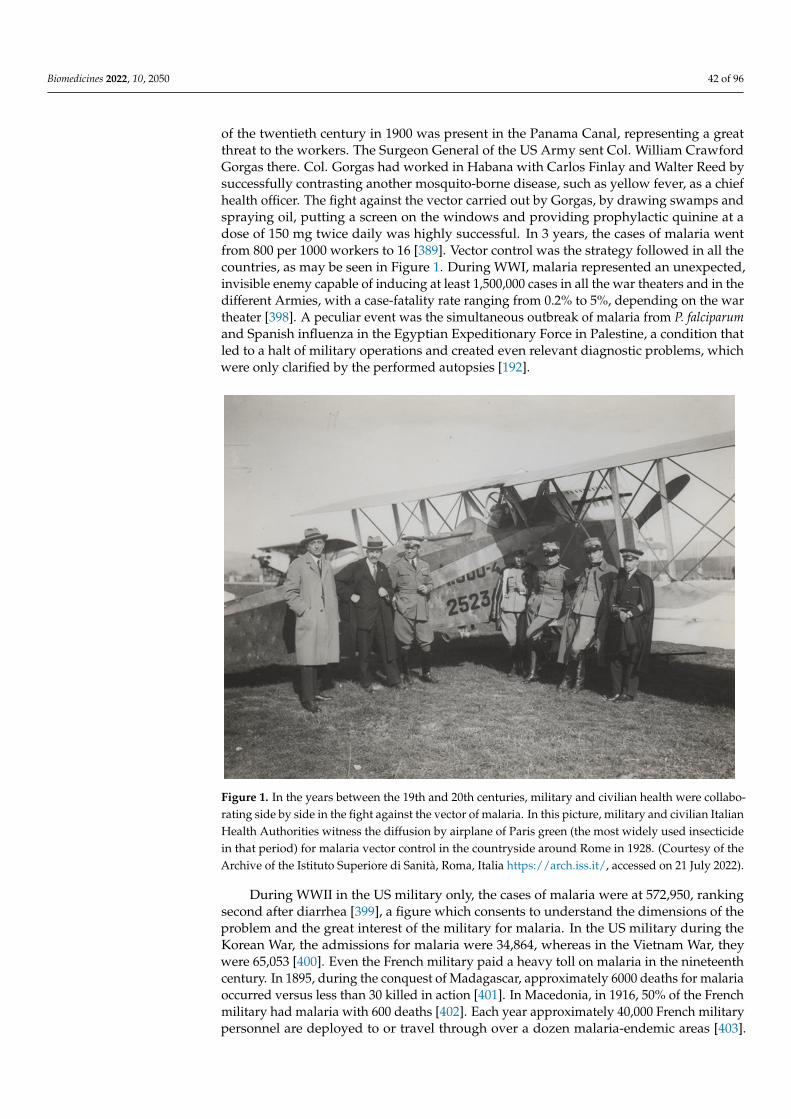
Biomedicines 2022, 10, 2050 42 of 96
of the twentieth century in 1900 was present in the Panama Canal, representing a greatthreat to the workers. The Surgeon General of the US Army sent Col. William CrawfordGorgas there. Col. Gorgas had worked in Habana with Carlos Finlay and Walter Reed bysuccessfully contrasting another mosquito-borne disease, such as yellow fever, as a chiefhealth officer. The fight against the vector carried out by Gorgas, by drawing swamps andspraying oil, putting a screen on the windows and providing prophylactic quinine at adose of 150 mg twice daily was highly successful. In 3 years, the cases of malaria wentfrom 800 per 1000 workers to 16 [389]. Vector control was the strategy followed in all thecountries, as may be seen in Figure 1. During WWI, malaria represented an unexpected,invisible enemy capable of inducing at least 1,500,000 cases in all the war theaters and in thedifferent Armies, with a case-fatality rate ranging from 0.2% to 5%, depending on the wartheater [398]. A peculiar event was the simultaneous outbreak of malaria from P. falciparumand Spanish influenza in the Egyptian Expeditionary Force in Palestine, a condition thatled to a halt of military operations and created even relevant diagnostic problems, whichwere only clarified by the performed autopsies [192].
Biomedicines 2022, 10, x FOR PEER REVIEW 42 of 98
epitopes (RTS-S) of P. falciparum, whose study started in 1984 from a collaboration be-tween WRAIR and SmithKlineBecham, currently GlaxoSmithKline, and the vaccine was developed in 1987 [393,394]. Although this vaccine, Mosquirix™, has only shown partial protection against the infection in children 5–17 months, it has been approved by the WHO, which in October 2021, has recommended its widespread use, with a four-admin-istration schedule, after a trial on over 800,000 children in Africa showed a substantial safety and partial protection [395]. It is the first approved anti-parasite vaccine. This rep-resents a milestone, even though prevention for the military continues to be based on chemoprophylaxis and protective personal measures, including insecticide-impregnated uniforms, sleeping under bed-nets, and applying insect repellents on the skin [396]. The researchers of WRAIR are involved even in the research on monoclonal antibodies. In fact, they have recently been coauthors of a small trial on a low-dose subcutaneous or intrave-nous monoclonal antibody for preventing malaria, which resulted to be safe and protec-tive [397]. Malaria at the start of the twentieth century in 1900 was present in the Panama Canal, representing a great threat to the workers. The Surgeon General of the US Army sent Col. William Crawford Gorgas there. Col. Gorgas had worked in Habana with Carlos Finlay and Walter Reed by successfully contrasting another mosquito-borne disease, such as yellow fever, as a chief health officer. The fight against the vector carried out by Gorgas, by drawing swamps and spraying oil, putting a screen on the windows and providing prophylactic quinine at a dose of 150 mg twice daily was highly successful. In 3 years, the cases of malaria went from 800 per 1000 workers to 16 [389]. Vector control was the strat-egy followed in all the countries, as may be seen in Figure 1. During WWI, malaria repre-sented an unexpected, invisible enemy capable of inducing at least 1,500,000 cases in all the war theaters and in the different Armies, with a case-fatality rate ranging from 0.2% to 5%, depending on the war theater [398]. A peculiar event was the simultaneous out-break of malaria from P. falciparum and Spanish influenza in the Egyptian Expeditionary Force in Palestine, a condition that led to a halt of military operations and created even relevant diagnostic problems, which were only clarified by the performed autopsies [192].
Figure 1. In the years between the 19th and 20th centuries, military and civilian health were collabo-rating side by side in the fight against the vector of malaria. In this picture, military and civilian Figure 1. In the years between the 19th and 20th centuries, military and civilian health were collabo-rating side by side in the fight against the vector of malaria. In this picture, military and civilian ItalianHealth Authorities witness the diffusion by airplane of Paris green (the most widely used insecticidein that period) for malaria vector control in the countryside around Rome in 1928. (Courtesy of theArchive of the Istituto Superiore di Sanità, Roma, Italia https://arch.iss.it/, accessed on 21 July 2022).
During WWII in the US military only, the cases of malaria were at 572,950, rankingsecond after diarrhea [399], a figure which consents to understand the dimensions of theproblem and the great interest of the military for malaria. In the US military during theKorean War, the admissions for malaria were 34,864, whereas in the Vietnam War, theywere 65,053 [400]. Even the French military paid a heavy toll on malaria in the nineteenthcentury. In 1895, during the conquest of Madagascar, approximately 6000 deaths for malariaoccurred versus less than 30 killed in action [401]. In Macedonia, in 1916, 50% of the Frenchmilitary had malaria with 600 deaths [402]. Each year approximately 40,000 French militarypersonnel are deployed to or travel through over a dozen malaria-endemic areas [403].
Biomedicines 2022, 10, 2050 43 of 96
From 1986 to 2011, 13,543 malaria cases were observed in the French Armed Forces, 2.2%were serious cases with eleven deaths, five of which occurred in Gabon or upon returnfrom this country. The P. falciparum species was responsible for 78.1% of attacks andP. vivax for 16.4% [403]. It should not be forgotten that the military may even becomeinvoluntary malaria vectors by going to endemic areas and returning home. This maymore easily happen with P. vivax, considering that the disease may manifest for months oryears after the infection. This was observed in the USA during the Korean War [404,405]and the Vietnam War [406], while the Russians imported thousands of malaria cases fromP. falciparum during the war in Afghanistan [407]. The military contribution to malaria is inthe etiology and vector identification as well as in some therapies and vaccine development.
3.6. Lymphatic Filariasis
Lymphatic filariasis is a highly incapacitating mosquito-borne disease, the second mostcommon after malaria among vector-borne diseases [408]. The vectors may be Anopheline,Culex and Mansonia mosquitoes, which bite at night, whereas in the Pacific, the vector is theAedes mosquito, which bites during the day [409]. The etiological agents are three species ofnematode parasites, Wuchereria bancrofti, responsible for 90% of cases, Brugia malayi, presentin East-Southeast Asia, and Brugia timori, present in the Timor area [408]. The worms have acomplex cycle, are transmitted by the bite of the vector and localize in the lymphatic vessels.The adult worms generate microfilariae, which migrate into lymph and blood channels.The disease may be acute or chronic, and its main manifestations are fever, lymphangitis,lymphadenopathy with pain, and in males, scrotal edema of the acute form, whereas thechronic form is highly invalidating with elephantiasis, lymphedema and hydrocele [409].Nearly one billion people in tropical areas are at risk of lymphatic filariasis, and it isestimated that approximately 36 million people are incapacitated as a consequence ofchronic lymphatic filariasis [409]. There are three effective drugs, albendazole, ivermectin,and diethylcarbamazine citrate, which may prophylactically be administered to at-riskpopulations, as recommended by the WHO, with annual mass drug administration (MDA),a strategy that aims to eradicate filariasis [410].
Filariasis was an unexpected invisible enemy for the US troops during WWII in thePacific war theater, with many thousands of hit men, more than 10,000 among Navy andMarine Corps, and an unspecified number of illnesses among the Army troops, with aglobal amount estimated at 14,000–16,000 men having clinical evidence of infection andsome battalions with 70% of their contingent out of service [368,408]. This acute form oflymphatic filariasis showed a trend of spontaneous clinical resolution once the diseasewas recognized and the patients were moved away from the endemic countries [409]. Thecontributions of the US military researchers to the fight against filariasis ranged fromdiagnosis to treatment, where the evidence that eradication strategy was possible througha safe and effective treatment was provided [368].
3.7. Schistosomiasis
Schistosomiasis is an infectious disease caused by a trematode parasite, which maypenetrate the human skin in contaminated freshwater. Humans are the main definitivehost, whereas intermediate hosts are snails living in freshwater. The cercariae are releasedfrom the snails and penetrate the human skin and, after 5–7 weeks, mature into adultworms, capable of generating eggs, which are retained in the body if not excreted by fecesor urine. The excreted eggs become miracidia, which infect the snails, thus maintainingthe cycle [411]. Three main species of schistosomes infect humans, Schistosoma mansoni,Schistosoma haematobium, and Schistosoma japonicum. The first two are found in Africa andthe Middle East, S. mansoni is also found in the Americas, while S. japonicum is found in Asia,mainly in China and the Philippines [411]. Eggs that remain in the body are responsiblefor the host inflammatory response, which may create granulomatous lesions in the hosttissues, intestine and liver in the case of S. mansoni and japonicum, and in the bladder andurogenital tract in the case of S. haematobium. The infection is more frequent in adolescents
Biomedicines 2022, 10, 2050 44 of 96
and tends to decrease in adults and older adults; it may be acute, with sudden onset of fever,malaise, abdominal pain, myalgia, headache, eosinophilia (Katayama syndrome), and itmay be chronic, with symptomatology that may be hepatosplenic or urinary, dependingon the type of Schistosoma [411]. It is estimated that nearly 800 million people are atrisk, and over 250 million are infected, 90% of whom live in Sub-Saharan Africa [412].Even for schistosomiasis, there is the possibility of safe and effective chemotherapy withpraziquantel, which may even work for prevention; thus, its annual administration isrecommended by the WHO with the objective of eradicating the disease, in associationwith the fight against the intermediate host [411]. Vaccines for human use are not availableyet [412].
Schistosomiasis has accompanied the military in war since the period of Napoleonicwars. In WWI, several hundreds of British and Australian troops were infected in Egypt andthe Middle East, whereas in WWII, over 1500 British and African troops became infected inNigeria. Hundreds of US soldiers were infected during the liberation of the Philippines.Although schistosomiasis in the military is far from the number of cases of malaria andfilariasis, it may represent a further obstacle to military readiness during operations inendemic areas. Thus, the US military provided relevant contributions to the diagnosis, inthe demonstration that praziquantel therapy could revert advanced hepatosplenomegalyand obstructive uropathy, and in the indications of the right conditions of salinity andchlorination for inactivating cercariae [368].
3.8. Trypanosomiasis
There are two types of trypanosomiases, the African and the American trypanosomi-asis. African trypanosomiasis is caused by the parasites Trypanosoma brucei gambiense orrhodesiense transmitted by the bite of the tsetse fly, Glossina spp. T. brucei is a flagellatedprotozoan parasite whose transmission by the bite of the tsetse fly was discovered in 1895in Zululand by the British military physician David Bruce [413]. Trypanosomes were firstfound in the blood of a European man in the Gambia by Dr. Robert Michael Forde [414] andDr. Joseph Everett Dutton [415], whereas trypanosomes in cerebrospinal fluid were firstobserved, described and put in connection with sleeping sickness by Dr. Aldo Castellani,later on, full Professor of Tropical Medicine at the University of Rome and General ofthe Italian Navy [416]. T. brucei gambiense is responsible for 97% of the human Africantrypanosomiasis and is present in western and central Africa, whereas T. brucei rhodesiense isresponsible for under 3% of African trypanosomiasis and is present in eastern and southernAfrica [417]. T. brucei rhodesiense and gambiense induce an invariably fatal disease if nottreated; however, the first has a quicker clinical course of approximately one year, whereasthe other has a more chronic course of a few years. During the twentieth century, threesevere epidemics of sleeping sickness were registered, the first in 1896 until 1906, mainly inUganda and Congo, the second in 1920, and the last in 1970 through the late 1990s [417].Considering the high number of deaths in the order of hundreds of thousands, the mostrenowned scientists were engaged in the search for effective therapies, including NobelPrize winners, such as Charles Laveran and Robert Koch. Moreover, in the second outbreakof the 1920s, a winning move was suggested by the French military physician EugèneJamot, represented by the institution of mobile teams for capillary detection and treatmentof sleeping sickness cases; this strategy, organized in Cameroon, was highly successful,considering that the sleeping sickness prevalence decreased from 60% in 1919 to 0.2–4.1%in 1930 [418]. The progress in therapy, the organization of the mobile teams as well as thevector control achieved good results, as witnessed by the prevalence in 2009, for the firsttime under 10,000 cases, and in 2019 and 2020, when 992 and 663 cases, respectively, hadbeen reported [417].
American trypanosomiasis is caused by T. cruzi and is transmitted by the feces of blood-sucking bugs belonging to the subfamily of Triatominae, as discovered by the Brazilianhygienist Carlos Chagas in 1909 [419]. The infection, named Chagas disease, has twoclinical phases, the first is clinically nonspecific and is 4–8 weeks long, during which
Biomedicines 2022, 10, 2050 45 of 96
parasitemia is present, whereas the second phase is clinically symptomatic in 15–30% ofthe infected people, and presents organ damage usually 10–25 years after the first clinicalphase, at cardiac level, with potentially fatal cardiomyopathy, and at the digestive level,with megaesophagus and megacolon [419]. Currently, 7–8 million people are estimatedto be infected by T. cruzi in Latin America, and 25 million are at risk. In 2008, morethan 10,000 people died from Chagas disease [419]. Moreover, due to migration, it isestimated that 400,000 people infected by T. cruzi are present in the world outside LatinAmerica, three-quarters of whom are in the USA [419]. As for African trypanosomiasis,no vaccines are available, but only drugs for therapy. Despite that trypanosomiasis hasrarely represented a problem for the military, the intervention of the military in the studyof the disease and in the means for fighting it is demonstrated by the relevant discoveriesmade by military physicians, including David Bruce, Charles Laveran and Eugène Jamot,as well as by the studies performed by the military researchers at the US Army MedicalResearch Unit (USAMRU) in Kenia on epidemiological and clinical aspects of Africantrypanosomiasis and at WRAIR in the immunology, but mainly therapy, of the two types oftrypanosomiasis [368].
3.9. Other Parasitic Diseases
Other parasitic diseases able to heavily influence the operational readiness of themilitary are intestinal parasites, such as Entamoeba histolytica, which is a leading cause ofdiarrhea worldwide [420]. It is estimated that over 500 million people are infected and40,000–100,000 individuals annually die from amebiasis worldwide [22] (Table 1). Themilitary may be exposed to infection if deployed to endemic areas [420,421]. The US militaryphysician Charles F Craig in the first half of the last century provided relevant contributionsto the serological diagnosis as well as to clinical and pathological observations [422–424],whereas the military physician E. Vedder could demonstrate the amebicidal power ofemetine in vitro, thus paving the way to its application in the treatment of amebiasis [425].
Hookworm is a soil-transmitted helminth disease caused by Ancylostoma duodenale orNecator americanus. It is estimated that worldwide 740 million people are infested, mainlyin China, Sub-Saharan Africa, East Asia and the Pacific Islands [426]. These worms reachthe digestive mucosa where they feed themselves by inducing chronic hyposideremicanemia. At the start of the last century, the US military physician Bailey K Ashford deeplyinvestigated the hookworm from Necator americanus in Puerto Rico, which was responsiblefor the death of almost 12,000 people per year [426,427]. Although in general this parasitosisdoes not represent a threat for the military deployed to endemic areas, nonetheless, it wasreported in some USA military soldiers deployed to Grenada [428] and Vietnam [368] andin the Singaporean military deployed in jungle training activities in Brunei [429].
3.10. Human Immunodeficiency Virus (HIV)
Human immunodeficiency virus (HIV) is a retrovirus discovered in 1983 by FrançoiseBarré-Sinoussi and Luc Montagnier at Pasteur Institute in Paris [430], for which they wereawarded the Nobel Prize for Physiology or Medicine in 2008. HIV is the etiological agentof the acquired immunodeficiency syndrome (AIDS), first described in 1981 in the USAin homosexual men [431]. The virus was independently isolated and described a fewmonths later by Robert Gallo, then working at the National Cancer Institute, in Maryland,USA, who provided further evidence of the association between the virus, which wascalled human T-lymphotropic virus (HTLV)-III, considering that Gallo had previouslydescribed HTLV-I and II, and AIDS [432]. A long controversy ensued over which of thetwo scientists had discovered the virus, which ended in 1987 with an agreement reached byUSA President Ronald Reagan and French Prime Minister Jacques Chirac, with a division ofthe royalties of the diagnostic test between the two countries [433]. It was only in 1986 thatthe virus was named HIV. HIV enters the host following sexual intercourse, blood/bloodderivatives administration, or from mother to fetus and through its envelope glycoprotein120 links to the CD4 helper T-lymphocytes, which are slowly but progressively destroyed,
Biomedicines 2022, 10, 2050 46 of 96
thus reaching low values, <200/µL, which are incompatible with maintaining good health,thus paving the way to opportunistic infections and cancers, until patient death [434]. Fromits first description in 1981 until 1996, when an effective therapy was made available [435],AIDS was invariably fatal and present at a global level, particularly in Sub-Saharan Africa,where HIV infection still represents more than 70% of the estimated global infections, whichwere 36,848,000 in 2019, 0.5% of the world population, showing a slow general decline.Except for in the Americas and Europe, where the rate of infection increases each year; themortality is instead in global decline [436]. One million and a half new HIV infections werereported in 2020 and 680,000 AIDS-related deaths [13] (Table 1). Currently, therapy is notresolutive; however, it allows a good quality of life with only one pill per day. Instead, noeffective vaccine is yet licensed, although therapeutic and prophylactic vaccines have beendeveloped and tested, in one case until phase 3.
HIV infection has been considered of high interest for the military since the firstdescription, considering that it is a sexually transmitted disease, a type of disease thatis particularly widespread among the military. It is severe and invariably fatal and itmay be transmitted by blood, thus representing a risk for the soldier as a “walking bloodbank” [437]. However, in the US military, this interest increased and became an organicscientific HIV program after the death of a recruit for generalized vaccinia after smallpoxvaccination due to an unrecognized HIV infection [438]. This dramatic event promptedthe US military authorities to introduce the screening for HIV dating from October 1985to all applicants for military service as well as to all active-duty forces, in order to protectthe recruits from the side effects of the living vaccines in case of HIV positivity and toprotect the “walking blood bank” [437]. The HIV-positive applicants were considered unfitfor military service, whereas the infected military members, even if retired, were offereda program of periodical checks, which represented an opportunity for helping infectedpatients and obtaining information on this new disease [437]. This was even more relevantbecause it occurred during a period of poor knowledge of the disease, due to the fact thatgeneralized HIV screening was difficult, rather nearly impossible, to be carried out for thestrong resistance of the associations of patients, who had fear of social stigma, consideringthat the disease was first described in homosexual men and drug addicts [439]. Thispopular resistance represented a strong brake everywhere to the possibility of knowingat least the epidemiology of the infection by generalized screening, which could notbe performed. Thus, the strategy of the US military of introducing compulsory HIVscreening for the applicants to military service allowed for one to collect information onthe spread of the infection among adolescents and young adults, and it was estimated thatthe rate of positivity in the military could be lower than in the corresponding general USApopulation [437,440,441]. Meanwhile, even relevant pathogenetic and clinical observationswere collected, including the defective regulation of the Epstein–Barr virus (EBV), whichcould expose AIDS patients to the risk of developing EBV-containing lymphomas [442], thedefective response to vaccinations in early stage HIV-infected patients [443], the systemicinflammation and immune activation as hallmarks of the disease, as witnessed by thehigh interleukin-6 levels [444], and finally the Walter–Reed staging classification system,which has represented a guide for the judgements of fitness to the military service and forclinicians during their clinical activity [445]. Moreover, the experience of the US militaryresearchers allowed one to calculate the rate of decline of CD4+ lymphocytes dependenton the stage of the disease [446]. Furthermore, the Walter–Reed Retrovirus ResearchGroup made relevant observations even in the field of malignancies in the course of AIDS,neurocognitive impairments, and anti-retroviral therapy [437]. The WRAIR was even thelead agency to test in phase 3 an HIV-1 vaccine in Thailand, which showed only a modestbenefit but was useful for future research [447,448]. Despite great advancements havingbeen achieved in a relatively short time, HIV infection remains a public health global threat.However, the US military researchers at WRAIR allowed for gaining early precious insightsinto the knowledge of the disease, which have represented a reference point everywhere.

Biomedicines 2022, 10, 2050 47 of 96
3.11. Hepatitis C
Hepatitis C virus (HCV) is an RNA flavivirus for the first time identified in April1989 [449,450], which is estimated to have infected 58 million people in all six WHOregions, with an annual incidence of 1.5 million new cases and 290,000 deaths per year, as aconsequence of cirrhosis and hepatocellular carcinoma [14] (Table 1). HCV may induce anacute, usually asymptomatic and spontaneously clearing, disease in approximately 30% ofcases, whereas in the remaining 70%, the disease becomes chronic, with 15–30% of thesechronic cases evolving to cirrhosis in approximately 20 years [14]. HCV is first transmittedby exposure to blood or blood derivatives, more rarely by sexual intercourse, and theincubation time ranges from 2 weeks to 6 months; however, acute infection is asymptomaticin approximately 80% of subjects; thus, it is generally undetected. The diagnosis is basedon a serological approach for the research of specific antibodies and a confirmation test incase of positivity with a molecular approach to identify the viral RNA. No vaccine is stillavailable; the direct-acting antivirals (DAAs) are highly effective, able to clear over 95% ofthe chronically infected patients in a period of 12–24 weeks, depending on the presence ornot of cirrhosis [14]. However, currently, access to DAAs is still too limited, even in affluentcountries [451], and it is estimated that less than 10 million patients have completed thetreatment with DAAs [14].
The interest of the military in HCV is mainly due to the need of protecting the “walkingblood bank” and, despite that no compulsory HCV screening at enrollment in the militaryis generally required, the percentage of HCV positivity is usually low, around 0.5% or evenless in the USA, where it is lower than in the civilian corresponding population [452] or inthe few European countries for which data are available [453,454]. Even in Morocco [455],India [456], and Brazil [457], HCV prevalence in the military is quite low, 0.245%, 0.45%,and 0.7%, respectively. Conversely, in the civilian blood donors of Sierra Leone, HCVpositivity rate was 1.2% [458], and in the patients of a military hospital in Rwanda, the rateof HCV-infected patients was 9.6% [459]. The highest world prevalence of HCV viremia at6.3% is in Egypt [460], as a consequence of the anti-schistosomiasis campaign carried outwith unsafe injections [461]. However, a massive effort to voluntarily screen all the adultEgyptian population in order to offer the DAAs by the government allows for foreseeingthat in a short time Egypt may achieve an HCV viremic prevalence of 0.5%, which is similarto the nearby regions [462]. In the US military, a cost-effectiveness analysis of introducingcompulsory HCV screening at enrollment versus the cost of treatment for the militaryfor those who may be HCV positive, has established that screening is cost-effective [463].Moreover, a study carried out on the HCV prevalence in 10,000 soldiers returning frominternational missions to Iraq and Afghanistan allowed one to observe that 23 subjects wereHCV positive; the majority of them (18/23) were positive at enrollment, with only five casesof positivity being service associated, thus even more underlying the economic advantageof introducing compulsory screening [463]. With an effective vaccine still unavailable,the only policy for eradicating the disease is to actively search for positive subjects andto offer them the DAAs. This policy may even help the military to protect the “walkingblood bank”.
3.12. Hepatitis E
Hepatitis E virus (HEV) has been first suggested to be the cause of an outbreak ofhepatitis in Kashmir (India) at the end of the 1970s and reported in 1980 [464]. In 1991, thevirus E was cloned [465], a relevant step not only for the precise viral description but evenfor its use in diagnosis and vaccine development. HEV is responsible for a generally acutehepatitis, transmitted by fecal–oral route, through contaminated water in hyperendemicand endemic countries. However, HEV is even present in the developed world as a zoonosis,for ingestion of undercooked meat from infected animals, such as swine, boar and deer.Four viral genotypes exist, the first two mainly occurring in hyperendemic and endemiccountries (Africa, Asia, Mexico and Brazil) and the 3–4 especially occurring as sporadiccases in developed countries [466]. Despite that HEV generally induces acute hepatitis,
Biomedicines 2022, 10, 2050 48 of 96
chronic hepatitis may be observed in immunocompromised people, such as transplantedpatients. Pregnant women present high mortality, ranging from 10% to 50% [467]. It isestimated that globally each year, 20 million people are HEV infected, 3.3 million of whomare symptomatic, with 44,000 deaths [23] (Table 1). Although the fecal–oral route is themain way of transmission, the persistent viremic phase does not exclude that HEV mayeven be transmitted by blood [466]. Two recombinant vaccines for HEV were developed,the first of which has been studied by the US military researchers of WRAIR togetherwith GlaxoSmithKline in phase 2 [468], but it did not further progress, possibly for lack ofcommercial value [467]; the other seems to be safe and effective, and it has been licensedin China, but it is not recommended by the WHO for extensive use due to insufficientinformation about safety and efficacy in different categories of patients [469]. Thus, HEVhas been included in this review among the non-vaccine-preventable infectious diseases.
The interest of the military for HEV is witnessed by having found different outbreaksin deployed troops in hyperendemic countries, the first of which was among Russian troopsin Afghanistan in 1981 [470], and others were described by the US military [471–476]. Acomplete resolution of the HEV issue for the military may only come from the availabilityof a safe and effective vaccine that protects against all viral genotypes. Meanwhile, carefulcontrol of water and food is the better prevention for deployed troops. Furthermore, doubton possible blood transmission is relevant for the military in consideration of the principleof the “walking blood bank”.
3.13. Chikungunya
Chikungunya is an RNA viral disease transmitted by Aedes aegypti and Aedes albopictusmosquitoes, characterized by fever, arthralgia, and skin rash [477]. The disease is generallyself-limiting, with mortality way lower than 1%. The arthralgia is incapacitating and innearly two-thirds of patients, may last for more than one year after the infection [478]. Thedisease was described for the first time in Tanzania in 1952 and is present in Africa and SouthAsia, where relevant outbreaks have been observed. However, the disease has even beendescribed in temperate climates, considering that an outbreak of more than 200 patientshas been reported in Italy in 2007 [479]. No licensed vaccine is available, although the USmilitary at WRAIR has worked on a project for developing an inactivated and subsequentlya live attenuated vaccine, approved as an investigational new drug (IND) [480]. TheUS military considers Chikungunya a relevant threat even though currently no higherrisk for the military compared to the civilian population has been observed [480]. WhenChikungunya cases were considered among military personnel deployed to endemic areas,low prevalence was observed [481]. Conversely, the situation is different when observedamong the military stationed in areas with outbreaks. The most accurate study on themilitary was on the French policemen during the outbreak on Reunion Island, where266,000 out of the 775,000 inhabitants presented symptoms of the disease [477]. Out of662 policemen, 128 (19.3%) were serologically positive for the Chikungunya virus, and3.2% were asymptomatic. Chronic arthralgia was reported in over 90%, acute fever innearly 90%, skin rash in over 50% and tiredness in all [482]. During this outbreak, theFrench health authorities asked the US military about the possibility of using the IND livevaccine [480]. Among the US military, 78 cases of Chikungunya were reported in PuertoRico between 2010 and 2016 and 118 in 2014–2015 [481].
3.14. Zika
Zika is an RNA Flavivirus transmitted by the bite of Aedes mosquitoes, first isolated in1947 in Uganda from a macaque monkey and in 1954 in Nigeria from human cases. Sincethen, sporadic cases were reported, whereas it reemerged in this century, with the firstoutbreak occurring in 2007 in Yap, Federated States of Micronesia, followed by anotheroutbreak in 2013 in French Polynesia [339,483]. In May 2015, Zika virus reached Brazil,where an estimated 440,000–1,300,000 persons were infected [483]. The WHO declaredthe Zika virus a public health emergence based on the net increase in microcephaly in
Biomedicines 2022, 10, 2050 49 of 96
Brazil (20/10,000 live births vs. 0.5/10,000 live births in the previous years) [483]. Anotherdreadful complication is Guillain–Barré Syndrome. Despite that no vaccine is still available,many candidate vaccines are studied, seven of which are in phase 1. WRAIR is studying aninactivated vaccine in collaboration with the Beth Israel Deaconess Medical Center [338].
3.15. Crimean–Congo Hemorrhagic Fever
Crimean–Congo Hemorrhagic Fever (CCHF) is the most widespread tick-borne dis-ease in the world and the second (after dengue) viral hemorrhagic fever in the world [484].The disease is caused by an RNA virus transmitted by the bite of a hard tick of the speciesHyalomma. However, even contact with infected animals may transmit the infection. More-over, inter-human transmission has also been reported. The infection may run asymp-tomatic in a large percentage of cases, up to 90% in hyperendemic areas [485]; however,symptomatic cases may be severe with high mortality of up to 40% [484]. First describedduring WWII in Soviet military personnel in Crimea [486] and in 1956 in Congo (hence thename), CCHF is now present in Asia, Africa, and Eastern and Southern Europe, includingSpain [484,487]. There is no therapy nor available vaccine, even though in Bulgaria, aninactivated vaccine from suckling mouse brain was licensed and used for at-risk categoriesof workers, including the military. However, less reactogenic and more effective are consid-ered the inactivated vaccines from cell cultures [484]. The Turkish military has reportedthe preparation and successful use in patients with high-level CCHFV viremia of hyper-immunoglobulins. They were prepared by the sera of 22 convalescent subjects and only oneadministration of 400 Kubar Units of hyperimmune immunoglobulins to 15 patients with ahigh level of viral copies (≥108/mL), allowing 13 of them (86.6%) to be cleared from thevirus, whereas two died [488]. Passive immunotherapy may be a promising tool in CCHF,in consideration of the severity of the disease and the current lack of effective therapy orvaccines. Based on the high mortality of the infection, the possibility of dissemination byaerosols and the current lack of therapy or vaccine prevention, CCHFV has been includedin the high biohazard agents, to be handled only in biosafety levels 3–4 [489] and amongbiological agents of category C by the CDC [57,484].
3.16. Hantaviruses
Hantaviruses are RNA viruses of the family Bunyaviridae, of which three main typesare known, one present in Finland and Scandinavia, called Puumala virus, able to induce amilder disease, the nephropathia epidemica (NE), the Hantaan (HTNV) old world type,which causes the hemorrhagic fever with renal syndrome (HFRS), a severe clinical conditionwith a mortality of approximately 12%, and the new world type, able to induce the mostsevere hantavirus cardiopulmonary syndrome (HCPS), burdened with a mortality ofapproximately 40% [489]. The first outbreak of over 10,000 cases of HFRS was reportedin WWII in Finland, among Finnish and German troops [490], and during the KoreanWar, over 3000 United Nations troops developed HFRS [491]. However, the etiologicagent was only identified in the 1980s, whereas the new world type was only identified in1993 [489]. The viruses are present in rodents and are transmitted to humans by aerosolof contaminated biological fluids or feces, a condition which may induce consideration ofhantaviruses as potential biological weapons, for easy dissemination, high clinical severityand lethality, lack of therapy and vaccine [487,492]. The military seems to be particularlyexposed, due to the easier contact with rodents, which is associated with the disruptionof the rodent habitat linked to the war and with the operational training in the field [493].During military training activity in Germany in 1990, a hantavirus outbreak from thePuumala virus was observed with an attack rate of 8.5% [493]. In the former Yugoslavia,particularly in Bosnia–Herzegovina and Croatia, many outbreaks of HFRS, some of whichwere large, have been reported between the 1950s and the 1990s, thus representing a likelythreat for NATO forces deployed there, considering that over 50% of soldiers in the field areexposed to possible HFRS risk factors [491]. Moreover, in the military personnel in Europein war, activity or maneuver-theater overlapping symptomatology of HFRS and HCPS has

Biomedicines 2022, 10, 2050 50 of 96
been reported, irrespective of the isolated virus, but coherent with the possibility that twogenetically related viruses, infecting both through the same respiratory way, both inducinga sort of “cytokine storm”, may determine damages in two target organs, lung and kidney,which are generally addressed separately [494]. An inactivated vaccine, Hantavax®, hasbeen developed and licensed in Korea and has been used in the Korean army; however, astudy carried out in the period 2009–2017, for evaluating its effectiveness in the progressionof HFRS, failed to demonstrate significant protection against the progression of HFRS [495].US military researchers are working on a bivalent DNA vaccine against the Puumalavirus and HTNV and have successfully completed a phase 2a study, by demonstrating theimmunogenicity of this tentative vaccine [496]. Hantaviruses have been included amongthe biological agents, category C [57].
3.17. Other Arboviral Diseases
West Nile virus was first observed in Uganda in 1937. It is a flavivirus primarilytransmitted by the bite of Culex mosquitoes. The infection in 80% of cases is asymptomatic,and in symptomatic cases the clinical course is generally not severe, characterized byfever, rash and lymphadenopathy. More recently, at the end of the last century, in theoutbreaks registered in Romania and in New York, the trend to target the CNS withmeningoencephalitis was more pronounced, as well as gastrointestinal symptomatology.There is no specific approved therapy or effective human vaccines. Thus, for the military,the only prevention consists of a vector control strategy [497]. West Nile virus has beenconsidered a potential biological agent, category C [498].
Rift Valley virus was first described in the Rift Valley in Kenia in 1931. The RNAvirus is mainly transmitted by mosquitoes, and the infection is generally benign or pau-cisymptomatic. However, it may be complicated by encephalitis, hepatitis, ocular disorders,nephritis, and hemorrhages; the mortality is generally low, but percentages as high as22–28% have even been reported. In this case, no specific therapy nor effective vaccineis available [499]. The Rift Valley virus should be considered when observing fevers ofunknown origin in the military [500].
3.18. Acute Respiratory Syndrome
An acute respiratory syndrome is frequently observed in the military, particularlyin the first weeks of recruits in the training phase. It is calculated that 25,000–80,000US recruits suffer acute respiratory disease (ARD) each year and that 200,000–600,000US service members had ARD each year during the influenza seasons of the year 2012through 2014 [501]. The socio-environmental conditions favoring ARD for trainees areovercrowding, psychological and physical stress, sleep deprivation, exposure to dust,smoke and extremes of temperature [501]. ARD presents with a syndrome of commoncold or pneumonia depending on whether the higher or lower airways are interestedby infection-induced inflammation, irrespective of the etiological agent. Even though thecommon cold is primarily determined by rhinoviruses (approximately in 50% of cases), evencoronaviruses, adenoviruses, influenza and parainfluenza viruses, respiratory syncytialvirus (RSV), human metapneumovirus, and group A streptococcus (Streptococcus pyogenes)may be found. In analogy, pneumonia is primarily induced by Streptococcus pneumoniae;however, adenoviruses and influenza virus may even be responsible. The intermediateclinical picture of bronchitis with cough may be induced by adenoviruses, influenza virus,Mycoplasma pneumoniae, Chlamydophila pneumoniae, and Bordetella pertussis [501]. There isa large overlap of induced syndromes and etiological agents; thus, a precision diagnosiscannot be made based on the clinical picture only, but it has to be laboratory driven, a needthat may not always be satisfied in operational activities or, even more, in wartime. Despitethat several etiologic agents are potentially implicated in ARD induction, those that aremore frequently observed in trainees are adenoviruses, influenza virus, and S. pyogenes [502].For adenoviruses 4 and 7, a safe and effective, FDA-approved, live, oral vaccine is available,and is administered to US recruits as the only military in the world [501]. Influenza virus,

Biomedicines 2022, 10, 2050 51 of 96
similar to adenoviruses, has been treated among the vaccine-preventable infectious diseases,whereas S. pyogenes has not been treated yet. It has represented a threat for the militaryduring WWII, associated with scarlet fever, and more recently in the period 1990–2011in the US military, when at least 17 outbreaks of S. pyogenes have been observed, alltreated with antibiotic chemoprophylaxis, generally consisting of benzathine-penicillin G;however, even erythromycin and azithromycin may be used. S. pyogenes generally inducespharyngitis, which resolves without complications. Less commonly, S. pyogenes may inducesuppurative and invasive infections, including meningitis, brain abscesses, pneumonia,and necrotizing fasciitis. The mortality of uncomplicated pharyngitis is <1%, whereasin the complicated forms, it ranges between 15% and 25%. Finally, S. pyogenes in somesubjects may even induce autoimmune diseases, such as acute rheumatic disease, whichmay manifest with either endocarditis or glomerulonephritis [501]. Streptococcus pneumoniaehas already been treated among the vaccine-preventable infections as well as for Bordetellapertussis and coronavirus (as far as SARS-CoV-2 is concerned), whereas rhinoviruses, para-influenza viruses, RSV and human metapneumovirus, as well as Mycoplasma pneumoniaeand Chlamydophila pneumoniae, have not been treated yet. Paradigmatic of the difficultyof differential diagnosis in ARD and the need to set up an integrated surveillance systembased on a network of well-equipped laboratories and skilled researchers, as in the USmilitary, to respond to the new diagnostic challenges posed by emerging infectious diseases,is the request for help of the Jordanian Ministry of Health in April 2012 addressed to theNAMRU-3 in Cairo, Egypt, to clarify the cause of 11 cases hospitalized for ARD, two ofwhom died soon after hospitalization. These cases were carefully analyzed for influenza,parainfluenza types 1 and 3, human metapneumovirus, human coronaviruses (includingSARS-CoV), and adenovirus, but all the results were negative. In September 2012, Middle-East respiratory syndrome due to coronavirus (MERS-CoV) became widely known, andNAMRU-3 received from the CDC the biological material for making diagnoses. The caseswere investigated again, in agreement with the Jordanian Ministry of Health, and the twocases deceased were clearly positive for all three genes of MERS-CoV [501].
RSV is a worldwide respiratory virus, present in over 90% of 5-year-old children.The immunity against RSV is short; thus, it is not difficult to be reinfected even by thesame strain. The relevance for the military was first observed in the US military in 1959,thereafter confirmed in the military trainees and even during the Vietnam war. In somestudies, in US and UK military trainees, it was present in 11% and 14% of the trainees withrespiratory symptoms, respectively, behind adenovirus, which was prevalent in both, with48% and 35%, respectively, and influenza virus, which was 11% in the US and 19% in theUK trainees [503,504]. Conversely, in a large study on the Dutch military, RSV was foundin only 3% of radiologically confirmed pneumonia cases [505].
Mycoplasma pneumoniae was first isolated in 1942 from a recruit with primary atypicalpneumonia, and afterward, it was found in recruits with pneumonia with variable, butalways rather high, percentages, ranging from 6–10% to >50%. In the 1960s, primaryatypical pneumonia from M. pneumoniae was differentiated from that due to adenovirus,and a favorable response to di-methyl-chlortetracycline was observed [501].
Chlamydophila pneumoniae has been recently identified as a relevant etiologic agent forARD/pneumonia. Recent studies by US Navy researchers have observed C. pneumoniae asthe etiologic agent of 10–15% of all pneumonia cases in military recruits [501].
In conclusion, acute respiratory syndrome is one type of pathology of great interest forthe military, especially for recruits and trainees, probably for environmental living condi-tions. Acute respiratory syndrome may be due to a series of etiologic agents, for a minorityof which preventive vaccination is available. However, even in these cases, vaccine-inducedprotection is not absolute, considering that not all the possible pathogenic strains are in-cluded in the vaccine, as for S. pneumoniae, or the reaction of immune response is notcompletely protective against the infection, as in the case of influenza and SARS-CoV-2,due to the type of antigen and the high variability of these RNA viral agents. Finally, foradenovirus, as already commented in the dedicated paragraph, it is not understandable
Biomedicines 2022, 10, 2050 52 of 96
why the vaccine is only administered to the US military, although the epidemiologicalproblem is present in the military of other countries.
3.19. Acute Diarrheal Syndrome
Diarrheal diseases have accompanied the military for a long time, particularly whendeployed and in wartime. Depending on the place of deployment, the etiology may change,with cholera being a dreadful threat, mainly in Southeast Asia and in the nineteenth century.Currently, the main threats are represented by Shigella, Salmonella, Campylobacter jejuni,enterotoxigenic Escherichia coli, and the Norwalk virus. Cholera had a case-fatality rate ofover 60% without therapy, reduced to 20–30% with therapy before 1960. The studies of theUS military physician Robert A. Phillips started at the end of the 1940s and continued intothe 1950s, by introducing the intravenous rehydration treatment, which could reduce thedeath rate from 30% to 0.6% [297]. Currently, oral dehydration is an affordable treatmenteven in developing countries and in diarrheal syndromes not caused by Vibrio cholerae.The study of Phillips and his collaborators saved the life of millions of people. As cholerais a vaccine-preventable infection, in this work, it has already been treated in the sectiondedicated to vaccine-preventable infections. Even typhoid fever, in the past a relevantcause of morbidity and mortality for the military, particularly in wartime, has alreadybeen treated among the vaccine-preventable infections. Despite that diarrheal syndromewas already present during the American Revolutionary War and the American CivilWar, during the Spanish–American War, a board led by Walter Reed was able to identifytyphoid as the etiologic agent of diarrhea, and water sterilization, together with improvedsanitation, could significantly reduce the morbidity from 85/1000 in 1898 to 6/1000 in1900 [297]. During Operation Desert Shield (September–December 1990, Saudi Arabia), astudy on soldiers with diarrheal syndrome had shown that 57% of the US troops reportedat least one episode of diarrhea, and 20% were temporarily unable to perform their dutiesbecause of it. The cause of diarrhea was a bacterial pathogen in 49.5%, with the mostfrequently isolated enterotoxigenic E. coli, followed by Shigella sonnei. A few cases ofvomiting in addition to diarrhea were due to the Norwalk virus [506]. More recently, inthe US troops deployed to Iraq and Afghanistan in 2003, 76% had at least one episode ofdiarrhea, 45% were incapacitated for a median of 3 days, and 17% were confined to bedfor a median of 2 days [507]. Most cases were watery diarrhea, typical of enterotoxigenicE. coli, which represents the most frequent cause of travelers’ diarrhea, whereas 12% ofcases, who reported fever, and 3% of cases, who reported bloody stool, were probablycaused by either Campylobacter or Shigella [507]. Despite the efforts of the US militaryresearchers of WRAIR for developing effective vaccines for Shigella, Campylobacter, andenterotoxigenic E. coli, no vaccine is still available, and acute diarrheal syndrome continuesto be a relevant problem for deployed troops, and a damage reduction may only comefrom indirect hygienic measures [505]. Some of these infectious agents have been includedamong the biological threats of category B [57]. In Table 8, the military relevance for andthe military contribution to non-vaccine-preventable diseases is summarized.
Table 8. Relevance for the military of non-vaccine-preventable infectious diseases and militarycontribution to their control.
Disease Military Relevance Military Contribution
Epidemic typhusPresent in many
wars—biological agentcategory B
USA troops received Cox’svaccine in WWII
Scrub typhus The military deployed toendemic areas are at risk
Patented recombinant rickettsiaprotein
Trench fever The name itself witnessesmilitary relevance First description—etiology

Biomedicines 2022, 10, 2050 53 of 96
Table 8. Cont.
Disease Military Relevance Military Contribution
Leishmaniasis The military deployed toendemic areas are at risk
First description—personalprotection—vector control
Malaria The military deployed toendemic areas are at risk
Etiology—drugs, monoclonalantibody, and vaccine
development
Lymphatic filariasis The military deployed toendemic areas are at risk
Demonstration oferadicating treatment
Schistosomiasis The military deployed toendemic areas are at risk
Diagnosis—treatment—environ.
prevention
Trypanosomiasis The military deployed toendemic areas are at risk
Etiology—treatment,mobile teams
Other parasitic diseases The military deployed toendemic areas are at risk Treatment
HIV infection
The military is at risk ofsexually transmitteddiseases—soldiers as
“walking blood bank”
Epidemiology—diseasebiology—vaccine development
Hepatitis C Soldiers as “walkingblood bank”
Screening—monitoringpre/post-risk mission
Hepatitis E It is a risk for the militarydeployed to endemic areas Vaccine development
Chikungunia The military deployed toendemic areas are at risk Vaccine development
Zika The military deployed toendemic areas are at risk Vaccine development
Crimean–Congo Biological agent category C Passive immunotherapy
Hantaviruses
The military deployed toendemic areas are at
risk—biological agentcategory C
Vaccine development
Acute respiratorysyndrome (influenza,
rhinoviruses,para-influenza viruses,respiratory syncytialvirus, adenoviruses,
coronaviruses, humanmetapneumovirus
Streptococcus pyogenes,Streptococcus pneumoniae,
Bordetella pertussis,Mycoplasma pneumoniae,
C. pneumoniae)
It is one type of pathologyof great interest for the
military, especiallyrecruited trainees, probably
for environmental liveconditions. It may be due toa series of etiologic agents,
for a minority of whichpreventive vaccination is
available. However, even inthese cases, the
vaccine-induced protectionis not absolute, such as for
S. pneumoniae, influenzaand SARS-CoV-2, in the last
two cases because of thehigh variability of these
RNA viral agents. Finally,for adenovirus, the vaccineis only administered to theUS military, although the
epidemiological problem ispresent in the military of
other countries
Support to first flu vaccinedevelopment—first isolation of“Asian” virus—identification ofdrifts and shifts—organizationflu surveillance systems—first
adenovirus identificationand vaccine
development—co-discovery ofStreptococcus
pneumoniae—testing the firsthexavalent polysaccharide
vaccine—COVID-19 vaccinedevelopment—US military
have organized a network ofworldwide laboratories for
providing advanced diagnosticcapabilities, as proven with
MERS-CoV in Jordan in 2012
Biomedicines 2022, 10, 2050 54 of 96
Table 8. Cont.
Disease Military Relevance Military Contribution
Acute diarrheal syndrome(cholera, Salmonella,
Shigella, enterotoxigenicE. coli, C. jejuni,Norwalk virus)
This is a condition of greatconcern for the military.
Cholera and typhoid fever arenot a problem anymore. Someof these agents are consideredbiological threats category B
Vaccine development—WRAIR isworking to develop effective
vaccines for Shigella,Campylobacter, and
enterotoxigenic E. coli; however,no vaccines are available yet
4. Biological Agents for Bio-Warfare/Bioterrorism Category A–B
Biological weapons (BW) achieve their target effects through infectious agents causingdisease. Bioterrorism is defined as the deliberate release of viruses, bacteria, or otherbiologic agents used to cause illness or death in humans, animals or plants [508].
In 1999, biological agents that can be deliberately released as BW were classified bythe CDC into three categories, A, B, and C, based on a series of five criteria evaluatedfor each agent, such as: (a) impact on public health due to the ability to produce cases ofillness and death; (b) possibility of affecting large masses of the population by highly stablemicroorganisms, easily obtainable in large quantities, easily transported and disseminated;(c) direct person-to-person transmission capability; (d) ability to arouse alarm and panicreactions in the population, as they are perceived as highly dangerous; (e) need to takespecial public health measures for their control [509,510].
Category A includes the most dangerous agents, those that have the greatest potentialto create public health and national security problems because they: (a) can be easily dis-seminated or transmitted from person to person; (b) cause high mortality with potential formajor public health impact; (c) have the potential to cause panic in the population and socialdisruption; (d) require the adoption of special measures for public health preparedness.
Category B agents are moderately easy to disseminate; they have moderate morbidityand low mortality rates. However, they require enhancement of central diagnostic capacityand disease surveillance. Finally, Category C includes emerging microorganisms thatcould be engineered for mass dissemination in the future because of availability, ease ofproduction and dissemination, and potential for high morbidity and mortality rates andmajor health impact [509]. The complete list of biological agents of categories A, B, and C isreported in Table 9.
Despite that history has long been disseminated of episodes highly suggestive ofdeliberate trials of spreading dreadful diseases through contaminated objects or cadavers ofplague victims, only more recently, with the birth of bacteriology at the end of the nineteenthcentury, and particularly following WWI, during which Germany was suspected, but notlater confirmed, of trying to disseminate anthrax, glanders, cholera and plague in thedifferent war theaters, the attention of different countries was addressed to the issue of BW.For the first time, it was tried to contain the risk of BW use by a diplomatic initiative, suchas the Geneva Protocol, in 1925. Moreover, in the same period, different countries startedtheir scientific engagements in offensive and/or defensive BW programs [511]. DuringWWII, BW programs were active in Japan, France, Germany, the Soviet Union, the USA,the UK, and Canada, whereas, following WWII, only the ones in the Soviet Union, the USA,Canada, France, and the UK survived.
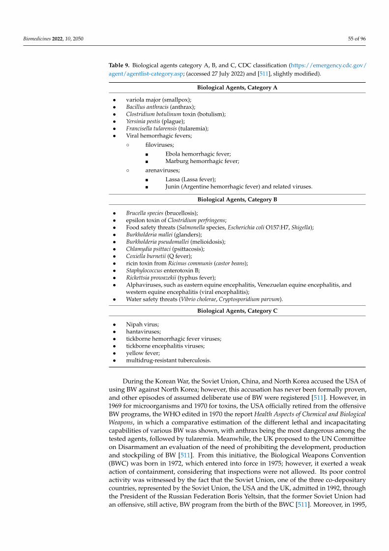
Biomedicines 2022, 10, 2050 55 of 96
Table 9. Biological agents category A, B, and C, CDC classification (https://emergency.cdc.gov/agent/agentlist-category.asp; (accessed 27 July 2022) and [511], slightly modified).
Biological Agents, Category A
• variola major (smallpox);• Bacillus anthracis (anthrax);• Clostridium botulinum toxin (botulism);• Yersinia pestis (plague);• Francisella tularensis (tularemia);• Viral hemorrhagic fevers;
◦ filoviruses;
� Ebola hemorrhagic fever;� Marburg hemorrhagic fever;
◦ arenaviruses;
� Lassa (Lassa fever);� Junin (Argentine hemorrhagic fever) and related viruses.
Biological Agents, Category B
• Brucella species (brucellosis);• epsilon toxin of Clostridium perfringens;• Food safety threats (Salmonella species, Escherichia coli O157:H7, Shigella);• Burkholderia mallei (glanders);• Burkholderia pseudomallei (melioidosis);• Chlamydia psittaci (psittacosis);• Coxiella burnetii (Q fever);• ricin toxin from Ricinus communis (castor beans);• Staphylococcus enterotoxin B;• Rickettsia prowazekii (typhus fever);• Alphaviruses, such as eastern equine encephalitis, Venezuelan equine encephalitis, and
western equine encephalitis (viral encephalitis);• Water safety threats (Vibrio cholerae, Cryptosporidium parvum).
Biological Agents, Category C
• Nipah virus;• hantaviruses;• tickborne hemorrhagic fever viruses;• tickborne encephalitis viruses;• yellow fever;• multidrug-resistant tuberculosis.
During the Korean War, the Soviet Union, China, and North Korea accused the USA ofusing BW against North Korea; however, this accusation has never been formally proven,and other episodes of assumed deliberate use of BW were registered [511]. However, in1969 for microorganisms and 1970 for toxins, the USA officially retired from the offensiveBW programs, the WHO edited in 1970 the report Health Aspects of Chemical and BiologicalWeapons, in which a comparative estimation of the different lethal and incapacitatingcapabilities of various BW was shown, with anthrax being the most dangerous among thetested agents, followed by tularemia. Meanwhile, the UK proposed to the UN Committeeon Disarmament an evaluation of the need of prohibiting the development, productionand stockpiling of BW [511]. From this initiative, the Biological Weapons Convention(BWC) was born in 1972, which entered into force in 1975; however, it exerted a weakaction of containment, considering that inspections were not allowed. Its poor controlactivity was witnessed by the fact that the Soviet Union, one of the three co-depositarycountries, represented by the Soviet Union, the USA and the UK, admitted in 1992, throughthe President of the Russian Federation Boris Yeltsin, that the former Soviet Union hadan offensive, still active, BW program from the birth of the BWC [511]. Moreover, in 1995,

Biomedicines 2022, 10, 2050 56 of 96
the United Nations Special Commission (UNSCOM) on Iraq obtained from Iraq, aftera long period of reticence, the admission that, despite being a signatory of the BWC, ithad developed biological and toxin weapons after the Gulf War in 1991 [512]. Recentevents in the international scenario have reminded the world of the danger of organizedterrorist attacks and the possibility that some biological agents may be deliberately releasedto be used as weapons. Following the disaster of 11 September 2001 in the USA, thepossible use of biological agents as aggressive was no longer considered a problem onlyfor the military, but a strategy for bioterrorism that represents a risk for the entire civilcommunity. Biotechnologies represent a means to improve the implementation of fasterdiagnostic methods and more effective tools of protection and therapy. However, thestrong development of biotechnologies in the last three decades and the internet-baseddiffusion of methods also make the production and development of biological agents moreaccessible to non-bio-terroristic groups. The development and production of geneticallymodified organisms, such as antibiotic-resistant bacterial strains, represent a concern in thefield of BW. A special category of human-made outbreaks of disease is the manipulationand dissemination of pathogens with the intention of disrupting societies. This may bepart of government policy in biological warfare, but it is also a means used by terroristgroups or criminals [511,513]. In this field, the military has always been heavily involvedboth in the development of offensive programs and in the search for countermeasures todeal with these diseases. Licensed vaccines are currently available for a few threats, suchas anthrax, Ebola virus, and smallpox; the last one has already been treated among thevaccine-preventable diseases, and research is underway to develop and produce vaccinesfor other threats, such as tularemia, botulism, plague, and Marburg virus. However, whilesmallpox was eradicated by a vaccine, vaccines are lacking or unsatisfactory, regardingsafety and effectiveness, for most biological agents. The biological agents of category Ahave all been treated, whereas only some biological agents of category B and C have beentreated, according to their major military contribution.
4.1. Anthrax
Anthrax, a potentially lethal zoonosis caused by Bacillus anthracis, a Gram-positive,rod-shaped, spore-forming and toxigenic bacterium, is one of the many infectious agentsidentified as a potential bioterrorist weapon [514], due to the stability of spores, whichcan persist for decades in the environment. Anthrax spores can be easily disseminated,as seen in the multiple releases via mailed letters or packages in the fall of 2001 in theUSA [52,515], and it is thus considered a biological agent, category A [57]. The disease maybe cutaneous, inhalational, and gastro-intestinal, with the first being milder and the othertwo, particularly the inhalational one, more severe. The estimation of the WHO about theconsequences of air dissemination of 50 kg of anthrax spores over a densely populated citywas 125,000 infections and 95,000 deaths [516].
During WWI, German scientists planned to infect livestock with anthrax [516,517].During the interwar period, in different world countries, including France, the UK, theformer Soviet Union, Canada and Japan, offensive biological weapon programs werelaunched. In those years, Japan, with the 731 unit led by the military physician GeneralShiro Ishii, was active in testing biological weapons in Manchuria. In particular, humanexperimentation on anthrax, which caused the death of thousands of Han Chinese people,was performed [516,518,519]. In 1943, Ishii used anthrax spores in villages south of Shang-hai in retaliation for the assistance to US soldiers. In 1942 and 1943, the USA, UK, andCanada evaluated the potential use of airborne spores of B. anthracis as a biological weaponcontaminating Gruinard Island, in Scotland. Most trials were conducted through missilesand aerials, and the effectiveness of the weapons was judged by exposing sheep, tethereddownwind of the detonation point, to the airborne cloud of spores [520,521]. Only in 1986was Gruinard Island decontaminated, and this episode represents now a well-known partof the history of BW development [522]. During the Cold War, the USA and the formerSoviet Union maintained active offensive biological weapon programs [523]. However, in

Biomedicines 2022, 10, 2050 57 of 96
1969, the USA unilaterally stopped their program [524], even if the former Soviet Unionwent on until 1992 with a covert offensive biological weapons program named Biopreparat,with a series of laboratories officially working for vaccine preparation, but actually onBW [525]. Anthrax spore production for military use was one of the main programs [526].A demonstration of this research program on B. anthracis is testified by the tragic conse-quences of anthrax spore inhalation by humans in Sverdlovsk in 1979 [527]. In this episode,the accidental release of a contaminated aerosol from a facility of the former Soviet Unionfor the production of weaponized anthrax spores caused the death of at least 68/77 (88%)exposed people who lived along the direction of airflow carrying the contaminated aerosol.Fear of the possible use of anthrax as a BW continued in the following years, so much so thatin 1991, during the Gulf War, the USA military was vaccinated [528]. Finally, weaponizedanthrax spores, deliberately released by mail, were used in 2001 in the USA, inducing elevencases (five of which were lethal) of inhalational and eleven cases of cutaneous anthrax.The analysis of these cases has enabled insights into better management of inhalationalanthrax [515,516].
Along with research and experimentation on anthrax as BW, and in the same way, thesearch for countermeasures has been active in the military environment, both in the use ofantibiotics and in the search for protective antibodies but, above all, in the developmentof vaccines.
During the 1930s, in the military laboratories of the Sanitary Technical Institute (STI)in Kirov, former Soviet Union, two avirulent, non-capsulated ST-1 and ST-3 strains derivedfrom a virulent anthrax strain were isolated [529]. The protective efficacy was tested inguinea pigs [530]; however, after WWII, these strains of suspended live spores were usedfor vaccine development in humans as well and were recommended for being admin-istered by scarification or subcutaneously [531]. In the period 1943–1950, the live sporevaccine was tested on 3500 volunteers; despite the adverse events not specified, it wasdefined as safe and immunogenic, and the protective potency of the vaccine was notspecifically determined [529]. In 1951 and 1952, the live spore vaccine was administered toover 140,000 subjects living in endemic areas for anthrax and as control, a population ofover 400,000 non-vaccinated matched subjects was analyzed. Fever in the first two dayswas observed in no more than 0.3% after both scarification and subcutaneous administra-tion, whereas only in this last case even local symptoms were reported. Moreover, only2.1/100,000 vaccinated subjects had anthrax versus 11.3/100,000 non-vaccinated ones, asignificant difference [530]. The live spore vaccine received a license for scarification in1953 and in 1959 for subcutaneous administration. In 1960, in the former Soviet Union,two million persons were yearly vaccinated and boostered with another dose after oneyear [529]. In those years, Russian military researchers tested the human anthrax live sporevaccine for aerogenic administration [532], whereas, in the period 1973–1975, repeatedstudies on the effectiveness of the human ST-1 vaccine were carried out using subcutaneousadministration, and even in this study, the difference between vaccinated and control groupwas highly significant [529].
Wright and other USA military researchers at Fort Detrick, Maryland, developed earlyvaccines against anthrax [533–535]. An aluminum-adsorbed anthrax vaccine was testedin a human field trial in the 1950s, demonstrating a 92.5% reduction in disease incidence(cutaneous and inhalation cases combined), and was licensed in 1970 as AVA (anthrax vac-cine adsorbed) [536,537]. Moreover, the military researchers from Fort Detrick challengedvaccinated nonhuman primates with lethal doses of anthrax spores, thus demonstratingthat AVA was protective in more than 90% of cases [320,538]. It consists of an acellularvaccine containing anthrax toxin antigens and results in protective immunity after threeto six doses [529]. In 1991, AVA was for the first time administered on a large scale to USmilitary personnel deployed to Iraq during the Persian Gulf War. Afterward, in 1998, theUS military started the Anthrax Vaccine Immunization Program to protect US militaryactive duty and reserve members [539]. In seven years, from 1998 through 2005, about5.6 million doses of AVA were administered to 1.5 million US military personnel [52]. De-
Biomedicines 2022, 10, 2050 58 of 96
spite this large experience and the evidence for protection, a certain level of reactogenicityand the unusually long vaccination schedule were accompanied by a general feeling of lackof confidence in the safety of AVA. Thus, many studies have been carried out not only bythe US military researchers, in order to clarify this crucial point. After evaluation of all theaccumulated scientific data, the National Academy of Sciences concluded that the anthraxvaccine has an adverse-reaction profile similar to that of other adult vaccines [540]. The USmilitary researchers even provided precious information on the need for a combined post-exposure prophylaxis of anti-microbials and vaccination, considering that anti-microbialprophylaxis alone for 1 month allowed 10–30% of cases to be infected [320], thus indicatingthe need of a combined treatment or alternatively 60 days of anti-microbial treatment, ascarried out in the at-risk subjects in the period of the anthrax letters in the USA [517].
In 1979 in Great Britain, an anthrax vaccine precipitated (AVP) using an avirulenttoxigenic, non-capsulating (pXO1+/pXO2−) 34F2 strain of Bacillus anthracis, originallyisolated by Sterne in 1937, was licensed. It contained protective antigen (PA) and traceamounts of lethal factor (LF) and edema factor (EF), the three toxin components, thusinducing better protection than AVA, which only contains PA [541]. It was developed inPorton Down, Salisbury, a Centre for Microbiological Research dependent on the Ministryof Defense until 1979, the year in which the center was split into two separately controlledlocations (one military and the other civilian). The research program for the use of airbornespores of B. anthracis as a BW in Gruinard Island, mentioned above, was conceived in theCentre of Porton Down.
At present, new second-generation vaccines in current research programs includerecombinant live vaccines and recombinant sub-unit vaccines. Even in the development ofthese innovative vaccines, military laboratories are engaged [320].
Anti-anthrax hyperimmune serum was obtained independently by immunizing ani-mals in France by the military physician Émile Marchoux and in Italy by Achille Sclavo atthe end of the nineteenth century [516], a few years after Emil von Behring and ShibasaburoKitasato in Germany developed anti-tetanus and anti-diphtheria hyper-immune sera, thusopening the era of passive immunotherapy. This serum was used in animals and even inhumans, representing the only protection for a long period. After the dramatic event ofthe letters contaminated with anthrax spores in 2001, the US government prepared hyper-immune intravenous immunoglobulins by collecting sera of immunized at-risk workersand military personnel [255], which, although not approved by the FDA and to be usedunder IND status, was protective, alone and even more together with anti-microbials [528].In 2012, a human anti-PA monoclonal antibody, raxibacumab, was approved by the FDAas prophylaxis and therapy of inhalational anthrax; in the same experimental conditions,it resulted as more protective than the intravenous polyclonal immunoglobulins [529].In 2016, another monoclonal antibody, obiltoxaximab, was approved by FDA. Currently,further monoclonal antibodies are under study, based on the assumption that targetingdifferent anthrax antigens may be more effective [542].
In addition to the study of vaccines, the contribution of the military is currently atthe forefront of the molecular genotyping of B. anthracis. For some years, the Italian ArmyMedical Research Center has been engaged in studies on differentiating and identifyingstrains from different geographic areas. This could be crucial for tracing strains deliberatelyreleased in a bioterrorism attack [516,543]. In a crisis of suspected bioterrorism, standard-ization, speed and accuracy, together with the availability of reference typing data, areimportant issues, as illustrated by the 2001 anthrax letters event. Along this line, severalresearch studies have described methods able to investigate genetic diversity betweenepidemic strains through single nucleotide repeat (SNR) analysis, in a fast and widelyaccessible way and particularly useful under field conditions [543–546].
In conclusion, pre-exposure anthrax vaccination has always been a target for militaryresearch; however, its use has been considered fundamental for selected population groupsfor which a calculable risk factor can be assessed. Only four of the NATO countries haveincluded the anthrax vaccine in the vaccination schedule of selected categories of military
Biomedicines 2022, 10, 2050 59 of 96
personnel [36]. The risk, now evident, for the possible use of anthrax as BW againstthe civilian population highlights the importance of the contribution of military medicalresearch in recent decades to obtain safer and more effective new anthrax vaccines [320].
4.2. Botulism
Botulism is a severe neurologic disease caused by neurotoxins produced by the bac-terium Clostridium botulinum. Botulinum neurotoxins (BoNTs) are the most potent naturallyoccurring toxins, with as little as 50 ng of neurotoxin sufficient to cause human botulismand to represent a significant biowarfare and bioterrorism threat [547]. BoNTs are currentlyrepresented by at least seven serotypes and more than 40 subtypes. Four of the sevenserotypes (A, B, C1, D, E, F, G) are pathogens for humans, in particular A, B, E, and F. Thesetoxins may enter the body through inhalation, ingestion and wounds. Due to its extremetoxicity, easy production and dissemination, BoNT has been classified as a category Abiothreat agent by the CDC [548]. New clostridial strains that produce novel neurotoxinvariants are being identified with increasing frequency, which presents challenges whenorganizing the nomenclature surrounding these neurotoxins [549].
As a prophylactic countermeasure, from 1959 until 2011, pentavalent (ABCDE) bo-tulinum toxoid (PBT) was available as IND. However, in 2011, the CDC stopped vaccineproduction considering its limited potency and high reactogenicity. At the present time,the most advanced candidates are recombinant nontoxic proteins. However, no licensedvaccines for prophylactic protection against botulism are currently available. In addition,vaccine development has been greatly complicated by the therapeutic use of BoNTs for agrowing number of indications including movement disorders, hemifacial spasm, essentialtremor, tics, writer’s cramp, cervical dystonia, cerebral palsy, vascular cerebral stroke, and,more recently, chronic pain, migraine, headache, and overactive bladder [550].
Since 2013, in Europe and the USA, a heptavalent F(ab)2 equine (ABCDEFG) botulismantitoxin was approved to treat individuals with symptoms of botulism after exposureor suspected exposure to botulinum neurotoxin. Even a trivalent (ABE) equine IgG isavailable, as well as a tetravalent (ABEF) equine, but only in Japan. Finally, human IgG forintravenous use (baby-BIG) is available for infant botulism. However, all these tools are inlimited supply, and their administration is finely regulated [550].
Military researchers also gave their contributions for this disease. Since 1941, BoNTshave become a military issue when an American Military Attaché in Berne, Switzerland,reported that “German experts and French collaborators in the Koch Foundation laborato-ries near Paris” were developing “botulinum toxin in an inert carrier for dissemination byair-burst bombs” [551]. This episode convinced the US Department of Defense to focus onmedical countermeasures, including vaccines, to immunize and protect personnel in thelaboratories at Camp Detrick against accidental exposure to biological warfare agents [552].The research at Camp Detrick, from 1943 to 1956, provided the foundation for the use ofBoNTs as a tool for studying the trophic regulation of skeletal muscle within motor neuronterminals and, more recently, for elucidation of the intricate details of neurotransmitterrelease at the molecular level [552]. Indirectly, Camp Detrick researchers also played asignificant role in studies that led to the use of BoNTs as a pharmaceutical product that hasbeen approved by the FDA for treating movement disorders, autonomic dysfunctions, andother conditions. It was also identified as a critical nutritional component for improvedgrowth of C. botulinum and increased production of BoNT serotype A. The purificationprocesses developed at Camp Detrick represent the base for the production of crystallinematerial required for the manufacture of the toxoid vaccine. Based on the research by CampDetrick investigators, a toxoid supply of over 1 million units was available to vaccinateabout 300,000 soldiers before the operations of D-Day in Normandy [552]. In 1956, CampDetrick was renamed Fort Detrick, and the US Army Medical Unit was established. Af-terward, in 1972, it was designated the US Army Medical Research Institute of InfectiousDiseases (USAMRIID), the lead military laboratory in the medical defense against biologicalwarfare threats [552]. Recently, US military researchers from USAMRIID reported on the
Biomedicines 2022, 10, 2050 60 of 96
production of recombinant BoNT toxin domain subunits as vaccine candidates againstmultiple serotypes. This ciBoNT HP (catalytically inactive holoprotein vaccines) vaccineelicited a more robust neutralizing antibody response, providing better protection against achallenge with parental toxins or with dissimilar subtypes [553].
In addition, a relevant contribution must be recognized to several studies of the ItalianArmy Medical and Veterinary Research Center, together with USAMRIID researchers inFort Detrick, in the field of epidemiology and genotyping of BoNT, with several studiesfocused on better understanding genetic variability among all of the C. botulinum serotypescoming from various geographic origins [549,554–556]. All these studies provide guidelinesfor botulinum neurotoxin subtype nomenclature.
4.3. Plague
Plague is caused by the facultative intracellular, Gram-negative, bacterial pathogen,Yersinia pestis. Plague is a severe, potentially lethal, disease that may manifest in three forms,bubonic, pneumonic and septicemic plague. It is a zoonosis, with rodents as reservoirsand fleas as vectors. Three great plague pandemics, resulting in nearly 200 million deathsin human history, have made Y. pestis one of the most virulent human pathogens. Even ifthe plague is often classified as a problem of the past, it remains a current threat in manyparts of the world, particularly in Africa [557,558]. In addition, the possible use of Y. pestisas a bioterrorist weapon is a serious threat due to its pathogenicity, easy dissemination,and human-to-human transmission. For this reason, it has been classified by the CDC asCategory A biological agent, also considering that different strains of Y. pestis showingresistance to the currently available antibiotics have been identified in Madagascar [559].The earliest recorded use of Y. pestis as a biological weapon dates back to the 14th century,when a Tatar army, in the attempt to conquer Caffa, in Crimea, reportedly catapultedvictims of plague over the city walls [560]. The Black Death, as the plague was named,which swept through Europe, the Near East, and North Africa in the mid-14th century,was probably the greatest public health disaster in recorded history and one of the mostdramatic examples ever of provoked pandemic. Caffa should be recognized as the siteof the most spectacular incident of biological warfare ever, with the Black Death as itsdisastrous consequence [561].
The first plague vaccine, developed at the end of the 19th century, consisted of killed,whole-cell, Y. pestis [562]. An immunogenic and somewhat less-reactogenic licensed vaccine(USP) containing a formalin-killed, highly virulent 195/P strain of Y. pestis, was effective inpreventing or ameliorating bubonic disease, as seen by the low incidence of plague cases inUS military personnel serving in Vietnam [563,564]. However, in vivo data on experimentalanimals suggested that this vaccine did not offer optimal protection against pneumonicplague [562]. Live attenuated Y. pestis vaccines, such as the EV strain (a virulent Y. pestis,derived from a patient identified as EV, and attenuated in the 1920s by serial passages), havebeen used in several countries for decades [565]. It was considered more immunogenicin the animals but more reactogenic than the inactivated one. The reactogenicity of theinactivated plague vaccine was evaluated to be lower than that of the whole-cell inactivatedtyphoid vaccine that was used in 1940 [320]. The live vaccines seemed to be able to protectagainst pneumonic and bubonic plague and induced high antibody titers, but unfortunately,they could have severe side effects and only induced short-lived protection that requiredannual boosters [566,567].
In analogy with all potential BWs, the military has invested resources in plagueresearch. At the end of the last century, the USAMRIID developed a recombinant fusionprotein, F1-V, comprising full-length capsular fraction 1 (F1) and low calcium responsevirulence protein (V) antigens [568–571]. This vaccine has long shown promise as a vaccinecandidate against both pneumonic and bubonic plague in rodents [568–570]. It was recentlyshown that F1-V adjuvanted with aluminum hydroxide (alum) using an IM/SC prime-boostregimen, provided complete protection against intranasal challenge with virulent Y. pestisCO92 in mice, guinea pigs, and macaques [572]. That F1 and V may be key molecules
Biomedicines 2022, 10, 2050 61 of 96
for active and passive immunization is also proven by the protective effect elicited notonly by a recombinant fusion protein F1-V acting as a vaccine but even by anti-F1 andV human monoclonal antibodies able to protect mice from challenge with Y. pestis [573].However, currently, no FDA-licensed vaccine [565], nor polyclonal/monoclonal antibodiesfor human use against plague [574] are available. In addition, a relevant contribution of theItalian Army Medical Research Center was in the field of strain differentiation of Y. pestis, adifficult challenge for these microorganisms considering their high intraspecies genomehomogeneity [545,575]. Fast strain identification and comparison with known genotypesmay be crucial for naturally occurring outbreaks versus bioterrorist events discrimination.In this regard, these studies were focused on assessing the inter-laboratory reproducibilityof in-house developed real-time PCR assays for the identification of Y. pestis [576].
4.4. Tularemia
Tularemia is a zoonosis caused by Francisella (formerly Pasteurella) tularensis, a faculta-tive intracellular, Gram-negative, coccobacillus, which may manifest in different clinicalforms, including the glandular and ulcero-glandular, oculo-glandular, oropharyngeal, ty-phoidal, and pneumonic. F. tularensis is present in some animals, in particular rabbits, andmay be transmitted to humans by drinking water contaminated by infected animals, drink-ing juice from contaminated fruits, or by aerosols and dust, or finally by the bite of insects,such as ticks and mosquitoes. It has been included among biological agents, category A [57].There are two types of F. tularensis, one more severe and contagious (10 cells represent theLD50 for rabbits and 50% infectious dose for humans), present in North America, and theother less virulent and contagious (10 million cells represent the LD50 for rabbits), presentin Europe and the former Soviet Union [320,577]. A great outbreak of tularemia, involving100,000 Soviet troops, occurred in Stalingrad during WWII. It started among the Nazimilitary and spread to the Soviet military. The large majority (over 95%) were pneumonicforms of tularemia. This unusual clinical presentation raised suspicions that this outbreakwas unnatural and deliberate. Whichever the origin of the Stalingrad outbreak, it remainsa military interest for tularemia, as even witnessed in the cases observed in Kosovo duringthe recent Balkan war [320].
The causative bacterium of tularemia, F. tularensis, was isolated in 1919 [578]. After thefirst few attempts with killed whole-cell vaccines, known as Foshay vaccines [579], whichwere not effective [580], a vaccine against tularemia was developed in the Soviet Union inthe 1940s. It was a live attenuated vaccine, and millions of people living in endemic areasunderwent immunization [581]. Afterward, in 1956, a mixture of two attenuated strains(155 and 15) of F. tularensis was brought from the Russian Institute of Epidemiology andMicrobiology (Gamaleia Institute, Moscow, Russia) to Fort Detrick, USA. From an ampouleof this product, a vaccine was produced, which resulted in protection for mice and guineapigs [582].
Studies at Fort Detrick continued into the 1960s; the strain was tested for safetyand efficacy in human volunteers and introduced as the F. tularensis live vaccine strain(LVS) [583–586]. This vaccine has been used since the mid-1960s and is associated witha significant decline in the rate of laboratory-acquired infections at Fort Detrick [587].However, considering the incomplete knowledge of the mutations in LVS and its residualvirulence, the vaccine remains under IND status and is administered only under protocoland with written informed consent.
Currently, in addition to military researchers, there are several groups involved indeveloping new-generation vaccines against tularemia, both by applying new technologiesand by using different routes of administration [588]. In particular, attempts are focusedon creating new live attenuated mutants [589,590], novel subunit vaccines [591] or gly-coconjugate of lipopolysaccharide O antigen of F. tularensis [592]. No tularemia vaccineis licensed yet, nor polyclonal/monoclonal antibodies for prophylaxis/therapy, despitethat polyclonal antibodies have been demonstrated to be protective in mice [593], whereasmonoclonal antibodies only result in partial protection [594].
Biomedicines 2022, 10, 2050 62 of 96
4.5. Filoviruses
The Filoviridae family is composed of enveloped RNA viruses with non-segmented,negative-sense genomes. Filoviruses are divided into three serologically distinct gen-era: Ebolavirus, Marburgvirus, and Cuevavirus [595]. They are among the most dangerouspathogens in the world, cause viral hemorrhagic fever, with case-fatality rates of up to 90%and are included in Category A of biological agents that could be used as weapons.
Before the 2014 West African Ebola virus outbreak, several filovirus vaccines hadbeen tested in small rodent models, but few candidates had moved into advanced devel-opment [596]. Even in this, case military research has always been on the front line, andin 1980, the first candidate vaccine on the basis of a heat- or formalin-inactivated Ebolavirus was tested at USAMRIID, Fort Detrick, on guinea pigs [597]. In the following years,Warfield and other military researchers worked on virus inactivation with preservation ofantigenic and structural integrity by a photoinducible alkylating agent [598]. The inacti-vated Ebola virus vaccine protected 80% of vaccinated mice from lethal disease. Similarlevels of protection were measured with inactivated Marburg virus studies. Virus-like par-ticles (VLPs) of Marburg virus vaccination completely protected guinea pigs [599]. Despitethe high efficacy in these animal models, the vaccine did not provide the proper level ofprotection to primates from lethal infection, both with Ebola and Marburg viruses [600,601].
More recently, several studies conducted always at USAMRIID investigated the possi-bility of using replicon-containing VLP filovirus vaccines [602–608]. Replicon-containingVLPs are generated by using a Venezuelan equine encephalitis virus (VEEV) vector toproduce replication-incompetent particles capable of entering a host cell. For both Ebolaand Marburg viruses, the antigen encoded is typically glycoprotein (GP), due to its capacityof inducing protective antibody responses [604,606]. Even though it is a promising topic,the protective efficacy of these vectored vaccines remains to be tested.
Another attractive strategy was the use of recombinant adenovirus vectors. Replication-deficient adenovirus vectors are highly immunogenic and can generate robust B and T cellresponses to viral antigens [609]. Among the studies in this area, a pan-filovirus vaccinewas tested, among others, by Swenson et al. at USAMRIID by using multiple adenovirusconstructs to express genes encoding Ebola GP, Ebola nucleoproteins, Marburg nucleo-proteins, and three Marburg GP combined into a single vaccine [610]. This approach isbelieved to provide widespread protection from multiple filovirus species [611].
DNA vaccine platforms have seen significant contributions from military researchers,such as the studies of Grant-Klein et al. on the evaluation of the ability of codon-optimizedDNA vaccines against the GP of Ebola and Marburg delivered by electroporation, individ-ually or as a mixture [612]. Further, they have also investigated different routes of vaccinedelivery and various DNA doses to optimize protection and to elicit the most robust an-tibody responses. Multiple clinical trials have been subsequently conducted to evaluatethe safety and immunogenicity of filovirus DNA vaccines, also with the contribution ofUS military researchers of WRAIR, as in the case of the first Ebola or Marburg vaccinetrials performed in Africa, showing that, given separately or together, both vaccines werewell tolerated, safe, and elicited antigen-specific humoral and cellular immune responses,suggesting limited immune interference [613].
Afterward, another relevant contribution of military researchers has been providedwith the approach by Filovirus VLPs, non-replicating vaccines generated by co-expressionof the GP and structural matrix protein VP40 in mammalian cells or insect cells [614,615].VLPs containing filovirus GP have successfully been used to vaccinate rodents, even in theabsence of adjuvants [616]. Moreover, it was demonstrated that VLPs activated cellularimmunity and that CD8+ T-cells are required for protection [617]. The protective efficacy ofVLPs seen in rodent models has also been observed in non-human primates by Warfieldand colleagues [618]. These studies showed encouraging results versus both Ebola andMarburg viruses [619,620]. The multitude of advantages afforded by VLPs make them apromising filovirus vaccine platform.

Biomedicines 2022, 10, 2050 63 of 96
USAMRIID researchers also investigated the possibility of using cytomegalovirusas a vaccine vector due to its strict species specificity and continuous replication withinthe host [621]. Likewise, rabies virus vectors have been explored as a vaccine platformagainst both Ebola and Marburg viruses by several groups of investigators, including themilitary [622].
Finally, the most encouraging candidate for a filovirus vaccine is currently representedby recombinant vesicular stomatitis virus (rVSV). One of the proteins encoded by rVSVis the G protein that, expressed on the surface of the virion, enables viral entry [623].At the beginning of the 2000s, Garbutt et al. produced the first rVSV-expressing EbolaGP, Marburg GP o Lassa GP [624]. Afterward, pre-clinical studies have established thatrVSV filovirus vaccines can rapidly induce protective immune responses in nonhumanprimates [625,626]. After the 2014 Ebola outbreak, there has been a notable acceleration inclinical trials in which there was a relevant role by USAMRIID researchers. Even militaryresearchers in the Russian Federation provided an important contribution to this vaccineapproach [627]. The 2014–2016 Ebola outbreak in West Africa is a paradigmatic example ofmilitary management of the epidemic both at the local level and on the part of the deployedmilitary by different western countries [242].
In all these studies, numerous partners from the public and private sectors havecombined efforts and resources to develop a Zaire ebolavirus (EBOV) vaccine candi-date (rVSV∆G-ZEBOV-GP) such that the rVSV∆G-ZEBOV-GP vaccine was approved asERVEBO® by the European Medicines Authority (EMA) and by the FDA in December2019 after five years of development [628]. No licensed vaccine for the Marburg virus isavailable yet.
Ebola virus disease has been faced with plasma from convalescent people. In 1995,during the Ebola outbreak in Kikwit, Democratic Republic of Congo, this treatment seemedpromising, considering that seven out of the eight patients who received convalescentplasma survived, a mortality of 12.5% versus 80% of the not treated patients [629]. A non-randomized study did not confirm this result and did not find a significant improvementin survival in patients who received convalescent plasma, whose anti-Ebola neutralizingtiter was unknown [630]. However, when the anti-Ebola antibodies were measured, itwas observed that the infusion of plasma with a high antibody titer was accompaniedby viral load reduction [631], thus confirming the usefulness of passive immunotherapyin Ebola disease. The military researchers of USAMRIID, who have collaborated in thisstudy, even participated in another study on the preparation of hyperimmune intravenousimmunoglobulins, which showed 5–6-fold increased potency compared to the pool ofconvalescent plasma and increased survivability of infected mice, when administered con-currently or 2 days after infection, thus hypothesizing that it may become a relevant tool forpost-exposure prophylaxis [632]. Recently, at the end of 2020, two monoclonal antibodies,INMAZEB® (consisting of three anti-Ebola glycoprotein monoclonal antibodies) [633] andEBANGATM (obtained by a convalescent patient and directed against the Ebola virusglycoprotein [634]) have been licensed by the FDA and, more recently, by the EMA.
4.6. Arenaviruses
Arenaviruses are divided into old world and new world viruses. All the members be-longing to the Arenavirus genus are linked to the progenitor lymphocytic choriomeningitisvirus (LCMV), which was discovered in 1933 and in 1964, and the similarities between thechronic disease caused in mice from LCMV and the one caused in hamsters from Machupovirus, a member of new world viruses, were noted [635]. They are named Arenavirusfrom the Latin arena, meaning sand [636], the most important member of the old world isthe Lassa virus, discovered in Nigeria in 1969, which is the most prevalent rodent-bornearenavirus circulating in West Africa. It is estimated that it is responsible for 300,000 in-fections per year and 5000 deaths [636]. The virus is present in some rodents and may betransmitted to humans by aerosols of the excreta. The mortality may be as high as 30%,and the same severity, and even more, is observed in the new world Arenaviruses, such
Biomedicines 2022, 10, 2050 64 of 96
as Junin, Machupo, and Sabia. For these characteristics of easy dissemination and highseverity, Arenaviruses are also included among Category A biological agents. The contri-bution of military researchers must be identified, as we have seen thus far, in the studiesat USAMRIID in Fort Detrick, especially on the pathogenetic mechanisms of the infection.From this point of view, a relevant work, born from the collaboration with other Europeanscientists, has allowed for clarification of the mechanisms of entry of the virus into infectedcells. Specifically, when the Lassa virus latches on to a receptor on the cell surface, it isfirst transported to a lysosome inside the cell, from which it can escape by hooking onto areceptor called LAMP1. This work, performed at USAMRIID using authentic Lassa viruses,was critical for validating the role of LAMP1 in Lassa virus infection, opening the door forthe development of directed therapies [637]. Currently, no licensed vaccine for the Lassavirus is available nor monoclonal antibodies for human use. Only for the Junin virus isthere a vaccine that has been registered in Argentina, where the virus is endemic.
4.7. Brucellosis
Brucellosis is a zoonosis caused by small Gram-negative coccobacilli of the genusBrucella. There are seven species of Brucella, of which four (B. abortus, B. melitensis, B. suis,and B. canis) are pathogenic to humans, with approximately 500,000 new human casesannually reported [638]. Brucella mainly infects cattle, swine, goats, sheep, and dogs, andthe disease may be transmitted to humans by eating or drinking unpasteurized contam-inated cheese or milk or by inhaling airborne agents. Person-to-person transmission israre. Considering its relative prolonged capacity for incapacitation, even in the presenceof a low mortality rate (5% of untreated cases), and given its significant contagiousnessby inhalation (10–100 bacteria sufficient to determine human disease), Brucella is consid-ered a potential biological weapon, so much so that it is included in Category B of CDCclassification [320]. Brucellosis is found globally and manifests with flu-like symptoms,including fever, weakness, malaise, and weight loss. In this case, the contribution of themilitary arises from the name itself of the disease. In 1887, the British military physicianDavid Bruce was able to isolate the germ, by him denominated Micrococcus melitensis, butlater re-denominated Brucella melitensis in his honor, from the spleens of patients who diedin the Malta isle due to a febrile illness, also known as Malta, Mediterranean or UndulantFever [639]. Due to the major role played by the Royal Army Medical Corps in clarifyingthe nature of the disease and its way of transmission, through the contaminated milkof the goats, thus leading to its prevention, the disease was also nicknamed the “CorpsDisease” [640]. However, brucellosis remained a constant threat in more recent times, inthe Mediterranean region and the Middle East during World War II and in the Middle Easttoday. In addition, considering its potential relevance as a weapon, in the 1950s, brucellosishas been included in various biological offensive programs in the USA, in South Africa,and in the former Soviet Union [524,641,642].
The contribution of military research also concerns vaccine development. The excellentresults obtained in the veterinary field allowed for dramatically reduced cases of humanbrucellosis. Current veterinary vaccines are based on live Rev1 B. melitensis strain andattenuated B. abortus strain 19. From the latter derived the preparations utilized for humanvaccines used in the past in some nations [643]. However, these human preparationsshowed a high incidence of cases of clinical disease and adverse reactions, as reportedboth in the USA and in the former Soviet Union, where this vaccination is still widelyused [644]. More recently, as part of the Brucella Vaccine Development program, WRAIRmilitary researchers have evaluated the effect as an adjuvant of a Neisseria Meningitidisouter membrane protein for an intranasal B. melitensis immunization in mice and guineapigs [645]. In China, military research developed a rapid and highly efficient method forthe identification of candidate antigens, using a combination of immunoproteomics withimmunization and bacterial challenge [646]. At present, no licensed human vaccine againstbrucellosis is available in the western world.
Biomedicines 2022, 10, 2050 65 of 96
4.8. Q Fever
Q fever is a zoonotic disease due to Gram-negative coccobacillus Coxiella burnetii, arickettsia-like organism capable of prolonged survival under harsh environmental condi-tions. Disease in humans is essentially caused by inhalation of dust from infected animalssuch as cattle, goats and sheep. The most frequent symptoms are flu-like syndrome, pneu-monia, and hepatitis [647]. Q fever has military relevance for both the risk of naturalinfection in military deployed abroad and for its potential use as a bioterrorism agent,considering that it is included in Category B of CDC classification. Thousands of casesof Q fever have been seen in military personnel since the disease was first reported in1937 [648,649]. However, it is believed that during the American Civil War, 1,765,000 casesof pneumonia and 45,000 deaths among the unionist troops might have occurred, includ-ing cases of Q fever, and the data are probably underestimated. This phenomenon wascertainly much more serious among the Confederates, but there are no reliable data onthe matter [650]. After being identified, it was first believed to be limited to Australia,hence the definition of Queensland fever, but afterward, it was found that the distributionwas worldwide, and the retrospective analysis of military researchers established thatmany British and US soldiers were affected by this disease during WWII in Greece andItaly [651,652]. After WWII, distinct outbreaks among US troops have been reported inLibya, in the USA (California and Texas) in the 1950s and, more recently, during OperationDesert Storm, Restore Hope, Enduring Freedom, and Iraqi Freedom [653–656]. In addition,an outbreak was reported among the Czech military in Bosnia in 1997 [657].
The military contribution was relevant after the identification of the germ, both inbetter defining the clinical aspects and the development of dedicated vaccines. Particularlynoteworthy, were the studies of US Col. AS Benenson concerning the development of thefirst effective Q fever vaccine [658,659]. This vaccine, based on whole-cell, formaldehyde-inactivated, ether-extracted C. burnetii with 10% egg yolk sac, was generally well tolerated,even though nonnegligible severe adverse reactions could be observed [660]. However, itproved useful for protection versus laboratory-acquired infections or for military personneldeployed to high-risk areas. Thereafter, this vaccine had been available at the US ArmyMedical Research and Materiel Command, Fort Detrick, under IND protocol, but due toits adverse side effects, it was (and is) not commercially licensed in the USA. Currently,the only licensed vaccine has been developed in Australia, Q-Vax® (Seqirus UK Limited,Maidenhead, UK) in 1989, and is used only in Australia in high-risk groups [661]. Even inthis case, the frequent hypersensitivity reactions to the vaccine have hindered the license ofQ-Vax® beyond Australia. Consequently, the research for a less reactogenic vaccine is stillin progress in military institutions, specifically at the USAMRIID [662,663].
4.9. New World Viral Encephalitis
Viral encephalitis poses a significant threat to military personnel employed in areas atrisk for these infectious agents. In addition, many of these agents are classified as CategoryB (Alphaviruses) and Category C (tick-borne encephalitis, West Nile fever) biological threatagents by the CDC and have reportedly been developed as biological weapons in thepast [664].
Alphavirus genera belonging to the Togaviridae family are positive-sense RNA viruses,usually transmitted by a mosquito vector. Infection may run asymptomatic but is usu-ally severely debilitating, with fever, malaise, headache, with risk of encephalitis and isoccasionally fatal, as in the case of the new world Alphaviruses Venezuelan, Eastern andWestern equine encephalitis viruses (VEEv, EEEv, WEEv) [665].
VEEv in humans may run asymptomatic or cause mild symptoms, including fever,headache, and, occasionally, convulsions and disorientation [257]. It was first isolatedin Venezuela, in 1938, and occurs mainly in South and Central America. In addition tomosquitoes, the virus is also transmitted in laboratories via aerosol [666].
There are currently no FDA-approved vaccines for VEEv, but two IND vaccines areavailable for at-risk laboratory personnel, TC-83 (live attenuated virus developed in 1961)
Biomedicines 2022, 10, 2050 66 of 96
and C-84 (a formalin-inactivated version of the TC-83 strain); however, neither vaccinecould protect experimental animals against inhalation infection [667–670]. Other engi-neered live attenuated vaccines have been investigated in mice and non-human primates.A candidate vaccine is the V3526, even if it protects against only a few genotypes ofVEEv [671]. For these reasons, the research has always continued over the years with adifferent approach, as DNA vaccines have proven particularly effective at eliciting pro-tective immune responses in animal models [672,673]. Laboratory workers at USAMRIIDare currently immunized with live (TC-83) and killed (C-84) vaccines. Moreover, militarystudies have increased knowledge on attenuation of VEEv, with a novel antigen expressionsystem based on VEEv genes, usable for other viruses [674]. Finally, military research isalso at the forefront of the study on the use of monoclonal antibodies, currently on animalmodels, as prophylactic and therapeutic approaches [257]. Particularly, at USAMRIID,it has been demonstrated that post-exposure administration of monoclonal antibodiesprotected macaques from the development of severe VEE disease even when administered48 h following aerosol exposure [675].
EEEv is rare; however, it is a serious disease, with 50% to 75% mortality and severeneurological sequelae in survivors [676]. Most of the cases occur in North, Central, andSouth America, and in the Caribbean. The military had paid attention to this virus as well,and in the 1970s, they investigated and produced a formalin-inactivated vaccine to protectlaboratory personnel working with EEEv [677,678]. This vaccine (TSI-GSD 104) is currentlyavailable as a US Army IND and has been administered to nearly 1000 workers [679].Moreover, the military research originated a chimeric vaccine containing genes of EEEvand WEEv [676] as well as a field-deployable RT-PCR assay [680].
WEEv is another alphavirus, and its basic description is the same as that for VEEv andEEEv. It has been isolated for the first time in California in 1930, and outbreaks still occurin the western USA [257]. In humans, it can present with fever, typical encephalitis andabnormal mental status, including focal neurological signs [681]. A formalin-inactivatedvaccine is available as IND and is administered to the laboratory workers who are exposedto the virus. This vaccine is well tolerated, considering that <5% of recipients report minorsymptoms [257]. Even in this case, military research contributed to various aspects, frompathogenetic mechanisms to the effects of the virus on different animal models [682–685]. Inaddition, a repertoire of mouse monoclonal antibodies against WEEv has been characterizedand shows promise for immunodetection and immunotherapy [686]. In the early 1990s, aformalin-inactivated vaccine (CM 4884), obtained from an attenuated strain, was developedby the US Army and made available under IND status [687]. The vaccine was administeredto laboratory workers at risk of contracting WEEv.
Finally, other military studies have investigated killed alphavirus vaccines and geneti-cally engineered, live attenuated virus vaccines [688–690]. In Table 10, the military interestfor and the military contribution to the biological agents, category A, and some biologicalagents, category B, are reported.
Table 10. Relevance for the military of possible biological weapons and military contribution totheir control.
Category A Military Interest Military Contribution
Smallpox Possible biological weapon Large vaccine useAnthrax Possible biological weapon Vaccine development—epidemiology—genotypingBotulism Possible biological weapon Vaccine development—epidemiology—genotypingPlague Possible biological weapon Vaccine development—epidemiology—genotyping
Tularemia Possible biological weapon Vaccine developmentFilovirus Possible biological weapon Vaccine development—polyclonal human Immunoglobulin
Arenavirus Possible biological weapon Pathogenesis

Biomedicines 2022, 10, 2050 67 of 96
Table 10. Cont.
Category A Military Interest Military Contribution
Category B
Brucellosis Possible biological weapon Etiology—vaccine developmentQ fever Possible biological weapon Vaccine development
Viral Encephalitis Possible biological weapons Vaccines and mAbs development—fieldable diagnosis
mAbs, monoclonal antibodies.
5. Aeromedical Evacuation of Patients with Highly Contagious, Severe Infectious Diseases
A particular mention must be reserved for aeromedical evacuation of patients withhighly contagious, severe infectious diseases. Evacuation of such patients is relevant tomilitary contingency operations because troops may be placed at risk for hemorrhagicfevers and other infections during deployment to tropical environments or by adversaries’use of biological warfare agents [691]. Historically, aeromedical evacuations with high-levelbiocontainment systems have been conducted by a limited number of military organiza-tions. Among these are the Deployable Air Isolator Team (DAIT) of the British Royal AirForce [692], the Aeromedical Isolation Team of the USAMRIID [693,694], and the Aeromed-ical Isolation Team of the Italian Air Force [695]. The Ebola Virus Disease (EVD) outbreak of2014–2016 provided the largest experience for the aeromedical evacuation of patients withviral hemorrhagic fevers [696]. Several healthcare workers who acquired the infection whilecaring for patients in West Africa were transported to EVD treatment facilities in the USAand Europe. While several of these patients were transported by military services, as inthe case of the Italian Air Force [695,697], civilian aircraft were contracted to provide evac-uation services by government agencies [698]; at least 10 nations conducted aeromedicalevacuations with high-level containment systems for at least 33 patients with EVD.
The aeromedical evacuation of COVID-19 patients during the pandemic was a new andunpredictable scenario. The new tasks were to move patients with acute respiratory distresssyndrome that caused an overflow of intensive care units regionally and to transport home,in a timely fashion, civilians or military personnel with suspected or confirmed COVID-19cases, from other countries or in deployed military units. In all these circumstances, theavailability of military organizations with a background in the transport of high infectiouspatients in the early stages enabled effective response to the many tasks received.
Aeromedical transport solutions for highly infectious patients to prevent transmissionare available as open and closed systems. In open systems, the medical crew, wearingfull personal protection equipment (PPE), and the patients are in a negative pressureisolation unit (container) with an airlock. Usually, open systems allow for easier access tothe patient and thus enable safer care for critically ill patients; however, the prohibitivelyhigh costs of open systems due to the need for a larger airframe are a limiting factor.Since the EVD epidemic, multiple organizations have developed isolated systems enablingsimultaneous isolation and care of multiple patients; these include the US Departmentof State Containerized Bio-Containment System [699] and the US Department of DefenseTransport Isolation System [700]. In closed systems, the patient is isolated in a smallernegative pressure isolation unit, with the medical crew outside that chamber. Patients aretreated by facilities that are fixed to several holes in the wall of the isolation unit. The mainlimitations of closed systems are limited access to the patient and reduced manual dexteritywhen delivering care through porthole gloves [698]. The Italian Air Force Isolation Unitemployed closed systems during the pandemic, such as the Aircraft Transit Isolator (ATI)and the IsoArk N-36 on their fixed and rotary wing assets (C-130J, Boeing KC-767A, C-27J,HH-101A) (Figure 2). In other countries, private air ambulance providers have carried outmany transfers of moderately to severely ill patients, by other cost-effective closed isolationsystem solutions [701,702].
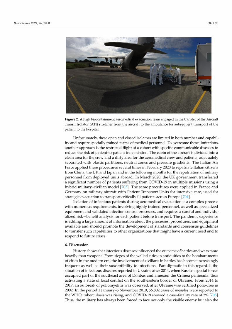
Biomedicines 2022, 10, 2050 68 of 96
Biomedicines 2022, 10, x FOR PEER REVIEW 68 of 98
negative pressure isolation unit, with the medical crew outside that chamber. Patients are treated by facilities that are fixed to several holes in the wall of the isolation unit. The main limitations of closed systems are limited access to the patient and reduced manual dexter-ity when delivering care through porthole gloves [698]. The Italian Air Force Isolation Unit employed closed systems during the pandemic, such as the Aircraft Transit Isolator (ATI) and the IsoArk N-36 on their fixed and rotary wing assets (C-130J, Boeing KC-767A, C-27J, HH-101A) (Figure 2). In other countries, private air ambulance providers have car-ried out many transfers of moderately to severely ill patients, by other cost-effective closed isolation system solutions [701,702].
Figure 2. A high biocontainment aeromedical evacuation team engaged in the transfer of the Air-craft Transit Isolator (ATI) stretcher from the aircraft to the ambulance for subsequent transport of the patient to the hospital.
Unfortunately, these open and closed isolators are limited in both number and capa-bility and require specially trained teams of medical personnel. To overcome these limi-tations, another approach is the restricted flight of a cohort with specific communicable diseases to reduce the risk of patient-to-patient transmission. The cabin of the aircraft is divided into a clean area for the crew and a dirty area for the aeromedical crew and pa-tients, adequately separated with plastic partitions, neutral zones and pressure gradients. The Italian Air Force applied these procedures several times in February 2020 to repatriate Italian citizens from China, the UK and Japan and in the following months for the repat-riation of military personnel from deployed units abroad. In March 2020, the UK govern-ment transferred a significant number of patients suffering from COVID-19 in multiple missions using a hybrid military–civilian model [703]. The same procedures were applied in France and Germany on military aircraft with Patient Transport Units for intensive care, used for strategic evacuation to transport critically ill patients across Europe [704].
Isolation of infectious patients during aeromedical evacuation is a complex process with numerous requirements, involving highly trained personnel, as well as specialized equipment and validated infection control processes, and requires a careful and individ-ualized risk−benefit analysis for each patient before transport. The pandemic experience is adding a large amount of information about the processes, procedures, and equipment available and should promote the development of standards and consensus guidelines to transfer such capabilities to other organizations that might have a current need and to respond to future crises.
6. Discussion
Figure 2. A high biocontainment aeromedical evacuation team engaged in the transfer of the AircraftTransit Isolator (ATI) stretcher from the aircraft to the ambulance for subsequent transport of thepatient to the hospital.
Unfortunately, these open and closed isolators are limited in both number and capabil-ity and require specially trained teams of medical personnel. To overcome these limitations,another approach is the restricted flight of a cohort with specific communicable diseases toreduce the risk of patient-to-patient transmission. The cabin of the aircraft is divided into aclean area for the crew and a dirty area for the aeromedical crew and patients, adequatelyseparated with plastic partitions, neutral zones and pressure gradients. The Italian AirForce applied these procedures several times in February 2020 to repatriate Italian citizensfrom China, the UK and Japan and in the following months for the repatriation of militarypersonnel from deployed units abroad. In March 2020, the UK government transferreda significant number of patients suffering from COVID-19 in multiple missions using ahybrid military–civilian model [703]. The same procedures were applied in France andGermany on military aircraft with Patient Transport Units for intensive care, used forstrategic evacuation to transport critically ill patients across Europe [704].
Isolation of infectious patients during aeromedical evacuation is a complex processwith numerous requirements, involving highly trained personnel, as well as specializedequipment and validated infection control processes, and requires a careful and individu-alized risk−benefit analysis for each patient before transport. The pandemic experienceis adding a large amount of information about the processes, procedures, and equipmentavailable and should promote the development of standards and consensus guidelinesto transfer such capabilities to other organizations that might have a current need and torespond to future crises.
6. Discussion
History shows that infectious diseases influenced the outcome of battles and wars moreheavily than weapons. From sieges of the walled cities in antiquities to the bombardmentsof cities in the modern era, the involvement of civilians in battles has become increasinglyfrequent as well as their susceptibility to infections. Paradigmatic in this regard is thesituation of infectious diseases reported in Ukraine after 2014, when Russian special forcesoccupied part of the southeast area of Donbas and annexed the Crimea peninsula, thusactivating a state of local conflict on the southeastern border of Ukraine. From 2014 to2017, an outbreak of poliomyelitis was observed, after Ukraine was certified polio-free in2002. In the period 1 January–5 November 2019, 56,802 cases of measles were reported tothe WHO, tuberculosis was rising, and COVID-19 showed a case-fatality rate of 2% [705].Thus, the military has always been forced to face not only the visible enemy but also the
Biomedicines 2022, 10, 2050 69 of 96
invisible agents of infectious diseases, which have frequently influenced the outcome ofbattles and wars more heavily than the strategic capacity. The possibility to neutralizethe consequences of the spreading of microorganisms became a military strategic capacityparticularly important after the birth of bacteriology—in Germany with Robert Koch andin France with Louis Pasteur. Military physicians from Germany, the UK, and Francewere the winners of the first, the second, and the seventh Nobel Prize for Physiology orMedicine and allowed passive immunotherapy to be a reality for defense against dreadfuldiseases such as tetanus and diphtheria and the discovery of etiology and the vector of anancient plague for humankind, such as malaria, allowing for its containment. Other militaryresearchers developed an effective typhoid vaccine and identified new pathogens, such asleishmaniasis. Meanwhile in the USA, Major Walter Reed contributed to the identificationof the yellow fever vector, thus paving the way for its containment, with the effectivepreventive measures designed by Major William Gorgas. The military from the countrieswith a strong attitude to be present in different war theaters worldwide had the need tocultivate and develop the then-rising bacteriology to protect the armies, and their researchand observations have represented useful conquests for all humankind. The two WWs andthe other wars of the twentieth century have represented the opportunity to test in the fieldthe scientific acquisitions in infectious diseases and to speed their application [706], with agrowing role of the US military medical researchers, considering that the USA has organizeda stable military network for the study of infectious diseases, with prestigious institutes inthe USA and overseas; one of the research Institutes is entitled to Walter Reed, the WRAIR,which was created in 1893 and without interruptions has continued its scientific activity,by launching specific organic programs, such as the HIV/AIDS program for the military,which has obtained many different and useful observations for the advancement of sciencein HIV, even for the general population [448]. Moreover, the financial resources madeavailable to the US Department of Defense for medical research is considerable; recently, ithas been USD 2 billion per year [706]. The military has historically been engaged in thesearch and early application of preventive tools, such as vaccines. This has led to relevantepidemiological results, as in the case of typhoid fever, meningococcal meningitis, tetanus,measles, and rubella. In other conditions, instead, the adoption (or lack of adoption) ofthe available new vaccinations has not been in line with the traditional timely militaryintervention, as in the case of meningococcal meningitis B, HPV, pneumococcus, adenovirusand influenza. In the last three cases, a responsibility may be attributed to the vaccinesthemselves, which may not be considered either fully protective or easily obtainable (this isthe case for the adenovirus vaccine, which has only been approved by the FDA for the USmilitary, and even for leptospirosis and dengue vaccines), whereas for meningitis B andHPV, the reason may be due to scarce awareness of the risk of infection.
However, the situation of infectious diseases is highly moving and is influenced by thevariability of socio-environmental conditions, including climate change [707], urbanizationand deforestation, which induced the emergence of new pathogens for humans and the re-emergence of pathogens that seemed to have disappeared. Between the last decades of thelast century and the beginning of this century, at least 30 new, potentially lethal, pathogenshave been described [222,708]. Such a moving situation induced the WHO, in the first halfof the 1990s, to launch a project on the emerging and re-emerging infectious diseases andto propose an organic and stable collaboration with the global military health services forsurveillance and control of infectious diseases, by creating inside the WHO, a new formalposition for a military physician liaison officer. This position was first covered, in theperiod 1995–2000, by one of the authors of this review (R.D.), and afterward, by US militaryphysicians up to 2012 [709]. The first military liaison officer could demonstrate, by a globalsurvey to which 76 world countries replied, that the collaboration between civilian andmilitary health services was already a precious and productive reality in some places, andthat in some developing countries, military health infrastructures could replace the lackingcivilian ones [7]. The subsequent activity of the US military physicians consolidated thiscivil–military collaboration, by strengthening collaborative projects with developing coun-
Biomedicines 2022, 10, 2050 70 of 96
tries, through the network of the US military research laboratories in developing countries,such as in Peru and Thailand [709]. However, this collaboration is becoming closer andcloser, making the fields of collaboration larger, from research and development to disastersand health emergencies management [710]. In this context, aeromedical evacuation ofpatients with highly infectious diseases, a medical, technical, and organizational challengewith significant risks for the safety of the patient, the crew, and the population, was analmost exclusively military activity until the Ebola outbreak of 2014–2016 [696,711–713].The heritage of engagement of the military in the fight against infectious diseases, evenbefore the birth of bacteriology [714], in particular for some dreadful diseases, such asinfluenza [190], and currently with the COVID-19 pandemic [242], legitimizes a sort of“militarization” of the management of infectious health emergencies, which have the po-tential for deeply undermining societal functioning and stability [708], as observed duringthe recent Ebola outbreak in West Africa in 2014–2016, the Zika virus outbreak in Brazil,or the COVID-19 pandemic worldwide [242]. This sort of “militarization” of the fightagainst infectious diseases, which is based on historical military involvement and a seriesof characteristics the military has, including quick mobilization, logistic organization andthe possibility to impose lockdowns by law enforcement, needed during the pandemic,is not agreed upon by everybody [190,242]. However, this international engagement ofthe military in humanitarian health initiatives has been favored by different governmentsin the context of global health diplomacy, a sort of soft power able to achieve relevantinternational security results with lesser expenses compared with the agencies officiallydedicated to military–military or civilian–military international collaborations. Militaryphysicians should specifically be formed for this innovative duty, and models of traininghave already been proposed [715,716]. Moreover, terrorism and bioterrorism are furtherdramatic phenomena that stimulate a closer and closer military–civilian collaboration, inorder to set up an effective response [717].
Another aspect that should be here underlined is that the research and developmentof new effective tools for the prevention and control of infectious diseases representing athreat to the military may be assimilated into the research and development of weaponsand armaments. In this context, the research and development of vaccines and/or poly-clonal/monoclonal antibodies against neglected infectious diseases, which is considerednot cost-effective by the civilian industry for lack of market, may be carried out or finan-cially supported by the military, which is engaged in this activity for strategic defensiveobjectives, regardless of market logic. However, the costs for developing a vaccine are,with today’s rules, prohibitive even for the military of affluent countries, such as for theUSA [513], thus inducing to think that such efforts may only be faced through internationalcollaboration among the militaries of allied countries, and a strengthened civil–militarycollaboration. In conclusion, a close civil–military collaboration for health promotion is ofreciprocal interest, particularly in the field of infectious diseases, irrespective of immediateor delayed danger caused by the disease. An example of this is, among others, HPV vac-cination, which may prevent a non-acute disease, such as cancer; however, the extensionof vaccination to male subjects, as observed in the military, may be a relevant measure ofpublic health.
7. Conclusions
In conclusion, war and infectious diseases have a reciprocal influence, because warcreates socio-environmental conditions favoring the spreading of infectious diseases, whichconversely may heavily influence the outcome of wars, by drastically reducing the op-erational readiness of the military. Paradigmatic of the influence of conflicts and socialdisruption on infectious diseases is the complex and difficult story of polio eradication.Conflicts are frequently accompanied by a re-emergence of the eradicated polio, as inUkraine, which was declared polio-free in 2002, and a polio outbreak was registered follow-ing the conflict with the Russian Federation in 2014 [705]. The same situation was observedin Syria in 2013, when polio, which had been eradicated 18 years before, re-emerged as a

Biomedicines 2022, 10, 2050 71 of 96
consequence of the civil war [140,718]. Even in Afghanistan, Pakistan, Nigeria, Somaliaand Kenia, the persistence or re-emergence of polio is generated by local conflicts andsocio-environmental, economic and health disruption [719]. An excellent historical reviewdocumenting the direct relationship between conflicts, wars and social disruption withre-emerging infectious diseases has recently been published [720]. The military healthservices have historically been forced to effectively combat infectious diseases, and thisspecific expertise should be put in common with the civilian counterpart to maximizethe efforts and the results in the control of infectious diseases. The quick mobilization,the logistic organization and the power of imposing restrictions by law enforcement arecharacteristics of the military worldwide, which have been recognized and used by thegovernments in recent outbreaks and pandemics, and which have the potential for deeplyundermining societal functioning and stability, inside the country and in internationalmilitary–military and civilian–military collaboration, in a sort of global health diplomacy.
Author Contributions: All authors have agreed on the design of the manuscript. The text has beenwritten by R.D. and R.B., whereas A.A. and M.L. have written the chapter on the high biocontainmentaeromedical evacuation; finally, G.D.L. oversaw the part relating to the vaccinations of the NATOcountries. R.N., M.S.P., T.S. and F.L. have repeatedly revised and coordinated the text. All authorshave read and agreed to the published version of the manuscript.
Funding: This research received no external funding.
Institutional Review Board Statement: Not applicable.
Informed Consent Statement: Not applicable.
Data Availability Statement: Not applicable.
Acknowledgments: The authors want to sincerely thank Angelo Di Pietro, who has collected some ofthe material used in this manuscript with enthusiasm, clever commitment and high professionalism.Angelo recently passed away from an illness before his 53rd birthday. Thanks also to Giulia Biselli forthe support in the final revision of the manuscript.
Conflicts of Interest: The authors declare no conflict of interest.
References1. Spier, R.E. Vaccines and the military. Vaccine 1993, 11, 491. [CrossRef]2. Connolly, M.A.; Heymann, D.L. Deadly comrades: War and infectious diseases. Lancet 2002, 360, S23–S24. [CrossRef]3. Wax, R.G. Manipulation of human history by microbes. Clin. Microbiol. Newsl. 2007, 29, 9–16. [CrossRef] [PubMed]4. Papagrigorakis, M.J.; Yapijakis, C.; Synodinos, P.N.; Baziotopoulou-Valavani, E. DNA examination of ancient dental pulp
incriminates typhoid fever as a probable cause of the Plague of Athens. Int. J. Infect. Dis. 2006, 10, 206–214. [CrossRef] [PubMed]5. Mura, M.; Haus-Cheymol, R.; Tournier, J.N. Immunization on the French Armed Forces: Impact, organization, limits and
perspectives. Infect. Dis. Now 2021, 51, 583–589. [CrossRef] [PubMed]6. Councell, C.E. War and infectious disease. Public Health Rep. 1941, 56, 547–573. [CrossRef]7. D’Amelio, R.; Heymann, D.L. Can the military contribute to global surveillance and control of infectious diseases? Emerg. Infect.
Dis. 1998, 4, 704–705. [CrossRef]8. Zumla, A.; Raviglione, M.; Hafner, R.; von Reyn, C.F. Tuberculosis. N. Engl. J. Med. 2013, 368, 745–755. [CrossRef]9. Krammer, F.; Smith, G.J.D.; Fouchier, R.A.M.; Peiris, M.; Kedzierska, K.; Doherty, P.C.; Palese, P.; Shaw, M.L.; Treanor, J.; Webster,
R.G.; et al. Influenza. Nat. Rev. Dis. Primers 2018, 4, 3. [CrossRef]10. Jafri, R.Z.; Ali, A.; Messonnier, N.E.; Tevi-Benissan, C.; Durrheim, D.; Eskola, J.; Fermon, F.; Klugman, K.P.; Ramsay, M.;
Sow, S.; et al. Global epidemiology of invasive meningococcal disease. Popul. Health Metr. 2013, 11, 17. [CrossRef]11. Coughlin, M.M.; Beck, A.S.; Bankamp, B.; Rota, P.A. Perspective on Global Measles Epidemiology and Control and the Role of
Novel Vaccination Strategies. Viruses 2017, 9, 11. [CrossRef] [PubMed]12. Available online: https://www.who.int/news-room/fact-sheets/detail/hepatitis-b (accessed on 29 May 2022).13. Confronting Inequalities. Lessons for Pandemic Responses from 40 Years of AIDS. Global AIDS Update. 2021. Available online:
https://www.unaids.org/sites/default/files/media_asset/2021-global-aids-update_en.pdf (accessed on 1 May 2022).14. Available online: https://www.who.int/news-room/fact-sheets/detail/hepatitis-c (accessed on 1 May 2022).15. World Health Organization. World Malaria Report 2021. Global Malaria Programme. WHO Geneva. 2021. Available online:
https://www.who.int/publications/i/item/9789240040496 (accessed on 10 March 2022).16. Available online: https://www.who.int/news-room/fact-sheets/detail/yellow-fever (accessed on 23 March 2022).
Biomedicines 2022, 10, 2050 72 of 96
17. Campbell, G.L.; Hills, S.L.; Fischer, M.; Jacobson, J.A.; Hoke, C.H.; Hombach, J.M.; Marfin, A.A.; Solomon, T.; Tsai, T.F.;Tsu, V.D.; et al. Estimated global incidence of Japanese encephalitis: A systematic review. Bull. World Health Organ. 2011, 89,766–774, 774A–774E. [CrossRef]
18. Bhatt, S.; Gething, P.W.; Brady, O.J.; Messina, J.P.; Farlow, A.W.; Moyes, C.L.; Drake, J.M.; Brownstein, J.S.; Hoen, A.G.;Sankoh, O.; et al. The global distribution and burden of dengue. Nature 2013, 496, 504–507. [CrossRef] [PubMed]
19. DengueNet—WHO’s Internet-based System for the global surveillance of dengue fever and dengue haemorrhagic fever(dengue/DHF). Dengue/DHF—Global public health burden. Wkly. Epidemiol. Rec. 2002, 77, 300–304.
20. Available online: https://www.who.int/news-room/fact-sheets/detail/typhoid (accessed on 1 June 2022).21. Harris, J.B.; LaRocque, R.C.; Qadri, F.; Ryan, E.T.; Calderwood, S.B. Cholera. Lancet 2012, 379, 2466–2476. [CrossRef]22. Dans, L.F.; Martínez, E.G. Amoebic dysentery. BMJ Clin. Evid. 2007, 2007, 0918. [PubMed]23. Global Hepatitis Report 2017; World Health Organization: Geneva, Switzerland, 2017.24. Cao, G.; Jing, W.; Liu, J.; Liu, M. The global trends and regional differences in incidence and mortality of hepatitis A from 1990 to
2019 and implications for its prevention. Hepatol. Int. 2021, 15, 1068–1082. [CrossRef]25. Costa, F.; Hagan, J.E.; Calcagno, J.; Kane, M.; Torgerson, P.; Martinez-Silveira, M.S.; Stein, C.; Abela-Ridder, B.; Ko, A.I. Global
morbidity and mortality of leptospirosis: A systematic review. PLoS Negl. Trop. Dis. 2015, 9, e0003898. [CrossRef]26. Weiss, M.M.; Weiss, P.D.; Mathisen, G.; Guze, P. Rethinking smallpox. Clin. Infect. Dis. 2004, 39, 1668–1673. [CrossRef]27. Fenner, F.; Henderson, D.A.; Arita, I.; Jezek, Z.; Ladnyi, I.D. Smallpox and Its Eradication/F. Fenner... [et al.]; World Health Orga-
nization: 1988. Chapter 6. Available online: https://apps.who.int/iris/handle/10665/39485 (accessed on 28 December 2021).28. Bayne-Jones, S. The Evolution of Preventive Medicine in the United States Army, 1607–1939; Office of the Surgeon General, Department
of the Army: Washington, DC, USA, 1968.29. Artenstein, A.W.; Opal, J.M.; Opal, S.M.; Tramont, E.C.; Peter, G.; Russell, P.K. History of U.S. military contributions to the study
of vaccines against infectious diseases. Mil. Med. 2005, 170 (Suppl. S4), 3–11. [CrossRef]30. Jenner, E. An Inquiry into the Causes and Effects of the Variolae Vaccinae, Discovered in Some of the Wester Counties of England,
Particularly Gloucestershire, and Known by the Name of the Cow Pox. 1798. Available online: http://resource.nlm.nih.gov/2559001R (accessed on 28 December 2021).
31. Rezza, G. Mandatory vaccination for infants and children: The Italian experience. Pathog. Glob. Health 2019, 113, 291–296.[CrossRef] [PubMed]
32. Cavallo, J.D. Des fièvres aux maladies infectieuses, trois siècles de lutte contre l’infection. Med. Armées 2008, 36, 517–526.33. Hüntelmann, A.C. Smallpox vaccination in the German Empire. Vaccination between biopolitics and moral economy. Asclepio
2020, 72, 292. [CrossRef]34. Meynell, E. French reactions to Jenner’s discovery of smallpox vaccination: The primary sources. Soc. Hist. Med. 1995, 8, 285–303.
[CrossRef]35. Hopkins, R.J.; Lane, J.M. Clinical efficacy of intramuscular vaccinia immune globulin: A literature review. Clin. Infect. Dis. 2004,
39, 819–826. [CrossRef]36. Standards Related Document. SRD-7 to AJMedP-4. Vaccinations Catalogue within the Nato & PfP forces. Edition A Version 2
July 2021. Available online: https://nso.nato.int/nso (accessed on 10 January 2022).37. Voigt, E.A.; Kennedy, R.B.; Poland, G.A. Defending against smallpox: A focus on vaccines. Expert Rev. Vaccines 2016, 15, 1197–1211.
[CrossRef] [PubMed]38. Gröschel, D.H.; Hornick, R.B. Who introduced typhoid vaccination: Almroth Write or Richard Pfeiffer? Rev. Infect. Dis. 1981, 3,
1251–1254. [CrossRef]39. Williamson, J.D.; Gould, K.G.; Brown, K. Richard Pfeiffer’s typhoid vaccine and Almroth Wright’s claim to priority. Vaccine 2021,
39, 2074–2079. [CrossRef]40. Pfeiffer, R.; Kolle, W. Experimental Investigations on Protective Inoculation of Men against Typhus Abdominalis. Ind. Med. Gaz.
1897, 32, 41–44.41. Wright, A.E. On the association of serous hæmorrhages with conditions of defective blood-coagulability. Lancet 1896, 148, 807–809.
[CrossRef]42. Wright, A.E.; Semple, D. Remarks on vaccination against typhoid fever. BMJ 1897, 1, 256–259. [CrossRef] [PubMed]43. Cantlie, N. History of the Army Medical Department; Churchill Livingstone: London, UK, 1974; Volume II.44. Leishman, W.B. Enteric Fevers in the British Expeditionary Force, 1914–1918. Glasgow Med. J. 1921, 95, 81–109. [PubMed]45. Torrens, J. Enteric group of fevers. In Medical Services Diseases of the War; MacPhearson, W., Herringham, W., Elliott, T., Balfour, A.,
Eds.; HM Stationery Office: London, UK, 1922; pp. 11–63.46. Gradmann, C.; Harrison, M.; Rasmussen, A. Typhoid and the Military in the Early 20th Century. Clin. Infect. Dis. 2019,
69 (Suppl. S5), S385–S387. [CrossRef] [PubMed]47. Castellani, A. Typhoid-paratyphoid vaccination with mixed vaccines. BMJ 1913, 2, 1577–1578. [CrossRef]48. D’Amelio, R.; Biselli, R.; Natalicchio, S.; Lista, F.; Peragallo, M.S. Vaccination programmes in the Italian military. Vaccine 2003, 21,
3530–3533. [CrossRef]49. D’Amelio, R.; Tagliabue, A.; Nencioni, L.; Di Addario, A.; Villa, L.; Manganaro, M.; Boraschi, D.; Le Moli, S.; Nisini, R.; Matricardi,
P.M. Comparative analysis of immunological responses to oral (Ty21a) and parenteral (TAB) typhoid vaccines. Infect. Immun.1988, 56, 2731–2735. [CrossRef]
Biomedicines 2022, 10, 2050 73 of 96
50. Haus-Cheymol, R.; Kraemer, P.; Simon, F. Les risques infectieux en opérations extérieures. Med. Armées 2009, 37, 435–452.51. Rasmussen, A. A corps défendant: Vacciner les troupes contre la typhoïde pendant la Grande Guerre. Corps 2008, 2, 41–48.
[CrossRef]52. Grabenstein, J.D.; Pittman, P.R.; Greenwood, J.T.; Engler, R.J. Immunization to protect the US Armed Forces: Heritage, current
practice, and prospects. Epidemiol. Rev. 2006, 28, 3–26. [CrossRef]53. Shanks, G.D. How World War 1 changed global attitudes to war and infectious diseases. Lancet 2014, 384, 1699–1707. [CrossRef]54. Germanier, R.; Füer, E. Isolation and characterization of Gal E mutant Ty 21a of Salmonella typhi: A candidate strain for a live,
oral typhoid vaccine. J. Infect. Dis. 1975, 131, 553–558. [CrossRef]55. Szu, S.C.; Stone, A.L.; Robbins, J.D.; Schneerson, R.; Robbins, J.B. Vi capsular polysaccharide-protein conjugates for prevention
of typhoid fever. Preparation, characterization, and immunogenicity in laboratory animals. J. Exp. Med. 1987, 166, 1510–1524.[CrossRef] [PubMed]
56. Cartee, R.T.; Thanawastien, A.; Griffin Iv, T.J.; Mekalanos, J.J.; Bart, S.; Killeen, K.P. A phase 1 randomized safety, reactogenicity,and immunogenicity study of Typhax: A novel protein capsular matrix vaccine candidate for the prevention of typhoid fever.PLoS Negl. Trop. Dis. 2020, 14, e0007912. [CrossRef]
57. Barras, V.; Greub, G. History of biological warfare and bioterrorism. Clin. Microbiol. Infect. 2014, 20, 497–502. [CrossRef]58. Hassel, B. Tetanus: Pathophysiology, treatment, and the possibility of using botulinum toxin against tetanus-induced rigidity and
spasms. Toxins 2013, 5, 73–83. [CrossRef] [PubMed]59. Von Behring, E.; Kitasato, S. Ueber das Zustandekommen der Diphtherie-Immunitat and der Tetanus-Immunitat bei Thieren.
Dtsch. Med. Wochenschr. 1890, 16, 1113–1114.60. Von Behring, E. Untersuchungen uber das Zustandekommen der Diphtherie-Immunitat and der Tetanus-Immunitat bei Thieren.
Dtsch. Med. Wochenschr. 1890, 16, 1145–1148.61. Bracha, A.; Tan, S.Y. Emil von Behring (1854–1917): Medicine’s first Nobel laureate. Singapore Med. J. 2011, 52, 1–2.62. Schlessinger, B.S.; Schlessinger, J.H. The Who’s Who of Nobel Prize Winners; Oryx Press: Phoenix, AZ, USA, 1986; p. 79.63. Bruce, D. Tetanus: Analysis of 1458 Cases, which occurred in Home Military Hospitals during the years 1914–1918. J. Hyg. 1920,
19, 1–32. [CrossRef]64. Linton, D.S. Emil von Behring. Infectious Disease, Immunology, Serum Therapy; American Philosophical Society: Philadelphia,
PA, USA, 2005; p. 357e62.65. Ferrajoli, F. Il servizio Sanitario Militare nella Guerra 1915–1918 (Nel Cinquantenario della Vittoria). G. Med. Mil. 1968,
118, 501–516.66. Editorial. Tetanus in the US Army in World War II. N. Engl. J. Med. 1947, 237, 411–413. [CrossRef]67. Hammarlund, E.; Thomas, A.; Poore, E.A.; Amanna, I.J.; Rynko, A.E.; Mori, M.; Chen, Z.; Slifka, M.K. Durability of vaccine-
induced immunity against tetanus and diphtheria toxins: A cross-sectional analysis. Clin. Infect. Dis. 2016, 62, 1111–1118.[CrossRef]
68. Ferlito, C.; Biselli, R.; Mariotti, S.; von Hunolstein, C.; Teloni, R.; Ralli, L.; Pinto, A.; Pisani, G.; Tirelli, V.; Biondo, M.I.; et al.Tetanus-diphtheria vaccination in adults: The long-term persistence of antibodies is not dependent on polyclonal B-cell activationand the defective response to diphtheria toxoid re-vaccination is associated to HLADRB1∗01. Vaccine 2018, 36, 6718–6725.[CrossRef]
69. Gentili, G.; D’Amelio, R.; Wirz, M.; Matricardi, P.M.; Nisini, R.; Collotti, C.; Pasquini, P.; Stroffolini, T. Prevalence of hyperimmu-nization against tetanus in Italians born after the introduction of mandatory vaccination of children with tetanus toxoid in 1968.Infection 1993, 21, 80–82. [CrossRef] [PubMed]
70. Sharma, N.C.; Efstratiou, A.; Mokrousov, I.; Mutreja, A.; Das, B.; Ramamurthy, T. Diphtheria. Nat. Rev. Dis. Primers 2019, 5, 81.[CrossRef]
71. Hardy, I.R.; Dittmann, S.; Sutter, R.W. Current situation and control strategies for resurgence of diphtheria in newly independentstates of the former Soviet Union. Lancet 1996, 347, 1739–1744. [CrossRef]
72. Rappuoli, R.; Podda, A.; Giovannoni, F.; Nencioni, L.; Peragallo, M.; Francolini, P. Absence of protective immunity againstdiphtheria in a large proportion of young adults. Vaccine 1993, 11, 576–577. [CrossRef]
73. Bordet, J.; Gengou, O. Le microbe de la coqueluche. Ann. Inst. Pasteur 1906, 20, 731.74. Kilgore, P.E.; Salim, A.M.; Zervos, M.J.; Schmitt, H.J. Pertussis: Microbiology, Disease, Treatment, and Prevention. Clin. Microbiol.
Rev. 2016, 29, 449–486. [CrossRef]75. Jansen, D.L.; Gray, G.C.; Putnam, S.D.; Lynn, F.; Meade, B.D. Evaluation of pertussis in U.S. Marine Corps trainees. Clin. Infect.
Dis. 1997, 25, 1099–1107. [CrossRef]76. Vincent, J.M.; Cherry, J.D.; Nauschuetz, W.F.; Lipton, A.; Ono, C.M.; Costello, C.N.; Sakaguchi, L.K.; Hsue, G.; Jackson, L.A.;
Tachdjian, R.; et al. Prolonged afebrile nonproductive cough illnesses in American soldiers in Korea: A serological search forcausation. Clin. Infect. Dis. 2000, 30, 534–539. [CrossRef]
77. Klement, E.; Grotto, I.; Srugo, I.; Orr, N.; Gilad, J.; Cohent, D. Pertussis in soldiers, Israel. Emerg. Infect. Dis. 2005, 11, 506–508.[CrossRef] [PubMed]
78. Aase, A.; Herstad, T.K.; Merino, S.; Brandsdal, K.T.; Berdal, B.P.; Aleksandersen, E.M.; Aaberge, I.S. Opsonophagocytic activityand other serological indications of Bordetella pertussis infection in military recruits in Norway. Clin. Vaccine Immunol. 2007, 14,855–862. [CrossRef]
Biomedicines 2022, 10, 2050 74 of 96
79. Mayet, A.; Brossier, C.; Haus-Cheymol, R.; Verret, C.; Meynard, J.B.; Migliani, R.; Pommier de Santi, V.; Decam, C.; Deparis,X. Pertussis surveillance within the French armed forces: A new system showing increased incidence among young adults(2007–2009). J. Infect. 2011, 62, 322–324. [CrossRef] [PubMed]
80. Rota, M.C.; Ausiello, C.M.; D’Amelio, R.; Cassone, A.; Giammanco, A.; Molica, C.; Lande, R.; Greco, D.; Salmaso, S. Prevalence ofmarkers of exposure to Bordetella pertussis among Italian young adults. Clin. Infect. Dis. 1998, 26, 297–302. [CrossRef] [PubMed]
81. Zepp, F.; Heininger, U.; Mertsola, J.; Bernatowska, E.; Guiso, N.; Roord, J.; Tozzi, A.E.; Van Damme, P. Rationale for pertussisbooster vaccination throughout life in Europe. Lancet Infect. Dis. 2011, 11, 557–570. [CrossRef]
82. Villemin, J.A. Étude sur la Tuberculose: Preuves Rationnelles et Expérimentales de sa Spécificité et son Inoculabilité; Baillère:Paris, France, 1868.
83. Koch, R. The Current State of the Struggle against Tuberculosis. Nobel Lecture December 12 1905. From Nobel lectures. Physiology orMedicine 1901–1921; Elsevier Publishing Company: Amsterdam, The Netherlands, 1967.
84. Behr, M.A.; Edelstein, P.H.; Ramakrishnan, L. Revisiting the timetable of tuberculosis. BMJ 2018, 362, k2738. [CrossRef] [PubMed]85. Tan, S.Y.; Kwok, E. Albert Calmette (1863–1933): Originator of the BCG vaccine. Singapore Med. J. 2012, 53, 433–434.86. Kimbrough, W.; Saliba, V.; Dahab, M.; Haskew, C.; Checchi, F. The burden of tuberculosis in crisis-affected populations: A
systematic review. Lancet Infect. Dis. 2012, 12, 950–965. [CrossRef]87. Mancuso, J.D. Tuberculosis Screening and Control in the US Military in War and Peace. Am. J. Public Health. 2017, 107, 60–67.
[CrossRef]88. Nevin, R.L. Active tuberculosis and recent overseas deployment in the U.S. Military. Am. J. Prev. Med. 2010, 39, e39–e40, author
reply e40. [CrossRef] [PubMed]89. Bergman, B.P.; Mackay, D.F.; Pell, J.P. Tuberculosis in Scottish military veterans: Evidence from a retrospective cohort study of
57,000 veterans and 173,000 matched non-veterans. J. R. Army Med. Corps 2017, 163, 53–57. [CrossRef]90. D’Amelio, R.; Stroffolini, T.; Biselli, R.; Molica, C.; Cotichini, R.; Bernardini, G.; Vellucci, A. Tuberculin skin reactivity in Italian
military recruits tested in 1996–1997. Eur. J. Clin. Microbiol. Infect. Dis. 2000, 19, 200–204. [CrossRef] [PubMed]91. Camarca, M.M.; Krauss, M.R. Active tuberculosis among U.S. Army personnel, 1980 to 1996. Mil. Med. 2001, 166, 452–456.
[CrossRef] [PubMed]92. D’Amelio, E.; Salemi, S.; D’Amelio, R. Anti-Infectious Human Vaccination in Historical Perspective. Int. Rev. Immunol. 2016, 35,
260–290. [CrossRef] [PubMed]93. D’Amelio, R.; Molica, C.; Biselli, R.; Stroffolini, T. Surveillance of infectious diseases in the Italian military as pre-requisite for
tailored vaccination programme. Vaccine 2001, 19, 2006–2011. [CrossRef]94. Weichselbaum, A. Ueber die Aetiologie der akuten meningitis cerebro-spinalis. Fortschr. Med. 1887, 5, 573.95. Goldschneider, I.; Gotschlich, E.C.; Artenstein, M.S. Human immunity to the meningococcus. I. The role of humoral antibodies. J.
Exp. Med. 1969, 129, 1307–1326. [CrossRef]96. Goldschneider, I.; Gotschlich, E.C.; Artenstein, M.S. Human immunity to the meningococcus. II. The development of natural
immunity. J. Exp. Med. 1969, 129, 1327–1348. [CrossRef]97. Gotschlich, E.C.; Liu, T.Y.; Artenstein, M.S. Human immunity to the meningococcus. III. Preparation and immunochemical
properties of the group A, group B, and group C meningococcal polysaccharides. J. Exp. Med. 1969, 129, 1349–1365. [CrossRef]98. Gotschlich, E.C.; Goldschneider, I.; Artenstein, M.S. Human immunity to the meningococcus. IV. Immunogenicity of group A and
group C meningococcal polysaccharides in human volunteers. J. Exp. Med. 1969, 129, 1367–1384. [CrossRef] [PubMed]99. Gotschlich, E.C.; Goldschneider, I.; Artenstein, M.S. Human immunity to the meningococcus. V. The effect of immunization with
meningococcal group C polysaccharide on the carrier state. J. Exp. Med. 1969, 129, 1385–1395. [CrossRef] [PubMed]100. Savona-Ventura, C. An Outbreak of Cerebrospinal Fever in a 19th Century British Mediterranean Naval Base. J. R. Army Med.
Corps 1994, 140, 155–158. [CrossRef]101. Brundage, J.F.; Ryan, M.A.; Feighner, B.H.; Erdtmann, F.J. Meningococcal disease among United States military service members
in relation to routine uses of vaccines with different serogroup-specific components, 1964–1998. Clin. Infect. Dis. 2002, 35,1376–1381. [CrossRef] [PubMed]
102. Artenstein, M.S.; Gold, R. Current status of prophylaxis of meningococcal disease. Mil. Med. 1970, 135, 735–739. [CrossRef][PubMed]
103. Artenstein, M.S.; Gold, R.; Zimmerly, J.G.; Wyle, F.A.; Schneider, H.; Harkins, C. Prevention of meningococcal disease by group Cpolysaccharide vaccine. N. Engl. J. Med. 1970, 282, 417–420. [CrossRef] [PubMed]
104. Biselli, R.; Fattorossi, A.; Matricardi, P.M.; Nisini, R.; Stroffolini, T.; D’Amelio, R. Dramatic reduction of meningococcal meningitisamong military recruits in Italy after introduction of specific vaccination. Vaccine 1993, 11, 578–581. [CrossRef]
105. Stroffolini, T.; Curianó, C.M.; Congiu, M.E.; Occhionero, M.; Mastrantonio Gianfrilli, P. Trends in meningococcal disease in Italy1987. Public Health 1989, 103, 31–34. [CrossRef]
106. Stroffolini, T. Vaccination campaign against meningococcal disease in army recruits in Italy. Epidemiol. Infect. 1990, 25, 1–5.[CrossRef]
107. Le Moli, S.; Matricardi, P.M.; Quinti, I.; Stroffolini, T.; D’Amelio, R. Clonotype analysis of human antibodies specific for Neisseriameningitidis polysaccharides A and C in adults. Clin. Exp. Immunol. 1991, 83, 460–465. [CrossRef]
Biomedicines 2022, 10, 2050 75 of 96
108. Ferlito, C.; Biselli, R.; Cattaruzza, M.S.; Teloni, R.; Mariotti, S.; Tomao, E.; Salerno, G.; Peragallo, M.S.; Lulli, P.; Caporuscio, S.; et al.Immunogenicity of meningococcal polysaccharide ACWY vaccine in primary immunized or revaccinated adults. Clin. Exp.Immunol. 2018, 194, 361–370. [CrossRef] [PubMed]
109. Ferlito, C.; Visco, V.; Biselli, R.; Cattaruzza, M.S.; Carreras, G.; Salerno, G.; Lista, F.; Capobianchi, M.R.; Castilletti, C.; Lapa, D.;et al. Safety of Multiple Vaccinations and Durability of Vaccine-Induced Antibodies in an Italian Military Cohort 5 Years afterImmunization. Biomedicines 2022, 10, 6. [CrossRef] [PubMed]
110. Spiegel, A.; Quenel, P.; Sperber, G.; Meyran, M. Evaluation de l’efficacité de la stratégie de vaccination systématique an-timéningococcique chez les appelés de l’armée française [Evaluation of systematic anti-meningococcal vaccination strategy inFrench military recruits]. Santé 1996, 6, 383–388. [PubMed]
111. Finne, J.; Leinonen, M.; Mäkelä, P.H. Antigenic similarities between brain components and bacteria causing meningitis. Implica-tions for vaccine development and pathogenesis. Lancet 1983, 2, 355–357. [CrossRef]
112. Kelly, D.F.; Rappuoli, R. Reverse vaccinology and vaccines for serogroup B Neisseria meningitidis. Adv. Exp. Med. Biol. 2005, 568,217–223. [CrossRef] [PubMed]
113. Rappuoli, R.; Pizza, M.; Masignani, V.; Vadivelu, K. Meningococcal B vaccine (4CMenB): The journey from research to real worldexperience. Expert Rev. Vaccines 2018, 17, 1111–1121. [CrossRef]
114. Parikh, S.R.; Andrews, N.J.; Beebeejaun, K.; Campbell, H.; Ribeiro, S.; Ward, C.; White, J.M.; Borrow, R.; Ramsay, M.E.; Ladhani,S.N. Effectiveness and impact of a reduced infant schedule of 4CMenB vaccine against group B meningococcal disease in England:A national observational cohort study. Lancet 2016, 388, 2775–2782. [CrossRef]
115. European Centre for Disease Prevention and Control. Expert Opinion on the Introduction of the Meningococcal B (4CMenB) Vaccine inthe EU/EEA; ECDC: Stockholm, Sweden, 2017.
116. Millar, B.C.; Moore, P.J.A.; Moore, J.E. Meningococcal disease: Has the battle been won? J. R. Army Med. Corps 2017, 163, 235–241.[CrossRef]
117. Dooley, D.P. History of U.S. military contributions to the study of viral hepatitis. Mil. Med. 2005, 170 (Suppl. S4), 71–76. [CrossRef]118. Hawkins, R.E.; Malone, J.D.; Cloninger, L.A.; Rozmajzl, P.J.; Lewis, D.; Butler, J.; Cross, E.; Gray, S.; Hyams, K.C. Risk of viral
hepatitis among military personnel assigned to US Navy ships. J. Infect. Dis. 1992, 165, 716–719. [CrossRef]119. D’Amelio, R.; Mele, A.; Mariano, A.; Romanò, L.; Biselli, R.; Lista, F.; Zanetti, A.; Stroffolini, T. Hepatitis A, Italy. Emerg. Infect. Dis.
2005, 11, 1155–1156. [CrossRef] [PubMed]120. Joussemet, M.; Bourin, P.; Lebot, O.; Fabre, G.; Deloince, R. Evolution of hepatitis A antibodies prevalence in young French
military recruits. Eur. J. Epidemiol. 1992, 8, 289–291. [CrossRef] [PubMed]121. MacCallum, F.O. Hepatitis. Br. Med. Bull. 1953, 9, 221–225. [CrossRef]122. Sawyer, W.A.; Meyer, K.F.; Eaton, M.D.; Bauer, J.H.; Putnam, P.; Schwentker, F.F. Jaundice in Army personnel in the Western
Region of the United States and its relation to vaccination against yellow fever. Parts II, III & IV. Am. J. Hyg. 1944, 40, 35–107.123. Teo, C.G. 19th-century and early 20th-century jaundice outbreaks, the USA. Epidemiol. Infect. 2018, 146, 138–146. [CrossRef]
[PubMed]124. Kitchen, L.W.; Vaughn, D.W. Role of U.S. military research programs in the development of U.S.-licensed vaccines for naturally
occurring infectious diseases. Vaccine 2007, 25, 7017–7030. [CrossRef]125. Gellis, S.S.; Stokes, J., Jr.; Brother, G.M.; Hall, W.M.; Gilmore, H.R.; Beyer, E.; Morrissey, R.A. The use of human immune serum
globulin (γ globulin) in infectious (epidemic) hepatitis in the Mediterranean theater of operations. I: Studies on prophylaxis intwo epidemics of infectious hepatitis. JAMA 1945, 128, 1062–1063. [CrossRef]
126. Conrad, M.E.; Lemon, S.M. Prevention of endemic icteric viral hepatitis by administration of immune serum gamma globulin. J.Infect. Dis. 1987, 156, 56–63. [CrossRef]
127. Innis, B.L.; Snitbhan, R.; Kunasol, P.; Laorakpongse, T.; Poopatanakool, W.; Kozik, C.A.; Suntayakorn, S.; Suknuntapong, T.;Safary, A.; Tang, D.B.; et al. Protection against hepatitis A by an inactivated vaccine. JAMA 1994, 271, 1328–1334. [CrossRef]
128. Trepò, C.; Chan, H.L.Y.; Lok, A. Hepatitis B virus infection. Lancet 2014, 384, 2053–2063. [CrossRef]129. Hrezo, R.J.; Clark, J. The walking blood bank: An alternative blood supply in military mass casualties. Disaster Manag. Response
2003, 1, 19–22. [CrossRef]130. Zanetti, A.R.; Mariano, A.; Romanò, L.; D’Amelio, R.; Chironna, M.; Coppola, R.C.; Cuccia, M.; Mangione, R.; Marrone, F.;
Negrone, F.S.; et al. Long-term immunogenicity of hepatitis B vaccination and policy for booster: An Italian multicentre study.Lancet 2005, 366, 1379–1384. [CrossRef]
131. Mazokopakis, E.; Vlachonikolis, J.; Philalithis, A.; Lionis, C. Seroprevalence of hepatitis A, B and C markers in Greek warshippersonnel. Eur. J. Epidemiol. 2000, 16, 1069–1072. [CrossRef]
132. Kupcinskas, L.; Petrauskas, D.; Petrenkiene, V.; Saulius, K. Prevalence of hepatitis B virus chronic carriers and risk factors forhepatitis B virus infection among Lithuanian army soldiers. Mil. Med. 2007, 172, 625–627. [CrossRef] [PubMed]
133. Scott, P.T.; Cohen, R.L.; Brett-Major, D.M.; Hakre, S.; Malia, J.A.; Okulicz, J.F.; Beckett, C.G.; Blaylock, J.M.; Forgione, M.A.;Harrison, S.A.; et al. Hepatitis B seroprevalence in the U.S. military and its impact on potential screening strategies. Mil. Med.2020, 185, e1654–e1661. [CrossRef]
134. D’Amelio, R.; Matricardi, P.M.; Biselli, R.; Stroffolini, T.; Mele, A.; Spada, E.; Chionne, P.; Rapicetta, M.; Ferrigno, L.; Pasquini, P.Changing epidemiology of hepatitis B in Italy: Public health implications. Am. J. Epidemiol. 1992, 135, 1012–1018. [CrossRef]

Biomedicines 2022, 10, 2050 76 of 96
135. D’Amelio, R.; Stroffolini, T.; Nisini, R.; Matricardi, P.M.; Rapicetta, M.; Spada, E.; Napoli, A.; Pasquini, P. Incidence of hepatitisB virus infection among an Italian military population: Evidence of low infection spread. Eur. J. Epidemiol. 1994, 10, 105–107.[CrossRef] [PubMed]
136. Kidd, D.; Williams, A.J.; Howard, R.S. Poliomyelitis. Postgrad. Med. J. 1996, 72, 641–647. [CrossRef]137. Centers for Disease Control and Prevention. Epidemiology and prevention of vaccine preventable diseases. In Poliomyelitis,
11th ed.; Atkinson, W., Wolfe, S., Hamborsky, J., McIntyre, L., Eds.; Public Health Foundation: Washington, DC, USA, 2015;pp. 297–310.
138. Ehreth, J. The Global Value of Vaccination. Vaccine 2003, 21, 596–600. [CrossRef]139. Ferlito, C.; Biselli, R.; Visco, V.; Cattaruzza, M.S.; Capobianchi, M.R.; Castilletti, C.; Lapa, D.; Nicoletti, L.; Marchi, A.; Magurano,
F.; et al. Immunogenicity of Viral Vaccines in the Italian Military. Biomedicines 2021, 9, 87. [CrossRef]140. Akil, L.; Ahmad, H.A. The recent outbreaks and reemergence of poliovirus in war and conflict-affected areas. Int. J. Infect. Dis.
2016, 49, 40–46. [CrossRef] [PubMed]141. Nathanson, N.; Kew, O.M. From emergence to eradication: The epidemiology of poliomyelitis deconstructed. Am. J. Epidemiol.
2010, 172, 1213–1229. [CrossRef] [PubMed]142. Bryant, J.O. The Invisible Enemy: The Effects of Polio on the American War Effort during World War II, 1941–1945. Electronic
Theses and Dissertations. Paper 1404. 2012. Available online: https://dc.etsu.edu/etd/1404 (accessed on 4 February 2022).143. World Health Organization. Polio vaccines: WHO position paper, March 2016. Wkly. Epidemiol. Rec. 2016, 91, 145–168.144. Keeling, M.J.; Grenfell, B.T. Disease extinction and community size: Modeling the persistence of measles. Science 1997, 275, 65–67.
[CrossRef] [PubMed]145. Moss, W.J. Measles. Lancet 2017, 390, 2490–2502. [CrossRef]146. Steffens, I.; Martin, R.; Lopalco, P.L. Spotlight on measles 2010: Measles elimination in Europe e a new commitment to meet the
goal by 2015. Euro Surveill. 2010, 15, piiZ19749. [CrossRef]147. Hinman, A.R.; Orenstein, W.A.; Bloch, A.B.; Bart, K.J.; Eddins, D.L.; Amler, R.W.; Kirby, C.D. Impact of measles in the United
States. Rev. Infect. Dis. 1983, 5, 439–444. [CrossRef]148. Woodward, J.J. Outlines of the Chief Camp Diseases of the United States Armies as Observed during the Present War; J B Lippincott:
Philadelphia, PA, USA, 1863.149. Shanks, D.G. Epidemiological Isolation as an Infection Mortality Risk Factor in U.S. Soldiers from Late Nineteenth to Early
Twentieth Centuries. Am. J. Trop. Med. Hyg. 2019, 101, 980–983. [CrossRef]150. Morens, D.M.; Taubenberger, J.K. A forgotten epidemic that changed medicine: Measles in the US Army, 1917–1918. Lancet Infect
Dis. 2015, 15, 852–861. [CrossRef]151. Shanks, G.D. Measles Mortality in the Armies of the Early 20th Century. J. Mil. Veterans’ Health 2020, 28, 79–82.152. Shanks, G.D.; Hu, Z.; Waller, M.; Lee, S.E.; Terfa, D.; Howard, A.; van Heyningen, E.; Brundage, J.F. Measles epidemics of variable
lethality in the early 20th century. Am. J. Epidemiol. 2014, 179, 413–422. [CrossRef] [PubMed]153. Mayet, A.; Genicon, C.; Duron, S.; Haus-Cheymol, R.; Ficko, C.; Bédubourg, G.; Laporal, S.; Trichereau, J.; Meynard, J.B.; Deparis,
X.; et al. The measles outbreak in the French military forces—2010–2011: Results of epidemiological surveillance. J. Infect. 2013,66, 271–277. [CrossRef] [PubMed]
154. D’Ancona, F.; D’Amario, C.; Maraglino, F.; Rezza, G.; Iannazzo, S. The law on compulsory vaccination in Italy: An update 2 yearsafter the introduction. Euro Surveill. 2019, 24, 1900371. [CrossRef] [PubMed]
155. Muscat, M.; Bang, H.; Wohlfahrt, J.; Glismann, S.; Mølbak, K.; EUVAC.NET Group. Measles in Europe: An epidemiologicalassessment. Lancet 2009, 373, 383–389. [CrossRef]
156. Glover, D.J.; DeMain, J.; Herbold, J.R.; Schneider, P.J.; Bunning, M. Comparative measles incidence among exposed military andnonmilitary persons in Anchorage, Alaska. Mil. Med. 2004, 169, 515–517. [CrossRef]
157. Levine, H.; Zarka, S.; Ankol, O.E.; Rozhavski, V.; Davidovitch, N.; Aboudy, Y.; Balicer, R.D. Seroprevalence of measles, mumpsand rubella among young adults, after 20 years of universal 2-dose MMR vaccination in Israel. Hum. Vaccin. Immunother. 2015, 11,1400–1405. [CrossRef]
158. Avramovich, E.; Indenbaum, V.; Haber, M.; Amitai, Z.; Tsifanski, E.; Farjun, S.; Sarig, A.; Bracha, A.; Castillo, K.; Markovich,M.P.; et al. Measles Outbreak in a Highly Vaccinated Population—Israel, July–August 2017. Morb. Mortal Wkly. Rep. 2018, 67,1186–1188. [CrossRef]
159. Galazka, A.M.; Robertson, S.E.; Kraigher, A. Mumps and mumps vaccine: A global review. Bull. World Health Organ. 1999,77, 3–14.
160. Hviid, A.; Rubin, S.; Mühlemann, K. Mumps. Lancet 2008, 371, 932–944. [CrossRef]161. Buynak, E.B.; Hilleman, M.R. Live attenuated mumps virus vaccine 1. Vaccine development. Proc. Soc. Exp. Biol. Med. 1966, 123,
768–775. [CrossRef]162. Hirsch, A. Mumps (Parotitis epidemica s. polymorpha). In Handbook of Geographical and Historical Pathology; New Syndenham
Society: London, UK, 1886; Volume III, pp. 277–283.163. Gordon, J.E.; Kilham, L. Ten years in the epidemiology of mumps. Am. J. Med. Sci. 1949, 218, 338–359. [CrossRef] [PubMed]164. Brooks, H. Epidemic parotitis as a military disease. Med. Clin. N. Am. 1918, 2, 492–505.165. Radin, M.J. The epidemic of mumps at Camp Wheeler, October, 1917 to March, 1918. Arch. Int. Med. 1918, 22, 354–369. [CrossRef]

Biomedicines 2022, 10, 2050 77 of 96
166. Barskey, A.E.; Glasser, J.W.; LeBaron, C.W. Mumps resurgences in the United States: A historical perspective on unexpectedelements. Vaccine 2009, 27, 6186–6195. [CrossRef] [PubMed]
167. Lista, F.; Faggioni, G.; Peragallo, M.S.; Tontoli, F.; Stella, A.; Salvatori, P.; Pusino, M.; Germani, M.A.; Contreas, V.; D’Amelio, R.Molecular analysis of early postvaccine mumps-like disease in Italian military recruits. JAMA 2002, 287, 1114–1115. [CrossRef]
168. D’Amelio, R.; Biselli, R.; Fascia, G.; Natalicchio, S. Measles-mumps-rubella vaccine in the Italian armed forces. JAMA 2000, 284,2059. [CrossRef]
169. Demicheli, V.; Rivetti, A.; Debalini, M.G.; Di Pietrantonj, C. Vaccines for measles, mumps and rubella in children. CochraneDatabase Syst. Rev. 2012, 2, CD004407. [CrossRef]
170. Gobet, A.; Mayet, A.; Journaux, L.; Dia, A.; Aigle, L.; Dubrous, P.; Michel, R. Mumps among highly vaccinated people:Investigation of an outbreak in a French Military Parachuting Unit, 2013. J. Infect. 2014, 68, 101–102. [CrossRef]
171. Eick, A.A.; Hu, Z.; Wang, Z.; Nevin, R.L. Incidence of mumps and immunity to measles, mumps and rubella among US militaryrecruits, 2000–2004. Vaccine 2008, 26, 494–501. [CrossRef]
172. Connell, A.R.; Connell, J.; Leahy, T.R.; Hassan, J. Mumps Outbreaks in Vaccinated Populations—Is It Time to Re-assess the ClinicalEfficacy of Vaccines? Front. Immunol. 2020, 11, 2089. [CrossRef]
173. Lambert, N.; Strebel, P.; Orenstein, W.; Icenogle, J.; Poland, G.A. Rubella. Lancet 2015, 385, 2297–2307. [CrossRef]174. Hilleman, M.R.; Buynak, E.B.; Weibel, R.E.; Stokes, J., Jr. Live, attenuated rubella-virus vaccine. N. Engl. J. Med. 1968, 279, 300–303.
[CrossRef] [PubMed]175. Parkman, P.D.; Buescher, E.L.; Artenstein, M.S. Recovery of rubella virus from army recruits. Proc. Soc. Exp. Biol. Med. 1962, 111,
225–230. [CrossRef] [PubMed]176. Anderson, R.M.; May, R.M. Immunisation and herd immunity. Lancet 1990, 335, 641–645. [CrossRef]177. Gershon, A.A.; Breuer, J.; Cohen, J.I.; Cohrs, R.J.; Gershon, M.D.; Gilden, D.; Grose, C.; Hambleton, S.; Kennedy, P.G.; Oxman,
M.N.; et al. Varicella zoster virus infection. Nat. Rev. Dis. Primers 2015, 1, 15016. [CrossRef]178. Suryam, V.; Khera, A.; Patrikar, S. Susceptibility of cadets and recruits to chickenpox: A seroprevalence study. Med. J. Armed
Forces India 2021, 77, 474–478. [CrossRef]179. Takahashi, M.; Otsuka, T.; Okuno, Y.; Asano, Y.; Yazaki, T. Live vaccine used to prevent the spread of varicella in children in
hospital. Lancet 1974, 2, 1288–1290. [CrossRef]180. Avrahami-Heller, Y.; Cohen, D.; Orr, N.; Slepon, R.; Ashkenazi, I.; Danon, Y.L. Immunity to varicella zoster virus in young Israeli
adults. Isr. Med. Assoc. J. 2000, 2, 196–199.181. Duncan, J.R.; Witkop, C.T.; Webber, B.J.; Costello, A.A. Varicella seroepidemiology in United States air force recruits: A
retrospective cohort study comparing immunogenicity of varicella vaccination and natural infection. Vaccine 2017, 35, 2351–2357.[CrossRef]
182. Gray, G.C.; Palinkas, L.A.; Kelley, P.W. Increasing incidence of varicella hospitalizations in United States Army and Navypersonnel: Are today’s teenagers more susceptible? Should recruits be vaccinated? Pediatrics 1990, 86, 867–873. [CrossRef]
183. Holmes, C.N. Predictive value of a history of varicella infection. Can. Fam. Physician 2005, 51, 60–65. [PubMed]184. Gaitonde, D.Y.; Moore, F.C.; Morgan, M.K. Influenza: Diagnosis and Treatment. Am. Fam. Physician 2019, 100, 751–758. [PubMed]185. Nuwarda, R.F.; Alharbi, A.A.; Kayser, V. An Overview of Influenza Viruses and Vaccines. Vaccines 2021, 9, 1032. [CrossRef]
[PubMed]186. Kosik, I.; Yewdell, J.W. Influenza Hemagglutinin and Neuraminidase: Yin-Yang Proteins Coevolving to Thwart Immunity. Viruses
2019, 11, 346. [CrossRef]187. Taubenberger, J.K.; Morens, D.M. 1918 Influenza: The mother of all pandemics. Emerg. Infect. Dis. 2006, 12, 15–22. [CrossRef]188. Liu, Y.C.; Kuo, R.L.; Shih, S.R. COVID-19: The first documented coronavirus pandemic in history. Biomed. J. 2020, 43, 328–333.
[CrossRef]189. Molgaard, C.A. Military vital statistics. The Spanish flu and the First World War. Significance 2019, 16, 33–37. [CrossRef]190. Watterson, C.; Kamradt-Scott, A. Figthing flu: Securitization and the military role in combating influenza. Armed Forces Soc. 2016,
42, 145–168. [CrossRef]191. The 1918 flu virus is resurrected. Nature 2005, 437, 794–795. [CrossRef]192. Shanks, G.D. Simultaneous epidemics of influenza and malaria in the Australian Army in Palestine in 1918. Med. J. Aust. 2009,
191, 654–657. [CrossRef]193. Francis, T.; Salk, J.E.; Pearson, H.E.; Brown, P.N. Protective effect of vaccination against induced influenza A. J. Clin. Investig. 1945,
24, 536–546. [CrossRef]194. Salk, J.E.; Pearson, H.E.; Brown, P.N.; Francis, T. Protective effect of vaccination against induced influenza B. J. Clin. Investig. 1945,
24, 547–553. [CrossRef] [PubMed]195. Meiklejohn, G.; Zajac, R.A.; Evans, M.E. Influenza at Lowry Air Force Base in Denver, 1982–1986. J. Infect. Dis. 1987, 156, 649–651.
[CrossRef]196. Meiklejohn, G. Viral respiratory disease at Lowry Air Force Base in Denver, 1952–1982. J. Infect. Dis. 1983, 148, 775–784. [CrossRef]
[PubMed]197. Meiklejohn, G.; Eickhoff, T.C.; Graves, P.I.J. Antigenic drift and efficacy of influenza virus vaccines, 1976–1977. J. Infect. Dis. 1978,
138, 618–624. [CrossRef] [PubMed]
Biomedicines 2022, 10, 2050 78 of 96
198. Meiklejohn, G.; Eickhoff, T.C.; Graves, P. Antibody response of young adults to experimental influenza A/New Jersey/76 virusvaccines. J. Infect. Dis. 1977, 136, S456–S459. [CrossRef]
199. Hoke, C.H., Jr.; Hopkins, J.A.; Meiklejohn, G.; Mostow, S.R. Comparison of sevral wild-type influenza viruses in the ferret trachealorgan culture system. Rev. Infect. Dis. 1979, 1, 946–954. [CrossRef]
200. Gremillion, D.H.; Meiklejohn, G.; Graves, P.I.J. Efficacy of single-dose influenza in Air Force recruits. J. Infect. Dis. 1983, 147, 1099.[CrossRef]
201. D’Amelio, R.; Biselli, R.; Calì, G.; Peragallo, M.S. Vaccination policies in the military: An insight on influenza. Vaccine 2002, 20(Suppl. S5), B36–B39. [CrossRef]
202. Sanchez, J.L.; Johns, M.C.; Burke, R.L.; Vest, K.G.; Fukuda, M.M.; Yoon, I.K.; Lon, C.; Quintana, M.; Schnabel, D.C.;Pimentel, G.; et al. Capacity-building efforts by the AFHSC-GEIS program. BMC Public Health 2011, 11 (Suppl. S2), S4. [CrossRef]
203. Duron, S.; Mayet, A.; Lienhard, F.; Haus-Cheymol, R.; Verret, C.; Védy, S.; Le Guen, P.; Berbineau, L.; Brisou, P.; Dubrous, P.; et al.The French Military influenza surveillance system (MISS): Overview of epidemiological and virological results during fourinfluenza seasons—2008–2012. Swiss. Med. Wkly. 2013, 143, w13848. [CrossRef]
204. Demicheli, V.; Jefferson, T.; Ferroni, E.; Rivetti, A.; Di Pietrantonj, C. Vaccines for preventing influenza in healthy adults. CochraneDatabase Syst. Rev. 2018, 2, CD001269. [CrossRef] [PubMed]
205. Lynch, J.P., 3rd; Kajon, A.E. Adenovirus: Epidemiology, Global Spread of Novel Serotypes, and Advances in Treatment andPrevention. Semin. Respir. Crit. Care Med. 2016, 37, 586–602. [CrossRef] [PubMed]
206. Enders, J.F.; Bell, J.A.; Dingle, J.H.; Francis, T., Jr.; Hilleman, M.R.; Huebner, R.J.; Payne, A.M. Adenoviruses: Group nameproposed for new respiratory-tract viruses. Science 1956, 124, 119–120. [CrossRef] [PubMed]
207. Hilleman, M.R.; Werner, J.H. Recovery of new agent from patients with acute respiratory illness. Proc. Soc. Exp. Biol. Med. 1954,85, 183–188. [CrossRef]
208. Dudding, B.A.; Top, F.H., Jr.; Winter, P.E.; Buescher, E.L.; Lamson, T.H.; Leibovitz, A. Acute respiratory disease in military trainees:The adenovirus surveillance program, 1966–1971. Am. J. Epidemiol. 1973, 97, 187–198. [CrossRef]
209. Top, F.H., Jr.; Grossman, R.A.; Bartelloni, P.J.; Segal, H.E.; Dudding, B.A.; Russell, P.K.; Buescher, E.L. Immunization with livetypes 7 and 4 adenovirus vaccines. I. Safety, infectivity, antigenicity, and potency of adenovirus type 7 vaccine in humans. J. Infect.Dis. 1971, 124, 148–154. [CrossRef]
210. Top, F.H., Jr.; Buescher, E.L.; Bancroft, W.H.; Russell, P.K. Immunization with live types 7 and 4 adenovirus vaccines. II. Antibodyresponse and protective effect against acute respiratory disease due to adenovirus type 7. J. Infect. Dis. 1971, 124, 155–160.[CrossRef]
211. Kuschner, R.A.; Russell, K.L.; Abuja, M.; Bauer, K.M.; Faix, D.J.; Hait, H.; Henrick, J.; Jacobs, M.; Liss, A.; Lynch, J.A.; et al. Aphase 3, randomized, double-blind, placebo-controlled study of the safety and efficacy of the live, oral adenovirus type 4 andtype 7 vaccine, in U.S. military recruits. Vaccine 2013, 31, 2963–2971. [CrossRef]
212. Radin, J.M.; Hawksworth, A.W.; Blair, P.J.; Faix, D.J.; Raman, R.; Russell, K.L.; Gray, G.C. Dramatic decline of respiratory illnessamong US military recruits after the renewed use of adenovirus vaccines. Clin. Infect. Dis. 2014, 59, 962–968. [CrossRef]
213. Hierholzer, J.C.; Pumarola, A.; Rodriguez-Torres, A.; Beltran, M. Occurrence of respiratory illness due to an atypical strain ofadenovirus type 11 during a large outbreak in Spanish military recruits. Am. J. Epidemiol. 1974, 99, 434–442. [CrossRef]
214. Chmielewicz, B.; Benzler, J.; Pauli, G.; Krause, G.; Bergmann, F.; Schweiger, B. Respiratory disease caused by a species B2adenovirus in a military camp in Turkey. J. Med. Virol. 2005, 77, 232–237. [CrossRef]
215. Jeon, K.; Kang, C.I.; Yoon, C.H.; Lee, D.J.; Kim, C.H.; Chung, Y.S.; Kang, C.; Choi, C.M. High isolation rate of adenovirus serotype7 from South Korean military recruits with mild acute respiratory disease. Eur. J. Clin. Microbiol. Infect. Dis. 2007, 26, 481–483.[CrossRef] [PubMed]
216. Zhu, Z.; Zhang, Y.; Xu, S.; Yu, P.; Tian, X.; Wang, L.; Liu, Z.; Tang, L.; Mao, N.; Ji, Y.; et al. Outbreak of acute respiratory disease inChina caused by B2 species of adenovirus type 11. J. Clin. Microbiol. 2009, 47, 697–703. [CrossRef] [PubMed]
217. Kajon, A.E.; Dickson, L.M.; Metzgar, D.; Houng, H.S.; Lee, V.; Tan, B.H. Outbreak of febrile respiratory illness associated withadenovirus 11a infection in a Singapore military training cAMP. J. Clin. Microbiol. 2010, 48, 1438–1441. [CrossRef] [PubMed]
218. Ko, J.H.; Woo, H.T.; Oh, H.S.; Moon, S.M.; Choi, J.Y.; Lim, J.U.; Kim, D.; Byun, J.; Kwon, S.H. Ongoing outbreak of humanadenovirus-associated acute respiratory illness in the Republic of Korea military, 2013 to 2018. Korean J. Intern. Med. 2021, 36,205–213. [CrossRef] [PubMed]
219. Jin, Y.; Yang, H.; Ji, W.; Wu, W.; Chen, S.; Zhang, W.; Duan, G. Virology, Epidemiology, Pathogenesis, and Control of COVID-19.Viruses 2020, 12, 372. [CrossRef]
220. Cucinotta, D.; Vanelli, M. WHO Declares COVID-19 a Pandemic. Acta Biomed. 2020, 91, 157–160. [CrossRef]221. Fraser, C.; Cummings, D.A.; Klinkenberg, D.; Burke, D.S.; Ferguson, N.M. Influenza transmission in households during the 1918
pandemic. Am. J. Epidemiol. 2011, 174, 505–514. [CrossRef]222. Weiss, R.A.; McMichael, A.J. Social and environmental risk factors in the emergence of infectious diseases. Nat. Med. 2004, 10
(Suppl. S12), S70–S76. [CrossRef]223. D’Amelio, R.; Asero, R.; Cassatella, M.A.; Laganà, B.; Lunardi, C.; Migliorini, P.; Nisini, R.; Parronchi, P.; Quinti, I.; Racanelli,
V.; et al. Anti-COVID-19 Vaccination in Patients with Autoimmune-Autoinflammatory Disorders and Primary/SecondaryImmunodeficiencies: The Position of the Task Force on Behalf of the Italian Immunological Societies. Biomedicines 2021, 9, 1163.[CrossRef] [PubMed]

Biomedicines 2022, 10, 2050 79 of 96
224. Zhu, F.C.; Guan, X.H.; Li, Y.H.; Huang, J.Y.; Jiang, T.; Hou, L.H.; Li, J.X.; Yang, B.F.; Wang, L.; Wang, W.J.; et al. Immunogenicity andsafety of a recombinant adenovirus type-5-vectored COVID-19 vaccine in healthy adults aged 18 years or older: A randomised,double-blind, placebo-controlled, phase 2 trial. Lancet 2020, 396, 479–488. [CrossRef]
225. Lewis, D. China’s coronavirus vaccine shows military’s growing role in medical research. Nature 2020, 585, 494–495. [CrossRef][PubMed]
226. Fiolet, T.; Kherabi, Y.; MacDonald, C.J.; Ghosn, J.; Peiffer-Smadja, N. Comparing COVID-19 vaccines for their characteristics,efficacy and effectiveness against SARS-CoV-2 and variants of concern: A narrative review. Clin. Microbiol. Infect. 2022, 28,202–221. [CrossRef] [PubMed]
227. Gad, M.; Kazibwe, J.; Quirk, E.; Gheorghe, A.; Homan, Z.; Bricknell, M. Civil-military cooperation in the early response to theCOVID-19 pandemic in six European countries. BMJ Mil. Health 2021, 167, 234–243. [CrossRef]
228. Riley, P.; Ben-Nun, M.; Turtle, J.; Bacon, D.; Owens, A.N.; Riley, S. COVID-19: On the Disparity in Outcomes Between Militaryand Civilian Populations. Mil. Med. 2021. [CrossRef]
229. Kasper, M.R.; Geibe, J.R.; Sears, C.L.; Riegodedios, A.J.; Luse, T.; Von Thun, A.M.; McGinnis, M.B.; Olson, N.; Houskamp, D.;Fenequito, R.; et al. An Outbreak of Covid-19 on an Aircraft Carrier. N. Engl. J. Med. 2020, 383, 2417–2426. [CrossRef]
230. Kordsmeyer, A.C.; Mojtahedzadeh, N.; Heidrich, J.; Militzer, K.; von Münster, T.; Belz, L.; Jensen, H.J.; Bakir, S.; Henning, E.;Heuser, J.; et al. Systematic Review on Outbreaks of SARS-CoV-2 on Cruise, Navy and Cargo Ships. Int. J. Environ. Res. PublicHealth 2021, 18, 5195. [CrossRef]
231. Servies, T.E.; Larsen, E.C.; Lindsay, R.C.; Jones, J.S.; Cer, R.Z.; Voegtly, L.J.; Lueder, M.R.; Malagon, F.; Bishop-Lilly, K.A.;Riegodedios, A.J. Notes from the Field: Outbreak of COVID-19 Among a Highly Vaccinated Population Aboard a U.S. Navy ShipAfter a Port Visit—Reykjavik, Iceland, July 2021. Morb. Mortal. Wkly. Rep. 2022, 71, 279–281. [CrossRef]
232. Elston, R.J.S.; Pennyfather, C.; Peppin, S. COVID-19 outbreak in a vaccinated deployed military population. BMJ Mil. Health 2021.[CrossRef]
233. Letizia, A.G.; Ge, Y.; Vangeti, S.; Goforth, C.; Weir, D.L.; Kuzmina, N.A.; Balinsky, C.A.; Chen, H.W.; Ewing, D.; Soares-Schanoski,A.; et al. SARS-CoV-2 seropositivity and subsequent infection risk in healthy young adults: A prospective cohort study. LancetRespir. Med. 2021, 9, 712–720. [CrossRef]
234. Letizia, A.G.; Ramos, I.; Obla, A.; Goforth, C.; Weir, D.L.; Ge, Y.; Bamman, M.M.; Dutta, J.; Ellis, E.; Estrella, L.; et al. SARS-CoV-2Transmission among Marine Recruits during Quarantine. N. Engl. J. Med. 2020, 383, 2407–2416. [CrossRef] [PubMed]
235. Escalera-Antezana, J.P.; Mariaca-Cerball, C.A.; Alvarado-Arnez, L.E.; Balderrama-Saavedra, M.A.; Bonilla-Aldana, D.K.;Rodriguez-Morales, A.J. Incidence of SARS-CoV-2/COVID-19 in military personnel of Bolivia. BMJ Mil. Health. 2021, 167,215–216. [CrossRef] [PubMed]
236. Pasqualotto, A.C.; Pereira, P.C.; Lana, D.F.D.; Schwarzbold, A.V.; Ribeiro, M.S.; Riche, C.V.W.; Castro, C.P.P.; Korsack, P.L.; Ferreira,P.E.B.; Domingues, G.C.; et al. COVID-19 seroprevalence in military police force, Southern Brazil. PLoS ONE 2021, 16, e0249672.[CrossRef]
237. Shin, D.H.; Oh, H.S.; Jang, H.; Lee, S.; Choi, B.S.; Kim, D. Analyses of Confirmed COVID-19 Cases Among Korean MilitaryPersonnel After Mass Vaccination. J. Korean Med. Sci. 2022, 37, e23. [CrossRef] [PubMed]
238. Dai, L.; Zheng, T.; Xu, K.; Han, Y.; Xu, L.; Huang, E.; An, Y.; Cheng, Y.; Li, S.; Liu, M.; et al. A Universal Design of BetacoronavirusVaccines against COVID-19, MERS, and SARS. Cell 2020, 182, 722–733.e11. [CrossRef]
239. Madsen, A.; Cox, R.J. Prospects and Challenges in the Development of Universal Influenza Vaccines. Vaccines 2020, 8, 361.[CrossRef]
240. Taquet, M.; Dercon, Q.; Luciano, S.; Geddes, J.R.; Husain, M.; Harrison, P.J. Incidence, co-occurrence, and evolution of long-COVIDfeatures: A 6-month retrospective cohort study of 273,618 survivors of COVID-19. PLoS Med. 2021, 18, e1003773. [CrossRef]
241. Davis, H.E.; Assaf, G.S.; McCorkell, L.; Wei, H.; Low, R.J.; Re’em, Y.; Redfield, S.; Austin, J.P.; Akrami, A. Characterizing longCOVID in an international cohort: 7 months of symptoms and their impact. EClinicalMedicine 2021, 38, 101019. [CrossRef]
242. Kalkman, J.P. Military crisis responses to COVID-19. J. Contingencies Crisis Manag. 2021, 29, 99–103. [CrossRef]243. Sternberg, G.M. A fatal form of septicaemia in the rabbit, produced by the subcutaneous injection of human saliva. Annu. Rep.
Natl. Board Health 1881, 3, 87–108.244. Sternberg, G.M. A fatal form of septicaemia in the rabbit, produced by the subcutaneous injection of human saliva. Natl. Board
Health Bull. 1881, 2, 781–783.245. Pasteur, L. Note sur la maladie nouvelle provoqué par la salive d’un enfant mort de la rage. Bull. I’Acad. Med. (Paris) 1881,
10, 94–103.246. Pasteur, L.; Chamberland, M.M.; Roux. Sur une maladie nouvelle, provoqué par la salive d’un enfant mort de la rage. C. R. Acad.
Sci. 1881, 92, 156–159.247. Pichichero, M.E. Pneumococcal whole-cell and protein-based vaccines: Changing the paradigm. Expert Rev. Vaccines 2017, 16,
1181–1190. [CrossRef]248. Moffitt, K.L.; Yadav, P.; Weinberger, D.M.; Anderson, P.W.; Malley, R. Broad antibody and T cell reactivity induced by a
pneumococcal whole-cell vaccine. Vaccine 2012, 30, 4316–4322. [CrossRef]249. Drijkoningen, J.J.; Rohde, G.G. Pneumococcal infection in adults: Burden of disease. Clin. Microbiol. Infect. 2014, 20 (Suppl. S5),
45–51. [CrossRef]
Biomedicines 2022, 10, 2050 80 of 96
250. Austrian, R. Some observations on the pneumococcus and on the current status of pneumococcal disease and its prevention. Rev.Infect. Dis. 1981, 3, S1–S17. [CrossRef]
251. Watson, D.A.; Musher, D.M.; Jacobson, J.W.; Verhoef, J. A brief history of the pneumococcus in biomedical research: A panoply ofscientific discovery. Clin. Infect. Dis. 1993, 17, 913–924. [CrossRef]
252. Russell, K.L.; Baker, C.I.; Hansen, C.; Poland, G.A.; Ryan, M.A.; Merrill, M.M.; Gray, G.C. Lack of effectiveness of the 23-valentpolysaccharide pneumococcal vaccine in reducing all-cause pneumonias among healthy young military recruits: A randomized,double-blind, placebo-controlled trial. Vaccine 2015, 33, 1182–1187. [CrossRef]
253. Jackson, A.C. Update on rabies. Res. Rep. Trop. Med. 2011, 2, 31–43. [CrossRef] [PubMed]254. World Health Organization. WHO Expert Consultation on Rabies: First Report; World Health Organization: Geneva, Switzerland, 2005.255. Salemi, S.; Markovic, M.; Martini, G.; D’Amelio, R. The expanding role of therapeutic antibodies. Int. Rev. Immunol. 2015, 34,
202–264. [CrossRef] [PubMed]256. Moe, C.D.; Keiser, P.B. Should U.S. troops routinely get rabies pre-exposure prophylaxis? Mil. Med. 2014, 179, 702–703. [CrossRef]257. Hoke, C.H., Jr. History of U.S. military contributions to the study of viral encephalitis. Mil. Med. 2005, 170 (Suppl. S4), 92–105.
[CrossRef] [PubMed]258. Williams, V.F.; Taubman, S.B.; Stahlman, S. Animal bites and rabies post-exposure prophylaxis, active and reserve components,
U.S. Armed Forces, 2011–2018. MSMR 2019, 26, 13–20. [PubMed]259. Dehner, L.P. Human rabies encephalitis in Vietnam. Ann. Intern. Med. 1970, 72, 375–378. [CrossRef]260. Dembert, M.L.; Lawrence, W.B.; Weinberg, W.G.; Granger, D.D.; Sanderson, R.D.; Garst, P.D.; Eighmy, J.J.; Wells, T.E. Epidemiology
of human rabies post-exposure prophylaxis at the US Naval Facility, Subic Bay, Philippines. Am. J. Public Health 1985, 75, 1440–1441.[CrossRef]
261. McCarthy, M. A century of the US Army yellow fever research. Lancet 2001, 357, 1772. [CrossRef]262. Clements, A.N.; Harbach, R.E. History of the discovery of the mode of transmission of yellow fever virus. J. Vector Ecol. 2017, 42,
208–222. [CrossRef]263. Theiler, M.; Smith, H.H. The use of yellow fever virus by in vitro cultivation for human immunization. J. Exp. Med. 1937, 65,
787–800. [CrossRef]264. Poland, J.D.; Calisher, C.H.; Monath, T.P.; Downs, W.G.; Murphy, K. Persistence of neutralizing antibody 30–35 years after
immunization with 17D yellow fever vaccine. Bull. World Health Organ. 1981, 59, 895–900. [PubMed]265. Erlanger, T.E.; Weiss, S.; Keiser, J.; Utzinger, J.; Wiedenmayer, K. Past, present, and future of Japanese encephalitis. Emerg. Infect.
Dis. 2009, 15, 1–7. [CrossRef] [PubMed]266. Sabin, A.B. Epidemic encephalitis in military personnel; isolation of Japanese B virus on Okinawa in 1945: Serologic diagnosis,
clinical manifestations, epidemic aspects and use of mouse brain vaccine. JAMA 1947, 133, 281–293. [CrossRef] [PubMed]267. Sabin, A.B.; Tigertt, W.D. Evaluation of Japanese B encephalitis vaccine. I. General background and methods. Am. J. Hyg. 1956,
63, 217–227. [CrossRef]268. Pages, F.; Faulde, M.; Orlandi-Pradines, E.; Parola, P. The past and present threat of vector-borne diseases in deployed troops.
Clin. Microbiol. Infect. 2010, 16, 209–224. [CrossRef]269. Buescher, E.L.; Scherer, W.F. The ecology of Japanese encephalitis virus in Japan. In Proceedings of the Sixth International
Congresses on Tropical Medicine and Malaria, Lisbon, Portugal, 5–13 September 1958.270. Hoke, C.H.; Nisalak, A.; Sangawhipa, N.; Jatanasen, S.; Laorakapongse, T.; Innis, B.L.; Kotchasenee, S.; Gingrich, J.B.; Latendresse,
J.; Fukai, K.; et al. Protection against Japanese encephalitis by inactivated vaccines. N. Engl. J. Med. 1988, 319, 608–614. [CrossRef]271. Sanchez, J.L.; Hoke, C.H.; McCown, J.; DeFraites, R.F.; Takafuji, E.T.; Diniega, B.M.; Pang, L.W. Further experience with Japanese
encephalitis vaccine. Lancet 1990, 335, 972–973. [CrossRef]272. Burke, D.S.; Nisalak, A. Detection of Japanese encephalitis virus immunoglobulin M antibodies in serum by antibody capture
radioimmunoassay. J. Clin. Microbiol. 1982, 15, 353–361. [CrossRef]273. Burke, D.S.; Nisalak, A.; Ussery, M.A. Antibody capture immunoassay detection of japanese encephalitis virus immunoglobulin
m and g antibodies in cerebrospinal fluid. J. Clin. Microbiol. 1982, 16, 1034–1042. [CrossRef]274. Defraites, R.F.; Gambel, J.M.; Hoke, C.H., Jr.; Sanchez, J.L.; Withers, B.G.; Karabatsos, N.; Shope, R.E.; Tirrell, S.; Yoshida, I.; Takagi,
M.; et al. Japanese encephalitis vaccine (inactivated, BIKEN) in U.S. soldiers: Immunogenicity and safety of vaccine administeredin two dosing regimens. Am. J. Trop. Med. Hyg. 1999, 61, 288–293. [CrossRef]
275. Hegde, N.R.; Gore, M.M. Japanese encephalitis vaccines: Immunogenicity, protective efficacy, effectiveness, and impact on theburden of disease. Hum. Vaccines Immunother. 2017, 13, 1–18. [CrossRef] [PubMed]
276. Hills, S.L.; Walter, E.B.; Atmar, R.L.; Fischer, M.; ACIP Japanese Encephalitis Vaccine Work Group. Japanese Encephalitis Vaccine:Recommendations of the Advisory Committee on Immunization Practices. MMWR Recomm. Rep. 2019, 68, 1–33. [CrossRef][PubMed]
277. Peragallo, M.S.; Nicoletti, L.; Lista, F.; D’Amelio, R.; East Timor Dengue Study Group. Probable dengue virus infection amongItalian troops, East Timor, 1999–2000. Emerg. Infect. Dis. 2003, 9, 876–880. [CrossRef] [PubMed]
278. European Centre for Disease Prevention and Control. Epidemiological Situation of Tick-Borne Encephalitis in the European Union andEuropean Free Trade Association Countries; ECDC: Stockholm, Sweden, 2012.
279. Haglund, M.; Gunther, G. Tick-borne encephalitis—Pathogenesis, clinical course and long-term follow-up. Vaccine 2003, 21(Suppl. S1), S11–S18. [CrossRef]

Biomedicines 2022, 10, 2050 81 of 96
280. Zavadska, D.; Anca, I.; Andre, F.; Bakir, M.; Chlibek, R.; Cizman, M.; Ivaskeviciene, I.; Mangarov, A.; Meszner, Z.; Pokorn, M.; et al.Recommendations for tick-borne encephalitis vaccination from the Central European Vaccination Awareness Group (CEVAG).Hum. Vaccin. Immunother. 2013, 9, 362–374. [CrossRef]
281. Lindquist, L.; Vapalahti, O. Tick-borne encephalitis. Lancet 2008, 371, 1861–1871. [CrossRef]282. World Health Organization. Vaccines against tick-borne encephalitis: WHO position paper. Wkly. Epidemiol. Rec. 2011,
86, 241–256.283. Rampa, J.E.; Askling, H.H.; Lang, P.; Zens, K.D.; Gültekin, N.; Stanga, Z.; Schlagenhauf, P. Immunogenicity and safety of the
tick-borne encephalitis vaccination (2009–2019): A systematic review. Travel Med. Infect. Dis. 2020, 37, 101876. [CrossRef]284. McNeil, J.G.; Lednar, W.M.; Stansfield, S.K.; Prier, R.E.; Miller, R.N. Central European tick-borne encephalitis: Assessment of risk
for persons in the armed services and vacationers. J. Infect. Dis. 1985, 152, 650–651. [CrossRef]285. Craig, S.C.; Pittman, P.R.; Lewis, T.E.; Rossi, C.A.; Henchal, E.A.; Kuschner, R.A.; Martinez, C.; Kohlhase, K.F.; Cuthie, J.C.; Welch,
G.E.; et al. An accelerated schedule for tick-borne encephalitis vaccine: The American Military experience in Bosnia. Am. J. Trop.Med. Hyg. 1999, 61, 874–878. [CrossRef]
286. Luria, L.; Cardoza-Favarato, G. Human Papillomavirus. [Updated 24 January 2022]. In StatPearls [Internet]; StatPearls Publishing:Treasure Island, FL, USA, 2022.
287. Duron, S.; Panjo, H.; Bohet, A.; Bigaillon, C.; Sicard, S.; Bajos, N.; Meynard, J.B.; Mérens, A.; Moreau, C. Prevalence and riskfactors of sexually transmitted infections among French service members. PLoS ONE 2018, 13, e0195158. [CrossRef] [PubMed]
288. Agan, B.K.; Macalino, G.E.; Nsouli-Maktabi, H.; Wang, X.; Gaydos, J.C.; Ganesan, A.; Kortepeter, M.G.; Sanchez, J.L. Humanpapillomavirus seroprevalence among men entering military service and seroincidence after ten years of service. MSMR 2013,20, 21–24.
289. Update: Sexually Transmitted Infections, Active Component, U.S. Armed Forces, 2012–2020. MSMR 2021, 28, 13–22.290. Spayne, J.; Hesketh, T. Estimate of global human papillomavirus vaccination coverage: Analysis of country-level indicators. BMJ
Open 2021, 11, e052016. [CrossRef] [PubMed]291. Hall, M.T.; Simms, K.T.; Lew, J.B.; Smith, M.A.; Brotherton, J.M.; Saville, M.; Frazer, I.H.; Canfell, K. The projected timeframe until
cervical cancer elimination in Australia: A modelling study. Lancet Public Health. 2019, 4, e19–e27. [CrossRef]292. Collins, M.K.; Tarney, C.; Craig, E.R.; Beltran, T.; Han, J. Human Papillomavirus Vaccination Rates of Military and Civilian Male
Respondents to the Behavioral Risk Factors Surveillance System Between 2013 and 2015. Mil. Med. 2019, 184 (Suppl. S1), 121–125.[CrossRef] [PubMed]
293. Seay, J.; Matsuno, R.; Buechel, J.; Tannenbaum, K.; Wells, N. HPV-Related Cancers: A Growing Threat to U.S. Military Health andReadiness. Mil. Med. 2021. [CrossRef]
294. Sitler, C.A.; Weir, L.F.; Keyser, E.A.; Casablanca, Y.; Hope, E. Mandatory HPV Vaccination; Opportunity to Save Lives, ImproveReadiness and Cut Costs. Mil. Med. 2021, 186, 305–308. [CrossRef]
295. Chesson, H.W.; Meites, E.; Ekwueme, D.U.; Saraiya, M.; Markowitz, L.E. Updated medical care cost estimates for HPV-associatedcancers: Implications for cost-effectiveness analyses of HPV vaccination in the United States. Hum. Vaccin. Immunother. 2019, 15,1942–1948. [CrossRef]
296. Ali, M.; Nelson, A.R.; Lopez, A.L.; Sack, D.A. Updated global burden of cholera in endemic countries. PLoS Negl. Trop. Dis. 2015,9, e0003832. [CrossRef]
297. Lim, M.L.; Murphy, G.S.; Calloway, M.; Tribble, D. History of U.S. military contributions to the study of diarrheal diseases. Mil.Med. 2005, 170 (Suppl. S4), 30–38. [CrossRef] [PubMed]
298. Giunchi, G. Alcune osservazioni, interpretazioni ed ipotesi in tema di storiografia medica degli eventi bellici [Observations,interpretations and hypotheses concerning medical historiography of war events]. Riv. Med. Aeronaut. Spaz. 1983, 48, 49–68.[PubMed]
299. Parish, H.J. A History of Immunization; E & S Livingstone: Edinburgh, UK, 1965.300. Hajj Hussein, I.; Chams, N.; Chams, S.; El Sayegh, S.; Badran, R.; Raad, M.; Gerges-Geagea, A.; Leone, A.; Jurjus, A. Vaccines
through Centuries: Major Cornerstones of Global Health. Front. Public Health 2015, 3, 269. [CrossRef] [PubMed]301. De Filippi, F. On some Special Problems of the Italian Medical War Services. Proc. R. Soc. Med. 1918, 11, 40–60. [CrossRef]
[PubMed]302. Phillips, R.A.; Van Slyke, D.D.; Hamilton, P.B.; Dole, V.P.; Emerson, K.; Archibald, R.M. Measurement of specific gravity of whole
blood and plasma by standard copper sulfate solutions. J. Biol. Chem. 1950, 183, 305. [CrossRef]303. Watten, R.H.; Morgan, F.M.; Songkhla, Y.N.; Vanikiati, B.; Phillips, R.A. Water and electrolyte studies in cholera. J. Clin. Investig.
1959, 38, 1879–1889. [CrossRef]304. Finkelstein, R.A.; Norris, H.T.; Dutta, N.K. Pathogenesis of experimental cholera in infant rabbits. 1. Observations on the
intraintestinal infective and experimental cholera produced with cell-free products. J. Infect. Dis. 1964, 1124, 203–216. [CrossRef]305. Sanchez, J.L.; Trofa, A.F.; Taylor, D.N.; Kuschner, R.A.; DeFraites, R.F.; Craig, S.C.; Rao, M.R.; Clemens, J.D.; Svennerholm, A.M.;
Sadoff, J.C.; et al. Safety and immunogenicity of the oral, whole cell/recombinant B subunit cholera vaccine in North Americanvolunteers. J. Infect. Dis. 1993, 167, 1446–1449. [CrossRef]
306. Sanchez, J.L.; Vasquez, B.; Begue, R.E.; Meza, R.; Castellares, G.; Cabezas, C.; Watts, D.M.; Svennerholm, A.M.; Sadoff, J.C.; Taylor,D.N. Protective efficacy of oral whole-cell/recombinant-B-subunit cholera vaccine in Peruvian military recruits. Lancet 1994, 344,1273–1276. [CrossRef]

Biomedicines 2022, 10, 2050 82 of 96
307. Taylor, D.N.; Killeen, K.P.; Hack, D.C.; Kenner, J.R.; Coster, T.S.; Beattie, D.T.; Ezzell, J.; Hyman, T.; Trofa, A.; Sjogren, M.H.; et al.Development of a live, oral, attenuated vaccine against El Tor cholera. J. Infect. Dis. 1994, 170, 1518–1523. [CrossRef]
308. Coster, T.S.; Killeen, K.P.; Waldor, M.K.; Beattie, D.T.; Spriggs, D.R.; Kenner, J.R.; Trofa, A.; Sadoff, J.C.; Mekalanos, J.J.; Taylor,D.N. Safety, immunogenicity, and efficacy of live attenuated Vibrio cholerae O139 vaccine prototype. Lancet 1995, 345, 949–952.[CrossRef] [PubMed]
309. Su-Arehawaratana, P.; Singharaj, P.; Taylor, D.N.; Hoge, C.; Trofa, A.; Kuvanont, K.; Migasena, S.; Pitisuttitham, P.; Lim, Y.L.;Losonsky, G.; et al. Safety and immunogenicity of different immunization regimens of CVD 103-HgR live oral cholera vaccine insoldiers and civilians in Thailand. J. Infect. Dis. 1992, 165, 1042–1048. [CrossRef] [PubMed]
310. Richie, E.E.; Punjabi, N.H.; Sidharta, Y.Y.; Peetosutan, K.K.; Sukandar, M.M.; Wasserman, S.S.; Lesmana, M.M.; Wangsasaputra,F.F.; Pandam, S.S.; Levine, M.M.; et al. Efficacy trial of single-dose live oral cholera vaccine CVD 103-HgR in North Jakarta,Indonesia, a cholera-endemic area. Vaccine 2000, 18, 2399–2410. [CrossRef]
311. Cholera vaccines: WHO position paper—August 2017. Wkly. Epidemiol. Rec. 2017, 92, 477–498.312. Levett, P.N. Leptospirosis. Clin. Microbiol. Rev. 2001, 14, 296–326. [CrossRef] [PubMed]313. Haake, D.A.; Levett, P.N. Leptospirosis in humans. Curr. Top. Microbiol. Immunol. 2015, 387, 65–97. [CrossRef] [PubMed]314. McBride, A.J.; Athanazio, D.A.; Reis, M.G.; Ko, A.I. Leptospirosis. Curr. Opin. Infect. Dis. 2005, 18, 376–386. [CrossRef] [PubMed]315. Inada, R.; Ido, Y.; Hoki, R.; Kaneko, R.; Ito, H. The etiology, mode of infection, and specific therapy of Weil’s disease (spirochaetosis
icterohaemorrhagica). J. Exp. Med. 1916, 23, 377–402. [CrossRef]316. Uhlenhuth, P.; Fromme, W. Experimentelle Untersuchungen über die sogenannte Weilsche Krankheit (ansteckende Gelbsucht).
Med. Klin. 1915, 44, 1202–1203.317. Hübener, E.A.; Reiter, H. Beiträge zur Aetiologie der Weilschen Krankheit. Dtsch. Med. Wochenschr. 1915, 41, 1275–1277.318. Weil, A. Ueber eine eigentümliche, mit Milztumor, Icterus und Nephritis einhergehende akute Infektionskrankheit. Dtsch. Arch.
Klin. Med. 1886, 39, 209–232.319. Tarantola, A.; Goarant, C. Leptospirosis in French Historical Medical Literature: Weil’s Disease or Kelsch’s Disease? Am. J. Trop.
Med. Hyg. 2018, 99, 1366–1368. [CrossRef] [PubMed]320. Christopher, G.W.; Agan, M.B.; Cieslak, T.J.; Olson, P.E. History of U.S. military contributions to the study of bacterial zoonoses.
Mil. Med. 2005, 170 (Suppl. S4), 39–48. [CrossRef] [PubMed]321. Gochenour, W.S., Jr.; Smadel, J.E.; Jackson, E.B.; Evans, L.B.; Yager, R.H. Leptospiral etiology of Fort Bragg fever. Public Health Rep.
1952, 67, 811–813.322. Gauld, R.L.; Crouch, W.L.; Kaminsky, A.L.; Hullinghorst, R.L.; Gochenour, W.S., Jr.; Yager, R.H. Leptospiral meningitis: Report of
outbreak among American troops on Okinawa. J. Am. Med. Assoc. 1952, 149, 229–231. [CrossRef] [PubMed]323. Takafuji, E.T.; Kirkpatrick, J.W.; Miller, R.N.; Karwacki, J.J.; Kelley, P.W.; Gray, M.R.; McNeill, K.M.; Timboe, H.L.; Kane, R.E.;
Sanchez, J.L. An efficacy trial of doxycycline chemoprophylaxis against leptospirosis. N. Engl. J. Med. 1984, 310, 497–500.[CrossRef]
324. Gentile, G.; Tong, C.; Renaud, C.; Menoud, N.; Casanova, L.; Blatteau, J.E.; Christen, J.R.; Texier, G.; Mayet, A.; Simon, F. Incidenceof leptospirosis in the French armed forces from 2004 to 2018: Retrospective analysis. Travel Med. Infect. Dis. 2021, 39, 101951.[CrossRef]
325. Katzelnick, L.C.; Harris, E. Participants in the Summit on Dengue Immune Correlates of Protection. Immune correlates ofprotection for dengue: State of the art and research agenda. Vaccine 2017, 35, 4659–4669. [CrossRef]
326. Malavige, G.N.; Fernando, S.; Fernando, D.J.; Seneviratne, S.L. Dengue viral infections. Postgrad. Med. J. 2004, 80, 588–601.[CrossRef]
327. Kabra, S.K.; Jain, Y.; Pandey, R.M.; Madhulika; Singhal, T.; Tripathi, P.; Broor, S.; Seth, P.; Seth, V. Dengue haemorrhagic fever inchildren in the 1996 Delhi epidemic. Trans. R. Soc. Trop. Med. Hyg. 1999, 93, 294–298. [CrossRef]
328. Burnette, W.N.; Hoke, C.H., Jr.; Scovill, J.; Clark, K.; Abrams, J.; Kitchen, L.W.; Hanson, K.; Palys, T.J.; Vaughn, D.W. Infectiousdiseases investment decision evaluation algorithm: A quantitative algorithm for prioritization of naturally occurring infectiousdisease threats to the U.S. military. Mil. Med. 2008, 173, 174–181. [CrossRef] [PubMed]
329. Vannice, K.S.; Durbin, A.; Hombach, J. Status of vaccine research and development of vaccines for dengue. Vaccine 2016, 34,2934–2938. [CrossRef] [PubMed]
330. Innis, B.L.; Eckels, K.H. Progress in development of a live-attenuated, tetravalent dengue virus vaccine by the United States ArmyMedical Research and Materiel Command. Am. J. Trop. Med. Hyg. 2003, 69 (Suppl. S6), 1–4. [CrossRef] [PubMed]
331. Dussupt, V.; Modjarrad, K.; Krebs, S.J. Landscape of Monoclonal Antibodies Targeting Zika and Dengue: Therapeutic Solutionsand Critical Insights for Vaccine Development. Front. Immunol. 2021, 11, 621043. [CrossRef]
332. Izurieta, R.O.; Macaluso, M.; Watts, D.M.; Tesh, R.B.; Guerra, B.; Cruz, L.M.; Galwankar, S.; Vermund, S.H. Anamnestic immuneresponse to dengue and decreased severity of yellow Fever. J. Glob. Infect. Dis. 2009, 1, 111–116. [CrossRef] [PubMed]
333. Dussupt, V.; Sankhala, R.S.; Gromowski, G.D.; Donofrio, G.; De La Barrera, R.A.; Larocca, R.A.; Zaky, W.; Mendez-Rivera,L.; Choe, M.; Davidson, E.; et al. Potent Zika and dengue cross-neutralizing antibodies induced by Zika vaccination in adengue-experienced donor. Nat. Med. 2020, 26, 228–235. [CrossRef]
334. Imrie, A.; Meeks, J.; Gurary, A.; Sukhbaatar, M.; Truong, T.T.; Cropp, C.B.; Effler, P. Antibody to dengue 1 detected more than 60years after infection. Viral Immunol. 2007, 20, 672–675. [CrossRef]
Biomedicines 2022, 10, 2050 83 of 96
335. Burke, D.S.; Nisalak, A.; Johnson, D.E.; Scott, R.M. A prospective study of dengue infections in Bangkok. Am. J. Trop. Med. Hyg.1988, 38, 172–180. [CrossRef]
336. Gibbons, R.V.; Streitz, M.; Babina, T.; Fried, J.R. Dengue and US military operations from the Spanish-American War throughtoday. Emerg. Infect. Dis. 2012, 18, 623–630. [CrossRef]
337. Kuno, G. Research on dengue and dengue-like illness in East Asia and the Western Pacific during the First Half of the 20thcentury. Rev. Med. Virol. 2007, 17, 327–341. [CrossRef]
338. Smallman-Raynor, M.R.; Cliff, A.D. Impact of infectious diseases on war. Infect. Dis. Clin. N. Am. 2004, 18, 341–368. [CrossRef][PubMed]
339. de Laval, F.; Dia, A.; Plumet, S.; Decam, C.; Leparc Goffart, I.; Deparis, X. Dengue surveillance in the French armed forces: Adengue sentinel surveillance system in countries without efficient local epidemiological surveillance. J. Travel Med. 2013, 20,259–261. [CrossRef]
340. Ratto-Kim, S.; Yoon, I.K.; Paris, R.M.; Excler, J.L.; Kim, J.H.; O’Connell, R.J. The US Military Commitment to Vaccine Development:A Century of Successes and Challenges. Front. Immunol. 2018, 9, 1397. [CrossRef]
341. Angelakis, E.; Bechah, Y.; Raoult, D. The History of Epidemic Typhus. Microbiol. Spectr. 2016, 4. [CrossRef] [PubMed]342. Bavaro, M.F.; Kelly, D.J.; Dasch, G.A.; Hale, B.R.; Olson, P. History of U.S. military contributions to the study of rickettsial diseases.
Mil. Med. 2005, 170 (Suppl. S4), 49–60. [CrossRef]343. Duma, R.J.; Sonenshine, D.E.; Bozeman, F.M.; Veazey, J.M., Jr.; Elisberg, B.L.; Chadwick, D.P.; Stocks, N.I.; McGill, T.M.; Miller,
G.B., Jr.; MacCormack, J.N. Epidemic typhus in the United States associated with flying squirrels. JAMA 1981, 245, 2318–2323.[CrossRef] [PubMed]
344. Raoult, D.; Roux, V. Rickettsioses as paradigms of new or emerging infectious diseases. Clin. Microbiol. Rev. 1997, 10, 694–719.[CrossRef]
345. Parola, P.; Raoult, D. Tropical rickettsioses. Clin. Dermatol. 2006, 24, 191–200. [CrossRef]346. Zinsser, R. Rats, Lice and History; Little, Brown & Co.: Boston, MA, USA, 1963.347. Weigl, R. Untersuchungen und Experimente an Fleckfieberläuse, Die Technik der Rickettsia-Forschung, Beitr. z. Klin. Infektionskr.
1920, 8, 353–376.348. Sadusk, J.F., Jr. The immunization of troops with typhus vaccine and the characteristics of typhus in immunized individuals. Yale
J. Biol. Med. 1949, 21, 211–232.349. Cox, H.R. Use of Yolk Sac for Developing Chick Embryo as Medium for Growing Rickettsiae of Rocky Mountain Spotted Fever
and Typhus Groups. Public Health Rep. 1938, 53, 2241–2247. [CrossRef]350. Cox, H.R.; Bell, E.J. Epidemic and Endemic Typhus: Protective Value for Guinea Pigs of Vaccines Prepared from Infected Tissues
of the Developing Chick Embryo. Public Health Rep. 1940, 55, 110–115. [CrossRef]351. Woodward, T.E. A historical account of the rickettsial diseases with a discussion of the unsolved problems. J. Infect. Dis. 1973,
127, 583–594. [CrossRef] [PubMed]352. Allen, B.M. The Effects of Infectious Disease on Napoleon’s Russian Campaign. A Research Report Submitted to the Faculty in Partial
Fulfillment of the Graduation Requirements; Air University: Maxwell Air Force Base, AL, USA, 1998.353. Tschanz, D.W. Typhus Fever on the Eastern Front in World War I. Insects, Disease and History Website, Entomology Group
of Montana State University. 2008. Available online: https://www.montana.edu/historybug/wwi-tef.html (accessed on11 April 2022).
354. Patterson, K.D. Typhus and its control in Russia, 1870–1940. Med. Hist. 1993, 37, 361–381. [CrossRef] [PubMed]355. Pennington, H. The impact of infectious disease in war time: A look back at WW1. Future Microbiol. 2019, 14, 165–168. [CrossRef]356. Raoult, D.; Ndihokubwayo, J.B.; Tissot-Dupont, H.; Roux, V.; Faugere, B.; Abegbinni, R.; Birtles, R.J. Outbreak of epidemic typhus
associated with trench fever in Burundi. Lancet 1998, 352, 353–358. [CrossRef]357. Tarasevich, I.; Rydkina, E.; Raoult, D. Outbreak of epidemic typhus in Russia. Lancet 1998, 352, 1151. [CrossRef]358. Osterloh, A. Vaccine Design and Vaccination Strategies against Rickettsiae. Vaccines 2021, 9, 896. [CrossRef]359. Graham, J.H.P. A note on a relapsing febrile illness of unknown origin. Lancet 1915, 186, 703–704. [CrossRef]360. Anstead, G.M. The centenary of the discovery of trench fever, an emerging infectious disease of World War 1. Lancet Infect. Dis.
2016, 16, e164–e172. [CrossRef]361. Bruce, D. Trench fever. Final report of the War Office Trench Fever Investigation Committee. J. Hyg. 1921, 20, 258–288. [CrossRef]
[PubMed]362. Osler, W. Trench fever: A critical analysis of the report of the American Commission. Lancet 1918, 192, 496–498. [CrossRef]363. Töpfer, H. Zur aetiologie de “Febris Wolhynica”. Berl. Klin. Wochen. 1916, 53, 323–324.364. Arkwright, J.A.; Bacot, A.W.; Martin Duncan, F. The association of Rickettsial Bodies in lice with trench fever. BMJ 1918, 2, 307.
[CrossRef] [PubMed]365. Vinson, J.W.; Fuller, H.S. Studies on trench fever. I. Propagation of rickettsia-like from a patient’s blood. Pathol. Microbiol. 1961,
24, 152–166.366. Mai, B.H.; Barbieri, R.; Chenal, T.; Castex, D.; Jonvel, R.; Tanasi, D.; Georges-Zimmermann, P.; Dutour, O.; Peressinotto, D.;
Demangeot, C.; et al. Five millennia of Bartonella quintana bacteraemia. PLoS ONE 2020, 15, e0239526. [CrossRef]367. Foucault, C.; Brouqui, P.; Raoult, D. Bartonella quintana characteristics and clinical management. Emerg. Infect. Dis. 2006, 12,
217–223. [CrossRef]

Biomedicines 2022, 10, 2050 84 of 96
368. Crum, N.F.; Aronson, N.E.; Lederman, E.R.; Rusnak, J.M.; Cross, J.H. History of U.S. military contributions to the study ofparasitic diseases. Mil. Med. 2005, 170 (Suppl. S4), 17–29. [CrossRef]
369. Ross, R. Note on the Bodies Recently Described by Leishman and Donovan. Br. Med. J. 1903, 2, 1261–1262. [CrossRef]370. Ross, R. Further Notes on Leishman’s Bodies. Br. Med. J. 1903, 2, 1401. [CrossRef]371. Burza, S.; Croft, S.L.; Boelaert, M. Leishmaniasis. Lancet 2018, 392, 951–970. [CrossRef]372. Desjeux, P. Leishmaniasis. Nat. Rev. Microbiol. 2004, 2, 692. [CrossRef] [PubMed]373. Berman, J. Current treatment approaches to leishmaniasis. Curr. Opin. Infect. Dis. 2003, 16, 397–401. [CrossRef] [PubMed]374. Coutinho De Oliveira, B.; Duthie, M.S.; Alves Pereira, V.R. Vaccines for leishmaniasis and the implications of their development
for American tegumentary leishmaniasis. Hum. Vaccin. Immunother. 2020, 16, 919–930. [CrossRef] [PubMed]375. Soto, J.; Medina, F.; Dember, N.; Berman, J. Efficacy of permethrin-impregnated uniforms in the prevention of malaria and
leishmaniasis in Columbian soldiers. Clin. Infect. Dis. 1995, 21, 599–602. [CrossRef]376. Beiter, K.J.; Wentlent, Z.J.; Hamouda, A.R.; Thomas, B.N. Nonconventional opponents: A review of malaria and leishmaniasis
among United States Armed Forces. Peer J. 2019, 7, e6313. [CrossRef]377. Kitchen, L.W.; Lawrence, K.L.; Coleman, R.E. The role of the United States military in the development of vector control products,
including insect repellents, insecticides, and bed nets. J. Vector Ecol. 2009, 34, 50–61. [CrossRef]378. Stahlman, S.; Williams, V.F.; Taubman, S.B. Incident diagnoses of leishmaniasis, active and reserve components, U.S. Armed
Forces, 2001–2016. MSMR 2017, 24, 2–7.379. Harman, D.R.; Hooper, T.I.; Gackstetter, G.D. Aeromedical evacuations from Operation Iraqi Freedom: A descriptive study. Mil.
Med. 2005, 170, 521–527. [CrossRef]380. Beaumier, C.M.; Gomez-Rubio, A.M.; Hotez, P.J.; Weina, P.J. United States military tropical medicine: Extraordinary legacy,
uncertain future. PLoS Negl. Trop. Dis. 2013, 7, e2448. [CrossRef]381. Alawieh, A.; Musharrafieh, U.; Jaber, A.; Berry, A.; Ghosn, N.; Bizri, A.R. Revisiting leishmaniasis in the time of war: The Syrian
conflict and the Lebanese outbreak. Int. J. Infect. Dis. 2014, 29, 115–119. [CrossRef] [PubMed]382. Henry, K.; Mayet, A.; Hernandez, M.; Frechard, G.; Blanc, P.A.; Schmitt, M.; André, N.; Loreau, J.M.; Ginouves, M.; Prévot, G.; et al.
Outbreak of Cutaneous Leishmaniasis among military personnel in French Guiana, 2020: Clinical, phylogenetic, individual andenvironmental aspects. PLoS Negl. Trop. Dis. 2021, 15, e0009938. [CrossRef] [PubMed]
383. Oré, M.; Sáenz, E.; Cabrera, R.; Sanchez, J.F.; De Los Santos, M.B.; Lucas, C.M.; Núñez, J.H.; Edgel, K.A.; Sopan, J.;Fernández, J.; et al. Outbreak of Cutaneous Leishmaniasis in Peruvian Military Personnel Undertaking Training Activities in theAmazon Basin, 2010. Am. J. Trop. Med. Hyg. 2015, 93, 340–346. [CrossRef] [PubMed]
384. van Thiel, P.P.; Zeegelaar, J.E.; van Gool, T.; Faber, W.R.; Kager, P.A. Cutaneous leishmaniasis in three Dutch military cohortsfollowing jungle training in Belize. Travel Med. Infect. Dis. 2011, 9, 153–160. [CrossRef] [PubMed]
385. Bailey, M.S. Cutaneous leishmaniasis in British troops following jungle training in Belize. Travel Med. Infect. Dis. 2011, 9, 253–254.[CrossRef] [PubMed]
386. Patino, L.H.; Mendez, C.; Rodriguez, O.; Romero, Y.; Velandia, D.; Alvarado, M.; Pérez, J.; Duque, M.C.; Ramírez, J.D. Spatialdistribution, Leishmania species and clinical traits of Cutaneous Leishmaniasis cases in the Colombian army. PLoS Negl. Trop. Dis.2017, 11, e0005876. [CrossRef]
387. Greenwood, B.M.; Bojang, K.; Whitty, C.J.; Targett, G.A. Malaria. Lancet 2005, 365, 1487–1498. [CrossRef]388. Plewes, K.; Leopold, S.J.; Kingston, H.W.F.; Dondorp, A.M. Malaria: What’s New in the Management of Malaria? Infect. Dis. Clin.
N. Am. 2019, 33, 39–60. [CrossRef]389. Ockenhouse, C.F.; Magill, A.; Smith, D.; Milhous, W. History of U.S. military contributions to the study of malaria. Mil. Med.
2005, 170 (Suppl. S4), 12–16. [CrossRef]390. Ross, R. On some Peculiar Pigmented Cells Found in Two Mosquitos Fed on Malarial Blood. Br. Med. J. 1897, 2, 1786–1788.
[CrossRef]391. Tan, S.Y.; Ahana, A. Charles Laveran (1845–1922): Nobel laureate pioneer of malaria. Singapore Med. J. 2009, 50, 657–658.
[PubMed]392. Birkett, A.J. Status of vaccine research and development of vaccines for malaria. Vaccine 2016, 34, 2915–2920. [CrossRef] [PubMed]393. Maher, B. Malaria: The end of the beginning. Nature 2008, 451, 1042–1046. [CrossRef] [PubMed]394. Laurens, M.B. RTS,S/AS01 vaccine (Mosquirix™): An overview. Hum. Vaccin. Immunother. 2020, 16, 480–489. [CrossRef]395. Balakrishnan, V.S. WHO recommends malaria vaccine for children. Lancet Infect. Dis. 2021, 21, 1634. [CrossRef]396. Teneza-Mora, N.; Lumsden, J.; Villasante, E. A malaria vaccine for travelers and military personnel: Requirements and top
candidates. Vaccine 2015, 33, 7551–7558. [CrossRef]397. Wu, R.L.; Idris, A.H.; Berkowitz, N.M.; Happe, M.; Gaudinski, M.R.; Buettner, C.; Strom, L.; Awan, S.F.; Holman, L.A.; Mendoza,
F.; et al. Low-Dose Subcutaneous or Intravenous Monoclonal Antibody to Prevent Malaria. N. Engl. J. Med. 2022, 387, 397–407.[CrossRef]
398. Brabin, B.J. Malaria’s contribution to World War One—The unexpected adversary. Malar. J. 2014, 13, 497. [CrossRef]399. Mackie, T.T. Tropical Disease Problems Among Veterans of World War II: Preliminary Report. Trans Am. Clin. Climatol. Assoc.
1947, 59, 108–121.400. Beadle, C.; Hoffman, S.L. History of malaria in the United States Naval Forces at war: World War I through the Vietnam conflict.
Clin. Infect. Dis. 1993, 16, 320–329. [CrossRef]
Biomedicines 2022, 10, 2050 85 of 96
401. Aubry, P. L’expédition française de Madagascar de 1895. Un désastre sanitaire. Pourquoi? Med. Armees 1979, 7, 745e51.402. Sergent, E. L’armée d’Orient délivrée du paludisme; Charles-Lavauzelle: Paris, France, 1932; p. 91.403. Migliani, R.; Pradines, B.; Michel, R.; Aoun, O.; Dia, A.; Deparis, X.; Rapp, C. Malaria control strategies in French armed forces.
Travel Med. Infect. Dis. 2014, 12, 307–317. [CrossRef] [PubMed]404. Brunetti, R.; Fritz, R.F.; Hollister, A.C., Jr. An outbreak of malaria in California, 1952–1953. Am. J. Trop. Med. Hyg. 1954, 3, 779–792.
[CrossRef] [PubMed]405. Coatney, G.R.; Alving, A.S.; Jones, R., Jr.; Hankey, D.D.; Robinson, D.H.; Garrison, P.L.; Coker, W.G.; Donovan, W.N.; Di Lorenzo,
A.; Marx, R.L.; et al. Korean vivax malaria. V. Cure of the infection by primaquine administered during long-term latency. Am. J.Trop. Med. Hyg. 1953, 2, 985–988. [CrossRef]
406. Schultz, M.G. Imported malaria. Bull. World Health Organ. 1974, 50, 329–336.407. Sergiev, V.P.; Baranova, A.M.; Orlov, V.S.; Mihajlov, L.G.; Kouznetsov, R.L.; Neujmin, N.I.; Arsenieva, L.P.; Shahova, M.A.;
Glagoleva, L.A.; Osipova, M.M. Importation of malaria into the USSR from Afghanistan, 1981–1989. Bull. World Health Organ.1993, 71, 385–388.
408. Leggat, P.A.; Melrose, W. Lymphatic filariasis: Disease outbreaks in military deployments from World War II. Mil. Med. 2005, 170,585–589. [CrossRef] [PubMed]
409. Melrose, W.D.; Leggat, P.A. Acute Lymphatic Filariasis Infection in United States Armed Forces Personnel Deployed to the PacificArea of Operations during World War II Provides Important Lessons for Today. Trop. Med. Infect. Dis. 2020, 5, 63. [CrossRef][PubMed]
410. Available online: https://www.who.int/news-room/fact-sheets/detail/lymphatic-filariasis (accessed on 24 April 2022).411. Colley, D.G.; Bustinduy, A.L.; Secor, W.E.; King, C.H. Human schistosomiasis. Lancet 2014, 383, 2253–2264. [CrossRef]412. McManus, D.P.; Bergquist, R.; Cai, P.; Ranasinghe, S.; Tebeje, B.M.; You, H. Schistosomiasis-from immunopathology to vaccines.
Semin. Immunopathol. 2020, 42, 355–371. [CrossRef]413. Bruce, D. Preliminary Report on the Tsetse Fly Disease or Nagana in Zululand Durban: Bennett and Davis. 1895. Available online:
https://iiif.wellcomecollection.org/pdf/b21364655 (accessed on 10 March 2022).414. Forde, R.M. Some clinical notes on a European patient in whose blood a trypanosome was observed. J. Trop. Med. 1902, 5, 261–263.415. Dutton, J.E. Preliminary note upon a trypanosome occurring in the blood of man. Thompson Yates Lab. Rep. 1902, 4, 455–468.416. Castellani, A. On the discovery of a species of Trypanosoma in the cerebro-spinal fluid of cases of sleeping sickness. Proc. R. Soc.
Lond. 1903, 71, 501–508.417. Available online: https://www.who.int/news-room/fact-sheets/detail/trypanosomiasis-human-african-(sleeping-sickness)
(accessed on 26 April 2022).418. Steverding, D. The history of African trypanosomiasis. Parasit Vectors 2008, 1, 3. [CrossRef] [PubMed]419. Steverding, D. The history of Chagas disease. Parasit Vectors 2014, 7, 317. [CrossRef]420. Shirley, D.T.; Watanabe, K.; Moonah, S. Significance of amebiasis: 10 reasons why neglecting amebiasis might come back to bite
us in the gut. PLoS Negl. Trop. Dis. 2019, 13, e0007744. [CrossRef]421. Kasper, M.R.; Lescano, A.G.; Lucas, C.; Gilles, D.; Biese, B.J.; Stolovitz, G.; Reaves, E.J. Diarrhea outbreak during U.S. military
training in El Salvador. PLoS ONE 2012, 7, e40404. [CrossRef]422. Craig, C.F. Amebiasis and amebic dysentery. Proc. Inst. Med. Chic. 1934, 10, 21–38. [CrossRef]423. Craig, C.F. The complications of amoebic and specific dysentery, as observed at autopsy. Am. J. Med. Sci. 1904, 128, 145–156.
[CrossRef]424. Craig, C.F. Amebiasis and Amebic Dysentery; Charles C Thomas: Springfield, IL, USA, 1934; pp. 267–304.425. Vedder, E.B. Origin and present status of the emetine treatment of amebic dysentery. JAMA 1914, 62, 501–506. [CrossRef]426. Brooker, S.; Bethony, J.; Hotez, P.J. Human hookworm infection in the 21st century. Adv. Parasitol. 2004, 58, 197–288. [CrossRef]
[PubMed]427. Ashford, B.K.; Igaravidez, P.G. Summary of 10 years’ campaign against hookworm disease in Puerto Rico. JAMA 1910, 54,
1757–1761. [CrossRef]428. Takafuji, E.T.; Kelley, P.W.; Weiner, H.; Milhous, N.; Miller, R. An Outbreak of Hookworm Infection in the 82nd Airborne Division follow-
ing Operation Urgent Fury in Grenada, November 1983 to January 1984; Epidemiology Consultant Service (EPICON): Washington,DC, USA, 1984.
429. Lee, V.J.; Ong, A.; Lee, N.G.; Lee, W.T.; Fong, K.L.; Lim, P.L. Hookworm infections in Singaporean soldiers after jungle training inBrunei Darussalam. Trans. R. Soc. Trop. Med. Hyg. 2007, 101, 1214–1218. [CrossRef] [PubMed]
430. Barré-Sinoussi, F.; Chermann, J.C.; Rey, F.; Nugeyre, M.T.; Chamaret, S.; Gruest, J.; Dauguet, C.; Axler-Blin, C.; Vézinet-Brun, F.;Rouzioux, C.; et al. Isolation of a T-lymphotropic retrovirus from a patient at risk for acquired immune deficiency syndrome(AIDS). Science 1983, 220, 868–871. [CrossRef] [PubMed]
431. CDC. Pneumocystis pneumonia—Los Angeles. MMWR 1981, 30, 250–252.432. Gallo, R.C.; Salahuddin, S.Z.; Popovic, M.; Shearer, G.M.; Kaplan, M.; Haynes, B.F.; Palker, T.J.; Redfield, R.; Oleske, J.;
Safai, B.; et al. Frequent detection and isolation of cytopathic retroviruses (HTLV-III) from patients with AIDS and at risk forAIDS. Science 1984, 224, 500–503. [CrossRef]
433. Ledford, H. Obituary. Luc Montagnier (1932–2022). Virologist who won a Nobel prize for discovering HIV. Nature 2022, 603, 223.[CrossRef]
Biomedicines 2022, 10, 2050 86 of 96
434. Sheppard, H.W.; Ascher, M.S. The natural history and pathogenesis of HIV infection. Annu. Rev. Microbiol. 1992, 46, 533–564.[CrossRef]
435. Gulick, R.M.; Mellors, J.W.; Havlir, D.; Eron, J.J.; Gonzalez, C.; McMahon, D.; Richman, D.D.; Valentine, F.T.; Jonas, L.; Meibohm,A.; et al. Treatment with indinavir, zidovudine, and lamivudine in adults with human immunodeficiency virus infection andprior antiretroviral therapy. N. Engl. J. Med. 1997, 337, 734–739. [CrossRef]
436. Govender, R.D.; Hashim, M.J.; Khan, M.A.; Mustafa, H.; Khan, G. Global Epidemiology of HIV/AIDS: A Resurgence in NorthAmerica and Europe. J. Epidemiol. Glob. Health 2021, 11, 296–301. [CrossRef]
437. Agan, B.K.; Ganesan, A.; Byrne, M.; Deiss, R.; Schofield, C.; Maves, R.C.; Okulicz, J.; Chu, X.; O’Bryan, T.; Lalani, T.; et al. The USMilitary HIV Natural History Study: Informing Military HIV Care and Policy for Over 30 Years. Mil. Med. 2019, 184 (Suppl. S2),6–17. [CrossRef] [PubMed]
438. Redfield, R.R.; Wright, D.C.; James, W.D.; Jones, T.S.; Brown, C.; Burke, D.S. Disseminated vaccinia in a military recruit withhuman immunodeficiency virus (HIV) disease. N. Engl. J. Med. 1987, 316, 673–676. [CrossRef] [PubMed]
439. D’Amelio, R.; Tuerlings, E.; Perito, O.; Biselli, R.; Natalicchio, S.; Kingma, S. A global review of legislation on HIV/AIDS: Theissue of HIV testing. J. Acquir. Immune Defic. Syndr. (1988) 2001, 28, 173–179. [CrossRef]
440. Burke, D.S.; Brundage, J.F.; Herbold, J.R.; Berner, W.; Gardner, L.I.; Gunzenhauser, J.D.; Voskovitch, J.; Redfield, R.R. Humanimmunodeficiency virus infections among civilian applicants for United States military service, October 1985 to March 1986.Demographic factors associated with seropositivity. N. Engl. J. Med. 1987, 317, 131–136. [CrossRef]
441. McNeil, J.G.; Brundage, J.F.; Wann, Z.F.; Burke, D.S.; Miller, R.N. Direct measurement of human immunodeficiency virusseroconversions in a serially tested population of young adults in the United States Army, October 1985 to October 1987. WalterReed Retrovirus Research Group. N. Engl. J. Med. 1989, 320, 1581–1585. [CrossRef] [PubMed]
442. Birx, D.L.; Redfield, R.R.; Tosato, G. Defective regulation of Epstein-Barr virus infection in patients with acquired immunodefi-ciency syndrome (AIDS) or AIDS-related disorders. N. Engl. J. Med. 1986, 314, 874–879. [CrossRef]
443. Birx, D.L.; Rhoads, J.L.; Wright, J.C.; Burke, D.S.; Redfield, R.R. Immunologic parameters in early-stage HIV-seropositive subjectsassociated with vaccine responsiveness. J. Acquir. Immune Defic. Syndr. (1988) 1991, 4, 188–196.
444. Birx, D.L.; Redfield, R.R.; Tencer, K.; Fowler, A.; Burke, D.S.; Tosato, G. Induction of interleukin-6 during human immunodeficiencyvirus infection. Blood 1990, 76, 2303–2310. [CrossRef]
445. Redfield, R.R.; Wright, D.C.; Tramont, E.C. The Walter Reed staging classification for HTLV-III/LAV infection. N. Engl. J. Med.1986, 314, 131–132. [CrossRef]
446. Brundage, J.F.; McNeil, J.G.; Miller, R.N.; Gardner, L.I., Jr.; Harrison, S.M.; Hawkes, C.; Craig, D.B.; Redfield, R.; Burke, D.S.The current distribution of CD4+ T-lymphocyte counts among adults in the United States with human immunodeficiency virusinfections: Estimates based on the experience of the U.S. Army. U.S. Army Retrovirus Research Group. J. Acquir. Immune Defic.Syndr. (1988) 1990, 3, 92–94.
447. Rerks-Ngarm, S.; Pitisuttithum, P.; Nitayaphan, S.; Kaewkungwal, J.; Chiu, J.; Paris, R.; Premsri, N.; Namwat, C.; de Souza, M.;Adams, E.; et al. Vaccination with ALVAC and AIDSVAX to prevent HIV-1 infection in Thailand. N. Engl. J. Med. 2009, 361,2209–2220. [CrossRef]
448. Alving, C.R. Walter Reed Army Institute of Research (WRAIR): Fifty Years of Achievements That Impact Science and Society. Mil.Med. 2021, 186, 72–77. [CrossRef] [PubMed]
449. Choo, Q.L.; Kuo, G.; Weiner, A.J.; Overby, L.R.; Bradley, D.W.; Houghton, M. Isolation of a cDNA clone derived from a blood-bornenon-A, non-B viral hepatitis genome. Science 1989, 244, 359–362. [CrossRef] [PubMed]
450. Kuo, G.; Choo, Q.L.; Alter, H.J.; Gitnick, G.L.; Redeker, A.G.; Purcell, R.H.; Miyamura, T.; Dienstag, J.L.; Alter, M.J.;Stevens, C.E.; et al. An assay for circulating antibodies to a major etiologic virus of human non-A, non-B hepatitis. Science 1989,244, 362–364. [CrossRef]
451. Cecil, B. Why 88% of US military veterans with HCV are not treated. J. Hepatol. 2012, 57, 924. [CrossRef] [PubMed]452. Hyams, K.C.; Riddle, J.; Rubertone, M.; Trump, D.; Alter, M.J.; Cruess, D.F.; Han, X.; Nainam, O.V.; Seeff, L.B.; Mazzuchi, J.F.; et al.
Prevalence and incidence of hepatitis C virus infection in the US military: A seroepidemiologic survey of 21,000 troops. Am. J.Epidemiol. 2001, 153, 764–770. [CrossRef]
453. D’Amelio, R.; Mele, A.; Mariano, A.; Romanò, L.; Biselli, R.; Lista, F.; Zanetti, A.; Stroffolini, T. Stable low levels of hepatitis Cvirus infection among Italian young males over the past decade. Dig. Liver Dis. 2006, 38, 64–65. [CrossRef]
454. Vucetic, D.; Kecman, G.; Ilic, V.; Balint, B. Blood donors’ positivity for transfusion-transmissible infections: The Serbian MilitaryMedical Academy experience. Blood Transfus. 2015, 13, 569–575. [CrossRef]
455. Uwingabiye, J.; Zahid, H.; Unyendje, L.; Hadef, R. Séroprévalence des marqueurs viraux sur les dons du sang au Centre deTransfusion Sanguine, Hôpital Militaire d’Instruction Mohammed V de Rabat [Seroprevalence of viral markers among blooddonors at the Blood Donor Center of Mohammed V Military Teaching Hospital of Rabat, Morocco]. Pan Afr. Med. J. 2016, 25, 185.[CrossRef]
456. Singh, M.; Kotwal, A.; Gupta, R.M.; Adhya, S.; Chatterjee, K.; Jayaram, J. Sero-Epidemiological and Behavioural Survey of HIV,HBV and HCV amongst Indian Armed Forces Trainees. Med. J. Armed Forces India 2010, 66, 50–54. [CrossRef]
457. Villar, L.M.; do Ó, K.M.; Scalioni, L.P.; Cruz, H.M.; Portilho, M.M.; Mendonça, A.C.; Miguel, J.C.; Figueiredo, A.S.; Almeida,A.J.; Lampe, E. Prevalence of hepatitis B and C virus infections among military personnel. Braz. J. Infect. Dis. 2015, 19, 285–290.[CrossRef] [PubMed]

Biomedicines 2022, 10, 2050 87 of 96
458. Tognon, F.; Sevalie, S.; Gassimu, J.; Sesay, J.; Hann, K.; Sheku, M.; Bearse, E.; Di Gennaro, F.; Marotta, C.; Pellizzer, G.; et al.Seroprevalence of hepatitis B and hepatitis C among blood donors in Sierra Leone: A multi-year retrospective study. Int. J. Infect.Dis. 2020, 99, 102–107. [CrossRef]
459. Umumararungu, E.; Ntaganda, F.; Kagira, J.; Maina, N. Prevalence of Hepatitis C Virus Infection and Its Risk Factors amongPatients Attending Rwanda Military Hospital, Rwanda. Biomed. Res. Int. 2017, 2017, 5841272. [CrossRef] [PubMed]
460. Polaris Observatory HCV Collaborators. Global prevalence and genotype distribution of hepatitis C virus infection in 2015: Amodelling study. Lancet Gastroenterol. Hepatol. 2017, 2, 161–176. [CrossRef]
461. Frank, C.; Mohamed, M.K.; Strickland, G.T.; Lavanchy, D.; Arthur, R.R.; Magder, L.S.; El Khoby, T.; Abdel-Wahab, Y.; Aly Ohn,E.S.; Anwar, W.; et al. The role of parenteral antischistosomal therapy in the spread of hepatitis C virus in Egypt. Lancet 2000, 355,887–891. [CrossRef]
462. Waked, I.; Esmat, G.; Elsharkawy, A.; El-Serafy, M.; Abdel-Razek, W.; Ghalab, R.; Elshishiney, G.; Salah, A.; Abdel Megid, S.; Kabil,K.; et al. Screening and Treatment Program to Eliminate Hepatitis C in Egypt. N. Engl. J. Med. 2020, 382, 1166–1174. [CrossRef]
463. Brett-Major, D.M.; Frick, K.D.; Malia, J.A.; Hakre, S.; Okulicz, J.F.; Beckett, C.G.; Jagodinski, L.L.; Forgione, M.A.; Gould, P.L.;Harrison, S.A.; et al. Costs and consequences: Hepatitis C seroprevalence in the military and its impact on potential screeningstrategies. Hepatology 2016, 63, 398–407. [CrossRef]
464. Khuroo, M.S. Study of an epidemic of non-A, non-B hepatitis. Possibility of another human hepatitis virus distinct frompost-transfusion non-A, non-B type. Am. J. Med. 1980, 68, 818–824. [CrossRef]
465. Tam, A.W.; Smith, M.M.; Guerra, M.E.; Huang, C.C.; Bradley, D.W.; Fry, K.E.; Reyes, G.R. Hepatitis E virus (HEV): Molecularcloning and sequencing of the full-length viral genome. Virology 1991, 185, 120–131. [CrossRef]
466. Khuroo, M.S. Discovery of hepatitis E: The epidemic non-A, non-B hepatitis 30 years down the memory lane. Virus Res. 2011, 161,3–14. [CrossRef]
467. Li, Y.; Huang, X.; Zhang, Z.; Li, S.; Zhang, J.; Xia, N.; Zhao, Q. Prophylactic Hepatitis E Vaccines: Antigenic Analysis andSerological Evaluation. Viruses 2020, 12, 109. [CrossRef] [PubMed]
468. Shrestha, M.P.; Scott, R.M.; Joshi, D.M.; Mammen, M.P., Jr.; Thapa, G.B.; Thapa, N.; Myint, K.S.; Fourneau, M.; Kuschner, R.A.;Shrestha, S.K.; et al. Safety and efficacy of a recombinant hepatitis E vaccine. N. Engl. J. Med. 2007, 356, 895–903. [CrossRef][PubMed]
469. Innis, B.L.; Lynch, J.A. Immunization against Hepatitis E. Cold Spring Harb. Perspect. Med. 2018, 8, a032573. [CrossRef] [PubMed]470. Balayan, M.S.; Andjaparidze, A.G.; Savinskaya, S.S.; Ketiladze, E.S.; Braginsky, D.M.; Savinov, A.P.; Poleschuk, V.F. Evidence for a
virus in non-A, non-B hepatitis transmitted via the fecal-oral route. Intervirology 1983, 20, 23–31. [CrossRef]471. Ticehurst, J.; Popkin, T.J.; Bryan, J.P.; Innis, B.L.; Duncan, J.F.; Ahmed, A.; Iqbal, M.; Malik, I.; Kapikian, A.Z.; Legters, L.J.; et al.
Association of hepatitis E virus with an outbreak of hepatitis in Pakistan: Serologic responses and pattern of virus excretion. J.Med. Virol. 1992, 36, 84–92. [CrossRef] [PubMed]
472. Burans, J.P.; Sharp, T.; Wallace, M.; Longer, C.; Thornton, S.; Batchelor, R.; Clemens, V.; Hyams, K.C. Threat of hepatitis E virusinfection in Somalia during Operation Restore Hope. Clin. Infect. Dis. 1994, 18, 100–102. [CrossRef]
473. Clayson, E.T.; Vaughn, D.W.; Innis, B.L.; Shrestha, M.P.; Pandey, R.; Malla, D.B. Association of hepatitis E virus with an outbreakof hepatitis at a military training camp in Nepal. J. Med. Virol. 1998, 54, 178–182. [CrossRef]
474. Drabick, J.J.; Gambel, J.M.; Gouvea, V.S.; Caudill, J.D.; Sun, W.; Hoke, C.H., Jr.; Innis, B.L. A cluster of acute hepatitis E infectionin United Nations Bangladeshi peacekeepers in Haiti. Am. J. Trop. Med. Hyg. 1997, 57, 449–454. [CrossRef]
475. Bryan, J.P.; Iqbal, M.; Tsarev, S.; Malik, I.A.; Duncan, J.F.; Ahmed, A.; Khan, A.; Khan, A.; Rafiqui, A.R.; Purcell, R.H.; et al.Epidemic of hepatitis E in a military unit in Abbotrabad, Pakistan. Am. J. Trop. Med. Hyg. 2002, 67, 662–668. [CrossRef]
476. Eick, A.; Ticehurst, J.; Tobler, S.; Nevin, R.; Lindler, L.; Hu, Z.; MacIntosh, V.; Jarman, R.; Gibbons, R.; Myint, K.; et al. HepatitisE seroprevalence and seroconversion among US military service members deployed to Afghanistan. J. Infect. Dis. 2010, 202,1302–1308. [CrossRef]
477. Pialoux, G.; Gaüzère, B.A.; Jauréguiberry, S.; Strobel, M. Chikungunya, an epidemic arbovirosis. Lancet Infect. Dis. 2007, 7,319–327. [CrossRef]
478. Borgherini, G.; Poubeau, P.; Jossaume, A.; Gouix, A.; Cotte, L.; Michault, A.; Arvin-Berod, C.; Paganin, F. Persistent arthralgiaassociated with chikungunya virus: A study of 88 adult patients on reunion island. Clin. Infect. Dis. 2008, 47, 469–475. [CrossRef][PubMed]
479. Rezza, G.; Nicoletti, L.; Angelini, R.; Romi, R.; Finarelli, A.C.; Panning, M.; Cordioli, P.; Fortuna, C.; Boros, S.; Magurano, F.; et al.CHIKV study group. Infection with chikungunya virus in Italy: An outbreak in a temperate region. Lancet 2007, 370, 1840–1846.[CrossRef]
480. Hoke, C.H., Jr.; Pace-Templeton, J.; Pittman, P.; Malinoski, F.J.; Gibbs, P.; Ulderich, T.; Mathers, M.; Fogtman, B.; Glass, P.;Vaughn, D.W. US Military contributions to the global response to pandemic chikungunya. Vaccine 2012, 30, 6713–6720. [CrossRef][PubMed]
481. Frickmann, H.; Herchenröder, O. Chikungunya Virus Infections in Military Deployments in Tropical Settings-A NarrativeMinireview. Viruses 2019, 11, 550. [CrossRef]
482. Queyriaux, B.; Simon, F.; Grandadam, M.; Michel, R.; Tolou, H.; Boutin, J.P. Clinical burden of chikungunya virus infection. LancetInfect. Dis. 2008, 8, 2–3. [CrossRef]
483. Plourde, A.R.; Bloch, E.M. A Literature Review of Zika Virus. Emerg. Infect. Dis. 2016, 22, 1185–1192. [CrossRef]

Biomedicines 2022, 10, 2050 88 of 96
484. Portillo, A.; Palomar, A.M.; Santibáñez, P.; Oteo, J.A. Epidemiological Aspects of Crimean-Congo Hemorrhagic Fever in WesternEurope: What about the Future? Microorganisms 2021, 9, 649. [CrossRef]
485. Bodur, H.; Akinci, E.; Ascioglu, S.; Öngürü, P.; Uyar, Y. Subclinical infections with Crimean-Congo hemorrhagic fever virus,Turkey. Emerg. Infect. Dis. 2012, 18, 640–642. [CrossRef]
486. Sidwell, R.W.; Smee, D.F. Viruses of the Bunya- and Togaviridae families: Potential as bioterrorism agents and means of control.Antivir. Res. 2003, 57, 101–111. [CrossRef]
487. Shahhosseini, N.; Wong, G.; Babuadze, G.; Camp, J.V.; Ergonul, O.; Kobinger, G.P.; Chinikar, S.; Nowotny, N. Crimean-CongoHemorrhagic Fever Virus in Asia, Africa and Europe. Microorganisms 2021, 9, 1907. [CrossRef] [PubMed]
488. Kubar, A.; Haciomeroglu, M.; Ozkul, A.; Bagriacik, U.; Akinci, E.; Sener, K.; Bodur, H. Prompt administration of Crimean-Congohemorrhagic fever (CCHF) virus hyperimmunoglobulin in patients diagnosed with CCHF and viral load monitorization byreverse transcriptase-PCR. Jpn. J. Infect. Dis. 2011, 64, 439–443. [CrossRef] [PubMed]
489. Avšic-Županc, T.; Saksida, A.; Korva, M. Hantavirus infections. Clin. Microbiol. Infect. 2019, 21S, e6–e16. [CrossRef] [PubMed]490. Clement, J.; Heyman, P.; McKenna, P.; Colson, P.; Avšic-Županc, T. The hantaviruses of Europe: From the bedside to the bench.
Emerg. Infect. Dis. 1997, 3, 205–211. [CrossRef] [PubMed]491. Markotic, A.; LeDuc, J.W.; Hlaca, D.; Rabatic, S.; Sarcevic, A.; Dasic, G.; Gagro, A.; Kuzman, I.; Barac, V.; Avšic-Županc, T.; et al.
Hantaviruses are likely threat to NATO forces in Bosnia and Herzegovina and Croatia. Nat. Med. 1996, 2, 269–270. [CrossRef]492. Bui-Mansfield, L.T.; Cressler, D.K. Imaging of hemorrhagic fever with renal syndrome: A potential bioterrorism agent of military
significance. Mil. Med. 2011, 176, 1327–1334. [CrossRef]493. Clement, J.; Underwood, P.; Ward, D.; Pilaski, J.; LeDuc, J. Hantavirus outbreak during military manoeuvres in Germany. Lancet
1996, 347, 336. [CrossRef]494. Clement, J.; Maes, P.; Saegeman, V.; Lagrou, K.; Van Ranst, M.; Lundkvist, Å. Comment on “A Cluster of Three Cases of Hantavirus
Pulmonary Syndrome among Canadian Military Personnel”. Can. J. Infect. Dis. Med. Microbiol. 2016, 2016, 7458409. [CrossRef]495. Yi, Y.; Park, H.; Jung, J. Effectiveness of inactivated hantavirus vaccine on the disease severity of hemorrhagic fever with renal
syndrome. Kidney Res. Clin. Pract. 2018, 37, 366–372. [CrossRef]496. Hooper, J.; Paolino, K.M.; Mills, K.; Kwilas, S.; Josleyn, M.; Cohen, M.; Somerville, B.; Wisniewski, M.; Norris, S.; Hill, B.;
et al. A Phase 2a Randomized, Double-Blind, Dose-Optimizing Study to Evaluate the Immunogenicity and Safety of a BivalentDNA Vaccine for Hemorrhagic Fever with Renal Syndrome Delivered by Intramuscular Electroporation. Vaccines 2020, 8, 377.[CrossRef]
497. Brandt, A.L.; Martyak, N.; Westhoff, J.; Kang, C. West Nile virus. Mil. Med. 2004, 169, 261–264. [CrossRef]498. Mushtaq, A.; El-Azizi, M.; Khardori, N. Category C potential bioterrorism agents and emerging pathogens. Infect. Dis. Clin. N.
Am. 2006, 20, 423–441. [CrossRef] [PubMed]499. Javelle, E.; Lesueur, A.; Pommier de Santi, V.; de Laval, F.; Lefebvre, T.; Holweck, G.; Durand, G.A.; Leparc-Goffart, I.; Texier, G.;
Simon, F. The challenging management of Rift Valley Fever in humans: Literature review of the clinical disease and algorithmproposal. Ann. Clin. Microbiol. Antimicrob. 2020, 19, 4. [CrossRef] [PubMed]
500. Durand, J.P.; Bouloy, M.; Richecoeur, L.; Peyrefitte, C.N.; Tolou, H. Rift Valley fever virus infection among French troops in Chad.Emerg. Infect. Dis. 2003, 9, 751–752. [CrossRef] [PubMed]
501. Sanchez, J.L.; Cooper, M.J.; Myers, C.A.; Cummings, J.F.; Vest, K.G.; Russell, K.L.; Sanchez, J.L.; Hiser, M.J.; Gaydos, C.A.Respiratory Infections in the U.S. Military: Recent Experience and Control. Clin. Microbiol. Rev. 2015, 28, 743–800. [CrossRef][PubMed]
502. Wang, Z.; Malanoski, A.P.; Lin, B.; Long, N.C.; Leski, T.A.; Blaney, K.M.; Hansen, C.J.; Brown, J.; Broderick, M.; Stenger, D.A.; et al.Broad spectrum respiratory pathogen analysis of throat swabs from military recruits reveals interference between rhinovirusesand adenoviruses. Microb. Ecol. 2010, 59, 623–634. [CrossRef]
503. O’Shea, M.K.; Ryan, M.A.; Hawksworth, A.W.; Alsip, B.J.; Gray, G.C. Symptomatic respiratory syncytial virus infection inpreviously healthy young adults living in a crowded military environment. Clin. Infect. Dis. 2005, 41, 311–317. [CrossRef]
504. O’Shea, M.K.; Pipkin, C.; Cane, P.A.; Gray, G.C. Respiratory syncytial virus: An important cause of acute respiratory illnessamong young adults undergoing military training. Influenza Other Respir. Viruses 2007, 1, 193–197. [CrossRef]
505. Hers, J.F.; Masurel, N.; Gans, J.C. Acute respiratory disease associated with pulmonary involvement in military servicemen inThe Netherlands. A serologic and bacteriologic survey, January 1967 to January 1968. Am. Rev. Respir. Dis. 1969, 100, 499–506.[CrossRef]
506. Hyams, K.C.; Bourgeois, A.L.; Merrell, B.R.; Rozmajzl, P.; Escamilla, J.; Thornton, S.A.; Wasserman, G.M.; Burke, A.; Echeverria,P.; Green, K.Y.; et al. Diarrheal disease during Operation Desert Shield. N. Engl. J. Med. 1991, 325, 1423–1428. [CrossRef]
507. Sanders, J.W.; Putnam, S.D.; Riddle, M.S.; Tribble, D.R. Military importance of diarrhea: Lessons from the Middle East. Curr.Opin. Gastroenterol. 2005, 21, 9–14. [PubMed]
508. Jansen, H.J.; Breeveld, F.J.; Stijnis, C.; Grobusch, M.P. Biological warfare, bioterrorism, and biocrime. Clin. Microbiol. Infect. 2014,20, 488–496. [CrossRef] [PubMed]
509. Biological and chemical terrorism: Strategic plan for preparedness and response. Recommendations of the CDC StrategicPlanning Workgroup. MMWR Recomm. Rep. 2000, 49, 1–14.
510. Kimmel, S.R.; Mahoney, M.C.; Zimmerman, R.K. Vaccines and bioterrorism: Smallpox and anthrax. J. Fam. Pract. 2003,52 (Suppl. S1), S56–S61.

Biomedicines 2022, 10, 2050 89 of 96
511. Riedel, S. Biological warfare and bioterrorism: A historical review. Proceedings 2004, 17, 400–406. [CrossRef]512. Black, S. UNSCOM and the Iraqi Biological Weapons Program: Implications for Arms Control. Politics Life Sci. 1999, 18, 62–69.
[CrossRef]513. Schmaljohn, C.S.; Smith, L.A.; Friedlander, A.M. Military vaccines in today’s environment. Hum. Vaccin. Immunother. 2012, 8,
1126–1128. [CrossRef]514. Blanchard, J.C.; Haywood, Y.; Stein, B.D.; Tanielian, T.L.; Stoto, M.; Lurie, N. In their own words: Lessons learned from those
exposed to anthrax. Am. J. Public Health. 2005, 95, 489–495. [CrossRef]515. Jernigan, D.B.; Raghunathan, P.L.; Bell, B.P.; Brechner, R.; Bresnitz, E.A.; Butler, J.C.; Cetron, M.; Cohen, M.; Doyle, T.; Fischer,
M.; et al. Investigation of bioterrorism-related anthrax, United States, 2001: Epidemiologic findings. Emerg. Infect. Dis. 2002, 8,1019–1028. [CrossRef]
516. D’Amelio, E.; Gentile, B.; Lista, F.; D’Amelio, R. Historical evolution of human anthrax from occupational disease to potentiallyglobal threat as bioweapon. Environ. Int. 2015, 85, 133–146. [CrossRef]
517. Wheelis, M. Biological sabotage in World War I. In Biological and Toxin Weapons: Research, Development, and Use from the MiddleAges to 1945. SIPRI Chemical and Biological Warfare Studies, No. 18; Geissler, E., van Courtland Moon, J., Eds.; Oxford UniversityPress: Oxford, UK, 1999; pp. 35–62.
518. Guillemin, J. Scientists and the history of biological weapons. A brief historical overview of the development of biologicalweapons in the twentieth century. EMBO Rep. 2006, 7, S45–S49. [CrossRef] [PubMed]
519. Harris, S.H. Factories of Death: Japanese Biological Warfare, 1932–1945, and the American Cover-Up; Routledge: London, UK, 1994.520. Willis, E.A. Landscape with dead sheep: What they did to Gruinard Island. Med. Confl. Surviv. 2002, 18, 199–210. [CrossRef]
[PubMed]521. Manchee, R.J.; Broster, M.G.; Stagg, A.J.; Hibbs, S.E. Formaldehyde Solution Effectively Inactivates Spores of Bacillus anthracis on
the Scottish Island of Gruinard. Appl. Environ. Microbiol. 1994, 60, 4167–4171. [CrossRef] [PubMed]522. Willis, E.A. Contamination and compensation: Gruinard as a ‘menace to the mainland’. Med. Confl. Surviv. 2004, 20, 334–343.
[CrossRef]523. Beierlein, J.M.; Anderson, A.C. New developments in vaccines, inhibitors of anthrax toxins, and antibiotic therapeutics for Bacillus
anthracis. Curr. Med. Chem. 2011, 18, 5083–5094. [CrossRef] [PubMed]524. Roffey, R.; Tegnell, A.; Elgh, F. Biological warfare in a historical perspective. Clin. Microbiol. Infect. 2002, 8, 450–454. [CrossRef]
[PubMed]525. Leitenberg, M. Biological weapons in the twentieth century: A review and analysis. Crit. Rev. Microbiol. 2001, 27, 267–320.
[CrossRef]526. Alibek, K.; Handelman, S. Biohazard; Random House: New York, NY, USA, 1999.527. Martin, J.W.; Christopher, G.W.; Eitzen, E.M. History of biological weapons: From poisoned darts to intentional epidemics. In
Medical Aspects of Biological Warfare (Series: Textbooks of Military Medicine); Dembek, F.Z., Ed.; The Borden Institute: Washington,DC, USA, 2007; p. 11.
528. Hendricks, K.A.; Wright, M.E.; Shadomy, S.V.; Bradley, J.S.; Morrow, M.G.; Pavia, A.T.; Rubinstein, E.; Holty, J.E.; Messonnier,N.E.; Smith, T.L.; et al. Centers for disease control and prevention expert panel meetings on prevention and treatment of anthraxin adults. Emerg. Infect. Dis. 2014, 20, e130687. [CrossRef]
529. Splino, M.; Patocka, J.; Prymula, R.; Chlibek, R. Anthrax vaccines. Ann. Saudi Med. 2005, 25, 143–149. [CrossRef]530. Shlyakhov, E.N.; Rubinstein, E. Human live anthrax vaccine in the former USSR. Vaccine 1994, 12, 727–730. [CrossRef]531. Aleksandrov, N.I.; Gefen, N.E.; Garin, N.S.; Gapochko, K.G.; Sergev, V.M.; Smirnov, M.S.; Tamarin, A.L.; Shliakhov, E.N.
Experience in massive aerogenic vaccination of humans against anthrax. Voen. Med. Zh. 1959, 8, 27–32. [PubMed]532. Baillie, L.; Hebdon, R.; Flick-Smith, H.; Williamson, D. Characterisation of the immune response to the UK human anthrax
vaccine. FEMS Immunol. Med. Microbiol. 2003, 36, 83–86. [CrossRef]533. Wright, G.G.; Green, T.W.; Kanode, R.G. Studies on immunity in anthrax. V. Immunizing activity of alum-precipitated protective
antigen. J. Immunol. 1954, 73, 387–391.534. Wright, G.G.; Puziss, M.; Neely, W.B. Studies on immunity in anthrax. IX. Effect of variations in cultural conditions on elaboration
of protective antigen by strains of Bacillus anthracis. J. Bacteriol. 1962, 83, 515–522. [CrossRef]535. Puziss, M.; Wright, G.G. Studies on immunity in anthrax. X. Gel-adsorbed protective antigen for immunization of man. J. Bacteriol.
1963, 85, 230–236. [CrossRef] [PubMed]536. Brachman, P.S.; Gold, H.; Plotkin, S.A.; Fekety, F.R.; Werrin, M.; Ingraham, N.R. Field Evaluation of a Human Anthrax Vaccine.
Am. J. Public Health Nations Health 1962, 52, 632–645. [CrossRef]537. Advisory Committee on Immunization Practices. Use of anthrax vaccine in the United States. MMWR Recomm. Rep. 2000,
49, 1–20.538. Ivins, B.E.; Pitt, M.L.; Fellows, P.F.; Farchaus, J.W.; Benner, G.E.; Waag, D.M.; Little, S.F.; Anderson, G.W., Jr.; Gibbs, P.H.;
Friedlander, A.M. Comparative efficacy of experimental anthrax vaccine candidates against inhalation anthrax in rhesus macaques.Vaccine 1998, 16, 1141–1148. [CrossRef]
539. Mazzuchi, J.F.; Claypool, R.G.; Hyams, K.C.; Trump, D.; Riddle, J.; Patterson, R.E.; Bailey, S. Protecting the health of U.S. militaryforces: A national obligation. Aviat. Space Environ Med. 2000, 71, 260–265.

Biomedicines 2022, 10, 2050 90 of 96
540. Institute of Medicine (US) Committee to Assess the Safety and Efficacy of the Anthrax Vaccine. The Anthrax Vaccine: Is It Safe?Does It Work? Joellenbeck, L.M., Zwanziger, L.L., Durch, J.S., Strom, B.L., Eds.; National Academies Press (US): Washington,DC, USA, 2002.
541. Baillie, L.W. Is new always better than old?: The development of human vaccines for anthrax. Hum. Vaccin. 2009, 5, 806–816.[CrossRef]
542. Avril, A.; Tournier, J.N.; Paucod, J.C.; Fournes, B.; Thullier, P.; Pelat, T. Antibodies against Anthrax Toxins: A Long Way fromBenchlab to the Bedside. Toxins 2022, 14, 172. [CrossRef] [PubMed]
543. Lista, F.; Faggioni, G.; Valjevac, S.; Ciammaruconi, A.; Vaissaire, J.; le Doujet, C.; Gorgé, O.; De Santis, R.; Carattoli, A.; Ciervo,A.; et al. Genotyping of Bacillus anthracis strains based on automated capillary 25-loci multiple locus variable-number tandemrepeats analysis. BMC Microbiol. 2006, 6, 33. [CrossRef] [PubMed]
544. Fortini, D.; Ciammaruconi, A.; De Santis, R.; Fasanella, A.; Battisti, A.; D’Amelio, R.; Lista, F.; Cassone, A.; Carattoli, A.Optimization of high-resolution melting analysis for low-cost and rapid screening of allelic variants of Bacillus anthracis bymultiple-locus variable-number tandem repeat analysis. Clin. Chem. 2007, 53, 1377–1380. [CrossRef] [PubMed]
545. Ciammaruconi, A.; Grassi, S.; De Santis, R.; Faggioni, G.; Pittiglio, V.; D’Amelio, R.; Carattoli, A.; Cassone, A.; Vergnaud, G.; Lista,F. Fieldable genotyping of Bacillus anthracis and Yersinia pestis based on 25-loci Multi Locus VNTR Analysis. BMC Microbiol. 2008,8, 21. [CrossRef] [PubMed]
546. Garofolo, G.; Ciammaruconi, A.; Fasanella, A.; Scasciamacchia, S.; Adone, R.; Pittiglio, V.; Lista, F. SNR analysis: Molecularinvestigation of an anthrax epidemic. BMC Vet. Res. 2010, 6, 11. [CrossRef] [PubMed]
547. Smith, T.J.; Roxas-Duncan, V.I.; Smith, L.A. Botulinum neurotoxins as biothreat agents. J. Bioterrorism Biodefense 2012. [CrossRef]548. Arnon, S.S.; Schechter, R.; Inglesby, T.V.; Henderson, D.A.; Bartlett, J.G.; Ascher, M.S.; Eitzen, E.; Fine, A.D.; Hauer, J.;
Layton, M.; et al. Botulinum toxin as a biological weapon: Medical and public health management. JAMA 2001, 285, 1059–1070. [CrossRef]
549. Peck, M.W.; Smith, T.J.; Anniballi, F.; Austin, J.W.; Bano, L.; Bradshaw, M.; Cuervo, P.; Cheng, L.W.; Derman, Y.; Dorner, B.G.; et al.Historical Perspectives and Guidelines for Botulinum Neurotoxin Subtype Nomenclature. Toxins 2017, 9, 38. [CrossRef]
550. Rasetti-Escargueil, C.; Popoff, M.R. Antibodies and Vaccines against Botulinum Toxins: Available Measures and Novel Ap-proaches. Toxins 2019, 11, 528. [CrossRef]
551. Lebeda, F.J.; Adler, M.; Dembek, Z.F. Yesterday and Today: The Impact of Research Conducted at Camp Detrick on BotulinumToxin. Mil. Med. 2018, 183, 85–95. [CrossRef]
552. Covert, N.M. Cutting Edge: A History of Fort Detrick, Maryland, 3rd ed.; Public Affairs Office, Headquarters US Army Garrison:Fort Detrick, MD, USA, 1997.
553. Webb, R.P.; Smith, T.J.; Smith, L.A.; Wright, P.M.; Guernieri, R.L.; Brown, J.L.; Skerry, J.C. Recombinant botulinum neurotoxinhc subunit (bont hc) and catalytically inactive clostridium botulinum holoproteins (cibont hps) as vaccine candidates for theprevention of botulism. Toxins 2017, 9, 269. [CrossRef] [PubMed]
554. Anniballi, F.; Auricchio, B.; Fiore, A.; Lonati, D.; Locatelli, C.A.; Lista, F.; Fillo, S.; Mandarino, G.; De Medici, D. Botulism in Italy,1986 to 2015. Euro Surveill. 2017, 22, 30550. [CrossRef]
555. Fillo, S.; Giordani, F.; Anniballi, F.; Gorgé, O.; Ramisse, V.; Vergnaud, G.; Riehm, J.M.; Scholz, H.C.; Splettstoesser, W.D.; Kieboom,J.; et al. Clostridium botulinum group I strain genotyping by 15-locus multilocus variable-number tandem-repeat analysis. J. Clin.Microbiol. 2011, 49, 4252–4263. [CrossRef] [PubMed]
556. Anniballi, F.; Fillo, S.; Giordani, F.; Auricchio, B.; Tehran, D.A.; di Stefano, E.; Mandarino, G.; De Medici, D.; Lista, F. Multiple-locusvariable number of tandem repeat analysis as a tool for molecular epidemiology of botulism: The Italian experience. Infect. Genet.Evol. 2016, 46, 28–32. [CrossRef] [PubMed]
557. Sun, W.; Singh, A.K. Plague vaccine: Recent progress and prospects. NPJ Vaccines 2019, 4, 11. [CrossRef] [PubMed]558. Stenseth, N.C.; Atshabar, B.B.; Begon, M.; Belmain, S.R.; Bertherat, E.; Carniel, E.; Gage, K.L.; Leirs, H.; Rahalison, L. Plague: Past,
present, and future. PLoS Med. 2008, 5, e3. [CrossRef]559. Guiyoule, A.; Gerbaud, G.; Buchrieser, C.; Galimand, M.; Rahalison, L.; Chanteau, S.; Courvalin, P.; Carniel, E. Transferable
plasmid-mediated resistance to streptomycin in a clinical isolate of Yersinia pestis. Emerg. Infect. Dis. 2001, 7, 43–48. [CrossRef]560. Cartwright, F. Disease and History; Barnes and Noble: New York, NY, USA, 1972.561. Wheelis, M. Biological warfare at the 1346 siege of Caffa. Emerg. Infect. Dis. 2002, 8, 971–975. [CrossRef]562. Haffkine, W.M. Remarks on the plague prophylactic fluid. Br. Med. J. 1897, 1, 1461–1462. [CrossRef]563. Rosario-Acevedo, R.; Biryukov, S.S.; Bozue, J.A.; Cote, C.K. Plague Prevention and Therapy: Perspectives on Current and Future
Strategies. Biomedicines 2021, 9, 1421. [CrossRef]564. Bartelloni, P.J.; Marshall, J.D., Jr.; Cavanaugh, D.C. Clinical and serological responses to plague vaccine U.S.P. Mil. Med. 1973, 138,
720–722. [CrossRef] [PubMed]565. Girard, G.; Robic, J. Current status of the plague in Madagascar and vaccinal prophylaxis with the aid of the EV virus-vaccine.
Bull. Soc. Path. Exot. 1942, 35, 43–49.566. Meyer, K.F.; Cavanaugh, D.C.; Bartelloni, P.J.; Marshall, J.D., Jr. Plague immunization. I. Past and present trends. J. Infect. Dis.
1974, 129, S13–S18. [CrossRef] [PubMed]567. Feodorova, V.A.; Sayapina, L.V.; Corbel, M.J.; Motin, V.L. Russian vaccines against especially dangerous bacterial pathogens.
Emerg. Microbes Infect. 2014, 3, e86. [CrossRef] [PubMed]

Biomedicines 2022, 10, 2050 91 of 96
568. Heath, D.G.; Anderson, G.W.; Mauro, J.M.; Welkos, S.L.; Andrews, G.P.; Adamovicz, J.; Friedlander, A.M. Protection againstexperimental bubonic and pneumonic plague by a recombinant capsular F1-V antigen fusion protein vaccine. Vaccine 1998, 16,1131–1137. [CrossRef]
569. Goodin, J.L.; Nellis, D.F.; Powell, B.S.; Vyas, V.V.; Enama, J.T.; Wang, L.C.; Clark, P.K.; Giardina, S.L.; Adamovicz, J.J.; Michiel,D.F. Purification and protective efficacy of monomeric and modified Yersinia pestis capsular F1-V antigen fusion proteins forvaccination against plague. Protein Expr. Purif. 2007, 53, 63–79. [CrossRef]
570. Powell, B.S.; Andrews, G.P.; Enama, J.T.; Jendrek, S.; Bolt, C.; Worsham, P.; Pullen, J.K.; Ribot, W.; Hines, H.; Smith, L.; et al.Design and testing for a nontagged F1-V fusion protein as vaccine antigen against bubonic and pneumonic plague. Biotechnol.Prog. 2005, 21, 1490–1510. [CrossRef]
571. Quenee, L.E.; Ciletti, N.A.; Elli, D.; Hermanas, T.M.; Schneewind, O. Prevention of pneumonic plague in mice, rats, guinea pigsand non-human primates with clinical grade rV10, rV10-2 or F1-V vaccines. Vaccine 2011, 29, 6572–6583. [CrossRef]
572. Xiao, X.; Zhu, Z.; Dankmeyer, J.L.; Wormald, M.M.; Fast, R.L.; Worsham, P.L.; Cote, C.K.; Amemiya, K.; Dimitrov, D.S. Humananti-plague monoclonal antibodies protect mice from Yersinia pestis in a bubonic plague model. PLoS ONE 2010, 5, e13047.[CrossRef]
573. Rosenzweig, J.A.; Hendrix, E.K.; Chopra, A.K. Plague vaccines: New developments in an ongoing search. Appl. Microbiol.Biotechnol. 2021, 105, 4931–4941. [CrossRef]
574. Wang-Lin, S.X.; Balthasar, J.P. Pharmacokinetic and Pharmacodynamic Considerations for the Use of Monoclonal Antibodies inthe Treatment of Bacterial Infections. Antibodies 2018, 7, 5. [CrossRef] [PubMed]
575. Ciammaruconi, A.; Grassi, S.; Faggioni, G.; De Santis, R.; Pittiglio, V.; D’Amelio, R.; Vergnaud, G.; Lista, F. A rapid allele variantdiscrimination method for Yersinia pestis strains based on high-resolution melting curve analysis. Diagn. Microbiol. Infect. Dis.2009, 65, 7–13. [CrossRef] [PubMed]
576. Tomaso, H.; Jacob, D.; Eickhoff, M.; Scholz, H.C.; Al Dahouk, S.; Kattar, M.M.; Reischl, U.; Plicka, H.; Olsen, J.S.; Nikkari, S.; et al.Preliminary validation of real-time PCR assays for the identification of Yersinia pestis. Clin. Chem. Lab. Med. 2008, 46, 1239–1244.[CrossRef]
577. Faber, M.; Heuner, K.; Jacob, D.; Grunow, R. Tularemia in Germany—A Re-emerging Zoonosis. Front. Cell. Infect. Microbiol. 2018,8, 40. [CrossRef]
578. Francis, E. Deer-fly or Pahvant Valley Plague: A disease of man of hitherto unknown etiology. Public Health Prev. 1919, 34,2061–2062. [CrossRef]
579. Foshay, L.; Hesselbrock, W.H.; Wittenberg, H.J.; Rodenberg, A.H. Vaccine Prophylaxis against Tularemia in Man. Am. J. PublicHealth Nations Health 1942, 32, 1131–1145. [CrossRef] [PubMed]
580. Wayne Conlan, J.; Oyston, P.C. Vaccines against Francisella tularensis. Ann. N. Y. Acad. Sci. 2007, 1105, 325–350. [CrossRef][PubMed]
581. Tigertt, W.D. Soviet viable Pasteurella tularensis vaccines. A review of selected articles. Bacteriol. Rev. 1962, 26, 354–373. [CrossRef]582. Eigelsbach, H.T.; Downs, C.M. Prophylactic effectiveness of live and killed tularemia vaccines, I: Production of vaccine and
evaluation in the white mouse and guinea pig. J. Immunol. 1961, 87, 415–425.583. Hornick, R.B. Studies on Pasteurella tularensis: Evaluation of a Living Vaccine for Tularemia; US Army Medical Unit: Ft. Detrick, MD,
USA, 1958; Section II; pp. 1–5.584. Saslaw, S.; Carhart, S. Studies with tularemia vaccines in volunteers, III: Serologic aspects following intracutaneous or respiratory
challenge in both vaccinated and nonvaccinated volunteers. Am. J. Med. Sci. 1961, 241, 689–699. [CrossRef]585. McCrumb, F.R.J. US Army, Armed Forces Epidemiological Board, Office of the Surgeon General, Annual Report; Commission on
Epidemiological Survey: Washington, DC, USA, 1962; pp. 81–86.586. Sawyer, W.D.; Tigertt, W.D.; Crozier, D. U.S. Annual Progress Report; Army Medical Unit: Ft. Detrick, MD, USA, 1962.587. Burke, D.S. Immunization against tularemia: Analysis of the effectiveness of live Francisella tularensis vaccine in prevention of
laboratory-acquired tularemia. J. Infect. Dis. 1977, 135, 55–60. [CrossRef] [PubMed]588. Pittman, P.R.; Plotkin, S.A. Biodefense and Special Pathogen Vaccines. Plotkin’s Vaccines 2018, 149–160.e7. [CrossRef]589. Jia, Q.; Lee, B.Y.; Bowen, R.; Dillon, B.J.; Som, S.M.; Horwitz, M.A. A Francisella tularensis live vaccine strain (LVS) mutant with a
deletion in capB, encoding a putative capsular biosynthesis protein, is significantly more attenuated than LVS yet induces potentprotective immunity in mice against F. tularensis challenge. Infect. Immun. 2010, 78, 4341–4355. [CrossRef] [PubMed]
590. Kim, T.H.; Pinkham, J.T.; Heninger, S.J.; Chalabaev, S.; Kasper, D.L. Genetic modification of the O-polysaccharide of Francisellatularensis results in an avirulent live attenuated vaccine. J. Infect. Dis. 2012, 205, 1056–1065. [CrossRef] [PubMed]
591. Richard, K.; Mann, B.J.; Stocker, L.; Barry, E.M.; Qin, A.; Cole, L.E.; Hurley, M.T.; Ernst, R.K.; Michalek, S.M.; Stein, D.C.; et al.Novel catanionic surfactant vesicle vaccines protect against Francisella tularensis LVS and confer significant partial protectionagainst F. tularensis Schu S4 strain. Clin. Vaccine Immunol. 2014, 21, 212–226. [CrossRef]
592. Stefanetti, G.; Okan, N.; Fink, A.; Gardner, E.; Kasper, D.L. Glycoconjugate vaccine using a genetically modified O antigen inducesprotective antibodies to Francisella tularensis. Proc. Natl. Acad. Sci. USA 2019, 116, 7062–7070. [CrossRef]
593. Klimpel, G.R.; Eaves-Pyles, T.; Moen, S.T.; Taormina, J.; Peterson, J.W.; Chopra, A.K.; Niesel, D.W.; Carness, P.; Haithcoat, J.L.;Kirtley, M.; et al. Levofloxacin rescues mice from lethal intra-nasal infections with virulent Francisella tularensis and inducesimmunity and production of protective antibody. Vaccine 2008, 26, 6874–6882. [CrossRef]
Biomedicines 2022, 10, 2050 92 of 96
594. Savitt, A.G.; Mena-Taboada, P.; Monsalve, G.; Benach, J.L. Francisella tularensis infection-derived monoclonal antibodies providedetection, protection, and therapy. Clin. Vaccine Immunol. 2009, 16, 414–422. [CrossRef]
595. Kuhn, J.H.; Becker, S.; Ebihara, H.; Geisbert, T.W.; Johnson, K.M.; Kawaoka, Y.; Lipkin, W.I.; Negredo, A.I.; Netesov, S.V.; Nichol,S.T.; et al. Proposal for a revised taxonomy of the family Filoviridae: Classification, names of taxa and viruses, and virusabbreviations. Arch. Virol. 2010, 155, 2083–2103. [CrossRef]
596. Suschak, J.J.; Schmaljohn, C.S. Vaccines against Ebola virus and Marburg virus: Recent advances and promising candidates. Hum.Vaccin. Immunother. 2019, 15, 2359–2377. [CrossRef]
597. Lupton, H.W.; Lambert, R.D.; Bumgardner, D.L.; Moe, J.B.; Eddy, G.A. Inactivated vaccine for Ebola virus efficacious in guineapigmodel. Lancet 1980, 2, 1294–1295. [CrossRef]
598. Warfield, K.L.; Swenson, D.L.; Olinger, G.G.; Kalina, W.V.; Viard, M.; Aitichou, M.; Chi, X.; Ibrahim, S.; Blumenthal, R.; Raviv, Y.;et al. Ebola virus inactivation with preservation of antigenic and structural integrity by a photoinducible alkylating agent. J.Infect. Dis. 2007, 196 (Suppl. S2), S276–S283. [CrossRef] [PubMed]
599. Warfield, K.L.; Swenson, D.L.; Negley, D.L.; Schmaljohn, A.L.; Aman, M.J.; Bavari, S. Marburg virus-like particles protect guineapigs from lethal Marburg virus infection. Vaccine 2004, 22, 3495–3502. [CrossRef] [PubMed]
600. Geisbert, T.W.; Pushko, P.; Anderson, K.; Smith, J.; Davis, K.J.; Jahrling, P.B. Evaluation in nonhuman primates of vaccines againstEbola virus. Emerg. Infect. Dis. 2002, 8, 503–507. [CrossRef]
601. Ignatyev, G.M.; Agafonov, A.P.; Streltsova, M.A.; Kashentseva, E.A. Inactivated Marburg virus elicits a nonprotective immuneresponse in Rhesus monkeys. J. Biotechnol. 1996, 44, 111–118. [CrossRef]
602. Hoenen, T.; Groseth, A.; Feldmann, H. Current ebola vaccines. Expert Opin. Biol. Ther. 2012, 12, 859–872. [CrossRef]603. Pushko, P.; Parker, M.; Ludwig, G.V.; Davis, N.L.; Johnston, R.E.; Smith, J.F. Replicon-helper systems from attenuated Venezuelan
equine encephalitis virus: Expression of heterologous genes in vitro and immunization against heterologous pathogens in vivo.Virology 1997, 239, 389–401. [CrossRef]
604. Wilson, J.A.; Hevey, M.; Bakken, R.; Guest, S.; Bray, M.; Schmaljohn, A.L.; Hart, M.K. Epitopes involved in antibody-mediatedprotection from Ebola virus. Science 2000, 287, 1664–1666. [CrossRef]
605. Maruyama, T.; Parren, P.W.; Sanchez, A.; Rensink, I.; Rodriguez, L.L.; Khan, A.S.; Peters, C.J.; Burton, D.R. Recombinant humanmonoclonal antibodies to Ebola virus. J. Infect. Dis. 1999, 179 (Suppl. S1), S235–S239. [CrossRef]
606. Parren, P.W.; Geisbert, T.W.; Maruyama, T.; Jahrling, P.B.; Burton, D.R. Pre- and postexposure prophylaxis of Ebola virus infectionin an animal model by passive transfer of a neutralizing human antibody. J. Virol. 2002, 76, 6408–6412. [CrossRef]
607. Olinger, G.G.; Bailey, M.A.; Dye, J.M.; Bakken, R.; Kuehne, A.; Kondig, J.; Wilson, J.; Hogan, R.J.; Hart, M.K. Protective cytotoxicT-cell responses induced by venezuelan equine encephalitis virus replicons expressing Ebola virus proteins. J. Virol. 2005, 79,14189–14196. [CrossRef] [PubMed]
608. Pushko, P.; Bray, M.; Ludwig, G.V.; Parker, M.; Schmaljohn, A.; Sanchez, A.; Jahrling, P.B.; Smith, J.F. Recombinant RNA repliconsderived from attenuated Venezuelan equine encephalitis virus protect guinea pigs and mice from Ebola hemorrhagic fever virus.Vaccine 2000, 19, 142–153. [CrossRef]
609. Yang, Y.; Greenough, K.; Wilson, J.M. Transient immune blockade prevents formation of neutralizing antibody to recombinantadenovirus and allows repeated gene transfer to mouse liver. Gene Ther. 1996, 3, 412–420. [PubMed]
610. Swenson, D.L.; Wang, D.; Luo, M.; Warfield, K.L.; Woraratanadharm, J.; Holman, D.H.; Dong, J.Y.; Pratt, W.D. Vaccine to confer tononhuman primates complete protection against multistrain Ebola and Marburg virus infections. Clin. Vaccine Immunol. 2008, 15,460–467. [CrossRef] [PubMed]
611. Pratt, W.D.; Wang, D.; Nichols, D.K.; Luo, M.; Woraratanadharm, J.; Dye, J.M.; Holman, D.H.; Dong, J.Y. Protection of nonhumanprimates against two species of Ebola virus infection with a single complex adenovirus vector. Clin. Vaccine Immunol. 2010, 17,572–581. [CrossRef]
612. Grant-Klein, R.J.; Van Deusen, N.M.; Badger, C.V.; Hannaman, D.; Dupuy, L.C.; Schmaljohn, C.S. A multiagent filovirus DNAvaccine delivered by intramuscular electroporation completely protects mice from Ebola and Marburg virus challenge. Hum.Vaccin. Immunother. 2012, 8, 1703–1706. [CrossRef]
613. Kibuuka, H.; Berkowitz, N.M.; Millard, M.; Enama, M.E.; Tindikahwa, A.; Sekiziyivu, A.B.; Costner, P.; Sitar, S.; Glover, D.; Hu,Z.; et al. Safety and immunogenicity of Ebola virus and Marburg virus glycoprotein DNA vaccines assessed separately andconcomitantly in healthy Ugandan adults: A phase 1b, randomised, double-blind, placebo-controlled clinical trial. Lancet 2015,385, 1545–1554. [CrossRef]
614. Swenson, D.L.; Warfield, K.L.; Larsen, T.; Alves, D.A.; Coberley, S.S.; Bavari, S. Monovalent virus-like particle vaccine protectsguinea pigs and nonhuman primates against infection with multiple Marburg viruses. Expert Rev. Vaccines 2008, 7, 417–429.[CrossRef]
615. Warfield, K.L.; Posten, N.A.; Swenson, D.L.; Olinger, G.G.; Esposito, D.; Gillette, W.K.; Hopkins, R.F.; Costantino, J.; Panchal, R.G.;Hartley, J.L.; et al. Filovirus-like particles produced in insect cells: Immunogenicity and protection in rodents. J. Infect. Dis. 2007,196 (Suppl. S2), S421–S429. [CrossRef]
616. Warfield, K.L.; Bosio, C.M.; Welcher, B.C.; Deal, E.M.; Mohamadzadeh, M.; Schmaljohn, A.; Aman, M.J.; Bavari, S. Ebola virus-likeparticles protect from lethal Ebola virus infection. Proc. Natl. Acad. Sci. USA 2003, 100, 15889–15894. [CrossRef]
617. Warfield, K.L.; Olinger, G.; Deal, E.M.; Swenson, D.L.; Bailey, M.; Negley, D.L.; Hart, M.K.; Bavari, S. Induction of humoral andCD8+ T cell responses are required for protection against lethal Ebola virus infection. J. Immunol. 2005, 175, 1184–1191. [CrossRef]
Biomedicines 2022, 10, 2050 93 of 96
618. Warfield, K.L.; Swenson, D.L.; Olinger, G.G.; Kalina, W.V.; Aman, M.J.; Bavari, S. Ebola virus-like particle-based vaccine protectsnonhuman primates against lethal Ebola virus challenge. J. Infect. Dis. 2007, 196 (Suppl. S2), S430–S437. [CrossRef] [PubMed]
619. Warfield, K.L.; Howell, K.A.; Vu, H.; Geisbert, J.; Wong, G.; Shulenin, S.; Sproule, S.; Holtsberg, F.W.; Leung, D.W.; Amarasinghe,G.K.; et al. Role of Antibodies in Protection Against Ebola Virus in Nonhuman Primates Immunized with Three Vaccine Platforms.J. Infect. Dis. 2018, 218 (Suppl. S5), S553–S564. [CrossRef]
620. Dye, J.M.; Warfield, K.L.; Wells, J.B.; Unfer, R.C.; Shulenin, S.; Vu, H.; Nichols, D.K.; Aman, M.J.; Bavari, S. Virus-Like ParticleVaccination Protects Nonhuman Primates from Lethal Aerosol Exposure with Marburgvirus (VLP Vaccination Protects Macaquesagainst Aerosol Challenges). Viruses 2016, 8, 94. [CrossRef] [PubMed]
621. Friedrich, B.M.; Trefry, J.C.; Biggins, J.E.; Hensley, L.E.; Honko, A.N.; Smith, D.R.; Olinger, G.G. Potential vaccines and post-exposure treatments for filovirus infections. Viruses 2012, 4, 1619–1650. [CrossRef] [PubMed]
622. Blaney, J.E.; Marzi, A.; Willet, M.; Papaneri, A.B.; Wirblich, C.; Feldmann, F.; Holbrook, M.; Jahrling, P.; Feldmann, H.; Schnell, M.J.Antibody quality and protection from lethal Ebola virus challenge in nonhuman primates immunized with rabies virus basedbivalent vaccine. PLoS Pathog. 2013, 9, e1003389. [CrossRef]
623. Wagner, R.R.R. Rhabidoviridae: The Viruses and Their Replication In Fields Virology; Fields, B.N.K., Ed.; Lippincott-Raven: New York,NY, USA, 1996; pp. 1121–1136.
624. Garbutt, M.; Liebscher, R.; Wahl-Jensen, V.; Jones, S.; Möller, P.; Wagner, R.; Volchkov, V.; Klenk, H.D.; Feldmann, H.; Ströher, U.Properties of replication-competent vesicular stomatitis virus vectors expressing glycoproteins of filoviruses and arenaviruses. J.Virol. 2004, 78, 5458–5465. [CrossRef] [PubMed]
625. Geisbert, T.W.; Geisbert, J.B.; Leung, A.; Daddario-DiCaprio, K.M.; Hensley, L.E.; Grolla, A.; Feldmann, H. Single-injection vaccineprotects nonhuman primates against infection with marburg virus and three species of ebola virus. J. Virol. 2009, 83, 7296–7304.[CrossRef]
626. Geisbert, T.W.; Daddario-Dicaprio, K.M.; Lewis, M.G.; Geisbert, J.B.; Grolla, A.; Leung, A.; Paragas, J.; Matthias, L.; Smith, M.A.;Jones, S.M.; et al. Vesicular stomatitis virus-based ebola vaccine is well-tolerated and protects immunocompromised nonhumanprimates. PLoS Pathog. 2008, 4, e1000225. [CrossRef]
627. Dolzhikova, I.V.; Zubkova, O.V.; Tukhvatulin, A.I.; Dzharullaeva, A.S.; Tukhvatulina, N.M.; Shcheblyakov, D.V.; Shmarov, M.M.;Tokarskaya, E.A.; Simakova, Y.V.; Egorova, D.A.; et al. Safety and immunogenicity of GamEvac-Combi, a heterologous VSV- andAd5-vectored Ebola vaccine: An open phase I/II trial in healthy adults in Russia. Hum. Vaccin. Immunother. 2017, 13, 613–620.[CrossRef]
628. Wolf, J.; Jannat, R.; Dubey, S.; Troth, S.; Onorato, M.T.; Coller, B.A.; Hanson, M.E.; Simon, J.K. Development of Pandemic Vaccines:ERVEBO Case Study. Vaccines 2021, 9, 190. [CrossRef] [PubMed]
629. Mupapa, K.; Massamba, M.; Kibadi, K.; Kuvula, K.; Bwaka, A.; Kipasa, M.; Colebunders, R.; Muyembe-Tamfum, J.J. Treatment ofEbola hemorrhagic fever with blood transfusions from convalescent patients. International Scientific and Technical Committee. J.Infect. Dis. 1999, 179 (Suppl. S1), S18–S23. [CrossRef] [PubMed]
630. van Griensven, J.; Edwards, T.; de Lamballerie, X.; Semple, M.G.; Gallian, P.; Baize, S.; Horby, P.W.; Raoul, H.; Magassouba, N.;Antierens, A.; et al. Evaluation of Convalescent Plasma for Ebola Virus Disease in Guinea. N. Engl. J. Med. 2016, 374, 33–42.[CrossRef] [PubMed]
631. Brown, J.F.; Dye, J.M.; Tozay, S.; Jeh-Mulbah, G.; Wohl, D.A.; Fischer, W.A., 2nd; Cunningham, C.K.; Rowe, K.; Zacharias, P.; vanHasselt, J.; et al. Anti-Ebola Virus Antibody Levels in Convalescent Plasma and Viral Load After Plasma Infusion in Patients withEbola Virus Disease. J. Infect. Dis. 2018, 218, 555–562. [CrossRef] [PubMed]
632. Ciencewicki, J.M.; Herbert, A.S.; Storm, N.; Josleyn, N.M.; Huie, K.E.; McKay, L.G.A.; Griffiths, A.; Dye, J.M.; Willis, T.; Arora, V.Characterization of an Anti-Ebola Virus Hyperimmune Globulin Derived from Convalescent Plasma. J. Infect. Dis. 2022, 225,733–740. [CrossRef]
633. Markham, A. REGN-EB3: First Approval. Drugs 2021, 81, 175–178. [CrossRef]634. Lee, A. Ansuvimab: First Approval. Drugs 2021, 81, 595–598. [CrossRef]635. Murphy, F.A. Arenavirus taxonomy: A review. Bull. World Health Organ. 1975, 52, 389–391.636. Brisse, M.E.; Ly, H. Hemorrhagic Fever-Causing Arenaviruses: Lethal Pathogens and Potent Immune Suppressors. Front. Immunol.
2019, 10, 372. [CrossRef]637. Jae, L.T.; Raaben, M.; Herbert, A.S.; Kuehne, A.I.; Wirchnianski, A.S.; Soh, T.K.; Stubbs, S.H.; Janssen, H.; Damme, M.; Saftig, P.;
et al. Lassa virus entry requires a trigger-induced receptor switch. Science 2014, 344, 1506–1510. [CrossRef]638. Pappas, G.; Papadimitriou, P.; Akritidis, N.; Christou, L.; Tsianos, E.V. The new global map of human brucellosis. Lancet Infect.
Dis. 2006, 6, 91–99. [CrossRef]639. Tan, S.Y.; Davis, C. David Bruce (1855–1931): Discoverer of brucellosis. Singapore Med. J. 2011, 52, 138–139.640. Vassallo, D.J. The corps disease: Brucellosis and its historical association with the Royal Army Medical Corps. J. R. Army Med.
Corps 1992, 138, 140–150. [CrossRef] [PubMed]641. Franz, D.R.; Parrott, C.D.; Takafuji, E.T. The U.S. biological warfare and biological defense programs. In Medical Aspects of Chemical
and Biological Warfare; Sidell, F.R., Takafuji, E.T., Franz, D.R., Eds.; Borden Institute, Office of the Surgeon General, Department ofthe Army: Washington, DC, USA, 1997; pp. 425–436.
642. Mangold, T.; Goldberg, J. Plague Wars; St Martins Griffin: New York, NY, USA, 1999.643. Roux, J. Brucella vaccines in humans. In Brucellosis; Madkour, M.M., Ed.; Butterworths: London, UK, 1989; pp. 244–249.

Biomedicines 2022, 10, 2050 94 of 96
644. Spink, W.W.; Hall, J.W.; Finstad, J.; Mallet, E. Immunization with viable Brucella organisms. Bull. World Health Organ. 1962, 26,409–419. [PubMed]
645. Van De Verg, L.L.; Hartman, A.B.; Bhattacharjee, A.K.; Tall, B.D.; Yuan, L.; Sasala, K.; Hadfield, T.L.; Zollinger, W.D.; Hoover,D.L.; Warren, R.L. Outer membrane protein of Neisseria meningitidis as a mucosal adjuvant for lipopolysaccharide of Brucellamelitensis in mouse and guinea pig intranasal immunization models. Infect. Immun. 1996, 64, 5263–5268. [CrossRef] [PubMed]
646. Yang, Y.; Wang, L.; Yin, J.; Wang, X.; Cheng, S.; Lang, X.; Wang, X.; Qu, H.; Sun, C.; Wang, J.; et al. Immunoproteomic analysisof Brucella melitensis and identification of a new immunogenic candidate protein for the development of brucellosis subunitvaccine. Mol. Immunol. 2011, 49, 175–184. [CrossRef] [PubMed]
647. Anderson, A.D.; Smoak, B.; Shuping, E.; Ockenhouse, C.; Petruccelli, B. Q fever and the US military. Emerg. Infect. Dis. 2005, 11,1320–1322. [CrossRef]
648. Derrick, E.H. ‘Q’ fever, a new fever entity: Clinical features, diagnosis and laboratory investigation. Med. J. Aust. 1937, 2, 281–299.[CrossRef]
649. Burnet, F.M.; Freeman, M. Experimental studies on the virus of ‘Q’ fever. Med. J. Aust. 1937, 2, 299–305. [CrossRef]650. Sartin, J.S. Infectious diseases during the Civil War: The triumph of the “Third Army”. Clin. Infect. Dis. 1993, 16, 580–584.
[CrossRef]651. Robbins, F.C.; Gauld, R.L.; Warner, F.B. Q fever in the Mediterranean area: Report of its occurrence in Allied troops. II.
Epidemiology. Am. J. Hyg. 1946, 44, 23–50. [PubMed]652. Feinstein, M.; Yesner, R.; Marks, J.L. Epidemics of Q fever among troops returning from Italy in spring of 1945: Clinical aspects of
epidemic at Camp Patrick Henry, VA. Am. J. Hyg. 1946, 44, 72–87. [PubMed]653. Fellers, F.X. An outbreak of Q fever. US Armed Forces Med. J. 1952, 3, 287–295.654. DeLay, P.D.; Lennette, E.H.; DeOme, K.B. Q fever in California. J. Immunol. 1950, 65, 211–220. [PubMed]655. Snodgrass, P.J. Endemic Q fever in south Texas. US Armed Forces Med. J. 1956, 10, 1457–1463.656. Byrne, W.R. Q fever. In Medical Aspects of Chemical and Biological Warfare; Sidell, F.R., Takafuji, E.T., Franz, D.R., Eds.; Borden
Institute, Office of the Surgeon General, Department of the Army: Washington, DC, USA, 1997; pp. 523–537.657. Splino, M.; Beran, J.; Chlibek, R. Q fever outbreak during the Czech Army deployment in Bosnia. Mil. Med. 2003, 168, 840–842.
[CrossRef] [PubMed]658. Benenson, A.S.; Tigertt, W.D. Studies on Q fever in man. Trans. Assoc. Am. Physicians 1956, 69, 98–104.659. Vivona, A.S.; Lowenthal, J.P.; Berman, S.; Benenson, A.S.; Smadel, J.E. Report of a field study with Q Fever vaccine. Am. J. Hyg.
1964, 79, 143–153. [CrossRef]660. Smadel, J.E.; Snyder, M.J.; Robbins, F.C. Vaccination against Q fever. Am. J. Hyg. 1948, 47, 71–81.661. Sellens, E.; Bosward, K.L.; Willis, S.; Heller, J.; Cobbold, R.; Comeau, J.L.; Norris, J.M.; Dhand, N.K.; Wood, N. Frequency of
Adverse Events Following Q Fever Immunisation in Young Adults. Vaccines 2018, 6, 83. [CrossRef]662. Waag, D.M.; England, M.J.; Pitt, L.M. Comparative efficacy of a Coxiella burnetii chloroform:methanol residue (CMR) vaccine
and a licensed cellular vaccine (QVax) in rodents challenged by aerosol. Vaccine 1997, 15, 1779–1783. [CrossRef]663. Fries, L.F.; Waag, D.M.; Williams, J.C. Safety and immunogenicity in human volunteers of a chloroform-methanol residue vaccine
for Q fever. Infect. Immun. 1993, 61, 1251–1258. [CrossRef] [PubMed]664. Franz, D.R.; Jahrling, P.B.; Friedlander, A.M.; McClain, D.J.; Hoover, D.L.; Bryne, W.R.; Pavin, J.A.; Christopher, G.W.; Eitzen, E.M.
Clinical recognition and management of patients exposed to biological warfare agents. JAMA 1997, 278, 399–411. [CrossRef][PubMed]
665. Strauss, J.H.; Strauss, E.G. The alphaviruses: Gene expression, replication, evolution. Microbiol. Rev. 1994, 58, 491–562. [CrossRef][PubMed]
666. Reichert, E.; Clase, A.; Bacetty, A.; Larsen, J. Alphavirus antiviral drug development: Scientific gap analysis and prospectiveresearch areas. Biosecur. Bioterror. 2009, 7, 413–427. [CrossRef]
667. Sutton, L.S.; Brooke, C.C. Venezuelan equine encephalomyelitis due to vaccination in man. JAMA 1954, 155, 1473–1476. [CrossRef]668. Berge, T.O.; Gleiser, C.A.; Gochenour, W.S.; Miesse, M.L.; Tigertt, W.D. Studies on the virus of Venezuelan equine encephalomyeli-
tis. J. Immunol. 1961, 87, 509–517.669. Burke, D.S.; Ramsburg, H.H.; Edelman, R. Persistence in humans of antibody to subtypes of Venezuelan equine encephalomyelitis
(VEE) virus after immunization with attenuated (TC-83) VEE virus vaccine. J. Infect. Dis. 1977, 136, 354–359. [CrossRef]670. Cole, F.E.; Pedersen, C.E.; Robinson, D.M.; Eddy, G.A. Improved method for production of attenuated Venezuelan equine
encephalomyelitis (TC-83 strain) vaccine. J. Clin. Microbiol. 1976, 4, 460–462. [CrossRef]671. McClain, D.J.; Pittman, P.R.; Ramsburg, H.H.; Nelson, G.O.; Rossi, C.A.; Mangiafico, J.A.; Schmaljohn, A.L.; Malinoski, F.J.
Immunologic interference from sequential administration of live attenuated alphavirus vaccines. J. Infect. Dis. 1998, 177, 634–641.[CrossRef]
672. Suschak, J.J.; Bagley, K.; Six, C.; Shoemaker, C.J.; Kwilas, S.; Spik, K.W.; Dupuy, L.C.; Schmaljohn, C.S. The genetic adjuvant IL-12enhances the protective efficacy of a DNA vaccine for Venezuelan equine encephalitis virus delivered by intramuscular injectionin mice. Antivir. Res. 2018, 159, 113–121. [CrossRef] [PubMed]
673. Suschak, J.J.; Bixler, S.L.; Badger, C.V.; Spik, K.W.; Kwilas, S.A.; Rossi, F.D.; Twenhafel, N.; Adams, M.L.; Shoemaker, C.J.; Spiegel,E.; et al. A DNA vaccine targeting VEE virus delivered by needle-free jet-injection protects macaques against aerosol challenge.NPJ Vaccines 2022, 7, 46. [CrossRef]

Biomedicines 2022, 10, 2050 95 of 96
674. Parker, M.D.; Smith, J.L.; Crise, B.J.; Oberste, M.S.; Schmura, S.M. Live Attenuated Virus Vaccines for Western Equine EncephalitisVirus, Eastern Equine Encephalitis Virus, and Venezuelan Equine Encephalitis Virus IE and IIIA Variants. U.S. Patent 6261570,17 July 2001.
675. Burke, C.W.; Froude, J.W.; Rossi, F.; White, C.E.; Moyer, C.L.; Ennis, J.; Pitt, M.L.; Streatfield, S.; Jones, R.M.; Musiychuk, K.; et al.Therapeutic monoclonal antibody treatment protects nonhuman primates from severe Venezuelan equine encephalitis virusdisease after aerosol exposure. PLoS Pathog. 2019, 15, e1008157. [CrossRef]
676. Schoepp, R.J.; Smith, J.F.; Parker, M.D. Recombinant chimeric Western and Eastern equine encephalitis viruses as potential vaccinecandidates. Virology 2002, 302, 299–309. [CrossRef] [PubMed]
677. Cole, F.E. Inactivated Eastern equine encephalomyelitis vaccine propagated in rolling-bottle cultures of chick embryo cells. Appl.Microbiol. 1971, 22, 842–845. [CrossRef] [PubMed]
678. Maire, L.F.; McKinney, R.W.; Cole, F.E. An inactivated Eastern equine encephalomyelitis vaccine propagated in chick-embryo cellculture. I. Production and testing. Am. J. Trop. Med. Hyg. 1970, 19, 119–122. [CrossRef]
679. Meadors, G.; Pittman, P.R.; Makuch, R.S.; Cannon, T.L.; Mangiafico, J.; Gibbs, P.H. Eastern equine encephalitis virus vaccine:An analysis of 16 years of experience with the safety and immunogenicity at a research institute. In In Proceedings of the 41stAnnual Meeting of the American Society of Tropical Medicine and Hygiene, Seattle, WA, USA, 15–19 November 1992.
680. O’Guinn, M.L.; Lee, J.S.; Kondig, J.P.; Fernandez, R.; Carbajal, F. Field detection of Eastern equine encephalitis virus in the AmazonBasin region of Peru using reverse transcription-polymerase chain reaction adapted for field identification of arthropod-bornepathogens. Am. J. Trop. Med. Hyg. 2004, 70, 164–171. [CrossRef]
681. Anderson, B.A. Focal neurologic signs in Western equine encephalitis. Can. Med. Assoc. J. 1984, 130, 1019–1021.682. Watts, D.M.; Tammariello, R.F.; Dalrymple, J.M.; Eldridge, B.F.; Russell, P.K.; Top, F.H., Jr. Experimental infection of vertebrates
of the Pocomoke Cypress Swamp, Maryland with Keystone and Jamestown Canyon viruses. Am. J. Trop. Med. Hyg. 1979, 28,344–350. [CrossRef]
683. Watts, D.M.; Williams, J.E. Experimental infection of bobwhite quail (Colinus virginianus) with western equine encephalitis(WEE) virus. J. Wildl. Dis. 1972, 8, 44–48. [CrossRef]
684. Williams, J.E.; Watts, D.M.; Young, O.P.; Reed, T.M. Transmission of Eastern (EEE) and Western (WEE) equine encephalitis virusesto Bobwhite sentinels in relation to density of Culiseta melanura mosquitoes. Mosq. News 1972, 32, 188–192.
685. Jahrling, P.B.; Hesse, R.A.; Anderson, A.O.; Gangemi, J.D. Opsonization of alphaviruses in hamsters. J. Med. Virol. 1983, 12, 1–16.[CrossRef] [PubMed]
686. Long, M.C.; Nagata, L.P.; Ludwig, G.V.; Alvi, A.Z.; Conley, J.D.; Bhatti, A.R.; Suresh, M.R.; Fulton, R.E. Construction andcharacterization of monoclonal antibodies against western equine encephalitis virus. Hybridoma 2000, 19, 121–127. [CrossRef][PubMed]
687. Cannon, T.L.; Makuch, R.S.; Gibbs, P.H.; Mangiafico, J.; Pittman, P.R. A 15-year review (1976–1990) of the safety and immuno-genicity of Western equine encephalitis, Formalin-inactivated vaccine. In In Proceedings of the 41st Annual Meeting of theAmerican Society of Tropical Medicine and Hygiene, Seattle, WA, USA, 15–19 November 1992.
688. Cole, F.E.; McKinney, R.W. Use of hamsters of potency assay of Eastern and Western equine encephalitis vaccines. Appl. Microbiol.1969, 17, 927–958. [CrossRef]
689. Robinson, D.M.; Berman, S.; Lowenthal, J.P. Mouse potency assay for Western equine encephalomyelitis vaccines. Appl. Microbiol.1972, 23, 104–107. [CrossRef]
690. Turell, M.J.; O’Guinn, M.L.; Parker, M.D. Limited potential for mosquito transmission of genetically engineered, live-attenuatedWestern equine encephalitis virus vaccine candidates. Am. J. Trop. Med. Hyg. 2003, 68, 218–221. [CrossRef]
691. North Atlantic Treaty Organization (NATO), STANAG 3204, STANDARD AAMedP-1.1 AEROMEDICAL EVACUATION Edition BVersion 1 July 2020. Available online: https://www.coemed.org/files/stanags/04_AAMEDP/AAMedP-1.1_EDB_V1_E_3204.pdf(accessed on 10 July 2022).
692. Nicol, E.D.; Mepham, S.; Naylor, J.; Mollan, I.; Adam, M.; d’Arcy, J.; Gillen, P.; Vincent, E.; Mollan, B.; Mulvaney, D.; et al.Aeromedical Transfer of Patients with Viral Hemorrhagic Fever. Emerg. Infect. Dis. 2019, 25, 5–14. [CrossRef]
693. Clayton, A.J. Containment aircraft transit isolator. Aviat. Space Environ. Med. 1979, 50, 1067–1072.694. Christopher, G.W.; Eitzen, E.M., Jr. Air evacuation under high-level biosafety containment: The aeromedical isolation team.
Emerg. Infect. Dis. 1999, 5, 241–246. [CrossRef]695. Biselli, R. Aeromedical Evacuation of Patients with Hemorrhagic Fevers: The Experience of Italian Air Force Aeromedical
Isolation Team. J. Hum. Virol. Retrovirol. 2015, 2, 00058. [CrossRef]696. Garibaldi, B.T.; Conger, N.G.; Withers, M.R.; Hatfill, S.J.; Gutierrez-Nunez, J.J.; Christopher, G.W. Aeromedical Evacuation of
Patients with Contagious Infections. Aeromed. Evacuation 2019, 317–335. [CrossRef]697. Ewington, I.; Nicol, E.; Adam, M.; Cox, A.T.; Green, A.D. Transferring patients with Ebola by land and air: The British military
experience. J. R. Army Med. Corps 2016, 162, 217–221. [CrossRef] [PubMed]698. Phoenix Air Group. Contagious Disease Transport. 2022. Available online: https://phoenixair.com/air-ambulance/contagious-
disease-transport/ (accessed on 8 July 2022).699. Cornelius, B.; Cornelius, A.; Crisafi, L.; Collins, C.; McCarthy, S.; Foster, C.; Shannon, H.; Bennett, R.; Brown, S.;
Rodriguez, K.; et al. Mass Air Medical Repatriation of Coronavirus Disease 2019 Patients. Air Med. J. 2020, 39, 251–256.[CrossRef] [PubMed]
Biomedicines 2022, 10, 2050 96 of 96
700. Reynolds, K.; Bornales, R.B. Historic Firsts: Aeromedical Evacuation and the Transportation Isolation System. Air Med. J. 2021, 40,76–78. [CrossRef]
701. Schwabe, D.; Kellner, B.; Henkel, D.; Pilligrath, H.J.; Krummer, S.; Zach, S.; Rohrbeck, C.; Diefenbach, M.; Veldman, A. Long-Distance Aeromedical Transport of Patients with COVID-19 in Fixed-Wing Air Ambulance Using a Portable Isolation Unit:Opportunities, Limitations and Mitigation Strategies. Open Access Emerg. Med. 2020, 12, 411–419. [CrossRef]
702. Albrecht, R.; Knapp, J.; Theiler, L.; Eder, M.; Pietsch, U. Transport of COVID-19 and other highly contagious patients by helicopterand fixed-wing air ambulance: A narrative review and experience of the Swiss air rescue Rega. Scand. J. Trauma Resusc. Emerg.Med. 2020, 28, 40. [CrossRef]
703. Dagens, A.B.; Mckinnon, J.; Simpson, R.; Calvert, C.; Keast, T.; Hart, N.; Almond, M. Trans-Atlantic aeromedical repatriation ofmultiple COVID-19 patients: A hybrid military-civilian model. BMJ Mil. Health 2020. [CrossRef]
704. Sammito, S.; Turc, J.; Post, J.; Beaussac, M.; Hossfeld, B.; Boutonnet, M. Analysis of European Air Medical Evacuation Flights ofCoronavirus Disease 2019 Patients. Air Med. J. 2021, 40, 211–215. [CrossRef]
705. Quinn, V.J.M.; Dhabalia, T.J.; Roslycky, L.L.; Wilson, V.J.M.; Hansen, J.C.; Hulchiy, O.; Golubovskaya, O.; Buriachyk, M.; Vadim,K.; Zauralskyy, R.; et al. COVID-19 at War: The Joint Forces Operation in Ukraine. Disaster Med. Public Health Prep. 2021, 25, 1–8.[CrossRef]
706. Barr, J.; Podolsky, S.H. A National Medical Response to Crisis—The Legacy of World War II. N. Engl. J. Med. 2020, 383, 613–615.[CrossRef]
707. Cavicchioli, R.; Ripple, W.J.; Timmis, K.N.; Azam, F.; Bakken, L.R.; Baylis, M.; Behrenfeld, M.J.; Boetius, A.; Boyd, P.W.; Classen,A.T.; et al. Scientists’ warning to humanity: Microorganisms and climate change. Nat. Rev. Microbiol. 2019, 17, 569–586. [CrossRef][PubMed]
708. Morens, D.M.; Fauci, A.S. Emerging infectious diseases: Threats to human health and global stability. PLoS Pathog. 2013, 9,e1003467. [CrossRef] [PubMed]
709. Chretien, J.P.; Blazes, D.L.; Coldren, R.L.; Lewis, M.D.; Gaywee, J.; Kana, K.; Sirisopana, N.; Vallejos, V.; Mundaca, C.C.; Montano,S.; et al. The importance of militaries from developing countries in global infectious disease surveillance. Bull. World HealthOrgan. 2007, 85, 174–180. [CrossRef] [PubMed]
710. Michaud, J.; Moss, K.; Licina, D.; Waldman, R.; Kamradt-Scott, A.; Bartee, M.; Lim, M.; Williamson, J.; Burkle, F.; Polyak, C.S.; et al.Militaries and global health: Peace, conflict, and disaster response. Lancet 2019, 393, 276–286. [CrossRef]
711. Gibbs, S.G.; Herstein, J.J.; Le, A.B.; Beam, E.L.; Cieslak, T.J.; Lawler, J.V.; Santarpia, J.L.; Stentz, T.L.; Kopocis-Herstein, K.R.;Achutan, C.; et al. Review of Literature for Air Medical Evacuation High-Level Containment Transport. Air Med. J. 2019, 38,359–365. [CrossRef]
712. Thoms, W.E., Jr.; Wilson, W.T.; Grimm, K.; Conger, N.G.; Gonzales, C.G.; DeDecker, L.; Hatzfeld, J.J. Long-range transportation ofEbola exposed patients: An evidence-based protocol. Am. J. Infect. Dis. Microbiol. 2015, 2, 19–24. [CrossRef]
713. Biselli, R.; Lastilla, M.; Arganese, F.; Ceccarelli, N.; Tomao, E.; Manfroni, P. The added value of preparedness for aeromedicalevacuation of a patient with Ebola. Eur. J. Intern. Med. 2015, 26, 449–450. [CrossRef]
714. Bailey, M.S. A brief history of British military experiences with infectious and tropical diseases. J. R. Army Med. Corps 2013, 159,150–157. [CrossRef]
715. Katz, R.; Blazes, D.; Bae, J.; Puntambekar, N.; Perdue, C.L.; Fischer, J. Global health diplomacy training for military medicalresearchers. Mil. Med. 2014, 179, 364–369. [CrossRef]
716. Nang, R.N.; Martin, K. Global Health Diplomacy: A New Strategic Defense Pillar. Mil. Med. 2017, 182, 1456–1460. [CrossRef]717. Moore, E.E.; Knudson, M.M.; Schwab, C.W.; Trunkey, D.D.; Johannigman, J.A.; Holcomb, J.B. Military-civilian collaboration in
trauma care and the senior visiting surgeon program. N. Engl. J. Med. 2007, 357, 2723–2727. [CrossRef]718. Al-Moujahed, A.; Alahdab, F.; Abolaban, H.; Beletsky, L. Polio in Syria: Problem still not solved. Avicenna J. Med. 2017, 7, 64–66.
[CrossRef] [PubMed]719. Bhutta, Z.A. Conflict and polio: Winning the polio wars. JAMA 2013, 310, 905–906. [CrossRef] [PubMed]720. Goniewicz, K.; Burkle, F.M.; Horne, S.; Borowska-Stefanska, M.; Wisniewski, S.; Khorram-Manesh, A. The Influence of War
and Conflict on Infectious Disease: A Rapid Review of Historical Lessons We Have Yet to Learn. Sustainability 2021, 13, 10783.[CrossRef]
Related Documents