A High-Performance Nanobio Photocatalyst for Targeted Brain Cancer Therapy Elena A. Rozhkova,* ,† Ilya Ulasov, ‡ Barry Lai, § Nada M. Dimitrijevic, †,| Maciej Lesniak, ‡ and Tijana Rajh † The Center for Nanoscale Materials, Argonne National Laboratory, Argonne, Illinois 60439, The UniVersity of Chicago Brain Tumor Center, the UniVersity of Chicago, Chicago, Illinois 60637, The AdVanced Photon Source, Argonne National Laboratory, Argonne, Illinois 60439, and Chemical Sciences and Engineering DiVision, Argonne National Laboratory, Argonne, Illinois 60439 Received May 21, 2009; Revised Manuscript Received July 21, 2009 ABSTRACT We report pronounced and specific antiglioblastoma cell phototoxicity of 5 nm TiO 2 particles covalently tethered to an antibody via a dihydroxybenzene bivalent linker. The linker application enables absorption of a visible part of the solar spectrum by the nanobio hybrid. The phototoxicity is mediated by reactive oxygen species (ROS) that initiate programmed death of the cancer cell. Synchrotron X-ray fluorescence microscopy (XFM) was applied for direct visualization of the nanobioconjugate distribution through a single brain cancer cell at the submicrometer scale. The vibrant development of modern nanotechnology and nanobiotechnology opens novel horizons for diagnosis, imaging, and therapy of diseases that have traditionally been recognized as incurable via basic therapies or surgical methods. Malignant glioma, in particular, glioblastoma multiforme (GBM), represents a devastating form of primary brain cancer characterized by resistance to conventional adjuvant therapies. Despite surgery, radiation, and chemo- therapy treatments the median survival is measured in months rather than years. 1,2 Cases of primary malignant brain and other nervous system tumors were estimated at ∼23 000 annually, and about 13 000 people die of malignant brain tumors each year in the United States. 3 In light of this prognosis, innovative adjuvant technologies include gene and immuno-therapy, and nanotechnology platforms. The ability to integrate the advanced properties of nanoscaled materials with the unique recognition capability of biomolecules to achieve active transport, imaging, and, finally, specific elimination of malignancies, makes emerging nanoplatforms attractive for the development of rationally designed modali- ties for neuro-oncology. 4 Semiconductor TiO 2 is well known as a photocatalyst in the degradation of organic substrates 5 and the deactivation of microorganisms 6-11 and viruses. 12 Under ultraviolet light (UV) excitation, TiO 2 nanoparticles of various sizes and morphologies have been reported to exhibit cytotoxicity toward some tumors. 13-22 Although nanomaterials tend to passively accumulate in tumors due to the so-called enhanced “permeability and retention effect” and often serve as “nanocarriers” for chemotherapeutics, this passive strategy has limitations because of its random delivery mode. 23 In this work, we propose a technique to overcome the passive transport drawbacks by integrating the hard inorganic nanomaterial with a biological soft material, an antibody that is able to recognize GBM cells. The interleukin-13R2 receptor domain (IL13R2R) has been widely studied because of its importance in tumor biology. 24 It binds to interleukin-13 (IL13), a key signaling molecule in malignancy and inflammation, with consequent internal- ization of the ligand-receptor complex inside the tumor cell. 25-27 IL13R2R has been reported to be exclusively overexpressed on the surface of certain tumors, including GBM. 28-30 Therefore IL13R2R is an ideal candidate to serve as a marker and a glioma-targeting vehicle for cytotox- ic elements, such as toxins, 28 viruses, 31 and immunonano- shells. 32 We focus on the development of a polychromatic visible-light inducible nanobio hybrid system based on 5 nm TiO 2 nanocrystals covalently tethered to a biological vehicle capable of selective recognition of the GBM (Figure 1). Like photodynamic therapy (PDT), our approach includes three * Corresponding author. E-mail: [email protected]. † The Center for Nanoscale Materials, Argonne National Laboratory. ‡ University of Chicago. § The Advanced Photon Source, Argonne National Laboratory. | Chemical Sciences and Engineering Division, Argonne National Laboratory. NANO LETTERS XXXX Vol. xx, No. x - 10.1021/nl901610f CCC: $40.75 XXXX American Chemical Society Downloaded by CARLI CONSORTIUM on July 29, 2009 Published on July 29, 2009 on http://pubs.acs.org | doi: 10.1021/nl901610f

Welcome message from author
This document is posted to help you gain knowledge. Please leave a comment to let me know what you think about it! Share it to your friends and learn new things together.
Transcript

A High-Performance NanobioPhotocatalyst for Targeted Brain CancerTherapyElena A. Rozhkova,*,† Ilya Ulasov,‡ Barry Lai,§ Nada M. Dimitrijevic,†,|
Maciej Lesniak,‡ and Tijana Rajh†
The Center for Nanoscale Materials, Argonne National Laboratory, Argonne, Illinois60439, The UniVersity of Chicago Brain Tumor Center, the UniVersity of Chicago,Chicago, Illinois 60637, The AdVanced Photon Source, Argonne National Laboratory,Argonne, Illinois 60439, and Chemical Sciences and Engineering DiVision,Argonne National Laboratory, Argonne, Illinois 60439
Received May 21, 2009; Revised Manuscript Received July 21, 2009
ABSTRACT
We report pronounced and specific antiglioblastoma cell phototoxicity of 5 nm TiO2 particles covalently tethered to an antibody via adihydroxybenzene bivalent linker. The linker application enables absorption of a visible part of the solar spectrum by the nanobio hybrid. Thephototoxicity is mediated by reactive oxygen species (ROS) that initiate programmed death of the cancer cell. Synchrotron X-ray fluorescencemicroscopy (XFM) was applied for direct visualization of the nanobioconjugate distribution through a single brain cancer cell at the submicrometerscale.
The vibrant development of modern nanotechnology andnanobiotechnology opens novel horizons for diagnosis,imaging, and therapy of diseases that have traditionally beenrecognized as incurable via basic therapies or surgicalmethods. Malignant glioma, in particular, glioblastomamultiforme (GBM), represents a devastating form of primarybrain cancer characterized by resistance to conventionaladjuvant therapies. Despite surgery, radiation, and chemo-therapy treatments the median survival is measured in monthsrather than years.1,2 Cases of primary malignant brain andother nervous system tumors were estimated at ∼23 000annually, and about 13 000 people die of malignant braintumors each year in the United States.3 In light of thisprognosis, innovative adjuvant technologies include gene andimmuno-therapy, and nanotechnology platforms. The abilityto integrate the advanced properties of nanoscaled materialswith the unique recognition capability of biomolecules toachieve active transport, imaging, and, finally, specificelimination of malignancies, makes emerging nanoplatformsattractive for the development of rationally designed modali-ties for neuro-oncology.4 Semiconductor TiO2 is well knownas a photocatalyst in the degradation of organic substrates5
and the deactivation of microorganisms6-11 and viruses.12
Under ultraviolet light (UV) excitation, TiO2 nanoparticlesof various sizes and morphologies have been reported toexhibit cytotoxicity toward some tumors.13-22 Althoughnanomaterials tend to passively accumulate in tumors dueto the so-called enhanced “permeability and retention effect”and often serve as “nanocarriers” for chemotherapeutics, thispassive strategy has limitations because of its randomdelivery mode.23 In this work, we propose a technique toovercome the passive transport drawbacks by integrating thehard inorganic nanomaterial with a biological soft material,an antibody that is able to recognize GBM cells. Theinterleukin-13R2 receptor domain (IL13R2R) has beenwidely studied because of its importance in tumor biology.24
It binds to interleukin-13 (IL13), a key signaling moleculein malignancy and inflammation, with consequent internal-ization of the ligand-receptor complex inside the tumorcell.25-27 IL13R2R has been reported to be exclusivelyoverexpressed on the surface of certain tumors, includingGBM.28-30 Therefore IL13R2R is an ideal candidate to serveas a marker and a glioma-targeting vehicle for cytotox-ic elements, such as toxins,28 viruses,31 and immunonano-shells.32 We focus on the development of a polychromaticvisible-light inducible nanobio hybrid system based on 5 nmTiO2 nanocrystals covalently tethered to a biological vehiclecapable of selective recognition of the GBM (Figure 1). Likephotodynamic therapy (PDT), our approach includes three
* Corresponding author. E-mail: [email protected].† The Center for Nanoscale Materials, Argonne National Laboratory.‡ University of Chicago.§ The Advanced Photon Source, Argonne National Laboratory.| Chemical Sciences and Engineering Division, Argonne National
Laboratory.
NANOLETTERS
XXXXVol. xx, No. x
-
10.1021/nl901610f CCC: $40.75 XXXX American Chemical Society
Dow
nloa
ded
by C
AR
LI
CO
NSO
RT
IUM
on
July
29,
200
9Pu
blis
hed
on J
uly
29, 2
009
on h
ttp://
pubs
.acs
.org
| do
i: 10
.102
1/nl
9016
10f

main components: light, oxygen, and a photoreactive ma-terial. The hybrid semiconductor particles absorb energy fromlight, which is then transferred to molecular oxygen, produc-ing cytotoxic reactive oxygen species (ROS). While braintumors can not be exposed to light directly, even the deepestbrain tumors may become accessible during surgery, andlight-based techniques may serve as an excellent intraop-erative adjuvant therapy.4 The advantages of nanoscalephotosensitization compared to “classical” PDT are the resultof a synergistic combination of advanced physical propertiesof inorganic materials with the targeting abilities of biomol-ecules and the multiple functions of drugs and imagingpayloads in one ideal therapeutic system.33 Furthermore,nanoparticles may overcome biological barriers, includingthe blood-brain barrier (BBB).33
Initially, we synthesized 5 nm TiO2 nanoparticles in ac-cordance with previous reports.34 The particles were cappedwith 1,2-epoxy-3-isopropoxypropane (glycidyl isopropylether) to prevent undesirable reactions of hydroxyl groupsat the TiO2 surface with biomolecules or cell membranes.The capped particles were covalently conjugated with theIL13R2R-targeting antibody (antihuman-IL13R2R, hereafterreferred as mAb) through amide linkage via a bidentatesurface linker under conditions selected to retain both theimmune reactivity and the photocatalytic activity of the finalTiO2-mAb conjugates. Approaches for tethering biomoleculesto the surface of TiO2 particles utilize the ability of oxygen-containing functional groups, such as carboxy-, hydroxyl-,and phosphate, to bind to the surface of nanoparticles.35-37
Our strategy to construct bio-TiO2 hybrids is based ondihydroxybenzenes, for example dopamine (DA), as linkers.As a result of the presence of two OH- groups in the orthoposition, the catecholate group forms a strong bidentate
complex with the coordinatively unsaturated Ti atoms at thesurface of nanoparticles.36 Furthermore, it has been shownthat when DNA or proteins are covalently bound to DA, DAacts as a conductive bridge between biomolecules and TiO2
nanocrystals, allowing transport of photogenerated holes tothe biomolecules.38,39 In this study, we used another dihy-droxybenzene linker, a naturally occurring metabolite of DA:3,4-dihydroxyphenilacetic acid (DOPAC). Similar to DA, theene-diol containing bivalent linker DOPAC simultaneouslyserves two key functions. First, chemisorption of DOPAC“heals” the semiconductor surface, enhancing and optimizingthe nanocrystal exterior charge-transfer dynamics, enablingabsorption of a Visible part of solar spectrum. Second,DOPAC chemisorption modifies the particle surface withcarboxylic functional groups, which are useful for furthercovalent tethering to a biomolecule. After instant chemi-sorption of DOPAC on the nanoparticles surface (using 100-fold excess), the resulting carboxylic groups of the TiO2-DOPAC particles were preactivated using 3-sulfo-1-hydroxy-succinimide/1-ethyl-3-[3-dimethylaminopropyl]carbodiimi-de hydrochloride (sulfo-NHS/EDAC), and then coupled toaminogroups of mAb via carbodiimide chemistry. The finalproduct, TiO2-DOPAC-anti-IL13R2R (hereafter referred asTiO2-mAb), was purified and characterized. Next, we testedwhether the mAb retains its recognition activity aftertethering to the TiO2 particles by the standard ELISAtechnique. The binding affinity was determined to becomparable to that of an unconjugated antibody (FigureS2A). Some reduction (up to 10%) of a conjugated antibodyactivity can originate from steric hindrances as a result ofthe nanoparticle attachment, or possibly by partial blockageof mAb recognition sites. Both the conjugated and uncon-jugated mAb showed binding affinity, with A172 high- andU87MG low-IL13R2R expressing human GBM cell lines(Figure S2B,C). In contrast, isotypical immunoglobulin IgG1,either conjugated or unconjugated, did not recognize isolatedor cellular IL13R2R. In cell experiments, TiO2-mAb bindingprofiles depended on the level of IL13R2R expression. Onaverage, concentrations of unconjugated or conjugated mAbunder ∼1 µg/mL responded to the antigen linearly, and datafor concentrations above 1 µg/mL indicated saturation. Forthis reason, for both high- and low- IL13R2R expressingcells, we chose 6-600 ng/mL of the conjugate as an averageworking concentration for subsequent toxicity studies.
To obtain direct visualization of the ligand-receptorinteraction and map the location and distribution of specifichuman GBM receptors throughout a single brain cancer cell,we utilized X-ray fluorescence microscopy (XFM) using theAdvanced Photon Source (APS). Third-generation synchro-trons with spatially coherent high-brilliance X-rays allowelemental mapping of biological specimens in near-nativeenvironments with submicrometer spatial resolution, whichprovides valuable complementary information to visible lightmicroscopy. Thus, XFM has been proven to be a powerfultechnique for the analysis of metals distribution within singlebacterial or mammalian cell or cellular compartments.40-42
We used A172 cells as a model for the XFM studiesbecause of their high IL13R2R expression. An incident X-ray
Figure 1. General scheme. Nanobiocomposites consisted of 5 nmTiO2 and IL13R-recognizing antibody linked via DOPAC linkerto recognize and bind exclusively to surface IL13R. Visible lightphoro-excitation of the nanobio hybrid in an aqueous solution resultsin the formation of various ROS. ROS, mainly superoxide, causecell membrane damage, permeability changes, and cell death.
B Nano Lett., Vol. xx, No. x, XXXX
Dow
nloa
ded
by C
AR
LI
CO
NSO
RT
IUM
on
July
29,
200
9Pu
blis
hed
on J
uly
29, 2
009
on h
ttp://
pubs
.acs
.org
| do
i: 10
.102
1/nl
9016
10f
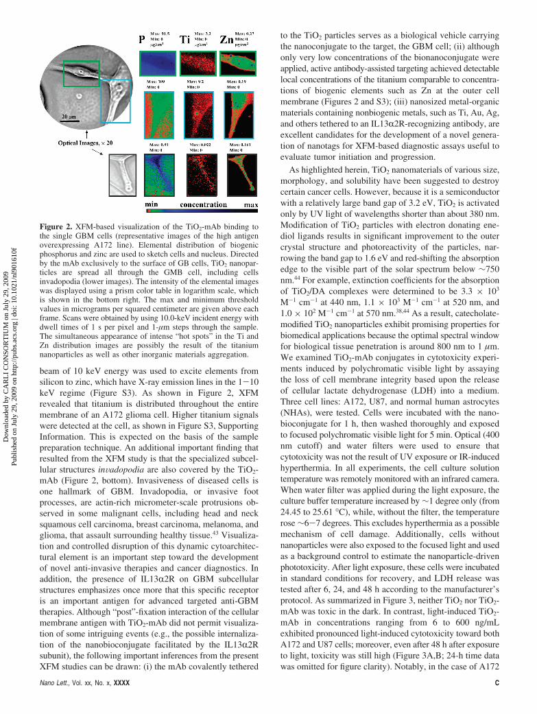
beam of 10 keV energy was used to excite elements fromsilicon to zinc, which have X-ray emission lines in the 1-10keV regime (Figure S3). As shown in Figure 2, XFMrevealed that titanium is distributed throughout the entiremembrane of an A172 glioma cell. Higher titanium signalswere detected at the cell, as shown in Figure S3, SupportingInformation. This is expected on the basis of the samplepreparation technique. An additional important finding thatresulted from the XFM study is that the specialized subcel-lular structures inVadopodia are also covered by the TiO2-mAb (Figure 2, bottom). Invasiveness of diseased cells isone hallmark of GBM. Invadopodia, or invasive footprocesses, are actin-rich micrometer-scale protrusions ob-served in some malignant cells, including head and necksquamous cell carcinoma, breast carcinoma, melanoma, andglioma, that assault surrounding healthy tissue.43 Visualiza-tion and controlled disruption of this dynamic cytoarchitec-tural element is an important step toward the developmentof novel anti-invasive therapies and cancer diagnostics. Inaddition, the presence of IL13R2R on GBM subcellularstructures emphasizes once more that this specific receptoris an important antigen for advanced targeted anti-GBMtherapies. Although “post”-fixation interaction of the cellularmembrane antigen with TiO2-mAb did not permit visualiza-tion of some intriguing events (e.g., the possible internaliza-tion of the nanobioconjugate facilitated by the IL13R2Rsubunit), the following important inferences from the presentXFM studies can be drawn: (i) the mAb covalently tethered
to the TiO2 particles serves as a biological vehicle carryingthe nanoconjugate to the target, the GBM cell; (ii) althoughonly very low concentrations of the bionanoconjugate wereapplied, active antibody-assisted targeting achieved detectablelocal concentrations of the titanium comparable to concentra-tions of biogenic elements such as Zn at the outer cellmembrane (Figures 2 and S3); (iii) nanosized metal-organicmaterials containing nonbiogenic metals, such as Ti, Au, Ag,and others tethered to an IL13R2R-recognizing antibody, areexcellent candidates for the development of a novel genera-tion of nanotags for XFM-based diagnostic assays useful toevaluate tumor initiation and progression.
As highlighted herein, TiO2 nanomaterials of various size,morphology, and solubility have been suggested to destroycertain cancer cells. However, because it is a semiconductorwith a relatively large band gap of 3.2 eV, TiO2 is activatedonly by UV light of wavelengths shorter than about 380 nm.Modification of TiO2 particles with electron donating ene-diol ligands results in significant improvement to the outercrystal structure and photoreactivity of the particles, nar-rowing the band gap to 1.6 eV and red-shifting the absorptionedge to the visible part of the solar spectrum below ∼750nm.44 For example, extinction coefficients for the absorptionof TiO2/DA complexes were determined to be 3.3 × 10³
M-1 cm-1 at 440 nm, 1.1 × 10³ M-1 cm-1 at 520 nm, and1.0 × 102 M-1 cm-1 at 570 nm.38,44 As a result, catecholate-modified TiO2 nanoparticles exhibit promising properties forbiomedical applications because the optimal spectral windowfor biological tissue penetration is around 800 nm to 1 µm.We examined TiO2-mAb conjugates in cytotoxicity experi-ments induced by polychromatic visible light by assayingthe loss of cell membrane integrity based upon the releaseof cellular lactate dehydrogenase (LDH) into a medium.Three cell lines: A172, U87, and normal human astrocytes(NHAs), were tested. Cells were incubated with the nano-bioconjugate for 1 h, then washed thoroughly and exposedto focused polychromatic visible light for 5 min. Optical (400nm cutoff) and water filters were used to ensure thatcytotoxicity was not the result of UV exposure or IR-inducedhyperthermia. In all experiments, the cell culture solutiontemperature was remotely monitored with an infrared camera.When water filter was applied during the light exposure, theculture buffer temperature increased by ∼1 degree only (from24.45 to 25.61 °C), while, without the filter, the temperaturerose ∼6-7 degrees. This excludes hyperthermia as a possiblemechanism of cell damage. Additionally, cells withoutnanoparticles were also exposed to the focused light and usedas a background control to estimate the nanoparticle-drivenphototoxicity. After light exposure, these cells were incubatedin standard conditions for recovery, and LDH release wastested after 6, 24, and 48 h according to the manufacturer’sprotocol. As summarized in Figure 3, neither TiO2 nor TiO2-mAb was toxic in the dark. In contrast, light-induced TiO2-mAb in concentrations ranging from 6 to 600 ng/mLexhibited pronounced light-induced cytotoxicity toward bothA172 and U87 cells; moreover, even after 48 h after exposureto light, toxicity was still high (Figure 3A,B; 24-h time datawas omitted for figure clarity). Notably, in the case of A172
Figure 2. XFM-based visualization of the TiO2-mAb binding tothe single GBM cells (representative images of the high antigenoverexpressing A172 line). Elemental distribution of biogenicphosphorus and zinc are used to sketch cells and nucleus. Directedby the mAb exclusively to the surface of GB cells, TiO2 nanopar-ticles are spread all through the GMB cell, including cellsinvadopodia (lower images). The intensity of the elemental imageswas displayed using a prism color table in logarithm scale, whichis shown in the bottom right. The max and minimum thresholdvalues in micrograms per squared centimeter are given above eachframe. Scans were obtained by using 10.0-keV incident energy withdwell times of 1 s per pixel and 1-µm steps through the sample.The simultaneous appearance of intense “hot spots” in the Ti andZn distribution images are possibly the result of the titaniumnanoparticles as well as other inorganic materials aggregation.
Nano Lett., Vol. xx, No. x, XXXX C
Dow
nloa
ded
by C
AR
LI
CO
NSO
RT
IUM
on
July
29,
200
9Pu
blis
hed
on J
uly
29, 2
009
on h
ttp://
pubs
.acs
.org
| do
i: 10
.102
1/nl
9016
10f

cells with high IL13R2R overexpression, the toxicity had aconcentration-dependent character and reached its maximum
of 80% 6 h after exposure to light, whereas the photoin-duced toxicity toward U87 cells with low receptor-expressionreached a plateau around ∼50% and did not changesignificantly with increasing nanoconjugate concentration.This observation correlates well with prior binding experi-ments, and can be explained assuming saturation of all vacantIL13R2 receptors on U87 cells by the lower concentrationsof the conjugate. It should be pointed out that TiO2 particlestethered to the isotypical immunoglobulin were unable torecognize IL13R2R and did not show notable cytotoxicity,as shown in Figure 3A,B. It is also significant that TiO2-mAb conjugate did not show cytotoxicity toward NHAs(Figure 3C). Passive transport of free TiO2 nanoparticlesresulted in some light-induced cytotoxicity (maximum∼15%) for all cells tested (see Figure 3 A-C). This factclearly demonstrates the benefits of precisely controlledactive transport of nanoparticles to the target cells in contrastto passive nanomaterial-cancer cell interfacing.
It is well established that UV-photoexcitation of TiO2
in an aqueous solution results in the formation of variousROS, such as hydroxyl (OH) and peroxy (HO2) radicals,superoxide anions (O2
-), hydrogen peroxide (H2O2), andsinglet oxygen (1O2).45-49 Recently, using the spin-trapelectron paramagnetic resonance (EPR) and radical-inducedfluorescence techniques, we demonstrated that formation ofROS arises from mechanically distinct multiple redoxchemistries on the surface of DA-modified and bare TiO2
particles.50 The major ROS produced upon illumination ofTiO2/DA or TiO2 linked to a biomolecule through a DAbridge detected was the superoxide anion, formed by reactionof photogenerated electrons with molecular oxygen, whilethe measured yield of 1O2 was very low compared to bareTiO2.50 In this study, we investigated whether the observedcytotoxicity was mediated by photoinduced generation ofROS and whether our previous “pure” physicochemical inVitro and cellular in situ studies are consistent. We foundthat cellular LDH release was remarkably inhibited by 10unit/mL superoxide dismutase (SOD; superoxide naturalscavenger), or 2 mM sodium azide (singlet oxygen and OH•
trap), somewhat less inhibited by 100 unit/mL catalase(quencher of hydrogen peroxide), and only partially sup-pressed by 50 mM mannitol (a quencher of hydroxylradicals), as shown in Figure 3D. Unlike sodium azide,histidine (another singlet oxygen quencher) only inhibitedthe cytotoxicity moderately, (refer to Figure 3D). The higherefficiency of azide in singlet oxygen quenching probablyarises from its large rate constant, which in vitro is 2 ordersof magnitude higher than for histidine.51 These results suggestthat, under illumination with visible light, the photoinducedcytotoxicity of TiO2 tethered to mAb through a DOPAClinker is mediated by oxygen-centered active radicals. Takinginto account our recent EPR studies50 and the results ofcurrent cell toxicity experiments, we believe that the super-oxide anion is the primary ROS responsible for cellmembrane damage, permeability changes, and cell death.Further radical reactions of superoxide can lead to singletoxygen, hydrogen peroxide, and hydroxyl radical forma-tion.
Figure 3. Phototoxicity of the TiO2-mAb toward (A) A172 GBMcells (high ILR2R expression) (B) U87 GBM cells (low ILR2Rexpression), (C) NHAs, and (D) A172 cells in the presence ofvarious free oxygen-centered radical quenchers. Isotype-matchednegative control antibody immunoglobulin IgG1, either conjugatedor unconjugated, did not recognize isolated or cellular IL13a2Rand did not show photoinduced toxicity.
D Nano Lett., Vol. xx, No. x, XXXX
Dow
nloa
ded
by C
AR
LI
CO
NSO
RT
IUM
on
July
29,
200
9Pu
blis
hed
on J
uly
29, 2
009
on h
ttp://
pubs
.acs
.org
| do
i: 10
.102
1/nl
9016
10f
Identification of the ROS involved in the light-inducedtoxicity is important because it reveals mechanisms of celldeath allowing rational development of cancer therapeutics.The formation of relatively short-lived (nanoseconds) ROSwith relatively low diffusion lengths, e.g. hydroxyl radicals(<20 nm) and singlet oxygen (20-220 nm in a cell), canresult in necrosis.52,53 On the other hand, ROS with longerhalf-lifetimes (milliseconds), such as O2
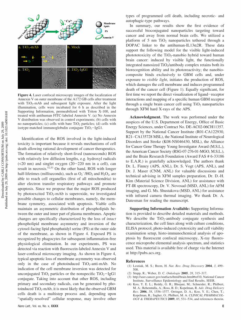
-/HO2 and H2O2, areable to reach cell organelles (first of all mitochondria) toalter electron transfer respiratory pathways and promoteapoptosis. Since we propose that the major ROS producedby photoexcited TiO2-mAb is superoxide, we investigatedpossible changes to cellular membranes, namely, the mem-brane symmetry, associated with apoptosis. Viable cellsmaintain an asymmetric distribution of phospholipids be-tween the outer and inner part of plasma membranes. Apopticchanges are specifically characterized by the loss of intactphospholipid membrane asymmetry and inversion of thecytosol-facing lipid phosphatidyl serine (PS) at the outer sideof the membrane, as shown in Figure 4. Exposed PS isrecognized by phagocytes for subsequent inflammation-freephysiological elimination. In our experiments, PS wasdetected via reaction with fluorescein-labeled Annexin V andlaser-confocal microscopy imaging. As shown in Figure 4,typical apoptotic loss of membrane asymmetry was observedonly in the case of photoinduced TiO2-anti-mAb. Noindication of the cell membrane inversion was detected forunconjugated TiO2 particles or the nonspecific TiO2-IgG1conjugate. Taking into account that other ROS, includingprimary and secondary radicals, can be generated by pho-toinduced TiO2-mAb, it is most likely that the observed GBMcells death is a multistep process and, depending upon“spatially-resolved” cellular response, may involve other
types of programmed cell death, including necrotic- andautophagic-type pathways.
In summary, our results show the first evidence ofsuccessful bioconjugated nanoparticles targeting towardcancer and away from normal brain cells. We utilized aplatform of 5 nm TiO2 nanoparticles tethered through aDOPAC linker to the antihuman-IL13R2R. These datasupport the following model for the visible light-inducedphotototoxicity of the TiO2-nanobio hybrid toward humanbrain cancer: induced by visible light, the functionallyintegrated nanosized TiO2/antibody complex retains both itsbiorecognition ability and its photoreactivity; the nanobio-composite binds exclusively to GBM cells and, underexposure to Visible light, initiates the production of ROS,which damages the cell membrane and induces programmeddeath of the cancer cell (Figure 1). Equally significant, forfirst time we report the direct visualization of ligand-receptorinteractions and mapping of a specific human GBM receptorthrough a single brain cancer cell using TiO2 nanoparticlesthrough XFM hard X-rays of the APS.
Acknowledgment. The work was performed under theauspices of the U.S. Department of Energy, Office of BasicEnergy Sciences, under Contract No. DE-AC02-06CH11357.Support by the National Cancer Institute (R01-CA122930,R21- CA135728 MSL), the National Institute of NeurologicalDisorders and Stroke (K08-NS046430, MSL), the Alliancefor Cancer Gene Therapy Young Investigator Award (M.S.L.),the American Cancer Society (RSG-07-276-01-MGO, M.S.L.),and the Brain Research Foundation (Award FAS # 6-33186to E.A.R.) is gratefully acknowledged. The authors thankDr. L. Finney (APS, ANL), Dr. S. Vogt (APS, ANL), andDr. J. Maser (CNM, ANL) for valuable discussions andtechnical advising in XFM samples preparation, Dr. D.-H.Kim (Material Science Division, ANL) for assistance withFT-IR spectroscopy, Dr. V. Novosad (MSD, ANL) for AFMimaging, and G. Ms. Shustakova (MSD, ANL) for assistancewith infrared camera thermal imaging. We thank Dr. A.Datesman for reading the manuscript.
Supporting Information Available: Supporting Informa-tion is provided to describe detailed materials and methods.We describe the TiO2-antibody conjugate synthesis andcharacterization, the cell lines along with culture conditions,ELISA protocol, photo-induced cytotoxicity and cell viabilityexamination setup, histo-immunochemical analysis of apo-ptosis by fluorescent confocal microscopy, X-ray fluores-cence microprobe elemental analysis spectrum, and statisticsused. This material is available free of charge via the Internetat http://pubs.acs.org.
References(1) Lesniak, M. S.; Brem, H. Nat. ReV. Drug DiscoVery 2004, 3, 499–
508.(2) Stupp, R.; Weber, D. C. Onkologie 2005, 28, 315–317.(3) http://seer.cancer.gov/statfacts/html/brain.html#ref10, National Cancer
Institute, Surveillance Epidemiology and End Results, SEER.(4) Koo, Y. E. L.; Reddy, G. R.; Bhojani, M.; Schneider, R.; Philbert,
M. A.; Rehemtulla, A.; Ross, B. D.; Kopelman, R. AdV. Drug DeliVeryReV. 2006, 58, 1556–1577. Orringer, D. A.; Koo, Y. E.; Chen, T.;Kopelman, R.; Sagher, O.; Philbert, M. A. CLINICAL PHARMACOL-OGY & THERAPEUTICS 2009, 85, 531–534, and references therein.
Figure 4. Laser confocal microscopy images of the localization ofAnnexin V on outer membrane of the A172 GB cells after treatmentwith TiO2-mAb and subsequent light exposure. After the lightillumination, cells were incubated for 6 h as described in theSupporting Information, permeabilized with Triton X-100, andtreated with antihuman FITC-labeled Annexin V. (a) No AnnexinV distribution was observed in control experiments; (b) cells withno nanoparticles; (c) cells with bare TiO2 particles; (d) cells withisotype-matched immunoglobulin conjugate TiO2-IgG1.
Nano Lett., Vol. xx, No. x, XXXX E
Dow
nloa
ded
by C
AR
LI
CO
NSO
RT
IUM
on
July
29,
200
9Pu
blis
hed
on J
uly
29, 2
009
on h
ttp://
pubs
.acs
.org
| do
i: 10
.102
1/nl
9016
10f

(5) Szaciowski, K.; Macyk, W.; Drzewiecka-Matuszek, A.; Brindell, M.;Stochel, G. Chem. ReV. 2005, 105, 2647–2694.
(6) Sunada, K.; Watanabe, T.; Hashimoto, K. J. Photochem. Photobiol.A: Chem. 2003, 156, 227–233.
(7) Sokmen, M.; Candan, F.; Sumer, Z. J. Photochem. Photobiol. A: Chem.2001, 143, 241–244.
(8) Theron, J.; Walker, J. A.; Cloete, T. E. Crit. ReV. Microbiol. 2008,34, 43–69.
(9) Amezaga-Madrid, P.; Nevarez-Moorillon, G. V.; Orrantia-Borunda,E.; Miki-Yoshida, M. FEMS Microbiol. Lett. 2002, 211, 183–188.
(10) Shah, R. R.; Kaewgun, S.; Lee, B. I.; Tzeng, T.-R. J. J. Biomed.Nanotechnol. 2008, 3, 339–348.
(11) Linkous, C. A.; Carter, G. J.; Locuson, D. B.; Ouellette, A. J.; Slattery,D. K.; Smitha, L. A. EnViron. Sci. Technol. 2000, 34, 4754–4758.
(12) Yamaguchi, K.; Sugiyama, T.; Kato, S.; Kondo, Y.; Ageyama, N.;Kanekiyo, M.; Iwata, M.; Koyanagi, Y.; Yamamoto, N.; Honda, M.;et al. J. Med. Virol. 2008, 80, 1322–1331.
(13) Cai, R.; Hashimoto, K.; Itoh, K.; Kubota, Y.; Fujishima, A. Bull. Chem.Soc. Jpn. 1991, 64, 1268–1273.
(14) Cai, R. X.; Kubota, Y.; Shuin, T.; Sakai, H.; Hashimoto, K.; Fujishima,A. Cancer Res. 1992, 52, 2346–2348.
(15) Sakai, H.; Ito, E.; Cai, R. X.; Yoshioka; Kubota, Y.; Hashimoto, K.;Fujishima, A. Biochim. Biophys. Acta 1994, 1201, 259–265.
(16) Kubota, Y.; Shuin, T.; Kawasaki, C.; Hosaka, M.; Kitamura, H.; Cai,R.; Sakai, H.; Hashimoto, K.; Fujishima, A. Br. J. Cancer 1994, 70,1107–1111.
(17) Sakai, H.; Baba, R.; Hashimoto, K.; Kubota, Y.; Fujishima, A. Chem.Lett. 1995, 3, 185–186.
(18) Lu, P.-J.; Ho, I.-C.; Lee, T.-C. Mutat. Res., Genet. Toxicol. EnViron.Mutagen. 1998, 414, 15–20.
(19) Uchino, T.; Tokunaga, H.; Ando, M.; Utsumi, H. Toxicol. in Vitro2002, 16, 629–635.
(20) Seo, J. W.; Chung, H.; Kim, M. Y.; Lee, J.; Choi, I. H.; Cheon, J.Small 2007, 3, 850–853.
(21) Juan, X.; Yi, S.; Junjie, H.; Chunmei, C.; Guoyuan, L.; Yan, J.;Yaomin, Z.; Zhiyu, J. Bioelectrochemistry 2007, 71, 217–222.
(22) Kalbacova, M.; Macak, J. M.; Schmidt-Stein, F.; Mierke, C. T.;Schmuki, P. Phys. Status Solidi RRL: Rapid Res. Lett. 2008, 2, 194–196.
(23) Peer, D.; Karp, J. M.; Hong, S.; FarokHzad, O. C.; Margalit, R.;Langer, R. Nat. Nanotechnol. 2007, 2, 751–760.
(24) Debinski, W.; Gibo, D. M. Mol. Med. 2000, 6, 440–449.(25) Mintz, A.; Gibo, D. M.; Slagle-Webb, B.; Christensen, N. D.; Debinski,
W. Neoplasia 2002, 4, 388–399.(26) Kawakami, K.; Taguchi, J.; Murata, T.; Puri, R. K. Blood 2001, 97,
2673–2679.(27) Kawakami, K.; Takeshita, F.; Puri, R. K. J. Biol. Chem. 2001, 276,
25114–25120.(28) Debinski, W.; Gibo, D.; Hulet, S.; Connor, J.; Gillespie, G. Cancer
Res. 1999, 5, 985–990.
(29) Kawakami, K.; Kawakami, M.; Snoy, P. J.; Husain, S. R.; Puri, R. K.J. Exp. Med. 2001, 194, 1743–1754.
(30) Wykosky, J.; Gibo, D. M.; Stanton, C.; Debinski, W. Clin. CancerRes. 2008, 14, 199–208.
(31) Zhou, G.; Yet, G. J.; Debinski, W.; Roizman, B. Proc. Natl. Acad.Sci. U.S.A. 2002, 99, 15124–15129.
(32) Bernardi, R. J.; Lowery, A. R.; Thompson, P. A.; Blaney, S. M.; West,J. L. J. Neurooncol. 2008, 86, 165–172.
(33) Ferrari, M. Nat. ReV. Cancer 2005, 5, 161–171.(34) Rajh, T.; Tiede, D. M.; Thurnauer, M. C. J. Non-Cryst. Solids 1996,
207, 815–820.(35) O’Regan, B.; Gratzel, M. Nature 1991, 353, 737–740.(36) Rajh, T.; Chen, L. X.; Lukas, K.; Liu, T.; Thurnauer, M. C.; Tiede,
D. M. J. Phys. Chem. B 2002, 106, 10543–10552.(37) Duncan, W. R.; Prezhdo, O. V. Annu. ReV. Phys. Chem. 2007, 58,
143–184.(38) Rajh, T.; Saponjic, Z.; Liu, J.; Dimitrijevic, N. M.; Scherer, N. F.;
Vega-Arroyo, M.; Zapol, P.; Curtiss, L. A.; Thurnauer, M. C. NanoLett. 2004, 4, 1017–1023.
(39) Dimitrijevic, N. M.; Saponjic, Z. V.; Rabatic, B. M.; Rajh, T. J. Am.Chem. Soc. 2005, 127, 1344–1345.
(40) Kemner, K. M.; Kelly, S. D.; Lai, B.; Maser, J.; O’Loughlin, E. J.;Sholto-Douglas, D.; Cai, Z. H.; Schneegurt, M. A.; Kulpa, C. F.;Nealson, K. H. Science 2004, 306, 686–687.
(41) Paunesku, T.; Rajh, T.; Wiederrecht, G.; Maser, J.; Vogt, S.; Stojicevic,N.; Protic, M.; Lai, B.; Oryhon, J.; Thurnauer, M.; Woloschak, G.Nat. Mater. 2003, 2, 343–346.
(42) Finney, L.; Mandava, S.; Ursos, L.; Zhang, W.; Rodi, D.; Vogt, S.;Legnini, D.; Maser, J.; Ikpatt, F.; Olopade, O. I.; Glesne, D. Proc.Natl. Acad. Sci. U.S.A. 2007, 104, 2247–2252.
(43) Stylli, S. S.; Kaye, A. H.; Lock, P. J. Clin. Neurosci. 2008, 15, 725–737.
(44) de la Garza, L.; Saponjic, Z. V.; Dimitrijevic, N. M.; Thurnauer, M. C.;Rajh, T. J. Phys. Chem. B 2006, 110, 680–686.
(45) Hoffmann, M. R.; Martin, S. T.; Choi, W.; Bahnemann, D. W. Chem.ReV. 1995, 95, 69–96.
(46) Mills, A.; Le Hunte, S. J. Photochem. Photobiol. A: Chem. 1997, 108,1–35.
(47) Schwarz, P. F.; Turro, N. J.; Bossmann, S. H.; Braun, A. M.; Wahab,A.-M. A. A.; Durr, H. J. Phys. Chem. B 1997, 101, 7127–7134.
(48) Daimon, T.; Nosaka, Y. J. Phys. Chem. C 2007, 111, 4420–4424.(49) Tachikawa, T.; Majima, T. J. Fluoresc. 2007, 17, 727–738.(50) Dimitrijevic, N. M.; Rozhkova, E. A.; Rajh, T. J. Am. Chem. Soc.
2009, 131, 2893–2899.(51) Basumodak, S.; Tyrrell, R. M. Cancer Res. 1993, 53, 4505–4510.(52) Redmond, R. W.; Kochevar, I. E. Photochem. Photobiol. 2006, 82,
1178–1186.(53) Schweitzer, C.; Schmidt, R. Chem. ReV. 2003, 103, 1685–1757.
NL901610F
F Nano Lett., Vol. xx, No. x, XXXX
Dow
nloa
ded
by C
AR
LI
CO
NSO
RT
IUM
on
July
29,
200
9Pu
blis
hed
on J
uly
29, 2
009
on h
ttp://
pubs
.acs
.org
| do
i: 10
.102
1/nl
9016
10f
Related Documents