A hedgehog survival pathway in ‘undead’ lipotoxic hepatocytes Keisuke Kakisaka, Sophie C. Cazanave, Nathan W. Werneburg, Nataliya Razumilava, Joachim C. Mertens, Steve F. Bronk, Gregory J. Gores ⇑ Division of Gastroenterology and Hepatology, College of Medicine, Mayo Clinic, Rochester, MN, United States Background & Aims: Ballooned hepatocytes in non-alcoholic steatohepatitis (NASH) generate sonic hedgehog (SHH). This observation is consistent with a cellular phenotype in which the cell death program has been initiated but cannot be executed. Our aim was to determine whether ballooned hepatocytes have potentially disabled the cell death execution machinery, and if so, can their functional biology be modeled in vitro. Methods: Immunohistochemistry was performed on human NASH specimens. In vitro studies were performed using HuH-7 cells with shRNA targeted knockdown of caspase 9 (shC9 cells) or primary hepatocytes from caspase 3 / mice. Results: Ballooned hepatocytes in NASH display diminished expression of caspase 9. This phenotype was modeled using shC9 cells; these cells were resistant to lipoapoptosis by palmi- tate (PA) or lysophosphatidylcholine (LPC) despite lipid droplet formation. During lipid loading by either PA or LPC, shC9 cells activate JNK which induces SHH expression via AP-1. An auto- crine hedgehog survival signaling pathway was further delin- eated in both shC9 and caspase 3 / cells during lipotoxic stress. Conclusions: Ballooned hepatocytes in NASH downregulate cas- pase 9, a pivotal caspase executing the mitochondrial pathway of apoptosis. Hepatocytes engineered to reduce caspase 9 expres- sion are resistant to lipoapoptosis, in part, due to a hedgehog autocrine survival signaling pathway. Ó 2012 European Association for the Study of the Liver. Published by Elsevier B.V. All rights reserved. Introduction Human hepatocellular injury is ubiquitous due to the prevalence of steatohepatitis syndromes, viral hepatitis, immune mediated insults, genetic diseases, etc. [1]. In these hepatic diseases, hepa- tocyte fate is often modeled as a binary process – cells either sur- vive or die to be replaced by liver regenerative processes [2]. Yet, cells may resist cell death by downregulating or inhibiting cell death programs, thereby surviving in an altered state. This new cell fate does not merely reflect an escape from death, but rather results in the formation of a unique, functional cell phenotype. The cell phenotype of non-lethal cell injury has been best charac- terized in model systems where it has been termed the ‘undead cell’ [3,4]. Because cell death by proapoptotic stimuli requires activation of executioner caspases [5], the undead cell can be engineered by deletion of these downstream caspases. For exam- ple, in Drosophila melanogaster genetic deletion of the executioner caspase DrICE or Dcp-1 results in a cell type in which otherwise toxic stimuli activate alternative cell signaling cascades produc- ing signals which enhance tissue remodeling [3,4,6]. A candidate undead cell in the liver is the ballooned hepato- cyte in non-alcoholic steatohepatitis (NASH). Ballooned hepato- cytes are thought to be important in the pathogenesis of NASH and the presence of such cells is used to score disease severity [7,8]. Little is known about ballooned hepatocytes other than they are enlarged cells which have lost cell polarity, frequently contain Mallory–Denk bodies, store neutral triglycerides, contain oxidized phospholipids, no longer stain for cytokeratin 18, and generate the ligand sonic hedgehog [8–10]. The seminal work by Diehl and colleagues demonstrating sonic hedgehog (SHH) generation by ballooned hepatocytes suggests this cell may, in fact, be a functional cell with an altered phenotype [8,11]. For example, in D. melanogaster retinal cells in which the cell death program is initiated but cannot be executed also generate SHH [6]. The mechanisms by which undead cells continue to evade cell death remain elusive, but given the potency of SHH as a sur- vival factor, it is possible that this ligand serves as an autocrine survival factor. Herein, we demonstrate that ballooned cells have reduced expression of a potent downstream caspase, caspase 9, suggest- ing they may not efficiently execute cell death pathways. To understand the functional implications of this observation, we modeled lipotoxic stress in HuH-7 cells in which caspase 9 had been knocked down by short hairpin RNA technology (shC9 cells). Knockdown of caspase 9 protected hepatocytes from lipotoxicity by the saturated free fatty acid (FFA), palmitate (PA) or the Journal of Hepatology 2012 vol. 57 j 844–851 Keywords: Caspase 3; c-Jun-N-terminal kinase (JNK); Lipoapoptosis; Lysophos- phatidylcholine; Palmitate. Received 30 March 2012; received in revised form 11 May 2012; accepted 21 May 2012, available online 26 May 2012 ⇑ Corresponding author. Address: College of Medicine, Mayo Clinic, 200 First Street SW, Rochester, MN 55905, United States. Tel.: +1 507 284 0686; fax: +1 507 284 0762. E-mail address: [email protected] (G.J. Gores). Abbreviations: NASH, non-alcoholic steatohepatitis; SHH, sonic hedgehog; FFA, free fatty acids; PA, palmitate; LPC, lysophosphatidylcholine; JNK, c-Jun N-ter- minal kinase; shRNA, short-hairpin RNA; MPH, mouse primary hepatocytes; D- API, 4 0 ,6-diamidine-2’-phenylindole dihydrochloride; EMSA, electrophoretic mobility shift assay; ChIP, chromatin immunoprecipitation; OA, oleate; C3 / , caspase 3 genetic knockout; AP-1, activator protein-1; ACOX-1, acyl-CoA oxidase 1, palmitoyl; CPT, carnitine palmitoyltransferase; ACACA, acetyl-CoA carboxylase alpha; ACACB, acetyl-CoA carboxylase beta. Research Article

Welcome message from author
This document is posted to help you gain knowledge. Please leave a comment to let me know what you think about it! Share it to your friends and learn new things together.
Transcript
Research Article
A hedgehog survival pathway in ‘undead’ lipotoxic hepatocytes
Keisuke Kakisaka, Sophie C. Cazanave, Nathan W. Werneburg, Nataliya Razumilava,Joachim C. Mertens, Steve F. Bronk, Gregory J. Gores⇑
Division of Gastroenterology and Hepatology, College of Medicine, Mayo Clinic, Rochester, MN, United States
Background & Aims: Ballooned hepatocytes in non-alcoholic Introduction
steatohepatitis (NASH) generate sonic hedgehog (SHH). Thisobservation is consistent with a cellular phenotype in whichthe cell death program has been initiated but cannot be executed.Our aim was to determine whether ballooned hepatocytes havepotentially disabled the cell death execution machinery, and ifso, can their functional biology be modeled in vitro.Methods: Immunohistochemistry was performed on humanNASH specimens. In vitro studies were performed using HuH-7cells with shRNA targeted knockdown of caspase 9 (shC9 cells)or primary hepatocytes from caspase 3�/� mice.Results: Ballooned hepatocytes in NASH display diminishedexpression of caspase 9. This phenotype was modeled usingshC9 cells; these cells were resistant to lipoapoptosis by palmi-tate (PA) or lysophosphatidylcholine (LPC) despite lipid dropletformation. During lipid loading by either PA or LPC, shC9 cellsactivate JNK which induces SHH expression via AP-1. An auto-crine hedgehog survival signaling pathway was further delin-eated in both shC9 and caspase 3�/� cells during lipotoxic stress.Conclusions: Ballooned hepatocytes in NASH downregulate cas-pase 9, a pivotal caspase executing the mitochondrial pathwayof apoptosis. Hepatocytes engineered to reduce caspase 9 expres-sion are resistant to lipoapoptosis, in part, due to a hedgehogautocrine survival signaling pathway.� 2012 European Association for the Study of the Liver. Publishedby Elsevier B.V. All rights reserved.Journal of Hepatology 20
Keywords: Caspase 3; c-Jun-N-terminal kinase (JNK); Lipoapoptosis; Lysophos-phatidylcholine; Palmitate.Received 30 March 2012; received in revised form 11 May 2012; accepted 21 May2012, available online 26 May 2012⇑ Corresponding author. Address: College of Medicine, Mayo Clinic, 200 FirstStreet SW, Rochester, MN 55905, United States. Tel.: +1 507 284 0686; fax: +1 507284 0762.E-mail address: [email protected] (G.J. Gores).Abbreviations: NASH, non-alcoholic steatohepatitis; SHH, sonic hedgehog; FFA,free fatty acids; PA, palmitate; LPC, lysophosphatidylcholine; JNK, c-Jun N-ter-minal kinase; shRNA, short-hairpin RNA; MPH, mouse primary hepatocytes; D-API, 40 ,6-diamidine-2’-phenylindole dihydrochloride; EMSA, electrophoreticmobility shift assay; ChIP, chromatin immunoprecipitation; OA, oleate; C3�/�,caspase 3 genetic knockout; AP-1, activator protein-1; ACOX-1, acyl-CoA oxidase1, palmitoyl; CPT, carnitine palmitoyltransferase; ACACA, acetyl-CoA carboxylasealpha; ACACB, acetyl-CoA carboxylase beta.
Human hepatocellular injury is ubiquitous due to the prevalenceof steatohepatitis syndromes, viral hepatitis, immune mediatedinsults, genetic diseases, etc. [1]. In these hepatic diseases, hepa-tocyte fate is often modeled as a binary process – cells either sur-vive or die to be replaced by liver regenerative processes [2]. Yet,cells may resist cell death by downregulating or inhibiting celldeath programs, thereby surviving in an altered state. This newcell fate does not merely reflect an escape from death, but ratherresults in the formation of a unique, functional cell phenotype.The cell phenotype of non-lethal cell injury has been best charac-terized in model systems where it has been termed the ‘undeadcell’ [3,4]. Because cell death by proapoptotic stimuli requiresactivation of executioner caspases [5], the undead cell can beengineered by deletion of these downstream caspases. For exam-ple, in Drosophila melanogaster genetic deletion of the executionercaspase DrICE or Dcp-1 results in a cell type in which otherwisetoxic stimuli activate alternative cell signaling cascades produc-ing signals which enhance tissue remodeling [3,4,6].
A candidate undead cell in the liver is the ballooned hepato-cyte in non-alcoholic steatohepatitis (NASH). Ballooned hepato-cytes are thought to be important in the pathogenesis of NASHand the presence of such cells is used to score disease severity[7,8]. Little is known about ballooned hepatocytes other thanthey are enlarged cells which have lost cell polarity, frequentlycontain Mallory–Denk bodies, store neutral triglycerides, containoxidized phospholipids, no longer stain for cytokeratin 18, andgenerate the ligand sonic hedgehog [8–10]. The seminal workby Diehl and colleagues demonstrating sonic hedgehog (SHH)generation by ballooned hepatocytes suggests this cell may, infact, be a functional cell with an altered phenotype [8,11]. Forexample, in D. melanogaster retinal cells in which the cell deathprogram is initiated but cannot be executed also generate SHH[6]. The mechanisms by which undead cells continue to evadecell death remain elusive, but given the potency of SHH as a sur-vival factor, it is possible that this ligand serves as an autocrinesurvival factor.
Herein, we demonstrate that ballooned cells have reducedexpression of a potent downstream caspase, caspase 9, suggest-ing they may not efficiently execute cell death pathways. Tounderstand the functional implications of this observation, wemodeled lipotoxic stress in HuH-7 cells in which caspase 9 hadbeen knocked down by short hairpin RNA technology (shC9 cells).Knockdown of caspase 9 protected hepatocytes from lipotoxicityby the saturated free fatty acid (FFA), palmitate (PA) or the
12 vol. 57 j 844–851
JOURNAL OF HEPATOLOGY
phospholipid lysophosphatidylcholine (LPC). Interestingly, PA orLPC treated shC9, but not wild type cells, generate sonic hedge-hog by a c-Jun-N-terminal kinase (JNK)-dependent pathway.Blockade of hedgehog signaling resulted in cell death of shC9cells following exposure to lipotoxic agents. Similar observationswere confirmed in primary caspase 3�/� mouse hepatocytes. Wespeculate that these engineered cells model the functional phe-notype of ballooned hepatocytes, albeit not the specific morphol-ogy, and, in turn, support the concept of ballooned hepatocytes asan altered, but functional cell population which has escaped celldeath.Materials and methods
Immunohistochemistry (IHC)
Formalin-fixed, paraffin-embedded liver sections (5 lm thick) from threepatients with non-alcoholic steatohepatitis (NASH) were obtained followingapproval by the Institutional Review Board (IRB), Mayo Clinic, Rochester MN,USA. In conjunction with the clinical core of P30DK084576, the sections werepre-selected by an experienced hepatopathologist for their abundance of bal-looned hepatocytes. These sections were deparaffinized in xylene and rehydratedthrough graded alcohols. After tissue permeabilization in 0.1% Triton X100 for2 min, antigen retrieval was performed by incubation in sodium citrate buffer(0.01 M sodium citrate, 0.05% Tween 20; pH 6.0) for 30 min utilizing a vegetablesteamer. Samples were cooled down, and endogenous peroxidase activity wasquenched by incubation for 15 min in 3% H2O2 (diluted in deionized H2O). Sec-tions were washed in changes of phosphate-buffered saline (PBS), three timesfor 5 min. The VECTASTAIN Elite ABC and ImmPACT VIP peroxidase substrate kits(Vector laboratories, Burlingame, CA, USA) were used in further steps according tomanufacturer’s instructions. After blocking non-specific binding for 1 h, sectionswere incubated with anti-caspase-9 p10 (F-7) mouse monoclonal primary anti-bodies (1:50; sc-17784) or anti-caspase-2L (C-20) goat monoclonal primary anti-bodies (1:50; sc-626-G, Santa Cruz Biotechnology, Santa Cruz, CA, USA) for 30 minat room temperature. Prior to mounting, the sections were counterstained withhematoxylin for 3 min (Sigma–Aldrich, St. Louis, MO, USA). Samples were exam-ined with light microscopy. Analysis of at least 20 cells of each type from eachpatient was conducted using Image J version 1.44 software (NIH, Bethesda, MD,USA). Ballooned cells were confirmed in hematoxylin and eosin (H&E) stainedsections.
Cells
HuH-7 cells, a human hepatocellular carcinoma cell line, were maintained in Dul-becco’s modified Eagle’s medium containing glucose (25 mM) supplemented with10% fetal bovine serum, 100,000 IU/L penicillin and 100 mg/L streptomycin. Wealso employed HuH-7 cells stably expressing a short-hairpin RNA (shRNA) target-ing caspase 9, shC9 cells. Mouse primary hepatocytes (MPH) were isolated fromC57BL/6 wild type (Jackson Laboratory, Bar Harbor, ME, USA) and caspase 3�/�
mice by collagenase perfusion and plated as primary cultures [12]. Developmentand characterization of the caspase 3�/� mice has been previously reported [13].MPH were maintained in Waymouth medium supplemented with 10% fetalbovine serum, 100,000 IU/L penicillin, 100 mg/L streptomycin, and 100 nMinsulin.
Phase contrast, fluorescent and confocal microscopy
HuH-7 cells and shC9 cells were grown on glass bottom culture dishes for phasecontrast and fluorescence microscopy, or glass coverslips for confocal microscopy.The following studies were conducted in conjunction with the microscopy core ofthe P30DK084567. For live cell imaging, lipid droplets were fluorescently labeledby incubation with 100 lg/ml of Bodipy 505/515 (Invitrogen, Camarillo, CA, USA)for 1 min at 37 �C. Identical cells were viewed using phase contrast and then fluo-rescent optics on a Zeiss Axiovert 200 epifluorescent microscope (Zeiss, Thorn-wood, NJ, USA). Fluorescence was visualized using excitation and emissionwavelengths of 488 and 507 nm, respectively. Both phase contrast and fluores-cent digital images were acquired and analyzed using a cooled-CCD Orca II cam-era (Hamamatsu Photonics, Hamamatsu City, Japan) with IPLab software
Journal of Hepatology 201
(Scanalytics, Fairfax, VA, USA). Fluorescent lipid droplet area as a percent of thetotal cellular area was quantified using Image J version 1.44 software (NIH,Bethesda, MD, USA).
In separate studies, cells were fixed with 3% paraformaldehyde for 30 min atroom temperature [14]. Intracellular lipid droplets were labeled with 10 lg/ml ofBodipy 505/515 for 10 min at room temperature, and mounted in Prolong Antif-ade mounting media (Invitrogen, Camarillo, CA, USA). Lipid droplet fluorescenceintensity per cell was quantified using an inverted phase/fluorescence microscope(Axiovert 35M; Carl Zeiss, Inc., Thornwood, NY, USA) and Metafluor quantitativefluorescence imaging software from Universal Imaging Corp. (West Chester, PA,USA) [15]. For confocal microscopy, cells were fixed and labeled as describedabove and visualized using an inverted laser-scanning confocal microscope (ZeissLSM 510, Carl Zeiss, Jena, Germany) with excitation and emission wavelengths of488 and 507 nm, respectively.
For SHH or ubiquitin immunocytochemistry, cells plated on coverslips werefixed with 4% paraformaldehyde in PBS for SHH staining or with cold acetoneon dry ice for ubiquitin staining, respectively, and permeabilized with 0.0125%(w/v) CHAPS in PBS for both stainings. The primary antiserum was rabbit anti-SHH (sc-9024, Santa Cruz Biotechnology, Santa Cruz, CA, USA) at a dilution of1:300 or rabbit anti-ubiquitin (Z0458, Dako, Carpinteria, CA, USA) at a dilutionof 1:500. The secondary antiserum was Alexa Fluor 488-conjugated or 568-con-jugated anti-rabbit IgG (Molecular Probes, Eugene, OR, USA), and ProLong Antif-ade with DAPI (Molecular Probes) was used as mounting medium. Images wereacquired by confocal microscopy employing excitation and emission wavelengthsof 488 and 507 nm for SHH staining or excitation and emission wavelengths of558 and 583 nm for ubiquitin staining. Cells with positive immunoreactivity forthe active conformation of SHH or cells with ubiquitin-punctate staining, respec-tively, were counted and expressed as a percentage of total cells.
Electrophoretic mobility shift assay (EMSA)
EMSA was performed as previously described by us in detail [16]. Briefly,nuclear extracts from shC9 cells were prepared using NE-PER nuclear and cyto-plasmic extraction reagents (Pierce Biotechnology, Rockford, IL, USA) accordingto the manufacturer’s instructions. For the EMSA, 20 lg of nuclear proteins wasincubated at room temperature for 20 min in binding buffer (25 mM HEPES, pH7.5, 0.1 M NaCl, 2 mM EDTA, 6% glycerol, 0.1% Triton X-100, 0.1 mM phenyl-methylsulfonyl fluoride, 0.4 mM dithiothreitol, 0.5 lg/ll poly(dI-dC), 0.5 lg/llsalmon sperm) with 0.04 pmol of CY 5.5-labeled double-stranded DNA oligonu-cleotides containing either one of the putative activator protein-1 (AP-1) bind-ing sequences (BS1) or (BS2). Each sequence with the mutated putative AP-1binding sequence (mutant BS1 or mutant BS2) was also employed as a negativecontrol (Supplementary Table 1). Protein-DNA complexes were separated fromthe unbound DNA probe by electrophoresis through 5% native polyacrylamidegels containing 0.5x Tris borate-EDTA. Fluorescence was visualized directly onthe gel using an Odyssey fluorescent imager (Licor Biosciences, Lincoln, NE,USA). For competition assays, a 200-fold molar excess of unlabeled double-stranded oligonucleotide was added to the reaction mixture 20 min before theaddition of the fluorescent probe. In the supershift assays, nuclear cell extractwas first incubated at room temperature for 25 min with 2 lg of anti-pho-pho-c-Jun antibody (Santa Cruz Biotechnology). The entire antibody/proteinmixture was then incubated with CY 5.5-labeled probe and processed for thegel shift as described above.
Chromatin immunoprecipitaion (ChIP) assay
ChIP assays were performed employing shC9 (6–10 million cells) using a com-mercially available assay (Active Motif, Carlsbad, CA, USA) according to the man-ufacturer’s instructions [17]. Briefly, shC9 cells were treated for 6 h with 800 lMpalmitate or for 4 h with 85 lM LPC; the cells were then fixed with 1% formalde-hyde in PBS at room temperature for 10 min. The formaldehyde crosslinking reac-tion was quenched with 125 mM glycine, the cells were lysed, and chromatin wassheared into small and uniform fragments by an enzymatic reaction. Followingcentrifugation, the DNA concentration in the supernatant was quantified by mea-suring absorbance at 260 nm. Samples containing 30 lg of DNA were next incu-bated with Protein G magnetic beads, ChIP Buffer 1, Protease Inhibitor Cocktail,the phospho-c-Jun antibody at 2 lg and dH2O at 4 �C overnight. The immunecomplex was separated from the sample buffer by a magnetic stand and washedsequentially with ChIP Buffer1 and ChIP Buffer 2. The sample was next elutedwith Elution Buffer AM2 and then added to the Reverse Cross-linking Bufferand heated at 95 �C for 15 min. The proteins were digested by proteinase K,and the DNA in the ChIP sample was used as template for quantitative real-timePCR. The primers used in this assay are depicted in Supplementary Table 2.
2 vol. 57 j 844–851 845
Research Article
Immunoblot analysisWhole cell lysates were prepared as previously described [12]. Nuclear cellextracts were prepared using NE-PER nuclear and cytoplasmic extractionreagents (Pierce Biotechnology, Rockford, IL, USA) according to the manufac-turer’s instructions. An equal amount of protein (50–80 lg) was resolved bySDS–PAGE on a 12.5–18% acrylamide gel, transferred to nitrocellulose mem-branes, and incubated with primary antibodies. Membranes were incubated withappropriate horseradish peroxidase-conjugated secondary antibodies (BiosourceInternational, Camarillo, CA, USA). The bound antibody was visualized using achemiluminescent substrate (ECL; Amersham, Arlington Heights, IL, USA) andexposure to Kodak X-OMAT film (Eastman Kodak, Rochester, NY, USA).
Materials and methods for treatment of hepatocytes with lipids, quantitationof cell death, and quantititative real-time PCR, Gli reporter assay, antibodies andreagents, and statistics are included in the Supplementary data.
Results
Ballooned hepatocytes in NASH display reduced caspase 9 expression
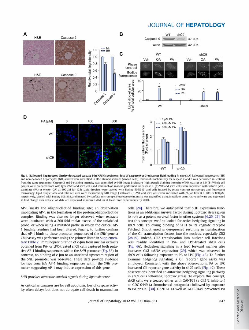
Loss of executioner caspase function is sufficient to prevent celldeath in developmental systems. Therefore, we performed immu-nohistochemistry in human NASH specimens for caspase 9, apotent executioner caspase. Ballooned hepatocytes, but notneighboring hepatocytes, displayed diminished caspase 9 expres-sion (Fig. 1A). In contrast, expression of the upstream caspase 2was readily identified in ballooned hepatocytes (Fig. 1A). Caspase2 has a long prodomain similar to caspase 9, and therefore, servedas a control for a caspase of similar size as caspase 9. These sug-gest ballooned hepatocytes are not merely damaged cells on thepath to cellular demise, but likely are functional cells which havedownregulated cell death pathways.
Diminished cell death in hepatocytes deficient in caspase 9 or 3despite lipid droplet accumulation
We sought to model the ballooned hepatocyte phenotype withdownregulation of caspase 9, and therefore, generated shC9HuH-7 cells (shC9 cells) (Fig. 1B). As assessed by phase contrastmorphology, the shC9 cells did not appear different from the par-ent cell line. The shC9 cells (Fig. 1C), however, actually demon-strated more robust lipid droplet formation as compared towild type cells following incubation with oleate (OA) or palmitate(PA) (Fig. 1C and D). The enhanced lipid loading of shC9 cellscould not be explained by decreased lipolysis as expression ofthe rate limiting enzyme for lipolysis[18,19], acyl-CoA oxidase1, palmitoyl (ACOX-1), or b-oxidation, carnitine palmitoyltrans-ferase 1A and 2 (CPT-1A and -2), acetyl-CoA carboxylase alpha(ACACA) and acetyl-CoA carboxylase beta (ACACB) was notdownregulated in these cells (Supplementary Fig. 1). Ratherenhanced lipid droplet accumulation by shC9 cells likely reflectstheir ability to resist lipoapoptosis by free fatty acids allowingfurther lipid accumulation over time. As the saturated free fattyacid PA induces lipoapoptosis via a metabolite lysophosphatidyl-choline (LPC) [12], we quantified apoptosis in shC9 and wild typecells using both PA and LPC. Using either lipotoxic agent, shC9cells were more resistant to PA or LPC cytotoxicity (Supplemen-tary Fig. 2A–C). To confirm these results in primary hepatocytes,we employed hepatocytes from wild type and caspase 3 deficient(C3�/�) mice. We posited that, given the dominant role of caspase3 as an executioner caspase downstream of caspase 9, these cellsmay also be resistant to lipotoxicity (Supplementary Fig. 3B), and
846 Journal of Hepatology 201
indeed, they were apoptosis resistant, despite lipid droplet accu-mulation (Supplementary Fig. 3A). Finally, because balloonedhepatocytes in the NASH liver display enhanced ubiquitin chainaccumulation [20], we performed immunocytochemistry forubiquitin chains following PA treatment. shC9 cells manifestincreased punctate ubiquitin staining following PA treatment,further suggesting their phenotypic similarity to ballooned hepa-tocytes (Supplementary Fig. 4). Taken together, these data dem-onstrate that knockdown or deletion of caspases downstream ofmitochondrial dysfunction results in cells resistant to lipoapopto-sis in spite of lipid droplet accumulation and ubiquitin chainaccumulation. However, we note that these cells do not com-pletely model the ballooned cellular morphology observed inhuman NASH.
PA and LPC induce SHH expression in lipoapoptosis resistanthepatocytes by a JNK-dependent pathway
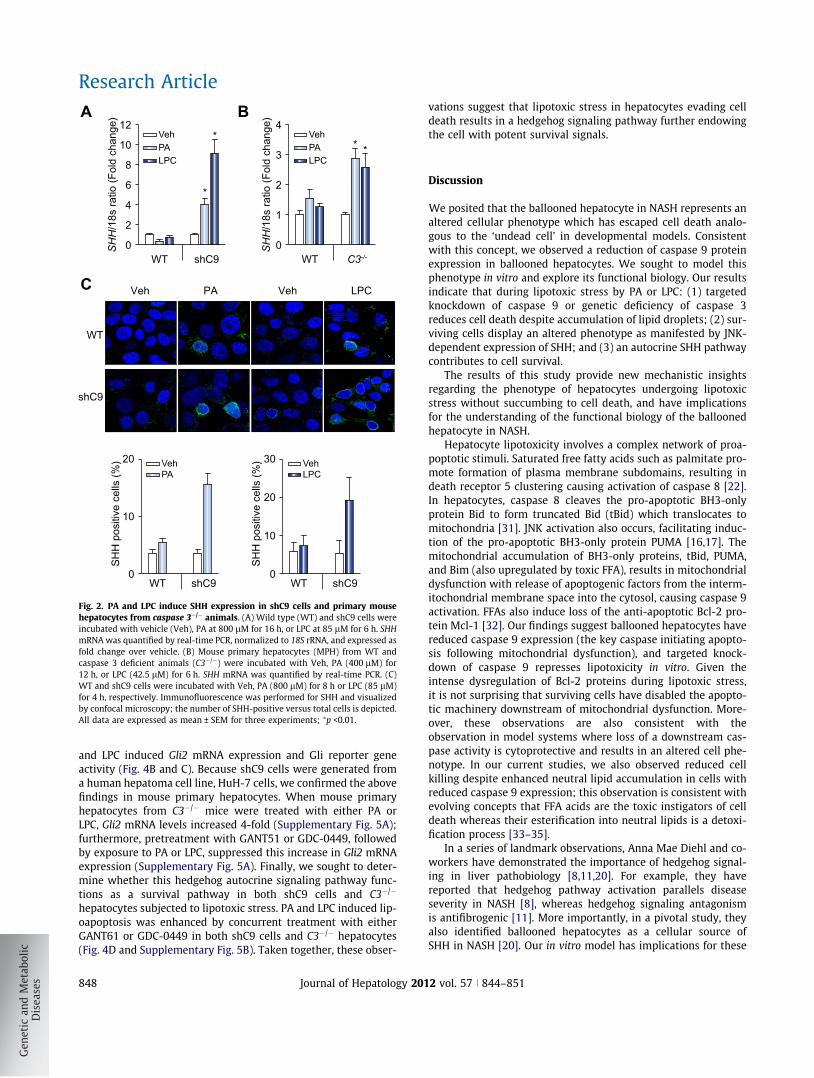
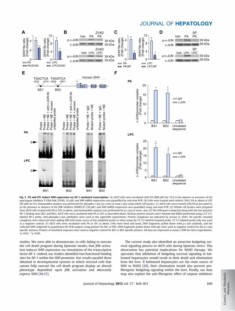
Next, we examined SHH generation by shC9 cells subjected tolipotoxic stress by PA or LPC. shC9 and caspase 3�/�, but not wildtype HuH-7 cells, expressed SHH at both mRNA and protein levelfollowing treatment with either PA or LPC (Fig. 2A–C). In D. mel-anogaster, the undead cell phenotype requires activation of initi-ator caspases which induce alternative signaling pathways in theabsence of downstream caspases [3,4]. Therefore, we nextexplored the effect of the pan-caspase inhibitor Z-VAD-fmk onSHH generation by shC9 cells treated with PA or LPC. Z-VAD-fmk treatment blunted SHH expression in shC9 cells exposed toeither PA or LPC (Fig. 3A). During lipoapoptosis, c-Jun N-terminalkinase (JNK) activation occurs by a mechanism downstream ofcaspase 8 activity [12,17,21]. Consistent with this information,Z-VAD-fmk treatment reduced phosphorylation of c-Jun, a JNKsubstrate, during incubation of shC9 cells with PA or LPC(Fig. 3B). As lipotoxic stress is associated with death receptor 5-dependent activation of caspase 8 [22], presumably Z-VAD-fmkis inhibiting this initiator caspase. To determine whether JNKactivation directly contributes to SHH expression during lipotoxicstress, we employed the pharmacological JNK inhibitorSP600125. SP600125, at a concentration which blocks c-Jun phos-phorylation, reduced SHH expression in this model (Fig. 3C andD). Collectively, these observations implicate JNK activation inthe generation of SHH in hepatocytes resistant to apoptosis.
Activator protein-1 (AP-1) mediates SHH transcription duringlipotoxic stress
Phosphorylated c-Jun heterodimerizes with other proteins toform the AP-1 transcription factor complex [23]. To ascertain ifAP-1 may directly upregulate SHH transcription, computationalanalysis of the human SHH gene sequence was performed toidentify consensus AP-1 binding sites. Two putative AP-1 bindingsites were identified within the human SHH promoter; one at�4525/�4518 (Binding Sequence 1, BS1) and a second at�2608/�2601 (Binding Sequence 2, BS2) (Fig. 3E). To testwhether AP-1 complexes bind to these sites, an EMSA was per-formed using CY5.5-labeled oligonucleotide probes for each puta-tive binding site (Supplementary Table 1). Using nuclear extractsobtained from either PA or LPC treated shC9 cells, protein bindingto both BS1 and BS2 was observed (Fig. 3E). This binding was nolonger observed when extracts were incubated with anti-phos-pho-c-Jun antisera, as presumably the antiserum binding to
2 vol. 57 j 844–851
shC9Caspase 9
Actin 42 kDa
47 kDa
PhasecontrastBodipy
fluorescence
Lipi
d dr
ople
t are
a,%
of t
otal
cel
lula
r are
a
Tota
l cel
lula
r flu
ores
cenc
e(F
old
chan
ge)
0
0 400 800
20
40
*
*PA [μM]
PAPAOA OAVeh Veh
Caspase 2
Caspase 9
H&E
H&E
*
*
Rel
ativ
e st
aini
ng in
tens
ity0
0.2
0.4
0.6
0.8
1.0
1.2
*
Caspa
se 2
Caspa
se 9
NHBH
OAVeh
PA
400 µM PA0 µM PA
800 µM PA
WT
WT
WT
WT
WT
shC9
shC9
shC9
shC9
A B
C
D
0
1
2
3
*
Fig. 1. Ballooned hepatocytes display decreased caspase 9 in NASH specimens; loss of caspase 9 or 3 enhances lipid loading in vitro. (A) Ballooned hepatocytes (BH)and non-balloned hepatocytes (NH, arrow) were identified in H&E stained sections (circled cells). Immunohistochemistry for caspase 2 and 9 was performed in sectionsfrom the same specimens. Caspase 2 and 9 staining intensity was quantified by NIH Image J software (right panel). Staining intensity of NH was set at 1.0. (B) Whole celllysates were prepared from wild type (WT) and shC9 cells and immunoblot analysis performed for caspase 9. (C) WT and shC9 cells were incubated with vehicle (Veh),palmitate (PA) or oleate (OA) at 400 lM for 12 h. Lipid droplets were labeled with Bodipy 505/515, and cells imaged by phase contrast microscopy and fluorescentmicroscopy. Lipid droplet area and total cell area were measured by NIH Image J software. (D) WT and shC9 cells were incubated with PA for 12 h at 0, 400, or 800 lM,respectively, labeled with Bodipy 505/515, and imaged by confocal microscopy. Fluorescence intensity was quantified using Metafluor quantitative software and expressedas fold change over vehicle. All data are expressed as mean ± SEM for at least three experiments; ⁄p <0.01.
JOURNAL OF HEPATOLOGY
AP-1 masks the oligonucleotide binding site; an observationimplicating AP-1 in the formation of the protein:oligonucleotidecomplex. Binding was also no longer observed when extractswere incubated with a 200-fold molar excess of the unlabeledprobe, or when using a mutated probe in which the critical AP-1 binding residues had been altered. Finally, to further confirmthat AP-1 binds to these promoter sequences of the SHH gene, aChIP assay was performed using the primers listed in Supplemen-tary Table 2. Immunoprecipitation of c-Jun from nuclear extractsobtained from PA- or LPC-treated shC9 cells captured both puta-tive AP-1 binding sequences within the SHH promoter (Fig. 3F). Incontrast, no binding of c-Jun to an unrelated upstream region ofthe SHH promoter was observed. These data provide evidencefor two bona fide AP-1 binding sequences within the SHH pro-moter suggesting AP-1 may induce expression of this gene.
SHH provides autocrine survival signals during lipotoxic stress
As critical as caspases are for cell apoptosis, loss of caspase activ-ity often delays but does not abrogate cell death in mammalian
Journal of Hepatology 201
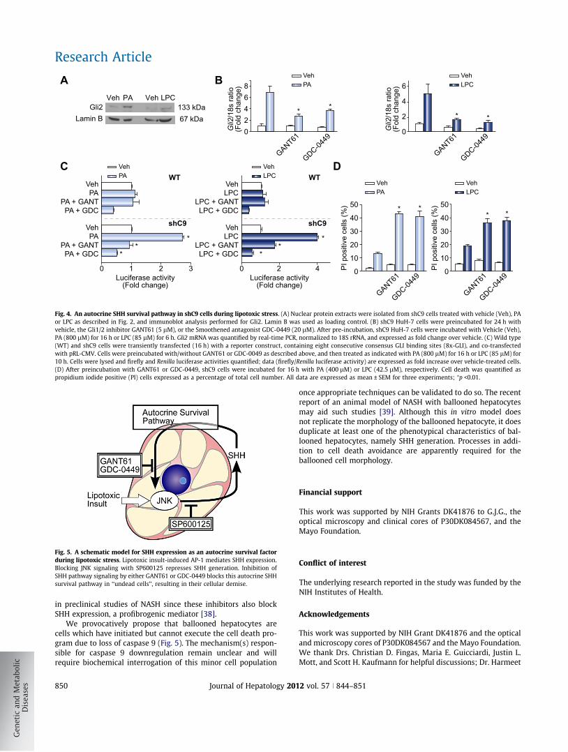
cells [24]. Therefore, we anticipated that SHH expression func-tions as an additional survival factor during lipotoxic stress givenits role as a potent survival factor in other systems [6,25–27]. Totest this concept, we first looked for active hedgehog signaling inshC9 cells. Following binding of SHH to its cognate receptorPatched, Smoothened is derepressed resulting in translocationof the Gli transcription factors into the nucleus, especially Gli2[28,29]. Indeed, Gli2 translocation into nuclear cell fractionswas readily identified in PA- and LPC-treated shC9 cells(Fig. 4A). Hedgehog signaling in a feed forward manner alsoincreases Gli2 mRNA expression [29], which was observed inshC9 cells following exposure to PA or LPC (Fig. 4B). To furtherexamine hedgehog signaling, a Gli reporter gene assay wasemployed. Consistent with the above observations, PA or LPCincreased Gli reporter gene activity in shC9 cells (Fig. 4C). Theseobservations identified an autocrine hedgehog signaling pathwayin shC9 cells following lipotoxic stress. To explore this concept,shC9 cells were treated either with GANT61 (a Gli1/2 inhibitor)or GDC-0449 (a Smoothened antagonist) followed by exposureto PA or LPC [30]. GANT61 as well as GDC-0449 prevented PA
2 vol. 57 j 844–851 847
SHH
/18s
ratio
(Fol
d ch
ange
)
0
2
4
6
8
12
10
*
*PAVeh
LPC
WT
WT
shC9
shC9
SHH
pos
itive
cel
ls (%
)
0
10
20PAVeh
WT shC9
SHH
/18s
ratio
(Fol
d ch
ange
)
0
1
2
4
3* *PA
Veh
LPC
WT C3-/-
A B
C PA LPCVehVeh
SHH
pos
itive
cel
ls (%
)
0
20
10
30 VehLPC
WT shC9
Fig. 2. PA and LPC induce SHH expression in shC9 cells and primary mousehepatocytes from caspase 3�/� animals. (A) Wild type (WT) and shC9 cells wereincubated with vehicle (Veh), PA at 800 lM for 16 h, or LPC at 85 lM for 6 h. SHHmRNA was quantified by real-time PCR, normalized to 18S rRNA, and expressed asfold change over vehicle. (B) Mouse primary hepatocytes (MPH) from WT andcaspase 3 deficient animals (C3�/�) were incubated with Veh, PA (400 lM) for12 h, or LPC (42.5 lM) for 6 h. SHH mRNA was quantified by real-time PCR. (C)WT and shC9 cells were incubated with Veh, PA (800 lM) for 8 h or LPC (85 lM)for 4 h, respectively. Immunofluorescence was performed for SHH and visualizedby confocal microscopy; the number of SHH-positive versus total cells is depicted.All data are expressed as mean ± SEM for three experiments; ⁄p <0.01.
Research Article
and LPC induced Gli2 mRNA expression and Gli reporter geneactivity (Fig. 4B and C). Because shC9 cells were generated froma human hepatoma cell line, HuH-7 cells, we confirmed the abovefindings in mouse primary hepatocytes. When mouse primaryhepatocytes from C3�/� mice were treated with either PA orLPC, Gli2 mRNA levels increased 4-fold (Supplementary Fig. 5A);furthermore, pretreatment with GANT51 or GDC-0449, followedby exposure to PA or LPC, suppressed this increase in Gli2 mRNAexpression (Supplementary Fig. 5A). Finally, we sought to deter-mine whether this hedgehog autocrine signaling pathway func-tions as a survival pathway in both shC9 cells and C3�/�
hepatocytes subjected to lipotoxic stress. PA and LPC induced lip-oapoptosis was enhanced by concurrent treatment with eitherGANT61 or GDC-0449 in both shC9 cells and C3�/� hepatocytes(Fig. 4D and Supplementary Fig. 5B). Taken together, these obser-
848 Journal of Hepatology 201
vations suggest that lipotoxic stress in hepatocytes evading celldeath results in a hedgehog signaling pathway further endowingthe cell with potent survival signals.
Discussion
We posited that the ballooned hepatocyte in NASH represents analtered cellular phenotype which has escaped cell death analo-gous to the ‘undead cell’ in developmental models. Consistentwith this concept, we observed a reduction of caspase 9 proteinexpression in ballooned hepatocytes. We sought to model thisphenotype in vitro and explore its functional biology. Our resultsindicate that during lipotoxic stress by PA or LPC: (1) targetedknockdown of caspase 9 or genetic deficiency of caspase 3reduces cell death despite accumulation of lipid droplets; (2) sur-viving cells display an altered phenotype as manifested by JNK-dependent expression of SHH; and (3) an autocrine SHH pathwaycontributes to cell survival.
The results of this study provide new mechanistic insightsregarding the phenotype of hepatocytes undergoing lipotoxicstress without succumbing to cell death, and have implicationsfor the understanding of the functional biology of the balloonedhepatocyte in NASH.
Hepatocyte lipotoxicity involves a complex network of proa-poptotic stimuli. Saturated free fatty acids such as palmitate pro-mote formation of plasma membrane subdomains, resulting indeath receptor 5 clustering causing activation of caspase 8 [22].In hepatocytes, caspase 8 cleaves the pro-apoptotic BH3-onlyprotein Bid to form truncated Bid (tBid) which translocates tomitochondria [31]. JNK activation also occurs, facilitating induc-tion of the pro-apoptotic BH3-only protein PUMA [16,17]. Themitochondrial accumulation of BH3-only proteins, tBid, PUMA,and Bim (also upregulated by toxic FFA), results in mitochondrialdysfunction with release of apoptogenic factors from the interm-itochondrial membrane space into the cytosol, causing caspase 9activation. FFAs also induce loss of the anti-apoptotic Bcl-2 pro-tein Mcl-1 [32]. Our findings suggest ballooned hepatocytes havereduced caspase 9 expression (the key caspase initiating apopto-sis following mitochondrial dysfunction), and targeted knock-down of caspase 9 represses lipotoxicity in vitro. Given theintense dysregulation of Bcl-2 proteins during lipotoxic stress,it is not surprising that surviving cells have disabled the apopto-tic machinery downstream of mitochondrial dysfunction. More-over, these observations are also consistent with theobservation in model systems where loss of a downstream cas-pase activity is cytoprotective and results in an altered cell phe-notype. In our current studies, we also observed reduced cellkilling despite enhanced neutral lipid accumulation in cells withreduced caspase 9 expression; this observation is consistent withevolving concepts that FFA acids are the toxic instigators of celldeath whereas their esterification into neutral lipids is a detoxi-fication process [33–35].
In a series of landmark observations, Anna Mae Diehl and co-workers have demonstrated the importance of hedgehog signal-ing in liver pathobiology [8,11,20]. For example, they havereported that hedgehog pathway activation parallels diseaseseverity in NASH [8], whereas hedgehog signaling antagonismis antifibrogenic [11]. More importantly, in a pivotal study, theyalso identified ballooned hepatocytes as a cellular source ofSHH in NASH [20]. Our in vitro model has implications for these
2 vol. 57 j 844–851
SHH
/18s
ratio
(Fol
d ch
ange
)
0
4
2**
SHH
/18s
ratio
(Fol
d ch
ange
)
0
8
12
4
PAPA/ZVAD
LPCLPC/ZVAD
*
PAZVAD
PA
ZVADCPL CPL
Veh
Veh
p-c-JUNc-JUN
p-c-JUNc-JUN 39 kDa
39 kDa
39 kDa39 kDa
PAPA/SP
SHH
/18s
ratio
(Fol
d ch
ange
)
0
4
2 *
LPCLPC/SP
SHH
/18s
ratio
(Fol
d ch
ange
)
0
8
12
4 *
PASPPA
SPCPL CPL
Veh
Veh
p-c-JUNc-JUN
p-c-JUNc-JUN 39 kDa
39 kDa
39 kDa39 kDa
Ex1 Ex2 Ex3
TGACTCA TGAGTCA
BS1
BS1 BS1
BS2
BS2 BS2
-4525 -4518 -2608 -2601 +1Human SHH
Prob
ePr
obe
+ N
EPr
obe
+ N
E +
anti
c-JU
N A
bPr
obe
+ N
E +
exce
ssun
labe
led
prob
e
Prob
e
Prob
e +
NE
+ Ig
G
Prob
e +
NE
+ Ig
G
Mut
ant p
robe
+ N
E
Mut
ant p
robe
+ N
E
Prob
e +
NE
Prob
e +
NE
+ an
ti c-
JUN
Ab
Prob
e +
NE
+ ex
cess
unla
bele
d pr
obe
S
S
PA
PA
LPC
LPC
A B C D
E F
BS1 BS2 Unrelatedsequence
c-JU
N/p
rom
oter
com
plex
(Fol
d ch
ange
)
0
5
10
20
15
* *
c-JUNIgG
BS1 BS2 Unrelatedsequence
c-JU
N/p
rom
oter
com
plex
(Fol
d ch
ange
)
0
5
10
25
20
15
*
*
c-JUNIgG
Fig. 3. PA and LPC induce SHH expression via AP-1 mediated transcription. (A) shC9 cells were incubated with PA (800 lM) for 16 h in the absence or presence of thepancaspase inhibitor Z-VAD-fmk (ZVAD, 10 lM) and SHH mRNA expression was quantified by real-time PCR. (B) Cells were treated with vehicle (Veh), PA as above or LPC(85 lM) for 8 h. Immunoblot analysis was performed for phospho-c-Jun (p-c-Jun) or total c-Jun using whole cell lysates. (C) shC9 cells were treated with PA as per panel A,in the presence or absence of the JNK inhibitor SP600125 (50 lM), and SHH mRNA expression was quantified using real-time PCR. (D) Whole cell lysates were preparedfrom shC9 cells treated with PA or LPC as above, and immunoblot analysis was performed for p-c-Jun or total c-Jun. (E) The SHH gene is depicted along with the two putativeAP-1 binding sites (BS1 and BS2). shC9 cells were incubated with PA or LPC as described above. Nuclear protein extracts were isolated and EMSA performed using a CY 5.5-labeled AP-1 probe. Anti-phospho-c-Jun antibodies were used in the supershift experiments. Protein complexes are indicated by arrows (S, shift). No specific retardedcomplexes were observed when adding 200-fold molar excess of the unlabeled probe or when using the CY 5.5-labeled mutated probe. CY 5.5-labeled probe only was usedas a negative control. (F) shC9 cells were incubated with PA or LPC as above. Cells were fixed and lysed, DNA fragments pulled down with p-c-Jun antibody, and thecollected DNA subjected to quantitative RT-PCR analysis using primers for BS1 or BS2. DNA fragments pulled down with IgG were used as negative control for the p-c-Junspecific antisera. Primers of unrelated sequence were used as negative control for BS1 or BS2 specific primers. All data are expressed as mean ± SEM for three experiments;⁄p <0.01, ⁄⁄p <0.05.
JOURNAL OF HEPATOLOGY
studies. We were able to demonstrate, in cells failing to executethe cell death program during lipotoxic insults, that JNK activa-tion induces SHH expression via stimulation of the transcriptionfactor AP-1; indeed, our studies identified two functional bindingsites for AP-1 within the SHH promoter. Our results parallel thoseobtained in developmental systems in which stressed cells thatcannot fully execute the cell death program display an alteredphenotype dependent upon JNK activation and aberrantlyexpress SHH [36,37].
Journal of Hepatology 201
The current study also identified an autocrine hedgehog sur-vival signaling process in shC9 cells during lipotoxic stress. Thisobservation has potential implications for NASH therapy. Wespeculate that inhibition of hedgehog survival signaling in bal-looned hepatocytes would result in their death and eliminationfrom the liver. If ballooned hepatocytes are the main source ofSHH in NASH [20], their elimination would also prevent pro-fibrogenic hedgehog signaling within the liver. Finally, our datamay also explain the anti-fibrogenic effect of caspase inhibitors
2 vol. 57 j 844–851 849
heVheV AP LPCGli2
Lamin B 67 kDa133 kDa
GANT61
GDC-0449
Gli2
/18s
ratio
(Fol
d ch
ange
)
0
42
68
* *
PAVeh
GANT61
GDC-0449
Gli2
/18s
ratio
(Fol
d ch
ange
)
0
4
2
6
* *
LPCVeh
GANT61
GDC-0449
PI p
ositi
ve c
ells
(%) * *
PAVeh
PAVeh
0
50
40
30
20
10
GANT61
GDC-0449
PI p
ositi
ve c
ells
(%)
0
50
40
30
20
10
* *
LPCVeh
LPCVeh
**
*
VehPA
PA + GANTPA + GDC
VehPA
PA + GANTPA + GDC
WT
shC9
0 1 2 3Luciferase activity
(Fold change)Luciferase activity
(Fold change)
**
*
VehLPC
LPC + GANTLPC + GDC
VehLPC
LPC + GANTLPC + GDC
WT
shC9
0 2 4
A B
C D
Fig. 4. An autocrine SHH survival pathway in shC9 cells during lipotoxic stress. (A) Nuclear protein extracts were isolated from shC9 cells treated with vehicle (Veh), PAor LPC as described in Fig. 2, and immunoblot analysis performed for Gli2. Lamin B was used as loading control. (B) shC9 HuH-7 cells were preincubated for 24 h withvehicle, the Gli1/2 inhibitor GANT61 (5 lM), or the Smoothened antagonist GDC-0449 (20 lM). After pre-incubation, shC9 HuH-7 cells were incubated with Vehicle (Veh),PA (800 lM) for 16 h or LPC (85 lM) for 6 h. Gli2 mRNA was quantified by real-time PCR, normalized to 18S rRNA, and expressed as fold change over vehicle. (C) Wild type(WT) and shC9 cells were transiently transfected (16 h) with a reporter construct, containing eight consecutive consensus GLI binding sites (8x-GLI), and co-transfectedwith pRL-CMV. Cells were preincubated with/without GANT61 or GDC-0049 as described above, and then treated as indicated with PA (800 lM) for 16 h or LPC (85 lM) for10 h. Cells were lysed and firefly and Renilla luciferase activities quantified; data (firefly/Renilla luciferase activity) are expressed as fold increase over vehicle-treated cells.(D) After preincubation with GANT61 or GDC-0449, shC9 cells were incubated for 16 h with PA (400 lM) or LPC (42.5 lM), respectively. Cell death was quantified aspropidium iodide positive (PI) cells expressed as a percentage of total cell number. All data are expressed as mean ± SEM for three experiments; ⁄p <0.01.
Fig. 5. A schematic model for SHH expression as an autocrine survival factorduring lipotoxic stress. Lipotoxic insult-induced AP-1 mediates SHH expression.Blocking JNK signaling with SP600125 represses SHH generation. Inhibition ofSHH pathway signaling by either GANT61 or GDC-0449 blocks this autocrine SHHsurvival pathway in ‘‘undead cells’’, resulting in their cellular demise.
Research Article
in preclinical studies of NASH since these inhibitors also blockSHH expression, a profibrogenic mediator [38].
We provocatively propose that ballooned hepatocytes arecells which have initiated but cannot execute the cell death pro-gram due to loss of caspase 9 (Fig. 5). The mechanism(s) respon-sible for caspase 9 downregulation remain unclear and willrequire biochemical interrogation of this minor cell population
850 Journal of Hepatology 201
once appropriate techniques can be validated to do so. The recentreport of an animal model of NASH with ballooned hepatocytesmay aid such studies [39]. Although this in vitro model doesnot replicate the morphology of the ballooned hepatocyte, it doesduplicate at least one of the phenotypical characteristics of bal-looned hepatocytes, namely SHH generation. Processes in addi-tion to cell death avoidance are apparently required for theballooned cell morphology.
Financial support
This work was supported by NIH Grants DK41876 to G.J.G., theoptical microscopy and clinical cores of P30DK084567, and theMayo Foundation.
Conflict of interest
The underlying research reported in the study was funded by theNIH Institutes of Health.
Acknowledgements
This work was supported by NIH Grant DK41876 and the opticaland microscopy cores of P30DK084567 and the Mayo Foundation.We thank Drs. Christian D. Fingas, Maria E. Guicciardi, Justin L.Mott, and Scott H. Kaufmann for helpful discussions; Dr. Harmeet
2 vol. 57 j 844–851
JOURNAL OF HEPATOLOGY
Malhi for reading the manuscript and providing insightful com-ments and suggestions; Eugene W. Krueger for aiding the studiesemploying contrast microscopy; and Courtney N. Hoover for herexcellent secretarial assistance.Supplementary data
Supplementary data associated with this article can be found, inthe online version, at http://dx.doi.org/10.1016/j.jhep.2012.05.011.
References
[1] Malhi H, Gores GJ. Cellular and molecular mechanisms of liver injury.Gastroenterology 2008;134:1641–1654.
[2] Gouw AS, Clouston AD, Theise ND. Ductular reactions in human liver:diversity at the interface. Hepatology 2011;54:1853–1863.
[3] Ryoo HD, Gorenc T, Steller H. Apoptotic cells can induce compensatory cellproliferation through the JNK and the Wingless signaling pathways. Dev Cell2004;7:491–501.
[4] Kondo S, Senoo-Matsuda N, Hiromi Y, Miura M. DRONC coordinates celldeath and compensatory proliferation. Mol Cell Biol 2006;26:7258–7268.
[5] Tait SW, Green DR. Mitochondria and cell death: outer membrane perme-abilization and beyond. Nat Rev Mol Cell Biol 2010;11:621–632.
[6] Fan Y, Bergmann A. Distinct mechanisms of apoptosis-induced compensa-tory proliferation in proliferating and differentiating tissues in the Drosoph-ila eye. Dev Cell 2008;14:399–410.
[7] Lackner C, Gogg-Kamerer M, Zatloukal K, Stumptner C, Brunt EM, Denk H.Ballooned hepatocytes in steatohepatitis: the value of keratin immunohis-tochemistry for diagnosis. J Hepatol 2008;48:821–828.
[8] Guy CD, Suzuki A, Zdanowicz M, Abdelmalek MF, Burchette J, Unalp A, et al.Hedgehog pathway activation parallels histologic severity of injury andfibrosis in human non-alcoholic fatty liver disease. Hepatology2012;55:1711–1721.
[9] Caldwell S, Ikura Y, Dias D, Isomoto K, Yabu A, Moskaluk C, et al.Hepatocellular ballooning in NASH. J Hepatol 2010;53:719–723.
[10] Jung Y, Witek RP, Syn WK, Choi SS, Omenetti A, Premont R, et al. Signals fromdying hepatocytes trigger growth of liver progenitors. Gut 2010;59:655–665.
[11] Syn WK, Choi SS, Liaskou E, Karaca GF, Agboola KM, Oo YH, et al. Osteopontinis induced by hedgehog pathway activation and promotes fibrosis progres-sion in non-alcoholic steatohepatitis. Hepatology 2011;53:106–115.
[12] Kakisaka K, Cazanave SC, Fingas CD, Guicciardi ME, Bronk SF, WerneburgNW, et al. Mechanisms of lysophosphatidylcholine-induced hepatocytelipoapoptosis. Am J Physiol Gastrointest Liver Physiol 2012;302:G77–84.
[13] Kuida K, Zheng TS, Na S, Kuan C, Yang D, Karasuyama H, et al. Decreasedapoptosis in the brain and premature lethality in CPP32-deficient mice.Nature 1996;384:368–372.
[14] Listenberger LL, Brown DA. Fluorescent detection of lipid droplets andassociated proteins. Curr Protoc Cell Biol 2007: Unit 24 2 [chapter 24].
[15] Higuchi H, Bronk SF, Bateman A, Harrington K, Vile RG, Gores GJ. Viralfusogenic membrane glycoprotein expression causes syncytia formationwith bioenergetic cell death: implications for gene therapy. Cancer Res2000;60:6396–6402.
[16] Cazanave SC, Elmi NA, Akazawa Y, Bronk SF, Mott JL, Gores GJ. CHOP and AP-1 cooperatively mediate PUMA expression during lipoapoptosis. Am JPhysiol Gastrointest Liver Physiol 2010;299:G236–243.
[17] Cazanave SC, Mott JL, Elmi NA, Bronk SF, Werneburg NW, Akazawa Y, et al.JNK1-dependent PUMA expression contributes to hepatocyte lipoapoptosis.J Biol Chem 2009;284:26591–26602.
Journal of Hepatology 201
[18] Reddy JK, Hashimoto T. Peroxisomal beta-oxidation and peroxisome prolif-erator-activated receptor alpha: an adaptive metabolic system. Annu RevNutr 2001;21:193–230.
[19] Schreurs M, Kuipers F, van der Leij FR. Regulatory enzymes of mitochondrialbeta-oxidation as targets for treatment of the metabolic syndrome. Obes Rev2010;11:380–388.
[20] Rangwala F, Guy CD, Lu J, Suzuki A, Burchette JL, Abdelmalek MF, et al.Increased production of sonic hedgehog by ballooned hepatocytes. J Pathol2011;224:401–410.
[21] Malhi H, Bronk SF, Werneburg NW, Gores GJ. Free fatty acids induce JNK-dependent hepatocyte lipoapoptosis. J Biol Chem 2006;281:12093–12101.
[22] Cazanave SC, Mott JL, Bronk SF, Werneburg NW, Fingas CD, Meng XW, et al.Death receptor 5 signaling promotes hepatocyte lipoapoptosis. J Biol Chem2011;286:39336–39348.
[23] Eferl R, Wagner EF. AP-1: a double-edged sword in tumorigenesis. Nat RevCancer 2003;3:859–868.
[24] Kroemer G, Martin SJ. Caspase-independent cell death. Nat Med2005;11:725–730.
[25] Han ME, Lee YS, Baek SY, Kim BS, Kim JB, Oh SO. Hedgehog signalingregulates the survival of gastric cancer cells by regulating the expression ofBcl-2. Int J Mol Sci 2009;10:3033–3043.
[26] Syn WK, Jung Y, Omenetti A, Abdelmalek M, Guy CD, Yang L, et al. Hedgehog-mediated epithelial-to-mesenchymal transition and fibrogenic repair innon-alcoholic fatty liver disease. Gastroenterology 2009;137:1478–1488,e1478.
[27] Shin K, Lee J, Guo N, Kim J, Lim A, Qu L, et al. Hedgehog/Wnt feedbacksupports regenerative proliferation of epithelial stem cells in bladder.Nature 2011;472:110–114.
[28] Regl G, Kasper M, Schnidar H, Eichberger T, Neill GW, Philpott MP, et al.Activation of the BCL2 promoter in response to Hedgehog/GLI signaltransduction is predominantly mediated by GLI2. Cancer Res2004;64:7724–7731.
[29] Walterhouse DO, Yoon JW, Iannaccone PM. Developmental pathways: sonichedgehog-Patched-GLI. Environ Health Perspect 1999;107:167–171.
[30] Mahindroo N, Punchihewa C, Fujii N. Hedgehog–Gli signaling pathwayinhibitors as anticancer agents. J Med Chem 2009;52:3829–3845.
[31] Malhi H, Guicciardi ME, Gores GJ. Hepatocyte death: a clear and presentdanger. Physiol Rev 2010;90:1165–1194.
[32] Masuoka HC, Mott J, Bronk SF, Werneburg NW, Akazawa Y, Kaufmann SH,et al. Mcl-1 degradation during hepatocyte lipoapoptosis. J Biol Chem2009;284:30039–30048.
[33] Alkhouri N, Dixon LJ, Feldstein AE. Lipotoxicity in non-alcoholic fatty liverdisease: not all lipids are created equal. Expert Rev Gastroenterol Hepatol2009;3:445–451.
[34] Monetti M, Levin MC, Watt MJ, Sajan MP, Marmor S, Hubbard BK, et al.Dissociation of hepatic steatosis and insulin resistance in mice overexpress-ing DGAT in the liver. Cell Metab 2007;6:69–78.
[35] Listenberger LL, Han X, Lewis SE, Cases S, Farese Jr RV, Ory DS, et al.Triglyceride accumulation protects against fatty acid-induced lipotoxicity.Proc Natl Acad Sci U S A 2003;100:3077–3082.
[36] Fan Y, Bergmann A. Apoptosis-induced compensatory proliferation. The Cellis dead. Long live the Cell! Trends Cell Biol 2008;18:467–473.
[37] Wells BS, Yoshida E, Johnston LA. Compensatory proliferation in Drosophilaimaginal discs requires Dronc-dependent p53 activity. Curr Biol2006;16:1606–1615.
[38] Witek RP, Stone WC, Karaca FG, Syn WK, Pereira TA, Agboola KM. Pan-caspase inhibitor VX-166 reduces fibrosis in an animal model of non-alcoholic steatohepatitis. Hepatology 2009;50:1421–1430.
[39] Yimin, Furumaki H, Matsuoka S, Sakurai T, Kohanawa M, Zhao S, et al. Anovel murine model for non-alcoholic steatohepatitis developed by combi-nation of a high-fat diet and oxidized low-density lipoprotein. Lab Invest2012;92:265–281.
2 vol. 57 j 844–851 851
Related Documents