RESEARCH ARTICLE Open Access A gene-derived SNP-based high resolution linkage map of carrot including the location of QTL conditioning root and leaf anthocyanin pigmentation Pablo F Cavagnaro 1,2† , Massimo Iorizzo 1† , Mehtap Yildiz 3 , Douglas Senalik 1,4 , Joshua Parsons 1 , Shelby Ellison 1 and Philipp W Simon 1,4* Abstract Background: Purple carrots accumulate large quantities of anthocyanins in their roots and leaves. These flavonoid pigments possess antioxidant activity and are implicated in providing health benefits. Informative, saturated linkage maps associated with well characterized populations segregating for anthocyanin pigmentation have not been developed. To investigate the genetic architecture conditioning anthocyanin pigmentation we scored root color visually, quantified root anthocyanin pigments by high performance liquid chromatography in segregating F 2 ,F 3 and F 4 generations of a mapping population, mapped quantitative trait loci (QTL) onto a dense gene-derived single nucleotide polymorphism (SNP)-based linkage map, and performed comparative trait mapping with two unrelated populations. Results: Root pigmentation, scored visually as presence or absence of purple coloration, segregated in a pattern consistent with a two gene model in an F 2 , and progeny testing of F 3 -F 4 families confirmed the proposed genetic model. Purple petiole pigmentation was conditioned by a single dominant gene that co-segregates with one of the genes conditioning root pigmentation. Root total pigment estimate (RTPE) was scored as the percentage of the root with purple color. All five anthocyanin glycosides previously reported in carrot, as well as RTPE, varied quantitatively in the F 2 population. For the purpose of QTL analysis, a high resolution gene-derived SNP-based linkage map of carrot was constructed with 894 markers covering 635.1 cM with a 1.3 cM map resolution. A total of 15 significant QTL for all anthocyanin pigments and for RTPE mapped to six chromosomes. Eight QTL with the largest phenotypic effects mapped to two regions of chromosome 3 with co-localized QTL for several anthocyanin glycosides and for RTPE. A single dominant gene conditioning anthocyanin acylation was identified and mapped. Comparative mapping with two other carrot populations segregating for purple color indicated that carrot anthocyanin pigmentation is controlled by at least three genes, in contrast to monogenic control reported previously. Conclusions: This study generated the first high resolution gene-derived SNP-based linkage map in the Apiaceae. Two regions of chromosome 3 with co-localized QTL for all anthocyanin pigments and for RTPE, largely condition anthocyanin accumulation in carrot roots and leaves. Loci controlling root and petiole anthocyanin pigmentation differ across diverse carrot genetic backgrounds. Keywords: Carrot, Anthocyanins, QTL mapping, Linkage map, Single nucleotide polymorphism * Correspondence: [email protected] † Equal contributors 1 Department of Horticulture, University of Wisconsin-Madison, 1575 Linden Drive, Madison, WI 53706, USA 4 USDA-Agricultural Research Service, Vegetable Crops Unit, University of Wisconsin-Madison, 1575 Linden Drive, Madison, WI 53706, USA Full list of author information is available at the end of the article © 2014 Cavagnaro et al.; licensee BioMed Central. This is an Open Access article distributed under the terms of the Creative Commons Attribution License (http://creativecommons.org/licenses/by/4.0), which permits unrestricted use, distribution, and reproduction in any medium, provided the original work is properly credited. The Creative Commons Public Domain Dedication waiver (http://creativecommons.org/publicdomain/zero/1.0/) applies to the data made available in this article, unless otherwise stated. Cavagnaro et al. BMC Genomics 2014, 15:1118 http://www.biomedcentral.com/1471-2164/15/1118

Welcome message from author
This document is posted to help you gain knowledge. Please leave a comment to let me know what you think about it! Share it to your friends and learn new things together.
Transcript

Cavagnaro et al. BMC Genomics 2014, 15:1118http://www.biomedcentral.com/1471-2164/15/1118
RESEARCH ARTICLE Open Access
A gene-derived SNP-based high resolutionlinkage map of carrot including the location ofQTL conditioning root and leaf anthocyaninpigmentationPablo F Cavagnaro1,2†, Massimo Iorizzo1†, Mehtap Yildiz3, Douglas Senalik1,4, Joshua Parsons1, Shelby Ellison1
and Philipp W Simon1,4*
Abstract
Background: Purple carrots accumulate large quantities of anthocyanins in their roots and leaves. These flavonoidpigments possess antioxidant activity and are implicated in providing health benefits. Informative, saturated linkagemaps associated with well characterized populations segregating for anthocyanin pigmentation have not beendeveloped. To investigate the genetic architecture conditioning anthocyanin pigmentation we scored root colorvisually, quantified root anthocyanin pigments by high performance liquid chromatography in segregating F2, F3 and F4generations of a mapping population, mapped quantitative trait loci (QTL) onto a dense gene-derived single nucleotidepolymorphism (SNP)-based linkage map, and performed comparative trait mapping with two unrelated populations.
Results: Root pigmentation, scored visually as presence or absence of purple coloration, segregated in a patternconsistent with a two gene model in an F2, and progeny testing of F3-F4 families confirmed the proposed geneticmodel. Purple petiole pigmentation was conditioned by a single dominant gene that co-segregates with one of thegenes conditioning root pigmentation. Root total pigment estimate (RTPE) was scored as the percentage of the rootwith purple color.All five anthocyanin glycosides previously reported in carrot, as well as RTPE, varied quantitatively in the F2 population.For the purpose of QTL analysis, a high resolution gene-derived SNP-based linkage map of carrot was constructed with894 markers covering 635.1 cM with a 1.3 cM map resolution. A total of 15 significant QTL for all anthocyanin pigmentsand for RTPE mapped to six chromosomes. Eight QTL with the largest phenotypic effects mapped to two regions ofchromosome 3 with co-localized QTL for several anthocyanin glycosides and for RTPE. A single dominant geneconditioning anthocyanin acylation was identified and mapped.Comparative mapping with two other carrot populations segregating for purple color indicated that carrotanthocyanin pigmentation is controlled by at least three genes, in contrast to monogenic control reported previously.
Conclusions: This study generated the first high resolution gene-derived SNP-based linkage map in the Apiaceae. Tworegions of chromosome 3 with co-localized QTL for all anthocyanin pigments and for RTPE, largely conditionanthocyanin accumulation in carrot roots and leaves. Loci controlling root and petiole anthocyanin pigmentationdiffer across diverse carrot genetic backgrounds.
Keywords: Carrot, Anthocyanins, QTL mapping, Linkage map, Single nucleotide polymorphism
* Correspondence: [email protected]†Equal contributors1Department of Horticulture, University of Wisconsin-Madison, 1575 LindenDrive, Madison, WI 53706, USA4USDA-Agricultural Research Service, Vegetable Crops Unit, University ofWisconsin-Madison, 1575 Linden Drive, Madison, WI 53706, USAFull list of author information is available at the end of the article
© 2014 Cavagnaro et al.; licensee BioMed CenCommons Attribution License (http://creativecreproduction in any medium, provided the orDedication waiver (http://creativecommons.orunless otherwise stated.
tral. This is an Open Access article distributed under the terms of the Creativeommons.org/licenses/by/4.0), which permits unrestricted use, distribution, andiginal work is properly credited. The Creative Commons Public Domaing/publicdomain/zero/1.0/) applies to the data made available in this article,

Cavagnaro et al. BMC Genomics 2014, 15:1118 Page 2 of 17http://www.biomedcentral.com/1471-2164/15/1118
BackgroundAnthocyanins, a subclass of water-soluble colored flavo-noids, provide red, blue and purple pigmentation to dif-ferent organs of a wide range of higher plants [1]. Theseplant pigments play important roles, such as attractionof pollinators and seed dispersers and, given their anti-oxidant properties, protection against ultraviolet (UV)and high intensity light, drought, wounding, cold tem-peratures and phytopathogen attack [2-5]. In addition,the consumption of anthocyanin-rich fruits and vegeta-bles is implicated to confer a number of health-relatedbenefits, including protection against oxidative stress,coronary heart disease, inflammation, some types of can-cer and other age-related diseases [6]. However, the healtheffects of dietary anthocyanins depend on amounts con-sumed and on their bioavailability. Previous studies sug-gest that the bioavailability and excretion of anthocyaninsand other polyphenols is highly influenced by their chem-ical structure [7], including the nature of the sugar conju-gate, the phenolic aglycone and acylation.Anthocyanins are synthesized via the flavonoid path-
way, a late branch of the shikimic acid pathway [8]. Al-though the biosynthetic pathways in different anthocyanin-containing species share a majority of common reactions,there are important differences between the types of an-thocyanins produced and accumulated by each species.For example, bilberry (Vaccinium myrtillus) fruits accu-mulate a balanced mixture of five major anthocyanin agly-cones (delphinidin, petunidin, cyanidin, peonidin andmalvidin) and lack pelargonidin [9], whereas snapdragonand maize are incapable of producing delphinidin [10] andcarrot (Daucus carota) almost exclusively accumulatescyanidin derivatives [11,12] . Regardless of the species, twoclasses of genes are involved in anthocyanin biosynthesis:structural genes encoding the enzymes that directly par-ticipate in the formation of anthocyanin pigments, andregulatory genes that control the transcription of struc-tural genes. Many of the genes involved in anthocyaninbiosynthesis, both structural and regulatory, have beenidentified and characterized for several model species,such as petunia (Petunia hybrida), snapdragon (Antirrhi-num majus) and maize (Zea mays), and both regulatoryand structural genes vary widely across species [3]. Conse-quently, information regarding the genetic control ofanthocyanin biosynthesis may not be reliably extrapolatedacross species.The genetics underlying anthocyanin pigmentation has
been most extensively studied in flowers, fruit andleaves. Only a few reports of genetic control of antho-cyanin biosynthesis are published for underground or-gans –tubers, bulbs and storage roots [13-18].Anthocyanins may undergo a series of chemical modifi-
cations including glycosylation, acylation and methylation.These changes are usually performed by glycosyl-, acyl-
and methyl-transferase enzymes, respectively. Several stud-ies have indicated that these modifications produce enor-mous chemical diversity (reviewed by Pojer et al. [19])which influence anthocyanin stability and bioavailability.For example, clinical human feeding studies using raw andcooked purple carrots revealed that, compared to the an-thocyanins in carrots consumed, the percentage recoveryof non-acylated anthocyanins in serum was significantlygreater than acylated anthocyanins, suggesting that acyl-ation reduces anthocyanin bioavailability [11,20-22]. Con-versely, acylated anthocyanins are more stable thannon-acylated anthocyanins, providing the former with anadvantage for their use as colorants in the food industry[19,21,23]. Despite their importance, the genetic factorscontrolling these chemical modifications are still relativelyunexplored.Carrot is a species that can accumulate large quantities
of anthocyanins in its storage roots (up to 17–18 mg/100 g fresh weight) [12]. Purple or “black” carrots accu-mulate almost exclusively cyanidin glycosides, both acyl-ated and non-acylated, with five cyanidin pigmentsreported in most studies [11,24] (Table 1). Substantialvariation in anthocyanin profiles and in total anthocya-nin concentration, and low amounts of derivatives ofanthocyanidins other than cyanidin, have been reportedamong carrot cultivars [12]. In addition, tissue distribu-tion of root purple pigmentation varies greatly acrosscarrot genotypes, ranging from a few pigmented celllayers in the periderm to a completely- and intensively-colored root (Figure 1). The significance of dietary an-thocyanins in human health and the extensive naturalvariation for the above traits has attracted the attentionof carrot breeders. Current breeding programs in purplecarrot aim at increasing total anthocyanin content aswell as achieving favorable ratios of acylated versus non-acylated anthocyanins depending on the end-marketpurpose, with a preference for high content of acylatedanthocyanins for their use as food colorants, but con-versely, high level of non-acylated forms for increasingbioavailability and nutraceutical value.To date, a single dominant gene controlling anthocya-
nin accumulation in carrot roots, P1, has been described[17]. Recently, P1 was genetically mapped, as were sev-eral anthocyanin biosynthetic genes (five structural andthree regulatory genes), but no tight linkage was foundbetween P1 and any of the candidate structural genesevaluated [18]. However, the five structural genes (CHS1,DFR1, F3H, LDOX2, PAL3) were expressed differentiallyand in decreasing order among those listed when com-paring solid-purple (purple in phloem and xylem), purpleorange (purple phloem and orange xylem) and orangecarrots, with transcript accumulation coinciding with an-thocyanin accumulation. These studies suggested a coordi-nated regulatory control of anthocyanin biosynthesis in

Table 1 Carrot cyanidin derivatives with approximate HPLC retention times and molecular masses
Compound Abbreviation RT MW
Cy-3-(2”-xylose-6-glucose-galactoside) Cy3XGG 14.0 743
Cy-3-(2”-xylose-galactoside) Cy3XG 15.1 581
Cy-3-(2”-xylose-6”-sinapoyl-glucose-galactoside) Cy3XSGG 15.4 949
Cy-3-(2”-xylose-6”-feruloyl-glucose-galactoside Cy3XFGG 16.0 919
Cy-3-(2”-xylose-6”-(4-coumuroyl)glucose-galactoside) Cy3XCGG 16.4 889
RT is retention time (min) for the chromatographic procedure described in the Materials and Methods section. MW is molecular weight.
Cavagnaro et al. BMC Genomics 2014, 15:1118 Page 3 of 17http://www.biomedcentral.com/1471-2164/15/1118
the carrot root, but the molecular and biochemical basisof genetic factors controlling the presence vs. absence ofcarrot root anthocyanin pigmentation remain unknown.Furthermore, very little is known about the genetics ofanthocyanin accumulation in carrot organs and tissuesother than the tap root. Variable expression of purple pig-mentation in leaves, nodes and flowers was reported butnot genetically characterized, and a simply inherited dom-inant locus controlling purple versus green pigmentationin petioles, P2, was also described [17] and linkage be-tween P1 and P2 was suggested but not mapped.Phenotypic evaluations of P1 and P2 in previous stud-
ies of carrot were performed on the basis of visuallyscoring for presence or absence of purple color in theroot and petiole, without providing a quantitative meas-urement on the extent of purple pigmentation in theseorgans. In addition to presence or absence of pigmenta-tion, anthocyanin biosynthetic genes can also modify theprofile of pigments and tissue-specific accumulation [3,25]and to date, few studies in carrot have explored these fac-tors affecting the relative accumulation of different antho-cyanin pigments in purple-colored tissues. One exceptionis a study of variation in pigment profiles of four com-mercial carrot cultivars which found more than 10-foldvariation in total anthocyanin content and significant
P9547 B7262 PI652188
Figure 1 Purple root phenotypes of three diverse carrotparental genetic stocks. Root phenotypes of the purple root colorsource progenitors of carrot mapping populations 70349 (P9547),10117 (B7262), and 2170 (PI652188), used for comparative mappingof loci controlling anthocyanin pigmentation in root and leaves.
variation in percentage cyanidin-3-(2’-xylose-galactoside)and cyanidin-3-(2’-xylose-6’-sinapoyl-glucose-galactoside)relative to the most abundant compound they found,cyanidin-3-(2’-xylose-6’-feruloyl-glucose-galactoside [12].Understanding the genetic architecture conditioning bothpigment concentration and the relative accumulation ofdifferent anthocyanin glycosides in the carrot root wouldbe of great value for carrot breeding and genetics. Further-more, since mapping of P1 was performed in a singlegenetic background, comparative analysis of the loci con-trolling anthocyanin pigmentation across diverse carrotbackgrounds would be of interest, considering the broadvariation for phenotype, genotype and geographical originobserved among purple carrot genetic stocks [17].High density linkage maps constructed with inform-
ative sequence-based markers, such as SNPs and SSRs,are essential for fine mapping of QTL, comparative ana-lysis of synteny, searching for candidate genes, facilitat-ing genome sequence assembly, and for marker assistedbreeding. The majority of the carrot maps constructedto date were unsaturated and used anonymous dominantmarkers, such as amplified fragment length polymor-phisms and randomly amplified polymorphic DNAs, al-though in some cases a few codominant SSR or restrictionfragment length polymorphisms markers were included[18,26-32]. A DArT map with the highest map resolution(1.1 cM) achieved to date in carrot was recently reportedby Grzebelus et al. [33]. However, the anonymous anddominant nature of DArT markers do not allow straightforward comparative map analysis or identification of can-didate genes associated with QTL.In the current study, we developed a SNP-based link-
age map with an unprecedented high map resolution inentire Apiaceae family, using a carrot population segre-gating for anthocyanin pigmentation in root and leaves.Through detailed phenotyping including HPLC analysiswe detected and mapped significant QTL for the differ-ent root anthocyanin glycosides. Their map positionrelative to the position of a large-effect QTL for rootpigment content revealed two regions on chromosome 3largely conditioning purple root pigmentation and antho-cyanin acylation, respectively. In addition, linkage mappingof the locus controlling purple pigmentation in leaves wasperformed, with its position being tightly linked to one of
Cavagnaro et al. BMC Genomics 2014, 15:1118 Page 4 of 17http://www.biomedcentral.com/1471-2164/15/1118
the QTL for root anthocyanins. A similar genetic controlfor purple pigmentation was found in an unrelated(Chinese) genetic background. Finally, we mapped thegenetic loci controlling both root and petiole purple pig-mentation in three unrelated carrot backgrounds and dis-covered that root and petiole anthocyanin pigmentationin diverse genetic backgrounds mapped to not only P1but also other loci.
ResultsInheritance of purple root and petiole colorIn population 70349, a total of 519 F2 plants were grownin two contrasting environments and phenotyped forpresence/absence of purple pigmentation in carrot roots.In the largest 70349 subset (N = 497), evaluated undergreenhouse conditions, purple pigmentation deviatedsignificantly (P < 0.001) from the 3:1 ratio expected for asingle dominant gene in an F2, that was reported previ-ously for P1 in other carrot backgrounds [17,18,26](Table 2). A smaller subset (N = 22) of field-grown 70349plants also deviated from the expected 3:1 ratio (χ2 = 4.91,P < 0.03). Additional analyses of the data considering othergenetic models revealed a good fit for a 9:7 segregation ra-tio in both greenhouse (χ2 = 0.003, P = 0.96) and field-grown (χ2 = 0.03, P = 0.87) plants of 70349. These datasuggest that two dominant loci interact epistatically in thegenetic control of root purple pigmentation in the 70349background.Further segregation analysis of purple root color in F3
and F4 derivatives of 70349 revealed a general agreementwith the genetic model proposed based upon segregationratios observed in the F2. Under the proposed model,derivative families of purple rooted carrots are expectedto segregate in purple: non-purple ratios of 1:0 (if theprogenitor is AABB), 9:7 (if the progenitor is AaBb) or3:1 (if the progenitor is either AABb or AaBB), whereasall derivatives from non-purple rooted carrots (withgenotype A_bb, aaB_, aabb) are expected to be 100%non-purple, since both loci at a dominant state are re-quired for pigment accumulation. Of the 46 70349-derived populations analyzed, 19 were developed fromself-pollinating single purple-rooted plants and 27 popu-lations derived from non-purple rooted progenitors(Table 2).All population 70349 derived F3s and F4s fit expected
ratios for the two-gene model proposed for the geneticcontrol of root purple pigmentation (Table 2). Amongthe 19 populations derived from purple carrots, 13 F3ssegregated for ‘purple: non-purple’ in a 9:7 ratio, 5 popu-lations (four F3s and one F4) showed a 3:1 ratio, and oneF3 was fixed for purple (1:0). A good fit was observedamong F3 families for the various expected ratios (rangingfrom χ2 = 0.00 to 3.65; P = 0.06 to 1.00) (mean χ2 = 0.46,P = 0.66). As expected, all the progenies derived from
non-purple carrots (25 F3s and 2 F4s) were 100% non-purple rooted (χ2 = 0.00, P = 1.00).In population 70349, anthocyanin pigmentation in pet-
ioles segregated as a simply inherited trait, with purplebeing dominant over green (χ2 = 2.27, P = 0.14). Amongthe 90 plants with non-purple petioles, all but two hadnon-purple roots. Were it not for those two exceptions,these data would indicate that one of the two genes re-sponsible for purple root pigmentation also conditionedpurple petioles. If those two exceptions are due to vari-able penetrance, that model would be valid, but the twoexceptions could also indicate that the gene conditioningpetiole pigmentation is very closely linked to one of thegenes for root pigmentation.Anthocyanin pigmentation was also evaluated in F2
population 2170, genetically unrelated to population70349 (Table 2). In this population, purple pigmentationwas dominant over non-purple and segregated as a sin-gle gene in both roots (χ2 = 0.01, P = 0.94) and petioles(χ2 = 0.13, P = 0.72). A previous study by Yildiz et al. [18]also found simple inheritance for root purple color inpopulation 10117, an F2 with no petiole pigmentationthat is unrelated to both the 70349 and 2170 popula-tions. Figure 1 presents root characteristics of the purplesource progenitor in these three populations.
Root pigment analysisIn contrast to the deep pigmentation of P9547 (Figure 1),the purple pigmentation in the 70349 F1 and F2 popula-tions, derived from P9547, was not as intense and pri-marily on the root exterior. Root total pigment estimate(RTPE) varied quantitatively in the F2 and ranged from0% (no purple pigmentation) to 100% (root surface com-pletely purple) (Additional file 1) with most purple rootshaving RTPE values between 10% and 89% (Additionalfile 2). All plants with dark purple petioles had purpleroots while 21.7% of the plants with purple petioles hadnon-purple roots, and 38.1% of the plants with pale pur-ple petioles had non-purple roots. RTPE values wererelatively evenly distributed from 5% to 100% regardlessof petiole color intensity (Table 3).HPLC data for root anthocyanins were obtained from
208 plants of population 70349 with >5% RTPE purplepigmentation. Pigment analyses in F2 individuals with5% or less of their tap root surface colored with purplewas not performed due to an insufficient amount of pig-mented tissue available for HPLC analysis.HPLC data demonstrated the accumulation of five cya-
nidin derivatives in roots of population 70349; two non-acylated (Cy3XG, Cy3XGG) and three acylated pigments(Cy3XFGG, Cy3XSGG, Cy3XCGG) (see Table 1 for com-plete name of compounds; and chemical structure inAdditional file 3). Analysis of frequency distributions forthe quantitative data revealed clear bimodal distributions

Table 2 Segregation of purple root pigmentation in carrot F2 families 70349, 2170, and 10117, and in F3 and F4 familiesderived from population 70349
Purplerootsource
Generation† Phenotypeof parent
selfed plantζ
Number of progeny
Purple Non-purple Total Expectedseg. ratioβ
χ2 P
P9547 F2 (70349) - gh P 279 218 497 3:1 94.3* <0.001
9:7 0.003 0.96
F2 (70349) -f P 12 10 22 3:1 4.91* 0.03
9:7 0.03 0.87
F3 (9534) - f P 77 58 135 1:0, 9:7, 3:1 0.03 0.85
F3 (9535) - f P 62 53 115 1:0, 9:7, 3:1 0.26 0.61
F3 (9553) - f P 20 17 37 1:0, 9:7, 3:1 0.07 0.79
F3 (9555) - f P 28 29 57 1:0, 9:7, 3:1 1.18 0.28
F3 (9556) - f P 18 13 31 1:0, 9:7, 3:1 0.04 0.84
F3 (9557) - f P 73 61 134 1:0, 9:7, 3:1 0.17 0.68
F3 (9561) - f P 70 54 124 1:0, 9:7, 3:1 0.00 0.96
F3 (9562) - f P 40 30 70 1:0, 9:7, 3:1 0.02 0.88
F3 (9567) - f P 32 30 62 1:0, 9:7, 3:1 0.54 0.46
F3 (9571) - f P 38 41 79 1:0, 9:7, 3:1 2.13 0.14
F3 (8598) - gh P 11 9 20 1:0, 9:7, 3:1 0.01 0.91
F3 (8599) - gh P 20 15 35 1:0, 9:7, 3:1 0.01 0.92
F3 (9563) - gh P 35 42 77 1:0, 9:7, 3:1 3.65 0.06
F3 (8596) - gh P 19 0 19 1:0, 9:7, 3:1 0.00 0.59
F3 (9536) - f P 30 12 42 1:0, 9:7, 3:1 0.29 0.72
F3 (9539) - f P 8 2 10 1:0, 9:7, 3:1 0.13 0.91
F3 (9551) - f P 83 27 110 1:0, 9:7, 3:1 0.01 0.73
F3 (9564) - f P 18 7 25 1:0, 9:7, 3:1 0.12 0.64
F4 (2313) - gh P 19 5 24 1:0, 9:7, 3:1 0.22 0.59
F3 (9533) - f NP 0 75 75 0:1 0.00 1.00
F3 (9538) - f NP 0 51 51 0:1 0.00 1.00
F3 (9540) - f NP 0 24 24 0:1 0.00 1.00
F3 (9541) - f NP 0 62 62 0:1 0.00 1.00
F3 (9543) - f NP 0 39 39 0:1 0.00 1.00
F3 (9544) - f NP 0 74 74 0:1 0.00 1.00
F3 (9546) - f NP 0 65 65 0:1 0.00 1.00
F3 (9547) - f NP 0 109 109 0:1 0.00 1.00
F3 (9548) - f NP 0 46 46 0:1 0.00 1.00
F3 (9549) - f NP 0 88 88 0:1 0.00 1.00
F3 (9550) - f NP 0 52 52 0:1 0.00 1.00
F3 (9552) - f NP 0 20 20 0:1 0.00 1.00
F3 (9554) - f NP 0 60 60 0:1 0.00 1.00
F3 (9560) - f NP 0 78 78 0:1 0.00 1.00
F3 (9565) - f NP 0 80 80 0:1 0.00 1.00
F3 (9566) - f NP 0 57 57 0:1 0.00 1.00
F3 (9568) - f NP 0 49 49 0:1 0.00 1.00
F3 (9569) - f NP 0 77 77 0:1 0.00 1.00
F3 (9570) - f NP 0 42 42 0:1 0.00 1.00
Cavagnaro et al. BMC Genomics 2014, 15:1118 Page 5 of 17http://www.biomedcentral.com/1471-2164/15/1118

Table 2 Segregation of purple root pigmentation in carrot F2 families 70349, 2170, and 10117, and in F3 and F4 familiesderived from population 70349 (Continued)
F3 (9572) - f NP 0 48 48 0:1 0.00 1.00
F3 (9573) - f NP 0 59 59 0:1 0.00 1.00
F3 (9574) - f NP 0 76 76 0:1 0.00 1.00
F3 (8593) - gh NP 0 32 32 0:1 0.00 1.00
F3 (8597) - gh NP 0 15 15 0:1 0.00 1.00
F3 (8600) - gh NP 0 30 30 0:1 0.00 1.00
F4 (2304) - gh NP 0 23 23 0:1 0.00 1.00
F4 (2307) - gh NP 0 42 42 0:1 0.00 1.00
PI652188 F2 (2170) P 49 16 65 3:1 0.01 0.94
B7262 F2 (10117) P 59 16 75 3:1 0.54 0.46†Generation followed by the population name, in parenthesis, and the cultivation environment where ‘f’ denotes field-grown plants at El Centro, California; ‘gh’denotes greenhouse conditions (University of Wisconsin). ζRoot phenotype was either purple (P) or non-purple (NP). βWhen multiple segregation ratios wereconsidered, based on the progenitor phenotype, the χ2 tested is indicated in bold. *P < 0.05.
Cavagnaro et al. BMC Genomics 2014, 15:1118 Page 6 of 17http://www.biomedcentral.com/1471-2164/15/1118
for percentage Cy3XGG and percentage Cy3XFGG, withpurple-rooted individuals having either less than 22%, orhigher than 60% of Cy3XGG, while RTPE and the othercyanidin derivatives more closely approximated normaldistributions (Additional file 2). All individuals with low% Cy3XGG had high % acylated (Cy3XSGG and Cy3XFGG)anthocyanins (in total, 52% to 98%) while all high %Cy3XGG individuals had low % acylated anthocyanins (4%to 24%). In fact, the relative content of the non-acylatedCy3XGG was strongly and inversely correlated with thetotal relative content of acylated anthocyanins (r = −0.99,P < 0.001), as is evident in the clustering of the relative dis-tributions of these compounds among F2 individuals. Thebimodal distribution of % Cy3XGG/% acylated was furtherinvestigated. Two clear groups of 38 individuals with ‘highCy3XGG/low acylated’ and 170 individuals with ‘lowCy3XGG/high acylated’ were identified (Additional file 4:Figure S1.A). These results suggest that acylation ofCy3XGG to produce Cy3XSGG and Cy3XFGG results inthe shift from ‘low’ to ‘high’ content of acylated anthocya-nins and a relatively simple genetic basis underlies thisacylation. Total acylated anthocyanin content was not
Table 3 Frequency distribution of petiole color, root total pigmin population 70349
Petiolecolor
Root total p
0 5 10 20 30 40
Dark Purple 0 3 9 4 3 4
High Σacyl* - - 5/5 4/4 2/3 3/3
Purple 69 21 18 23 24 17
High Σacyl - 5/7 8/13 13/20 14/19 16/17
Pale Purple 8 4 4 1 - -
High Σacyl - 1/4 2/3 1/1 - -
*Ratios reflect number of plants with high % acylated anthocyanins (>50% acylated
associated with RTPE values (r = 0.20), but plants withdark purple petioles tended to be in the high acylated cat-egory (Table 3).Among carrots included in HPLC analysis, the 38:170
segregation ratio did not fit a single gene model for anF2 (χ2 = 4.48, P = 0.034), with ‘low Cy3XGG/high acyl-ated’ being dominant over ‘high Cy3XGG/low acylated’.This distorted segregation ratio may suggest a morecomplex genetic basis underlying this trait, or may re-flect the fact that HPLC analysis was only performed ona subset of purple roots in the population.Among individuals with high content of acylated an-
thocyanins (i.e., those with low Cy3XGG) the feruloyl-containing pigment (Cy3XFGG) was the most abundantanthocyanin in most plants, with a concentration rangeof 25-85%, followed by Cy3XSGG (range: 9-60%) andCy3XCGG (range: 0-5%). Since in most plants, Cy3XCGGwas detected in trace amounts and therefore its geneticanalysis may not be accurate, this pigment was excludedfrom QTL analysis. More detailed analyses of relationshipsamong the individual anthocyanin pigments and RTPE arein Additional file 4: Figure S1.B and Table S2.
ent estimate (RTPE) and high total acylated anthocyanins
igment estimate (RTPE)
50 60 70 80 90 100 Total
1 2 2 3 2 1 34
1/1 2/2 2/2 3/3 2/2 1/1 25/26
19 21 21 17 10 4 264
16/19 14/15 19/21 13/15 8/9 1 128/157
- 3 1 - - - 21
- 2/3 1/1 - - - 7/12
)/total number of plants evaluated by HPLC for a given RTPE category.

Cavagnaro et al. BMC Genomics 2014, 15:1118 Page 7 of 17http://www.biomedcentral.com/1471-2164/15/1118
Construction of the framework linkage map in population70349Out of the 4000 SNPs evaluated, 3611 (91%) produced dis-tinct genotypic clusters using the Kbioscience (Hoddesdon,England) platform that functions with the KASPar assay.Of these, 1099 were polymorphic and were used for link-age mapping analysis. Screening of 60 SSR markers withknown chromosome location in a subset of the 70349population allowed the identification of 40 polymorphicSSRs.At LOD > 10, of the 1139 polymorphic loci, 894 markers
were grouped in nine linkage groups (LGs), consistentwith the carrot haploid chromosome number (n = 9). Allmarkers with known chromosome location grouped as ex-pected and all groups were unambiguously anchored tothe nine carrot chromosomes (Figure 2).Evaluation of marker order in each linkage group by
CheckMatrix revealed minimal or no error in scores,therefore indicating a high quality linkage map (Additionalfile 5). In order to reduce the complexity of the linkagemap figure, redundant markers (i.e., markers sharingexactly the same map position and having no recombin-ation among them) were removed from the image, leavinga single marker per locus or bin. The resulting carrot
Figure 2 Carrot genetic linkage map of the 70349 mapping populatiocorresponding physical chromosomes (markers in blue indicate anchoringcorresponds to the phenotypic loci controlling purple pigmentation in roolinkage groups represent support intervals of QTL for individual anthocyanipigment estimate (RTPE). The RTPE QTL with largest phenotypic effects, expindicate markers that were distorted from expected segregation ratios for P
genetic map includes 482 non-redundant data points(Figure 2). A complete list of all the mapped markers, in-cluding redundant markers, is provided in Additional file 6.Marker distribution across the nine chromosomes (CHs)
is presented in Table 4. The number of markers per chro-mosome ranged from 50 (35 non-redundant) in CH9 to141 (82 non-redundant) in CH3. The entire map covered635.1 cM, with map length ranging from 59.7 cM (CH9)to 88.9 cM (CH1). The overall map had an average linkagegroup size of 70.8 cM and a map resolution of 1.3 cM.Three clusters of distorted markers were identified on
CH1, CH8 and CH9. A major portion of CH8 had highsegregation distortion (p < 0.01). These results are consist-ent with recent mapping of DArT markers in this same F2population where high segregation distortion of markersin chromosome 8 was found, resulting in the mapping of avery low number of non-distorted markers [33].The relative content of the predominant non-acylated
anthocyanin pigment Cy3XGG, had a clear bimodal dis-tribution in the F2 compatible with a simply-inheriteddominant trait, and for mapping purposes was scored asa dominant phenotypic marker. As such, for purposes ofmap construction, individuals with ≥ 60% Cy3XGG (‘highCy3XGG/low acylated’) were considered as the recessive
n. Linkage groups were oriented and named according to theirmarkers with known chromosome location) [34]. Markers in redt (P3) and acylation of root anthocyanins (Raa1). Bars to the right of then pigments (Cy3XG, Cy3XGG, Cy3XSGG, Cy3XFGG) and for total rootlaining 50.5% of the variation, is denoted in red on CH3. Asterisks≤ 0.01 (*) and P ≤ 0.005 (**).

Table 4 Summary of the carrot F2 population 70349genetic map
Chr No. markers Non redundantmarkers
Sizeof LG (cM)
Mean mapresolution1 (cM)
1 89 56 88.8 1.6
2 103 47 69.3 1.5
3 141 82 86.1 1.1
4 107 52 69.2 1.3
5 114 65 70.7 1.1
6 128 57 61.3 1.1
7 92 52 65.5 1.3
8 65 36 66.5 1.8
9 50 35 57.7 1.6
Total 894 482 635.1 1.31Mean map resolution is the average distance between non-redundant markersfor a given chromosome.
Cavagnaro et al. BMC Genomics 2014, 15:1118 Page 8 of 17http://www.biomedcentral.com/1471-2164/15/1118
status (coded as “A”), and those with ≤ 22% (‘low Cy3XGG/high acylated’) as dominant (coded as “C”), based on theirrelative frequency distribution which nearly fit a 3:1 ratio(170:38) for individuals with ‘≤22% Cy3XGG’: ‘≥60%Cy3XGG’ or ‘low Cy3XGG/high acylated’: ‘high Cy3XGG/low acylated’. Mapping only included 70349 individualswith >5% pigmented roots (RTPE). With this approach,the genetic factor conditioning accumulation of Cy3XGGwas mapped to CH3 and co-localized with marker K0149(Figure 2). Cy3XGG, mapped as a single dominant genecontrolling high versus low content of acylated anthocya-nins, was denoted Raa1 (for ‘root anthocyanin acylation’)to distinguish it from Cy3XGG content evaluated as aquantitative trait.
Table 5 Summary of QTL for root total pigment estimate (RTPand Cy3XFGG) in the 70349 F2 population
Trait QTL ID Chromosome Position (cM) LOD value 1
RTPE Q1 3 49.1 26.7
Q2 1 63.0 3.7
Cy3XG Q1 3 50.7 14.8
Q2 8 56.1 7.2
Q3 2 68.5 6.2
Q4 6 0 3.6
Cy3XGG Q1 3 27.1 104.7
Q2 3 50.1 84.2
Q3 1 74.0 71.4
Cy3XSGG Q1 3 44.1 41.6
Q2 3 25.9 17.5
Q3 6 49.0 8.4
Cy3XFGG Q1 3 44.1 66.8
Q2 3 27.1 38.3
Q3 4 19.1 13.9
QTL analysis and mapping in population 70349QTL analysis was carried out for four anthocyanin gly-cosides (Cy3XG, Cy3XGG, Cy3XSGG and Cy3XFGG)and for total pigment content (RTPE) in roots of 70349.Significant QTL (p < 0.1) were detected for all traits(Table 5). In total, 15 QTL were mapped onto the carrotlinkage map (Figure 2). Consistent with the two-genemodel observed for root purple color segregation in F2-F4 families, two QTL were detected for RTPE on chro-mosomes 3 and 1. The QTL in chromosome 3 had astrong statistical support (LOD = 26.7) and the largest ef-fect on phenotype, explaining 50.5% of the observedvariation. The support interval for this large-effect QTLwas delimited within a 1.4 cM map region. The otherRTPE QTL, located in chromosome 1, had less statisticalsupport (LOD = 3.7) and only accounted for 4.9% of thevariation, and its support interval covered a large region(28 cM) of chromosome 1.The locus controlling anthocyanin pigmentation in
leaves mapped to CH3 and was tightly linked (at 1.9 cM)to marker K2309 This trait was earlier noted to be eithertightly linked (<5 cM) to one of the genes for RTPE, orit may correspond to the same gene, with the excep-tional plants with purple roots and non-purple petiolesdue to reduced penetrance of petiole pigmentation. Anexamination of the markers flanking the purple petiolelocus for these two exceptional plants indicated allelesfor only the purple-rooted parent. This suggests reducedpenetrance of petiole pigmentation rather than recom-bination of a second gene within this region accountingfor these exceptions. Based upon this evidence, we sur-mise that the same gene controls both petiole and root
E) and anthocyanin pigments (Cy3XG, Cy3XGG, Cy3XSGG
.5 LOD support interval Nearest marker % variation explained
48.1-49.5 K2287 50.5
53.0-81.0 K1664 4.9
47.9-55.1 GSSR-017 26.4
47.1-64.1 K0973 11.3
67.8-69.3 K2669 9.6
0-7.0 K1768 5.3
27.0-27.7 Raa1/K0149 73.3
49.5-50.4 K0627 36.6
73.0-75.0 K1964 23.4
43.1-46.1 P3 51.7
24.1-27.7 K1323 13.9
30.1-41.5 K2617 5.7
43.1-45.0 P3 59.2
25.1-27.7 K0149 18.9
17.1-21.1 K3591 4.4

Cavagnaro et al. BMC Genomics 2014, 15:1118 Page 9 of 17http://www.biomedcentral.com/1471-2164/15/1118
pigmentation, and we name that gene P3. P3 mappedwithin the 43.1-55.1 cM region harboring four QTL forroot anthocyanins, suggesting that this 12 cM regionharbors the genetic determinants conditioning both rootand petiole pigmentation.QTL for individual anthocyanin pigments were distrib-
uted across six chromosomes (CH1, CH2, CH3, CH4,CH6 and CH8) (Table 4). The number of QTL detectedfor each pigment ranged from three to four. The pheno-typic variation explained by QTL with the largest effectfor each trait was –from smallest to largest- 26.4% forCy3XG, 51.7% for Cy3XSGG, 59.2% for Cy3XFGG and73.3% for Cy3XGG.Eight QTL with the highest LOD values (ranging from
14.8 to 104.7) and the largest phenotypic effects weremapped in two regions of chromosome 3. These tworegions covered by the QTL support intervals are 24.1-27.7 cM and 43.1-55.1 cM, respectively. The 43.1-55.1 cMregion harbors co-localized QTL for all five traits (i.e., thefour anthocyanin glycosides and RTPE). Allelic interac-tions for markers in this region of chromosome 3 were ex-amined (Additional file 7). For all the QTL in this region,the ‘A’ allele at the nearest QTL marker had a recessive ef-fect on phenotype. Root total pigment content estimatewas highest in heterozygote (AB) individuals for markerK0627, the closest marker linked to the RTPE-QTL. Rootpigmentation in these heterozygous individuals was onaverage nearly ten-fold higher (42.6 ± 27.6%; mean ± SD)than in individuals homozygous for the ‘B’ allele (4.75 ±8.75%). These results suggest an over-dominant effect inindividuals heterozygous (AB) for the “RTPE QTL”.The 24.1-27.7 cM region of chromosome 3 harbors
overlapping QTL for Cy3XSGG, Cy3XFGG and Cy3XGG(Fig. 2 and Table 5). The Cy3XGG QTL had the higheststatistical support (LOD = 104.7) and the largest pheno-typic effect (73.3%) of all 15 mapped QTL. In addition, thesupport interval of Cy3XGG overlapped the shortest mapdistance (0.7 cM) of all QTL. Interestingly, the narrowQTL region that influenced Cy3XGG content co-localizedwith Raa1 in the linkage map, supporting the idea that asingle chromosomal region may control anthocyanin acyl-ation in carrot roots. For this gene, the dominant allele(A_) conditions acylation of anthocyanins [and this resultsin a low content (<22%) of Cy3XGG (‘low Cy3XGG/highacylated’), the chemical substrate which is acylated]while the recessive (BB) genotype conditions low acyl-ation [which results in a relatively high (>60%) contentof Cy3XGG] (Additional file 7).While it was noted that the Raa1 gene, with a segrega-
tion ratio of 38:170, did not fit a single gene modelabove, an examination of the allelic status of those plantswith purple roots that were not included in HPLC ana-lysis (44 plants) observed that 14 of these individualshad homozygous markers for the recessive allele, and 30
had at least one dominant allele. The inclusion of thesedata in our earlier segregation analysis for Raa1 did fit asingle gene model for an F2 (χ
2 = 2.56; P = 0.11), suggest-ing that anthocyanin acylation is, in fact, a single geneconditioned by Raa1.As expected based upon their co-localization in the
linkage map, analysis of allele interactions for Cy3XSGGand Cy3XFGG QTL that mapped in the 24.1-27.7 cMregion (Figure 2) coincided with the observed allelic in-teractions for Cy3XGG. The ‘A’ allele at the nearestmarker for Cy3XSGG and Cy3XFGG QTL was domin-ant over the ‘B’ allele, where the dominant genotypes(A_) had high content of acylated anthocyanins (mainlyCy3XSGG and Cy3XFGG) and the recessive genotypes(BB) had low content of acylated pigments (and highlevel of the non-acylated Cy3XGG). To account forquantitative variation in these two pigments, the pheno-typic cutoff for the dominant (A_) and recessive (BB)genotypic classes was ~10% for Cy3XSGG (i.e., all ‘A_’individuals had >10% and all BB individuals had <10%of this pigment), and ~26% for Cy3XFGG (‘A_’: >26%Cy3XFGG; ‘BB’: <26% Cy3XFGG) (Additional file 7).
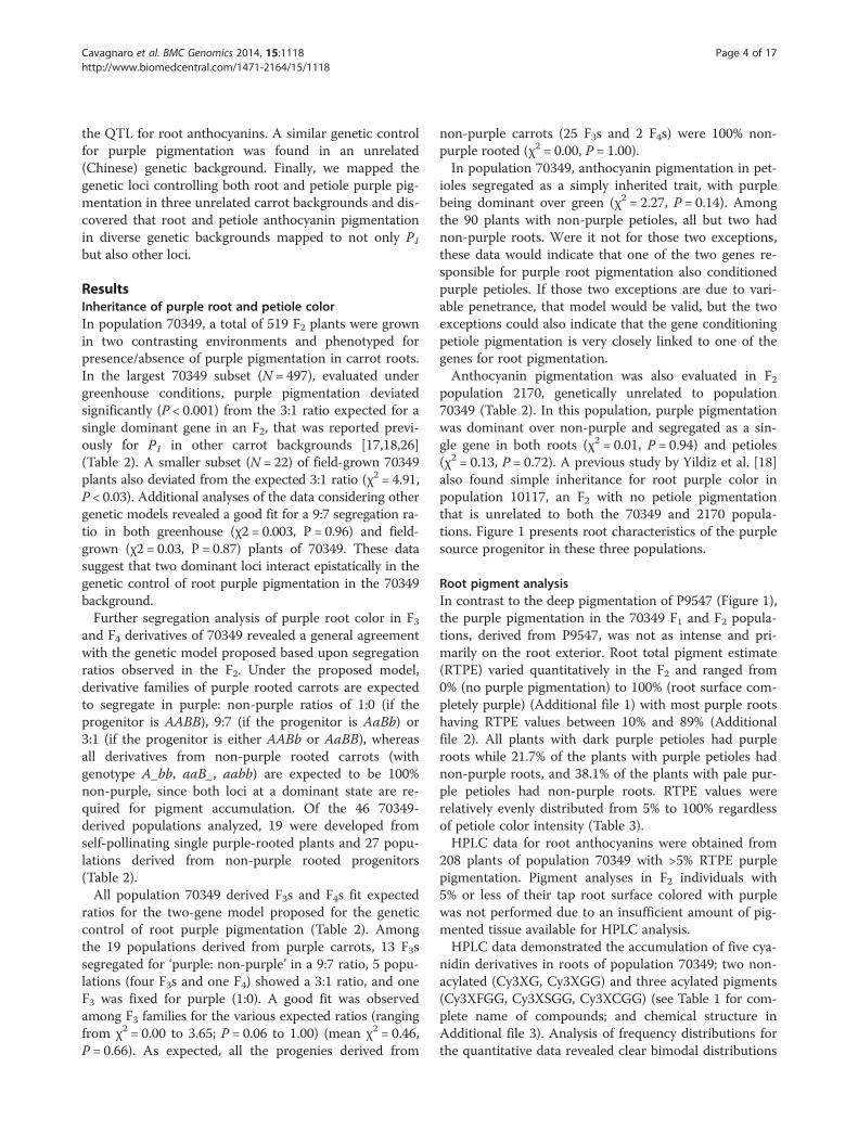
Comparative mapping of anthocyanin traitsIn addition to the 70349 mapping population, the pheno-typic traits controlling purple root and petiole pigmenta-tion, and several common markers (SNPs, SSRs) andgenes (FLS, F3H, PSY2) were mapped in two other popula-tions, 10117 and 2170. Both developed from crosses be-tween purple- and orange-rooted carrots, where thepurple progenitor was genetically and phenotypically dif-ferent from the genetic source of purple pigmentation inpopulation 70349 and from each other (see MethodsFigure 1).Comparative map analysis of CH3 from 70349, 2170
and 10117 was performed (Figure 3). The use of sixdata-points in common across the three maps, includingmarkers tightly linked (<5 cM) to all the anthocyanintraits mapped in each chromosome allowed accuratemap comparisons and revealed high marker collinearityacross the three maps.In population 10117, purple root pigmentation was
previously found to be conditioned by a single dominantgene, P1, [17] which mapped to CH3 [18]. The SNPmarker K0627 tightly linked to P3 was included in the43.1-55.1 cM region of chromosome 3 in population70349, and was also tightly linked to four co-localizedQTL for anthocyanin glycosides. This region (i.e., markerK0627) also mapped to CH3 of population 10117 but wasrelatively distant (27.3 cM) from P1 (Figure 3). In addition,two markers (K0149 and PSY2) in chromosome 3 ofpopulation 70349 that were tightly linked to Raa1, thelocus controlling anthocyanin acylation, and to three over-lapping QTL for acylated and non-acylated anthocyanin

Figure 3 Comparative mapping of loci on chromosome 3 controlling anthocyanin pigmentation in three carrot genetic backgrounds.The F2 mapping populations 70349, 2170 and 10117 were developed using different purple-rooted genotypes as progenitors. All linkage groupscorrespond to CH3 of the respective maps. Anthocyanin traits, indicated in red and italic font, include the loci controlling purple root and petiolepigmentation in populations 70349 and 2170 (P3), root pigmentation in 10117 (P1), and Raa1, the locus conditioning root anthocyanin acylationin population 70349. The support interval for the QTL for root total pigment estimate (RTPE) in population 70349 is indicated in red. Commonmarkers across CHs are denoted in bold and connected by horizontal dotted lines. The following genes are denoted in italics: F3H (flavanone3-hydroxylase), FLS (flavanone 3'hydroxylase), PSY2 (phytoene synthase-2).
Cavagnaro et al. BMC Genomics 2014, 15:1118 Page 10 of 17http://www.biomedcentral.com/1471-2164/15/1118
pigments mapped to CH3 of population 10117 at a dis-tance of 16.8-17.8 cM away from P1. These data clearly in-dicated that the loci controlling anthocyanin pigmentationin roots of diverse genetic backgrounds differ, and those inthe 70349 population are different from P1.In population 2170 the F1 plants were all purple-
rooted and had purple petioles, whereas in the F2, purplepigmentation segregation was consistent with a single
gene model (χ2 = 0.01, P = 0.94) with dominance for pur-ple over non-purple.. Root and petiole pigmentationcompletely co-segregated in this population, indicatingthat the same gene controls pigmentation in both or-gans. This gene mapped with 16 molecular markers (6SSRs, 8 SNPs and 2 gene-specific markers), conforminga linkage group of 49 cM, with an average distance be-tween adjacent markers of 2.45 cM (Figure 3). The use

Cavagnaro et al. BMC Genomics 2014, 15:1118 Page 11 of 17http://www.biomedcentral.com/1471-2164/15/1118
of chromosome-anchoring markers [34] allowed the orien-tation and assignment of this LG to carrot chromosome 3.In CH3 of 2170, the locus controlling purple pigmen-
tation in roots and petioles was tightly linked to K0627,the same marker that was tightly linked to P3, RTPE andfour other anthocyanin QTL in 70349, suggesting thatthe same chromosome region controls root and petioleanthocyanin pigmentation in 2170 and 70349, and thisregion clearly differs from P1. Thus, we surmise that thelocus controlling root purple and petiole pigmentationin 2170 was P3 (Figure 3).
DiscussionDevelopment of the first high resolution gene-derivedSNP-based linkage map in the ApiaceaeThe development of transcriptome-base SNP markersfrom D. carota next-generation sequencing data [35] hasopened the possibility for performing cost-effective high-throughput genotyping of carrot mapping and breedingpopulations. From an initial screening of 4000 SNPmarkers developed from Expressed Sequence Tag (EST)data [35], we constructed a carrot linkage map with mapresolution for a sequence-based marker map unprece-dented in the Apiaceae. The resulting genetic map har-bored 895 polymorphic molecular markers covering635.1 cM, with an average map resolution of 1.3 cM. Todate, only 117 markers with known sequence informa-tion have been mapped in carrot [18,30,31] with themost dense map containing 73 sequence-based markers(mainly SSRs) and a resolution of 7.4 cM [30]. Othermaps were mainly based on anonymous dominant AFLP,RAPD, RFLP and DArT markers [26,28,33]. The totallength of the map (635.1 cM) was similar to the maplength found in other studies (534.4 cM [26]; 668.7 cM[31]) but smaller than the linkage maps developed byCavagnaro et al. [30] (1180 – 1273.2 cM), Just et al.[29] (1055–1210.5 cM), Grzebelus et al. [32] (1297.9 -1504.2 cM). The observed differences in total map lengthamong these studies are most likely attributable to the dif-ferent software and algorithms used for linkage analysis.While the present study, as well as those of Vivek andSimon [26] and Alessandro et al. [31] used JoinMap [36],the maps of Cavagnaro et al. [30], Just et al. [29] andGrzebelus et al. [32] used Mapmaker [37]. These two pro-grams use very different mathematical and statistical pro-cedures to construct the linkage groups [38] and these canaccount for the observed variation among carrot mapsizes. Several linkage mapping studies have noted thatmarker distance and linkage group lengths were consist-ently larger with Mapmaker than JoinMap, regardless ofthe organism and the type of marker used [39-43]. Thisdifference was observed even when both programs usedthe same mapping function [42,43]. Because the multilocus-likelihood method used by Mapmaker assumes an absence
of crossover interference while linkage maps constructedwith JoinMap are more accurate and shorter when suchinterference is present [39,42].Another source of variation that may contribute to
size differences observed among the carrot maps is thetype of marker used. Uncorrected genotyping errorsoften detected with dominant AFLP markers, such asthose predominantly used in earlier carrots maps withhigher map size, can drastically increase the calculatedmap lengths [44,45]. Such error is unlikely in our co-dominant SNP-based map. Marker scores across eachlinkage group were verified using CheckMatrix to elimin-ate miss-scored markers that may generate false doublecross-overs (false double cross-overs can substantially in-crease linkage group length), therefore providing a solidsupport to the marker order.The linkage map constructed in this study provides an
unprecedented opportunity for integrating carrot geneticmaps. Most of the SSR and gene-specific markers in-cluded in our map represent common data points acrossseveral linkage maps constructed for various carrot traits[18,29-31] as well as chromosome anchoring markers[34]. In addition, over 300 SNPs mapped in this studyare shared with a recently developed linkage map con-taining QTL for nematode resistance [46]. Integration ofcarrot genetic maps, including our dense SNP-based map,will rapidly increase marker density and include inform-ative sequence-based markers relative to previously unsat-urated map regions containing important traits. This willallow for straight forward identification of candidate genesto characterize traits with the upcoming carrot genome se-quence [47].High resolution linkage maps constructed with sequence-
based markers are crucial for aiding in the assembly ofnext-generation sequence data used in genome sequencingprojects. The map developed in this study, with 895sequence-based markers and a resolution of 1.3 cM, iscurrently being used to facilitate the assembly of the carrotgenome sequence.
Genetics of anthocyanin pigmentation in carrotBased on examination of the structure of the five cyani-din glycosides present in carrot roots (Additional file 3),together with available information on anthocyanin bio-chemistry in carrot [48,49] and other species [10,50-53],a proposed scheme for cyanidin modification in carrot ispresented in Figure 4. Based on this scheme, an inversebalance between the content of nonacylated Cy3XGGand acylated Cy3XCGG, Cy3XFGG, and Cy3XSGG isexpected, with the balance modulated by the activity ofan acyltransferase.In the present study, the strong negative correlation
found between the non acylated Cy3XGG and the totalcontent of acylated anthocyanin forms (r = −0.99, P < 0.001)

Figure 4 Proposed anthocyanin biochemical pathway noting pigments and genes (in italics) involved in anthocyanin formation andcompound modification in carrot roots. In bold are anthocyanin pigments detected by HPLC in this and previous studies. Abbreviationscorrespond to the pigments full names, as in Table 1.
Cavagnaro et al. BMC Genomics 2014, 15:1118 Page 12 of 17http://www.biomedcentral.com/1471-2164/15/1118
strongly suggests that acylation of Cy3XGG, presumablyby an acyltransferase (in agreement with the proposedscheme in Figure 4), causes this shift from non-acylatedto acylated anthocyanin forms. In addition, the co-localization of a large-effect QTL for Cy3XGG and twoQTL for the major acylated anthocyanins (Cy3XSGG andCy3XFGG) in a very narrow map region (3.6 cM) ofchromosome 3, not only reinforces the notion thatCy3XGG is the chemical substrate for acylation, givingrise to Cy3XSGG and Cy3XFGG, but also suggests thatthis region is largely responsible for the genetic controlof root anthocyanin acylation. Furthermore, our com-bined pigment and linkage analysis data for Cy3XGGprovide evidence that suggests a single dominant genecontrolling this trait. The clear bimodal distribution ofCy3XGG in the F2, a segregation ratio compatible with asimply inherited dominant trait, and the co-localizationof this trait mapped both as a QTL and as single gene, allsuggest that indeed a single dominant gene controls‘high’ versus ‘low’ content of acylated anthocyanins inroots of 70349. We name this gene Raa1.In addition to Raa1, two QTL with relatively smaller
phenotypic effects conditioning anthocyanin acylationwere detected in CH3, 23 cM apart from Raa1, andCH1, suggesting a complex regulation for modificationof carrot pigments, with several genes probably influen-cing acylation.Because acylation of carrot anthocyanins influences
bioavailability [11,20-22], an important property of dietarynutrients in human health, and it also influences pigmentstability [19,21,23], important for the use of pigments infoods and as dyes, further characterization of Raa1 is ofinterest. With the carrot genome sequence nearing com-pletion [47], in combination with the sequence-basedmarkers tightly linked to Raa1 in the current 70349 gen-etic map, searching for candidate genes will be straightforward. A detailed characterization of Raa1 may have apositive impact in carrot breeding programs aiming atdeveloping carrots with higher nutraceutical value (e.g.,with increased content of non-acylated anthocyanins)and also carrots with more stable pigments (i.e., withhigh proportion of acylated anthocyanins) for the foodindustry.A previous study in carrot demonstrated that acylation
of anthocyanins is important for transport into the vacu-oles [54], suggesting that acylation may play an important
role for the accumulation of stable pigments in carrotroots. However, in the present study, only a weak signifi-cant correlation (0.34) was found between purple rootpigmentation (RTPE) and one of the acylated compounds(Cy3XSGG). In addition, Raa1, which conditions highversus low content of acylated anthocyanins, did not co-localize with either of the two RTPE QTL in the linkagemap, suggesting that acylation does not have a major ef-fect on the accumulation of root anthocyanins in the70349 genetic background.
Mapping of anthocyanin QTLThe study presented here represents the first compre-hensive QTL analysis of anthocyanin pigmentation andpigment modification performed in carrot. Using quanti-tative data we identified 15 QTL covering eight genomicregions associated with accumulation of four cyanidinderivatives and with the percentage of purple pigmenta-tion (RTPE) in carrot root. Consistent with the two-genemodel observed for root purple color segregation in F2-F4 families, two QTL on chromosomes 3 and 1 were de-tected to account for variation in RTPE.With the exception of the Cy3XGG QTL in the 27.0-
27.7 cM region of CH3, the QTL with the largest pheno-typic effect for all the anthocyanin glycosides and forroot total pigment content, which explained 36.6 - 59.2%of the variation, were associated with the 43.1-50.1 cMregion of CH3 and best associated with marker K0627.We propose that this QTL region includes a gene thatcontrols the primary regulatory mechanism underlyinganthocyanin accumulation. Detection of ten additionalQTL covering six genomic regions associated with theaccumulation of individual anthocyanin compounds, lo-cated on chromosome 2, 3, 4, 6 and 8 indicates the com-plexity of the genetic control of anthocyanin accumulationin carrot. Based on studies in other crops these QTL couldbe involved in anthocyanin biosynthesis, accumulation,and chemical modification (e.g., glycosylation, acylation)necessary for the stable storage of these pigments, as hasbeen reported for anthocyanin accumulation and diver-sification in other species. For example, in raspberry,Bushakra et al. [55] detected 24 QTL associated with theaccumulation and diversity of different cyanidin deriva-tives, while in grapes, Fournier-Level et al. [56] detectedmultiple QTL associated with cyanidin methylation andaccumulation.

Cavagnaro et al. BMC Genomics 2014, 15:1118 Page 13 of 17http://www.biomedcentral.com/1471-2164/15/1118
Synteny and putative orthologous anthocyanin lociamong carrot mapsSeveral carrot maps for various traits of interest, includ-ing anthocyanin and carotenoid pigmentation, have beendeveloped [18,26,29,30] but common markers acrosslinkage maps carrying similar traits in different geneticbackgrounds were usually lacking in these previousstudies.In the present study, the use of sequence-based markers
for chromosome 3 in common across three linkage mapsof carrot harboring root and petiole anthocyanin traits re-vealed high synteny among the maps (Figure 3). This, incombination with the use of markers tightly linked to theanthocyanin mapped traits, revealed that the genetic fac-tors conditioning root pigmentation in populations 70349(QTL-RTPE) and 2170 (P3) are different from P1, previ-ously reported in 10117 [18]. The syntenic correspond-ence between RTPE (in 70349) and P3 (in 2170) and thetight linkage of both traits to marker K0627 in both mapssuggests that this region controls root anthocyanin accu-mulation in both genetic backgrounds.Purple carrots played an important role in the domes-
tication of carrot as a tap root crop [63]. Based uponhistorical documents, it is believed that the first domes-ticated carrot roots were purple and yellow and suchdomestication occurred in Central Asia, with orange car-rots representing a secondary and European domestica-tion event [63]. Our comparative map analysis foranthocyanin accumulation in carrot represents not onlya breeding tool but also provides further insight into car-rot domestication. The fact that two different genes con-trol anthocyanin root pigmentation in unrelated geneticbackgrounds suggests that independent mutations andhuman selection events may have contributed to thedomestication of purple carrots. Petiole anthocyanin pig-mentation segregated as a simply-inherited trait in popu-lations 70349 and 2170, and was mapped for the firsttime. Furthermore, in this study, we present clear evi-dence for pleiotropy at a single gene controlling bothpetiole and root pigmentation in these two populations.This is in contrast to a previous study which found, in apopulation not included in this study, that root pig-mentation and petiole pigmentation were controlled bydifferent genes [17]. Coincidently with our results inpopulations 70349 and 2170, a single gene, AN1, has beenfound to condition anthocyanin pigmentation in differenttissues (tuber skin and foliage) of potato [63-65]. To-gether, these findings emphasize the observation thatanthocyanin biosynthesis, accumulation and structure is acomplex trait in carrot, as has been observed in severalother plant species. Even beyond identifying candidategenes for QTL identified in this study, more genes condi-tioning this trait are likely to be discovered elsewhere inthe carrot genome.
Despite the high synteny observed among the threemaps, their total length varied considerably. While themap lengths were comparable between population 2170(49.0 cM) and population 10117 (55.5 cM), the formermaps were shorter than the 70349 map of CH3 (86.1 cM).This is likely due to the fewer number of markers mappedin 2170 and 10117 compared to 70349, with the formermaps lacking markers in one (10117) or both (2170) ofthe terminal chromosome ends, as is evident from mapcomparisons in figure 3. The mapping of these eightsequence-based and common markers allowed for a closecomparison of these 3 maps, and will contribute to mapintegration while increasing marker informativeness andmarker saturation in the 2170 and 10117 backgrounds.More importantly, this will result in an immediate in-crease in marker saturation in nearby regions of P1, P3and Raa1, which will facilitate tracking these genes inbreeding programs, and will further facilitate searchingfor candidate genes in the upcoming carrot genomesequence.
ConclusionsAn informative high resolution SNP-based genetic mapof carrot was constructed and used for detection andmapping of root anthocyanin QTL. In population 70349,the mapping of 15 QTL across 6 carrot chromosomessuggests a more complex regulation for this trait thanpreviously described in a previously unstudied geneticbackground, with many genes probably influencing pig-ment biosynthesis, compound modification and/or accu-mulation. Of particular interest, a 12 cM region ofchromosome 3 harboring co-localized QTL with largestphenotypic effect for root total pigment content (RTPE)and four anthocyanin glycosides, as well as the simply-inherited gene P3, which was largely responsible for thegenetic control of anthocyanin pigmentation in bothroots and petioles. In addition, quantitative data analysisfor each anthocyanin compound provided the opportun-ity to identify a large effect QTL and a simply-inheritedgene, Raa1, that was also on chromosome 3 and associ-ated with anthocyanin acylation. Comparative map ana-lysis revealed that neither of these chromosome regionsthat were associated with anthocyanin pigmentation andacylation, respectively, corresponded to the P1 gene ini-tially reported for root purple pigmentation.The present study increases the current knowledge on
the genetic control for this complex trait in carrot.These data, together with the molecular resources gener-ated herein (e.g. the high-resolution SNP map and thedevelopment of markers in common across differentlinkage maps) and the upcoming carrot genome se-quence will facilitate cloning of the genetic factors con-trolling both anthocyanin accumulation and acylation.Characterization of such genes will have a positive impact

Cavagnaro et al. BMC Genomics 2014, 15:1118 Page 14 of 17http://www.biomedcentral.com/1471-2164/15/1118
on carrot breeding programs aiming at increasing pigmentconcentration and anthocyanin profiles, depending on theend-market use.
MethodsPlant materialInheritance of purple pigmentation was studied in F2, F3and F4 families derived from an initial cross betweenP4201 and B6320. P4201 is an inbred line with purpleouter phloem and yellow xylem storage roots and purpleleaves that was derived from a cross between inbredP9547 (Figure 1), with purple xylem and phloem rootcolor derived from Central Anatolia, and B2566, an in-bred with orange root color from diverse Europeansources. B6320 is an inbred with orange roots and greenpetioles derived from the European open-pollinated cul-tivars Nantes and Camberly. A single F1 plant with pur-ple root outer phloem and yellow xylem, and purpleleaves, was self-pollinated to produce the F2 population70349 (N = 519), which was used for genetic mappingstudies. For phenotyping, plants of the F2 populationwere grown in pots under greenhouse conditions in2007. After phenotyping as described below, rootswere vernalized at 1–2°C, planted at the University ofWisconsin West Madison Agricultural Research Station(WMARS), and individual plants were self-pollinated toproduce F3 families. A subset of the F2 and the F3 familieswere field-grown at El Centro, California, in 2009, andphenotyped for root pigmentation. Individual F3 plantsunderwent another cycle of self-pollination and their F4progenies were grown in Madison-WI and phenotypedin 2012.For comparison purposes, linkage analyses were also
performed in two mapping populations unrelated topopulation 70349 but also segregating for root and peti-ole purple pigmentation. Population 2170 was an F3family (N = 65) derived from a cross between a purplerooted carrot with purple leaves derived from an inter-cross between PI652188 (a purple carrot from China,and the ultimate purple source in 2170) and PI326011,an orange-rooted European carrot with green leaves. AnF2 plant of this family with the same phenotype as thepurple parent was self-pollinated to generate the F3 fam-ily evaluated. The other mapping population, 10117, wasan F2 family (N = 72) derived from a cross betweenB1896 (a true-breeding inbred with yellow roots derivedfrom a cross between PI173687, a population from east-ern Turkey segregating for presence or absence of pur-ple root color, and B493, an inbred with orange rootsand green petioles derived from diverse European sources)and B7262 (Figure 1), an inbred with purple outer phloemand orange inner phloem and xylem, and purple leavesfrom the same cross as B1896). This population was previ-ously characterized in more detail by Yildiz et al. [18].
Thus, the ultimate genetic source of purple color in thethree mapping populations in this study varied geograph-ically and phenotypically.
Phenotyping and segregation analysisPhenotyping purple color in 70349 and its F3 - F4 deriva-tives was performed on the basis of the presence or ab-sence of purple pigmentation in carrot roots as describedby Simon [17]. In addition, the percentage of purple pig-mentation covering the root surface, which estimates theroot total pigment content in the 70349 background, wasscored visually and recorded in 70349 (from here on re-ferred to as root “RTPE” for “root total pigment estimate”).Root tissues from purple carrots were sampled for antho-cyanin HPLC analysis. Purple pigmentation in leaves of70349 was evaluated on the basis of presence or absenceof purple color in the leaf petioles, as described previously[17]. Phenotyping was done on a presence/absence basisas in this previous study for anthocyanin pigmentation inroots and petioles of population 2170 and roots of popula-tion 10117. Data from the F2, F3 and F4 carrot segregatingpopulations were analyzed using the χ2 method to test theratios expected for each character.
HLPC analysisTissue samples containing visible purple pigmentationwere obtained from roots of the 70349 mapping popula-tion. The samples were lyophilized and anthocyaninswere extracted with acidified methanol, followed byhigh-performance liquid chromatography (HPLC) ana-lysis of anthocyanin pigments as described by Kurilichet al. [11]. Table 1 lists the five major carrot anthocyaninpigments (all cyanidin derivatives) identified and quanti-fied in the present study. The data were expressed aspercentage concentration of a given pigment relative tothe total anthocyanin content, which derived from thesum of the content of the five individual anthocyanincompounds. Pairwise Spearman rank correlation valuesamong anthocyanin pigments and traits were obtainedusing SAS v.9.1 [57].
Construction of the framework linkage map in 70349Total genomic DNA of individual plants from 70349,2170 and 10117 populations was isolated from lyophi-lized leaves following the protocol described by Murrayand Thompson [58] and quantified using Pico Green(Invitrogen, Paisley, UK).The linkage map was constructed using 187 F2 individ-
uals from 70349. A collection of 4000 published SNPsdeveloped from carrot transcriptome data [35] and 40published SSR markers with known chromosome location[30] were used. SNPs were genotyped using the KASParchemistry, which is a competitive allele-specific PCR SNPgenotyping system using FRET quencher cassette oligos

Cavagnaro et al. BMC Genomics 2014, 15:1118 Page 15 of 17http://www.biomedcentral.com/1471-2164/15/1118
(http://www.lgcgroup.com). SNPs were coded with a ‘K’followed by four digit numbers (e.g. K0001). SNPs locatedwithin carotenoid or anthocyanin biosynthetic genes werelabeled according to the gene abbreviation. SSR primerpairs were evaluated using a fluorescent method asdescribed before [59].JoinMap 4.0 software [36] was used for mapping. Scores
of all markers used for mapping were converted intogenotype codes using the A/H/B system for co-dominantand A/C, B/D system for dominant markers segregating inF2 population. The linkage groups (LGs) were obtained ata LOD threshold value >3.0. Regression mapping algo-rithm and Haldane’s mapping function was used to calcu-late genetic distances among marker loci. Markers andgenotypes with more than 10% of missing data were ex-cluded from the analysis. The degree of marker segrega-tion distortion in the F2 was determined by marker datacomparison against the expected 3:1 and 1:2:1 ratio fordominant and codominant markers, respectively, usingChi square tests, where significant distortion was declaredat P < 0.01 [60]. The marker order in each linkage groupwas examined for inconsistencies leading to false doublerecombination events using CheckMatrix (http://www.atgc.org/XLinkage). Markers with more than one incon-sistent score were removed. In order to reduce the com-plexity of the final map figure, redundant markers,considering as such those that had no recombinationamong them and therefore shared the same map position,were removed from the image, leaving a single marker -themarker with the least amount of missing data- per mapposition. A complete list with all the mapped markers,including redundant markers, is provided in Additionalfile 6.Map positions of SSR markers and SNP markers cor-
responding to anthocyanin and carotenoid biosyntheticgenes with known chromosome location [18,30] wereused to anchor the linkage groups to carrot chromo-somes. After chromosome assignment, LGs were ori-ented and numbered according to the chromosomeorientation and classification of Iovene et al. [34].
QTL analysisQTL analysis was performed using R/qtl with the mul-tiple imputations method [61]. QTL detection includedpreliminary QTL identification using scanone followedby QTL modeling. Briefly, the QTL with the largestLOD value from the ‘scanone’ analysis was added to theQTL model and if the model was significant, the QTLwas retained. This process was then repeated using‘addqtl’ instead of ‘scanone’, followed by QTL modelingand testing for interactions among the QTL, until add-ing additional QTL to the model was no longer signifi-cant. The support intervals for the map locations ofQTL were calculated using a 1.5 LOD drop interval
which is considered the best approximation to a 95%support interval in QTL mapping.
Comparative mapping of anthocyanin traits acrossdiverse carrot genetic backgroundsIn addition to the 70349 mapping population, the pheno-typic traits controlling root and petiole purple pigmenta-tion, and several common markers (SNPs, SSRs) andgenes (FLS, F3H, PSY2) were mapped in F2 populations10117 and 2170, which were unrelated to 70349 (see sub-section ‘plant material’). The anthocyanin genes F3H (fla-vanone 3-hydroxylase) and FLS (flavanone 3'hydroxylase),and the carotenoid gene PSY2 (phytoene synthase-2), aswell as the SSRs used, were known to locate in chromo-some 3 based upon results from previous linkage studiesin carrot [18,29].Primer pairs for 6 SNPs selected from CH3 of 70349,
as well as for FLS, F3H and PSY2 were designed usingPrimer3 software [62] and used in polymerase chain re-actions (PCR) of 20 μl final volume containing 1X DNApolymerase buffer, 0.02 mM of each dNTP, 0.25 μM ofeach primer, 0.2 μl Taq polymerase (MBI, Fermentas,USA) and ~50 ng of genomic DNA. PCR conditionswere: initial denaturation at 94°C for 2 min, followed by25 cycles of 94°C for 30 sec, appropriate annealingtemperature for 30 sec, and 72°C for 45 sec, and a finalstep at 72°C for 10 min. Sequencing reactions were per-formed in 5 μl final volume consisting of 1.75 μl ofwater, 1 μl of 5 μM primer, 0.75 μl of 5 × BigDye®3.1 se-quencing buffer, 0.5 μl of Big-Dye®3.1 and 1 μl of PCRproduct, previously diluted 1:10 with water. Big dye re-action conditions were 25 cycles of 96°C for 10 sec, and58°C for 2 min, and a final step at 72°C for 5.0 min. Se-quencing was performed at the University of WisconsinBiotechnology Center, and sequence analysis usedSequencher software version 4.8 (GeneCodes Corpor-ation, Ann Arbor, MI). SSR markers were evaluatedusing a fluorescent method as described by Iorizzo et al.[59]. Phenotyping and mapping of purple pigmentationin roots and leaves of 2170, and in roots of 10117, wasdescribed above.
Additional files
Additional file 1: Phenotypic variation for root total pigmentcontent (RTPE) in 70349.
Additional file 2: Frequency distribution of ‘root total pigmentcontent’ (RTPE) and of individual anthocyanin pigment compoundsin roots of 70349.
Additional file 3: Molecular structure of anthocyanins from carrotroot.
Additional file 4: Figure S1. Pigment distribution and correlationanalysis among cyanidin derivatives and ‘root total pigment content’(RTPE) in 70349 population. Table S2. Pair-wise Spearman rank correlationvalues among root anthocyanin pigments in 70349.

Cavagnaro et al. BMC Genomics 2014, 15:1118 Page 16 of 17http://www.biomedcentral.com/1471-2164/15/1118
Additional file 5: Marker genotype analysis in individuals of 70349mapping population displayed vertically across the nine carrotchromosomes using Checkmatrix.
Additional file 6: Complete list of mapped markers per chromosome.
Additional file 7: Allele interaction analysis for closest markersassociated with QTL for anthocyanin compounds and root totalpigmentation (RTPE).
AbbreviationsCHs: Chromosomes; Cy3XGG: Cy-3-(2”-xylose-6-glucose-galactoside);Cy3XG: Cy-3-(2”-xylose-galactoside); Cy3XSGG: Cy-3-(2”-xylose-6”-sinapoyl-glucose-galactoside); Cy3XFGG: Cy-3-(2”-xylose-6”-feruloyl-glucose-galactoside; Cy3XCGG: Cy-3-(2”-xylose-6”-(4-coumuroyl)glucose-galactoside);DArT: Diversity arrays technology; HPLC: High-performance liquidchromatography; QTL: Quantitative trait loci; RT: Retention time; RTPE: Roottotal pigment estimate; SNP: Single nucleotide polymorphism; SSR: Simplesequence repeat.
Competing interestsThe authors declare that they have no competing interests.
Authors’ contributionsPC and MI participated in designing experiments, generated phenotypicdata, carried out the molecular genetic and QTL studies and analyses, anddrafted the manuscript. MY participated in plant phenotyping, HPLC analysis,and mapping. DS participated in HPLC, marker, and mapping datageneration and analysis. JP and SE participated in data generation, analysis,mapping, and data interpretation. PS developed genetic stocks, conceived ofthe study, participated in its design and coordination, and helped to draftthe manuscript. All authors read and approved the final manuscript.
AcknowledgementsThe authors acknowledge Anne E. Atkins for valuable assistance on plantphenotyping and marker evaluation. This work was supported by theAgricultural Research Service, United States Department of Agriculture.
Author details1Department of Horticulture, University of Wisconsin-Madison, 1575 LindenDrive, Madison, WI 53706, USA. 2CONICET, Facultad de Ciencias Agrarias –Universidad Nacional de Cuyo, and INTA E.E.A. La Consulta, Ex Ruta 40. km96, La Consulta CC 8, Mendoza 5567, Argentina. 3Department of AgriculturalBiotechnology, Faculty of Agriculture, Yuzuncu Yil University, 65080 Van,Turkey. 4USDA-Agricultural Research Service, Vegetable Crops Unit, Universityof Wisconsin-Madison, 1575 Linden Drive, Madison, WI 53706, USA.
Received: 11 September 2014 Accepted: 11 December 2014Published: 16 December 2014
References1. Harborne JB, Williams CA: Advances in flavonoid research since 1992.
Phytochemistry 2000, 55:481–504.2. Koes RE, Quattrocchio F, Mol JNM: The flavonoid biosynthetic pathway in
plants: function and evolution. Bioessays 1993, 16:123–132.3. Holton TA, Cornish EC: Genetics and biochemistry of anthocyanin
biosynthesis. Plant Cell 1995, 7:1071–1083.4. Dixon RA, Palva NL: Stress-induced phenylpropanoid metabolism. Plant
Cell 1995, 7:1085–1097.5. Shirley BW: Flavonoid biosynthesis: “new” functions for an “old” pathway.
Trends Plant Sci 1996, 1:377–382.6. Ross JA, Kasum CM: Dietary flavonoids: bioavailability, metabolic effects,
and safety. Annu Rev Nutr 2002, 22:19–34.7. Manach C, Williamson G, Morand C, Scalbert A, Rémésy C: Bioavailability
and bioefficacy of polyphenols in humans. I. review of 97 bioavailabilitystudies. Am J Clin Nutr 2005, 81:230S–242S.
8. Herrmann KM, Weaver LM: The shikimate pathway. Annu Rev Plant PhysiolPlant Mol Biol 1999, 50:473–503.
9. Jaakola L, Määttä K, Pirttilä AM, Törrönen R, Kärenlampi S, Hohtola A:Expression of genes involved in anthocyanin biosynthesis in relation toanthocyanin, proanthocyanidin, and flavonol levels during bilberry fruitdevelopment. Plant Physiol 2002, 130:729–739.
10. Gould KS, Davies K, Winefield C: Anthocyanins: Biosynthesis, Functions, andApplications. New York: Springer; 2008.
11. Kurilich AC, Clevidence BA, Britz SJ, Simon PW, Novotny JA: Plasma and urineresponses are lower for acylated vs nonacylated anthocyanins from rawand cooked purple carrots. J Agric Food Chem 2005, 5:6537–6542.
12. Montilla EC, Arzaba MR, Hillebrand S, Winterhalter P: Anthocyanincomposition of black carrot (Daucus carota ssp. sativus var. atrorubensAlef.) cultivars antonina, beta sweet, deep purple, and purple haze.J Agric Food Chem 2011, 59:3385–3390.
13. Jung CS, Griffith HM, De Jong DM, Cheng S, Bodis M, De Jong WS: Thepotato P locus codes for flavonoid 3050-hydroxylase. Theor Appl Genet2005, 110:269–275.
14. Lalusin AG, Ohta M, Fujimura T: Temporal and spatial expression of genesinvolved in anthocyanin biosynthesis during sweet potato (Ipomoeabatatas (L.) Lam.) root development. Int J Plant Sci 2006, 167:249–256.
15. Lu QN, Yang Q: cDNA cloning and expression of anthocyaninbiosynthetic genes in wild potato (Solanum pinnatisectum). Afr JBiotechnol 2006, 5:811–818.
16. Mano H, Ogasawara F, Sato K, Higo H, Minobe Y: Isolation of a regulatorygene of anthocyanin biosynthesis in tuberous roots of purple-fleshedsweet potato. Plant Physiol 2007, 143:1252–1268.
17. Simon PW: Inheritance and expression of purple and yellow storage rootcolor in carrot. J Hered 1996, 87:63–66.
18. Yildiz M, Willis DK, Cavagnaro PF, Iorizzo M, Abak K, Simon PW: Expressionand mapping of anthocyanin biosynthesis genes in carrot. Theor AppliedGenetics 2013, 126:1689–1702.
19. Pojer E, Mattivi F, Johnson D, Stockley CS: The case for anthocyaninconsumption to promote human health: a review. Compr Rev Food Sci F2013, 12:483–508.
20. Novotny JA, Clevidence BA, Kurilich AC: Anthocyanin kinetics aredependent on anthocyanin structure. Br J Nutr 2012, 107:504–509.
21. Charron CS, Kurilich AC, Clevidence BA, Simon PW, Harrison DJ, Britz AJ,Baer DJ, Novotny JA: Bioavailability of anthocyanins from purple carrotjuice: effects of acylation and plant matrix. J Agric Food Chem 2009,57:1226–1230.
22. Netzel M, Netzel G, Kammerer DR, Schieber A, Carle R, Simons L, Bitsch I,Bitsch R, Konczak I: Cancer cell antiproliferation activity and metabolismof black carrot anthocyanins. Innov Food Sci Emerg Technol 2007,8:365–372.
23. Malien-Aubert C, Dangles O, Amiot MJ: Color stability of commercialanthocyanin-based extracts in relation to the phenolic composition.protective effects by intra- and intermolecular copigmentation. J AgricFood Chem 2001, 49:170–176.
24. Harborne JB: A unique pattern of anthocyanins in Daucus carota andother Umbelliferae. Biochem Syst Ecol 1976, 4:31–35.
25. Dooner HK, Robbins TP, Jorgensen RA: Genetic and developmental controlof anthocyanin biosynthesis. Annu Rev Genet 1991, 25:173–199.
26. Vivek BS, Simon PW: Linkage relationships among molecular markers andstorage root traits of carrot (Daucus carota L. ssp. sativus). Theor ApplGenet 1999, 99:58–64.
27. Boiteux LS, Belter JG, Roberts PA, Simon PW: RAPD linkage map of thegenomic region encompassing the rootknot nematode (Meloidogynejavanica) resistance locus in carrot. Theor Appl Genet 2000, 100:439–446.
28. Santos CAF, Simon PW: QTL analyses reveal clustered loci foraccumulation of major provitamin A carotenes and lycopene in carrotroots. Mol Genet Genomics 2002, 268:122–129.
29. Just BJ, Santos CAF, Fonseca MEN, Boiteux LS, Oloizia BB, Simon PW:Carotenoid biosynthesis structural genes in carrot (Daucus carota): isolation,sequence-characterization, single nucleotide polymorphism (SNP) markersand genome mapping. Theor Appl Genet 2007, 114:693–704.
30. Cavagnaro PF, Chung SM, Manin S, Yildiz M, Ali A, Alessandro MS, Iorizzo M,Senalik DA, Simon PW: Microsatellite isolation and marker developmentin carrot-genomic distribution, linkage mapping, genetic diversityanalysis and marker transferability across Apiaceae. BMC Genomics 2011,12:386.
31. Alessandro MS, Galmarini CR, Iorizzo M, Simon PW: Molecular mapping ofvernalization requirement and fertility restoration genes in carrot. TheorAppl Genet 2013, 126:415–423.
32. Grzebelus D, Jagosz B, Simon PW: The DcMaster transposon display mapspolymorphic insertion sites in the carrot (Daucus carota L.) genome.Gene 2007, 390:67–74.

Cavagnaro et al. BMC Genomics 2014, 15:1118 Page 17 of 17http://www.biomedcentral.com/1471-2164/15/1118
33. Grzebelus D, Iorizzo M, Senalik D, Ellison S, Cavagnaro PF, Macko-PodgorniA, Heller-Uszynska K, Kilian A, Nothnagel T, Allender C, Simon PW, BaranskiR: Diversity, genetic mapping, and signatures of domestication in thecarrot (Daucus carota L.) genome, as revealed by Diversity ArraysTechnology (DArT) markers. Mol Breeding 2014, 33:625–637.
34. Iovene M, Cavagnaro PF, Senalik D, Buell CR, Jiang J, Simon PW:Comparative FISH mapping of Daucus species (Apiaceae family).Chromosome Res 2011, 19:493–506.
35. Iorizzo M, Senalik D, Ellison S, Grzebelus D, Cavagnaro PF, Spooner D, VanDeynze A, Simon PW: Development of a high-throughput SNP resource toadvance genomic, genetic and breeding research in carrot (Daucus carota L.).In Plant and Animal Genome XXI Conference, 12–16 January. San Diego,California: Scherago International; 2013.
36. Van Ooijen JW: JoinMap 4.0: Software for the Calculation of Genetic LinkageMaps in Experimental Populations. Wageningen, Netherlands: Kyazma B.V;2006.
37. Lander E, Green P, Abrahamson J, Barlow A, Daly M, Lincoln S, Newburg L:MAPMAKER: an interactive computer package for constructing primarygenetic linkage maps of experimental and natural populations. Genomics1987, 1:174–181.
38. Cheema J, Dicks J: Computational approaches and software tools forgenetic linkage map estimation in plants. Brief Bioinform 2009,10:595–608.
39. Hong Y, Chen X, Liang X, Liu H, Zhou G, Li S, Wen S, Holbrook CC, Guo B:A SSR-based composite genetic linkage map for the cultivated peanut(Arachis hypogaea L.) genome. BMC Plant Biol 2010, 27:10–17.
40. Sewell MM, Sherman BK, Neale DB: A consensus map for loblolly pine(Pinus taeda L.). I. construction and integration of individual linkagemaps from two outbred three-generation pedigrees. Genetics 1999,151:321–330.
41. Gosselin I, Zhou Y, Bousquet J, Isabel N: Megagametophyte-derivedlinkage maps of white spruce (Picea glauca) based on RAPD, SCAR andESTP markers. Theor Appl Genet 2002, 104:987–997.
42. Qi X, Stam P, Lindhout P: Comparison and integration of four barleygenetic maps. Genome 1996, 39:379–394.
43. Liebhard R, Koller B, Gianfranceschi L, Gessler C: Creating a saturatedreference map for the apple (Malus x domestica Borkh.) genome. TheorAppl Genet 2003, 106:1497–508.
44. Cartwright DA, Troggio M, Velasco R, Gutin A: Genetic mapping in thepresence of genotyping errors. Genetics 2007, 176:2521–2527.
45. Hackett CA, Broadfoot LB: Effects of genotyping errors, missing valuesand segregation distortion in molecular marker data on the constructionof linkage maps. Heredity 2003, 90:33–38.
46. Parsons DJ: QTL for Meloidogyne Incognita Root Knot Nematode Resistanceand other Traits of Interest in Carrot (Daucus carota), PhD thesis. University ofWisconsin, Horticulture Department; 2014.
47. Iorizzo M, Senalik D, Ellison S, Cavagnaro PF, Cheng S, Zheng P, Zheng Z,Van Deynze A, Simon PW: The building of the first Apiaceae genome.In Plant and Animal Genome XXII Conference, 11–15 January. San Diego,California: Scherago International; 2014.
48. Gläßgen W, Seitz H: Acylation of anthocyanins with hydroxycinnamicacids via 1-O-acylglucosides by protein preparations from cell cultures ofDaucus carota L. Planta 1992, 186:582–585.
49. Rose A, Gläßgen W, Hopp W, Seitz H: Purification and characterization ofglycosyltransferases involved in anthocyanin biosynthesis in cell-suspension cultures of Daucus carota L. Planta 1996, 198:397–403.
50. He F, Mu L, Yan GL, Liang NN, Pan QH, Wang J, Reeves MJ, Duan CQ:Biosynthesis of anthocyanins and their regulation in colored grapes.Molecules 2010, 15:9057–9091.
51. Montefiori M, Espley RV, Stevenson D, Cooney J, Datson PM, Saiz A,Atkinson RG, Hellens RP, Allan AC: Identification and characterisation ofF3GT1 and F3GGT1, two glycosyltransferases responsible for anthocyaninbiosynthesis in red-fleshed kiwifruit (Actinidia chinensis). Plant J 2011,65:106–118.
52. Yonekura-Sakakibara K, Fukushima A, Nakabayashi R, Hanada K, Matsuda F,Sugawara S, Inoue E, Kuromori T, Ito T, Shinozaki K, Wangwattana B,Yamazaki M, Saito K: Two glycosyltransferases involved in anthocyaninmodification delineated by transcriptome independent componentanalysis in Arabidopsis thaliana. Plant J 2012, 69:154–167.
53. Goulas V, Vicente AR, Manganaris GA: Structural diversity of anthocyaninsin fruits. In Anthocyanins: Structure, Biosynthesis and Health Benefits.
Chapter 10. Edited by Motohashi N. Hauppauge: Nova Science PublishersInc; 2012:225–250.
54. Hopp W, Seitz HU: The uptake of acylated anthocyanin into isolatedvacuoles from a cell suspension culture of Daucus carota. Planta 1987,170:74–85.
55. Bushakra JM, Krieger C, Deng D, Stephens MJ, Allan AC, Storey R, SymondsVV, Stevenson D, McGhie T, Chagné D, Buck EJ, Gardiner SE: QTL involvedin the modification of cyanidin compounds in black and red raspberryfruit. Theor Appl Genet 2013, 126:847–865.
56. Fournier-Level A, Hugueney P, Verriès C, This P, Ageorges A: Geneticmechanisms underlying the methylation level of anthocyanins in grape(Vitis vinifera L.). BMC Plant Biol 2011, 11:179.
57. SAS 9.1.3 Help and Documentation. SAS Institute, Inc. Cary, NC; 2000–2004.58. Murray M, Thompson W: Rapid isolation of high-molecular weight plant
DNA. Nucleic Acids Res 1980, 8:4321–4325.59. Iorizzo M, Senalik DA, Grzebelus D, Bowman M, Cavagnaro PF, Matvienko M,
Ashrafi H, Van Deynze A, Simon PW: De novo assembly andcharacterization of the carrot transcriptome reveals novel genes, newmarkers, and genetic diversity. BMC Genomics 2012, 12:389.
60. Vuylsteke M, Mank R, Antonise R, Bastiaans E, Senior ML, Stuber CW,Melchinger AE, Lubberstedt T, Xia XC, Stam P, Zabeau M, Kuiper M: Twohigh-density AFLP linkage maps of Zea mays L.: analysis of distributionof AFLP markers. Theor Appl Genet 1999, 99:921–935.
61. Broman KW, Sen S: A Guide to QTL Mapping with R/qtl. New York: Springer;2009.
62. Rozen S, Skaletsky HJ: Primer3. 1998, [http://www-genome.wi.mit.edu/genome_software/other/primer3.html]
63. Banga O: Origin and domestication of the western cultivated carrot.Genetica Agraria 1963, 17:357–370.
64. De Jong H: Inheritance of anthocyanin pigmentation in the cultivatedpotato: a critical review. Am. Potato J 1991, 68:585–593.
65. D’Amelia V, Aversano R, Batelli G, Caruso I, Moreno MC, Castro-Sanz AB,Chiaiese P, Fasano C, Palomba F, Carputo D: High An1 variability andinteraction with basic helix-loop-helix co-factors related to anthocyninbiosynthesis in potato leaves. Plant J 2014, doi:10.1111/tpj.12653.
doi:10.1186/1471-2164-15-1118Cite this article as: Cavagnaro et al.: A gene-derived SNP-based highresolution linkage map of carrot including the location of QTLconditioning root and leaf anthocyanin pigmentation. BMC Genomics2014 15:1118.
Submit your next manuscript to BioMed Centraland take full advantage of:
• Convenient online submission
• Thorough peer review
• No space constraints or color figure charges
• Immediate publication on acceptance
• Inclusion in PubMed, CAS, Scopus and Google Scholar
• Research which is freely available for redistribution
Submit your manuscript at www.biomedcentral.com/submit
Related Documents