A Fusion Intermediate gp41 Immunogen Elicits Neutralizing Antibodies to HIV-1 * Received for publication, April 4, 2014, and in revised form, August 1, 2014 Published, JBC Papers in Press, August 26, 2014, DOI 10.1074/jbc.M114.569566 Rachel P. J. Lai ‡1,2 , Miriam Hock §¶1,3 , Jens Radzimanowski §¶4 , Paul Tonks ‡ , David Lutje Hulsik §¶5 , Gregory Effantin §¶6 , David J. Seilly ‡ , Hanna Dreja ‡7 , Alexander Kliche , Ralf Wagner , Susan W. Barnett**, Nancy Tumba ‡‡ , Lynn Morris ‡‡ , Celia C. LaBranche §§ , David C. Montefiori §§ , Michael S. Seaman ¶¶ , Jonathan L. Heeney ‡8 , and Winfried Weissenhorn §¶9 From the ‡ Department of Veterinary Medicine, University of Cambridge, Madingley Road, Cambridge CB3 0ES, United Kingdom, § Universite ´ Grenoble Alpes, Unit of Virus Host Cell Interactions (UVHCI), F-38000 Grenoble, France, ¶ CNRS, UVHCI, F-38000 Grenoble, France, Institute of Medical Microbiology and Hygiene, University of Regensburg, Franz-Josef-Strauss-Allee 11, 93053 Regensburg, Germany, **Novartis Vaccines and Diagnostics Inc., Cambridge, Massachusetts 02139, ‡‡ National Institute for Communicable Diseases, Centre for HIV and Sexually Transmitted Infections, 1 Modderfontein Road, Sandringham 2131, South Africa, §§ Department of Surgery, Duke University Medical Center, Durham, North Carolina 27710, and ¶¶ Division of Viral Pathogenesis, Beth Israel Deaconess Medical Center, Boston, Massachusetts 02115 Background: HIV-1 gp41 MPER is a target for inducing broadly neutralizing antibodies. Results: Gp41 int folds into a compact elongated structure that induces neutralizing antibodies upon immunization. Conclusion: Presentation of gp41 int in a lipid environment is beneficial to induce neutralizing antibodies. Significance: Membrane-anchored gp41 int is a promising antigen to improve breadth and potency of anti-gp41 antibody responses. The membrane-proximal external region (MPER) of the human immunodeficiency virus, type 1 (HIV-1) envelope glyco- protein subunit gp41 is targeted by potent broadly neutralizing antibodies 2F5, 4E10, and 10E8. These antibodies recognize lin- ear epitopes and have been suggested to target the fusion inter- mediate conformation of gp41 that bridges viral and cellular membranes. Anti-MPER antibodies exert different degrees of membrane interaction, which is considered to be the limiting factor for the generation of such antibodies by immunization. Here we characterize a fusion intermediate conformation of gp41 (gp41 int -Cys) and show that it folds into an elongated 12- nm-long extended structure based on small angle x-ray scatter- ing data. Gp41 int -Cys was covalently linked to liposomes via its C-terminal cysteine and used as immunogen. The gp41 int -Cys proteoliposomes were administered alone or in prime-boost regimen with trimeric envelope gp140 CA018 in guinea pigs and elicited high anti-gp41 IgG titers. The sera interacted with a peptide spanning the MPER region, demonstrated competition with broadly neutralizing antibodies 2F5 and 4E10, and exerted modest lipid binding, indicating the presence of MPER-specific antibodies. Although the neutralization potency generated solely by gp140 CA018 was higher than that induced by gp41 int - Cys, the majority of animals immunized with gp41 int -Cys pro- teoliposomes induced modest breadth and potency in neutral- izing tier 1 pseudoviruses and replication-competent simian/ human immunodeficiency viruses in the TZM-bl assay as well as responses against tier 2 HIV-1 in the A3R5 neutralization assay. Our data thus demonstrate that liposomal gp41 MPER formu- lation can induce neutralization activity, and the strategy serves to improve breadth and potency of such antibodies by improved vaccination protocols. The initial antibody response following acute HIV-1 10 infec- tion arises 13 days following the onset of viremia and is tar- geted to the gp41 region of the envelope glycoprotein (Env) (1). Although these antibodies are highly mutated and polyreactive (2), they are non-neutralizing and do not select for escape vari- ants (3). Neutralizing antibodies (nAbs) appear only months after infection when the antigen-specific B cells have under- gone multiple rounds of somatic hypermutation. The quest for new broadly neutralizing antibodies (bnAbs) over the last few years revealed that a substantial percentage of HIV-1-infected individuals eventually develop broad and potent neutralizing antibodies that act against a large variety of strains. These bnAbs map to four major antigenic sites on Env, including the * This work was supported in part by the Bill and Melinda Gates Foundation- funded Consortium of AIDS Vaccine Discovery (Grant 38637), the Compre- hensive Antibody-Vaccine Immune Monitoring Consortium (Grant 38619), and Sidaction (to W. W.). 1 Both authors contributed equally to this work. 2 Present address: Division of Mycobacterial Research, National Inst. for Med- ical Research, The Ridgeway, London NW7 1AA, UK. 3 Supported by a postdoctoral fellowship from Sidaction. 4 Supported by Labex Grenoble Alliance for Integrated Structural Cell Biology (GRAL) Grant ANR-10-LABX-49-01. 5 Present address: Bionovion B.V., Molenweg 50, 5349 AC Oss, The Netherlands. 6 Supported by a postdoctoral fellowship from the Agence nationale de recherche ´ sur le sida et les he ´ patites virales. 7 Present address: Abcam, 330 Cambridge Science Park, Cambridge CB4 0FL, UK. 8 To whom correspondence may be addressed. E-mail: [email protected]. 9 To whom correspondence may be addressed. E-mail: [email protected]. 10 The abbreviations used are: HIV-1, human immunodeficiency virus, type 1; MPER, membrane-proximal external region; SAXS, small angle x-ray scatter- ing; Env, envelope glycoprotein; SHIV, simian/human immunodeficiency virus; nAb, neutralizing antibody; bnAb, broadly neutralizing antibody; gp41 int , intermediate conformation of gp41; Bicine, N,N-bis(2-hydroxyethyl)- glycine; HR, heptad repeat region; fd, fold-on domain. THE JOURNAL OF BIOLOGICAL CHEMISTRY VOL. 289, NO. 43, pp. 29912–29926, October 24, 2014 © 2014 by The American Society for Biochemistry and Molecular Biology, Inc. Published in the U.S.A. 29912 JOURNAL OF BIOLOGICAL CHEMISTRY VOLUME 289 • NUMBER 43 • OCTOBER 24, 2014 at MRC National Institute for Medical Research on June 2, 2015 http://www.jbc.org/ Downloaded from

Welcome message from author
This document is posted to help you gain knowledge. Please leave a comment to let me know what you think about it! Share it to your friends and learn new things together.
Transcript

A Fusion Intermediate gp41 Immunogen Elicits NeutralizingAntibodies to HIV-1*
Received for publication, April 4, 2014, and in revised form, August 1, 2014 Published, JBC Papers in Press, August 26, 2014, DOI 10.1074/jbc.M114.569566
Rachel P. J. Lai‡1,2, Miriam Hock§¶1,3, Jens Radzimanowski§¶4, Paul Tonks‡, David Lutje Hulsik§¶5,Gregory Effantin§¶6, David J. Seilly‡, Hanna Dreja‡7, Alexander Kliche�, Ralf Wagner�, Susan W. Barnett**,Nancy Tumba‡‡, Lynn Morris‡‡, Celia C. LaBranche§§, David C. Montefiori§§, Michael S. Seaman¶¶,Jonathan L. Heeney‡8, and Winfried Weissenhorn§¶9
From the ‡Department of Veterinary Medicine, University of Cambridge, Madingley Road, Cambridge CB3 0ES, United Kingdom,§Universite Grenoble Alpes, Unit of Virus Host Cell Interactions (UVHCI), F-38000 Grenoble, France, ¶CNRS, UVHCI, F-38000Grenoble, France, �Institute of Medical Microbiology and Hygiene, University of Regensburg, Franz-Josef-Strauss-Allee 11, 93053Regensburg, Germany, **Novartis Vaccines and Diagnostics Inc., Cambridge, Massachusetts 02139, ‡‡National Institute forCommunicable Diseases, Centre for HIV and Sexually Transmitted Infections, 1 Modderfontein Road, Sandringham 2131, SouthAfrica, §§Department of Surgery, Duke University Medical Center, Durham, North Carolina 27710, and ¶¶Division of ViralPathogenesis, Beth Israel Deaconess Medical Center, Boston, Massachusetts 02115
Background: HIV-1 gp41 MPER is a target for inducing broadly neutralizing antibodies.Results: Gp41int folds into a compact elongated structure that induces neutralizing antibodies upon immunization.Conclusion: Presentation of gp41int in a lipid environment is beneficial to induce neutralizing antibodies.Significance: Membrane-anchored gp41int is a promising antigen to improve breadth and potency of anti-gp41 antibodyresponses.
The membrane-proximal external region (MPER) of thehuman immunodeficiency virus, type 1 (HIV-1) envelope glyco-protein subunit gp41 is targeted by potent broadly neutralizingantibodies 2F5, 4E10, and 10E8. These antibodies recognize lin-ear epitopes and have been suggested to target the fusion inter-mediate conformation of gp41 that bridges viral and cellularmembranes. Anti-MPER antibodies exert different degrees ofmembrane interaction, which is considered to be the limitingfactor for the generation of such antibodies by immunization.Here we characterize a fusion intermediate conformation ofgp41 (gp41int-Cys) and show that it folds into an elongated �12-nm-long extended structure based on small angle x-ray scatter-ing data. Gp41int-Cys was covalently linked to liposomes via itsC-terminal cysteine and used as immunogen. The gp41int-Cysproteoliposomes were administered alone or in prime-boostregimen with trimeric envelope gp140CA018 in guinea pigs andelicited high anti-gp41 IgG titers. The sera interacted with apeptide spanning the MPER region, demonstrated competitionwith broadly neutralizing antibodies 2F5 and 4E10, and exerted
modest lipid binding, indicating the presence of MPER-specificantibodies. Although the neutralization potency generatedsolely by gp140CA018 was higher than that induced by gp41int-Cys, the majority of animals immunized with gp41int-Cys pro-teoliposomes induced modest breadth and potency in neutral-izing tier 1 pseudoviruses and replication-competent simian/human immunodeficiency viruses in the TZM-bl assay as well asresponses against tier 2 HIV-1 in the A3R5 neutralization assay.Our data thus demonstrate that liposomal gp41 MPER formu-lation can induce neutralization activity, and the strategy servesto improve breadth and potency of such antibodies by improvedvaccination protocols.
The initial antibody response following acute HIV-110 infec-tion arises �13 days following the onset of viremia and is tar-geted to the gp41 region of the envelope glycoprotein (Env) (1).Although these antibodies are highly mutated and polyreactive(2), they are non-neutralizing and do not select for escape vari-ants (3). Neutralizing antibodies (nAbs) appear only monthsafter infection when the antigen-specific B cells have under-gone multiple rounds of somatic hypermutation. The quest fornew broadly neutralizing antibodies (bnAbs) over the last fewyears revealed that a substantial percentage of HIV-1-infectedindividuals eventually develop broad and potent neutralizingantibodies that act against a large variety of strains. ThesebnAbs map to four major antigenic sites on Env, including the
* This work was supported in part by the Bill and Melinda Gates Foundation-funded Consortium of AIDS Vaccine Discovery (Grant 38637), the Compre-hensive Antibody-Vaccine Immune Monitoring Consortium (Grant 38619),and Sidaction (to W. W.).
1 Both authors contributed equally to this work.2 Present address: Division of Mycobacterial Research, National Inst. for Med-
ical Research, The Ridgeway, London NW7 1AA, UK.3 Supported by a postdoctoral fellowship from Sidaction.4 Supported by Labex Grenoble Alliance for Integrated Structural Cell Biology
(GRAL) Grant ANR-10-LABX-49-01.5 Present address: Bionovion B.V., Molenweg 50, 5349 AC Oss, The Netherlands.6 Supported by a postdoctoral fellowship from the Agence nationale de
recherche sur le sida et les hepatites virales.7 Present address: Abcam, 330 Cambridge Science Park, Cambridge CB4 0FL,
UK.8 To whom correspondence may be addressed. E-mail: [email protected] To whom correspondence may be addressed. E-mail: [email protected].
10 The abbreviations used are: HIV-1, human immunodeficiency virus, type 1;MPER, membrane-proximal external region; SAXS, small angle x-ray scatter-ing; Env, envelope glycoprotein; SHIV, simian/human immunodeficiencyvirus; nAb, neutralizing antibody; bnAb, broadly neutralizing antibody;gp41int, intermediate conformation of gp41; Bicine, N,N-bis(2-hydroxyethyl)-glycine; HR, heptad repeat region; fd, fold-on domain.
THE JOURNAL OF BIOLOGICAL CHEMISTRY VOL. 289, NO. 43, pp. 29912–29926, October 24, 2014© 2014 by The American Society for Biochemistry and Molecular Biology, Inc. Published in the U.S.A.
29912 JOURNAL OF BIOLOGICAL CHEMISTRY VOLUME 289 • NUMBER 43 • OCTOBER 24, 2014
at MR
C N
ational Institute for Medical R
esearch on June 2, 2015http://w
ww
.jbc.org/D
ownloaded from

CD4 binding site (4 –9), the V1-V2 loop-specific antibodies thatrecognize glycopeptides (10 –13), glycan V3-directed antibod-ies (11), and the membrane-proximal external region (MPER)of gp41 (14). A common feature of these bnAbs is their highextent of somatic mutations, a prerequisite to achieve broadneutralization and high potency (15).
The MPER is an attractive target because it is a highly con-served, tryptophan-rich region that plays a critical role in mem-brane fusion and virus infectivity (16 –18). Anti-MPER anti-bodies 2F5, Z13e1, 4E10, and 10E8 target overlapping linearepitopes within MPER (14, 17, 19 –24). The nAbs 2F5 and 4E10contain hydrophobic heavy chain CDR3 residues that mediatelipid binding (25, 26) and are required for neutralization (27–30). This potential polyreactivity, which may be distinct frompathogenic autoantibodies (31), was suggested to be a majorhindrance to induce such antibodies because of their naturalelimination through B cell tolerance mechanisms (32–34). Themost potent anti-MPER antibody, 10E8, was initially reportedto lack autoreactivity (14), but a recent study challenges this anddemonstrates modest 10E8 membrane binding activity (35).However, 10E8-like activity is found in a large number ofpatient sera with broad neutralizing activity, demonstratingthat not all anti-MPER antibodies undergo clonal deletion (14),which is in contrast to other studies suggesting that bnAbs tothe MPER are rare in infected patients or vaccinated naïve indi-viduals (36 –39). Further underlining the importance of anti-MPER antibodies, passive immunization strategies containingnAbs 2F5 and 4E10 protected animals from SHIV infection(40).
A main target of anti-MPER antibodies is a fusion interme-diate conformation of gp41 (35, 41, 42) that forms during thereceptor binding-induced transitions from native Env (43– 46)to the postfusion conformation (47–50). The interaction with atransitional state of Env is further supported by the interactionof nAb Z13e1 with the open CD4-induced Env conformation(51). MPER may become membrane-embedded during entry,and it has been suggested that anti-MPER antibodies extractthe epitopes from the bilayer (52, 53).
A number of vaccine approaches targeting the gp41 MPERhave been attempted to elicit bnAbs (for a review, see Ref. 54).These include the generation of structurally stabilized peptides,scaffolds, or chimeras (55– 64); chimeric virus or virus-like par-ticles by engrafting the MPER antibody epitopes onto otherviruses (65–78); gp41 fusion proteins (79 – 84); and liposomescontaining MPER peptides (85). Different prime-boost strate-gies with peptides, DNA, and whole Env protein have also beenexplored in these studies. Although the described immunogensinduced antibody titers to gp41, only a few studies demon-strated modest neutralization activity depending on the neu-tralization assay used. Notably, guinea pigs immunized withvirus-like particles carrying chimeras of gp41 and hemaggluti-nin produced sera with modest neutralization of selected tier 1and tier 2 pseudoviruses using the stringent TZM-bl assay (65).Furthermore, immunization of llamas with gp41 proteolipo-somes led to the isolation of an anti-MPER nanobody that neu-tralized a small number of tier 1 and tier 2 viruses as a bihead(86). Other studies indicate that the postfusion conformationpresenting MPER is capable of inducing neutralizing antibodies
(87, 88), consistent with the finding that mAb 10E8 interactswith this conformation in vitro (35).
Here we structurally characterized the intermediate confor-mation of gp41 (gp41int) and coupled it covalently to liposomes,which were then administered alone or in combination withsoluble gp140 in guinea pigs. Immunization was performedwith a mixture of two adjuvants, Carbopol-971P and MF59,that preserved the liposomal structure prior to immunization.Analyses of the postimmune sera demonstrated strong gp41-specific IgG responses, the presence of antibodies targetingMPER, and neutralizing activity against a panel of tier 1 and tier2 HIV-1 viruses. Our study thus indicates the benefit of a mem-brane environment in the induction of neutralizing antibodiesby gp41int.
EXPERIMENTAL PROCEDURES
Ethics Statement—The animal study was carried out in strictaccordance with the United Kingdom Animals (Scientific Pro-cedure) Act 1986, and the protocol was approved by the localEthical and Welfare Committee of the University of Cambridgeand the United Kingdom Home Office (Project license number80/2238).
Recombinant Gp140 Purification—HIV-1 gp140CA018 is anA/G recombinant subtype Env derived from a Cameroonpatient. The gp140 glycoprotein was purified using a publishedprotocol with Galanthus nivalis agarose affinity column fol-lowed by diethylaminoethyl-Sepharose and ceramic hydroxy-apatite columns to remove all contaminants (89). The purifiedglycoprotein was concentrated using an Amicon YM-30 (30-kDa-cutoff) ultrafiltration disc (Millipore) and stored at �80 °Cuntil use.
Gp41 Protein Expression and Purification—The gp41 con-structs are based on the HXB2 group M subtype B sequence.Gp41int-Cys and gp41int-fd contain gp41 residues 584 – 684.Part of gp41 heptad repeat region 1 (HR1) is N-terminally fusedin-frame with the GCN4 trimerization domain (90). Gp41int-fdcontains a C-terminal fold-on trimerization domain renderinggp41int-fd similar to the reported GCN-gp41-inter construct(91). Both cysteines at positions 598 and 604 have been mutatedto serine. Gp41int-fd has the following sequence: MAQIED-KIEEILSKIYHIENEIARIKKLIGEAERYLKDQQLLGIWGSSG-KLISTTAVPWNASWSNKSLEQIWNNMTWMEWDREIN-NYTSLIHSLIEESQNQQEKNEQELLELDKWASLWNWFN-ITNWLWYIKGYIPEAPRDGQAYVRKDGEWVLLSTFL(underlined amino acids correspond to pIIGCN4, italicizedamino acids correspond to gp41 HR1, and underlined S corre-sponds to cystein residues changed to serine). The cDNA wascloned into the expression vector pETM-13.
Gp41int-Cys is similar to gp41int-fd but has the C-terminalfold-on domain replaced by a single cysteine. The sequence ofthe construct is as follows: MAQIEDKIEEILSKIYHIENEIARI-KKLIGEAERYLKDQQLLGIWGSSGKLISTTAVPWNASWS-NKSLEQIWNNMTWMEWDREINNYTSLIHSLIEESQNQQ-EKNEQELLELDKWASLWNWFNITNWLWYIKSC. ThecDNA encoding the fusion protein was cloned into the expres-sion vector pETM-13 (European Molecular Biology LaboratoryHeidelberg). For small angle x-ray scattering (SAXS) analysisgp41int-Cys contained an extra tobacco etch virus protease site
gp41int Induces Neutralizing Antibodies
OCTOBER 24, 2014 • VOLUME 289 • NUMBER 43 JOURNAL OF BIOLOGICAL CHEMISTRY 29913
at MR
C N
ational Institute for Medical R
esearch on June 2, 2015http://w
ww
.jbc.org/D
ownloaded from

and a FLAG tag at the C terminus. This construct shows aslightly higher solubility.
Gp41int-fd was expressed in Escherichia coli strain Rosetta 2(DE3) (Novagen). Cells were grown to an A600 nm of 0.6 andinduced with 1 mM isopropyl 1-thio-�-D-galactopyranoside at37 °C. After 4 h, cells were harvested by centrifugation and lysedin Tris buffer (20 mM Tris, pH 8.5, 0.1 M NaCl). The solublefraction was discarded, and the pellet was resuspended in Trisbuffer supplemented with 0.1% Triton X-100 (Sigma) overnightat 4 °C. Solubilized gp41int-fd in Tris, 0.1% Triton X-100 bufferwas bound to a Q-Sepharose column, and Triton X-100 wasremoved by excessive washing with Tris buffer. Gp41int-fd waseluted by applying a 0.05– 0.5 M NaCl gradient in Tris buffer. Afinal purification step included size exclusion chromatographyon a Superose 6 column in 20 mM Bicine, pH 9.3, 100 mM NaCl.Gp41int-Cys was expressed and purified by following the sameprotocol as for gp41int-fd with the addition of 10 mM DTT in allbuffers. A final purification step included size exclusion chro-matography on a Superose 6 column in 20 mM Tris, pH 8.5, 100mM NaCl, 10 mM DTT. Final gp41int-Cys yields were 10 mg/li-ter of E. coli culture, indicating that production can be scaled upand is suitable for good manufacturing practice production forfurther immunization trials.
SAXS Analysis of Gp41int-Cys—X-ray scattering data werecollected on beam line BM-29 (European Synchrotron Radia-tion Facility, Grenoble, France) at 20 °C, a wavelength of 0.9919Å, and a sample-to-detector (PILATUS 1M, DECTRIS) dis-tance of 2.849 m. The scattering intensities of the gp41int-Cyswere measured at concentrations of 0.75 and 3 mg/ml in the gelfiltration buffer. The data were normalized to the intensity ofthe incident beam, the scattering of the buffer was subtracted,and the resulting intensities were scaled for concentration.Data processing and analysis were performed using the ATSASpackage (92), and molecular weights were estimated based onthe method of Putnam et al. (93). The final merged scatteringdata were further evaluated using PRIMUS (94). The isotropicscattering intensity I(q) was transformed to the distance distri-bution function P(r) using the program GNOM, which was alsoused to calculate the particle maximum dimension Dmax (95).Dmax was considered to be optimal when the radius of gyration(Rg) obtained from the P(r) plot was equal to that obtained fromthe Guinier analysis. For ab initio modeling of the SAXS data,10 sets of independent models were calculated using Dammin(96).
Circular Dichroism Spectroscopy—All spectra were recordedon a Jasco J-810 spectropolarimeter at 20 °C. Prior to analysis,proteins were dialyzed in 10 mM potassium phosphate, pH 8.0,and the secondary structure content was calculated with theJasco Spectra Manager 2 software package.
Surface Plasmon Resonance—Surface plasmon resonanceanalysis was performed with a Biacore X3000 (GE Healthcare).As a flow buffer, 10 mM HEPES, 150 mM NaCl, pH 7.4 with0.005% P-20 was used. Gp41int-Cys was immobilized to 1000response units using 50 �g/ml protein in flow buffer on anactivated CM-5 sensor chip (GE Healthcare, BR-1000-50)according to the manufacturer’s instructions. Specific bindingto the target protein was corrected for nonspecific binding tothe deactivated control channel. The flow rate was 50 �l/min.
Regeneration of the sensor chip was achieved with 100 mM
glycine, pH 3.3, 20 mM NaOH for 60 s at 50 �l/min. mAb 2F5was injected at concentrations of 50, 100, and 250 nM, and bnAb10E8 was injected at concentrations of 1.3 and 6 �M. Data wereanalyzed with BIAevalution software version 4.1. The curveswere fitted with separate association and dissociation to obtainreasonable �2 scores and residuals.
Liposome Production and Characterization—Liposomeswere prepared with an HIV-1 virus-like lipid composition (97).The following lipids, phosphatidylserine, phosphatidylcholine,phosphatidylethanolamine, sphingomyelin, 1,2-dipalmitoyl-sn-glycero-3-phosphothioethanol (Avanti Polar Lipids, Inc.),and cholesterol were dissolved in chloroform and mixed in a1:1:1:2:0.5:4:5 ratio at a final concentration of 5 mg/ml. Aftersolvent evaporation, the lipid film was dried under vacuum.Multilamellar vesicles were obtained by resuspending the lipidfilm in PBS buffer to a final lipid concentration of 5 mg/ml. Theliposome suspension was extruded through a 100-nm mem-brane (Whatman). Gp41int-Cys was extensively dialyzedagainst Tris buffer without DTT and then mixed with the lipo-somes at a 2:1 ratio (w/w). Binding of gp41int-Cys to the lipo-somes was confirmed by sucrose gradient analysis (0 – 40%sucrose in PBS; 12 h at 40,000 rpm) and SDS-PAGE. Primingwas performed with proteoliposomes present in the 10 and 20%sucrose fractions, and boosting was performed using the com-plete mixture of gp41int-Cys and liposomes with an estimated30% of gp41int-Cys cross-linked to liposomes.
For immuno-electron microscopy (EM), gp41int-Cys proteo-liposomes were incubated with bnAb 10E8, and binding wasconfirmed by sucrose gradient centrifugation. Fractions con-taining 10E8, gp41int-Cys, and liposomes were used for immu-nogold labeling using an adapted protocol.11 A thin film of car-bon was floated in water, picked up with a bare copper grid, andallowed to dry. 4 �l of proteoliposome (gp41int-Cys and mAb10E8) solution was adsorbed for 1 min on the carbon-coatedgrid. Excess liquid was blotted away, and the grid was floated ontop of a 50-�l drop of 0.1% BSA in PBS for 1 min. After a mildblotting, the grid was floated on a 20-�l drop of protein A-golddiluted 1:10 or 1:20 in 0.1% BSA in PBS for 20 min and thenwashed in two successive 100-�l drops of water. The grid wasstained for 20 s in a 20-�l drop of 2% uranyl acetate, blotted, andallow to dry before EM observation on a JEOL 1200EX. Theexperiments in Carbopol/MF59 were identical except thatbefore staining with uranyl acetate the grid was floated on top ofa 20-�l drop of Carbopol/MF59 for 5 min and then washed infive consecutive 100-�l drops of water. Images were acquired ata nominal magnification of 20,000. The grid squares werescanned for the presence of gold on the liposomes and in thebackground. It was found that only liposomes displayed morethan three to four gold particles; concentration of gold particlesin one place was only observed on proteoliposomes and neverin the background. This observation was repeated on severalEM grids for proteoliposomes in sucrose or after dialysis in PBSand for two different concentrations of protein A-gold. A con-trol with liposomes only was performed, and it was found that
11 M. Schnos, personal communication.
gp41int Induces Neutralizing Antibodies
29914 JOURNAL OF BIOLOGICAL CHEMISTRY VOLUME 289 • NUMBER 43 • OCTOBER 24, 2014
at MR
C N
ational Institute for Medical R
esearch on June 2, 2015http://w
ww
.jbc.org/D
ownloaded from

they did not cluster the protein A-gold as did the gp41int-Cysproteoliposomes.
Immunization and Serum Collection—In the pilot study, twogroups of four Hartley white female guinea pigs were intramus-cularly immunized with gp41int-Cys (50 �g) with or withoutadjuvants at weeks 0 and 4. The adjuvant used was a combina-tion of Carbopol-971P (Lubrizol) and MF59 (Novartis Vaccinesand Diagnostics), formulated according to previous protocols(98). Carbopol-971P was first prepared as a 2% (w/v) suspen-sion in PBS before mixing 1:1 (v/v) with gp140CA018 or gp41int-Cys. The mixture was then added 1:1 (v/v) with 50 �l of MF59and incubated for another 10 min before injection. Serum sam-ples were collected at weeks �2, 2, and 6, and terminal sampleswere collected at week 8.
In the immunogenicity study, three groups of six Hartleywhite female guinea pigs each received four intramuscularimmunizations at weeks 0, 6, 12, and 18 (see Table 1). Animalsreceived gp140CA018 (50 �g) (group 1) and gp41int-Cys (50 �g)(group 2), respectively, for all four immunizations. Animals ingroup 3 were primed once at week 0 with gp140CA018 followedby three boosts with gp41int-Cys at weeks 6, 12, and 18. Serumsamples were collected 2 weeks before and 2 weeks after eachimmunization at weeks �2, 2, 8, 14, and 20, and a terminalsample was collected at week 22. All antigens were formulatedin Carbopol-971P and MF59 as described previously (98).
End Point Titer and Avidity Index ELISA—The end pointtiters of Env-specific antibodies in sera were measured bystandard ELISA. Briefly, Maxisorb 96-microwell plates (Nunc)were coated overnight with purified gp41int-fd protein (50ng/well) diluted in 100 mM carbonate-bicarbonate coatingbuffer, pH 9.6 and blocked with 5% nonfat milk in PBS contain-ing 1% Tween 20 (PBST). Serum samples were serially diluted3-fold with sample buffer (2% BSA in PBST) and added to trip-licate wells. A guinea pig-specific IgG conjugated to HRP (Jack-son ImmunoResearch Laboratories) was used as secondaryantibody (diluted 1:15,000 in sample buffer). Between eachstep, the plates were washed six times with wash buffer (PBST).The plate was developed with one-step 3,3,5,5-tetramethylben-zidine (Pierce Thermo Scientific) before being stopped with 2 M
H2SO4. Absorbance values were detected at 450 nm using aBio-Rad iMark microplate reader.
The avidity indices of vaccinated sera were measured by theirresistance to 8 M urea in binding to gp41int-fd antigen. Briefly,microwell plates were prepared and blocked as describedabove. Serum samples were diluted to give an A450 nm readoutbetween 1.0 and 1.5 in end point ELISA and were added to twosets of triplicate wells. The wells were then washed three timeswith either PBS-Tween 20 or 8 M urea in PBS-Tween 20 beforeincubation with an anti-guinea pig secondary antibody. Theplates were washed and developed with 3,3,5,5-tetramethyl-benzidine as described above. The avidity index was calculatedas the percentage of average urea-treated A450 nm/average PBS-Tween 20 A450 nm. Antisera with index values �50% were des-ignated high avidity, those with index values of 30 –50% weredesignated intermediate avidity, and those with index values�30% were designated low avidity.
Anti-lipid ELISA—Antibody response to lipids was measuredby ELISA using a protocol described previously (99). Briefly,
cholesterol, cardiolipins, phosphatidylcholine, and phosphati-dylserine (Sigma-Aldrich) were prepared in a 1:10 (v/v) solutionof chloroform and ethanol and coated onto microwell plates(200 ng/well) overnight. The ELISA was then carried out asdescribed above, and the absorbance values were measured at450 nm using a Bio-Rad iMark microplate reader.
Anti-MPER Peptide ELISA—Anti-MPER ELISAs were per-formed according to standard procedures. Briefly, MPER pep-tides having the sequence KKKNEQELLELDKWASLWN-WFDITNWLWYIRKKK (italicized amino acids are non-MPERamino acids) were coated at 50 ng/well onto microtiter plates andthen blocked with 2% BSA. Serial dilutions of serum were added intriplicate and incubated for 2 h at 16 °C. Detection of antibodybinding was carried out as described above.
Real Time Neutralizing Epitope Competition Assay—The realtime neutralizing epitope competition assay was performed asdescribed previously (98). Briefly, gp41int-fd protein was coatedat 100 ng/well onto microwell plates and then blocked with 2%BSA. The gp41int-fd protein (100 ng/well) was added in tripli-cate and incubated for 2 h at 16 °C. To determine the approxi-mate epitopes to which the polyclonal sera were raised, antiserafrom vaccinated animals were diluted according to their endpoint titer to �1 � 104 IgG to compete with Eu3�-labeledmonoclonal antibodies (mAbs) (100 ng/well) for binding toantigens. Enhancement solution (PerkinElmer Life Sciences)was added, and relative luciferase units at 620 nm were mea-sured in an Envision luminometer (PerkinElmer Life Sciences).Avidity of the antisera to the epitopes was determined by reduc-tion in the Eu3� luminescence intensity of the specific mAbwith which the serum was competing. The 2F5, 4E10, and 5F3mAbs were purchased from Polymun (Vienna, Austria), andZ13e1 was kindly provided by D. R. Burton (Scripps ResearchInstitute).
Neutralization Assay—The TZM-bl assay was carried outusing a standard protocol with molecularly cloned pseudovi-ruses (100). Briefly, pseudovirus was incubated with seriallydiluted guinea pig antisera for 1 h at 37 °C before being placedinto wells of 96-well plates seeded with TZM-bl cells. After a48-h incubation at 37 °C, the cells were lysed, and luciferasesignal in the lysate was developed with Britelite Plus substrate(1:1, v/v; PerkinElmer Life Sciences) and read in a luminometer.The A3R5 assay was done with replication-competent HIV-1inserted with Renilla luciferase reporter gene (101). The inhi-bition was carried out in the same way described above beforebeing exposed to A3R5 cells and incubated for 4 – 6 days at37 °C. The luciferase signal was developed with VivaRen sub-strate (Promega) and detected with a luminometer.
Statistical Analysis—Comparison of binding, avidity, andneutralization data between groups was performed using aMann-Whitney U test with GraphPad Prism 6.0 software.
RESULTS
Structural Characterization of Gp41int-Cys—Gp41int-Cyscontains the trimeric leucine zipper (pIIGCN4) fused in-framewith part of HR1, which prevents refolding of HR2 onto HR1,leading to the six-helical bundle postfusion conformation. ThepIIGCN4 chimera is followed by the Cys-loop region, HR2,MPER, and either the C-terminal trimerization fold-on domain
gp41int Induces Neutralizing Antibodies
OCTOBER 24, 2014 • VOLUME 289 • NUMBER 43 JOURNAL OF BIOLOGICAL CHEMISTRY 29915
at MR
C N
ational Institute for Medical R
esearch on June 2, 2015http://w
ww
.jbc.org/D
ownloaded from

(gp41int-fd) or a C-terminal cysteine (gp41int-Cys). Becausecysteines 598 and 604 formed mixed disulfide bonds asobserved previously (102), both residues were mutated to serine(Fig. 1A). Gp41int-fd and gp41int-Cys elute in single peaks at15.9 and 16.4 ml from a Superose 6 size exclusion chromatog-raphy column (Fig. 1B). Chemical cross-linking demonstratesthat they form trimers in solution. SDS-PAGE shows that themonomer band (migrating at �22 kDa) of gp41int-fd cross-links to a band migrating at �75 kDa representing the trimer.Likewise, gp41int-Cys cross-linking shifts the monomer band(�15 kDa) to a new broad band migrating at �50 kDa, whichcorresponds to a trimeric form (Fig. 1B). A small amount ofgp41int-fd and gp41int-Cys cross-link to higher molecularweight species under these conditions (Fig. 1B).
Gp41int-Cys was further analyzed by SAXS (Fig. 2A). Guinierevaluation resulted in an Rg of 3.33 nm and the maximal proteindimension (Dmax) of 117 Å was calculated by the distance dis-tribution function P(r) (Fig. 2B). The shape of gp41int-Cys wasdetermined ab initio, and the trimer model fits the experimen-tal data with the discrepancy �2 of 0.87 (Fig. 2A). Gp41int-Cys
FIGURE 2. Structural characterization of gp41int-Cys by SAXS. A, experi-mental scattering intensities obtained for gp41int-Cys are shown as a functionof resolution and after averaging and subtraction of solvent scattering. Thescattering intensities calculated from the ab initio model (presented in C) withthe lowest �2 values are shown as a blue line. B, the P(r) function of the gp41int-Cys data indicates maximal dimensions (Dmax) of 117 Å. C, molecular envelopeof the ab initio-calculated model of gp41int-Cys. The model corresponding topIIGCN4 (green ribbon) and of the gp41 part derived from the SOSIP.664gp140 trimer structure (Protein Data Bank code 4NC0; HR1, blue ribbon; HR2,red ribbon) have been docked into the SAXS envelope.
FIGURE 1. Expression constructs gp41int-fd and gp41int-Cys. A, schematicdrawing of gp41. FP, fusion peptide; C-C loop, cysteine loop; TM, transmem-brane region; Cytos., cytosolic domain; pIIGCN4, trimeric coiled coil derivedfrom the GCN4 leucine zipper fused to HR1 (90); fd, fold-on domain sequencederived from bacteriophage T4 fibritin. The numbering represents that offull-length HXB2 Env. B, both gp41int-fd and gp41int-Cys were purified on aSuperose 6 gel filtration column, and their respective profiles are shown.Notably, gp41int-fd is larger and elutes at 15.9 ml, whereas gp41int-Cys elutesat 16.4 ml. Protein from each peak fraction was cross-linked with 0.5 mM
glutaraldehyde and separated by 15% SDS-PAGE. The left panel of the insetshows that gp41int-fd is cross-linked to a band (*) migrating at �75 kDa,indicating trimer formation (the monomer band migrates at �22 kDa). Theright panel of the inset shows the cross-linking results on gp41int-Cys, whichcross-links to a new band (*) migrating at �50 kDa, representing the trimer(the monomer band migrates at �15 kDa).
gp41int Induces Neutralizing Antibodies
29916 JOURNAL OF BIOLOGICAL CHEMISTRY VOLUME 289 • NUMBER 43 • OCTOBER 24, 2014
at MR
C N
ational Institute for Medical R
esearch on June 2, 2015http://w
ww
.jbc.org/D
ownloaded from
has an elongated �106-Å-long structure with a maximal diam-eter of 37 Å at the top end, a �60-Å diameter of the middle part,and a 25-Å diameter of the bottom end (Fig. 2C). We proposethat the top end represents the HR1 coiled coil region, whichwas partly replaced by trimeric pIIGCN4. Docking thepIIGCN4-gp41 hybrid coiled coil into the SAXS envelope cor-roborates this position for the HR1 part (Fig. 2C). This is also inagreement with the molecular fit of the gp41 coordinates fromthe SOSIP.664 gp140 crystal structure (43), which fit into thetop end and the middle part of the SAXS envelope (Fig. 2C). TheSOSIP.664 gp140 crystal structure lacks the Cys-loop region,which could be accommodated by the middle region, and thebottom end would provide space for the missing MPER part(Fig. 2C). The relatively small 25-Å diameter of the bottom endindicates that all three chains are in close proximity prior toentering the membrane, consistent with a trimerization role forthe transmembrane region. We conclude that this potentialintermediate conformation of gp41 forms a compact and elon-gated trimer that will bridge the viral and cellular membranewith a distance of 110 –120 Å depending on the missing 30N-terminal amino acids (fusion peptide-proximal region andthe fusion peptide).
Secondary structure analysis by circular dichroism revealed�55% helices, 8% �-strands, 7% turns, and 30% random coilstructures. This indicates a relatively low secondary structurecontent for the rest of gp41 (the Cys-loop region and HR2)because the coiled coil structures of pIIGCN4 and part of HR1account for 29% helices.
Although the fusion intermediate construct gp41-inter,which is very similar to gp41int-Cys, was extensively tested forbinding to neutralizing anti-MPER antibodies (35, 91), wetested gp41int-Cys for binding to bnAbs 10E8 and 2F5. A nota-ble difference between gp41-inter and gp41int-Cys is that thelatter lacks the C-terminal fold-on trimerization domain (Fig.1A). Surface plasmon resonance measurements showed nano-molar affinities for both antibodies. MAb 2F5 interacts with adissociation constant of 5.3 nM, and mAb 8E10 interacts with adissociation constant of 57 nM. We conclude that gp41int-Cyshas a mostly �-helical, elongated structure that interacts withbnAbs 2F5 and 10E8.
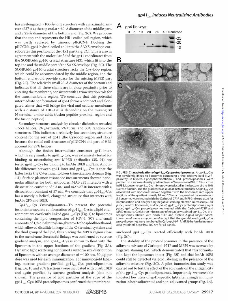
Gp41int-Cys Proteoliposomes—To present the potentialfusion intermediate conformation of gp41int-Cys in a lipid envi-ronment, we covalently linked gp41int-Cys (Fig. 1) to liposomescontaining the lipid composition of HIV-1 (97) and smallamounts of 1,2-dipalmitoyl-sn-glycero-3-phosphothioethanol,which allowed disulfide linkage of the C-terminal cysteine andthe thiol group of the lipid, thus placing the MPER region closeto the membrane. Successful linkage was confirmed by sucrosegradient analysis, and gp41int-Cys is shown to float with theliposomes in the upper fractions of the gradient (Fig. 3A).Dynamic light scattering confirmed a uniform size distributionof liposomes with an average diameter of �100 nm. 50 �g perdose was used for each immunization. For immunogold label-ing, sucrose gradient-purified gp41int-Cys proteoliposomes(Fig. 3A, 10 and 20% fractions) were incubated with bnAb 10E8and again purified by sucrose gradient analysis (data notshown). The presence of gold particles at the edge of thegp41int-Cys/10E8 proteoliposomes confirmed that membrane-
anchored gp41int-Cys reacted efficiently with bnAb 10E8(Fig. 3C).
The stability of the proteoliposomes in the presence of theadjuvant mixture of Carbopol-971P and MF59 was assessed bynegative staining EM, which demonstrated that this formula-tion kept the liposomes intact (Fig. 3B) and that bnAb 10E8could still be detected via gold labeling in the presence of theadjuvant mixture (Fig. 3C). A pilot immunization study wascarried out to test the effect of the adjuvants on the antigenicityof the gp41int-Cys proteoliposomes. Importantly, we were ableto detect low titers of gp41-specific IgG after a single immuni-zation in both adjuvanted and non-adjuvanted groups (Fig. 4A).
FIGURE 3. Characterization of gp41int-Cys proteoliposomes. A, gp41int-Cyswas covalently linked to liposomes containing a thiol-reactive lipid (1,2-fi-palmitoyl-sn-hlycero-3-phosphothioethanol), and proteoliposomes werepurified on a sucrose density gradient from 40% sucrose in PBS to 0% sucrosein PBS. Liposome gp41int-Cys mixtures were placed in the bottom of the 40%sucrose fraction, and the gradient was spun at 40,000 rpm for 6 h. Gp41int-Cysassociated with liposomes moved together with the liposomes into upperfractions of the gradient (mostly 10 and 20% sucrose; marked by an asterisk).B, liposomes were treated with the Carbopol-971P and MF59 mixture used forimmunization and analyzed by negative staining electron microscopy. Leftpanel, control liposomes; middle panel, gp41int-Cys proteoliposomes; rightpanel, gp41int-Cys proteoliposomes treated with the Carbopol-971P andMF59 mixture. C, electron microscopy of negatively stained gp41int-Cys pro-teoliposomes labeled with bnAb 10E8 and protein A-gold (upper panel).Lower panel, same as upper panel except that the gold-labeled gp41int-Cysproteoliposomes were incubated in Carbopol-971P/MF59 before being neg-atively stained. Scale bar, 200 nm for all panels.
gp41int Induces Neutralizing Antibodies
OCTOBER 24, 2014 • VOLUME 289 • NUMBER 43 JOURNAL OF BIOLOGICAL CHEMISTRY 29917
at MR
C N
ational Institute for Medical R
esearch on June 2, 2015http://w
ww
.jbc.org/D
ownloaded from

The gp41-specific IgG titers increased by a factor of �3 in theadjuvanted group compared with the unadjuvanted groupsafter two immunizations (Fig. 4A; mean � 4.10 � 106 versus1.35 versus 106, respectively; p � 0.05), indicating that the adju-vant combination was effective in enhancing antigen-specificantibody responses.
Gp41int-Cys Proteoliposomes Elicited Anti-MPER IgG Re-sponses—The immunogenicity of gp41int-Cys proteoliposomeswas evaluated in vivo by immunization of guinea pigs. Threegroups, six guinea pigs each, were immunized four times eitherwith gp41int-Cys proteoliposomes alone, with trimeric Envgp140CA018 alone, or with Env gp140CA018 boosted three timeswith gp41int-Cys proteoliposomes (Table 1). All immunizationswere done with the combined Carbopol-971P and MF59adjuvants.
Serum antigen-specific IgG was measured by ELISA at theterminal bleed, which is 4 weeks after the final (fourth) immu-nization. Although animals immunized with gp140CA018 alone(group 1) have high binding IgG titers to whole Env glycopro-tein (data not shown), little of these antibody responses wasdirected toward gp41 (mean � 3.6 � 104; Fig. 4B). In contrast,animals in group 2 (gp41int-Cys proteoliposomes) and group 3(gp140CA018 plus gp41int-Cys proteoliposomes) developedstrong, gp41-specific antibody responses that were 2–3 logshigher in magnitude (group 2 mean, 1.8 � 107; p � 0.01). Prim-ing with gp140CA018 followed by boosting with gp41int-Cys pro-teoliposomes decreased the overall gp41-specific IgG titers(mean, 7.3 � 106; p � 0.05). To estimate antibody maturation,avidity of the antisera was measured by the 8 M urea displace-ment method as described previously (103). Regardless of theimmunization regime, all antisera were found to have high rel-ative avidity indices, defined as �50% residual binding togp41int-fd (Fig. 4C).
Because anti-MPER nAbs 2F5 and 4E10 have been reportedto be polyreactive, we tested the serum IgG responses beforeand after immunization toward cardiolipin, phosphatidylcho-line, phosphatidylserine, and cholesterol. Although no signifi-cant binding to the three lipids and to cholesterol was observedin the sera from group 1 (gp140CA018), some animals fromgroup 2 (gp41int-Cys proteoliposomes) showed binding to car-diolipin (two of six), phosphatidylcholine (two of six), and phos-
FIGURE 4. IgG titers and relative avidity to gp41int. A, immunogenicityof gp41 proteoliposomes with or without adjuvants was compared. Thegp41int-Cys proteoliposomes were capable of eliciting modest titers ofgp41-specific IgG response after a single immunization regardless of theuse of adjuvants. However, the IgG titer was significantly higher after twoimmunizations when Carbopol-971P and MF59 adjuvants were used com-pared with the unadjuvanted group. B, the gp41int-Cys proteoliposome(group 2) elicited significantly higher gp41-specific IgG titers than recom-binant gp140CA018 or prime-boost with gp140CA018 and gp41int-Cys. Theweek �2 prebleed controls have IgG titers below the detection level (datanot shown). C, avidity of the antisera was determined by their resistance to8 M urea in binding to the gp41int-fd antigen. Regardless of the immuni-zation regimen, all antisera tested displayed high avidity, defined as�50% residual binding, as compared with the no-urea controls. Error barsrepresent S.D.
TABLE 1Guinea pig immunization regimen and scheduleAnimals (six per group) were immunized with different prime-boost regimens asnoted in the table. Each animal received four intramuscular injections of 50 �g ofrecombinant gp140CA018 and/or gp41int-Cys liposomes. Serum samples were taken2 weeks prior to the immunization study and 2 weeks after each immunization. Aterminal sample was collected 4 weeks after the final immunization.
gp41int Induces Neutralizing Antibodies
29918 JOURNAL OF BIOLOGICAL CHEMISTRY VOLUME 289 • NUMBER 43 • OCTOBER 24, 2014
at MR
C N
ational Institute for Medical R
esearch on June 2, 2015http://w
ww
.jbc.org/D
ownloaded from

phatidylserine (one of six) (Fig. 5). We conclude that the immu-nization scheme did not induce significant polyreactivity.
Gp41int-Cys Proteoliposomes Elicited Antibodies againstMPER—To determine whether the antibodies elicited by thegp41int-Cys proteoliposomes recognized the neutralizationepitopes on the fusion intermediate conformation, the poly-clonal antisera were placed to compete with nAbs 2F5, 4E10,and Z13e1 and the non-neutralizing antibody 5F3 for bindingto gp41int-fd. The result demonstrated that the sera from thegp41int-Cys liposome immunization (group 2) elicited both2F5- and 4E10-like antibodies as four of six animal antiseracould compete off at least 30% of mAb 2F5 (shown as �70%residual binding), six of six could displace �55% of the 4E10mAb, whereas none could efficiently compete with Z13e1.Group 3 (gp140CA018 plus gp41int-Cys proteoliposomes)revealed a lower activity against 2F5 (three of six) and 4E10(four of six), and sera from all animals showed cross-reactivityagainst mAb 5F3. In contrast, group 1 (gp140CA018) demon-strated no significant competition against all four antibodies(Fig. 6A). Consistent with the competition assay, group 1(gp140CA018) showed no reactivity against an MPER peptide,whereas all six sera from group 2 (gp41int-Cys proteolipo-somes) revealed significant binding to the MPER peptide up toa 1:1000 serum dilution (Fig. 6B). Together these results indi-cate that gp41int-Cys proteoliposomes but not gp140CA018 elic-ited anti-MPER antibodies.
Gp41int-Cys Proteoliposomes Induced Moderate NeutralizingAntibody Responses—To analyze the breadth and potency ofthe immune responses elicited by the two gp41 constructs,unfractionated sera from immunized guinea pigs were tested inthe TZM-bl neutralization assay with a panel of HIV and SHIVpseudoviruses (Table 2). Gp41int-Cys proteoliposomes (group2) induced moderate neutralization responses to DJ263.8 andMW965.26 with a mean 50% titer ranging from 58 to 276 (Table2); however, the sera failed to neutralize SF162.LS even thoughall antisera could modestly neutralize BaL.P4, SF162.P3, andSF162.P4 SHIVs (Table 2), all of which are subtype B HIV-1 Envchimeric viruses. We speculate that serial passages of these rep-lication-competent SHIVs have induced site mutations in gp41,rendering them more sensitive to neutralization by gp41-spe-cific antibodies; however, this would require confirmation bysequencing.
In contrast, gp140CA018 was more immunogenic and eliciteda good nAb response. All animals (six of six) immunized withthis recombinant glycoprotein (group 1) could neutralize 50%of MW965.26 pseudovirus with titers of 1:76,918 or higher(Table 2). They also potently neutralized DJ263.8 and SHIV-1157ipEL-P and moderately neutralized SF162.LS and SHIVsBaL.P4, SF162.P3/P4, and 1157ipd3N4 (Table 2). Priming withgp140CA018 appeared to have little effect on enhancing the mag-nitude of nAb responses compared with using gp41int-Cys pro-teoliposomes alone. Although the 50% neutralization titers
FIGURE 5. Polyreactivity of the antisera. Non-antigen-specific binding to cholesterol, cardiolipin, phosphatidylcholine, and phosphatidylserine wasmeasured by ELISA. The prebleed (PB) sera of gp140 group 1 and gp41int-Cys group 2 were compared with the respective group sera after immunization(terminal bleed (TB)). Error bars represent S.D.
gp41int Induces Neutralizing Antibodies
OCTOBER 24, 2014 • VOLUME 289 • NUMBER 43 JOURNAL OF BIOLOGICAL CHEMISTRY 29919
at MR
C N
ational Institute for Medical R
esearch on June 2, 2015http://w
ww
.jbc.org/D
ownloaded from

against MW965.26 increased by 1 log when animals wereprimed with gp140CA018 (groups 3 versus groups 2; Table 2),this enhancement effect was not observed with other HIV orSHIV pseudoviruses.
To investigate whether tier 2 viruses could be neutralized,the antisera were tested against four replication-competentsubtype C isolates in the A3R5 assay. All animals (six of six)immunized with gp140CA018 (group 1) could neutralize at leastthree of four tier 2 isolates with low to modest titers (Table 3).Notably, three of six animals receiving gp41int-Cys proteolipo-somes (group 2) were also able to neutralize at least one of thefour viruses albeit with lower potency (Table 3). In contrast to
tier 1 neutralization, priming with gp140CA018 (group 3)enhanced the breadth of the immune response against tier 2viruses (Table 3). Although overall titers against tier 2 viruseswere only modest, the data indicate that the gp41int-Cys pro-teoliposomes were capable of eliciting nAbs that neutralizedtier 2 viruses in the absence of a gp140 prime.
DISCUSSION
We structurally characterized the conformation of gp41 thatwas previously suggested to represent the fusion intermediatestate of gp41 (41), which assembles during membrane fusion(104). The low resolution envelope of gp41int-Cys reveals anelongated structure that could span a distance of �10 nmbetween the viral and cellular membranes. Interestingly, theSAXS envelope fits the gp41 coordinates of the nativeSOSIP.664 gp140 trimer structure reasonably well (43),although the overall helical content of gp41int-Cys arguesagainst a complete helical conformation of HR2 if part of MPERis helical. The potential fitting might indicate either that HR2adopted the intermediate conformation in the crystal structuredue to the absence of MPER or that this region does not changesubstantially in the absence of gp120. Alternatively, because ofthe low resolution of the SAXS model, the fit may be by chance,and the Cys-loop region and HR2 may adopt a completely dif-ferent conformation and/or may accommodate MPER in anextended conformation (105). The bottom end of the SAXSstructure is not accounted for by the gp41 coordinates, and wehypothesize that it contains MPER connected to the transmem-brane region. This would be in agreement with the small stalkentering the viral membrane identified by EM tomography(106, 107).
Gp41int was suggested to be the main target for anti-MPERnAbs 2F5, 4E10, and 10E8 (35, 41), and the membrane is acrucial factor for anti-MPER antibody neutralization (27–29,108). We therefore used membrane-coupled gp41int-Cys forimmunization of guinea pigs, which may present the mem-brane-dependent epitopes such that gp41int might induce anti-bodies that either need to extract the epitope from the mem-brane (52, 53) or show significant (25, 26, 109) or lowmembrane binding activity (35) required for neutralization.
Although liposomes permit efficient antigen presentation toantigen-presenting cells and exhibit a depot-forming propertyat the injection site (110, 111), we improved the immunogenic-ity of the gp41int-Cys liposomes by the combination of twoadjuvants, an anionic polymer (Carbopol-971P) and an oilemulsion (MF59). Carbopol-971P is commonly used for oral ortopical application, and MF59 is a licensed adjuvant for influ-enza vaccine; both can be produced with the good manufactur-ing practice standard for potential clinical use. This combina-tion has been demonstrated previously to significantly increasebinding IgG and neutralization titers in immunization studieswith recombinant gp140 as an immunogen (98, 112). Formula-tion of gp41int-Cys proteoliposomes in this adjuvant combina-tion slightly enlarged the liposome diameter but left themintact, and bnAb 10E8 stayed bound to the gp41 proteolipo-somes in the adjuvant mixture. Overall the Carbopol/MF59mixture improved the immunogenicity of the gp41int-Cysproteoliposomes.
FIGURE 6. Interaction with the gp41 MPER sequence. A, competition ofserum IgG with anti-MPER antibodies upon binding to gp41int-fd. In contrastto those immunized with gp140CA018, antisera from animals that receivedgp41int-Cys were able to weakly out-compete broadly neutralizing mAb 2F5for binding to gp41int-fd antigen. Immunization with gp41int-Cys alone or inprime-boost combination with gp140CA018 also elicited antibodies that couldcompete with broadly neutralizing mAb 4E10 and the non-neutralizing 5F3antibody. Although the epitope of Z13e1 overlaps those of 2F5 and 4E10,none of the antisera displayed were able to out-compete this mAb. B, serialserum dilutions were tested for binding to an MPER peptide. Each serum wastested in triplicate. Each curve represents the average reactions of the serafrom group 1 (gp140CA018) and group 2 (gp41int-Cys proteoliposome) (thesera from 6 animals were pooled and tested 3 times). Error bars represent S.D.
gp41int Induces Neutralizing Antibodies
29920 JOURNAL OF BIOLOGICAL CHEMISTRY VOLUME 289 • NUMBER 43 • OCTOBER 24, 2014
at MR
C N
ational Institute for Medical R
esearch on June 2, 2015http://w
ww
.jbc.org/D
ownloaded from

Although gp140int-Cys was based on the peptide sequence ofHXB2, we selected gp140CA018 for the immunization study as itdisplayed superior immunogenicity to other gp140s and elic-ited robust neutralization responses in guinea pigs12 with theaim that it would enhance the neutralization response in aprime-boost strategy with the gp41int proteoliposomes. TheMPER sequences present in gp41int-Cys and CA018 are largelyidentical with three conservative changes (N675D, T679E, andN680H) and changes at positions 663 (Glu to Ala) and 669 (Serto Gly). These small differences might have contributed to thefact that priming with gp140 had no beneficial effect on thegeneration of anti-gp41 antibodies. In addition, it is likely thatgp41 present in gp140 adopts a different conformation thangp41 present in gp41int-Cys. Therefore the combination ofnative-like gp41, presumably present in gp140, and gp41 in anintermediate prefusion state might have no combinatorial ben-eficial effect. Because the immunogenicity of gp120 within
12 R. P. J. Lai, P. Tonks, D. J. Seilly, H. Dreja, and J. L. Heeney, unpublished data.
TABLE 2Serum neutralization measured by the TZM-bl assayThe 50% neutralization titers are colored-coded to reflect their potency range as indicated. Titers below 40 or less than 3-fold of the corresponding prebleed background areconsidered non-neutralizing and not color-coded.
TABLE 3Serum neutralization measured by the A3R5 assayAntisera were assessed for their ability to neutralize replication-competent tier2 viruses. The 50% neutralization titers are color-coded according to theirpotency as indicated. Titers �20 and at least 3-fold above the correspondingprebleed background are considered neutralizing. The uncolored block indi-cates no neutralization.
gp41int Induces Neutralizing Antibodies
OCTOBER 24, 2014 • VOLUME 289 • NUMBER 43 JOURNAL OF BIOLOGICAL CHEMISTRY 29921
at MR
C N
ational Institute for Medical R
esearch on June 2, 2015http://w
ww
.jbc.org/D
ownloaded from

gp140 might be dominant, most of the antibodies are directedto gp120 in this combinatorial approach.
Immunization with gp41int-Cys proteoliposomes producedsera that demonstrated competition with nAbs 2F5 and 4E10and interacted with an MPER-spanning peptide in a dose-de-pendent manner, indicating that the sera contained antibodiesdirected against MPER. Sera from all six animals showed mod-est neutralization titers against a panel of tier 1 viruses andSHIVs in the stringent TZM-bl assay. Because it has been notedthat anti-gp41 antibodies exhibit higher inhibitory activities inperipheral blood mononuclear cell-based assays than thestandard TZM-bl cell line-based assay (113) possibly due to alower surface concentration of CCR5 co-receptors in the for-mer (114), we tested neutralization of a few tier 2 viruses in theA3R5 neutralizing assay (101). This revealed that most animalsproduced modest nAb activities to replication-competent tier 2isolates albeit at lower titers. A gp140 prime-gp41int-Cys pro-teoliposome boost immunization regimen resulted in anincrease in neutralization of selected tier 1 and tier 2 viruses.However, gp41-specific antibody activities were significantlylower in the gp140CA018-primed group, and immunizationwith gp140CA018 alone already resulted in antisera that neu-tralized tier 1 and tier 2 HIV-1 pseudoviruses with potent tomodest titers. Therefore improvement in neutralization inthe gp140CA018 prime-gp41 boost group is largely due to thepresence of gp120-specific neutralizing antibodies. Thepotency of anti-gp120 nAbs might be due to an increasedhalf-life, although we did not measure antibody half-life inthis study. It has been shown in HIV-1 patients that anti-gp120 antibodies have an average half-life of 81 weeks,whereas anti-gp41 antibodies have an average half-life of 33weeks (115). Therefore prolonged immunization regimenswith repeated doses of gp41 might be required to furtherimprove the neutralization antibody breadth to a level com-parable with that induced by gp140.
Although many gp41 antigens have been tested in the past,only a few studies reported tier 1 and tier 2 virus neutralizationwith animal sera using the standard TZM-bl standard assay(65), enriched total IgGs (116), and isolated antibodies (86,117). Using a strategy similar to that described here, a gp140oligomer prime followed by MPER peptide-liposome booststrategy resulted in antibodies that reacted with the gp41 fusionintermediate construct described by Frey et al. (41) and the 2F5epitope; however, no neutralization was observed, indicatingthat the complete fusion intermediate conformation coupled toliposomes is a better immunogen than MPER peptides coupledto membranes (85).
Anti-MPER bnAbs bind antigen and membrane components(26, 52, 53). We therefore measured the IgG responses to threelipids, cardiolipin, phosphatidylcholine, phosphatidylserine,and to cholesterol. However, only a few animals that receivedgp41int-Cys proteoliposomes showed modest interaction withthe lipids, consistent with the elimination of polyreactive anti-bodies by B cell tolerance mechanisms (33, 34). Because of thesmall sample size, we cannot determine whether the lack ofpolyreactivity observed in the animal is associated with themodest to weak neutralization of tier 2 isolates. However, thesignificance of weak polyreactivity is difficult to determine as
shown for mAb 10E8 (14, 35). Notably, the immunization-in-duced llama nanobody 2H10 did not interact with lipids ormembranes in vitro but nevertheless required a tryptophanwithin its CDR3 for neutralization but not for antigen binding(86).
In summary, using the gp41int-Cys proteoliposomes, we wereable to obtain promising humoral responses against gp41 thatshowed neutralization using the stringent TZM-bl assay. Thisprovides an important proof of concept that liposome-basedapproaches carrying the fusion intermediate conformation ofgp41 have the potential to induce neutralizing antibodies inanimal models. The generation of antibodies with increasedbreadth and potency will require additional modifications suchas immunofocusing on the MPER region by using a set of dif-ferent gp41int-Cys sequences, enhanced coupling of gp41int-Cys to liposomes, prolonged immunization schemes, novelnanoparticles combined with proteoliposomes, or combina-tions thereof.
Acknowledgments—We thank C. Brown and M. Larsen for help on theanimal work and A. Hinz for a previous contribution. We thank KelliGreene and Hongmei Gao for organizing the neutralization assays.We further acknowledge the use of the platforms of the GrenobleInstruct center (Integrated Structural Biology, Grenoble; UMS 3518,CNRS-Commissariat a l’Energie Atomique-Universite Joseph Fouri-er-European Molecular Biology Laboratory), which is supported byFrench Infrastructure for Integrated Structural Biology Grant ANR-10-INSB-05– 02 and Grenoble Alliance for Integrated Structural CellBiology (GRAL) Grant ANR-10-LABX-49-01.
REFERENCES1. Alam, S. M., Scearce, R. M., Parks, R. J., Plonk, K., Plonk, S. G., Sutherland,
L. L., Gorny, M. K., Zolla-Pazner, S., Vanleeuwen, S., Moody, M. A., Xia,S. M., Montefiori, D. C., Tomaras, G. D., Weinhold, K. J., Karim, S. A.,Hicks, C. B., Liao, H. X., Robinson, J., Shaw, G. M., and Haynes, B. F.(2008) Human immunodeficiency virus type 1 gp41 antibodies that maskmembrane proximal region epitopes: antibody binding kinetics, induc-tion, and potential for regulation in acute infection. J. Virol. 82, 115–125
2. Liao, H. X., Chen, X., Munshaw, S., Zhang, R., Marshall, D. J., Vanderg-rift, N., Whitesides, J. F., Lu, X., Yu, J. S., Hwang, K. K., Gao, F., Markow-itz, M., Heath, S. L., Bar, K. J., Goepfert, P. A., Montefiori, D. C., Shaw,G. C., Alam, S. M., Margolis, D. M., Denny, T. N., Boyd, S. D., Marshal, E.,Egholm, M., Simen, B. B., Hanczaruk, B., Fire, A. Z., Voss, G., Kelsoe, G.,Tomaras, G. D., Moody, M. A., Kepler, T. B., and Haynes, B. F. (2011)Initial antibodies binding to HIV-1 gp41 in acutely infected subjects arepolyreactive and highly mutated. J. Exp. Med. 208, 2237–2249
3. Bar, K. J., Tsao, C. Y., Iyer, S. S., Decker, J. M., Yang, Y., Bonsignori, M.,Chen, X., Hwang, K. K., Montefiori, D. C., Liao, H. X., Hraber, P., Fischer,W., Li, H., Wang, S., Sterrett, S., Keele, B. F., Ganusov, V. V., Perelson,A. S., Korber, B. T., Georgiev, I., McLellan, J. S., Pavlicek, J. W., Gao, F.,Haynes, B. F., Hahn, B. H., Kwong, P. D., and Shaw, G. M. (2012) Earlylow-titer neutralizing antibodies impede HIV-1 replication and select forvirus escape. PLoS Pathog. 8, e1002721
4. Corti, D., Langedijk, J. P., Hinz, A., Seaman, M. S., Vanzetta, F., Fernan-dez-Rodriguez, B. M., Silacci, C., Pinna, D., Jarrossay, D., Balla-Jhagjhoorsingh, S., Willems, B., Zekveld, M. J., Dreja, H., O’Sullivan, E.,Pade, C., Orkin, C., Jeffs, S. A., Montefiori, D. C., Davis, D., Weissenhorn,W., McKnight, A., Heeney, J. L., Sallusto, F., Sattentau, Q. J., Weiss, R. A.,and Lanzavecchia, A. (2010) Analysis of memory B cell responses andisolation of novel monoclonal antibodies with neutralizing breadth fromHIV-1-infected individuals. PLoS One 5, e8805
5. Scheid, J. F., Mouquet, H., Feldhahn, N., Seaman, M. S., Velinzon, K.,
gp41int Induces Neutralizing Antibodies
29922 JOURNAL OF BIOLOGICAL CHEMISTRY VOLUME 289 • NUMBER 43 • OCTOBER 24, 2014
at MR
C N
ational Institute for Medical R
esearch on June 2, 2015http://w
ww
.jbc.org/D
ownloaded from

Pietzsch, J., Ott, R. G., Anthony, R. M., Zebroski, H., Hurley, A., Phogat,A., Chakrabarti, B., Li, Y., Connors, M., Pereyra, F., Walker, B. D., War-demann, H., Ho, D., Wyatt, R. T., Mascola, J. R., Ravetch, J. V., andNussenzweig, M. C. (2009) Broad diversity of neutralizing antibodiesisolated from memory B cells in HIV-infected individuals. Nature 458,636 – 640
6. Wu, X., Yang, Z. Y., Li, Y., Hogerkorp, C. M., Schief, W. R., Seaman, M. S.,Zhou, T., Schmidt, S. D., Wu, L., Xu, L., Longo, N. S., McKee, K., O’Dell,S., Louder, M. K., Wycuff, D. L., Feng, Y., Nason, M., Doria-Rose, N.,Connors, M., Kwong, P. D., Roederer, M., Wyatt, R. T., Nabel, G. J., andMascola, J. R. (2010) Rational design of envelope identifies broadly neu-tralizing human monoclonal antibodies to HIV-1. Science 329, 856 – 861
7. Scheid, J. F., Mouquet, H., Ueberheide, B., Diskin, R., Klein, F., Oliveira,T. Y., Pietzsch, J., Fenyo, D., Abadir, A., Velinzon, K., Hurley, A., Myung,S., Boulad, F., Poignard, P., Burton, D. R., Pereyra, F., Ho, D. D., Walker,B. D., Seaman, M. S., Bjorkman, P. J., Chait, B. T., and Nussenzweig, M. C.(2011) Sequence and structural convergence of broad and potent HIVantibodies that mimic CD4 binding. Science 333, 1633–1637
8. Diskin, R., Scheid, J. F., Marcovecchio, P. M., West, A. P., Jr., Klein, F.,Gao, H., Gnanapragasam, P. N., Abadir, A., Seaman, M. S., Nussenzweig,M. C., and Bjorkman, P. J. (2011) Increasing the potency and breadth ofan HIV antibody by using structure-based rational design. Science 334,1289 –1293
9. Liao, H. X., Lynch, R., Zhou, T., Gao, F., Alam, S. M., Boyd, S. D., Fire,A. Z., Roskin, K. M., Schramm, C. A., Zhang, Z., Zhu, J., Shapiro, L., NISCComparative Sequencing Program, Mullikin, J. C., Gnanakaran, S., Hra-ber, P., Wiehe, K., Kelsoe, G., Yang, G., Xia, S. M., Montefiori, D. C.,Parks, R., Lloyd, K. E., Scearce, R. M., Soderberg, K. A., Cohen, M., Ka-manga, G., Louder, M. K., Tran, L. M., Chen, Y., Cai, F., Chen, S., Moquin,S., Du, X., Joyce, M. G., Srivatsan, S., Zhang, B., Zheng, A., Shaw, G. M.,Hahn, B. H., Kepler, T. B., Korber, B. T., Kwong, P. D., Mascola, J. R., andHaynes, B. F. (2013) Co-evolution of a broadly neutralizing HIV-1 anti-body and founder virus. Nature 496, 469 – 476
10. Walker, L. M., Phogat, S. K., Chan-Hui, P. Y., Wagner, D., Phung, P.,Goss, J. L., Wrin, T., Simek, M. D., Fling, S., Mitcham, J. L., Lehrman, J. K.,Priddy, F. H., Olsen, O. A., Frey, S. M., Hammond, P. W., Protocol GPrincipal Investigators, Kaminsky, S., Zamb, T., Moyle, M., Koff, W. C.,Poignard, P., and Burton, D. R. (2009) Broad and potent neutralizingantibodies from an African donor reveal a new HIV-1 vaccine target.Science 326, 285–289
11. Walker, L. M., Huber, M., Doores, K. J., Falkowska, E., Pejchal, R., Julien,J. P., Wang, S. K., Ramos, A., Chan-Hui, P. Y., Moyle, M., Mitcham, J. L.,Hammond, P. W., Olsen, O. A., Phung, P., Fling, S., Wong, C. H., Phogat,S., Wrin, T., Simek, M. D., Protocol G Principal Investigators, Koff, W. C.,Wilson, I. A., Burton, D. R., and Poignard, P. (2011) Broad neutralizationcoverage of HIV by multiple highly potent antibodies. Nature 477,466 – 470
12. McLellan, J. S., Pancera, M., Carrico, C., Gorman, J., Julien, J. P., Khayat,R., Louder, R., Pejchal, R., Sastry, M., Dai, K., O’Dell, S., Patel, N.,Shahzad-ul-Hussan, S., Yang, Y., Zhang, B., Zhou, T., Zhu, J., Boyington,J. C., Chuang, G. Y., Diwanji, D., Georgiev, I., Kwon, Y. D., Lee, D.,Louder, M. K., Moquin, S., Schmidt, S. D., Yang, Z. Y., Bonsignori, M.,Crump, J. A., Kapiga, S. H., Sam, N. E., Haynes, B. F., Burton, D. R., Koff,W. C., Walker, L. M., Phogat, S., Wyatt, R., Orwenyo, J., Wang, L. X.,Arthos, J., Bewley, C. A., Mascola, J. R., Nabel, G. J., Schief, W. R., Ward,A. B., Wilson, I. A., and Kwong, P. D. (2011) Structure of HIV-1 gp120V1/V2 domain with broadly neutralizing antibody PG9. Nature 480,336 –343
13. Pancera, M., Shahzad-Ul-Hussan, S., Doria-Rose, N. A., McLellan, J. S.,Bailer, R. T., Dai, K., Loesgen, S., Louder, M. K., Staupe, R. P., Yang, Y.,Zhang, B., Parks, R., Eudailey, J., Lloyd, K. E., Blinn, J., Alam, S. M.,Haynes, B. F., Amin, M. N., Wang, L. X., Burton, D. R., Koff, W. C., Nabel,G. J., Mascola, J. R., Bewley, C. A., and Kwong, P. D. (2013) Structuralbasis for diverse N-glycan recognition by HIV-1-neutralizing V1-V2-directed antibody PG16. Nat. Struct. Mol. Biol. 20, 804 – 813
14. Huang, J., Ofek, G., Laub, L., Louder, M. K., Doria-Rose, N. A., Longo,N. S., Imamichi, H., Bailer, R. T., Chakrabarti, B., Sharma, S. K., Alam,S. M., Wang, T., Yang, Y., Zhang, B., Migueles, S. A., Wyatt, R., Haynes,
B. F., Kwong, P. D., Mascola, J. R., and Connors, M. (2012) Broad andpotent neutralization of HIV-1 by a gp41-specific human antibody. Na-ture 491, 406 – 412
15. Klein, F., Diskin, R., Scheid, J. F., Gaebler, C., Mouquet, H., Georgiev, I. S.,Pancera, M., Zhou, T., Incesu, R. B., Fu, B. Z., Gnanapragasam, P. N.,Oliveira, T. Y., Seaman, M. S., Kwong, P. D., Bjorkman, P. J., and Nussen-zweig, M. C. (2013) Somatic mutations of the immunoglobulin frame-work are generally required for broad and potent HIV-1 neutralization.Cell 153, 126 –138
16. Munoz-Barroso, I., Salzwedel, K., Hunter, E., and Blumenthal, R. (1999)Role of the membrane-proximal domain in the initial stages of humanimmunodeficiency virus type 1 envelope glycoprotein-mediated mem-brane fusion. J. Virol. 73, 6089 – 6092
17. Zwick, M. B., Labrijn, A. F., Wang, M., Spenlehauer, C., Saphire, E. O.,Binley, J. M., Moore, J. P., Stiegler, G., Katinger, H., Burton, D. R., andParren, P. W. (2001) Broadly neutralizing antibodies targeted to themembrane-proximal external region of human immunodeficiency virustype 1 glycoprotein gp41. J. Virol. 75, 10892–10905
18. Salzwedel, K., West, J. T., and Hunter, E. (1999) A conserved tryptophan-rich motif in the membrane-proximal region of the human immunode-ficiency virus type 1 gp41 ectodomain is important for Env-mediatedfusion and virus infectivity. J. Virol. 73, 2469 –2480
19. Muster, T., Steindl, F., Purtscher, M., Trkola, A., Klima, A., Himmler, G.,Ruker, F., and Katinger, H. (1993) A conserved neutralizing epitope ongp41 of human immunodeficiency virus type 1. J. Virol. 67, 6642– 6647
20. Stiegler, G., Kunert, R., Purtscher, M., Wolbank, S., Voglauer, R., Steindl,F., and Katinger, H. (2001) A potent cross-clade neutralizing humanmonoclonal antibody against a novel epitope on gp41 of human immu-nodeficiency virus type 1. AIDS Res. Hum. Retroviruses 17, 1757–1765
21. Nelson, J. D., Brunel, F. M., Jensen, R., Crooks, E. T., Cardoso, R. M.,Wang, M., Hessell, A., Wilson, I. A., Binley, J. M., Dawson, P. E., Burton,D. R., and Zwick, M. B. (2007) An affinity-enhanced neutralizing anti-body against the membrane-proximal external region of human immu-nodeficiency virus type 1 gp41 recognizes an epitope between those of2F5 and 4E10. J. Virol. 81, 4033– 4043
22. Ofek, G., Tang, M., Sambor, A., Katinger, H., Mascola, J. R., Wyatt, R.,and Kwong, P. D. (2004) Structure and mechanistic analysis of the anti-human immunodeficiency virus type 1 antibody 2F5 in complex with itsgp41 epitope. J. Virol. 78, 10724 –10737
23. Cardoso, R. M., Zwick, M. B., Stanfield, R. L., Kunert, R., Binley, J. M.,Katinger, H., Burton, D. R., and Wilson, I. A. (2005) Broadly neutralizinganti-HIV antibody 4E10 recognizes a helical conformation of a highlyconserved fusion-associated motif in gp41. Immunity 22, 163–173
24. Pejchal, R., Gach, J. S., Brunel, F. M., Cardoso, R. M., Stanfield, R. L.,Dawson, P. E., Burton, D. R., Zwick, M. B., and Wilson, I. A. (2009) Aconformational switch in human immunodeficiency virus gp41 revealedby the structures of overlapping epitopes recognized by neutralizing an-tibodies. J. Virol. 83, 8451– 8462
25. Haynes, B. F., Fleming, J., St Clair, E. W., Katinger, H., Stiegler, G., Kunert,R., Robinson, J., Scearce, R. M., Plonk, K., Staats, H. F., Ortel, T. L., Liao,H. X., and Alam, S. M. (2005) Cardiolipin polyspecific autoreactivity intwo broadly neutralizing HIV-1 antibodies. Science 308, 1906 –1908
26. Alam, S. M., McAdams, M., Boren, D., Rak, M., Scearce, R. M., Gao, F.,Camacho, Z. T., Gewirth, D., Kelsoe, G., Chen, P., and Haynes, B. F.(2007) The role of antibody polyspecificity and lipid reactivity in bindingof broadly neutralizing anti-HIV-1 envelope human monoclonal anti-bodies 2F5 and 4E10 to glycoprotein 41 membrane proximal envelopeepitopes. J. Immunol. 178, 4424 – 4435
27. Ofek, G., McKee, K., Yang, Y., Yang, Z. Y., Skinner, J., Guenaga, F. J.,Wyatt, R., Zwick, M. B., Nabel, G. J., Mascola, J. R., and Kwong, P. D.(2010) Relationship between antibody 2F5 neutralization of HIV-1 andhydrophobicity of its heavy chain third complementarity-determiningregion. J. Virol. 84, 2955–2962
28. Scherer, E. M., Leaman, D. P., Zwick, M. B., McMichael, A. J., and Burton,D. R. (2010) Aromatic residues at the edge of the antibody combining sitefacilitate viral glycoprotein recognition through membrane interactions.Proc. Natl. Acad. Sci. U.S.A. 107, 1529 –1534
29. Alam, S. M., Morelli, M., Dennison, S. M., Liao, H. X., Zhang, R., Xia,
gp41int Induces Neutralizing Antibodies
OCTOBER 24, 2014 • VOLUME 289 • NUMBER 43 JOURNAL OF BIOLOGICAL CHEMISTRY 29923
at MR
C N
ational Institute for Medical R
esearch on June 2, 2015http://w
ww
.jbc.org/D
ownloaded from

S. M., Rits-Volloch, S., Sun, L., Harrison, S. C., Haynes, B. F., and Chen, B.(2009) Role of HIV membrane in neutralization by two broadly neutral-izing antibodies. Proc. Natl. Acad. Sci. U.S.A. 106, 20234 –20239
30. Julien, J. P., Huarte, N., Maeso, R., Taneva, S. G., Cunningham, A., Nieva,J. L., and Pai, E. F. (2010) Ablation of the complementarity-determiningregion H3 apex of the anti-HIV-1 broadly neutralizing antibody 2F5 ab-rogates neutralizing capacity without affecting core epitope binding.J. Virol. 84, 4136 – 4147
31. Singh, H., Henry, K. A., Wu, S. S., Chruscinski, A., Utz, P. J., and Scott,J. K. (2011) Reactivity profiles of broadly neutralizing anti-HIV-1 anti-bodies are distinct from those of pathogenic autoantibodies. AIDS 25,1247–1257
32. Haynes, B. F., Nicely, N. I., and Alam, S. M. (2010) HIV-1 autoreactiveantibodies: are they good or bad for HIV-1 prevention? Nat. Struct. Mol.Biol. 17, 543–545
33. Verkoczy, L., Diaz, M., Holl, T. M., Ouyang, Y. B., Bouton-Verville, H.,Alam, S. M., Liao, H. X., Kelsoe, G., and Haynes, B. F. (2010) Autoreac-tivity in an HIV-1 broadly reactive neutralizing antibody variable regionheavy chain induces immunologic tolerance. Proc. Natl. Acad. Sci. U.S.A.107, 181–186
34. Chen, Y., Zhang, J., Hwang, K. K., Bouton-Verville, H., Xia, S. M., New-man, A., Ouyang, Y. B., Haynes, B. F., and Verkoczy, L. (2013) Commontolerance mechanisms, but distinct cross-reactivities associated withgp41 and lipids, limit production of HIV-1 broad neutralizing antibodies2F5 and 4E10. J. Immunol. 191, 1260 –1275
35. Chen, J., Frey, G., Peng, H., Rits-Volloch, S., Garrity, J., Seaman, M. S., andChen, B. (2014) Mechanism of HIV-1 neutralization by antibodies tar-geting a membrane-proximal region of gp41. J. Virol. 88, 1249 –1258
36. Shen, X., Parks, R. J., Montefiori, D. C., Kirchherr, J. L., Keele, B. F.,Decker, J. M., Blattner, W. A., Gao, F., Weinhold, K. J., Hicks, C. B.,Greenberg, M. L., Hahn, B. H., Shaw, G. M., Haynes, B. F., and Tomaras,G. D. (2009) In vivo gp41 antibodies targeting the 2F5 monoclonal anti-body epitope mediate human immunodeficiency virus type 1 neutraliza-tion breadth. J. Virol. 83, 3617–3625
37. Pietzsch, J., Scheid, J. F., Mouquet, H., Seaman, M. S., Broder, C. C., andNussenzweig, M. C. (2010) Anti-gp41 antibodies cloned from HIV-in-fected patients with broadly neutralizing serologic activity. J. Virol. 84,5032–5042
38. Zhu, Z., Qin, H. R., Chen, W., Zhao, Q., Shen, X., Schutte, R., Wang, Y.,Ofek, G., Streaker, E., Prabakaran, P., Fouda, G. G., Liao, H. X., Owens, J.,Louder, M., Yang, Y., Klaric, K. A., Moody, M. A., Mascola, J. R., Scott,J. K., Kwong, P. D., Montefiori, D., Haynes, B. F., Tomaras, G. D., andDimitrov, D. S. (2011) Cross-reactive HIV-1-neutralizing human mono-clonal antibodies identified from a patient with 2F5-like antibodies. J. Vi-rol. 85, 11401–11408
39. Morris, L., Chen, X., Alam, M., Tomaras, G., Zhang, R., Marshall, D. J.,Chen, B., Parks, R., Foulger, A., Jaeger, F., Donathan, M., Bilska, M., Gray,E. S., Abdool Karim, S. S., Kepler, T. B., Whitesides, J., Montefiori, D.,Moody, M. A., Liao, H. X., and Haynes, B. F. (2011) Isolation of a humananti-HIV gp41 membrane proximal region neutralizing antibody by an-tigen-specific single B cell sorting. PLoS One 6, e23532
40. Hessell, A. J., Rakasz, E. G., Tehrani, D. M., Huber, M., Weisgrau, K. L.,Landucci, G., Forthal, D. N., Koff, W. C., Poignard, P., Watkins, D. I., andBurton, D. R. (2010) Broadly neutralizing monoclonal antibodies 2F5 and4E10 directed against the human immunodeficiency virus type 1 gp41membrane-proximal external region protect against mucosal challengeby simian-human immunodeficiency virus SHIVBa-L. J. Virol. 84,1302–1313
41. Frey, G., Peng, H., Rits-Volloch, S., Morelli, M., Cheng, Y., and Chen, B.(2008) A fusion-intermediate state of HIV-1 gp41 targeted by broadlyneutralizing antibodies. Proc. Natl. Acad. Sci. U.S.A. 105, 3739 –3744
42. Dimitrov, A. S., Jacobs, A., Finnegan, C. M., Stiegler, G., Katinger, H., andBlumenthal, R. (2007) Exposure of the membrane-proximal external re-gion of HIV-1 gp41 in the course of HIV-1 envelope glycoprotein-medi-ated fusion. Biochemistry 46, 1398 –1401
43. Julien, J. P., Cupo, A., Sok, D., Stanfield, R. L., Lyumkis, D., Deller, M. C.,Klasse, P. J., Burton, D. R., Sanders, R. W., Moore, J. P., Ward, A. B., andWilson, I. A. (2013) Crystal structure of a soluble cleaved HIV-1 envelope
trimer. Science 342, 1477–148344. Lyumkis, D., Julien, J. P., de Val, N., Cupo, A., Potter, C. S., Klasse, P. J.,
Burton, D. R., Sanders, R. W., Moore, J. P., Carragher, B., Wilson, I. A.,and Ward, A. B. (2013) Cryo-EM structure of a fully glycosylated solublecleaved HIV-1 envelope trimer. Science 342, 1484 –1490
45. Bartesaghi, A., Merk, A., Borgnia, M. J., Milne, J. L., and Subramaniam, S.(2013) Prefusion structure of trimeric HIV-1 envelope glycoprotein de-termined by cryo-electron microscopy. Nat. Struct. Mol. Biol. 20,1352–1357
46. Merk, A., and Subramaniam, S. (2013) HIV-1 envelope glycoproteinstructure. Curr. Opin. Struct. Biol. 23, 268 –276
47. Chan, D. C., Fass, D., Berger, J. M., and Kim, P. S. (1997) Core structure ofgp41 from the HIV envelope glycoprotein. Cell 89, 263–273
48. Weissenhorn, W., Dessen, A., Harrison, S. C., Skehel, J. J., and Wiley,D. C. (1997) Atomic structure of the ectodomain from HIV-1 gp41. Na-ture 387, 426 – 430
49. Caffrey, M., Cai, M., Kaufman, J., Stahl, S. J., Wingfield, P. T., Gronen-born, A. M., and Clore, G. M. (1997) Determination of the secondarystructure and global topology of the 44 kDa ectodomain of gp41 of thesimian immunodeficiency virus by multidimensional nuclear magneticresonance spectroscopy. J. Mol. Biol. 271, 819 – 826
50. Buzon, V., Natrajan, G., Schibli, D., Campelo, F., Kozlov, M. M., andWeissenhorn, W. (2010) Crystal structure of HIV-1 gp41 including bothfusion peptide and membrane proximal external regions. PLoS Pathog. 6,e1000880
51. Oster, A. M., Pieniazek, D., Zhang, X., Switzer, W. M., Ziebell, R. A.,Mena, L. A., Wei, X., Johnson, K. L., Singh, S. K., Thomas, P. E., Elmore,K. A., and Heffelfinger, J. D. (2011) Demographic but not geographicinsularity in HIV transmission among young black MSM. AIDS 25,2157–2165
52. Sun, Z. Y., Oh, K. J., Kim, M., Yu, J., Brusic, V., Song, L., Qiao, Z., Wang,J. H., Wagner, G., and Reinherz, E. L. (2008) HIV-1 broadly neutralizingantibody extracts its epitope from a kinked gp41 ectodomain region onthe viral membrane. Immunity 28, 52– 63
53. Kim, M., Sun, Z. Y., Rand, K. D., Shi, X., Song, L., Cheng, Y., Fahmy, A. F.,Majumdar, S., Ofek, G., Yang, Y., Kwong, P. D., Wang, J. H., Engen, J. R.,Wagner, G., and Reinherz, E. L. (2011) Antibody mechanics on a mem-brane-bound HIV segment essential for GP41-targeted viral neutraliza-tion. Nat. Struct. Mol. Biol. 18, 1235–1243
54. Montero, M., van Houten, N. E., Wang, X., and Scott, J. K. (2008) Themembrane-proximal external region of the human immunodeficiencyvirus type 1 envelope: dominant site of antibody neutralization and targetfor vaccine design. Microbiol. Mol. Biol. Rev. 72, 54 – 84
55. Decroix, N., Hocini, H., Quan, C. P., Bellon, B., Kazatchkine, M. D., andBouvet, J. P. (2001) Induction in mucosa of IgG and IgA antibodiesagainst parenterally administered soluble immunogens. Scand. J. Immu-nol. 53, 401– 409
56. Joyce, J. G., Hurni, W. M., Bogusky, M. J., Garsky, V. M., Liang, X., Citron,M. P., Danzeisen, R. C., Miller, M. D., Shiver, J. W., and Keller, P. M.(2002) Enhancement of �-helicity in the HIV-1 inhibitory peptide DP178leads to an increased affinity for human monoclonal antibody 2F5 butdoes not elicit neutralizing responses in vitro. Implications for vaccinedesign. J. Biol. Chem. 277, 45811– 45820
57. Liao, M., Lu, Y., Xiao, Y., Dierich, M. P., and Chen, Y. (2000) Induction ofhigh level of specific antibody response to the neutralizing epitopeELDKWA on HIV-1 gp41 by peptide-vaccine. Peptides 21, 463– 468
58. McGaughey, G. B., Citron, M., Danzeisen, R. C., Freidinger, R. M., Gar-sky, V. M., Hurni, W. M., Joyce, J. G., Liang, X., Miller, M., Shiver, J., andBogusky, M. J. (2003) HIV-1 vaccine development: constrained peptideimmunogens show improved binding to the anti-HIV-1 gp41 MAb. Bio-chemistry 42, 3214 –3223
59. Ni, J., Powell, R., Baskakov, I. V., DeVico, A., Lewis, G. K., and Wang, L. X.(2004) Synthesis, conformation, and immunogenicity of monosaccha-ride-centered multivalent HIV-1 gp41 peptides containing the sequenceof DP178. Bioorg. Med. Chem. 12, 3141–3148
60. Matoba, N., Geyer, B. C., Kilbourne, J., Alfsen, A., Bomsel, M., and Mor,T. S. (2006) Humoral immune responses by prime-boost heterologousroute immunizations with CTB-MPR(649 – 684), a mucosal subunit
gp41int Induces Neutralizing Antibodies
29924 JOURNAL OF BIOLOGICAL CHEMISTRY VOLUME 289 • NUMBER 43 • OCTOBER 24, 2014
at MR
C N
ational Institute for Medical R
esearch on June 2, 2015http://w
ww
.jbc.org/D
ownloaded from

HIV/AIDS vaccine candidate. Vaccine 24, 5047–505561. Zhou, M., Kostoula, I., Brill, B., Panou, E., Sakarellos-Daitsiotis, M., and
Dietrich, U. (2012) Prime boost vaccination approaches with differentconjugates of a new HIV-1 gp41 epitope encompassing the membraneproximal external region induce neutralizing antibodies in mice. Vaccine30, 1911–1916
62. Guenaga, J., Dosenovic, P., Ofek, G., Baker, D., Schief, W. R., Kwong,P. D., Karlsson Hedestam, G. B., and Wyatt, R. T. (2011) Heterologousepitope-scaffold prime:boosting immuno-focuses B cell responses to theHIV-1 gp41 2F5 neutralization determinant. PLoS One 6, e16074
63. Ofek, G., Guenaga, F. J., Schief, W. R., Skinner, J., Baker, D., Wyatt, R., andKwong, P. D. (2010) Elicitation of structure-specific antibodies byepitope scaffolds. Proc. Natl. Acad. Sci. U.S.A. 107, 17880 –17887
64. Strasz, N., Morozov, V. A., Kreutzberger, J., Keller, M., Eschricht, M., andDenner, J. (2014) Immunization with hybrid proteins containing themembrane proximal external region of HIV-1. AIDS Res. Hum. Retrovi-ruses 30, 498 –508
65. Ye, L., Wen, Z., Dong, K., Wang, X., Bu, Z., Zhang, H., Compans, R. W.,and Yang, C. (2011) Induction of HIV neutralizing antibodies against theMPER of the HIV envelope protein by HA/gp41 chimeric protein-basedDNA and VLP vaccines. PLoS One 6, e14813
66. Muster, T., Ferko, B., Klima, A., Purtscher, M., Trkola, A., Schulz, P.,Grassauer, A., Engelhardt, O. G., García-Sastre, A., and Palese, P. (1995)Mucosal model of immunization against human immunodeficiency vi-rus type 1 with a chimeric influenza virus. J. Virol. 69, 6678 – 6686
67. Eckhart, L., Raffelsberger, W., Ferko, B., Klima, A., Purtscher, M.,Katinger, H., and Ruker, F. (1996) Immunogenic presentation of a con-served gp41 epitope of human immunodeficiency virus type 1 on recom-binant surface antigen of hepatitis B virus. J. Gen. Virol. 77, 2001–2008
68. Kusov, Y. Y., Zamjatina, N. A., Poleschuk, V. F., Michailov, M. I., Morace,G., Eberle, J., and Gauss-Muller, V. (2007) Immunogenicity of a chimerichepatitis A virus (HAV) carrying the HIV gp41 epitope 2F5. AntiviralRes. 73, 101–111
69. Marusic, C., Rizza, P., Lattanzi, L., Mancini, C., Spada, M., Belardelli, F.,Benvenuto, E., and Capone, I. (2001) Chimeric plant virus particles asimmunogens for inducing murine and human immune responses againsthuman immunodeficiency virus type 1. J. Virol. 75, 8434 – 8439
70. Muster, T., Guinea, R., Trkola, A., Purtscher, M., Klima, A., Steindl, F.,Palese, P., and Katinger, H. (1994) Cross-neutralizing activity againstdivergent human immunodeficiency virus type 1 isolates induced by thegp41 sequence ELDKWAS. J. Virol. 68, 4031– 4034
71. Zhang, H., Huang, Y., Fayad, R., Spear, G. T., and Qiao, L. (2004) Induc-tion of mucosal and systemic neutralizing antibodies against human im-munodeficiency virus type 1 (HIV-1) by oral immunization with bovinepapillomavirus-HIV-1 gp41 chimeric virus-like particles. J. Virol. 78,8342– 8348
72. Kim, M., Qiao, Z., Yu, J., Montefiori, D., and Reinherz, E. L. (2007) Im-munogenicity of recombinant human immunodeficiency virus type1-like particles expressing gp41 derivatives in a pre-fusion state. Vaccine25, 5102–5114
73. Luo, M., Yuan, F., Liu, Y., Jiang, S., Song, X., Jiang, P., Yin, X., Ding, M.,and Deng, H. (2006) Induction of neutralizing antibody against humanimmunodeficiency virus type 1 (HIV-1) by immunization with gp41membrane-proximal external region (MPER) fused with porcine endog-enous retrovirus (PERV) p15E fragment. Vaccine 24, 435– 442
74. Jain, S., Patrick, A. J., and Rosenthal, K. L. (2010) Multiple tandem copiesof conserved gp41 epitopes incorporated in gag virus-like particles elicitsystemic and mucosal antibodies in an optimized heterologous vectordelivery regimen. Vaccine 28, 7070 –7080
75. Kamdem Toukam, D., Tenbusch, M., Stang, A., Temchura, V., Storcks-dieck Genannt Bonsmann, M., Grewe, B., Koch, S., Meyerhans, A.,Nchinda, G., Kaptue, L., and Uberla, K. (2012) Targeting antibody re-sponses to the membrane proximal external region of the envelope gly-coprotein of human immunodeficiency virus. PLoS One 7, e38068
76. Arnold, G. F., Velasco, P. K., Holmes, A. K., Wrin, T., Geisler, S. C.,Phung, P., Tian, Y., Resnick, D. A., Ma, X., Mariano, T. M., Petropoulos,C. J., Taylor, J. W., Katinger, H., and Arnold, E. (2009) Broad neutraliza-tion of human immunodeficiency virus type 1 (HIV-1) elicited from hu-
man rhinoviruses that display the HIV-1 gp41 ELDKWA epitope. J. Virol.83, 5087–5100
77. Bomsel, M., Tudor, D., Drillet, A. S., Alfsen, A., Ganor, Y., Roger, M. G.,Mouz, N., Amacker, M., Chalifour, A., Diomede, L., Devillier, G., Cong,Z., Wei, Q., Gao, H., Qin, C., Yang, G. B., Zurbriggen, R., Lopalco, L., andFleury, S. (2011) Immunization with HIV-1 gp41 subunit virosomes in-duces mucosal antibodies protecting nonhuman primates against vaginalSHIV challenges. Immunity 34, 269 –280
78. Yi, G., Lapelosa, M., Bradley, R., Mariano, T. M., Dietz, D. E., Hughes, S.,Wrin, T., Petropoulos, C., Gallicchio, E., Levy, R. M., Arnold, E., andArnold, G. F. (2013) Chimeric rhinoviruses displaying MPER epitopeselicit anti-HIV neutralizing responses. PLoS One 8, e72205
79. Law, M., Cardoso, R. M., Wilson, I. A., and Burton, D. R. (2007) Antigenicand immunogenic study of membrane-proximal external region-graftedgp120 antigens by a DNA prime-protein boost immunization strategy.J. Virol. 81, 4272– 4285
80. Coeffier, E., Clement, J. M., Cussac, V., Khodaei-Boorane, N., Jehanno,M., Rojas, M., Dridi, A., Latour, M., El Habib, R., Barre-Sinoussi, F., Hof-nung, M., and Leclerc, C. (2000) Antigenicity and immunogenicity of theHIV-1 gp41 epitope ELDKWA inserted into permissive sites of the MalEprotein. Vaccine 19, 684 – 693
81. Liang, X., Munshi, S., Shendure, J., Mark, G., 3rd, Davies, M. E., Freed,D. C., Montefiori, D. C., and Shiver, J. W. (1999) Epitope insertion intovariable loops of HIV-1 gp120 as a potential means to improve immuno-genicity of viral envelope protein. Vaccine 17, 2862–2872
82. Mantis, N. J., Kozlowski, P. A., Mielcarz, D. W., Weissenhorn, W., andNeutra, M. R. (2001) Immunization of mice with recombinant gp41 in asystemic prime/mucosal boost protocol induces HIV-1-specific serumIgG and secretory IgA antibodies. Vaccine 19, 3990 – 4001
83. Hinz, A., Schoehn, G., Quendler, H., Hulsik, D. L., Stiegler, G., Katinger,H., Seaman, M. S., Montefiori, D., and Weissenhorn, W. (2009) Charac-terization of a trimeric MPER containing HIV-1 gp41 antigen. Virology390, 221–227
84. Behrendt, R., Fiebig, U., Kurth, R., and Denner, J. (2012) Induction ofantibodies binding to the membrane proximal external region of gp36 ofHIV-2. Intervirology 55, 252–256
85. Dennison, S. M., Sutherland, L. L., Jaeger, F. H., Anasti, K. M., Parks, R.,Stewart, S., Bowman, C., Xia, S. M., Zhang, R., Shen, X., Scearce, R. M.,Ofek, G., Yang, Y., Kwong, P. D., Santra, S., Liao, H. X., Tomaras, G.,Letvin, N. L., Chen, B., Alam, S. M., and Haynes, B. F. (2011) Induction ofantibodies in rhesus macaques that recognize a fusion-intermediate con-formation of HIV-1 gp41. PLoS One 6, e27824
86. Lutje Hulsik, D., Liu, Y. Y., Strokappe, N. M., Battella, S., El Khattabi, M.,McCoy, L. E., Sabin, C., Hinz, A., Hock, M., Macheboeuf, P., Bonvin,A. M., Langedijk, J. P., Davis, D., Forsman Quigley, A., Aasa-Chapman,M. M., Seaman, M. S., Ramos, A., Poignard, P., Favier, A., Simorre, J. P.,Weiss, R. A., Verrips, C. T., Weissenhorn, W., and Rutten, L. (2013) Agp41 MPER-specific llama VHH requires a hydrophobic CDR3 for neu-tralization but not for antigen recognition. PLoS Pathog. 9, e1003202
87. Samygina, V. R., Sokolov, A. V., Bourenkov, G., Petoukhov, M. V., Pulina,M. O., Zakharova, E. T., Vasilyev, V. B., Bartunik, H., and Svergun, D. I.(2013) Ceruloplasmin: macromolecular assemblies with iron-containingacute phase proteins. PLoS One 8, e67145
88. Wang, J., Tong, P., Lu, L., Zhou, L., Xu, L., Jiang, S., and Chen, Y. H. (2011)HIV-1 gp41 core with exposed membrane-proximal external region in-ducing broad HIV-1 neutralizing antibodies. PLoS One 6, e18233
89. Srivastava, I. K., Stamatatos, L., Legg, H., Kan, E., Fong, A., Coates, S. R.,Leung, L., Wininger, M., Donnelly, J. J., Ulmer, J. B., and Barnett, S. W.(2002) Purification and characterization of oligomeric envelope glyco-protein from a primary R5 subtype B human immunodeficiency virus.J. Virol. 76, 2835–2847
90. Weissenhorn, W., Calder, L. J., Dessen, A., Laue, T., Skehel, J. J., andWiley, D. C. (1997) Assembly of a rod-shaped chimera of a trimericGCN4 zipper and the HIV-1 gp41 ectodomain expressed in Escherichiacoli. Proc. Natl. Acad. Sci. U.S.A. 94, 6065– 6069
91. Frey, G., Chen, J., Rits-Volloch, S., Freeman, M. M., Zolla-Pazner, S., andChen, B. (2010) Distinct conformational states of HIV-1 gp41 are recog-nized by neutralizing and non-neutralizing antibodies. Nat. Struct. Mol.
gp41int Induces Neutralizing Antibodies
OCTOBER 24, 2014 • VOLUME 289 • NUMBER 43 JOURNAL OF BIOLOGICAL CHEMISTRY 29925
at MR
C N
ational Institute for Medical R
esearch on June 2, 2015http://w
ww
.jbc.org/D
ownloaded from
Biol. 17, 1486 –149192. Konarev, P. V., Petoukhov, M. V., Volkov, V. V., and Svergun, D. I. (2006)
ATSAS 2.1, a program package for small-angle scattering data analysis.J. Appl. Crystallogr. 39, 277–286
93. Putnam, C. D., Hammel, M., Hura, G. L., and Tainer, J. A. (2007) X-raysolution scattering (SAXS) combined with crystallography and compu-tation: defining accurate macromolecular structures, conformations andassemblies in solution. Q. Rev. Biophys. 40, 191–285
94. Konarev, P. V., Volkov, V. V., Sokolova, A. V., Koch, M. H. J., and Sver-gun, D. I. (2003) PRIMUS: a Windows PC-based system for small-anglescattering data analysis. J. Appl. Crystallogr. 36, 1277–1282
95. Svergun, D. I. (1992) Determination of the regularization parameter inindirect-transform methods using perceptual criteria. J. Appl. Crystal-logr. 25, 495–503
96. Svergun, D. I. (1999) Restoring low resolution structure of biologicalmacromolecules from solution scattering using simulated annealing.Biophys. J. 76, 2879 –2886
97. Brugger, B., Glass, B., Haberkant, P., Leibrecht, I., Wieland, F. T., andKrausslich, H. G. (2006) The HIV lipidome: a raft with an unusual com-position. Proc. Natl. Acad. Sci. U.S.A. 103, 2641–2646
98. Lai, R. P., Seaman, M. S., Tonks, P., Wegmann, F., Seilly, D. J., Frost, S. D.,LaBranche, C. C., Montefiori, D. C., Dey, A. K., Srivastava, I. K., Satten-tau, Q., Barnett, S. W., and Heeney, J. L. (2012) Mixed adjuvant formu-lations reveal a new combination that elicit antibody response compara-ble to Freund’s adjuvants. PLoS One 7, e35083
99. Freudenberg, M. A., Fomsgaard, A., Mitov, I., and Galanos, C. (1989)ELISA for antibodies to lipid A, lipopolysaccharides and other hydropho-bic antigens. Infection 17, 322–328
100. Li, M., Gao, F., Mascola, J. R., Stamatatos, L., Polonis, V. R., Koutsoukos,M., Voss, G., Goepfert, P., Gilbert, P., Greene, K. M., Bilska, M., Kothe,D. L., Salazar-Gonzalez, J. F., Wei, X., Decker, J. M., Hahn, B. H., andMontefiori, D. C. (2005) Human immunodeficiency virus type 1 envclones from acute and early subtype B infections for standardized assess-ments of vaccine-elicited neutralizing antibodies. J. Virol. 79,10108 –10125
101. Edmonds, T. G., Ding, H., Yuan, X., Wei, Q., Smith, K. S., Conway, J. A.,Wieczorek, L., Brown, B., Polonis, V., West, J. T., Montefiori, D. C.,Kappes, J. C., and Ochsenbauer, C. (2010) Replication competent molec-ular clones of HIV-1 expressing Renilla luciferase facilitate the analysis ofantibody inhibition in PBMC. Virology 408, 1–13
102. Weissenhorn, W., Wharton, S. A., Calder, L. J., Earl, P. L., Moss, B.,Aliprandis, E., Skehel, J. J., and Wiley, D. C. (1996) The ectodomain ofHIV-1 env subunit gp41 forms a soluble, �-helical, rod-like oligomer inthe absence of gp120 and the N-terminal fusion peptide. EMBO J. 15,1507–1514
103. Cole, K. S., Rowles, J. L., Jagerski, B. A., Murphey-Corb, M., Unangst, T.,Clements, J. E., Robinson, J., Wyand, M. S., Desrosiers, R. C., and Mon-telaro, R. C. (1997) Evolution of envelope-specific antibody responses inmonkeys experimentally infected or immunized with simian immuno-deficiency virus and its association with the development of protectiveimmunity. J. Virol. 71, 5069 –5079
104. Weissenhorn, W., Hinz, A., and Gaudin, Y. (2007) Virus membrane fu-sion. FEBS Lett. 581, 2150 –2155
105. Reardon, P. N., Sage, H., Dennison, S. M., Martin, J. W., Donald, B. R.,Alam, S. M., Haynes, B. F., and Spicer, L. D. (2014) Structure of an HIV-1-neutralizing antibody target, the lipid-bound gp41 envelope mem-brane proximal region trimer. Proc. Natl. Acad. Sci. U.S.A. 111,1391–1396
106. Liu, J., Bartesaghi, A., Borgnia, M. J., Sapiro, G., and Subramaniam, S.(2008) Molecular architecture of native HIV-1 gp120 trimers. Nature455, 109 –113
107. Meyerson, J. R., Tran, E. E., Kuybeda, O., Chen, W., Dimitrov, D. S.,Gorlani, A., Verrips, T., Lifson, J. D., and Subramaniam, S. (2013) Molec-ular structures of trimeric HIV-1 Env in complex with small antibodyderivatives. Proc. Natl. Acad. Sci. U.S.A. 110, 513–518
108. Julien, J. P., Bryson, S., Nieva, J. L., and Pai, E. F. (2008) Structural detailsof HIV-1 recognition by the broadly neutralizing monoclonal antibody2F5: epitope conformation, antigen-recognition loop mobility, and an-ion-binding site. J. Mol. Biol. 384, 377–392
109. Scherer, E. M., Zwick, M. B., Teyton, L., and Burton, D. R. (2007) Diffi-culties in eliciting broadly neutralizing anti-HIV antibodies are not ex-plained by cardiolipin autoreactivity. AIDS 21, 2131–2139
110. Buiting, A. M., van Rooijen, N., and Claassen, E. (1992) Liposomes asantigen carriers and adjuvants in vivo. Res. Immunol. 143, 541–548
111. Fortin, A., and Therien, H. M. (1993) Mechanism of liposome adjuvan-ticity: an in vivo approach. Immunobiology 188, 316 –322
112. Dey, A. K., Burke, B., Sun, Y., Hartog, K., Heeney, J. L., Montefiori, D.,Srivastava, I. K., and Barnett, S. W. (2012) Use of a polyanionic carbomer,Carbopol971P, in combination with MF59, improves antibody responsesto HIV-1 envelope glycoprotein. Vaccine 30, 2749 –2759
113. Binley, J. M., Wrin, T., Korber, B., Zwick, M. B., Wang, M., Chappey, C.,Stiegler, G., Kunert, R., Zolla-Pazner, S., Katinger, H., Petropoulos, C. J.,and Burton, D. R. (2004) Comprehensive cross-clade neutralization anal-ysis of a panel of anti-human immunodeficiency virus type 1 monoclonalantibodies. J. Virol. 78, 13232–13252
114. Choudhry, V., Zhang, M. Y., Harris, I., Sidorov, I. A., Vu, B., Dimitrov,A. S., Fouts, T., and Dimitrov, D. S. (2006) Increased efficacy of HIV-1neutralization by antibodies at low CCR5 surface concentration.Biochem. Biophys. Res. Commun. 348, 1107–1115
115. Bonsignori, M., Moody, M. A., Parks, R. J., Holl, T. M., Kelsoe, G., Hicks,C. B., Vandergrift, N., Tomaras, G. D., and Haynes, B. F. (2009) HIV-1envelope induces memory B cell responses that correlate with plasmaantibody levels after envelope gp120 protein vaccination or HIV-1 infec-tion. J. Immunol. 183, 2708 –2717
116. Pastori, C., Tudor, D., Diomede, L., Drillet, A. S., Jegerlehner, A., Rohn,T. A., Bomsel, M., and Lopalco, L. (2012) Virus like particle based strat-egy to elicit HIV-protective antibodies to the �-helical regions of gp41.Virology 431, 1–11
117. Dawood, R., Benjelloun, F., Pin, J. J., Kone, A., Chanut, B., Jospin, F.,Lucht, F., Verrier, B., Moog, C., Genin, C., and Paul, S. (2013) Generationof HIV-1 potent and broad neutralizing antibodies by immunization withpostfusion HR1/HR2 complex. AIDS 27, 717–730
gp41int Induces Neutralizing Antibodies
29926 JOURNAL OF BIOLOGICAL CHEMISTRY VOLUME 289 • NUMBER 43 • OCTOBER 24, 2014
at MR
C N
ational Institute for Medical R
esearch on June 2, 2015http://w
ww
.jbc.org/D
ownloaded from
Heeney and Winfried WeissenhornMontefiori, Michael S. Seaman, Jonathan L. Morris, Celia C. LaBranche, David C.Susan W. Barnett, Nancy Tumba, Lynn
Wagner,Hanna Dreja, Alexander Kliche, Ralf Hulsik, Gregory Effantin, David J. Seilly,Radzimanowski, Paul Tonks, David Lutje Rachel P. J. Lai, Miriam Hock, Jens Elicits Neutralizing Antibodies to HIV-1A Fusion Intermediate gp41 ImmunogenMicrobiology:
doi: 10.1074/jbc.M114.569566 originally published online August 26, 20142014, 289:29912-29926.J. Biol. Chem.
10.1074/jbc.M114.569566Access the most updated version of this article at doi:
.JBC Affinity SitesFind articles, minireviews, Reflections and Classics on similar topics on the
Alerts:
When a correction for this article is posted•
When this article is cited•
to choose from all of JBC's e-mail alertsClick here
http://www.jbc.org/content/289/43/29912.full.html#ref-list-1
This article cites 117 references, 48 of which can be accessed free at
at MR
C N
ational Institute for Medical R
esearch on June 2, 2015http://w
ww
.jbc.org/D
ownloaded from
Related Documents


